Embed Size (px)
Citation preview
Exploring Modifications and Identification of Neurolenin as a Potential Anti-filarial Drug Candidate for Lymphatic Filariasis
Meghna Purkayastha
Submitted to the Department of Biological Sciences of Smith College
In partial fulfillment Of the requirements for the degree of
Bachelor of the Arts
Steven A. Williams, Honors Project Advisor May, 2016
2
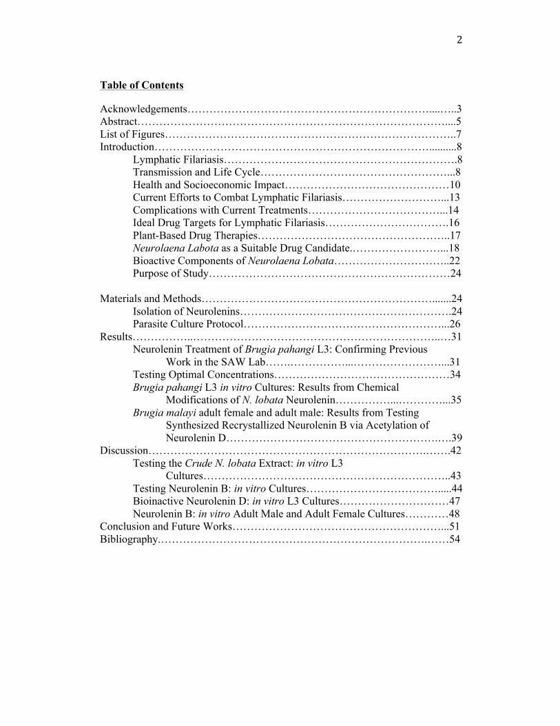
Table of Contents
Acknowledgements…………………………………………………………....…..3 Abstract…………………………………………………………………………....5 List of Figures……………………………………………………………………..7 Introduction…………………………………………………………………..........8 Lymphatic Filariasis……………………………………………………….8 Transmission and Life Cycle……………………………………………...8 Health and Socioeconomic Impact………………………………………10 Current Efforts to Combat Lymphatic Filariasis………………………...13 Complications with Current Treatments………………………………...14 Ideal Drug Targets for Lymphatic Filariasis…………………………….16 Plant-Based Drug Therapies……………………………………………..17 Neurolaena Labota as a Suitable Drug Candidate.……………………...18 Bioactive Components of Neurolaena Lobata…………………………..22 Purpose of Study…………………………………………………………24 Materials and Methods……………………………………………………….......24 Isolation of Neurolenins………………………………………………….24 Parasite Culture Protocol………………………………………………...26 Results……………..…………………………………………………………..…31
Neurolenin Treatment of Brugia pahangi L3: Confirming Previous Work in the SAW Lab…….……………...……………………...31
Testing Optimal Concentrations…………………………………………34 Brugia pahangi L3 in vitro Cultures: Results from Chemical
Modifications of N. lobata Neurolenin……………...…………...35 Brugia malayi adult female and adult male: Results from Testing
Synthesized Recrystallized Neurolenin B via Acetylation of Neurolenin D………………………………………………….….39
Discussion………………………………………………………………….…….42 Testing the Crude N. lobata Extract: in vitro L3
Cultures…………………………………………………………..43 Testing Neurolenin B: in vitro Cultures……………………………….....44 Bioinactive Neurolenin D: in vitro L3 Cultures…………………………47 Neurolenin B: in vitro Adult Male and Adult Female Cultures…………48 Conclusion and Future Works…………………………………………………...51 Bibliography.……………………………………………………………….……54
3
Acknowledgements
I would first like to acknowledge Steve Williams for being my thesis advisor and my special studies adviser for my three years at Smith College. You provoked my passion and willingness to be independent, learn new things, and view the world through the sphere of neglected tropical diseases. Without you I would not have structured a solidified career path post-graduation, and for that I am so grateful. Thank you to Sarah Moore, for agree to be my second reader. I have learned so much from your expertise and am inspired by your pedagogy and patience. You greatly augmented my passion for drug development and I am honored to present this body of work to you. Most graciously, I would like to thank Sue Haynes, the true mother of this project. For the past 6 years you have watched this project grow and develop into something so rewarding, and without you we would be nowhere. Thank you for your excellent worm culturing expertise, your patience with all the technical procedures, and for the number of times you cultured for me when I couldn’t possibly look at more worms. You are my true partner in crime! Thank you for all that you do, and I will miss you greatly. Thank you to Kevin Shea, Katie McGeough ‘16, and Megan Neubig ‘17 for your contributions to this project. Without the help from the Shea Lab, and the CHM 223 Experimental Teaching Laboratory course, we would not have progressed so quickly in this study, or at all. Thank you for all your chemistry expertise, and for answering my many, many chemistry-related questions. Without your help, there would be no project and our teamwork truly taught me the value of interdisciplinary collaboration. Thank you to Tiffani Chang ‘16, Stephanie Capsuto ‘18, and Susan Mishiyev ‘17, my teammates in the SAW Plant Group. Your unwavering support over the past three years pushed me to take on this project. Tiffani, thank you for your dedication to the intricacies of the project by conducting Ames and toxicity testing experiments, we have been the original troublemakers since the beginning! Stephanie, thank you for sticking by my side culturing and counting for hours on end. Your skilled lab techniques are so impressive and I cannot wait to observe the progress of this project with you in lead. Susan, I am so excited for you to join Stephanie to take on this task. I am inspired by your passion of tissue culturing; you will learn so many things. Thank you to Weam Zaky and everyone from the Smith College SAW Laboratory: you all inspire me everyday. Thank you for watching over me in the tissue culture room for days on end. I am so grateful to have such a great support system where we can bond over similar passions and share warm laughs in the cold hallways of Ford Hall.
4
Thank you to Allison Sirois (Sarah Moore Laboratory), Maria Delfin-Auza. Allison, for your constant support and your ear while I unloaded my stress. You are such a role-model, good luck at University of Massachusetts, Amherst next year. Maria, my beloved statistics counselor in the Spinelli Center. Thank you for your patience, I have learned so much from you! To Naina Zaman, who was my permanent lab “buddy.” You were by my side until the sunrise in Ford Hall.You gave me the motivation I thought I never had. Thank you for supporting me throughout my growth at Smith, I will never forget it. Thank you to my twin sister Kajori and my parents, Amma and Baba. Thank you to my parents for giving me the opportunity to send me to Smith College where I learned more than I could ever imagine. I learned so much about my strengths and passion. To my sister, who always knew the right thing to say. Love you all. Thank you to my funding sources, the National Institute of Health-Allergy and Infectious Disease Division, #HHSN2722010000301, the Filariasis Research Reagent Resource Center (FR3) at the University of Georgia, and the Tomlinson Memorial Fund
5
Abstract
Lymphatic filariasis (LF), a parasitic illness, is a globally neglected
tropical disease that is known to keep 1.4 billion people at risk of infection,
mostly in Southeast Asia and Africa (CDC, 2013). LF is a mosquito born disease
that is caused by the human parasites: Wuchereria bancrofti, Brugia malayi, and
Brugia timori. This study focuses on testing sesquiterpene lactones, called
neurolenins, which are secondary plant metabolites from Neurolaena lobata, a
medicinal herb, native to Guatemala and Central America. The Williams and Shea
Laboratories together plan to modify neurolenin to improve its potential as a drug
candidate that exhibits anti-filiarial activity against LF parasites.
Current drug treatments for LF do not target adult parasites residing in
human lymph nodes and vessels, and possible issues of genetic resistance
necessitate exploration of drug candidates that target all life stages of the parasite
including L3, adults, and microfilariae (mf). Neurolenin B, a sesquiterpene
lactone from N. lobata, is biologically active in vitro against L3, adult, and mf B.
pahangi and B. malayi nematodes. In this study, the Shea Laboratory converted
Neurolenin D, another sesquiterpene lactone found in N. lobata, to Neurolenin B
via acetylation and esterification methods that were analyzed for their efficacy in
killing filarial parasites in culture. Using the acetylation technique, synthesized
recrystallized Neurolenin B (modified from Neurolenin D) was biologically active
with significant killing against adult female, adult male, L3, and mf B. malayi and
B. paghangi parasites. The Neurolenin B product synthesized using an
esterification procedure (modified from Neurolenin D), also called isovaleric acid

6
Neurolenin D ester, was also biologically active with significant killing against L3
B. pahangi parasites. The ability for Neurolenin B to kill adult worms makes it a
novel drug for LF because current drugs only target mf. Future investigation will
continue with Ames testing to screen various forms of neurolenin for potential
mutagenicity. Future work with the Shea Laboratory will also examine
improvements in solubility for isovaleric Neurolenin D ester for further testing.
Both acetylated and esterified Neurolenin B products will be tested against
additional parasite cultures to validate the ability of these compounds to
efficiently kill filarial nematodes. We also plan to use rodent models to compare
the efficacy of the products in vivo in collaboration with Glaxo Smith Kline.

7
List of Figures Figure 1: Life cycle of Wuchereria bancrofti……………………………………10 Figure 2: Average percent mortality for adult female B. pahangi treated with
Neurolenin A and B ……………………………….………………….....20 Figure 3: Average percentage mortality for adult male B. pahangi treated with
Neurolenin A and B…...………………………………………………....21 Figure 4: Average percentage mortality for L3 B. pahangi treated
with Neurolenin A and B…….. ……………………………………...….22 Figure 5: Four main sesquiterpene lactones from N. lobata …………………….24 Figure 6: Isovaleric acid Neurolenin D ester
via esterification of Neurolenin D………………………………………..25 Figure 7: Synthesized Recrystallized Neurolenin B
via acetylation of Neurolenin D ……...………………………….………26 Figure 8: Example plate arrangement for adult female and
adult male B. pahangi …………………………………………………...28 Figure 9: Mortality rate for L3 B. pahangi testing crude extract
from N. lobata post-Charcoal Filtration ……………..………...……..…32 Figure 10: Mortality rate for L3 B. pahangi testing a
partially purified Neurolenin B………………………..............................34 Figure 11: Mortality rate for L3 B. pahangi testing crude extract
from N. lobata post-Charcoal Filtration at optimal concentrations…...…35 Figure 12: Mortality rate for L3 B. pahangi testing Neurolenin D………………37 Figure 13: Mortality rate for L3 B. pahangi testing synthesized
recrystallized Neurolenin B by acetylation of Neurolenin D…………...……………………………………………………..………38
Figure 14: Mortality rate for L3 B. pahangi testing isolvaleric acid Neurolenin D ester (Neurolenin B) by esterification of Neurolenin D ………………...…………………………….............…39
Figure 15: Average mortality rate for adult female B. malayi testing synthesized recrystallized Neurolenin B by acetylation of Neurolenin D…………………………………………………….……40
Figure 16: Average mortality rate for adult male B. malayi testing synthesized recrystallized Neurolenin B by acetylation of Neurolenin D…………………………………….......……41
8
Introduction: Lymphatic Filariasis
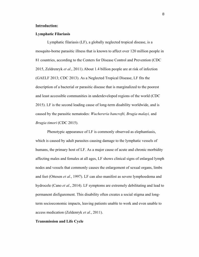
Lymphatic filariasis (LF), a globally neglected tropical disease, is a
mosquito-borne parasitic illness that is known to affect over 120 million people in
81 countries, according to the Centers for Disease Control and Prevention (CDC
2015, Zeldrenryk et al., 2011). About 1.4 billion people are at risk of infection
(GAELF 2013; CDC 2013). As a Neglected Tropical Disease, LF fits the
description of a bacterial or parasitic disease that is marginalized to the poorest
and least accessible communities in underdeveloped regions of the world (CDC
2015). LF is the second leading cause of long-term disability worldwide, and is
caused by the parasitic nematodes: Wuchereria bancrofti, Brugia malayi, and
Brugia timori (CDC 2015).
Phenotypic appearance of LF is commonly observed as elephantiasis,
which is caused by adult parasites causing damage to the lymphatic vessels of
humans, the primary host of LF. As a major cause of acute and chronic morbidity
affecting males and females at all ages, LF shows clinical signs of enlarged lymph
nodes and vessels that commonly causes the enlargement of sexual organs, limbs
and feet (Ottesen et al., 1997). LF can also manifest as severe lymphoedema and
hydrocele (Cano et al., 2014). LF symptoms are extremely debilitating and lead to
permanent disfigurement. This disability often creates a social stigma and long-
term socioeconomic impacts, leaving patients unable to work and even unable to
access medication (Zeldenryk et al., 2011).
Transmission and Life Cycle

9
According to the World Health Organization, the most prevalent
nematode, W. bancrofti is most commonly found in the Culex (urban areas),
Anopheles (rural Africa), and Aedes mosquito vectors (Pacific islands) (WHO
2015). B. malayi and other species in the Brugia genus are mostly transmitted by
the Mansonia mosquito vector, and are specific to Southeast and South Asia. Of
the 120 million people infected, one third of the people with LF reside in India,
one third reside in Africa, and one third reside in broader South Asia, the Pacific,
and the Americas (Anil and Talluri 2015).
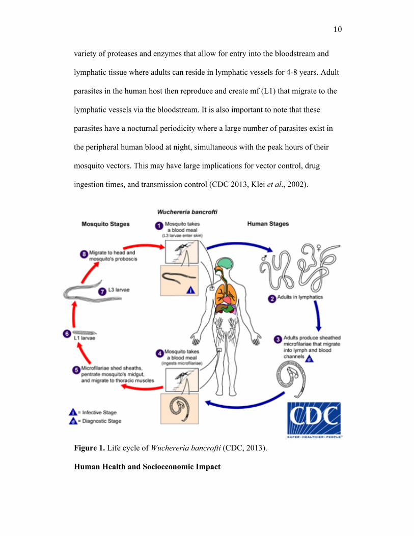
The mode of transmission of LF is stimulated by the life cycle of the
aforementioned parasites, which reside in mosquito and human hosts (Figure 1).
When an infected mosquito takes a blood meal from an uninfected person, it
deposits adolescent (L3) nematodes onto the skin around the puncture wound
(CDC 2013). Nematodes enter the human host bloodstream only when the
mosquito bites. Once in the body, nematodes mature through a physiological
molting process where, once adults, they enter the lymph nodes to replicate and
produce microfilariae (mf) offspring. The mf are later circulated through the
bloodstream and taken up by a subsequent mosquito blood meal (CDC 2013). The
mf begin their maturation in the gut and flight muscles of their host mosquito,
molting from L1 to L2 to L3 life stages. Once reaching the L3 stage, the
nematodes are ready for deposition into the skin of a human host through the
proboscis of the mosquito (CDC 2013). It takes about 7-9 days for the L3
adolescents to molt to the L4 stage that mature into adults and reside in the human
host lymphatic system. Before maturing into adults, L3/L4 adolescents secrete a
10
variety of proteases and enzymes that allow for entry into the bloodstream and
lymphatic tissue where adults can reside in lymphatic vessels for 4-8 years. Adult
parasites in the human host then reproduce and create mf (L1) that migrate to the
lymphatic vessels via the bloodstream. It is also important to note that these
parasites have a nocturnal periodicity where a large number of parasites exist in
the peripheral human blood at night, simultaneous with the peak hours of their
mosquito vectors. This may have large implications for vector control, drug
ingestion times, and transmission control (CDC 2013, Klei et al., 2002).
Figure 1. Life cycle of Wuchereria bancrofti (CDC, 2013).
Human Health and Socioeconomic Impact
11
The clinical course of LF disease is often as follows: asymptomatic
microfilariae, where patients are generally asymptomatic but may develop chronic
inflammatory granulomas depending on the density of mf. These chronic
symptoms could also be accompanied with splenic destruction and chyluria
(milky urine). The second phase includes adenolymphangitis (ADL) and episodic
fever attacks, and painful inflammation of lymph nodes, and testes and spermatic
cords in males. This stage is the most common and is often the most recurrent.
Secondary infections from ADL include bacterial infection in lesions of affected
areas that contribute to extreme pain (Wayangankar et al., 2015).
The third and final stage of the disease results in irreversible lymphedema.
Adult parasites are the main contributors of these symptoms as they reside in the
lymph nodes and release pro-inflammatory cytokines that contribute to skin
exfoliation, genital swelling, and pain. The third stage is the most debilitating and
often leads to elephantiasis that afflicts 15 million people worldwide (WHO,
2016). The lymphatic system is the main regulator for fluid balance between
blood and tissue and is physically blocked by the damage caused by adult worms.
Lymphatic vessel walls thicken and dilate, causing an imbalance in the lymphatic
tissue and vessels, preventing the proper function of the system. (CDC 2013;
Wayangankar et al., 2015; Zeldenryk et al., 2011).
According to the Global Programme to Eliminate Lymphatic Filariasis
(GPELF), morbidity management, one of its two main initiatives, remains less
widespread and successful. Only 26 of the 81 endemic countries even have
morbidity management programs and the programs lack a focus on supporting

12
patients in actively participating in their communities with daily activities both
independently and collaboratively. Most management programs only focus on
physical impairment and do not address emerging issues such as social stigma and
cultural norms. Because LF affects mostly impoverished communities, most
patients delay seeking assistance, and free drug availability do not mitigate other
costs like travel and time lost from work (Zeldenryk et al., 2011). Many families
consider LF patients to be a shameful social burden; LF patients are often hidden
from communities. Men who incur genital damage are severely handicapped and
have limitations in participating in physical labor, which is a significant challenge
since many patients live in communities that are largely agriculture with intensive
manual labor. Women pay a high price for social stigmatization because women
with lymphedema are restricted from marriage, which is essential for monetary
and physical security in many cultures in endemic regions (GAELF 2013).
The WHO indicates that LF causes an estimated annual loss of $1 billion
and inhibits up to 88% of total economic activity in endemic countries. GAELF
and the WHO also estimate that LF is responsible for the loss of 4.4 million
disability adjusted life years (DALYs) in men and over 1.3 million DALYs in
women. The DALY index measures the loss of a healthy productive life due to a
disability and is a metric for the measurement of the gap between current health
status and an ideal health situation where a population is free of disability and
disease. Most developing countries have high DALY numbers where large
populations persist in the cyclical nature of poverty and disease, particularly
exacerbated by neglected tropical diseases like LF (CDC 2013; GAELF 2013).

13
Current Efforts to Combat Lymphatic Filariasis
The Global Programme to Eliminate Lymphatic Filariasis, launched in
2002, was the first extensive action taken in hopes of eradicating LF by 2020. The
two main initiatives, as stated earlier, were to provide community-wide mass drug
administration (MDA) via Ministries of Health programmes and pharmaceutical
companies like GalxoSmithKline and Merck & Co., and to alleviate the suffering
of those with chronic LF symptoms through morbidity management (Zeldenryk et
al., 2011, WHO 2016).
One of the current drugs used to treat LF is Diethylcarbamazine (DEC).
First discovered in 1948, DEC critically reduces LF transmission, but does not
remove all mf, or adult nematodes from a single dose of 6mg of drug/kg of body
weight (Taylor, Hoerauf, and Bockarie 2010; Bockarie and Deb 2010). A one-day
treatment has been shown to be as effective as the regular 12-day treatment,
giving DEC an economic edge in populations that may be able to afford only one
trip to the clinic (CDC 2013). Used for more than 50 years, DEC has shown to
impact the nematode arachidonic acid signalling pathway in vivo. The drug aims
to target the mf in an infected patient to prevent transmission so that mosquitoes
cannot transmit the disease to others when taking a human bloodmeal (GAELF
2013; CDC 2013; Taylor, Hoerauf, and Bockarie 2010). DEC is responsive to the
density of mf in the blood and incurs side effects when a lower population of mf
is present. Common side effects include dizziness, nausea, and muscle pain (CDC
2013). Ivermectin, derived from avermectin, is another broad-spectrum anti-
filarial drug that has been popularly used in the treatment for LF since the 1970s
14
to today (Crump and Omura 2011). Ivermectin’s mechanism for action is the
activation of glutamate-activated chloride channels, causing paralysis of mf
(Bockarie and Deb 2010; Arena et al. 1995). Like DEC, ivermectin is only known
to have moderate effects on adult worms with quarterly administration, an
unreasonable number of dosages for those that cannot afford multiple trips to a
health center, which is a majority of the total LF population (Bockarie and Deb
2010).
The third most common drug treatment used is Albendazole, another
broad spectrum drug that is used in conjunction with DEC and ivermectin
(Taylor, Hoerauf, and Bockarie 2010). Developed in 1975, Albendazole causes
degenerative alterations in the LF parasite by inhibiting microtubule
polymerization and assembly during the parasite’s life cycle. Albendazole helps
to decrease glycogen stores in mf stage parasites, and is also effective with a
single dose at 400 mg with a half-life of 8-12 hours (DrugBank 2014).
Complications with Current Treatments
The World Health Organization and CDC have made major efforts in
Mass Drug Administration to combat LF, but emerging genetic resistance of the
parasites to anti-filarial medication has become a concern in efforts for effective
treatment. Although there is no formal evidence of resistance to drugs currently
used for LF, the recent resistance to ivermectin when used to combat
onchocerciasis has raised concerns (Molyneux et al., 2003). As stated earlier,
management of chronic symptoms has been largely underdeveloped, and MDA is
only undertaken annually and worldwide coverage has yet to be met (GAELF

15
2013). MDA relies heavily on community health workers to educate patients
about the disease and to get compliance by asking patients to taking the necessary
medication. MDA is only effective if there is at least 80% coverage within a 5-
year period, something that has shown to be variable since the inception of the
program (Plaisier et al., 2000). Compliance concerns not only create an issue for
those affected by LF, but they are also possibly related to drug resistance
development. In areas where compliance is low, community treatment exists at
low, sublethal levels and can induce accelerated development of resistance. Drug
regimens for LF often include a combination of two current drugs, making
resistance less likely when compared to diseases only treated with one drug
(Molyneux et al., 2003). However, given the time period (4-8 years) that the adult
worms reside in the human host, high compliance of about 80% is necessary for 5
years, a very lengthy compliance period (Michael et al., 2004).
Other than compliance and drug resistance mechanisms, various side
effects also contribute to the rising concerns for the current treatments of LF. For
example, DEC cannot be co-administered in populations that also have
onchocerciasis, another parasitic neglected tropical disease. Patients who are
inflicted with both diseases and take DEC incur a heightened inflammatory
response when both parasites are targeted (Bockarie and Deb 2010). Patients who
have both LF and loiasis cannot be treated with ivermectin as encephalopathy and
neurological degradation can often coexist as side effects (Bockarie and Deb
2010). It is evident that those affected with LF can also be prone to other NTDs
that inevitably will decrease the efficacy of current treatment options for LF.
16
Ultimately, the biggest “side effect” with the drugs on the market is that they are
not effective in killing adult nematodes. Only mf are efficiently killed by DEC,
Albendazol, and ivermectin.
However, MDA has increased coverage from 12 countries in 2000 to 59
countries in 2010, giving more than 500 million people treatment. In fact, 17
countries receive 100% of geographic coverage and have completed five or more
rounds of MDA programming. More successfully, in 2008, China and the
Republic of Korea have declared elimination of LF as a public health problem
(WHO 2016).
Ideal Drug Targets for Lymphatic Filariasis
Given the current treatments on the market, it is evident that there is a
need for new drug candidates for LF. Specifically, a new therapy that targets adult
nematodes has become alarmingly necessary. Current treatments only can work to
prevent transmission by killing mf parasites, which may not be sufficient to
eradicate LF. Adult worms are the producers of mf and reside in lymph nodes and
vessels where long-lived adults eventually can cause elephantiasis and
lymphedema. Targeting adult nematodes will not only prevent transmission, but
will also help patients that already have the disease (Murthy, Joseph, and Murthy
2011). L3 worms can also be considered as potential drug targets as they resemble
adolescent worms that molt to adults, and this is the stage that enters the human
host. By targeting L3, we can prevent worms from developing into adults that
cause the physical manifestations of the disease. Thus, an effective drug model
would target LF worms at three important stages: mf, L3, and adult worms. More
17
importantly, a gradual killing of worms is desired. Sudden death of worms may
lead to an acute inflammatory shock and drive the patient into further presentation
of clinical symptoms that may become fatal. Other useful drug properties include
the need for non-resistant therapies, regimens that do not require multiple doses,
and drugs that have minimal side effects. Additionally, a treatment that is also
effective against other NTDs, as are current treatments for LF, would result in a
nearly perfect drug candidate. As stated earlier, it is common for those with LF to
also contract other NTDs caused by worm-like parasites in human hosts. Some of
these NTDs include hookworm, whipworm (trichuriasis), and roundworm
(ascariasis) (Micromedex 2015). Thus, creating a drug that also targets other
diseases caused by nematodes not only helps a patient with LF, but will also help
patients with other worm-related NTDs.
Plant-Based Drug Therapies
Plant based medicine is most commonly used today in traditional Chinese
practices, Ayurvedic medicine in India and the Middle East, and in many
indigenous cultures of the Americas, which date as far back as 60,000 years ago
(Pan et al., 2014). Novel anti-filarial drugs with bioactive components from
traditional plants enable alternative methods in combating LF (Murthy, Joseph,
and Murthy 2011). New drug therapies to combat LF and other NTDs need to be
as potent as current drugs, but they also need to be effective at lower doses with
minimal side effects. These characteristics can be met with an exploration of plant
based drug therapies. Historically, 35,000-70,000 plant species have been
screened for possible drug therapies, and those that have enthnopharmacological
18
purposes have been used in medicine (Veeresham 2012). During the last 30 years
in particular, about 50% of the approved drugs are derived from natural products
to combat cancer. In the past 16 years alone, 11% of the 252 drugs considered as
“essential” according to the WHO were from a flowering plant origin. Plants
represent the core structures for numerous drugs on the market, regardless of
modern advances towards synthetic chemistry or molecular modeling design. For
example, Merck in 1826 released the first semi-synthetic pure aspirin drug that is
based on salicin, a natural product isolated from Salix alba (Veeresham 2012).
This product later led to the isolation of cocaine, codeine, quinine, and other
active compounds that are still in use today. Some plants used in historic
traditional practices have been shown to have therapeutic effects against ovarian
and breast cancer and even malaria. These plant extracts have been synthesized as
drugs approved by the FDA (like Paclitaxel from Taxus brevifolia for ovarian and
breast cancer and Artemisinin from Artemisia annua for malaria) (Veeresham
2012).
Neurolaena Labota as a Suitable Drug Candidate for Lymphatic Filariasis
The Steven A. Williams Lab at Smith College (SAW Lab) is using
Centella asiatica, Neurolaena lobata, and Quisqualis indica as medicinal herbs
native to East Asia, Guatemala, and China respectively to develop drugs that
exhibit anti-filarial activity on LF parasites. This investigation will focus on
Neurolaena lobata, as its extracts have shown to have anti-parastic properties in
vitro against Trypanosoma cruzi, T.vaginalis, Leishmania mexicana and
Plasmodium falciparum (Berger et al. 1998, 2001; Franssen et al. 1997; François

19
et al. 1996; Muelas-Serrano et al. 2000). Found in the Asteraceae family, N.
lobata is a weedy shrub that originates in Central and South America. It is known
to have therapeutic properties in treating malaria, diabetes, cancer, and fevers in
traditional medicinal practices of Mayan and Mesoamerican communities (Leon-
Levy Native Plant Preserve; Walshe-Roussel et al. 2013). The anti-parasitic
element of N. lobata is of most interest in the SAW Lab as it pertains to LF and
was sparked by a study conducted by Fujimaki et al (2005) that defined a crude
extract in vitro of N. lobata leaves as the bioactive component of the plant that
showed microfilaricidal properties on Brugia pahangi nematodes. A series of
concentrations of the extract at 500 µg/mL, 250 µg/mL, 100 µg/mL, 50 µg/mL
and 10 µg/mL were tested on mostly adult worms, both female and male. The
most promising results indicated a halt of motility at 6 hours post-treatment at 500
µg/mL, or at 24 hours post treatment at 250 µg/mL for both sexes of adult worms.
The study also revealed that female adult worms were eventually prevented from
further microfilarial release based on concentration and time variables, and that
adult males were more commonly reduced in their motility (Fujimaki et al.,
2005). In this study, Brugia pahangi was used as the model nematode because
they are almost genetically identical to Brugia malayi, but only infect cats and
other domesticated animals, making it useful for study in a laboratory. The results
from this paper show that N. lobata clearly does have anti-parasitic effects on
Brugia pahangi adult nematodes, an underdeveloped drug target that current
treatments do not offer impact.
20
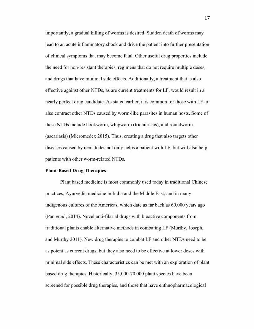
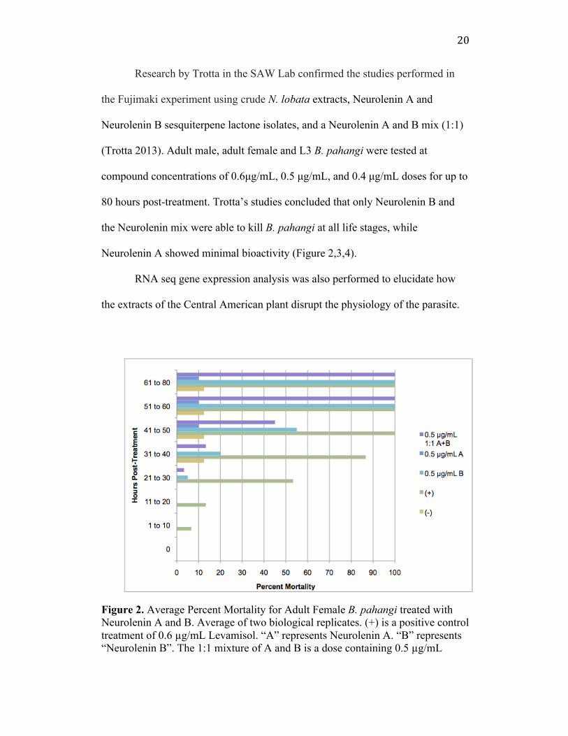
Research by Trotta in the SAW Lab confirmed the studies performed in
the Fujimaki experiment using crude N. lobata extracts, Neurolenin A and
Neurolenin B sesquiterpene lactone isolates, and a Neurolenin A and B mix (1:1)
(Trotta 2013). Adult male, adult female and L3 B. pahangi were tested at
compound concentrations of 0.6µg/mL, 0.5 µg/mL, and 0.4 µg/mL doses for up to
80 hours post-treatment. Trotta’s studies concluded that only Neurolenin B and
the Neurolenin mix were able to kill B. pahangi at all life stages, while
Neurolenin A showed minimal bioactivity (Figure 2,3,4).
RNA seq gene expression analysis was also performed to elucidate how
the extracts of the Central American plant disrupt the physiology of the parasite.
Figure 2. Average Percent Mortality for Adult Female B. pahangi treated with Neurolenin A and B. Average of two biological replicates. (+) is a positive control treatment of 0.6 µg/mL Levamisol. “A” represents Neurolenin A. “B” represents “Neurolenin B”. The 1:1 mixture of A and B is a dose containing 0.5 µg/mL

21
Neurolenin A and 0.5 µg/mL Neurolenin B (Trotta, 2014).
Figure 3. Average Percent Mortality for Adult Male B. pahangi treated with Neurolenin A and B. Average of two biological replicates. (+) is a positive control treatment of 0.6 µg/mL Levamisole. “A” represents Neurolenin A. “B” represents “Neurolenin B”. The 1:1 mixture of A and B is a dose containing 0.5 µg/mL Neurolenin A and 0.5 µg/mL Neurolenin B (Trotta, 2014).
22
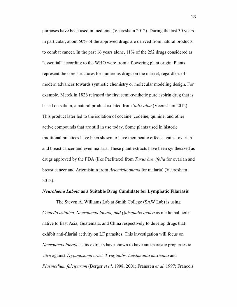
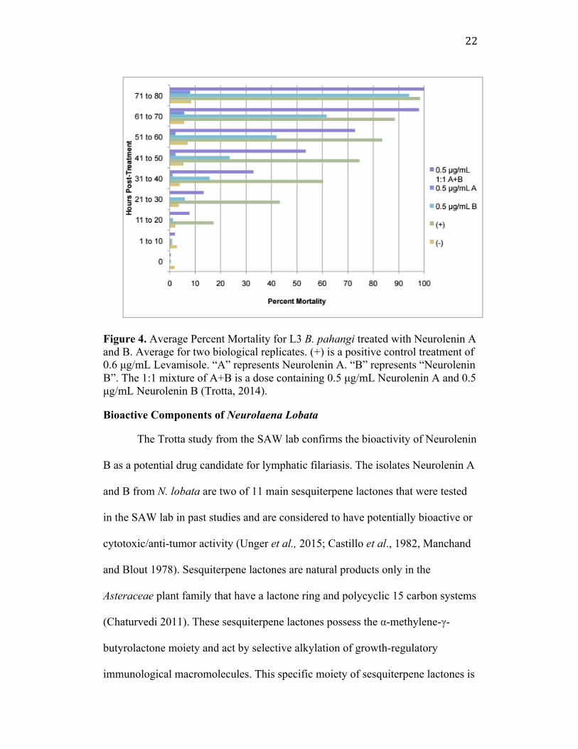
Figure 4. Average Percent Mortality for L3 B. pahangi treated with Neurolenin A and B. Average for two biological replicates. (+) is a positive control treatment of 0.6 µg/mL Levamisole. “A” represents Neurolenin A. “B” represents “Neurolenin B”. The 1:1 mixture of A+B is a dose containing 0.5 µg/mL Neurolenin A and 0.5 µg/mL Neurolenin B (Trotta, 2014). Bioactive Components of Neurolaena Lobata The Trotta study from the SAW lab confirms the bioactivity of Neurolenin
B as a potential drug candidate for lymphatic filariasis. The isolates Neurolenin A
and B from N. lobata are two of 11 main sesquiterpene lactones that were tested
in the SAW lab in past studies and are considered to have potentially bioactive or
cytotoxic/anti-tumor activity (Unger et al., 2015; Castillo et al., 1982, Manchand
and Blout 1978). Sesquiterpene lactones are natural products only in the
Asteraceae plant family that have a lactone ring and polycyclic 15 carbon systems
(Chaturvedi 2011). These sesquiterpene lactones possess the α-methylene-γ-
butyrolactone moiety and act by selective alkylation of growth-regulatory
immunological macromolecules. This specific moiety of sesquiterpene lactones is
23
what has been correlated with their bioactivity in past literature. This alkylation
process is known as the Michael-type reaction by the α-methylene lactone group
in particular (Manchand and Blout 1978). The Michael reaction involves a
conjugate addition to the alpha, beta unsaturated ketone system that reveals a
stable yet reactive nucleophilic enolate (Michael acceptor). Neurolenin A and B
in particular are found in N. lobata leaves and are of interest because they can be
isolated in great quantities and differ by one functional group (Manchand and
Blout 1978). The Trotta study and previous studies confirmed that under the
specified laboratory conditions, only Neurolenin B was biologically active (Unger
et al., 2015; Castillo et al., 1982, Manchand and Blout 1978). Experiments from
the Kevin Shea Experimental Teaching 223 Laboratory at Smith College further
confirmed these results, but it was found that starting materials were not pure and
the overall isolation of Neurolenin B in the Trotta experiment may not have
actually been pure Neurolenin B, but rather a mix of various sesquiterpene
lactones. The Shea Laboratory, in continuation of the Trotta study, has identified
four sesquiterpene lactones that should be the focus of the continued study of anti-
parasitic activity as they can be easily altered through the manipulation of one
functional group (Figure 5). As the Trotta study indicated, each lactone is

24
differentially bioactive, and further studies that explore the purification of 11 or
more sesquiterpene lactones of N. lobata will prove definitive in identifying the
most potent and effective drug candidate for lymphatic filariasis.
Figure 5. Four main sesquiterpene lactones from N. lobata (Manchand and Blout 1978). Purpose of Study
This project will further investigate the bioactive impact of Neurolenin B
(prepared from Neurolenin D) against adult female, adult male and L3 Brugia
pahangi parasites via in vitro drug testing. The ultimate goal of this work is to
contribute to the larger effort of discovering a novel potential drug candidate that
will kill not only mf, but also adult nematodes, potentially replacing or
supplementing current drugs that are being used in MDA for eliminating
lymphatic filariasis.
Materials and Methods Isolation of Neurolenins All isolations of Neurolenins were conducted by the Kevin Shea
laboratory, Smith College, and used the protocol from Manchand and Blout,
1978, to prepare the extraction of Neurolenin D from Neurolaena lobata leaves.
Solvents and reagents were purchased from Pharmco-AAPER, Sigma Aldrich,

25
Fisher Scientific, and Acros Organics and extracts from leaves were prepared with
nitrogen wand and magnetic strings. The Neurolaena lobata was purchased from
Rainforest Remedies or Grenada Market and were crushed and powdered using a
food processor. The powder was placed in a thimble in a soxhlet extractor. The
powder was extracted with dichloromethane and removed via rotary evaporation.
The remaining dark green gum was obtained and run through a column that
produced fractions of Neurolenin D that were recrystallized with cold ethyl
acetate. There were two main techniques used in this study to convert Neurolenin
D to Neurolenin B: acetylation and esterification techniques. The esterification
method (Figure 6) was performed using isovaleric acid and the acetylation
technique used acetic anhydride (Figure 7). However, the acetic anhydride was
later removed before the product was given to the SAW Lab because of its
potential toxic effects in the human body.
Figure 6. Esterification of Neurolenin D to Neurolenin B with Isovaleric Acid. The resulting product is called isovaleric acid neurolenin D ester (Shea Lab 2015).
O
O
O
HO O OOH
O
O
O
HO O OO
O
EDC, DMAP
NEt3, DCM
HO
O
Neurolenin D Neurolenin B

26
Figure 7. Acetylation of Neurolenin D to Neurolenin B. The resulting product is called synthesized recrystallized Neurolenin B (Shea Lab 2015). Parasite Culture (T.V. Rajan et al. 2003, J. Parasitology 89(4) p 868-870 with modifications made by Susan Haynes, Smith College)
Culture Media (10% FBS) 90 mL Minimum Essential Medium Alpha (no HEPES, just sodium bicarbonate buffer) (Gibco - Life Technologies) 10 mL Fetal Bovine Serum deactivated (Gibco - Life Technologies 1 mL Pen Strep 5000U (Gibco - Life Technologies) 100 µLGentamycin (10 mg/mL stock) (Gibco - Life Technologies)
20 µL Ciprofloxacin (10 mg/mL, ophthalmic stock) (TCI America) 20 µL Fortaz (Ceftazidime) (in PBS 10 mg/mL stock) (TCI America)
Total Volume: 101 mL Prepare under sterile conditions and store at 4°C. Complete medium includes 90mL of the culture medium, 10 mL Fetal Bovine, and 20 µL of fresh Fortaz added on the same day of culture.
Wash Solution
100 mL RPMI 1640 (HEPES Buffered) (Gibco - Life Technologies) 1 mL Pen Strep 5000U 100 µL Gentamycin (10 mg/mL stock) 20 µL Ciprofloxacin (10 mg/mL stock) 100 µL Fungizone (Amphotericin B) (2.5 g/mL stock) (Gibco - Life Technologies)
Total Volume: 100 mL
Prepare under sterile conditions and store at 4°C
Neurolenin B
O
O
O
HO O OOH
O
O
O
HO O OO
O
O
OO
NEt3 , DCM
Neurolenin D

27
Ascorbic Acid: dilute to final concentration of 15 µg/mL of culture medium. Make 1.5 mg/ml (100X) stock and freeze. Only unfreeze stock once and discard stock vial once it is added to the cultures. Prepare under sterile conditions and store at -20°C for single use. Nematode Preparation and Procedure:
Nematodes: B. pahangi nematodes (L3, adult males and adult females)
were obtained from the University of Georgia (Athens, Georgia), College of
Veterinary Medicine. Parasites were shipped to Smith College in conical 50 mL
tubes in RPMI 1640 solution.
Preparation: Wipe down the Tissue Culture room hood (Ford 130) with
70% EtOH. Expose any working surfaces with UV radiation for at least 30
minutes. Let the medium come to room temperature about 20 minutes before use.
Before opening the tube of worms, allow the tube to sit upright for 30-60 minutes
(at room temperature) thus ensuring the worms have settled to the bottom. Set the
incubator CO2 so the pH is 7.2 - 7.4 at 37°C.
Washing the Worms: Pipet the wash solution into (1) 100mm petri dish
and (3) 60 mm petri dishes, filling each two thirds full. For L3 worms, use a
transfer pipet to aspirate the worms from the bottom of the tube and put into the
100 mm petri dish wash solution. Avoiding any debris, use a p-200 pipet to
collect the worms from this wash and move them to the next wash. Do this three
times. Pick up as little RPMI 1640 solution as possible when transferring the
worms. The goal is to get 40 worms in the p-200 pipet. Pipet tips should be
changed between each wash. The number of washes performed depends on the
conditions/state the worms arrive in and also their experimental use. They should
28
be rinsed 3 times, more if necessary. All procedures for L3 worms must be done
under a light microscope. For adult females and adult males, follow the same
procedures, but instead of using a transfer pipet or p-200 to pick up the worms,
use EtOH sterilized forceps for easy transfer. A light microscope is not needed as
adult females and adult males can be seen with the naked eye. However, the
microscope must be used to view mf produced by adult female worms.
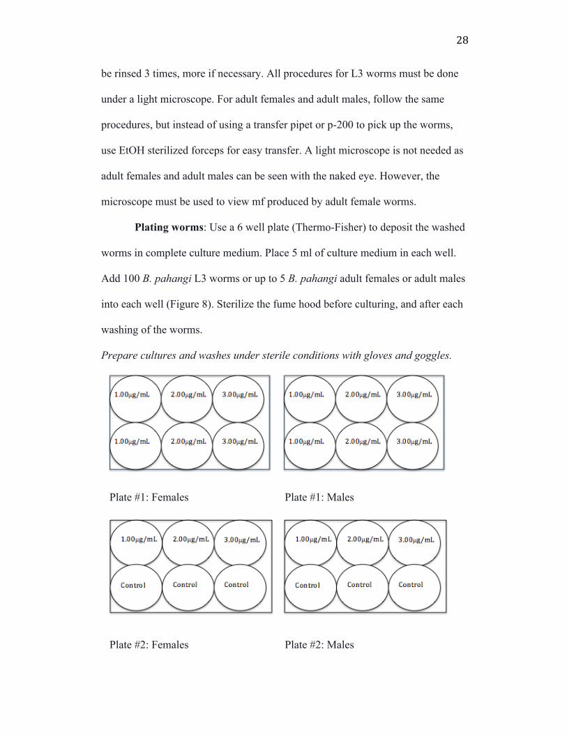
Plating worms: Use a 6 well plate (Thermo-Fisher) to deposit the washed
worms in complete culture medium. Place 5 ml of culture medium in each well.
Add 100 B. pahangi L3 worms or up to 5 B. pahangi adult females or adult males
into each well (Figure 8). Sterilize the fume hood before culturing, and after each
washing of the worms.
Prepare cultures and washes under sterile conditions with gloves and goggles.
Plate #1: Females Plate #1: Males
Plate #2: Females Plate #2: Males
29
Figure 8. Example plate arrangement for adult female and adult male Brugia pahangi worms. Each adult well contains 5 worms each in 5 mL of complete medium. Amount in µg/mL refers to the concentration of neurolenin added to each well. This example arrangement shows a replicate experiment.
Treatment and Observation: Plating occurs on Day 0, and drug
treatment is added on Day 5. Between Days 0-5, worms are observed and death is
noted and subtracted from death count treatment as “death due to shipping or
plating.” During day 5, 50 µL of ascorbic acid (Vitamin C) is added to each well
to ensure the molting and continuation of the life cycle of L3 to L4 B. pahangi
(Rajan, 2003). Adult worms do not receive Vitamin C (ascorbic acid) treatment as
they do not molt, however they must have old medium exchanged with new
medium as mf start appearing in female adult cultures at Day 5. Treatment is
added at indicated optimal concentrations that differed between L3 and adults.
The acetylated Neurolenin D (Neurolenin B) had optimal concentrations for L3
worms at 1.25 µg/mL, 1.00 µg/mL, and 0.800 µg/mL. The esterification of
Neurolenin D to Neurolenin B had optimal concentrations at 1.25 µg/mL, 1.00
µg/mL, 0.800 µg/mL, and 0.700 µg/mL. The adult females and adult males were
only treated with the acetylation of Neurolenin D to Neurolenin B at 1.00 µg/mL,
2.00 µg/mL, and 3.00 µg/mL. These optimal concentrations were determined by
choosing the top 3 or 4 concentrations that killed the most worms during the
desired period (50-70 hours post-treatment). Each compound received from the
Shea lab was prepared in a solvent, 70%EtOH, at a concentration of 1mg/mL.
Death is recorded when worms become completely immobile and appear
hairpin like and dark in morphology. At each observation time, worms were

30
recorded as either dead or alive. Live worms have sinusoidal motion and move
extremely fast if healthy. Before death, it is common for worms to move
extremely slowly. This behavior may explain results that appear to show a sudden
spike in death. This is not due to a “sudden death” but rather a slow, gradual
death. Each culture includes a negative control where no treatment is applied and
molt rate is observed (only in L3 cultures) to quantify the health of the culture. L3
worms were scored as either “partial molt” or “full molt” when transparent like-
cuticles are shed from worms and can be seen floating in the culture medium. A
worm with a cuticle still attached to worms is considered a partial molt. The drug
antibiotic Levamisole was used as a positive control for adult cultures. Solid
levamisole HCl (TCI America) was dissolved in MEM at a concentration of
1µg/mL for use with B. malayi cultures (only B. malayi parasites were available
for adult culture during the time of the investigation). The Levamisol was added
at 3.00µg/mL, the highest concentration tested on adult female and adult male
worms.
Analysis: All cultures were observed every 4 hours starting on Day 5 for
up to 72 hours or more (until Day 8 or longer). Average percentage mortality, was
recorded in each well by taking the total number of dead worms and dividing by
the total number of worms in the well (after accounting for death due to plating or
shipping of worms). Results were plotted with time in hours post-treatmen (hpt)
on the X-axis and and percentage death on the Y-axis. Average mortality for each
treatment condition was compared to the health of the average mortality of the
negative control and statistical significance of the differences between the
31
experimental condition and the control condition was calculated using a one-tailed
paired student T-test at the last time point. The last time check was at 72 hours
post-treatment (hpt) for L3 cultures, and was approximately 98 hours post drug
treatment for adult cultures. Significant drug bioactivity was defined as a p-value
less than 0.05.
Results
Neurolenin Treatment of Brugia pahangi L3: Confirming Previous Work in the SAW Lab All extracts from Neurolaena lobata during this investigation were
prepared from the Shea Laboratory with the help of Katie McGeough and Megan
Neubig. Results were tested for significant killing compared to the health of the
negative control time at 72 hpt, the last time check (for L3 cultures).
First we aimed to confirm the findings of previous work done by Kristine
Trotta in the SAW Lab, Smith College (Figure 9). Her results indicated that a
crude extract from dry N. lobata leaves was biologically active at the three highest
concentrations: 200 µg/mL, 300 µg/mL and 400 µg/mL compared to the control,
which received no treatment. For this investigation, the Shea lab helped us to
confirm the results by producing a new extract where it was tested in the SAW
Lab. Treatment groups molted quickly, at 20 hpt, where the ideal time of death is
expected at around 50-70 hpt (Figure 9). The mortality rate is measured as
1=100% death, where all worms in the well were dead at the indicated hour. For
this investigation, the Shea lab prepared the new extract using a charcoal filtration
technique. Past work in the SAW Lab did not test the charcoal-filtrated product
when working with the crude extract. Charcoal filtration is a method of filtering
32
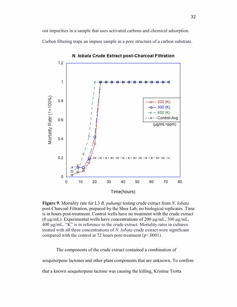
out impurities in a sample that uses activated carbons and chemical adsorption.
Carbon filtering traps an impure sample in a pore structure of a carbon substrate.
Figure 9. Mortality rate for L3 B. pahangi testing crude extract from N. lobata post-Charcoal Filtration, prepared by the Shea Lab, no biological replicates. Time is in hours post-treatment. Control wells have no treatment with the crude extract (0 µg/mL). Experimental wells have concentrations of 200 µg/mL, 300 µg/mL, 400 µg/mL. “K” is in reference to the crude extract. Mortality rates in cultures treated with all three concentrations of N. lobata crude extract were significant compared with the control at 72 hours post-treatment (p<.0001).
The components of the crude extract contained a combination of
sesquiterpene lactones and other plant components that are unknown. To confirm
that a known sesquiterpene lactone was causing the killing, Kristine Trotta
33
modified the crude extract method, and isolated Neurolenin B, a known
sesquiterpene lactone from N. lobata that had biological activity against B.
pahangi and B malayi. In this investigation, with collaboration from the Shea lab,
succeeded in isolating Neurolenin B in the same way, but it was later revealed by
the Shea laboratory that this extract also contained Neurolenin D and other
sesquiterpene lactones. Thus, this isolation was in fact not fully purified (Figure
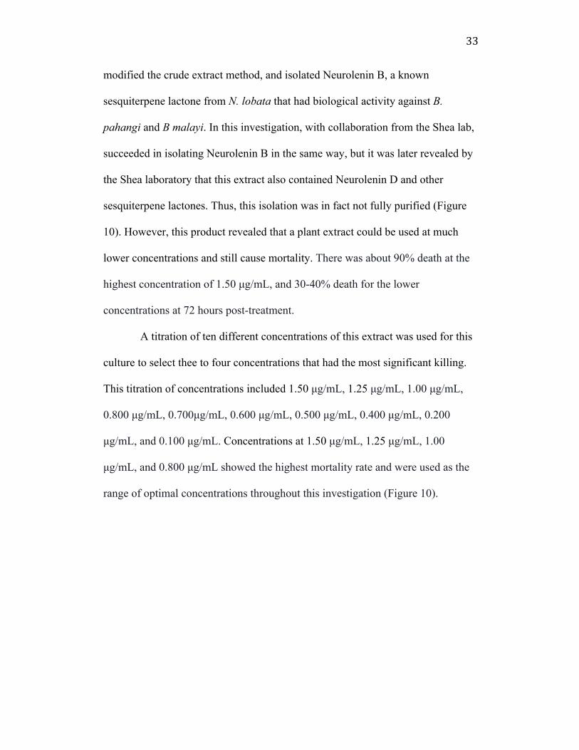
10). However, this product revealed that a plant extract could be used at much
lower concentrations and still cause mortality. There was about 90% death at the
highest concentration of 1.50 µg/mL, and 30-40% death for the lower
concentrations at 72 hours post-treatment.
A titration of ten different concentrations of this extract was used for this
culture to select thee to four concentrations that had the most significant killing.
This titration of concentrations included 1.50 µg/mL, 1.25 µg/mL, 1.00 µg/mL,
0.800 µg/mL, 0.700µg/mL, 0.600 µg/mL, 0.500 µg/mL, 0.400 µg/mL, 0.200
µg/mL, and 0.100 µg/mL. Concentrations at 1.50 µg/mL, 1.25 µg/mL, 1.00
µg/mL, and 0.800 µg/mL showed the highest mortality rate and were used as the
range of optimal concentrations throughout this investigation (Figure 10).
34
Figure 10. Mortality rate for L3 B. pahangi testing a partially purified Neurolenin B, extract (from Kristine Trotta, 2014, but prepared by the Shea Laboratory, 2015) with no biological replicates using a stock solution of 1 mg/mL. Time is in hours post-treatment. The control has 0 µg/mL of the partially purified Neurolenin B, and was tested against wells with concentrations of 1.5 µg/mL, 1.25 µg/mL, 1.00 µg/mL, 0.800 µg/mL. Mortality rates in cultures treated with 1.5 µg/mL partially purified Neurolenin B extract were significant compared with the control at 72 hours post-treatment (p<0.0001). Testing Optimal Concentrations Because relatively high mortality was seen using the partially purified
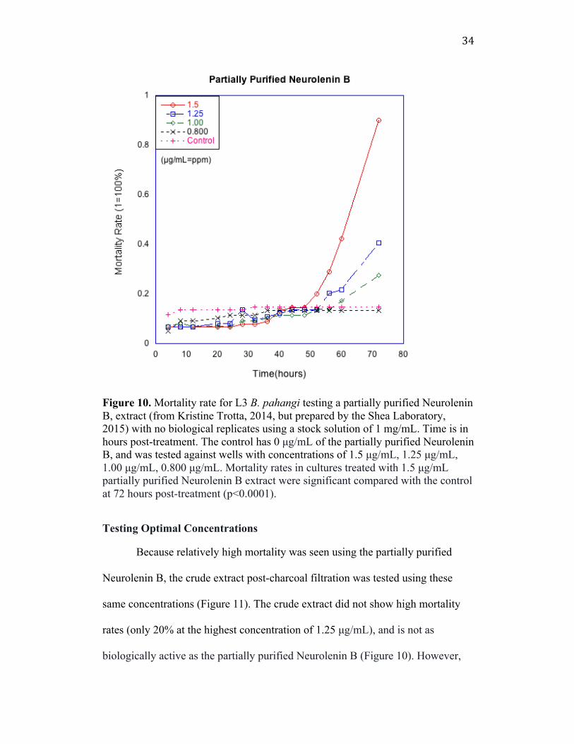
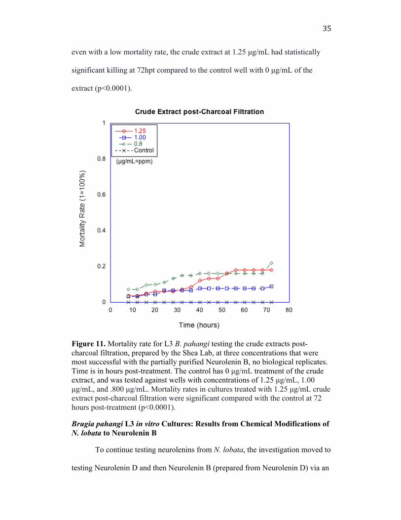
Neurolenin B, the crude extract post-charcoal filtration was tested using these
same concentrations (Figure 11). The crude extract did not show high mortality
rates (only 20% at the highest concentration of 1.25 µg/mL), and is not as
biologically active as the partially purified Neurolenin B (Figure 10). However,
35
even with a low mortality rate, the crude extract at 1.25 µg/mL had statistically
significant killing at 72hpt compared to the control well with 0 µg/mL of the
extract (p<0.0001).
Figure 11. Mortality rate for L3 B. pahangi testing the crude extracts post-charcoal filtration, prepared by the Shea Lab, at three concentrations that were most successful with the partially purified Neurolenin B, no biological replicates. Time is in hours post-treatment. The control has 0 µg/mL treatment of the crude extract, and was tested against wells with concentrations of 1.25 µg/mL, 1.00 µg/mL, and .800 µg/mL. Mortality rates in cultures treated with 1.25 µg/mL crude extract post-charcoal filtration were significant compared with the control at 72 hours post-treatment (p<0.0001). Brugia pahangi L3 in vitro Cultures: Results from Chemical Modifications of N. lobata to Neurolenin B
To continue testing neurolenins from N. lobata, the investigation moved to
testing Neurolenin D and then Neurolenin B (prepared from Neurolenin D) via an

36
acetylation and esterification technique. It was found that the Neurolenin D
sesquiterpene lactone purified from N. lobata by the Shea Laboratory did not
show high mortality rates against L3 Brugia pahangi at any tested concentrations
(Figure 12). Less than 20% of the worms at the highest concentration (1.25
µg/mL) died at 72 hpt. The control well here shows higher mortality rate than all
other concentrations due to the poor health of the worms from shipping and
handling.
The mortality rate in the culture testing the synthesis of Neurolenin B from
Neurolenin D via the acetylation technique, at 1.25 µg/mL had statistically
significant killing at 72 hpt compared to a control with 0 µg/mL (Figure 13). This
drug treatment was biologically active against L3 B. pahangi. The recrystallized
Neurolenin B via the acetylation technique showed 60% death at 72 hpt, at the
highest concentration tested, 1.25 µg/mL, and was later tested at higher
concentrations in adult B. malayi, which also showed statistically significant
killing.

37
Figure 12. Mortality rate for L3 B. pahangi testing Neurolenin D sesquiterpene lactone, using a 1 mg/mL stock solution prepared by the Shea Laboratory, with no biological replicates. Time is in hours post-treatment. The control has 0 µg/mL treatment of Neurolenin D. Mortality rates in cultures treated with 1.25 µg/mL Neurolenin D was significant compared with the control at 72 hours post-treatment (p<0.0005).
38
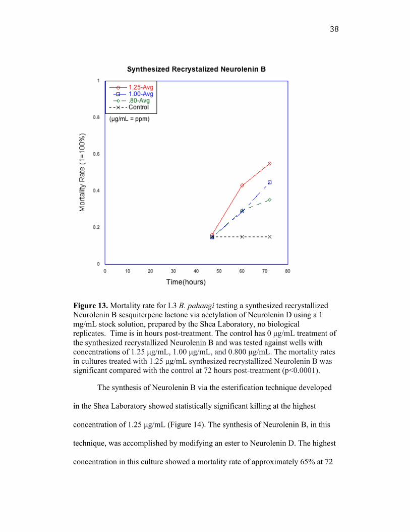
Figure 13. Mortality rate for L3 B. pahangi testing a synthesized recrystallized Neurolenin B sesquiterpene lactone via acetylation of Neurolenin D using a 1 mg/mL stock solution, prepared by the Shea Laboratory, no biological replicates. Time is in hours post-treatment. The control has 0 µg/mL treatment of the synthesized recrystallized Neurolenin B and was tested against wells with concentrations of 1.25 µg/mL, 1.00 µg/mL, and 0.800 µg/mL. The mortality rates in cultures treated with 1.25 µg/mL synthesized recrystallized Neurolenin B was significant compared with the control at 72 hours post-treatment (p<0.0001).
The synthesis of Neurolenin B via the esterification technique developed
in the Shea Laboratory showed statistically significant killing at the highest
concentration of 1.25 µg/mL (Figure 14). The synthesis of Neurolenin B, in this
technique, was accomplished by modifying an ester to Neurolenin D. The highest
concentration in this culture showed a mortality rate of approximately 65% at 72
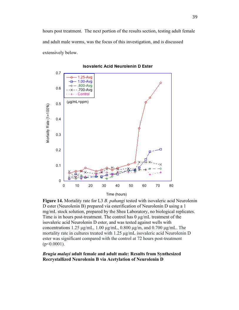
39
hours post treatment. The next portion of the results section, testing adult female
and adult male worms, was the focus of this investigation, and is discussed
extensively below.
Figure 14. Mortality rate for L3 B. pahangi tested with isovaleric acid Neurolenin D ester (Neurolenin B) prepared via esterification of Neurolenin D using a 1 mg/mL stock solution, prepared by the Shea Laboratory, no biological replicates. Time is in hours post-treatment. The control has 0 µg/mL treatment of the isovaleric acid Neurolenin D ester, and was tested against wells with concentrations 1.25 µg/mL, 1.00 µg/mL, 0.800 µg/m, and 0.700 µg/mL. The mortality rate in cultures treated with 1.25 µg/mL isovaleric acid Neurolenin D ester was significant compared with the control at 72 hours post-treatment (p<0.0001). Brugia malayi adult female and adult male: Results from Synthesized Recrystallized Neurolenin B via Acetylation of Neurolenin D
40
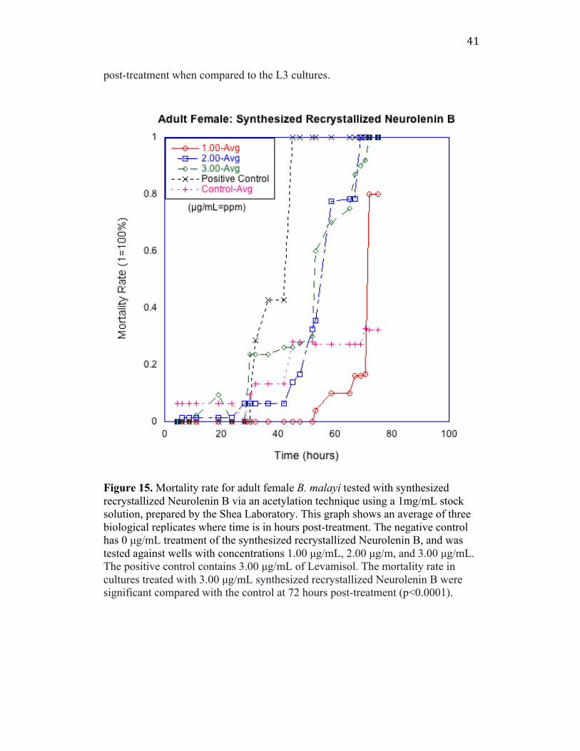
Mortality rates for adult females treated with recrystallized Neurolenin B
synthesized from Neurolenin D via the acetylation technique at the highest
concentration (3.00 µg/mL) showed statistically significant killing when
compared to the control (0 µg/mL) at 72hpt (Figure 15). At the highest
concentration, there was 100% mortality at both 72hpt. Similarly, mortality rates
for adult males treated with recrystallized Neurolenin B synthesized from
Neurolenin D via the acetylation technique at the highest concentrations (300
µg/mL) showed statistically significant killing when compared to the control (0
µg/mL) at 72hpt and 98 hours post-treatment (Figure 16). At the highest
concentration, there was 100% mortality at 98hpt. The adult cultures used B.
malayi as the experimental model and were observed for longer periods of time
41
post-treatment when compared to the L3 cultures.
Figure 15. Mortality rate for adult female B. malayi tested with synthesized recrystallized Neurolenin B via an acetylation technique using a 1mg/mL stock solution, prepared by the Shea Laboratory. This graph shows an average of three biological replicates where time is in hours post-treatment. The negative control has 0 µg/mL treatment of the synthesized recrystallized Neurolenin B, and was tested against wells with concentrations 1.00 µg/mL, 2.00 µg/m, and 3.00 µg/mL. The positive control contains 3.00 µg/mL of Levamisol. The mortality rate in cultures treated with 3.00 µg/mL synthesized recrystallized Neurolenin B were significant compared with the control at 72 hours post-treatment (p<0.0001).
42
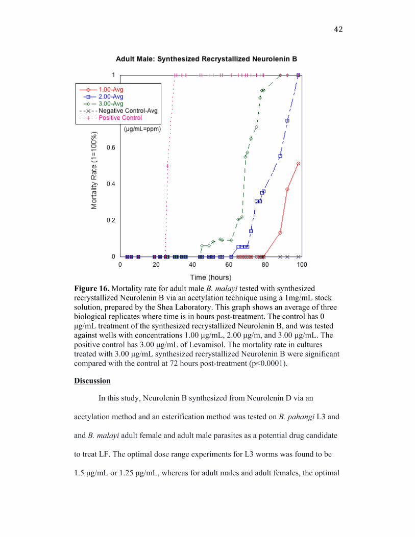
Figure 16. Mortality rate for adult male B. malayi tested with synthesized recrystallized Neurolenin B via an acetylation technique using a 1mg/mL stock solution, prepared by the Shea Laboratory. This graph shows an average of three biological replicates where time is in hours post-treatment. The control has 0 µg/mL treatment of the synthesized recrystallized Neurolenin B, and was tested against wells with concentrations 1.00 µg/mL, 2.00 µg/m, and 3.00 µg/mL. The positive control has 3.00 µg/mL of Levamisol. The mortality rate in cultures treated with 3.00 µg/mL synthesized recrystallized Neurolenin B were significant compared with the control at 72 hours post-treatment (p<0.0001).
Discussion In this study, Neurolenin B synthesized from Neurolenin D via an
acetylation method and an esterification method was tested on B. pahangi L3 and
and B. malayi adult female and adult male parasites as a potential drug candidate
to treat LF. The optimal dose range experiments for L3 worms was found to be
1.5 µg/mL or 1.25 µg/mL, whereas for adult males and adult females, the optimal
43
dose was found to be 3.00 µg/mL. This dose range reflects optimal concentrations
of treatment where worms die in a moderately gradual death in the desired range
of 50-100 hpt. Adult worms were observed for longer periods of time because this
study displayed the first time pure Neurolenin B (synthesized recrystallized
Neurolenin B) was tested on adult worms. A bioactive treatment must be potent
enough to kill worms gradually within a few days, but not kill the worms so
quickly where antigen from the worms is also released quickly. The faster the
antigen is released, the more likely it will lead to antigen dumping that would
shock the human host immune system. A treatment that kills all worms quickly
would be one where total worm death occurs between 10-30 hpt, and where death
is not gradual. These in vitro studies may not behave the same as in vivo studies,
but this investigation aims to explore dose-dependent mechanisms in the worms,
that must be considered in any future animal experiments.
All neurolenin products were generated by the Shea Laboratory and given
to the SAW Laboratory at a stock concentration of 1mg/mL in 70% EtOH unless
otherwise noted. These stocks were diluted to 0.100-3.00 µg/mL for L3 and adult
female and male cultures for this study. For L3 cultures, the concentrations
selected that showed the highest mortality rates were 1.50 µg/mL, 1.25 µg/mL,
1.00 µg/mL, .800 µg/mL, and .700 µg/mL. For adult cultures, the concentrations
selected that showed the highest mortality rates were 1.00 µg/mL, 2.00 µg/mL,
and 3.00 µg/mL. The positive control prepared for the adult cultures, Levamisol,
was prepared in a 1mg/mL stock solution in MEM at the highest concentration
tested in adults, 3.00 µg/mL The significance for the mortality rate at the highest
44
concentration in each adult and L3 culture was tested against 0 µg/mL in the
control well at 72hpt and 98hpt because we were following common
pharmaceutical reporting procedures. In these procedures, statistical significance
showing proof of the effectiveness of a drug at just one concentration is enough to
justify any kind of biological or anti-parasitic property.
The adult and L3 life stages of the parasite were the focus of this study
because adult worms are the primary cause for serious clinical symptoms in LF
and are not efficiently killed by current drugs. As aforementioned, current
treatments only target mf which prevent the early stages of transmission. At the
L3 stage, the worms enter the human host from the mosquito vector and by
targeting L3 worms, we can prevent the worms from making it to the adult stage.
L3 worms are also exemplary experimental parasites because they can grow and
molt in vitro without an animal host, giving an indication of worm health. This
study used B. pahangi in L3 cultures and B. malayi adult parasites because of
worm availability from the University of Georgia and also because the two
species are almost identical as experimental laboratory models.
Testing the crude N. labota Extracts: in vitro L3 Cultures
The crude extracts at the highest concentration, 400 µg/mL (p<. 0001) had
a mortality rate that showed statistically significant killing when compared to the
control (0 µg/mL) well at 72hpt (Figure 9). The worms were dying too rapidly, at
around 20 hpt, which, in this study is considered dangerous as it may lead to
immune shock or rapid “antigen dumping” in an animal or human host. The crude
extract was tested to determine if there was any kind of biological activity the
45
plant, before attempting isolation of specific sesquiterpene lactones or
neurolenins.
This experiment showed that L3s die when treated with the crude extract
at high concentrations. The crude extract was also tested at much lower
concentrations determined after purifying the crude extracts and testing partially
purified Neurolenin B and purified Neurolenin B via acetylation and esterification
methods (Figure 11). These concentrations for desired activity of the synthesized
compound were much lower, compared to crude extract, showing activity at 1.25
µg/mL, 1.00 µg/mL, 0.800 µg/mL, and 0.700 µg/mL. At these low concentrations,
the crude did not show high mortality rates, or strong biological activity.
Therefore in crude extracts, there are sesquiterpene lactones that have efficacy at
high doses, but do not have efficacy at much lower doses. Since the purified, non-
crude extracts have the ability to kill worms at much lower doses, this means that
the purified non-crude extracts must have much higher concentrations of the
biologically active form of neurolenin.
Testing Neurolenin B: in vitro L3 Cultures
This experiment aimed to first reconfirm the studies in previous SAW Lab
experiments. The first study confirmed that Neurolenin B was the most
biologically active sesquiterpene lactone from N. lobata, as shown in the literature
and as shown in the experiments from Kristine Trotta, Smith College (Trotta,
2014). However, the Shea lab, using Kristine Trotta’s method of isolating
Neurolenin B, confirmed that the Neurolenin B extract Trotta isolated was not in
fact pure Neurolenin B, but was a partially purified extract that still had traces of

46
other sesquiterpene lactones and other components of N. lobata. With this
partially purified Neurolenin B product, an optimal range of concentrations was
determined that killed the worms slowly and gradually, and within the 50-70 or
more hpt time period (Figure 10). The optimal range of concentrations was
determined by planning a titration experiment that tested a sequence of
concentrations designed to detect a range that killed the worms most effectively
(three to four concentrations), in the desired time frame. The range of optimal
concentrations lies in this range at 1.5 µg/mL, 1.25 µg/mL, 1.00 µg/mL, and 0.800
µg/mL where there was statically significant killing at 1.25 (p<. 0001) for the
partially purified Neurolenin B extract.
In collaboration with the Shea lab, a newly synthesized and purified
Neurolenin B product was used to test against the L3 B. pahangi worms. A fully
purified Neurolenin B was synthesized by acetylating Neurolenin D. This method
used acetic anhydride as a reagent, but this was later removed by recrystallizing
the purified product. This recrystallized product at 1.25 µg/mL had a mortality
rate that was statistically significant when compared to the control (0 at 1.25
µg/mL) at 72hpt. Although there was only a 60% mortality rate at the highest
concentration of 1.25 µg/mL by 72hpt (Figure 13), this marks the first time that a
fully purified Neurolenin B sesquiterpene lactone was synthesized in a laboratory
from the manipulation of Neurolenin D and tested on live parasites. In this
particular experiment, death recorded began at approximately 45 hpt due to
logistical and time complications, thus preventing the display of preceding data
points. It is evident that more purified samples of Neurolenin B may require a
47
higher dosage range to achieve 90% or more mortality rate at the 50-70 hour post-
treatment period for L3 larvae. However, if this culture was monitored for a
longer period of time, it is expected that a slow gradual death would be achieved
eventually. This culture proves that the acetylated product had effective killing
potential it was later tested at slightly higher concentrations on adult female and
male worms.
Another technique used to synthesize the Neurolenin B product was an
esterification procedure that also required modifications to Neurolenin D. The
modifications involved isovaleric acid and placement of an ester group on the
Neurolenin D sesquiterpene lactone. The mortality rate of the product with a
concentration of 1.25 µg/mL (p<. 0001) had statistically significant killing when
compared to the control well (0 µg/mL) at 72hpt. The optimal range of high
bioactivity and high mortality rates are similar to the previous experiment and are
as follows: 1.25 µg/mL, 1.00 µg/mL, 0.800 µg/mL, and 0.700 µg/mL (Figure 14).
However, there were solubility problems and the product was not fully dissolved
in the solvent when tested against L3 B. pahangi. Therefore, the product derived
from an esterification technique would likely be much more potent if a new
solvent was more effective in dissolving the product. Based on the collective data,
it is expected that a dissolved product would kill worms with a 90-100% mortality
rate, or higher than the 65% mortality rate achieved in this study on L3 larvae.
Future work should test the esterified product on adult worms after finding a safe
solvent candidate for the worms that also dissolves the esterified product
completely.
48
Regardless of the method used to synthesize Neurolenin B, the
sesquiterpene lactone itself had great efficacy across all cultures in terms of
interrupting the molting process. Each culture with a Neurolenin B product had
only partial molting, indicating that the L3 to L4 process was targeted. This may
lead to insights about the mechanism of action of Neurolenin B and its potentially
negative effects on cuticle formation. It may also lead to some conclusions
regarding effects on inhibiting the life cycle growth of these parasites as a whole.
Bio-inactive Neurolenin D: in vitro L3 Cultures
The study confirmed that Neurolenin D by itself was relatively bio-
inactive against L3 B. pahangi worms (Figure 12). This demonstrates that not all
sesquiterpene lactones in N. lobata extract contribute equally to its anti-parasitic
activity. It is also important to note that the worms in this culture were in poor
health after shipment, particularly the control well, where there was a higher
mortality rate compared to other wells with treatment. However, collaboration
with the Shea lab has led to some other conclusions about Neurolenin D. The N.
lobata plants were obtained from Belize, which were found to have high amounts
of Neurolenin D. N. lobata plants obtained from other locations apparently vary in
the amounts of different sesquiterpene lactones based on the genetic variability of
N. lobata in different geographic locations and climates (Sotes et al., 2015).
Because Neurolenin D was available in high amounts in the plants used in this
study, it was easy to isolate large quantities of Neurolenin D to synthesize
Neurolenin B, the most bioactive sesquiterpene lactone according to experiments
cited in the in literature (and also according to our results). Some of these papers
49
claim that Neurolenin B blocks the NPM/ALK pathway that triggers a signaling
cascade, typically leading to cell survival and “immortality” (Unger, 2015).
Neurolenin B: in vitro Adult Male and Adult Female Cultures
Both adult male and female cultures had mortality rates that showed
statistically significant killing at 3.0 µg/mL (p<0.0001) when testing synthesized
recrystallized Neurolenin B (using the modification of Neurolenin D to
Neurolenin B via the acetylation method) against B. malayi nematodes at 72 hours
post-treatment and 98hpt. In adult male cultures, 100% mortality was first
recorded at approximately 85hpt with a concentration of 3.00 µg/mL, and for
adult female cultures, 100% mortality was first recorded at approximately 70hpt
with a concentration of 3.00 µg/mL.
B. malayi nematodes were used in the adult cultures due to the lack of B.
pahangi nematodes available from the suppliers (University of Georgia). For the
adult female cultures, an average of three biological replicates showed moderate
increased death in the control wells due to worms injured in the process of plating
and likely also because of the poor health of the worms due to shipment. During
the course of monitoring the worms, mf were also monitored in the female
cultures, in vitro. Throughout the three adult cultures, the mf all died at 3.00
µg/mL alongside the death of the adult females. However, at 2.00 µg/mL and 1.00
µg/mL, mf slowed in motility, but did not die during the monitoring period. We
can assume here, that if this product was used at the lower concentrations (1.0
µg/mL and 2.0 µg/mL) as a suitable drug candidate for LF, it must be used in
conjunction with current drugs in the market that kill mf. For the adult worm
50
cultures, a positive control (Levamisol) and a negative control (no drug treatment)
were used. Levamisol is a known treatment for parasitic worm infections for
veterinary use that has many adverse side effects like the depletion of white blood
cells and complications with the nervous system. It is not approved for human use
(Drugs.com). The Levamisol treatment in both cultures achieved 100% mortality
at around 30-40hpt.
The adult cultures also were monitored well beyond 70 hpt, unlike the L3
cultures. The wells were monitored for a longer period of time because the wells
with concentrations 3.00 µg/mL, for both adult female and adult male worms,
died much faster than wells with the other concentrations. The adult cultures were
meant to be monitored as closely as possible, or at least until more worms died at
the lower concentrations to get an accurate estimate of total adult death in all
wells.
Another observation from the adult cultures includes a trend where the
adult female worms died at a faster rate than the adult male worms. The adult
female worms started showing death at approximately 30hpt, whereas adult males
started showing death at approximately 40hpt. This may give some insights about
the mechanism of action of the drug treatment.
Adult nematodes are the primary target of this study, as adult worms are
not efficiently killed by current drug treatments for LF. Developing a drug that
can efficiently kill adults, mf and L3 is a high priority of this project and these
results on adult make and female cultures are very encouraging that we are close
to achieving that objective.
51
Conclusions and Future Reseach
We conclude from this collaboration with the Shea Laboratory that both
acetylation and esterification techniques to convert Neurolenin D to Neurolenin B
produced active compounds with efficacy against adult, L3 and mf B. pahangi
and B. malayi nematodes. We can conclude from the results described in this
study that Neurolenin B is biologically active while Neurolenin D is relatively
biologically inactive. Synthesized recrystallized Neurolenin B (from Neurolenin
D using the acetylation method) showed statistically significant killing and was
biologically active when comparing mortality rates at the highest concentrations
(3.00 µg/mL and 1.25 µg/mL) with the control wells (0 µg/mL) against L3, adult
female and adult male nematodes, and mf at 72hpt and 98hpt. Synthesized
recrystallized Neurolenin B, produced in the Shea Laboratory using the
acetylation technique, is the purist form of Neurolenin B we have been able to test
so far. Isovaleric acid Neurolenin D ester (which results in Neurolenin B) showed
significant killing and was biologically active when comparing mortality rates at
the highest concentration, 1.25 µg/mL against L3 nematodes at 72hpt. There were
solubility issues discovered in the esterified product, and it is expected that the
product is more potent than our measurements indicate, due to these solubility
issues. Despite this solubility issue, significant L3 death was demonstrated at
~50hpt. The results from the esterified Neurolenin D product, along with its
solubility issues led us to use the acetylated Neurolenin D product on adult
nematodes, which showed significant killing and biological activity at 3.00
µg/mL.
52
We plan to continue conducting biological replicate experiments in adult
and L3 worms for both Neurolenin B produced from Neurolenin D by acetylation
and esterification techniques to confirm biological activity and significant killing
across all life stages of the parasite. It is clear that the required dosage differed for
L3 and adult nematodes. It would be helpful to perform RNA-seq expression
analysis on adult males and adult females, along with L3, to see how life stage
and sex can impact which genes are differentially expressed in response to
treatment. By observing gene expression analysis, we can better understand the
mechanism of action Neurolenin B has on filarial nematodes.
For future studies, we aim to explore growing techniques of N. lobata in a
location site at or near Smith College. The Shea Laboratory observed varying
concentrations of the different neurolenins based on the origin of the plant. The
plants purchased for this study were from Belize, and were observed to have a
high concentration of Neurolenin D. We expect that N. lobata found in other parts
of Central and South America will have genetic variability resulting in different
concentrations of the various neurolenins. We hope to grow N. lobata at the Smith
College Botanical Garden to have a ready and consistent supply of neurolenins.
We also plant to use DNA barcoding to confirm the origin of any plant material
we obtain for our work.
In addition to demonstrating worm killing, we also want to demonstrate
that Neurolenin B is not mutagenic or toxic. The Ames mutagenicity test will be
used to determine the mutagenicity of the synthesized Neurolenin B produced by
acetylation and esterification. Mutagens have the ability to alter DNA sequences
53
by deleting or changing nucleotides. The Ames test is used to test the
mutagenicity of different compounds in bacteria. It uses mutant strains of
Salmonella typhimurium bacteria that can detect many different mutagenic agents.
By detecting potential mutagenic agents, we can determine if Neurolenin B is
mutagenic before proceeding to testing in animal models, or even for human
ingestion.
N. lobata has been used historically in traditional and indigenous
medicine, and no signs of mutagenicity or toxicity have been previously reported.
However, testing for potential mutagenicity remains a necessary step when
considering Neurolenin B as a suitable drug candidate for LF. Furthermore, we
hope to observe more L3 and adult cultures testing both Neurolenin B products
that were derived from Neurolenin D via acetylation and esterification techniques
to gather more data, biological replicates, and further confirmation of the results
presented in this study.
Ultimately, we hope to eventually partner with Glaxo Smith Kline, a
pharmaceutical company, to start testing our Neurolenin B products on rodent
hosts infected with B. pahangi to validate Neurolenin B as a suitable drug
candidate for LF.
54
Bibliography
Anil, N., Talluri, V. R. Lymphatic Filariasis: Drug targets and Nematicidal Plants, J. Pharm. Sci. & Res, 7(11): 2015, 928-933. Arena, J.P., et al. The Mechanism of Action of Avermectins in Caenorhabditis elegans: Correlation between Activation of Glutamate-Sensitive Chloride Current, Membrane Binding, and Biological Activity. The American Society of Parasitologists. 81(2): 1995, 286-94. http://www.jstor.org/stable/3283936. Berger, I., et al. Plants used in Guatemala for the treatment of protozoal infections - II. Activity of extracts and fractions of five Guatemalan plants against Trypanosoma cruzi. Journal of Ethnopharmacology. 62:1998, 107-115. DOI: 10.1016/S0378-8741(98)00011 Berger, I., Passreiter, C.M., Cáceres, A., Kubelka, W. Antiprotozoal Activity of Neurolaena lobata. Phytotherapy Research. 15: 2001, 327-330. DOI: 10.1002/ptr.782 Bockarie, MJ, Deb, RM Elimination of lymphatic filariasis: do we have the drugs to complete the job? Current Opinion in Infectious Diseases 23: 2010, 617-620, DOI: 10.1097/QCO.0b013e32833fdee5. Cano, J., Rebollo, M.P., Golding, N., Pullan, R.L., Crellen, T., Soler, A., Hope., L.A.K., Lindsay, S.W., Hay, S.I., Bockarie, M.J., Brooker, S.J. The global distribution and transmission limits of lymphatic filariasis: past and present. BioMed Central, Parasites & Vectors, 2014, 466, DOI: 10.1186/s13071-014-0466-x. Castillo, J.B., Ferrero, T.M., Luis, F.R., Bueno, P.V., Gupta, M.P., Nathan, P.J., Panam Flora: New Sesquiterpene Lactones from Neurolaena Lobata. Journal of Natural Products, 45(6): 1982, 762-765. Centers for Disease Control and Prevention (CDC). Biology - Life Cycle of Wuchereria bancrofti. September 2013. http://www.cdc.gov/parasites/lymphaticfilariasis/biology_w_bancrofti.html Centers for Disease Control and Prevention (CDC). Global Health - Neglected Tropical Diseases. 2013. http://www.cdc.gov/globalhealth/ntd/ Centers for Disease Control and Prevention (CDC). Mass Drug Administration for the Elimination of Lymphatic Filariasis - Port-au-Prince, Haiti, 2011-2012. Centers for Disease Control and Prevention Morbidity and Mortality Weekly Report 62 (23):14 June 2013 http://www.cdc.gov/mmwr/pdf/wk/mm6223.pdf
55
Centers for Disease Control and Prevention (CDC). Parasites-Lymphatic Filariasis. 2013. http://www.cdc.gov/parasites/lymphaticfilariasis/biology.html Centers for Disease Control and Prevention (CDC). Parasites- Lymphatic Filariasis - Guidance for Evaluation and Treatment. 2013. http://www.cdc.gov/parasites/lymphaticfilariasis/health_professionals/dxtx.html Chaturvedi, D. Sesquiterpene lactones: Structural diversity and their biological activities. In Opportunity, Challenge and Scope of Natural Products in Medicinal Chemistry: Kerala India, Chapter 10: 2011, 313-334. http://www.trnres.com/ebook/uploads/tiwari/T_1302158793Tiwari-10.pdf Crump A, Omura S. Ivermectin, 'Wonder drug' from Japan: the human use perspective. Proceedings of the Japan Academy, Series B. 87: 2011, 13-28. DOI: 10.2183/pjab.87.13. Drug Bank. Identification, Albendazole. Drug and Drug Target Database, 2014. http://www.drugbank.ca/drugs/DB00518 Drugs.com, Levamisole, Conditions and Cancer, Levamisole. 2016. Drugs.com François, G., Passreiter, C.M., Woerdenbag. H.J., Van Looveren, M. Antiplasmodial activities and cytotoxic effects of aqueous extracts and sesquiterpene lactones from Neurolaena lobata. NCBI. 62(2): 1996, 126-9. http://www.ncbi.nlm.nih.gov/pubmed/8657743 Franssen F.F.J, et al. In Vivo and In Vitro Antiplasmodia Activities of Some Plants Traditionally Used in Guatemala against Malaria. Antimicrobial Agents and Chemotherapy. 41(7): 1997, 1500-1503. http://aac.asm.org/content/41/7/1500.long Fujimaki, Y., et al. Macrofilaricidal and macrofilaricidal effects of Neurolaena lobata, a Guatemalan medicinal plant, on Brugia pahangi. Journal of Helminthology 79: 2005, 23-28. DOI: 10.1079/JOH2004262. Global Alliance to Eliminate Lymphatic Filariasis (GAELF). Epidemiology. August 2013. http://www.filariasis.org/epidemiology.html Klei, T. R., & Rajan, T. V. (Eds.) The Filaria (5): 2002. Springer. http://www.springer.com/us/book/9781402070389 Leon-Levy Native Plant Preserve. Plant Information: Neurolaena lobata. August 2013. http://www.levypreserve.org/Plant-Listings/Neurolaena-lobata

56
Manchand, P.S., Blout, J.F., Chemical constituents of tropical plants. 11 stereostructures of neurolenins A and B, novel germacranolide sesquiterpenes from Neurolaena lobata. J. Org. Chem, 43(22): 1978, 4352-4354. Michael, E., Malecela-Lazaro, M. N., Simonsen, P. E., Pedersen, E. M., Barker, G., Kumar, A., and Kazura, J.W. Mathematical Modeling and the Control of Lymphatic Filariasis, The Lancet infectious diseases, 4:2004, 223-234. Micromedex, Albendazole (Oral Route), Drugs and Supplements. Mayo Clinic, Truven Health Analytics, 2015. http://www.mayoclinic.org/drugs-supplements/albendazole-oral-route/description/drg-20061505 Molyneux, D.H., Bradley, M., Hoeraul, A., Kyelem D., Taylor, M., Mass drug treatment for lymphatic filariasis and onchocerciasis, Trends in Parasitology. 19(10): 2003, 516-522. Muelas-Serrano S et al. In vitro screening of American plant extracts on Trypanosoma cruzi and Trichomonas vaginalis. Journal of Ethnopharmacology.71: 2000, 101-107. DOI: 10.1016/S0378-8741(99)00185-3. Murthy P.K., Joseph S.K., Murthy P.S.R. Plant products in the treatment and control of filariasis and other helminth infections and assay systems for antifilarial/antihelminthic activity. Planta Medica, 77 (6): 2011, 647-661. DOI: 10.1055/s-0030-1250452. Novaes, R.M.L., Rodrigues, J.G., and Lovato, M.B. “An efficient protocol for tissue sampling and DNA isolation from the stem bark of Leguminosae trees.” 8(1): February 2009, 86-96. Ottesen, E. A., Duke, B. O. L., Karam, M., Behbehani, K. Strategies and tools for the control/elimination of lymphatic filariasis. Bull. World Health Org. 75(6): 1997, 491-503 Rajan, T.V., Paciorkowski, N., Kalajzic, I., McGuiness, C.. Absorbic acid is a requirement for the morphogenesis of the human filarial parasite Brugia malayi. J. of Parasitology 89(4): 2003, 868-870. Shea, Kevin Laboratory. Katie McGeough and Megan Neubig at Smith College, Honors Thesis Project, 2015-16. Si-Yuan Pan, Gerhard Litscher, Si-Hua Gao, et al., “Historical Perspective of Traditional Indigenous Medical Practices: The Current Renaissance and Conservation of Herbal Resources,” Evidence-Based Complementary and Alternative Medicine, 2014, 1-20. Article ID 525340,DOI: 10.1155/2014/525340 Sotes, G.J., Lohengrin C.A., Montesinos, D., Coutinho, A.X., Pelaex, W.J., Lopes, S.M., Pinho e Melo, T. Inter-regional variation of leaf surface defenses in
57
native and non-native Centaurea solstitialis plants. Science Direct, Biochemical and Systematics and Ecology. 62 (208): 2015, 218. Plaisier, A. P., Stolk, W. A., Van Oortmarssen, G. J., & Habbema, J. D. F. Effectiveness of Annual Ivermectin Treatment for Wuchereria bancrofti infection. Parasitology Today, 16(7): 2000, 298-302. Taylor, M.J., Hoerauf, A., Bockarie, M. Lymphatic filariasis and onchocerciasis. Lancet. 376: 2010, 1175-85. DOI: 10.1016/S0140-6736(10)60586-7. Trotta, K.L, Analysis of Natural Products from Neurolaena lobata as Candidate Antifilarial Agents Against the Parasitic Nematode Brugia pahangi. Smith College Honors Thesis, Department of Biological Sciences, 2014. Unger, C., Kiss, I., Vasas, A., Lajter, I., Kramer, N., Astanosov, A.G., Nguyen, C.H., Chatuphonprasert W., Brenner, S., Krieger, S., et al., The germacranolide sesquiterpene lactone neurolenin B of the medical plan Neurolaena lobata (L.) R.Br. ex Cass inhibits NPM/ALK-driven cell expansion and NF-KB-driven tumour intravasation, Phytomedicine. 22: 2015, 862-874. Veeresham, C., Natural Products dervied from plants as a source of drugs. J. Adv. Pharm. Technol. Res. 3(4): 2012, 200-201. DOI: 10.4103/2231-4040.104709 Walshe-Roussel, B. et al. Potent anti-inflammatory activity of sesquiterpene lactones from Neurolaena lobata (L.) R. Br. ex Cass., a Q'eqchi Maya traditional medicine. Phytochemistry. 92: 2013, 122-127. DOI: 10.1016/j.phytochem/2013.05.004. Wayangankar, S., Chandrasekar P.H., Bronze M.S., Jackson, R.L.J., Filariasis Clinical Presentation. Medscape: Drugs and Diseases, 2015. http://emedicine.medscape.com/article/217776-clinical Wignall, J. et al. Medicinal Plants: Hoax or Potential Breakthrough in Drug Development for Lymphatic Filariasis? Presented at Celebrating Collaborations, Smith College, Northampton MA, April 2013. World Health Organization (WHO), Lymphatic Filariasis, Epidemiology, WHO, 2016. http://www.who.int/lymphatic_filariasis/epidemiology/en/ World Health Organization (WHO), Lymphatic Filariasis. Programmes, What is lymphatic Filariasis? WHO, 2016. http://www.who.int/lymphatic_filariasis/about/en/ Zeldenryk, L. M., Gray, M., Speare, R., Gordon, S., Melrose, W. The emerging story of disability associated with lymphatic filariasis: a critical review. PLoS neglected tropical diseases. 5(12): 2011, e1366