Embed Size (px)
Citation preview

From DEPARTMENT OF MEDICAL BIOCHEMISTRY AND
BIOPHYSICS
Karolinska Institutet, Stockholm, Sweden
EXPLORING NOVEL ROLES OF TUMOR PERICYTES
Jong Wook Hong
Stockholm 2015

All previously published papers were reproduced with permission from the publisher.
Published by Karolinska Institutet.
Printed by E-Print AB 2015
© Jong Wook Hong, 2015
ISBN 978-91-7676-132-8

EXPLORING NOVEL ROLES OF TUMOR PERICYTES
THESIS FOR DOCTORAL DEGREE (Ph.D.)
By
Jong Wook Hong
Principal Supervisor:
Associate Professor Guillem Genové
Karolinska Institutet
Department of Medical Biochemistry and
Biophysics
Division of Vascular Biology
Co-supervisor(s):
Professor Christer Betsholtz
Karolinska Institutet
Department of Medical Biochemistry and
Biophysics
Division of Vascular Biology
Uppsala University
Department of Immunology, Genetics and
Pathology
Division of Cancer and Vascular Biology
PhD Mirjana Poljakovic
Karolinska Institutet
Department of Molecular Medicine and Surgery
Division of Urology
Opponent:
Professor Sven Påhlman
Lund University
Department of Laboratory Medicine, Lund
Center for Molecular Pathology
Examination Board:
Associate Professor Anna Dimberg
Uppsala University
Department of Immunology, Genetics and
Pathology
Division of Cancer and Vascular Biology
Associate Professor Theodoros Foukakis
Karolinska Institutet
Department of Oncology-Pathology
Associate Professor Anna Darabi
Lund University
Department of Clinical Sciences, Lund
Division of Neurosurgery


To my family


ABSTRACT
Tumor biology has been extensively studied over the last few decades, with a principal focus
on how neoplastic cells obtain cellular immortality. A number of oncogenes and tumor
suppressor genes have been uncovered that regulate cancerous transformation. However,
tumors are now believed to be complex tissues consisting of various kinds of tumor stromal
cells as well as transformed cancer cells. The tumor stroma is mainly comprised of tumor-
infiltrating leukocytes, cancer-associated fibroblasts, vascular endothelial cells, lymphatic
endothelial cells, tumor pericytes, and extracellular matrix. These cells have an extensive
interplay with one another or with cancer cells, from the initiation stage of tumor
development to its metastatic dissemination.
The aim of this thesis was to investigate the tumor pericytes—one of the tumor stroma
constituents that has not been widely explored—in order to determine their novel roles in
tumor malignancy. The use of a pericyte-deficient mouse model (pdgfbret/ret), in paper I,
confirmed that the myeloid-derived suppressor cells (MDSCs), one of the most aggressive
types of tumor-infiltrating leukocytes, are significantly increased both at the tumor site and in
the peripheral blood in B16 melanoma and Lewis lung carcinoma (LLC) subcutaneous mouse
models of pdgfbret/ret, compared to their littermate controls. The increase in the MDSC
number was dependent on expression of tumor-derived IL-6, induced by the hypoxic tumor
microenvironment in pericyte-deficient B16 and LLC tumors. Analysis of gene expression in
human samples (253 breast cancer patients of an Uppsala dataset) showed an inverse
correlation between human pericyte-related genes and human MDSC markers and a
subsequent relevance to the survival rate of breast cancer patients.
The relevance of the tumor pericytes to other tumor stroma cells was studied in paper II,
which revealed a comparable abundance of PDGFRα- expressing perivascular cells in
pericyte-deficient B16 melanomas. A further investigation identified the PDGFRα-expressing
perivascular cells as “specialized myofibroblasts” with gene signature features of both
fibroblast-related (Fap, Pdgfra, Hgf) and pericyte-related (Cspg4, Pdgfrb, Asma) gene sets.
Moreover, pericyte-deficient, B16-melanoma-bearing mice showed an elevated level of the
serum S100B protein, which is widely considered to be a distinctive prognostic marker for
malignant melanoma patients. The B16 melanoma tumor cell-derived S100B was then
confirmed to pass into the peripheral blood. Presumably “activated endothelial cells” in
pdgfbret/ret mice would facilitate more rapid transport of S100B protein via endothelial
caveola-mediated transcytosis.
In conclusion, tumor pericytes directly interact with adjacent endothelial cells, thereby
controlling the tumor vasculature and further changing the tumor microenvironment. Tumor
pericytes favor tumor immunogenicity by blocking systemic MDSC bursts in experimental
mouse models (B16, LLC) and are also negatively involved in recruitment of the perivascular
myofibroblasts. However, the biological relevance of the perivascular myofibroblasts should
be further investigated.

LIST OF SCIENTIFIC PAPERS
I. Hong, J., N.P. Tobin, H. Rundqvist, T. Li, M. Lavergne, Y. Garcia-Ibanez, H. Qin, J. Paulsson, M. Zeitelhofer, M.Z. Adzemovic, I. Nilsson, P. Roswall, J. Hartman, R.S. Johnson, A. Ostman, J. Bergh, M. Poljakovic, and G. Genove. 2015. Role of tumor pericytes in the recruitment of myeloid-derived suppressor cells. Journal of the National Cancer Institute. 107(10):pii:djv209.
II. Hong, J., Y. Garcia-Ibanez, H. Qin, M. Lavergne, T. Li, J. Andrae, and G. Genove. 2015. Analysis of the melanoma perivascular cells reveals recruitment of myofibroblasts in the absence of pericytes. Manuscript in preparation
Other publications not included in the thesis
Niaudet, C., J.J. Hofmann, M.A. Mae, B. Jung, K. Gaengel, M. Vanlandewijck, E. Ekvarn, M.D. Salvado, A. Mehlem, S. Al Sayegh, L. He, T. Lebouvier, M. Castro-Freire, K. Katayama, K. Hultenby, C. Moessinger, P. Tannenberg, S. Cunha, K. Pietras, B. Lavina, J. Hong, T. Berg, and C. Betsholtz. 2015. Gpr116 receptor regulates distinctive functions in pneumocytes and vascular endothelium. PloS one. 10:e0137949.
Zang, G., K. Gustafsson, M. Jamalpour, J. Hong, G. Genove, and M. Welsh. 2015. Vascular dysfunction and increased metastasis of B16F10 melanomas in Shb deficient mice as compared with their wild type counterparts. BMC cancer. 15:234.

CONTENTS
1 Introduction ..................................................................................................................... 1
1.1 Tumor microenvironment (TME) ......................................................................... 1
1.1.1 Tumor-associated endothelial cells ........................................................... 3
1.1.2 Tumor pericytes......................................................................................... 6
1.1.3 Cancer-associated fibroblasts ................................................................... 9
1.1.4 Tumor-infiltrating leukocytes (TILs) ..................................................... 13
1.2 Cancer biomarkers ............................................................................................... 17
1.2.1 S100A8/9 and S100B .............................................................................. 17
1.2.2 Serum IL6 ................................................................................................ 18
2 Aim of the thesis ............................................................................................................ 21
3 Results and discussion ................................................................................................... 23
3.1 PAPER I ............................................................................................................... 23
3.2 PAPER II ............................................................................................................. 24
4 Acknowledgments ......................................................................................................... 26
5 References ..................................................................................................................... 29

LIST OF ABBREVIATIONS
ANG1, ANG2 Angiopoietin 1, Angiopoietin 2
Bregs Regulatory B cells
CAFs Cancer associated fibroblasts
CAV1, CAV2, CAV3 Caveolin1, Caveolin2, Caveolin3
CTLs Cytotoxic T lymphocytes
ECM Extracellular matrix
EMT Epithelial–mesenchymal transition
EndMT Endothelial-mesenchymal transition
LLC Lewis lung carcinoma
MDSCs Myeloid-derived suppressor cells
Mo-MDSCs Monocytic-myeloid-derived suppressor cells
G-MDSCs Granulocytic- myeloid-derived suppressor cells
NK cells Natural killer cells
NKT cells Natural killer T cells
pDCs Plasmacytoid dendritic cells
pdgfbret/ret PDGFB retention motif knock out
TERT Telomerase reverse transcriptase
TILs Tumor-infiltrating leukocytes
TAMs Tumor-associated macrophages
Th17 cells T helper 17 cells
TME Tumor microenvironment
Tregs Regulatory T cells

1
1 INTRODUCTION
1.1 TUMOR MICROENVIRONMENT (TME)
In the last few decades, extensive documentation has revealed how incipient cancer cells
acquire the traits of endless proliferation and aggressiveness, termed “tumor malignancy.” In
accordance with the advent of new molecular biology techniques, a series of discrete steps of
tumorigenesis has now been well established, so that the “multistep process of
tumorigenesis” is known to comprise the following: cellular transformation into neoplastic
cells; angiogenesis; motility and invasion via blood and lymphatic vessels; formation of multi
cell aggregates with platelets or leukocytes; embolism and circulation; arrest in capillary
beds; extravasation into organ parenchyma; colonization (adaptation to new
microenvironment, establishment of micrometastases and macrotumors); and the metastasis
of metastatic tumors to other organs (figure 1) [1].
Figure. 1. Multistep process of tumorigenesis
These complex serial processes always begin with genetic changes in normal cells, whereby
various kinds of extracellular or intracellular stimuli elicit aberrant gene expressions. The
combination of irregular genetic changes, in turn, converts neoplastic cells into transformed
cancerous cells. Many tumors show genetic instability that persists from the beginning (tumor

2
initiation) stage and this instability is even amplified at the late developmental stage of
tumorigenesis—metastasis—as the intrinsic intracellular maintenance and repair system
breaks down.
Classically, the genetic changes that occur subsequent to genetic instability in cancer cells are
categorized as follows: the activation of proto-oncogenes (e.g., RAS, EGFR, MYC, and
ABL1), the inactivation of tumor-suppressor genes (e.g., BRCA1, BRCA2, PTEN, RB, and
TP53), and the inactivation of genomic stability genes that encode telomerase or DNA
mismatch repair machinery-related proteins [2]. A number of cancer cell-associated genes,
oncogenes, and tumor suppressor genes have been reported in various cancer types and at
different developmental stages of tumorigenesis, but their expression patterns show
considerable heterogeneity. The recent advancements in techniques for high throughput
sequencing of DNA or mRNA (whole genome sequencing and whole transcriptome
sequencing) have allowed resolution and refinement of these heterogeneous patterns in
certain human tumors; for example, in prostate cancer, chromosomal rearrangements of the
ETS transcription factor genes with the androgen responsive promoter, TMPRSS2–ERG
fusions, were discovered in up to 50% of cases, while in melanoma, mutations in the TERT
promoter appear in approximately 70% [3].
The notion that gene expression profiles vary in different developmental stages of
tumorigenesis or in discrete cancer types can be explained by the following two features.
First, the traits of transformed cancer cells mostly resemble the traits of their normal
counterparts. Second, neoplastic cells cross talk with their normal neighboring cells and, in
turn, establish unique and divergent genetic aberrations. The latter concept has been
aggressively studied during the last two decades and is now well appreciated to be a crucial
part of the development of tumorigenesis. Various kinds of host-originated cells are
interconnected with neoplastic cancer cells during the whole process of tumorigenesis; these
include tumor cell-neighboring regional fibroblasts, new blood or lymphatic vessels sprouted
from neighboring tissues, pericytes, many types of bone marrow-derived cells, and
circulating or regional inflammatory cells. Their reciprocal crosstalk change both cancer cells
and host-derived cells, thereby establishing a unique tumor tissue. Most of the tumor stroma
components are reprogrammed and distorted from their normal counterparts to favor tumor
malignancy (figure 2). In some human solid tumors, patho-oncologists have measured tumor–
stroma ratios using conventional histopathological analysis with hematoxylin & eosin stained
tumor sections and have showed that stroma-enriched tumors are associated with a poor
prognosis in breast (triple negative), colon, esophagus, lung (non-small cell), liver
(hepatocarcinoma), and cervical cancers [4]. As such, the tumor microenvironment is
implicated in the support of tumor progression rather than in its restraint. This notion,
however, is still controversial and should be addressed with more research.
The recent study of pancreatic ductal adenocarcinoma in a mouse model can be exemplified
as countering the concept of a tumor-supportive role of the tumor microenvironment, as
depletion of αSMA-positive myofibroblasts in the tumor microenvironment by genetic
alteration induced greater malignancy in transformed pancreatic cancer cells [5]. As such,

3
some controversy exists regarding whether tumor-neighboring tissues are bystanders that
perform some passive actions or whether they actively participate in tumorigenesis in a
supportive or suppressive way. A better understanding of how the reprogramed tumor stroma,
termed the tumor microenvironment (TME), interplays with cancer cells should help in
establishing targeted therapies to eliminate tumor progression and metastasis.
Figure. 2. The tumor microenvironment (TME)
1.1.1 Tumor-associated endothelial cells
The multistep development of tumorigenesis requires that tumor cells receive a supply of
nutrients and oxygen, and that waste products be extravasated through blood vessels, in the
same manner as required by normal cells. The establishment of new blood vasculature within
tumor tissues therefore represents a key step in the excessive and abrupt proliferation of
cancer cells, which rarely occurs in normal cells under physiological conditions in adults.
A well-known concept in tumor biology is that the formation of new blood vasculature is
generated by “angiogenesis,” the sprouting of new vessels from pre-exiting ones in response
to pro-angiogenic signals (e.g., VEGFA, FGFs) that react with the endothelial cells via the
extracellular domains of their membrane-anchored receptors [6]. During the embryonal
development stage, angiogenesis occurs vigorously and triggers blood vessel-sprouting from
the existing vessel tubing, in which endothelial progenitor cells have assembled and
differentiated into endothelial cells (vasculogenesis). In adults, wound healing represents a
rare case that requires angiogenesis to form new vessels in lesions.
The normal vasculature is well organized, differentiated, and classified into arteries,
arterioles, veins, venules and capillaries, and each type of vasculature has its own phenotype
4
and function [7]. In contrast, tumor vasculature is chaotic, having considerable variations in
phenotype and is essentially unclassifiable. Phenotypically, tumor vessels appear irregularly
branched, enlarged, hemorrhaging, and irregular in their blood flow. During the whole
developmental stage of tumor progression and metastasis, the proangiogenic signals, which
are mainly secreted by tumor parenchyma and partially by the tumor stroma, overwhelm their
antiangiogenic counterparts, thereby causing the angiogenic switch to be invariably turned on
[8].
Previous reports have confirmed that tumor angiogenesis occurs from a capillary or venule in
the nearby tissue, where the tumor grows, and the newly formed vessels branch into the
intratumoral region. Recent studies, however, have suggested that vasculogenesis also
includes the new formation of blood vessels from endothelial progenitor cells during tumor-
associated neovascularization. Endothelial progenitor cells are mostly originated from bone
marrow-derived cells, including hematopoietic stem cells, myeloid progenitors, and
mesenchymal stem cells. Bone marrow transplantation using EGFP or LacZ-marked reporter
mice confirmed that various subsets of bone marrow-derived cells are recruited at an
implanted tumor locus and become integrated into nascent vessels [9]. Whether the
endothelial progenitor cells affect to any great extent the tumor-associated neovascularization
remains arguable, due to the very low rate of integration and the invalidated identification of
endothelial progenitor cells in tumor vessels. In addition, tumor-associated neovascularization
has been shown to be accomplished by the neoplastic cancer cells themselves. In
glioblastoma, a fraction of cancer cells expressing CD133, believed to be a cancer-stem cell
marker, differentiates into endothelial progenitor cells as well as tumor cells, which thereafter
have the features of endothelial hyperplasia in the glioblastoma tissue [10]. As such, tumor-
associated endothelial cells can be obtained from three different routes: neighboring vessels
and host-derived and cancer cell-derived endothelial progenitor cells.
In the context of cancer therapy, tumor-associated endothelial cells have been recognized as
an “attractive target.” Tremendous efforts have been made to block tumor angiogenesis in the
last few decades. A few drug candidates, including bevacizumab (a neutralizing monoclonal
anti VEGFA antibody) have gone through clinical trials and have been approved for clinical
use in combination with chemotherapy or cytostatic reagents for patients with some advanced
cancers, including metastatic colorectal cancer, advanced non-small cell lung cancer,
advanced ovarian cancer, advanced cervical cancer, metastatic renal cell carcinoma, and
recurrent glioblastoma, which reflects their limited efficacy [11-13].
Recent preclinical and clinical studies have focused on a newer aspect of angiogenesis
regulation: the endogenous inhibitors of angiogenesis (angiostatin and endostatin). These are
counter-balancing molecules against proangiogenic factors, and play important roles in the
development of tumorigenesis by suppressing tumor angiogenesis, thereby inducing
dormancy, especially in distant micrometastases. Nevertheless, single targets against tumor-
associated endothelial cells are recognized as still insufficient for cancer treatment [11].
5
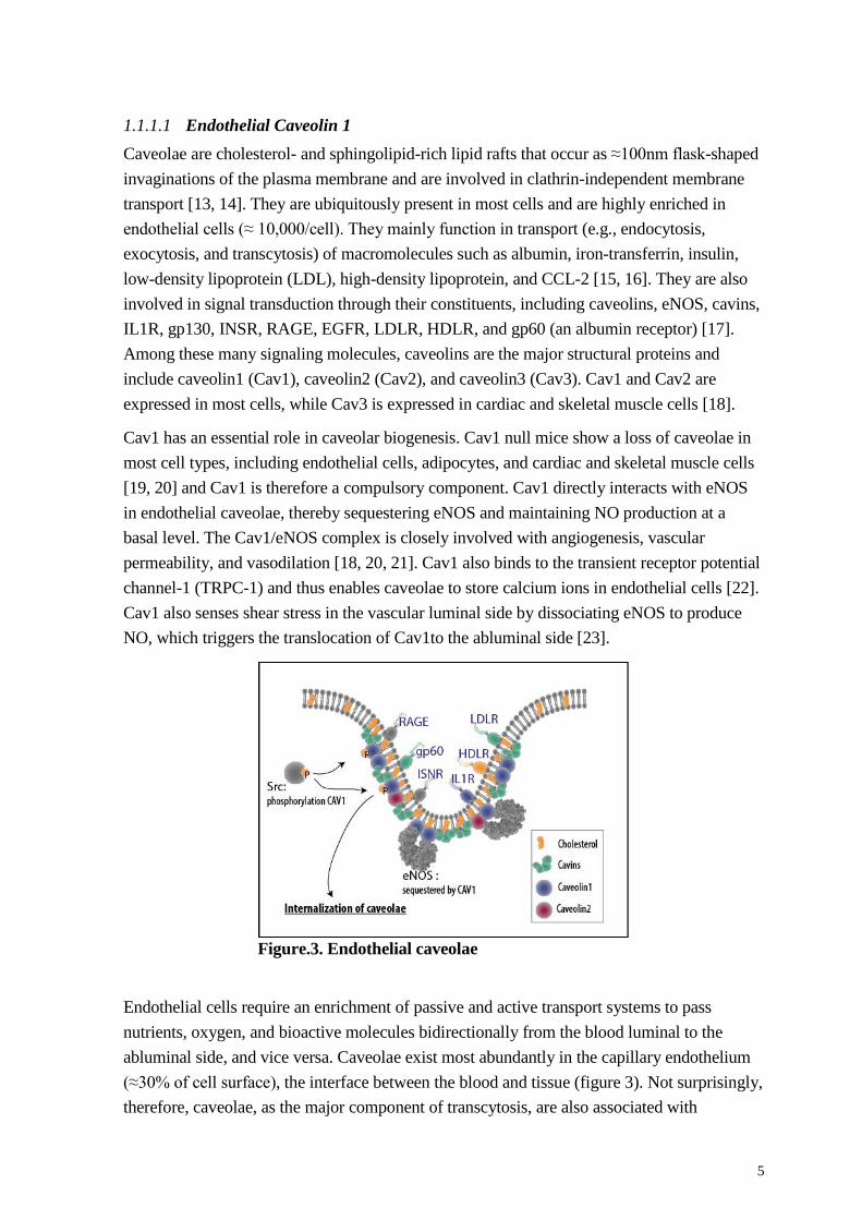
1.1.1.1 Endothelial Caveolin 1
Caveolae are cholesterol- and sphingolipid-rich lipid rafts that occur as ≈100nm flask-shaped
invaginations of the plasma membrane and are involved in clathrin-independent membrane
transport [13, 14]. They are ubiquitously present in most cells and are highly enriched in
endothelial cells (≈ 10,000/cell). They mainly function in transport (e.g., endocytosis,
exocytosis, and transcytosis) of macromolecules such as albumin, iron-transferrin, insulin,
low-density lipoprotein (LDL), high-density lipoprotein, and CCL-2 [15, 16]. They are also
involved in signal transduction through their constituents, including caveolins, eNOS, cavins,
IL1R, gp130, INSR, RAGE, EGFR, LDLR, HDLR, and gp60 (an albumin receptor) [17].
Among these many signaling molecules, caveolins are the major structural proteins and
include caveolin1 (Cav1), caveolin2 (Cav2), and caveolin3 (Cav3). Cav1 and Cav2 are
expressed in most cells, while Cav3 is expressed in cardiac and skeletal muscle cells [18].
Cav1 has an essential role in caveolar biogenesis. Cav1 null mice show a loss of caveolae in
most cell types, including endothelial cells, adipocytes, and cardiac and skeletal muscle cells
[19, 20] and Cav1 is therefore a compulsory component. Cav1 directly interacts with eNOS
in endothelial caveolae, thereby sequestering eNOS and maintaining NO production at a
basal level. The Cav1/eNOS complex is closely involved with angiogenesis, vascular
permeability, and vasodilation [18, 20, 21]. Cav1 also binds to the transient receptor potential
channel-1 (TRPC-1) and thus enables caveolae to store calcium ions in endothelial cells [22].
Cav1 also senses shear stress in the vascular luminal side by dissociating eNOS to produce
NO, which triggers the translocation of Cav1to the abluminal side [23].
Figure.3. Endothelial caveolae
Endothelial cells require an enrichment of passive and active transport systems to pass
nutrients, oxygen, and bioactive molecules bidirectionally from the blood luminal to the
abluminal side, and vice versa. Caveolae exist most abundantly in the capillary endothelium
(≈30% of cell surface), the interface between the blood and tissue (figure 3). Not surprisingly,
therefore, caveolae, as the major component of transcytosis, are also associated with

6
pathological conditions including atherosclerosis, diabetes, and hyperlipemia [17, 24]. The
manner in which caveolae-mediated transcytosis and endocytosis occur and which molecular
interactions control each step of the serial processes remain largely unresolved questions.
Recent evidence, in part, has shown that the initial step of the caveola-mediated transcellular
pathway requires the phosphorylation of Cav1 (pT14). In the lung microvessel, tyrosine
phosphorylation of Cav1, mediated by oxidant stress (hydrogen peroxidase), induced
transcellular albumin hyperpermeability, with Src identified as the upstream kinase that
performed the phosphorylation. Interestingly, a high degree of oxidant stress also induced
paracellular permeability by dissociating VE-cadherin from membrane-bound β-catenin. In
mouse embryonal fibroblasts (MEFs), phospho-Cav1 mediated the internalization of
cholesterol-dependent clusters of GM1 and GPI-linked protein (caveolae), implying that
phospho-Cav1 is not limited to the endothelial caveolae pathway [25, 26].
In tumor endothelial cells, the irregularly active state of transcellular signaling pathway is
presumed to occur in the tumor endothelium, where it positively modulates TME for tumor
malignancy, taking into account both notions that 1) the aberrant tumor vessels are the main
passage for the considerable amount of tumor-derived factors, and 2) only small molecules (<
3nm) pass through via the paracellular pathway.
1.1.2 Tumor pericytes
Pericytes are specialized mesenchymal cells that regulate angiogenesis and vascular integrity.
As inferred by their name, pericytes lie in the perivascular niche, where the basement
membrane anchors pericytes and endothelial cells, enabling their close interconnection.
Pericytes are present in the microvessels—capillaries, postcapillary venules, and terminal
arterioles [27]. Vascular smooth muscle cells (vSMCs) encircle the large blood vessels,
including precapillary arterioles, arterioles, veins, and arteries. Spindle-like and flattened
vSMCs constitute a dense layer surrounding the abluminal region of blood vessels and are
responsible for vasocontraction and vasodilation. By contrast, pericytes are umbrella-like
round cells, with protruding cytoplasmic processes on the abluminal surface of the
endothelial tubing. The possibility that pericytes also function in vasocontraction to control
blood flow in the microvessels remains controversial [28]. Nevertheless, pericytes are similar
to vSMCs, when gene and protein expression signatures are taken into account. Pericytes,
therefore, are considered to be the closest relatives to vSMCs.
Other cell types also anchor or stand in the vascular beds in similar fashion to pericytes; these
are called “perivascular mesenchymal cells” and include bone marrow mesenchymal stem
cells, bone marrow-derived progenitor cells, infiltrating leukocytes, fibroblasts,
myofibroblasts, and regional progenitor cells. An exact categorization of these cell types has
not been established. Under physiological conditions, pericytes are defined as already
described above; namely, as cells embedded in the basement membrane with endothelial
cells, present in the microvessels, morphologically featuring protruding-umbrella like shapes,
and expressing multiple pericyte marker proteins. So far, no single unique marker for

7
pericytes has been identified; instead, multiple marker proteins (e.g., NG2, PDGFRβ, CD13,
αSMA, DESMIN, RGS5, IPTG7, and CD248) are used to identify pericytes [29, 30]. NG2
has been used as the most reliable marker for pericytes, but it is also expressed in the skin,
bone, fat, and brain in regional progenitor cells, including oligodendrocyte precursor cells,
osteoblasts, chondroblasts, epidermal and hair follicle progenitor cells, and adipose stem cells
[31-33]. PDGFRβ is also expressed in other cell types, including myofibroblasts, neuronal
stem cells, and mesenchymal stem cells. Interestingly, PDGFRα, which has rarely been
considered as a pericyte marker, has recently been demonstrated by electron microscopy
analysis to be expressed in a considerable proportion of the pericytes in the mouse spinal
cord. In skeletal muscle, NG2+/NESTIN-perivascular cells express PDGFRα and function in
fat deposition during skeletal muscle regeneration, further indicating the complexity of
pericyte identification [34-36].
In their anatomical and biochemical respects, mature pericytes reside in close proximity to
the quiescent endothelium and are embedded and interconnected with the basement
membrane. At distinctive points in the basement membrane, pericytes directly contact the
endothelial cells by peg-pocket type interactions, where the pericyte cytoplasmic processes
(pegs) are inserted into endothelial invaginations (pockets), or where the two membranes of
the pericytes and endothelial cells align together. These mechanical close contacts mediate
active biochemical interactions in paracrine and juxtacrine manners, and vice versa.
Several signaling pathways are involved in pericyte/endothelium interactions:
PDGFB/PDGFRβ, TGFβ1/TGFRβ2/ALK1 or ALK5, TIE2/ANG1 or ANG2, and HB-
EGF/EGFR [28]. Classically, PDGFB/PDGFRβ is the best-known molecular axis, recruiting
the developing pericytes to the angiogenic endothelial tip cells. PDGFB is expressed in the
endothelial cells, and then retained in the extracellular matrix neighboring the endothelial
cells, where it binds to a receptor tyrosine kinase, PDGFRβ, in pericytes. This interaction is
the crucial and dominating factor for mural cell recruitment and subsequent vascular
integrity. In a mouse model, both Pdgfb and Pdgfrb null mice exhibited perinatal lethality,
mainly caused by vascular dysfunction, and particularly the loss of mural cell recruitment.
Interestingly, the pattern of mural cell deficiency was heterogeneous between different
organs, alluding to the fact that other pathways are also somehow actively implicated in the
mural cell recruitment and that the ontogeny of mural cells varies in different organs [37, 38].
A smarter attempt has also contributed to generate a pericyte-deficient mouse model
(pdgfbret/ret), in which the PDGFB retention motif is deleted to protect PDGFB binding to the
extracellular matrix, which leads to hypoplasia and partial detachment of the pericytes [39].
In TGFβ signaling, complex patterns of regulation arise between endothelial cells and various
kind of mesenchymal cells, including pericytes or vSMCs, Once TGFβ1 binds to the TGFβ
type 1 receptor (ALK1 or ALK5, respectively in the endothelium and pericytes), two
confronting signaling pathways are implicated. ALK1 leads to proliferation and ALK5 leads
to differentiation via phosphorylation of Smad1/5 and Smad2/3, respectively. The resulting
effects on vascular integrity depend on the expression ratio of ALK1/ALK5, which appears to
undergo a dynamic change depending on the vascular developmental stage.

8
The concept underlying the combinational or reciprocal TGFβ signaling networks would be
to control and maintain vascular integrity [40, 41]. TIE2 is a receptor tyrosine kinase, mostly
expressed in endothelial cells, that binds to angiopoietin 1 (ANG1) and angiopoietin 2
(ANG2). ANG1 is secreted by pericytes, perivascular mesenchymal cells, fibroblasts, or
cancer cells and functions as a key stabilizer for vascular permeability and integrity by
maintaining or increasing pericyte coverage in the endothelium. On the other hand, ANG2, in
large part, is secreted by endothelial cells and counterbalances ANG1 by competing to bind
the same receptor, TIE2, in the endothelium. ANG2 binding to TIE2 results in pericyte
detachment and angiogenic sprouting. Hypoxia upregulates ANG2 secretion in the
endothelium, as well as that of VEGFA, thereby turning on an angiogenic switch and forming
aberrant and immature vasculature. As such, TIE2/ANG1, or the ANG2 signaling pathway,
also participates in the homeostasis of the vascular system.
Interestingly, in the endocrine pancreas of pdgfbret/ret mice, which are pericyte deficient mice,
a similar level of pericyte coverage to that seen in littermate controls is observed,
representing an exception to recruitment by the PDGF-B/PDGFRβ axis (unpublished data). A
recent study in a mouse pancreatic neuroendocrine tumor model showed that endothelial- and
pericyte-derived HB-EGF binds to EGFR in the pericyte, thereby increasing pericyte
coverage and vascular stability and providing a clue to the identity of the alternative
recruiting factor in the pancreas [42-44].
Figure. 4. Tumor pericytes
The active state of the tumor vasculature has long been
postulated to lack pericyte coverage. Recent studies,
however, have revealed that tumor vasculature is covered by
a certain number of pericytes, and its integrity is also, in
large part, controlled by the tumor pericytes. Unique traits of
tumor pericytes appear to be a loose detachment from the
endothelium and a more heterogeneous frequency (figure 4).
The recruitment of tumor pericytes mostly occurs in an
analogous manner to that of their normal counterparts.
PDGF-B/PDGFRβ axis plays an important role in
recruitment and maintenance of tumor pericytes in the
endothelium. In the pdgfbret/ret mouse model, implanted
melanomas, lung carcinomas, and sarcomas caused a greater
than 50% reduction in pericyte coverage when compared to the littermate controls [45, 46].
The TIE2/ANG1 and ANG2 signaling pathways are also implicated in tumor pericyte
coverage and vascular integrity, as described above.
The gene and protein signatures of tumor pericytes might appear more variable that those of
normal pericytes, as inferred by their prolonged dynamic states in tumor vasculature. The
NG2 protein has been widely used as a marker for tumor pericytes in mouse models. This is
9
because other markers for pericytes—for example, αSMA, DESMIN and PDGFRβ—can be
expressed more often in major tumor stroma constituents, cancer associated fibroblasts, and
myofibroblasts. This is exemplified by the fact that NG2-tk+GCV transgenic mice, which
show inducible and transient pharmacological targeting of pericytes, are well appreciated as a
mouse model for tumor pericyte studies, in addition to the pdgfbret/ret pericyte-deficient mice
model [47].
The role of tumor pericytes in the multistep process of tumorigenesis has been recently
documented in several papers. Tumor pericytes clearly function to stabilize tumor
vasculature, thereby supporting tumor vessels that deliver more oxygen and nutrients, which
seems to favor tumor progression. This assumption is supported by two respective mouse
experimental models. In the 4T1 murine breast tumor model, tumor growth rate is decreased
when pericytes are targeted, while in T241 murine sarcoma, the ectopic expression of N-
cadherin stimulates pericyte coverage and tumor growth [48, 49]. By contrast, in murine lung
carcinoma in the pdgfbret/ret mice, the partial depletion of pericytes instead results in an
increased tumor growth, while in the same context of a mouse setting with a B16 murine
melanoma, targeting of pericytes does not result in any difference in tumor growth [45].
These discrepancies might imply that crosstalk by the tumor pericytes with other tumor
stroma constituents or tumor cells may differ among discrete tumors. However, regarding
metastasis or tumor cell dissemination, consistent outcomes have been that poor pericyte
coverage in tumor vasculature correlates with more advanced metastasis [47-49]. Paper I of
this thesis discusses tumor pericytes and tumor malignancy in greater detail.
Investigation of the identity, ontogeny, and function of pericytes or pericyte-like cells
continues in physiologic and the pathologic conditions, including tumor biology. With
respect to targeting tumor pericytes as a therapeutic approach, the following factors need to
be addressed in further investigations: 1) The difference between the closest relative, αSMCs,
and pericytes, regarding their recruitment and maintenance, 2) A more refined definition of
pericytes, focusing on how their discrete features differ from perivascular cells or pericyte-
like cells, 3) The origin of pericytes or tumor pericytes—whether they arise from endothelial
progenitor cells, pre-exiting pericytes, bone marrow-derived cells, or mesenchymal stem
cells, and 4) The role of tumor pericytes in the tumor microenvironment—whether they are
tumor supportive or suppressive.
1.1.3 Cancer-associated fibroblasts
Fibroblasts, the most abundant mesenchymal cells in the connective tissues, originate from
the mesodermal layer at the embryonal stage. Morphologically, they differ in various
locations and stages; thus, they are still characterized rather vaguely as non-vascular, non-
epithelial, and non-inflammatory cells in the connective tissue [50]. Fibroblasts reside in the
connective tissues and are embedded within the ECM, which is mostly synthesized and
established by the fibroblasts. Their roles in physiological conditions are to maintain ECM
homeostasis by secreting most of the ECM components, including various subtypes of

10
collagen fibers, fibronectin, and the collective proteases that degrade the ECM, such as the
family of matrix metalloproteases (MMPs). In addition, fibroblasts support the neighboring
tissue to keep its integrity in terms of paracrine and juxtacrine interactions [51].
The crucial supportive role of fibroblasts is emphasized in wound healing processes. Once an
injury occurs, regional fibroblasts or distant cells respond to lesion-derived stimuli,
subsequently enabling the reprograming of fibroblasts to invade the lesion, facilitate
contraction, and restore the ECM, which acts as the scaffold for other recruited cells. The
reprogramed fibroblasts, mostly composed of myofibroblasts defined as αSMA-expressing
fibroblasts, undergo apoptosis after completing the wound healing process [52]. In the
context of tumorigenesis of a solid tumor, the tumor tissue is considered to reflect a chronic
wound region, in which fibroblasts are constantly being stimulated by dynamically evolving
cancer cells. The fibroblasts in the tumor tissue are therefore termed “cancer-associated
fibroblast (CAFs).” The CAFs can be further subtyped as discrete cells that function in tumor
suppression and tumor promotion. However, CAFs, in large part, have been widely
documented to orchestrate cancer cells and the tumor microenvironment, promoting tumor
malignancy from the very beginning—from tumor initiation to the last metastatic
colonization [53].
Fibroblasts, as mentioned earlier, are the most common cell type in the connective tissue,
which means that the fibroblasts would be the first barrier that pre-transformed cells would
have to overcome during the multistep process of tumorigenesis. The fibroblasts (which, in
this case, are not the CAFs) have been reported in several studies to block the growth of
neoplastic cells. A recent in vitro and ex vivo study showed that isolation and co-culture of
normal human and mouse quiescent fibroblasts of diverse origins with the PC3 prostate
carcinoma cell line revealed a prominent suppression of the proliferation of PC3 cells by
secretion of different sets of ECM components and bioactive molecules responsible for cell to
cell contact-dependent inhibition of cell proliferation [54, 55]. The normal fibroblasts, though
not yet programmed, are somehow able to restrain the development of tumor initiation and
metastatic colonization. On the other hand, a few neoplastic cells continuously evolve and
acquire the ability to resist and destroy the fibroblast-derived defense armaments, thereby
changing the property of the fibroblasts in a tumor-favoring way. TGFβ is the major cancer
cell-derived factor affecting CAF activation and the PDGF, COX2, and IL6 signaling
pathways have also been implicated in CAF activation [53, 56, 57].
The manner by which CAFs orchestrate or modulate tumorigenesis remains in question. The
answer evidently lies in their unique feature as a dense reservoir of bioactive molecules,
including growth factors, proinflammatory cytokines, ECM-degrading enzymes, and
chemokines. Once activated, CAFs start secreting a plethora of bioactive molecules, which
stimulate not only neoplastic cells or transformed cancer cells, but also neighboring tumor
environment components or distant cells, in a paracrine manner. Various experiments using
in vitro co-culture and transplantation studies with tumor cells and CAFs have shown that
abundant grow factor secretions (e.g., EGF, bFGF and HGF) might support tumor growth. In
some cases, tumor cells can be stimulated by CAFs to express COX2 and elicit changes in the

11
tumor microenvironment, including angiogenesis and tumor immunity, although the exact
underlying mechanisms are not fully elucidated [53, 58]. In human ductal carcinoma in situ
(DCIS), in neoplasia of breast cancer, and in xenograft tumor experiments, the COX2 level
was highly upregulated by interaction with CAFs via the NF-kB mediated signaling pathway,
thus enabling tumor cells to secrete MMP14 and VEGF-A. Interestingly, a neoplastic skin
tumor in a transgenic mouse model showed that tumor cells stimulated neighboring CAFs to
express a variety of proinflammatory molecules, including COX2, via the NF-kB signaling
pathway. These two studies confirmed that CAFs are eventually implicated in tumor
progression and metastasis [59], thereby providing growing evidence of a complex reciprocal
crosstalk occurring between cancer cells and neighboring CAFs. The CAF-derived CXCL12
has also been well studied. CXCL12 behaves as the chemo-attractant to recruit cells
expressing its receptor CXCR4, such as bone marrow-derived endothelial progenitor cells,
mesenchymal stem cells, tumor cells, and tumor-infiltrating leukocytes, thereby confirming
its close involvement in angiogenesis, tumor immunity, and the EMT program [60].
Attempts to target CAFs for cancer therapy have not been successful. This might be because
CAFs are heterogeneous and have diverse origins, arising from regional fibroblasts, bone
marrow-derived progenitor cells, mesenchymal stem cells, specially differentiated stellate
cells (e.g., liver and pancreas), or fibroblasts from the epithelial-mesenchymal transition
(EMT) or the endothelial-mesenchymal transition (EndMT). Even though located in the same
region of tumor tissue, different CAFs have various traits and stages, some of which are
complementary, compensatory, or confronting in their functions regarding tumor malignancy.
For instance, FAP, a type II transmembrane protein functioning as a post-prolyl protease, has
been targeted for development of a CAF-related cancer therapeutic agent because the
expression of FAP protein is mostly detected on the cell surfaces of CAFs, but is absent or
rarely found on the normal counterparts. A study using FAP null mice showed a decrease in
tumor malignancy of lung carcinoma and colon tumor models. A preclinical study of a 4T1
breast cancer mouse model showed similar results, where a decreased tumor burden was
associated with the attenuated deposition of collagen I and increased recruitment of CD8
positive cells, collectively demonstrating the important role of FAP positive CAFs in
modulating tumorigenesis [50, 61]. However, in a clinical trial (phase II) in human patients
with metastatic colorectal cancer, humanized anti FAP-neutralizing antibody (sibrotuzumab)
failed to show any significant efficacy as a treatment [62]. In addition, a recent study with
αSMA-tk transgenic mice revealed the astonishing result that, in pancreatic ductal
adenocarcinoma (PDAC)—one of the most stroma- rich cancers with a lethal malignancy—
depletion of αSMA positive CAFs elicited a decrease in tumor vessel density, an increase in
hypoxia, a subsequent recruitment of tumor-promoting inflammatory subsets, regulatory T
cells (Tregs), and myeloid-derived suppressor cells (MDSCs), and eventually an increase in
tumor malignancy with reduced survival. This might highlight again the complexity of CAF
interplay within tumor tissues [5].
12
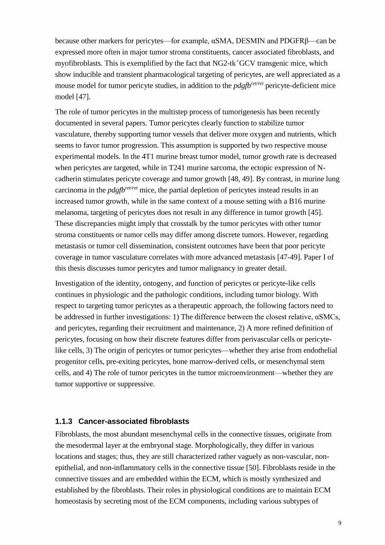
Table 1
Murine protein markers commonly used for identifying pericytes and fibroblasts
Marker
(gene symbol) Function
Cell types expressing the marker
Other cell types expressing the marker
Refer-ence
FSP1
(S100a4)
S100 calcium binding protein
Fibroblasts in normal and fibrotic tissues
Epithelial ovarian carcinoma cells and macrophages, activated lymphocytes
[63-65]
Fibroblast-activation protein, FAP
(Fap)
Serine endopeptidase
Cancer-associated fibroblasts and activated fibroblasts (myofibroblast)
Myoblast, mature cardiac fibroblasts, and activated melanocytes
[53, 66, 67]
Vimentin
(Vim)
Intermediate filament
Fibroblasts in normal and fibrotic tissues
Endothelial cells, mesenchymal cells, and most neuronal precursor cells
[68-70]
Platelet-derived
growth factor
receptor-alpha, PDGFRα
(Pdgfra)
Cell surface tyrosine kinase receptor for PDGFs (PDGFA and PDGFC)
Perivascular profibrotic cells in skeletal muscle, skin and small intestine, dermal fibroblasts, pericytes in spinal cord
Neural stem cells, oligodendrocytes precursor cells, renal interstitial cells
[35, 71, 72] [69, 73]
Platelet-derived
growth factor
receptor-beta, PDGFRβ
(Pdgfrb)
Cell surface tyrosine kinase receptor for PDGFs (PDGFB and PDGFD)
Bone marrow-derived or regional myofibroblasts (kidney, breast and liver), pericytes
Mesenchymal stem cells, neuronal progenitors, vSMCs
[28, 74-76]
Desmin
(Des)
Intermediate filament
Fibroblasts in skin, pericytes vSMCs, cardiomyocytes, skeletal muscle cells, and fetal hepatic stellate cells
[28, 53, 77]
Alpha-smooth
muscle actin, αSMA (Acta2)
Microfilament Myofibroblasts and pericytes vSMCs, myoepithelial cells [45, 78, 79]
Chondroitin sulfate
proteoglycan 4, NG2 (Cspg4)
Integral membrane proteoglycan
Perivascular myofibroblasts and pericytes
vSMCs, oligodendrocyte precursor cells, osteoblasts, chondroblasts, epidermal and hair follicle progenitor cells, and adipose stem cells
[31-33, 80]
Endosialin
(Cd248)
C-type lectin transmembrane receptors
Brain pericytes in embryonal stage and glioma, fibroblasts, and myofibroblasts
Vascular-associated leukocytes (CD45+/CD144+)
[81-84]
Regulator of
G protein signaling 5, RGS5*
(Rgs5)
Intracellular signal transducer (GTPase activator)
Pericytes vSMCs, breast cancer cells, multiple myeloma cells, colon cancer cells, renal cell carcinoma cells
[85, 86]
Alanine aminopeptidase, CD13 (Anpep)
Membrane-integrated aminopeptidase
Brain and retinal pericytes vSMCs, myeloid cells, and epithelial cells of normal tissues and malignant neoplasms
[87-89]
*Obscure yet, because of lack of a validated antibody for immunostaining

13
1.1.4 Tumor-infiltrating leukocytes (TILs)
The use of the currently available advanced methods to detect somatic mutations in tumor
biology using genome-wide analysis has indicated that some transformed cancer cells, such
as melanoma, lung carcinoma, and glioma, retain hundreds to even thousands of somatic
mutations on their chromosomes (somatic mutation prevalence), as well as aberrant
epigenetic changes, conferring an increase in the corresponding pool of antigens to be
recognized by the immune system [90]. This feature raises the question as to how pre-
neoplastic or transformed cancer cells circumvent the immune surveillance system, which has
evolved a sophisticated combination of innate and adaptive immune systems against external
and internal hindrances that threaten the organism’s lifespan. The antigens originating from
host-derived transformed tumor cells are presumed to be improperly recognized and primed
by the host immune system (immunologic ignorance). However, recent work with solid
tumors in experimental mouse models and from human patients has shown that spontaneous
tumor antigen-specific T cell responses are prevalently induced at tumor sites, even though
the response varies in terms of the individual patients, the location, and the grade of tumors
[91].
A complex series of anti-tumor immune response mechanisms, which are not yet fully
elucidated, have been reported to suppress tumor progression and malignancy. The cells
involved include granulocytes (the major immune component in the peripheral blood),
regional macrophages, dendritic cells (especially the CD8α+ subset, which is implicated in
type I interferon/STAT1 signaling pathway, responsible for the innate recognition of tumor
cells), cytotoxic T lymphocytes (CTLs), natural killer T (NKT) cells, and natural killer (NK)
cells (the complimentary innate arm to CTLs that destroy the tumor cells that downregulate
expression of the MHC I molecule and thus evade attacks from CTLs). As seen in bacterial
infections or skin injuries, the innate arm of the immune system first affects antitumor
immunity. Regional macrophages, natural killer cells, or circulating granulocytes sense tumor
cells as an aberrant stress, and some debris from dying tumor cells (DNA fragments or
aberrant peptide) are exposed and are recognized by antigen presenting cells (dendritic cells),
followed by the subsequent activation of the adaptive arm in host immune system.
Eventually, the activated CTLs start to destroy the tumor cells that present aberrant antigen-
MHC I on their membranes. Mice that are genetically targeted for enhancement or deficiency
in NK cells and CTLs confirm an inverse correlation between tumor burden and the activity
of the above immune effector cells, demonstrating the importance of antitumor immunity
functions against tumorigenesis [92-94]. Upon evasion of the host immune surveillance
system, some, but not many, of the neoplastic or transformed cells—which have already
acquired the aberrant genetic instability—proceed to evolve and thus adapt a strategy to
nullify or destroy antitumor immunity, in close cooperation with the neighboring TME.

14
The strategy for avoiding the intrinsic surveillance immune systems can be subdivided into 4
different categories: one is that tumor cells per se react to the CTLs. In the case of most
melanomas, the tumor cells highly express an inhibitory ligand, programmed death-ligand 1
(PD-L1), which, in turn, binds to the inhibitory receptor, PD1, in activated CD8+ CTLs or
NK cells, thereby inhibiting CTL or NK cell activation [95]. The tumor cells of many cancers
also attempt to evade T cell-mediated immune responses through down-regulation of MHC
class 1 [96].
The second category is the reprogramming of myeloid or dendritic cells in the innate immune
system by tumor-derived factors—the TME even polarizes the transition-state cells into
tumor-promoting state cells. Extensive investigations during the last decade have focused on
these tumor-educated and tumor-favoring cell types, including tumor- associated
macrophages (TAMs), tumor-associated neutrophils (TANs), and plasmacytoid dendritic
cells (pDCs). TAMs, a distinct population of macrophages in the TME, promote tumor
progression and metastasis. Most murine TAMs can be identified by the following markers:
CD11b+, CD34-, CD45+, CD68+, CCR2+, CXCR4+ and F4/80+. TAMs were first identified
as a negative regulator of antitumor immunity by suppressing CTLs, as they express PD-L1,
similarly to tumor cells and thus directly counteract CTLs or indirectly suppress CTLs by
CCL22-mediated recruitment of regulatory T cells (Tregs) [97, 98]. The TIE2+ TAM subset
also stimulates tumor angiogenesis by expressing a proangiogenic enzyme, thymidine
phosphorylase, and cathepsin B. In addition, in a mouse mammary tumor model, TAMs play
a crucial role of the intravasation of invasive tumor cells within the primary tumor to form
blood vessels by a paracrine interaction, termed the “paracrine loop,” which includes
signaling pathways such as EGF, CSF-1, and their corresponding receptors [99, 100].
Like TAMs, the tumor-educated neutrophils, the TANs, have been recently identified as an
immune-suppressive cell type in murine lung carcinoma, mainly functioning to reduce CTL
activity and thereby promote tumor progression. However, their actual contribution to
tumorigenesis remains unclear because neutrophils are also involved in antitumor immunity.
In the 1960s, patients with advanced cancer were reported to have a severe blood
neutrophilia, which today is considered to result from host-tumor interactions. The nature of
this blood neutrophilia, however, is still uncertain, as it may be largely tumor supportive or
tumor suppressive. TANs change their properties in either way, depending on their discrete
TME [101, 102].
Plasmacytoid dendritic cells (pDCs) are the antigen-presenting cells that circulate in the blood
and lie in the secondary lymphoid organs. The pDCs are the key regulators of antiviral
immunity as they sense external DNA fragments and produce type I interferon (e.g., IFNα,
IFNβ). They are identified in the mouse by the expression of the following markers: B220+,
CD11c+, PDCA1+, Siglec-H+, GR1+, and CD11b-. In the context of tumor immunity, pDCs
function as negative immune regulators. In human breast cancer and mouse xenograft
experiments, immature pDCs showed a considerable tumor-promoting effect through a
mechanism involving the endosomal Toll-like receptor (TLR7) in the pDCs, which binds to a
tumor-derived or TME-derived ligand, TLR7L. This activates pDCs to produce type I

15
interferon, followed by the subsequent recruitment of immune-regulatory T cells (Tregs)
within the tumor sites [103, 104].
A third category involves the adaptive immune arm in the immune surveillance system,
which is also often distorted by tumor cells or their neighboring TME, and thereafter
transformed into powerful tumor promoting constituents. The adaptive immune regulatory
cells are as follows: the regulatory T cells (Tregs), the regulatory B cells (Bregs), and the
IL17-producing CD4+ helper T cells (Th17). The Tregs are defined as the
immunosuppressive CD4+ T cells and express the alpha chain of the IL2 receptor, CD25, and
the immune-responsive transcription factor, forkhead box P3 (FOXP3). Tumor-derived or
TME-derived CCL22 and CCL5 were first reported as Treg-recruiting factors at tumor sites.
In a breast cancer model, prostaglandin E2 was also suggested to recruit Tregs, and the
pleiotropic cytokine, TNF, was reported to activate Tregs by upregulating FOXP3 via a
TNF/TNFR2 pathway in the presence of IL2.
The exact suppressive role of Tregs is not yet fully understood. However, it is well
appreciated that Tregs would be one of the major components of immunosuppressive
mechanisms for avoiding antitumor cytotoxicity. Their suppression, in large part, is directed
to the activity of CTLs, NK cells, and antigen-presenting dendritic cells, by a direct cell to
cell contact with FasL/Fas and PD1/PD-L1 interactions or by a TGFβ signaling pathway-
mediated paracrine interaction [105-107]. Bregs are also associated with tumorigenesis.
Bregs constitute a small population of B cells that have immunosuppressive functions in
autoimmune diseases. Tumor-related Bregs are characterized by expression of the markers
CD25+, B220+, and CD19+. Recently, lung metastases of 4T1 breast cancer mouse model
have confirmed that Bregs contribute to Treg conversion from naïve CD4+ helper cells and,
in the end, aid in growth of lung metastases [108]. IL17-producing Th17 cells are also
involved in immune-regulatory responses relating to tumorigenesis. However, the role of
Th17 cells in tumor biology remains controversial. One clear observation is that Th17 cells
are more abundant at tumor sites than in the peripheral blood, alluding to a possible
implication of Th17 in tumorigenesis. A recent report, using a VEGFA-blockade resistant
murine lymphoma model, showed that the tumor-promoting effect of Th17 cells, which
secrete IL17A, involves the modulation of the TME to induce G-CSF, thereby recruiting
MDSCs. Conversely, in ovarian tumors, the increased number of Th17 cells was associated
with an improved prognosis. Taken together, these data can be interpreted as indicating that
Th17 cells play different roles, depending on the context of the tumors [109, 110].
The last category involves the immature myeloid cell population as a strong tumor-promoting
factor for incipient cancerous cells or transformed malignant tumor cells. As mentioned
previously, neutrophilia in patients with the advanced cancers results, in part, from tumor-
educated, immune-regulatory activation. Tumor-bearing mice show an accumulation of
immature myeloid cell populations in the bone marrow, the spleen, the blood, and at tumor
sites. These populations are characterized by expression of CD11b and GR1, together with
the undifferentiated myeloid-expressing proteins, S100A8 and S100A9. Today, these cells
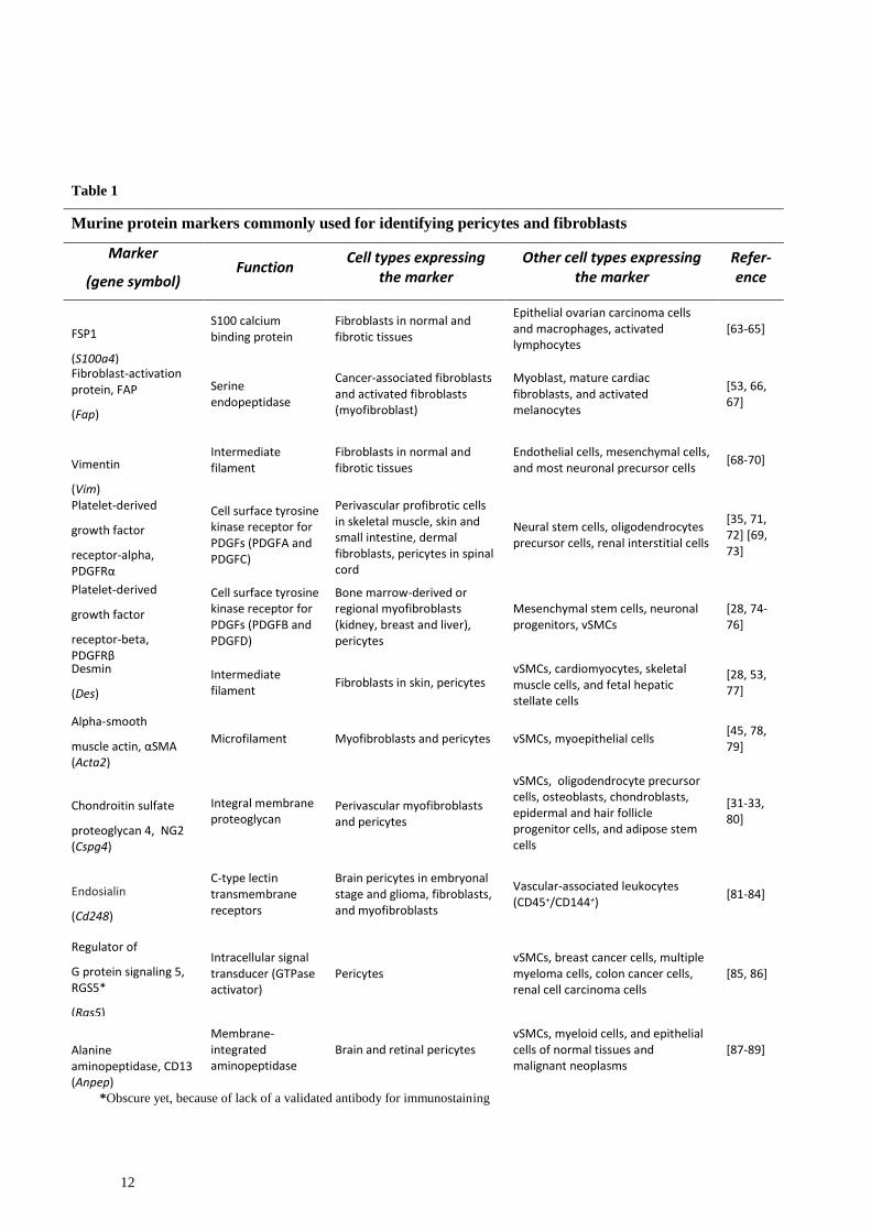
are referred to as myeloid-derived suppressor cells (MDSCs) [110, 111]. The MDSCs are a
16
heterogeneous population with immunosuppressive, proangiogenic, and metastasis-
supportive functions (figure 5). They are subdivided into two discrete subsets: monocytic
(Mo-MDSCs) and granulocytic (G-MDSCs) MDSCs. The marker proteins for murine
MDSCs are CD11b+/GR1+ (LY6C high LY6G low: Mo-MDSC, LY6C low LY6G+ : G-MDSC),
and those for human MDSCs are HLA-DR-/CD33+, sub-grouped into CD14+/dull for Mo-
MDSCs and CD15+ for G-MDSCs. Both MDSC subsets—G-MDSCs and Mo-MDSCs—
serve to suppress immune effectors, the CTLs and NK cells. G-MDSCs express a
considerable amount of ROS and little NO, whereas Mo-MDSCs express little ROS but a
considerable amount of NO, and both MDSC subsets express Arginase 1. When exposed to a
combination of three different pathways—ROS, NO, and Arginase1—both MDSC subsets
efficiently block antigen specific CTLs as well as NK cells. MDSCs can also directly sustain
proliferation of tumor cells or protect against the apoptotic death of tumor cells via the
S100A8/S100A9 signaling pathway in MDA231-LM2 breast cancer cells [112, 113].
Adaptively transferred MDSCs can also differentiate into F4/80+ TAMs in HIF1α-mediated
manner, thereby reflecting MDSC plasticity for the promotion of tumorigenesis in various
ways [114]. Tumor angiogenesis is also positively regulated by MDSCs at tumor sites, where
MDSCs interplay with the TME and thereby stimulate the production of proangiogenic
factors (e.g., IL8, IL10, PGE2, VEGF, and MMP8/9). Conversely, the activated MDSC
subsets can be transdifferentiated into endothelial-like cells [111, 115]. As described above,
the MDSCs systemically expand, once activated by tumor- or TME-derived factors (e.g.,
CCL2, CXCL5, IL6, IL17, G-CSF, GM-CSF, CSF, CXCL12, TGFβ, TNF, and VEGFA) that
were secreted and reached the bone marrow, a reservoir of immature myeloid cells. The
systemic MDSC expansion also contributes to pre-metastatic niche formation by promoting
the adherence and growth of disseminating tumor cells [112, 116].
Figure. 5. MDSCs: crosstalk in the tumor
17
Taken together, the available evidence supports reprogramming of TILs by tumor cells
through the suppression of immune effector cells and promotion of negative regulatory
leukocytes, which both intrinsically function in immune homeostasis. In this way, the
immune surveillance system is overcome. A better understanding of how TILs cross talk with
tumor cells, the TME, or the tumor macro-environment at different stages or at different
tumor loci will definitely benefit effective tumor targeting. This is especially the case when
considering the accumulating, but limited, knowledge of TILs that already points to a link to
clinical therapeutic approaches, such as the generation of genetically enhanced effector T
cells and clinical targeting to tumor-suppressing stroma (MDSCs, Tregs and pDCs),
combined with a cancer vaccine therapy [117, 118].
1.2 CANCER BIOMARKERS
1.2.1 S100A8/9 and S100B
The S100 protein family consists of 21 members: S100A1-S100A16, S100B, S100P, S100G,
S100Z, and FGL, which all have calcium-binding EF hand (helix E-loop-helix F) domains in
their N-terminal regions [119]. In 1965, two members of the S100 proteins, S100S1 and
S100B, were first identified and termed S100 proteins because they were soluble in 100%
saturated ammonium sulfate [120]. S100 proteins have bifunctional roles: In the cytoplasm,
they act as calcium sensors, by binding to intracellular calcium, and as signaling modulators,
by translating calcium influx into diverse target-connected signaling pathways. In the
extracellular spaces, S100 proteins appear as secreted proteins that are capable of binding to
several cell membrane receptors (e.g., RAGE, TLR4, FGFR1, and ALCAM), thereby
affecting nearby or distant cells in complex ways, depending on the composition of their cell
membrane receptors [121, 122]. The S100 proteins contribute to tumor malignancy in various
types of cancers, including brain (S100B, S100A8, S100A9) [122, 123], breast (S100A4,
S100A7, S100A8, S100A9) [124-127], and skin (S100A4, S100A9, S100B [121, 127])
cancers. Within the S100 protein family, S100A8/S100A9 and S100B have recently emerged
as attractive therapeutic targets, with inhibitors now in clinical trials for prostate and skin
cancer.
S100A8 and S100A9 are, in large part, expressed in immature CD11b+ myeloid cells, such as
the MDSCs in the tumor stroma. The prolonged expression of both S100A8 and S100A9
delays the differentiation of myeloid progenitor cells to their derivatives (e.g., macrophages
and dendritic cells). In the TME, the prevalence of S100A8 and S100A9 indicates a high
frequency of MDSCs and poor prognosis [128]. S100A8 and S100A9 mostly occur in vivo as
heterodimers (S100A8/9) and their binding to the cell membrane receptor, RAGE, stimulates
MAPK and NFκB signaling pathways, thereby enabling the greater expression of S100A8
and S100A9 as an autocrine feedback loop, or the maintenance of cell proliferation.
However, how S100A8/9 is involved in intracellular signaling networks in different cell types
remains unclear.

18
Tumor and serum S100B has been used as a diagnostic marker for advanced melanoma, and
a glioma xenograft mouse model has shown that an aberrantly upregulated tumor S100B
level promoted tumor growth and recruited tumor promoting myeloid cells to the tumor sites.
In contrast to S100A8 and S100A9 (tumor stromal origin), S100B is expressed in tumor cells
in melanoma and gliomas. As an intracellular regulator, the Ca2+-bound S100B protein
directly interacts with tumor suppressor protein p53, thereby sequestering p53 protein in the
cytoplasmic region, which prevents tumor cells from undergoing apoptosis or cell cycle arrest
[129, 130]. An association of S100B with cell migration and cell motility via a Src-mediated
pathway was also reported in glial cells [131]. Once secreted into the extracellular spaces,
S100B might affect the nearby cells by interacting with cell membrane receptors, as
mentioned above, and then induce secretion of the proinflammatory cytokines, TNFα, IL1,
and IL6 [123]. In patients with melanoma or in B16 melanoma-bearing mice, the S100B level
is considerably enhanced in the blood, where circulating melanoma cells would secret S100B,
but this serum S100b is viewed as a consequence of the advanced melanoma, not the cause of
it [132].
1.2.2 Serum IL6
In 1983, interleukin 6 (IL6) was first identified to induce the maturation of B cells into
immunoglobulin-producing cells. Accumulating evidence has now extended its role as a
pleiotropic bioactive molecule that has multiple functions in the human body [133]. Its
functions are associated with diverse physiologic and pathologic conditions, such as liver cell
regeneration and glucose and lipid metabolism: It is a key mediator of insulin resistance,
myocardial infarction, T cell priming in the secondary lymphoid organs, autoimmune disease
(e.g., rheumatoid arthritis), atherosclerosis, sepsis, and tumorigenesis [133-135].
The expressions of IL6 and its receptor IL6R are detected in several cell types: hepatocytes,
endothelial cells, fibroblasts, myocytes, and some leukocytes, including monocytes,
macrophages, T cells, and dendritic cells [136]. The IL6/IL6R complex, however, does not
turn on downstream signaling pathways; instead, this complex binds to the co-stimulatory
membrane-anchored receptor, gp130, which is ubiquitously expressed in most cell types.
Two IL6/IL6R /gp130 trimers form a hexamer complex, which acts as a signal transducer
that mainly activates the STAT3 transcription factor, as well as PI3K and MAPK (IL6
classical signaling) [137]. Adding more complexity, IL6 in the extracellular space and in the
blood can bind to the soluble form of the IL6 receptor (sIL6R), which is cleaved by the cell
membrane-bound metalloproteinases, ADAM10 and ADAM17, and subsequently shed from
the cellular membrane. Thereafter, this IL6/sIL6R complex binds to cell membrane-anchored
gp130 and turns on the alternative IL6-mediated signaling pathway (IL6 trans-signaling). The
alternative Il6 signaling mechanism enables IL6R-negative cells to respond to IL6, thereby
conferring multiple availabilities to IL6 [138, 139]. Moreover, a soluble form of gp130
(sgp130) is also found in the body and acts as an antagonist to block the circulating
IL6/sIL6R complex. In the steady state, sgp130 blunts the activation of IL6 signaling, but in

19
IL6-related pathological conditions, serum IL6 levels abruptly peak at the maximum and
overwhelm the activation of IL6 trans-signaling (figure 6) [140-144].
As an inflammatory cytokine, IL6, like IL1, TNF, and TGFβ, plays an important role in the
maintenance of immune homeostasis, especially in the control of the activation of T cell and
humoral responses. For example, in the lymph nodes, antigen-presenting dendritic cells
activate T cell priming by the IL6/IL6R/gp130 signaling pathway. By contrast, for T cell
trafficking, the IL6/sIL6R/gp130 signaling pathway is activated in the high endothelial
venules (HEV), increasing their adhesiveness through the expression of ICAM1. Chronic
inflammation is characterized by prolonged activation of the IL6 signaling, which is often
aberrantly distorted and amplified through the circulation system. In this context, IL6
signaling is presumed to be somehow closely associated with cancers, as some subtypes often
develop from chronic inflammation. Interestingly, a high concentration of serum IL6 has
been extensively correlated with poor prognosis in human melanoma and gastric, pancreatic,
lung, breast, and colorectal cancers. IL6 in the TME can be expressed in tumor parenchyma,
tumor cells, and the tumor stroma, as well as tumor-promoting cell types (e.g., CAFs,
MDSCs, TAMs, and CD4+ regulatory cells, Th17, and Tregs). In tumor cells, IL6 trans-
signaling is, in part, aberrantly activated by genetic and epigenetic changes, which helps
tumor cells maintain proliferation or block apoptotic signals. In the tumor stroma, some
tumor-promoting cells (e.g., MDSCs and Tregs) are present and serve as a source of sIL6R to
boost IL6 trans-signaling at tumor sites, thereby driving the establishment of a tumor-
favoring niche in the TME. This includes stimulation of angiogenesis by VEGFA and bFGF,
which are downstream cytokines of trans-IL6 signaling, and activation or recruitment of
tumor-promoting leukocyte subsets [134].
Figure. 6. Interaction of IL6 with IL6 receptor: classic (cis)-and trans-signaling

20
Taken together, the evidence points to an intimate involvement of cis- and trans-IL6 signaling
pathways in tumorigenesis; this unexpectedly lies on the tumor-promoting side rather than in
anti-tumor immunity. The origin and function of serum IL6 is still elusive in patients with
cancers, but many advanced cancer types show an enhanced serum IL6 level. In the light of
this, IL6 should be more extensively investigated as a therapeutic target.
21
2 AIM OF THE THESIS
In the last two decades, the concept of tumor biology has abruptly changed, so that tumors
are indeed appreciated as complex tissues, rather than as lumps of chaotic neoplastic cells.
Emerging evidence now points to an important role of the TME in regulating
tumorigenesis. In this thesis, we studied the role of tumor pericytes as one of the crucial
regulatory components in the TME.
PAPER I: To establish the role of tumor pericytes in regulating MDSC bursts
PAPER II: To confirm the close interplay of tumor pericytes and perivascular
myofibroblasts


23
3 RESULTS AND DISCUSSION
3.1 PAPER I
In paper I, tumor pericytes are studied with a focus especially on how they interplay with
TILs in the TME. The use of syngeneic subcutaneous tumors (B16 melanoma and Lewis lung
carcinoma) in pericyte-deficient transgenic mice (pdgfbret/ret) demonstrated that the numbers
of tumor-promoting TILs and MDSCs were increased both in the blood and at the tumor sites
in the pericyte-deficient, tumor-bearing group, when compared to the tumor-bearing
littermate control group. This systemic MDSC boost did not occur in the pericyte-rescued
B16 melanoma (B16-PDGF-B) cells, implying that the tumor pericytes did indeed regulate
MDSC activation and recruitment. The observed higher frequency of MDSCs in the blood of
pericyte-deficient, tumor-bearing mice suggested the occurrence of a release of MDSC-
related bioactive molecules from the TME or the circulating MDSC to the blood, thereby
indicating an involvement in MDSC recruitment and expansion. Surprisingly, the level of
serum IL6, one of the potential MDSC-recruiting factors, was also increased, together with
the MDSC population. The increased serum IL6 level was downregulated in the pericyte-
rescued B16 melanoma (B16-PDGF-B).
Serum IL6 is used as a prognostic marker for human melanoma, gastric, pancreatic, lung,
breast, and colorectal cancers and it has an as yet unexplained close relationship with TILs in
the TME. However, how serum IL6 affects tumorigenesis, and whether it is the cause or the
consequence of tumorigenesis, remains to be elucidated. In light of this, the underlying
mechanism of serum IL6 as it relates to tumor pericytes is worth studying. The following
mechanistic study was therefore performed using IL6 shRNA-mediated gene knockdown cell
lines (B16-IL6KD) and hypoxia experiments. The results confirmed that the increase in serum
IL6 is tumor cell-derived in the hypoxic TME. The reduction in IL6 expression from B16
melanoma, as expected, also reversed the systemic MDSC bursts to a similar level to that
seen in the tumor-bearing, littermate control group. These experiments confirm a role for
serum IL6 as a hypoxia-responsive, MDSC-recruiting factor.
Pericyte-deficient tumor vasculature serves to elicit a hypoxic TME, because of the lack of
integrity of this vasculature. As a tumor grows to an advanced stage, it tends to have a more
irregular tumor vasculature, with less pericyte coverage, and its TME tends to be more
hypoxic. This might explain why patients or animals with advanced solid tumors show a high
concentration of serum IL6. This would be an interesting topic for a future study, to
determine if the serum IL6 and the MDSCs in the patients are correlated with diverse types
and degrees of cancers. The T241 murine sarcoma showed a similar MDSC expansion,
independent of tumor pericyte coverage, which might reflect that T241 sarcoma cells, unlike
B16 and LLC cells, do not upregulate IL6 under hypoxic conditions. This is one example
where a different type of tumor cell might confer its own discrete TME.
Each B16 and LLC tumor in the pdgfbret/ret mice exhibited a different type of tumor
malignancy, even though both tumors acquired the same tumor-promoting armament in the

24
form of MDSCs. The LLC tumor in pdgfbret/ret mice grows more rapidly, but the B16 tumor
in pdgfbret/ret mice grows similarly, while more tumor cells disseminate into the peripheral
blood. This higher frequency of circulating tumor cells does not occur in the LLC tumor in
these mice. This discrepancy was explained by the fact that each tumor has a different subset
of MDSCs: B16 tumors have a relatively higher proportion of monocytic MDSCs, while LLC
tumors have mostly granulocytic MDSCs. In this paper, the exact mechanism underlying the
MDSC subset and different tumor phenotype is still elusive. Further study is needed to
determine which TME constituent interplays with tumor pericytes and activates MDSC in a
different way. In the blood, MDSCs belong to mostly granulocytic subset in both B16 and
LLC tumor model, implying that MDSCs are first recruited by the HIF1α/IL6 axis and then
activated in a different way at each tumor site.
The human study with breast cancer cohorts showed a fortunate, meaningful, and correlated
outcome, where the expression of pericyte-related genes (CSPG4, RGS5,ITGA7, CD248) are
inversely correlated with the expression of human MDSC markers (S100A9, CD33) and this
correlation appears to have consequential relevance to the survival rate of patients with breast
cancers. As discussed in the introduction, due to the lack of the specific markers for tumor
pericytes, the gene expression analysis used above is limited in its accuracy. Nevertheless, it
is still of importance, in that the clusters of pericyte-related genes displayed a significant
correlation with MDSC markers.
3.2 PAPER II
Recently, tumor pericytes have been shown to regulate tumor-favoring TILs and MDSCs via
aberrant tumor vasculature. In paper II, a new idea is proposed whereby tumor pericytes in
B16 melanoma play a tumor type-specific role by regulating myofibroblast recruitment. In
B16 tumors, PDGFRα-expressing cells were found in tumor vasculature and, in turn, these
perivascular PDGFRα-expressing cells appeared more frequently in pericyte-deficient tumor
vasculature. By contrast, in LLC tumors, PDGFRα-expressing cells were mostly localized
outside of the tumor vasculature and showed no difference in frequency between pericyte-
deficient, pdgfbret/ret and littermate control mice, indicating TME heterogeneity between the
tumor types.
The perivascular PDGFRα+ cells were further characterized using immunochemistry and
quantitative PCR analysis with FACS-sorted samples: PDGFRα+/PDGFRβ+ as the
perivascular PDGFRα+ cells and PDGFRα+/PDGFRβ- as the pericytes, and identified as a
subset of myofibroblasts that differentially express mRNA for FAP, HGF, CXCL12, and
Wnt5a. Interestingly, the perivascular myofibroblasts (PDGFRα+) express comparable
amounts of NG2, PDGFRβ, and αSMA to those expressed by pericytes, but the fibroblasts do
not express mRNA of RGS5, another marker for pericytes. As discussed in the introduction,
no pericyte-specific marker has yet been found. Correct detection of tumor pericytes,
especially by immunostaining, requires careful selection of marker proteins and must take
into consideration the tumor type and stage. In the light of this notion, RGS5 should be

25
considered as a pericyte marker in the melanoma context. However, this study was performed
with a mouse model, and requires verification by studies on human melanomas.
This study also showed that serum S100B is involved in the generation of the higher
frequency of the perivascular myofibroblasts observed in the pericyte-deficient B16 tumor, as
determined using the S100B-knockdown B16 (B16-S100BKD) tumor model. The S100B
protein is highly expressed in melanoma cells in humans and mice, and it is a bifunctional
protein, as discussed in the introduction. The S100B secreted into the extracellular space
binds to its receptor, RAGE, which is abundant in endothelial cells and leukocytes. This is
followed by cellular responses, including secretion of pro-inflammatory cytokines and
chemokines and possibly transcytosis through the endothelium. In the melanoma, serum
S100B is used as a prognostic marker. Nevertheless, the way that serum S100B affects
tumorigenesis in melanoma and in the blood is not yet understood. S100B expression is
considered to be a consequence of the increased tumor burden. Our experimental setting
revealed a distinctive function of serum S100B in recruiting the perivascular myofibroblasts.
The function of recruited myofibroblasts in terms of the increase in serum S100B remains to
be established. Even though the pericyte-deficient B16 tumor showed an increased serum
S100B (a marker of poor prognosis for melanoma), it did not exhibit any difference in tumor
malignancy. As shown in paper I, pericyte-deficient B16 melanoma showed more MDSC
bursts. Therefore, the MDSCs are presumed to behave as the major controller, driving
melanoma to greater malignancy, while the perivascular myofibroblasts, as a minor
modulators, react in accordance with MDSCs or against a tumor-promoting TME.
Conceivably, serum S100B may enhance the recruitment of perivascular myofibroblasts as
“defenders” of the host-defense system. As shown in the introduction, recent evidence
obtained in murine pancreatic adenocarcinoma has suggested a protective role of the TME
against tumor malignancy.
Lastly, this study has revealed a possible reason for the increased serum S100B levels
observed in pericyte-deficient B16 tumors. In pericyte-detached endothelial cells, tyrosine-14
of Cav1 had a higher phosphorylation level when compared to Cav1 of pericyte-attached
endothelial cells. This was confirmed both in the in vitro cell culture experiment and in the
B16 tumor vascular fragment in pdgfbret/ret and littermate control mice. Moreover, lung
vascular fragments in tumor-free, pericyte-deficient mice also showed an enhanced
phosphorylation of Cav1, indicating that pericyte coverage regulates the endothelial Cav1
state, but not in a tumor-limiting manner. The S100B receptor, RAGE, has been reported to
have a caveolar localization, so that, upon S100B binding, the RAGE/S100B complex is
internalized with the help of phospho-Cav1 (an essential prerequisite for S100B
internalization). The manner by which pericytes control endothelial Cav1 deserves further
investigation.

26
4 ACKNOWLEDGMENTS
I am finally writing the “Acknowledgment” section—which I never imagined I would get to
before. It reminds me of a time, 5 years ago, in March, 2010. I came here for an interview
with Guillem and some people in Christer’s group about entering the Ph.D. program. It was
actually the first time in my life that I had visited Europe. Everything was so different from
the place where I lived: people’s appearances, streets, downtowns, food, transportation, trees,
sky—the social system and the way people thought. The most impressive thing was the
coldness, around -15°C on the slushy street. As I remember, it was one of the coldest
Marches recorded. However, when I arrived in the lab, I realized that all the things inside
were warm and bright, even though outside was still cold and dark. Now, I am sitting in the
same lab where those bright people still work next to me. I would like to express my gratitude
to the bright people whom I have met during my last 5 years’ special journey.
Christer Betsholtz, my co-supervisor, was the reason that I came to Karolinska for the Ph.D.
program. Six years ago, in South Korea, I read his paper about pericytes, published in 2003. It
was so awesome and inspiring that I began to consider whether to quit my job and do a Ph.D.
So, I came here and met Christer, who was a great and humorous scientist. Even though I
haven’t talked much with him, his comments have always been concise and correct. I
sincerely appreciate his help.
To my principal supervisor, Guillem Genové—unbelievably, 5 years have already passed
since I joined here as your student; I’m now your “old student.” How can I express my
thankful feelings to you? I am not an outgoing person, so I have never shown my emotions to
you, and as you know, my language skills are sufficiently fluent to let me communicate with
you about our shared scientific topics in the pericyte world. So, I truly appreciate your
patience when discussing with me. During the last 5 years, I remember sometimes being a bit
frustrated by something, and I used to transfer that frustration on to you, in “individual
meetings,” but you always kept an open stance and then tried to figure out what I was
nagging about. Finally, we made it, with a nice publication and three babies (two are yours
and one is mine). I hope you are going to be the best scientist in KI, and will then upload your
photo more often on the KI web site.
Mirjana Poljakovic, my co-supervisor—I still remember what you said at dinner time with
Guillem and my wife, Seungmin, in the coldest March, 2010. It was a very pleasant time for
us, facing Sweden for the first time at close range, and you told us, at the last moment, to
“feel Stockholm.” I don’t know exactly why I still remember this short sentence, but actually
I am still feeling STOCKHOLM in many respects, and these 5 years of feelings have
changed my attitude and way of life in mostly positive ways. Many thanks to you—especially
for the fantastic B16 extracts, which we are still using.
During the first half of my Ph.D. period, I was part of Christer Betsholtz’s group, which was
full of people who were so nice and friendly: Johanna, Konstantin, Radiosa, Ying, Kazuhiro,
Sara, Miyuki, Maarja, Barbara, Lwaki, Colin, Helene, Cecilia, Jennifer, Thibaud, Michael,

27
Elisabeth, and BongNam. They all are talented, humorous, and generous. Christer’s group
was very international and they seemed know how to balance their lives with their lab work.
Especially, many thanks go to Sara, Colin, and Kazuhiro; they helped me a lot to merge into a
new society. I cannot forget these other colleagues; they were always positive, leading our
corridor to the bright side.
To my officemates, Michail, Yi, David, Manuel, Milena, and Agnieszka: It was an enormous
pleasure spending time with you. Talking, sometimes gossiping, drinking (?), laughing, and
also discussing science—all these things have made me a bit more relaxed and comfortable. I
truly cross my fingers for you all—for your great achievements, both in science and life
business.
Javier: I have to mention his name here in the “Acknowledgments.” I would say he is the
“FACS master” and also a “happy maker” for me. He is super-kind. FACS was the most
crucial equipment for my Ph.D., and I ran out of all my energy preparing cells from the
animal facility to the lab bench. Then, when I arrived at the CMB FACS facility, I would be
totally exhausted. At that time, Javier always welcomed me with open arms and energized me
again. Undoubtedly, I would never have finished my Ph.D. without his help. I feel especially
thankful to him.
Professor Ulf Eriksson—recently, I have come to realize that he supports us in many ways
with extensive generosity. I would like to express my great gratitude to him, and to his group,
the largest and most mature group in the division. I was continuously borrowing things or
obtaining good research ideas from Ulf’s group. Thanks for that to all the group members:
Annelie, Annika, Sofia, Karin, Benjamin, Isolde, Erika, Philip, Lars M., Hong, Sebastian,
Christine, and Ingrid.
Hanna and Patricia, I remember that we did a presentation 4.5 years ago at the same place. It
was a nerve-wracking, but exciting, moment. You both had each had a baby and had just
returned from a maternal leave. I hope you end up your Ph.D. programs with a great pleasure
soon.
To the new group leaders, Daniel, LarsY., Josefin, and Linda, I appreciate your help in
organizing many activities relating to our corridor business and participating actively in our
seminars all the time. It was actually a lot of help, learning science as well as mingling
together. Friendly colleagues in those groups were Yixin, Frank, Joanna, Mirela, Marta,
Christina, Aranzazu, Hassan, and Husain. I was very happy because you were my colleagues.
Jonas Fuxe was in my half-time board. At that time, I was very embarrassed because all my
supervisors were not attending the half-time seminar. Luckily, Jonas helped me to get through
it smoothly without more trouble. To Joel, Vedrana, Meifong, and the present Ph.D. student,
Azadeh, I wish the best for your futures.
In the matrix corridor, qPCR mates, Anne-May and Ann Sofie, a kind antibody supplier,
Juha, Japanese pub-mate, Kan, RNA-seq master, Jing, hard and steady-worker Sergey, kind
people, Bing, Susanne, Olle, Sonia, and Anna—you all were so nice and kind, making me
comfortable in the lab.

28
In Pietra’s group, Eliane and Pernilla, you were always pleasant and in good moods, and you
helped a lot in both my projects. I wish the best for you in Lund and KI.
To my collaborators, Helene and Nick—Thanks for your help. You both introduced new
aspects of science to me, and in the end, directed my project in a better way.
Claudia, Svante, Alessandra, Chad, and new administrator, Gizella, Thanks for your help! It
would have been very tough to get through and finish my PhD without your help!
Lastly (in English), a brilliant Chinese PhD student and my best colleague, Tian, and an
incredibly pleasant French Postdoc and another best colleague, Marion—now we are three in
Guillem’s group. I don’t know how to express my grateful feelings to you both. That is why
you are in the last paragraph in this Acknowledgment section. We have had great times
together in the lab as colleagues, co-workers, and friends.
Big thanks to you all! You always make me happy, standing in the positive and bright side. I
sincerely cheer for your life. Previous colleagues in Guillem’s group—Simona, Yaiza,
Luitgard, and Hanyu—I have been a lucky Ph.D. student because I worked with you, having
lots of help and fun with you.
To my family and friends,
지난 5년간의 스웨덴 생활이 유의미하도록 도와준 이곳의 많은분들께 먼저
인사를 드리고 싶네요. 처음 이곳 생활을 시작할 수 있게 도와준
홍문관님,송정엽 형님, 한글학교와 배드민턴 치며 만나 여러 도움 받은 효수형,
헨릭, 곽경환영사님 그리고 KI 동지들 재윤이, 우주교수님, 명지선생님,
호석부회장님, 동오박사님, 박정섭선생님, 이선재박사님, 이미영님, 다들
고맙습니다. 건승하십시오!
그리고 저에게 좋은 과학 가르쳐주신 아주대 임인경 교수님, 백운기 교수님,
이재호 교수님 그리고 경희대 김상훈교수님 늘 감사합니다. 한국에서 배운 바른
과학이 없었다면 이곳에 실린 논문은 단언코 없었을 것이라 생각합니다.
나의 가족, 이승민 고맙네, 운명처럼 스웨덴에 오게되서 이제껏 위태위태한
노학생이 즐거운 인생을 살 수있게 도와줘서 고맙고 사랑합니다. 과학자로서도
당신의 과학이 더욱 반짝반짝 빛나길 응원할께! 한국에 계신 부모님 늘 고맙고
죄송한 마음 함께 합니다. 저희를 잘 낳아 키워주셔서 고맙고 오랜시간 함께
하지 못해 죄송한 마음 가득합니다. 바르고 떳떳한 자식으로 잘 살겠습니다.
그리고 우리의 몫까지 늘상 챙겨주는 형제들 늘 고맙습니다.
마지막으로, 순환, 순동 형제 바르게 잘 자라주길 기원한다.

29
5 REFERENCES
1. Talmadge JE, Fidler IJ: AACR centennial series: the biology of cancer metastasis: historical perspective. Cancer research 2010, 70(14):5649-5669.
2. Hanahan D, Weinberg RA: Hallmarks of cancer: the next generation. Cell 2011, 144(5):646-674.
3. Watson IR, Takahashi K, Futreal PA, Chin L: Emerging patterns of somatic mutations in cancer. Nature reviews Genetics 2013, 14(10):703-718.
4. Dekker TJ, van de Velde CJ, van Pelt GW, Kroep JR, Julien JP, Smit VT, Tollenaar RA, Mesker WE: Prognostic significance of the tumor-stroma ratio: validation study in node-negative premenopausal breast cancer patients from the EORTC perioperative chemotherapy (POP) trial (10854). Breast cancer research and treatment 2013, 139(2):371-379.
5. Ozdemir BC, Pentcheva-Hoang T, Carstens JL, Zheng X, Wu CC, Simpson TR, Laklai H, Sugimoto H, Kahlert C, Novitskiy SV et al: Depletion of carcinoma-associated fibroblasts and fibrosis induces immunosuppression and accelerates pancreas cancer with reduced survival. Cancer cell 2014, 25(6):719-734.
6. Folkman J: Role of angiogenesis in tumor growth and metastasis. Seminars in oncology 2002, 29(6 Suppl 16):15-18.
7. Aird WC: Phenotypic heterogeneity of the endothelium: II. Representative vascular beds. Circulation research 2007, 100(2):174-190.
8. Nagy JA, Chang SH, Shih SC, Dvorak AM, Dvorak HF: Heterogeneity of the tumor vasculature. Seminars in thrombosis and hemostasis 2010, 36(3):321-331.
9. Patenaude A, Parker J, Karsan A: Involvement of endothelial progenitor cells in tumor vascularization. Microvascular research 2010, 79(3):217-223.
10. Wang R, Chadalavada K, Wilshire J, Kowalik U, Hovinga KE, Geber A, Fligelman B, Leversha M, Brennan C, Tabar V: Glioblastoma stem-like cells give rise to tumour endothelium. Nature 2010, 468(7325):829-833.
11. Mullard A: Phase III setback for lead angiopoietin inhibitor. Nature Reviews Drug Discovery 2014, 13(12):877-877.
12. Cascone T, Herynk MH, Xu L, Du Z, Kadara H, Nilsson MB, Oborn CJ, Park YY, Erez B, Jacoby JJ et al: Upregulated stromal EGFR and vascular remodeling in mouse xenograft models of angiogenesis inhibitor-resistant human lung adenocarcinoma. The Journal of clinical investigation 2011, 121(4):1313-1328.
13. Gu X, Fliesler SJ, Zhao YY, Stallcup WB, Cohen AW, Elliott MH: Loss of caveolin-1 causes blood-retinal barrier breakdown, venous enlargement, and mural cell alteration. The American journal of pathology 2014, 184(2):541-555.
14. Razani B, Woodman SE, Lisanti MP: Caveolae: from cell biology to animal physiology. Pharmacological reviews 2002, 54(3):431-467.
15. Frank PG, Pavlides S, Lisanti MP: Caveolae and transcytosis in endothelial cells: role in atherosclerosis. Cell and tissue research 2009, 335(1):41-47.
16. Ge S, Song L, Serwanski DR, Kuziel WA, Pachter JS: Transcellular transport of CCL2 across brain microvascular endothelial cells. Journal of neurochemistry 2008, 104(5):1219-1232.
17. Simionescu M, Popov D, Sima A: Endothelial transcytosis in health and disease. Cell and tissue research 2009, 335(1):27-40.
18. Sowa G: Caveolae, caveolins, cavins, and endothelial cell function: new insights. Frontiers in physiology 2012, 2:120.
19. Engelman JA, Chu C, Lin A, Jo H, Ikezu T, Okamoto T, Kohtz DS, Lisanti MP: Caveolin-mediated regulation of signaling along the p42/44 MAP kinase cascade in vivo. A role for the caveolin-scaffolding domain. FEBS letters 1998, 428(3):205-211.
20. Bauer PM, Yu J, Chen Y, Hickey R, Bernatchez PN, Looft-Wilson R, Huang Y, Giordano F, Stan RV, Sessa WC: Endothelial-specific expression of caveolin-1 impairs microvascular permeability and angiogenesis. Proceedings of the National Academy of Sciences of the United States of America 2005, 102(1):204-209.
21. Drab M, Verkade P, Elger M, Kasper M, Lohn M, Lauterbach B, Menne J, Lindschau C, Mende F, Luft FC et al: Loss of caveolae, vascular dysfunction, and pulmonary defects in caveolin-1 gene-disrupted mice. Science 2001, 293(5539):2449-2452.
22. Pani B, Singh BB: Lipid rafts/caveolae as microdomains of calcium signaling. Cell calcium 2009, 45(6):625-633.

30
23. Sun RJ, Muller S, Stoltz JF, Wang X: Shear stress induces caveolin-1 translocation in cultured endothelial cells. European biophysics journal : EBJ 2002, 30(8):605-611.
24. Mehta D, Malik AB: Signaling mechanisms regulating endothelial permeability. Physiological reviews 2006, 86(1):279-367.
25. del Pozo MA, Balasubramanian N, Alderson NB, Kiosses WB, Grande-Garcia A, Anderson RG, Schwartz MA: Phospho-caveolin-1 mediates integrin-regulated membrane domain internalization. Nature cell biology 2005, 7(9):901-908.
26. Sun Y, Hu G, Zhang X, Minshall RD: Phosphorylation of caveolin-1 regulates oxidant-induced pulmonary vascular permeability via paracellular and transcellular pathways. Circulation research 2009, 105(7):676-685, 615 p following 685.
27. Diaz-Flores L, Gutierrez R, Madrid JF, Varela H, Valladares F, Acosta E, Martin-Vasallo P, Diaz-Flores L, Jr.: Pericytes. Morphofunction, interactions and pathology in a quiescent and activated mesenchymal cell niche. Histology and histopathology 2009, 24(7):909-969.
28. Armulik A, Genove G, Betsholtz C: Pericytes: developmental, physiological, and pathological perspectives, problems, and promises. Developmental cell 2011, 21(2):193-215.
29. Armulik A, Abramsson A, Betsholtz C: Endothelial/pericyte interactions. Circulation research 2005, 97(6):512-523.
30. Gaengel K, Genove G, Armulik A, Betsholtz C: Endothelial-mural cell signaling in vascular development and angiogenesis. Arteriosclerosis, thrombosis, and vascular biology 2009, 29(5):630-638.
31. Kadoya K, Fukushi J, Matsumoto Y, Yamaguchi Y, Stallcup WB: NG2 proteoglycan expression in mouse skin: altered postnatal skin development in the NG2 null mouse. The journal of histochemistry and cytochemistry : official journal of the Histochemistry Society 2008, 56(3):295-303.
32. Fukushi J, Inatani M, Yamaguchi Y, Stallcup WB: Expression of NG2 proteoglycan during endochondral and intramembranous ossification. Developmental dynamics : an official publication of the American Association of Anatomists 2003, 228(1):143-148.
33. Polito A, Reynolds R: NG2-expressing cells as oligodendrocyte progenitors in the normal and demyelinated adult central nervous system. Journal of anatomy 2005, 207(6):707-716.
34. Uezumi A, Fukada S, Yamamoto N, Ikemoto-Uezumi M, Nakatani M, Morita M, Yamaguchi A, Yamada H, Nishino I, Hamada Y et al: Identification and characterization of PDGFRalpha+ mesenchymal progenitors in human skeletal muscle. Cell death & disease 2014, 5:e1186.
35. Goritz C, Dias DO, Tomilin N, Barbacid M, Shupliakov O, Frisen J: A pericyte origin of spinal cord scar tissue. Science 2011, 333(6039):238-242.
36. Chojnacki A, Mak G, Weiss S: PDGFRalpha expression distinguishes GFAP-expressing neural stem cells from PDGF-responsive neural precursors in the adult periventricular area. The Journal of neuroscience : the official journal of the Society for Neuroscience 2011, 31(26):9503-9512.
37. Soriano P: Abnormal kidney development and hematological disorders in PDGF beta-receptor mutant mice. Genes & development 1994, 8(16):1888-1896.
38. Lindahl P, Johansson BR, Leveen P, Betsholtz C: Pericyte loss and microaneurysm formation in PDGF-B-deficient mice. Science 1997, 277(5323):242-245.
39. Lindblom P, Gerhardt H, Liebner S, Abramsson A, Enge M, Hellstrom M, Backstrom G, Fredriksson S, Landegren U, Nystrom HC et al: Endothelial PDGF-B retention is required for proper investment of pericytes in the microvessel wall. Genes & development 2003, 17(15):1835-1840.
40. Goumans MJ, Valdimarsdottir G, Itoh S, Rosendahl A, Sideras P, ten Dijke P: Balancing the activation state of the endothelium via two distinct TGF-beta type I receptors. The EMBO journal 2002, 21(7):1743-1753.
41. Chen S, Kulik M, Lechleider RJ: Smad proteins regulate transcriptional induction of the SM22alpha gene by TGF-beta. Nucleic acids research 2003, 31(4):1302-1310.
42. Hammes HP, Lin J, Wagner P, Feng Y, Vom Hagen F, Krzizok T, Renner O, Breier G, Brownlee M, Deutsch U: Angiopoietin-2 causes pericyte dropout in the normal retina: evidence for involvement in diabetic retinopathy. Diabetes 2004, 53(4):1104-1110.
43. Falcon BL, Hashizume H, Koumoutsakos P, Chou J, Bready JV, Coxon A, Oliner JD, McDonald DM: Contrasting actions of selective inhibitors of angiopoietin-1 and angiopoietin-2 on the normalization of tumor blood vessels. The American journal of pathology 2009, 175(5):2159-2170.
44. Nolan-Stevaux O, Truitt MC, Pahler JC, Olson P, Guinto C, Lee DC, Hanahan D: Differential contribution to neuroendocrine tumorigenesis of parallel egfr signaling in cancer cells and pericytes. Genes & cancer 2010, 1(2):125-141.
31
45. Nisancioglu MH, Betsholtz C, Genove G: The absence of pericytes does not increase the sensitivity of tumor vasculature to vascular endothelial growth factor-A blockade. Cancer research 2010, 70(12):5109-5115.
46. Morikawa S, Baluk P, Kaidoh T, Haskell A, Jain RK, McDonald DM: Abnormalities in Pericytes on Blood Vessels and Endothelial Sprouts in Tumors. The American journal of pathology 2002, 160(3):985-1000.
47. Cooke VG, LeBleu VS, Keskin D, Khan Z, O'Connell JT, Teng Y, Duncan MB, Xie L, Maeda G, Vong S et al: Pericyte depletion results in hypoxia-associated epithelial-to-mesenchymal transition and metastasis mediated by met signaling pathway. Cancer cell 2012, 21(1):66-81.
48. Keskin D, Kim J, Cooke VG, Wu CC, Sugimoto H, Gu C, De Palma M, Kalluri R, LeBleu VS: Targeting vascular pericytes in hypoxic tumors increases lung metastasis via angiopoietin-2. Cell reports 2015, 10(7):1066-1081.
49. Xian X, Hakansson J, Stahlberg A, Lindblom P, Betsholtz C, Gerhardt H, Semb H: Pericytes limit tumor cell metastasis. The Journal of clinical investigation 2006, 116(3):642-651.
50. Ohlund D, Elyada E, Tuveson D: Fibroblast heterogeneity in the cancer wound. The Journal of experimental medicine 2014, 211(8):1503-1523.
51. Tomasek JJ, Gabbiani G, Hinz B, Chaponnier C, Brown RA: Myofibroblasts and mechano-regulation of connective tissue remodelling. Nature reviews Molecular cell biology 2002, 3(5):349-363.
52. Ozaki N, Ishizaki M, Ghazizadeh M, Yamanaka N: Apoptosis mediates decrease in cellularity during the regression of Arthus reaction in cornea. The British journal of ophthalmology 2001, 85(5):613-618.
53. Kalluri R, Zeisberg M: Fibroblasts in cancer. Nature reviews Cancer 2006, 6(5):392-401.
54. Stoker MG, Shearer M, O'Neill C: Growth inhibition of polyoma-transformed cells by contact with static normal fibroblasts. Journal of cell science 1966, 1(3):297-310.
55. Alkasalias T, Flaberg E, Kashuba V, Alexeyenko A, Pavlova T, Savchenko A, Szekely L, Klein G, Guven H: Inhibition of tumor cell proliferation and motility by fibroblasts is both contact and soluble factor dependent. Proceedings of the National Academy of Sciences of the United States of America 2014, 111(48):17188-17193.
56. Lohr M, Schmidt C, Ringel J, Kluth M, Muller P, Nizze H, Jesnowski R: Transforming growth factor-beta1 induces desmoplasia in an experimental model of human pancreatic carcinoma. Cancer research 2001, 61(2):550-555.
57. Elenbaas B, Weinberg RA: Heterotypic signaling between epithelial tumor cells and fibroblasts in carcinoma formation. Experimental cell research 2001, 264(1):169-184.
58. Anderberg C, Li H, Fredriksson L, Andrae J, Betsholtz C, Li X, Eriksson U, Pietras K: Paracrine signaling by platelet-derived growth factor-CC promotes tumor growth by recruitment of cancer-associated fibroblasts. Cancer research 2009, 69(1):369-378.
59. Erez N, Truitt M, Olson P, Arron ST, Hanahan D: Cancer-Associated Fibroblasts Are Activated in Incipient Neoplasia to Orchestrate Tumor-Promoting Inflammation in an NF-kappaB-Dependent Manner. Cancer cell 2010, 17(2):135-147.
60. Orimo A, Gupta PB, Sgroi DC, Arenzana-Seisdedos F, Delaunay T, Naeem R, Carey VJ, Richardson AL, Weinberg RA: Stromal fibroblasts present in invasive human breast carcinomas promote tumor growth and angiogenesis through elevated SDF-1/CXCL12 secretion. Cell 2005, 121(3):335-348.
61. Sounni NE, Noel A: Targeting the tumor microenvironment for cancer therapy. Clinical chemistry 2013, 59(1):85-93.
62. Hofheinz RD, al-Batran SE, Hartmann F, Hartung G, Jager D, Renner C, Tanswell P, Kunz U, Amelsberg A, Kuthan H et al: Stromal antigen targeting by a humanised monoclonal antibody: an early phase II trial of sibrotuzumab in patients with metastatic colorectal cancer. Onkologie 2003, 26(1):44-48.
63. Strutz F, Okada H, Lo CW, Danoff T, Carone RL, Tomaszewski JE, Neilson EG: Identification and characterization of a fibroblast marker: FSP1. The Journal of cell biology 1995, 130(2):393-405.
64. Cabezon T, Celis JE, Skibshoj I, Klingelhofer J, Grigorian M, Gromov P, Rank F, Myklebust JH, Maelandsmo GM, Lukanidin E et al: Expression of S100A4 by a variety of cell types present in the tumor microenvironment of human breast cancer. International journal of cancer Journal international du cancer 2007, 121(7):1433-1444.
65. Kikuchi N, Horiuchi A, Osada R, Imai T, Wang C, Chen X, Konishi I: Nuclear expression of S100A4 is associated with aggressive behavior of epithelial ovarian carcinoma: an important autocrine/paracrine factor in tumor progression. Cancer science 2006, 97(10):1061-1069.
66. Joe AW, Yi L, Natarajan A, Le Grand F, So L, Wang J, Rudnicki MA, Rossi FM: Muscle injury activates resident fibro/adipogenic progenitors that facilitate myogenesis. Nature cell biology 2010, 12(2):153-163.
67. Yan C, Grimm WA, Garner WL, Qin L, Travis T, Tan N, Han YP: Epithelial to mesenchymal transition in human skin wound healing is induced by tumor necrosis factor-alpha through bone morphogenic protein-2. The American journal of pathology 2010, 176(5):2247-2258.

32
68. Boyne LJ, Fischer I, Shea TB: Role of vimentin in early stages of neuritogenesis in cultured hippocampal neurons. International journal of developmental neuroscience : the official journal of the International Society for Developmental Neuroscience 1996, 14(6):739-748.
69. Driskell RR, Lichtenberger BM, Hoste E, Kretzschmar K, Simons BD, Charalambous M, Ferron SR, Herault Y, Pavlovic G, Ferguson-Smith AC et al: Distinct fibroblast lineages determine dermal architecture in skin development and repair. Nature 2013, 504(7479):277-281.
70. Xu B, deWaal RM, Mor-Vaknin N, Hibbard C, Markovitz DM, Kahn ML: The endothelial cell-specific antibody PAL-E identifies a secreted form of vimentin in the blood vasculature. Molecular and cellular biology 2004, 24(20):9198-9206.
71. Brittan M, Hunt T, Jeffery R, Poulsom R, Forbes SJ, Hodivala-Dilke K, Goldman J, Alison MR, Wright NA: Bone marrow derivation of pericryptal myofibroblasts in the mouse and human small intestine and colon. Gut 2002, 50(6):752-757.
72. Floege J, Eitner F, Alpers CE: A new look at platelet-derived growth factor in renal disease. Journal of the American Society of Nephrology : JASN 2008, 19(1):12-23.
73. Dulauroy S, Di Carlo SE, Langa F, Eberl G, Peduto L: Lineage tracing and genetic ablation of ADAM12(+) perivascular cells identify a major source of profibrotic cells during acute tissue injury. Nature medicine 2012, 18(8):1262-1270.
74. Iwaisako K, Jiang C, Zhang M, Cong M, Moore-Morris TJ, Park TJ, Liu X, Xu J, Wang P, Paik YH et al: Origin of myofibroblasts in the fibrotic liver in mice. Proceedings of the National Academy of Sciences of the United States of America 2014, 111(32):E3297-3305.
75. LeBleu VS, Taduri G, O'Connell J, Teng Y, Cooke VG, Woda C, Sugimoto H, Kalluri R: Origin and function of myofibroblasts in kidney fibrosis. Nature medicine 2013, 19(8):1047-1053.
76. Ronnov-Jessen L, Petersen OW, Koteliansky VE, Bissell MJ: The origin of the myofibroblasts in breast cancer. Recapitulation of tumor environment in culture unravels diversity and implicates converted fibroblasts and recruited smooth muscle cells. The Journal of clinical investigation 1995, 95(2):859-873.
77. Paulin D, Li Z: Desmin: a major intermediate filament protein essential for the structural integrity and function of muscle. Experimental cell research 2004, 301(1):1-7.
78. Gugliotta P, Sapino A, Macri L, Skalli O, Gabbiani G, Bussolati G: Specific demonstration of myoepithelial cells by anti-alpha smooth muscle actin antibody. The journal of histochemistry and cytochemistry : official journal of the Histochemistry Society 1988, 36(6):659-663.
79. Quante M, Tu SP, Tomita H, Gonda T, Wang SS, Takashi S, Baik GH, Shibata W, Diprete B, Betz KS et al: Bone marrow-derived myofibroblasts contribute to the mesenchymal stem cell niche and promote tumor growth. Cancer cell 2011, 19(2):257-272.
80. Greenhalgh SN, Iredale JP, Henderson NC: Origins of fibrosis: pericytes take centre stage. F1000prime reports 2013, 5:37.
81. Lax S, Hardie DL, Wilson A, Douglas MR, Anderson G, Huso D, Isacke CM, Buckley CD: The pericyte and stromal cell marker CD248 (endosialin) is required for efficient lymph node expansion. European journal of immunology 2010, 40(7):1884-1889.
82. MacFadyen J, Savage K, Wienke D, Isacke CM: Endosialin is expressed on stromal fibroblasts and CNS pericytes in mouse embryos and is downregulated during development. Gene expression patterns : GEP 2007, 7(3):363-369.
83. Bagley RG, Honma N, Weber W, Boutin P, Rouleau C, Shankara S, Kataoka S, Ishida I, Roberts BL, Teicher BA: Endosialin/TEM 1/CD248 is a pericyte marker of embryonic and tumor neovascularization. Microvascular research 2008, 76(3):180-188.
84. Li C, Chacko AM, Hu J, Hasegawa K, Swails J, Grasso L, El-Deiry WS, Nicolaides N, Muzykantov VR, Divgi CR et al: Antibody-based tumor vascular theranostics targeting endosialin/TEM1 in a new mouse tumor vascular model. Cancer biology & therapy 2014, 15(4):443-451.
85. Boss CN, Grunebach F, Brauer K, Hantschel M, Mirakaj V, Weinschenk T, Stevanovic S, Rammensee HG, Brossart P: Identification and characterization of T-cell epitopes deduced from RGS5, a novel broadly expressed tumor antigen. Clinical cancer research : an official journal of the American Association for Cancer Research 2007, 13(11):3347-3355.
86. Nisancioglu MH, Mahoney WM, Jr., Kimmel DD, Schwartz SM, Betsholtz C, Genove G: Generation and characterization of rgs5 mutant mice. Molecular and cellular biology 2008, 28(7):2324-2331.

33
87. Mishima Y, Matsumoto-Mishima Y, Terui Y, Katsuyama M, Yamada M, Mori M, Ishizaka Y, Ikeda K, Watanabe J, Mizunuma N et al: Leukemic cell-surface CD13/aminopeptidase N and resistance to apoptosis mediated by endothelial cells. Journal of the National Cancer Institute 2002, 94(13):1020-1028.
88. Dixon J, Kaklamanis L, Turley H, Hickson ID, Leek RD, Harris AL, Gatter KC: Expression of aminopeptidase-n (CD 13) in normal tissues and malignant neoplasms of epithelial and lymphoid origin. Journal of clinical pathology 1994, 47(1):43-47.
89. Armulik A, Genove G, Mae M, Nisancioglu MH, Wallgard E, Niaudet C, He L, Norlin J, Lindblom P, Strittmatter K et al: Pericytes regulate the blood-brain barrier. Nature 2010, 468(7323):557-561.
90. Greenman C, Stephens P, Smith R, Dalgliesh GL, Hunter C, Bignell G, Davies H, Teague J, Butler A, Stevens C et al: Patterns of somatic mutation in human cancer genomes. Nature 2007, 446(7132):153-158.
91. Gajewski TF, Schreiber H, Fu YX: Innate and adaptive immune cells in the tumor microenvironment. Nature immunology 2013, 14(10):1014-1022.
92. Fuertes MB, Kacha AK, Kline J, Woo SR, Kranz DM, Murphy KM, Gajewski TF: Host type I IFN signals are required for antitumor CD8+ T cell responses through CD8{alpha}+ dendritic cells. The Journal of experimental medicine 2011, 208(10):2005-2016.
93. Diamond MS, Kinder M, Matsushita H, Mashayekhi M, Dunn GP, Archambault JM, Lee H, Arthur CD, White JM, Kalinke U et al: Type I interferon is selectively required by dendritic cells for immune rejection of tumors. The Journal of experimental medicine 2011, 208(10):1989-2003.
94. Hildner K, Edelson BT, Purtha WE, Diamond M, Matsushita H, Kohyama M, Calderon B, Schraml BU, Unanue ER, Diamond MS et al: Batf3 deficiency reveals a critical role for CD8alpha+ dendritic cells in cytotoxic T cell immunity. Science 2008, 322(5904):1097-1100.
95. Gajewski TF: Failure at the effector phase: immune barriers at the level of the melanoma tumor microenvironment. Clinical cancer research : an official journal of the American Association for Cancer Research 2007, 13(18 Pt 1):5256-5261.
96. Campoli M, Ferrone S: HLA antigen changes in malignant cells: epigenetic mechanisms and biologic significance. Oncogene 2008, 27(45):5869-5885.
97. Wyckoff J, Wang W, Lin EY, Wang Y, Pixley F, Stanley ER, Graf T, Pollard JW, Segall J, Condeelis J: A paracrine loop between tumor cells and macrophages is required for tumor cell migration in mammary tumors. Cancer research 2004, 64(19):7022-7029.
98. Wyckoff JB, Wang Y, Lin EY, Li JF, Goswami S, Stanley ER, Segall JE, Pollard JW, Condeelis J: Direct visualization of macrophage-assisted tumor cell intravasation in mammary tumors. Cancer research 2007, 67(6):2649-2656.
99. Pardoll D: Metastasis-promoting immunity: when T cells turn to the dark side. Cancer cell 2009, 16(2):81-82.
100. Na YR, Yoon YN, Son DI, Seok SH: Cyclooxygenase-2 inhibition blocks M2 macrophage differentiation and suppresses metastasis in murine breast cancer model. PloS one 2013, 8(5):e63451.
101. Fridlender ZG, Albelda SM: Tumor-associated neutrophils: friend or foe? Carcinogenesis 2012, 33(5):949-955.
102. Mishalian I, Bayuh R, Levy L, Zolotarov L, Michaeli J, Fridlender ZG: Tumor-associated neutrophils (TAN) develop pro-tumorigenic properties during tumor progression. Cancer immunology, immunotherapy : CII 2013, 62(11):1745-1756.
103. Treilleux I, Blay JY, Bendriss-Vermare N, Ray-Coquard I, Bachelot T, Guastalla JP, Bremond A, Goddard S, Pin JJ, Barthelemy-Dubois C et al: Dendritic cell infiltration and prognosis of early stage breast cancer. Clinical cancer research : an official journal of the American Association for Cancer Research 2004, 10(22):7466-7474.
104. Le Mercier I, Poujol D, Sanlaville A, Sisirak V, Gobert M, Durand I, Dubois B, Treilleux I, Marvel J, Vlach J et al: Tumor promotion by intratumoral plasmacytoid dendritic cells is reversed by TLR7 ligand treatment. Cancer research 2013, 73(15):4629-4640.
105. Hamano R, Huang J, Yoshimura T, Oppenheim JJ, Chen X: TNF optimally activatives regulatory T cells by inducing TNF receptor superfamily members TNFR2, 4-1BB and OX40. European journal of immunology 2011, 41(7):2010-2020.
106. Chen X, Baumel M, Mannel DN, Howard OMZ, Oppenheim JJ: Interaction of TNF with TNF Receptor Type 2 Promotes Expansion and Function of Mouse CD4+CD25+ T Regulatory Cells. The Journal of Immunology 2007, 179(1):154-161.
107. Kitamura T, Qian BZ, Pollard JW: Immune cell promotion of metastasis. Nature reviews Immunology 2015, 15(2):73-86.

34
108. Olkhanud PB, Damdinsuren B, Bodogai M, Gress RE, Sen R, Wejksza K, Malchinkhuu E, Wersto RP, Biragyn A: Tumor-evoked regulatory B cells promote breast cancer metastasis by converting resting CD4(+) T cells to T-regulatory cells. Cancer research 2011, 71(10):3505-3515.
109. Chung AS, Wu X, Zhuang G, Ngu H, Kasman I, Zhang J, Vernes JM, Jiang Z, Meng YG, Peale FV et al: An interleukin-17-mediated paracrine network promotes tumor resistance to anti-angiogenic therapy. Nature medicine 2013, 19(9):1114-1123.
110. Zou W, Restifo NP: T(H)17 cells in tumour immunity and immunotherapy. Nature reviews Immunology 2010, 10(4):248-256.
111. Murdoch C, Muthana M, Coffelt SB, Lewis CE: The role of myeloid cells in the promotion of tumour angiogenesis. Nature reviews Cancer 2008, 8(8):618-631.
112. Talmadge JE, Gabrilovich DI: History of myeloid-derived suppressor cells. Nature reviews Cancer 2013, 13(10):739-752.
113. Ostrand-Rosenberg S, Sinha P: Myeloid-derived suppressor cells: linking inflammation and cancer. Journal of immunology 2009, 182(8):4499-4506.
114. Corzo CA, Condamine T, Lu L, Cotter MJ, Youn JI, Cheng P, Cho HI, Celis E, Quiceno DG, Padhya T et al: HIF-1alpha regulates function and differentiation of myeloid-derived suppressor cells in the tumor microenvironment. The Journal of experimental medicine 2010, 207(11):2439-2453.
115. Serafini P: Editorial: PGE2-producing MDSC: a role in tumor progression? Journal of leukocyte biology 2010, 88(5):827-829.
116. Kaplan RN, Riba RD, Zacharoulis S, Bramley AH, Vincent L, Costa C, MacDonald DD, Jin DK, Shido K, Kerns SA et al: VEGFR1-positive haematopoietic bone marrow progenitors initiate the pre-metastatic niche. Nature 2005, 438(7069):820-827.
117. Najjar YG, Finke JH: Clinical perspectives on targeting of myeloid derived suppressor cells in the treatment of cancer. Frontiers in oncology 2013, 3:49.
118. Sadelain M, Riviere I, Brentjens R: Targeting tumours with genetically enhanced T lymphocytes. Nature reviews Cancer 2003, 3(1):35-45.
119. Donato R: Functional roles of S100 proteins, calcium-binding proteins of the EF-hand type. Biochimica et Biophysica Acta (BBA) - Molecular Cell Research 1999, 1450(3):191-231.
120. Moore BW: A soluble protein characteristic of the nervous system. Biochemical and biophysical research communications 1965, 19(6):739-744.
121. Bresnick AR, Weber DJ, Zimmer DB: S100 proteins in cancer. Nature reviews Cancer 2015, 15(2):96-109.
122. Marenholz I, Heizmann CW, Fritz G: S100 proteins in mouse and man: from evolution to function and pathology (including an update of the nomenclature). Biochemical and biophysical research communications 2004, 322(4):1111-1122.
123. Wang H, Zhang L, Zhang IY, Chen X, Da Fonseca A, Wu S, Ren H, Badie S, Sadeghi S, Ouyang M et al: S100B promotes glioma growth through chemoattraction of myeloid-derived macrophages. Clinical cancer research : an official journal of the American Association for Cancer Research 2013, 19(14):3764-3775.
124. Cheng P, Corzo CA, Luetteke N, Yu B, Nagaraj S, Bui MM, Ortiz M, Nacken W, Sorg C, Vogl T et al: Inhibition of dendritic cell differentiation and accumulation of myeloid-derived suppressor cells in cancer is regulated by S100A9 protein. The Journal of experimental medicine 2008, 205(10):2235-2249.
125. Acharyya S, Oskarsson T, Vanharanta S, Malladi S, Kim J, Morris PG, Manova-Todorova K, Leversha M, Hogg N, Seshan VE et al: A CXCL1 paracrine network links cancer chemoresistance and metastasis. Cell 2012, 150(1):165-178.
126. Emberley ED, Niu Y, Njue C, Kliewer EV, Murphy LC, Watson PH: Psoriasin (S100A7) expression is associated with poor outcome in estrogen receptor-negative invasive breast cancer. Clinical cancer research : an official journal of the American Association for Cancer Research 2003, 9(7):2627-2631.
127. Gomes LH, Raftery MJ, Yan WX, Goyette JD, Thomas PS, Geczy CL: S100A8 and S100A9-oxidant scavengers in inflammation. Free radical biology & medicine 2013, 58:170-186.
128. Arai K, Takano S, Teratani T, Ito Y, Yamada T, Nozawa R: S100A8 and S100A9 overexpression is associated with poor pathological parameters in invasive ductal carcinoma of the breast. Current cancer drug targets 2008, 8(4):243-252.
129. Sawa H, Murakami H, Ohshima Y, Sugino T, Nakajyo T, Kisanuki T, Tamura Y, Satone A, Ide W, Hashimoto I et al: Histone deacetylase inhibitors such as sodium butyrate and trichostatin A induce apoptosis through an increase of the bcl-2-related protein Bad. Brain Tumor Pathol 2001, 18(2):109-114.
35
130. Ichikawa M, Williams R, Wang L, Vogl T, Srikrishna G: S100A8/A9 activate key genes and pathways in colon tumor progression. Molecular cancer research : MCR 2011, 9(2):133-148.
131. Steiner J, Bernstein HG, Bielau H, Berndt A, Brisch R, Mawrin C, Keilhoff G, Bogerts B: Evidence for a wide extra-astrocytic distribution of S100B in human brain. BMC neuroscience 2007, 8:2.
132. Vuoristo MS, Kellokumpu-Lehtinen P, Laine S, Parvinen LM, Hahka-Kemppinen M, Korpela M, Kumpulainen E: The value of serum S-100beta and interleukins as tumour markers in advanced melanoma. Melanoma research 2000, 10(3):237-241.
133. Mauer J, Denson JL, Bruning JC: Versatile functions for IL-6 in metabolism and cancer. Trends in immunology 2015, 36(2):92-101.
134. Fisher DT, Appenheimer MM, Evans SS: The two faces of IL-6 in the tumor microenvironment. Seminars in immunology 2014, 26(1):38-47.
135. Miyao Y, Yasue H, Ogawa H, Misumi I, Masuda T, Sakamoto T, Morita E: Elevated plasma interleukin-6 levels in patients with acute myocardial infarction. American heart journal 1993, 126(6):1299-1304.
136. Rose-John S: IL-6 trans-signaling via the soluble IL-6 receptor: importance for the pro-inflammatory activities of IL-6. International journal of biological sciences 2012, 8(9):1237-1247.
137. Lacroix M, Rousseau F, Guilhot F, Malinge P, Magistrelli G, Herren S, Jones SA, Jones GW, Scheller J, Lissilaa R et al: Novel insights into IL-6 cis- and trans-signaling pathways by differentially manipulating the assembly of the IL-6 signaling complex. The Journal of biological chemistry 2015.
138. Chalaris A, Rabe B, Paliga K, Lange H, Laskay T, Fielding CA, Jones SA, Rose-John S, Scheller J: Apoptosis is a natural stimulus of IL6R shedding and contributes to the proinflammatory trans-signaling function of neutrophils. Blood 2007, 110(6):1748-1755.
139. Matthews V, Schuster B, Schutze S, Bussmeyer I, Ludwig A, Hundhausen C, Sadowski T, Saftig P, Hartmann D, Kallen KJ et al: Cellular cholesterol depletion triggers shedding of the human interleukin-6 receptor by ADAM10 and ADAM17 (TACE). The Journal of biological chemistry 2003, 278(40):38829-38839.
140. Narazaki M, Yasukawa K, Saito T, Ohsugi Y, Fukui H, Koishihara Y, Yancopoulos GD, Taga T, Kishimoto T: Soluble forms of the interleukin-6 signal-transducing receptor component gp130 in human serum possessing a potential to inhibit signals through membrane-anchored gp130. Blood 1993, 82(4):1120-1126.
141. Bousso P, Robey E: Dynamics of CD8+ T cell priming by dendritic cells in intact lymph nodes. Nature immunology 2003, 4(6):579-585.
142. Girard JP, Moussion C, Forster R: HEVs, lymphatics and homeostatic immune cell trafficking in lymph nodes. Nature reviews Immunology 2012, 12(11):762-773.
143. Heikkila K, Ebrahim S, Lawlor DA: Systematic review of the association between circulating interleukin-6 (IL-6) and cancer. European journal of cancer 2008, 44(7):937-945.
144. Lippitz BE: Cytokine patterns in patients with cancer: a systematic review. The Lancet Oncology 2013, 14(6):e218-e228.