Embed Size (px)
Citation preview
EXPRESSION OF INTERLEUKIN 4, INTERLEUKIN 4
RECEPTOR AND IL-4 RECEPTOR RELATED
GP200-MR6 MOLECULE IN PTERYGIUM AND NORMAL
BULBAR CONJUNCTIVA TISSUE
By
Dr. Go Eng Soon
Dissertation Submitted In partial Fulfilment Of The
Requirements Fot ThetDegree Of Master Of Medicine
(Ophthalmology)
UNIVERSITI SAINS MALAYSIA
2001
DISCLAIMER
I hereby certify that the work in this dissertation is my own except for the quotations
which have been duly acknowledged.
Dated November 30th 2001
Dr. Go Eng Soon
P-UMOSJ6
ii

ACKNOWLEDGEMENT
;])ear cfol'(ll3uciJha, on4 in Jlwu J. ~e /ound ImJ refuge! SaJhu!
My deepest gratitude to my supervisors Dr.Raja Azmi Mohd. Noor and Dr. Mohtar
Ibrahim for their continuous guidance, concern and support through out my pursuit of
M.Med (Ophthalmology).
My most sincere gratitude to my co-supervisors, Associate Professor Dr. Ishak Bin Mat
and Dr. Meor Zamari Bin Meor Kamal whom have never fail to give me useful advice
and guidance culminating in the completion of this dissertation.
To the teaching faculty of The Department of Ophthalmology, thank you for walking
me through this very trying clinical journey.
Special thanks goes to Encik Rosli Jusoh and all my fellow colleagues in Department of
Ophthalmology, Immunology and Pathology. Your presence is like the reflection as
bright as the most priceless gem. Glare it may be, still I yearn to come back for more.
I am extremely grateful to Dr. Ranjeet Bhagwan Singh Medical Trust Fund for making
my dream a reality.
To my dad, my mum and my sisters, you will always be my pillars of strength.
To all the patients whom have consented for this project, may I say my heartfelt
'Gracias'.
May God bless!
iii

ABSTRACT
IL-4 has been shown to have anti-tumoural activity in many cell lines. IL-4 needs to
bind with IL-4 Receptor to exert its action. Down-regulation of IL-4 associated gp200-
MR6 expression has been associated with increasingly malignant tissues.
Fifty-one pterygium and forty-one superior bulbar conjunctiva tissues from patients
with pterygium, along with fourteen conjunctiva samples from pterygium-free subjects
are subjected to immunohistochemical analysis to detennined the expression of IL-4,
IL-4 receptor and IL-4 receptor associated gp200-MR6 molecule.
Analysis of paired pterygium and conjunctiva samples revealed a lower percentage of
pterygium stained positive for IL-4 and MR6. This trend is reversed for the expression
of IL-4 receptor. None of these findings are statistically significant.
This may represent a down-regulation of IL-4 and MR6 secondary to chronic actinic
assaults. The up-regulation of IL-4R expression may be a compensatory mechanism to
cope with less IL-4 concentration. The fact that differential expression of IL-4 and its
receptors in pterygium and 'normal' conjunctiva tissue is not significant statistically
points towards the field change sustained by conjunctiva when there is an actinic
damage to the limbal stem cells from the very beginning.
iv
The age of the patient and the size of pterygium tissues do not seem to influence the
expression of IL-4, IL-4 receptor and IL-4 receptor associated gp-200 MR6 molecule in
such tissues. Higher percentage of recurrent pterygium expressed IL-4 and MR6.
Subjects with hypertension, diabetes and allergy disorders has less IL-4 and gp-200
MR6 but more IL-4R expression on the pterygium tissues. Smoker seems to have higher
staining for IL-4. This may again be the reflection of our body's immune mechanism
coping with various different forms of stress.
Higher percentage of conjunctiva of pterygium-free patients express IL-4R (p<O.05).
Similar trend is true for MR6 molecule but statistically not significant.
In the light of these evidences, more studies should be carried out to determine the
presence of other cytokines or growth factors in ocular surface so that a composite
picture of this possible autocrine loop that regulates the homeostasis of ocular surface
can be elucidated.
v
ABSTRAK
IL-4 mempunyai activiti anti-ketumbuhan dalam sebilangan besar aliran sel yang telah
dikaji. Untuk menjalankan fungsinya, IL-4 mesti bergabung dengan reseptor IL-4 (IL-
4R). Penurunan regulasi expresi gp200-MR6 telah dikaitkan dengan peningkatan
malignansi tisu.
Sejumlah lima puluh satu tisu pterygium dan empat puluh satu tisu bulbar konjuntiva
telah diambil daripada pesakit-pesakit yang menjalani pembedahan pembuangan tisu
pterygium. Sejumlah empat belas sampel konjuntiva juga diperolehi daripada subjek
yang tidak mempunyai ketumbuhan pterygium. Analisa imunohistokimia telah
dijalankan ke atas semua sampel yang terkumpul untuk menentukan ekspresi IL-4, IL-
4R dan gp200-MR6.
Analisis yang dijalankan ke atas sampel berkembar tisu pterygium dan konjuntiva
mendapati peratusan keputusan positif yang lebih rendah untuk IL-4 dan gp200-MR6
dalam tisu pterygium. Manakala tisu pterygium didapati mempunyai peratusan
keputusan positif yang lebih tinggi untuk IL-4R berbanding tisu konjuntiva. Namun
demikian, ia tidak signifikan (bererti) mengikut analisa statistik.
Keputusan ini mungkin mencerminkan penurunan regulasi expresi IL-4 dan MR6
disebabkan pendedahan aktinik yang kronik. Peningkatan regulasi ekspresi IL-4R
vi

mungkin merupakan suatu mekanisma kompensatori tisu semasa menghadapi
konsentrasi IL-4 yang berkurangan. Perbezaan keputusan expresi IL-4 serta reseptor
reseptomya dalam tisu pterygium dan tisu konjuntiva yang tidak signifikan mengikut
tafsiran statistik besar kemungkinan disebabkan oleh perubahan peralihan medan
berperingkat yang dihadapi tisu konjuntiva apabila sel induk limbusnya mengalami
kerosakan actinik dari peringkat permulaan lagi.
Faktor umur subjek dan saiz tisu pterygium didapati tidak mempengaruhi ekspresi IL-4,
IL-4R dan MR6 dalam tisu pterygium. Peratusan tisu pterygium yang rekuren
(berulang) yand didapati positif terhadap IL-4 dan MR6 adalah lebih tinggi daripada
tisu pterygium primer. Subjek yang mengalami penyakit darah tinggi, diabetis dan
penyakit alergi didapati mempunyai expresi IL-4 dan MR6 yang kurang berbanding
dengan subjek yang normal. Perokok pula mempunyai expresi IL-4 yang lebih tinggi.
Semua ini mungkin menggambarkan mekanisma daya ketahanan tubuh kita apabila
menghadapi tekanan yang pelbagai.
Peratusan lebih tinggi untuk tisu konjuntiva daripada subjek yang tidak mempunyai
ketumbuhan pterygium menunjukkan ekspresi IL-4R yang lebih tinggi. Keputusan ini
adalah signifikan mengikut analisa ststistik.
Sesungguhnya, daripada kesimpulan keputusan-keputusan yang diperolehi dalam kajian
ini, lebih banyak kajian perIu dijalankan untuk menentukan kehadiran serta peranan
sebenar yang dimainkan oleh pelbagai sitokin dan faktor ketumbuhan dalam tisu
pennukaan okular. Ini diharapkan akan dapat memberi gambaran yang lebih jitu tentang
kemungkinan kehadiran suatu lengkaran autokrin yang mengawal proses homeostasis
pada pennukaan tisu ocular.
vii
Contents
TITLE
DISCLAIMER
ACKNOWLEDGEMENT
ABSTRACT
ABSTRAK
TABLE OF CONTENTS
LIST OF TABLES
LIST OF FIGURES
TABLE OF CONTENTS
LIST OF ABRREVIATIONS
CHAPTERS
1. INTRODUCTION
2. OBJECTIVES OF STUDY
2.1 GENERAL OBJECTIVE
2.2 SPECIFIC OBJECTIVES
2.3 NULL HYPOTHESIS
3. BACKGROUND
Page
i
ii
iii
iv
vi
viii
xi
xii
xiv
1
3
4
4
5
6
viii

4. RESEARCH MATERIAL AND METHODS
4.1 STUDY DESIGN
4.2 POPULATION, TIME AND PLACE OF STUDY
4.3 SAMPLING AND SAMPLE SIZE
4.4 SELECTION CRITERIA
4.4.1 Inclusion criteria
4.4.2 Exclusion criteria
4.5 CONSENT
4.6 VARIABLES OBSERVED
4.7 SPECIMEN
4.7.1 Storage
4.8 DEFINITION OF TERMS
4.9 ETHICAL APPROVAL
4.10 Il\I1MUNOHISTOCHEMISTRY
4.10.1 Tissue sectioning
4.10.2 Staining
4.10.3 . Immunohistochemical materials
4.10.4 Solution prepared for staining
4.10.5 Method of immunohistochemical staining
4.10.6 Interpretation of slides
4.11 STATISTICAL ANALYSIS
16
17
17
18
18
18
18
19
19
20
20
22
23
24
24
25
26
30
31
33
40
ix

5. RESULTS 41
5.1 DISTRIBUTION OF SAMPLE 42
5.2 DISTRIBUTION BY AGE OF THE SUBJECTS 43
5.3 DISTRIBUTION BY SEX AND RACE OF THE SUBJECTS 44
5.4 FREQUENCY OF MEDICAL DISORDERS OF THE SUBJECTS 46
5.5 ANALYSIS OF PAIRED SAMPLES OF PTERYGIUM AND
CONJUNCTN A 46
5.6 ANALYSIS OF PTERYGIUM SAMPLES 48
5.7 ANALYSIS OF CONJUNCTNA SAMPLES 53
5.8 ANALYSIS OF ASSOCIATION OF VARIABLES FOR
EXPRESSION OF CYTOKINES IN CONJUNCTN A TISSUE 55
6. DISCUSSION 56
7. CONCLUSION 65
8. REFERENCES 68
9. APPENDIX 77
x
LIST OF TABLES
Table Page
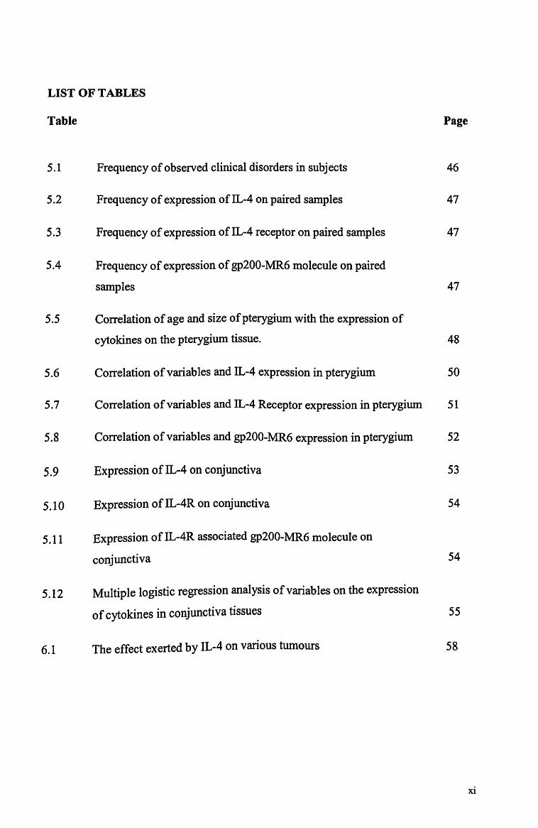
5.1 Frequency of observed clinical disorders in subjects 46
5.2 Frequency of expression of IL-4 on paired samples 47
5.3 Frequency of expression of IL-4 receptor on paired samples 47
5.4 Frequency of expression of gp200-MR6 molecule on paired
samples 47
5.5 Correlation of age and size of pterygium with the expression of
cytokines on the pterygium tissue. 48
5.6 Correlation of variables and IL-4 expression in pterygium 50
5.7 Correlation of variables and IL-4 Receptor expression in pterygium 51
5.8 Correlation of variables and gp200-MR6 expression in pterygium 52
5.9 Expression ofIL-4 on conjunctiva 53
5.10 Expression of IL-4R on conjunctiva 54
5.11 Expression ofIL-4R associated gp200-MR6 molecule on
conjunctiva 54
5.12 Multiple logistic regression analysis of variables on the expression
of cytokines in conjunctiva tissues 55
6.1 The effect exerted by IL-4 on various tumours 58
xi

LIST OF FIGURES
Figure Page
3.1 Diagramatic representation ofIL-4, IL4R and MR6 and anti-IL-4 Ab 14
3.2 Diagramatic representation ofIL-4 receptor associated gp-200
MR6 molecule and anti-MR6 Ab 15
4.1 Tissues stored in the cryovials while awaiting sectioning 21
4.2 Cryovials are stacked-up in the nitrogen tank at -70°C 21
4.3 Tissues are sectioned at 5 J.1m with the cryostats 24
4.4 Staining station for tissues section 25
4.5 The antibody used in the study 29
4.6 The immunostainer where the tissues are incubated 32
4.7 Documenting the slides with image analyser 33
4.8 H&E staining of the pterygium 34
4.9 Pterygium staining positive for IL-4 34
4.10 Pterygium with positive staining for IL-4R 35
4.11 Pterygium stained positive for gp200-MR6 molecule 35
4.12 conjunctiva with H&E staining 36
4.13 Conjunctiva stained positive for IL-4 36
4.14 Conjunctiva expressing IL-4 receptor 37
4.15 Conjunctiva showing expression of gp200-MR6 molecule 37
4.16 Well-differentiated colorectal CA expressing IL-4 R 38
xii

4.17 Breast CA expressing gp200-MR6 molecule 38
4.18 Negative control slide for pterygium 39
4.19 Negative control slide for conjunctiva tissue 39
5.1 Distribution of samples 42
5.2 Distribution by age of the subjects with pterygium 43
5.3 Distribution by age of the subjects without pterygium 43
5.4 Distribution by sex of the subjects with pterygium 44
5.5 Distribution by sex of the subjects without pterygium 44
5.6 Distribution by race of the subjects 45
xiii

LIST OF ABBREVIATIONS
Ab(s) Antibody( -ies)
Ag(s) Antigen(s)
DAB Diaminobenzedine
DH20 Distilled water
EGF Epidennal growth factor
EGFR epidennal growth factor receptor
H&E Hematoxylin and eosin
Ig(s) Imrnunoglobulin( s)
IL(s) Interleukin( s)
IL-R(s) Interleukin receptor( s)
KD kilo dalton
Mab(s) Monoclonal antibody(ies)
NS Not significant statistically
OCT Optimum Cold Temperature
PBS Phosphate-buffered saline
SPSS Statistical Package for Social Science
SS Statistically significant
TNF Tumour necrosis factor
xiv

INTRODUCTION
1
1. INTRODUCTION
Pterygium is not just a degenerative disease, but may be a proliferative disorder of the
ocular surface. This may explain the high incidence of recurrent rate after its primary
excision. Studies have consistently shown that countries nearer the equator have higher
rates of pterygium. A possible reason for this geographic variation is that the ultraviolet
B irradiation may be one of the most important risk factors for the development of
pterygium.
While many treatment modalities have been attempted to manage pterygium, it is
equally important to try to elucidate the consequences of chronic UV light exposure on
this tissue. This will provide us with valuable information regarding the changes in its
micro-environment which may form the basis of possible new and more effective
treatment modalities. In the light of better understanding of the epithelial biology of the
ocular surface and availability of improved materials and methods in immunological
research, potential plausible pathogenesis of this tissue can be identified.
This study involved the use of immunocytochemical technique to gauge the expression
ofIL-4, IL-4 Receptor and IL-4 Receptor related gp200-MR6 molecule, the presence of
all of which has been investigated and proven to playa role in the pathogenesis of
malignancy in other tissues and other cell lines. Their potential role in pterygium has
not been explored thus far.
2

OBJECTIVES OF STUDY
3
2. OBJECTIVES OF STUDY
2.1 GENERAL OBJECTIVE
The aim of this study is to investigate the expression and postulate the role of
IL-4 and its receptors in the pathogenesis of pterygium
2.2 SPECIFIC OBJECTIVES
1. To compare the expression of IL-4, IL-4 receptor and MR6 molecule in
pterygium and nonnal conjunctiva tissue unexposed to UV light in
patients with pterygium.
11. To compare the expression of IL-4, IL-4 receptor and MR6 molecule in
pterygium in relation to the age of the subjects, size of the pterygium and
other clinical parameters.
111. To compare the expression of IL-4, IL-4 receptor and MR6 molecule in
normal conjunctiva tissue unexposed to UV light in subjects with and
without pterygium.
4
2.3 NULL HYPOTHESIS
1. There is no significant difference of expression of IL-4, IL-4 receptor
and IL-4 receptor related gp200-MR6 molecule in pterygium and normal
conjunctiva tissue in patients with pterygium.
ii. There is no significant difference of expression of IL-4, IL-4 receptor
and IL-4 receptor related gp200-MR6 molecule in pterygium in relation
to the age of the subjects, size of the pterygium and other clinical
parameters.
111. There is no significant difference of expression of IL-4, IL-4 receptor
and IL-4 receptor related gp200-MR6 molecule in normal conjunctiva
tissue unexposed to UV light in subj ects with and without pterygium.
5

BACKGROUND
6

3. BACKGROUND
Ever since Susruta recorded the removal of a pterygium lesion in 1000 BC (Jaros PA et
ai, 1988), this triangular fold of vascularised bulbar conjunctiva still poses a challenge
to the practising ophthalmologists and researchers alike. Its histology and the
ultrastructure has been studied extensively (Duke-elder S, 1954; Spencer WH et ai,
1985; Hogan MJ et ai, 1967; Austin P et ai, 1983) and hypothesis put forth suggesting
its aetiopathogenesis (Moran DJ et ai, 1984; Karai I et ai, 1984; Wong WW, 1978;
Pinkerton OD et ai, 1984), hoping to find a more satisfactory management of this
disease process as it has a high recurrent rate after primary excision.
Duke-Elder has defined pterygium as a degenerative and hyperplastic process (Duke
Elder S, 1954) where there is accumulations of amorphous, eosinophilic, hyalinised or
granular-appearing material resembling degenerated collagen interspersed with coiled or
fragmented fibres resembling abnormal elastic tissue (Spencer WH et ai, 1985) which
interestingly is resistant to the nonproteolytic enzyme elastase, hence the terms elastoid
and elastotic degeneration (Cogan DG et ai, 1959). It is often found that the stromal
fibrocytes are increased in number as if proliferating in response to injury. Minor
aggregates of proteinaceous substance, acid mucopolysaccharide and calcific
concretions are also seen in older lesion.
7

Austin P, lackobiec et al concluded that a large component of pingueculae and pterygia
is the result of newly synthesised elastic fibre precursors with abnormal maturation
(elastodysplasia) that undergo secondary degeneration (elastodystrophy) (Austin P et ai,
1983). The histological features have uncanny resemblance to those found in
pingueculae and in the dermis of sun-exposed skin. Clinically, pterygium differs from
pinguecula by exhibiting progressive invasion of fibrovascular tissue into the cornea.
This actinic elastotosis are widely believed to arise from an abnormal elastogenesis
secondary to fibroblastic activation by UV radiation. (Spencer WH et ai, 1985; Ledoux
Corbusier M et a11979)
Immunologic basis for the pathogenesis of pterygium has gained substantial
momentum in the last decade. In addition to the early demonstration of plasma cell and
lymphocytes infiltration in the stroma with the presence of IgG and IgE, which is absent
in normal conjunctiva (Austin P. et ai, 1983). Liu L et al have also demonstrated that
most of the lymphocytes are T-cell (CD3+) and alteration of the helper-suppressor ratio
from 1 :2.7 in the normal conjunctiva to 1: 1.5 in pterygium, which lead them to believe
it to be a phenomenon of type 1,3 and 4 hypersensitivity (Liu L et ai, 1993). Mast cells
count in pterygium was found to be twice as high as in normal conjunctiva (Nakagami T
et ai, 1997, 1998).
In the quest of finding growth factors in pterygium, Kria et al has demonstrated with
immunohistochemical and ELISA analysis that basic fibroblast growth factor (b-FGF)
is strongly expressed in tissue cultures from recurrent pterygium as the platelet-derived
growth factor (PDGF) in primary pterygium. There is however only a weak expression
of both transforming growth factor beta (TGF -~) and tumour necrosis factor alpha (TNF
8

a.). All four growth factors are expressed strongly in pterygium tissue in vivo. All these
GFs were however sparse in nonnal conjunctival fibroblasts (Kria L et ai, 1998). These
findings provide evidence that b-FGF may participate in recurrence and rapid growth of
pterygium by its angiogenic activity. TGF-~, being a fibrogenic factor may enhance the
fibrotic process. (Kria L et ai, 1998).
TGF-~ family contains prototypic and potent cytokines involved in fibrosis (scarring)
during wound repair processes (Border W A et ai, 1992; Roberts AB et ai, 1993; Grande
JP et ai, 1997). They consist of 3 different isofonns and they exert their actions via
binding with three respective receptors.
Establishment of the limballocation of corneal epithelial stem cells has brought about
the concept of limbal stem cell deficiency. (Schenner A et ai, 1986; Tseng SCG et ai,
1989; Tseng SCG et aI, 1996). In nonnal subjects, the epithelial mass fonned at the
limbus constitutes a growth pressure and acts as a junctional barrier to prevent
conjunctival epithelial migration onto the corneal surface in the event of a total corneal
epithelial defect. When there is an insult to this region, a spectrum of abnonnal corneal
surfaces is produced (Chen JJY et ai, 1991). This can manifest as conjunctivalization
with vascularization, chronic inflammation, destruction of the basement membrane and
fibrous ingrowth.
K work LS et al has demonstrated in his animal model for pterygium, that an albedo
(indirect) light projected from the temporal sclera is focused and concentrated at the
nasal limbus, suggesting that UV light might cause focal alteration of the limbal tissue
thus causing pterygium fonnation (Kwork LS et ai, 1994). This can perhaps explained
9

the nasal bulbar conjunctiva predilection of pterygium and the intrinsic abnormalities in
DNA repair as a result of UV irradiation as evident by a high incidence of microsatellite
instability and loss of heterozygosity. (Spandidos DA et ai, 1997).
Pterygium can indeed be locally invasive, and the pterygium epithelium exhibits
varying degrees of abnormality, ranging from mild dysplasia to carcinoma in-situ (Clear
AS et ai, 1979). Tan DTH et al and Dushku et al have independently in their
immunohistochemical study with p53 monoclonal antibody found that there is increased
nuclear p53 gene product in the limbal epithelium of pterygium but with little or no
apoptosis. This, they concluded, is consistent with an activating mutation of p53 gene
secondary to UV irradiation (Tan DTH et ai, 1997; Dushku N et ai, 1997). p53 is a
tumour suppressor gene, it is the most common marker of human neoplastic growth. It
is thought to act as a transcription factor that activates or represses the expression of
growth-controlling genes and is abnormally expressed in a variety of human cancer
(Greenblatt MS et ai, 1994) as well as in actinic skin lesions (Sim CS et ai, 1992).
Dushku et al also demonstrated the expression of vimentin in limbal epithelium of
pterygium, which is normally found in mesenchymal cells and migrating epithelial cells.
(Dushku N et ai, 1994). In the related development, Kennedy Met al has demonstrated
that ultraviolet Blight (UV B) does induce the production of multiple cytokines (IL-l,
IL-6, IL-8 and TNFa) by the corneal stromal cells. (Kennedy Met ai, 1997).
Interleukins (IL) as a part of the cytokines family were first described as a group of
signalling polypeptides, controlling the activity of lymphoid and haemopoietic cells (0'
Garra A, 1989a; 1989b ). Various human tumours have been shown to express receptors
10
for many different ILs ( McMillan et ai, 1995; Topp et ai, 1995; Moore et ai, 1999).
Exposure of these cells to variety of ILs leads to positive or negetive receptor-mediated
regulation of cell growth. In the recent years, however, emphasis has been on the
possibility that human tumour cells of non-lymphoid origin may also be capable of
producing and / or responding to some of these cytokines. For example, IL-1 (a. & ~)
and IL-6 has been shown to stimulate the proliferation of certain tumour cell lines in
vitro (Tsai S.C. et ai, 1987; Howard Met aI1982).
IL of interest in this study is IL-4, a member of the cytokine family with multifunctional
activities on a variety of cell lines. It is a glycoprotein of 129 amino acids with a
molecular weight of 15 000 - 20 000 Dalton. IL-4 is glycosylated at two arginine
residues (position 38 and 105) and contains six cysteine residues involved in disulfide
bond formation, which is essential for its biological activity.
IL-4 is secreted by activated CD4 T cell of Th 2 subset, also produced by mast cells and
basophils. Initially described as a B-cell growth factor (Howard M et ai 1982), it has
subsequently shown to have a wide spectrum of activities on T lymphocytes,
macrophages, granulocytes and epithelial cells (O'Hara J et ai, 1987; Monroe JG et al
1988; Thornhill MH et ai, 1990; Toi M et al 1991). Being a prototypic type 2
immunoregulatory cytokine (Brown MA et aI, 1997; Chomarat P et al 1997), it
modulates the production of cytokine by endothelial cells, monocytes and macrophages,
also reducing inflammation by stimulating the production of monocytes IL-l Receptor
antagonist (Chomarat P et al 1995) and soluble 'decoy' IL-l receptor (Calotta F et ai,
1993).
11
IL-4 has been reported to exert anti-proliferative effect on some cancer cells. Hoon et ai
have demonstrated that IL-4 alone or in combination with other cytokines could
modulate cell surface Ag expression and inhibited growth of human melanoma cells in a
dose-dependent manner, its anti-proliferative effect synergises with IFN-y. IL-4 also
enhanced the anti-proliferative effect of TNF-a. and the additive effect was significantly
more potent than the individual cytokines or IL-4 plus IFN-y (Hoon et ai1991).
Accumulating data from transfection assays with the IL-4 gene or treatment of cell lines
with IL-4 show that IL-4 has a potent anti-tumoural activity e.g. B.16 melanoma cell
line in mice, mammary adenocarcinoma and Lewis Lung carcinoma (Zaloom Y et ai,
1993; Tepper RI et ai, 1989). Providing the evidence that IL-4 gene -transfected CA
cells can be used for CA immunotherapy (Ohira T et ai 1992). Synergism between IL-4
production and other growth inhibitors has also been described (Thornhill MH et ai,
1990). Expression of IL-4 can be studied using an IL-4 antibody with
immunohistochemistry technique. (Figure 3.1)
To enable IL-4 to exert its biological effects, it needs to bind with a specific IL-4
receptor. The human IL-4 receptor has an extracellular domain of 207 amino acids, a
transmembrane domain of 24 residues, and a large intracellular domain of 569 amino
acids. The IL-4 receptor is a complex consisting of two chains. One chain is high
affinity IL-4 binding (p140, a chain, CD 124) chain and the other chain is the y chain.
The high affinity IL-4 binding chain belongs to the cytokine receptor superfamily. The
cytoplasmic domain contains SerlPro-rich regions similar to those present in the IL-2
receptor and GM-CSF receptor p-chains. (Callard R et ai, 1994).
12

The evidence of such vital marriage between the IL-4 and IL-4 receptor is elucidated in
the study of proliferation of a number of breast cancer cell lines including anchorage
dependent and independent breast cancer cell lines MCF-7 and MDA-MB-231 were
inhibited by IL-4, which exerts its effect through IL-4Rs expressed on these cells. The
mechanism of IL-4-induced growth inhibition in human breast cancer was thought to be
the induction of apoptosis (Gooch et ai, 1998).
Further support of IL-4 role in the immunological surveillance of tumour comes from
the studies on the IL-4 receptor related gp200-MR6 molecule, using antibody for
gp200-MR6 (Figure 3.2). The facts that gp200-MR6 expression is lost with increasing
malignancy in lung and colonic CA, does suggest its role in tumour suppression
(Tungekar G et ai, 1991; Kaklamanis L et ai, 1992). In breast Ca however, both
upregulation and downregulation have been reported (Mat m et ai, 1993; AI Jabaari B
et ai, 1989). Lorenzen et al suggested that the loss of IL-4R related gp200-MR6
represent a mechanism of tumour escape from immune surveillance. To the best of our
knowledge, the expression of IL-4 and IL-4 receptor and IL-4 receptor associated
gp200-MR6 molecule on the ocular tissue has never been studied before. Thus, this
study was conducted to establish the presence of these cytokines on the ocular surface.
13

IL-4 and its membrane bound receptor which also have a gp 200 MR 6 molecule
I Anti-IL4 I Ab
IL-4R IL-4R
MR6
IL-4 binds to its receptor site
MR6
Adding antibody(Ab) to the complex or free IL-4 willlead to binding of the Ab onto the IL-4 molecule. The Ab can be identified under the microscope by conjugating it to a dye which will show up as immunoflourescein positive, thus facilitating the study ofIL-4 expression
Figure 3.1 Diagrammatic representation of IL-4, IL-4 receptor and IL-4 receptor associated gp-200 MR6 molecule with anti-IL-4 Ab
14

IL-4 and its membrane bound receptor which also have a gp 200 MR 6 molecule
MR6
IL-4 binds to its receptor site Addition ofMR6 antibody
MR GAb
The Ab to MR6 will bind to the IL-4 receptor-related gp200 MR6 molecule. The Ab can then be identified under the microscope by conjugating it to a dye which will show up as immunoflourescent positive, thus facilitating the study the expression ofIL-4 Receptor.
Figure 3.2 Diagramatic representation of IL-4, IL-4 receptor and MR6 with antiMR6Ab.
15
MATERIALS AND METHODS
16
4. MATERIAL AND METHODS
4.1 STUDY DESIGN
This is a laboratory-based comparative study. All the laboratory work is done in
the laboratory of Department of pathology and Department of hnmunology.
4.2.1 POPULATION, TIME AND PLACE OF STUDY
Study population:
Period of study:
Place of study:
Patients attending eye clinic in Hospital USM and
General Hospital Kota Bharu who have consented for
pterygium excision.
Patients undergoing extracapsular cataract extraction in
Hospital USM.
1st January 2000 to 30th November 2001
Department of Ophthalmology, School of Medical
Sciences, USM.
Department of Ophthalmology, Hospital Kota Bharu.
hnmunohistochemistry laboratory, Department of
Pathology, USM
17

4.3 SAMPLING AND SAMPLE SIZE
A convenient sampling size was taken as there was no previous study on
similar subj ect.
4.4 SELECTION CRITERIA
4.4.1 INCLUSION CRITERIA
All patients who underwent pterygium excision or cataract extraction with
otherwise normal eye were included in the study.
4.4.2 EXCLUSION CRITERIA
The following cases were excluded from the study:
a) Patients on long term (in the last six months) or current use of topical medicated
preparations (all preparation except eye lubricants without any active
ingredients), nasal spray (e.g. steroid etc.).
b) Patients on systemic steroid therapy or any other systemic immunomodulating
medication (e.g. systemic cyclosporin A ) or any form of chemotherapy.
c) Patients with history of trauma or insult to the eye within the last 6 months.
(postoperative, subconjunctival haemorrhage etc).
d) Patients with hisoty of any form of conjunctivitis or inflammation in the eyes
(uveitis etc).
e) Contact lens wearers.
18
~ I
f) Patients who has any form of keratoplasty performed.
g) Patients with a known history of autoimmune disorders. (rheumatoid arthritis,
SLE etc.)
4.5 CONSENT
All participating patients were explained in-depth regarding the nature and
objectives of this study and the potential complications associated with tissue
harvesting. This was done in patients' native language or dialects with the aid of
diagrammatic representations of the procedures. Written informed consent is
then obtained from the willing subjects. (Appendix C)
4.6 VARIABLES OBSERVED
The units of variables recorded include demographic datas e.g. the age, sex and
race of the patients involved. The laterality of the pterygium or conjunctiva
excised is duly recorded. Also recorded is the status of the pterygium, whether it
is a primary or recurrent lesion. The size of the pterygium is also measured with
slitlamp in millimetre (from the limbus of the cornea perpendicularly to the head
of the pterygium). Also recorded is the presence of systemic illness like
diabetes, hypertension, evidence of allergy or asthma and smoking.
(Data collection sheet - Appendix A and B)
19

I
,I
i! ,j I, I .1
4.7 SPECIMEN
Excision of tissue for the specimen is done with written consent (Appendix C).
Three groups of tissues are obtained.
Group 1:
Pterygium proper
Group 2:
Superior or inferior bulbar conjunctival tissue from patient with
pterygium who undergone cataract extraction or patient undergoing
pterygium excision with conjunctival transposition and consented for
bulbar conjunctival biopsy.
Group 3:
Superior bulbar conjunctival tissue from patient who has undergone
cataract extraction and no evidence of pterygium.
4.7.1 STORAGE
Specimens collected will be immediately put into a cryo-vial and snap freezed in
a portable nitrogen tank before being transferred to nitrogen tank for storage.
20

Figure 4.1 Tissues stored in the cryovials while awaiting sectioning.
Figure 4.2 Cryovials are stacked-up in a nitrogen tank preserving the tissues at
-70°C.
2 1
4.8 DEFINITION OF TERMS:
a. Diabetes Mellitus- records of diabetic treatment or newly diagnosed diabetes
with a fasting whole blood concentration of over 6.7mmolll, or a random value
exceeding 10mmolll.
b. Hypertension- clear records of treatment for hypertension or newly diagnosed
hypertension defined as blood pressure recording of systolic more than
140mmHg and/or diastolic of more than 90mmHg sustained over three
consecutive recordings at least 6 hours apart.
c. Allergy- clear history and/or record of treatment of allergy disorders. Clear
history or clinical sign and symptom of allergy illness, ego acute exacerbation of
bronchial asthma, allergic conjunctivitis, allergic rhinitis, history of urticaria or
rashes after food or drug ingestion etc.
d. Smoking- Clear history of active smoking or long duration of cigarette smoking
(e.g. more than 6 months).
e. Superior bulbar conjunctiva-- a small 3mm X 3mm conjunctival tissue harvested
anywhere from eleven to one O'clock positions.
f. Inferior bulbar conjunctiva-a small 3mm X 3mm conjunctival tissue harvested
anywhere from five to seven O'clock positions.
22

4.9 ETIDCAL APPROVAL
This Research protocol and methodology has been approved by the Research
and Ethics Committee, School of Medical Sciences, Universiti Sains Malaysia.
Reference: USM/PPSP®lEthics Com.l200 1 [54.4(4)]
23
4.10 IMMUNOmSTOCHEMISTRY - MATERIALS AND METHOD
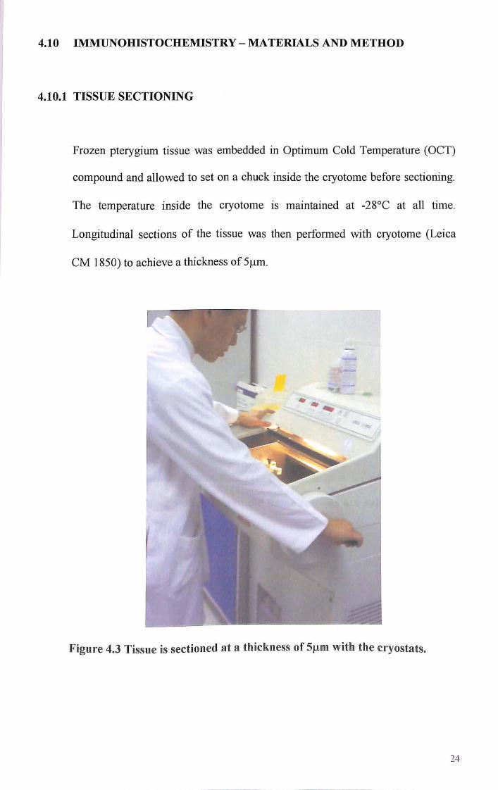
4.10.1 TISSUE SECTIONING
Frozen pterygium tissue was embedded in Optimum Cold Temperature (OCT)
compound and allowed to set on a chuck inside the cryotome before sectioning.
The temperature inside the cryotome is maintained at -28°C at all time.
Longitudinal sections of the tissue was then performed with cryotome (Leica
CM 1850) to achieve a thickness of 51lm.
Figure 4.3 Tissue is sectioned at a thickness of 51lm with the cryostats.
24
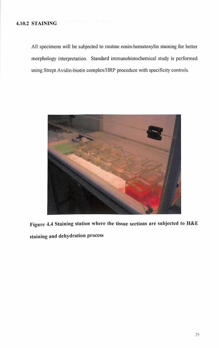
4.10.2 STAINING
All specimens will be subjected to routine eosin-hematoxylin staining for better
morphology interpretation. Standard immunohistochemical study is performed
using Strep!.Avidin-biotin complex/HRP procedure with specificity controls.
Figure 4.4 Staining station where the tissue sections are subjected to H&E
staining and dehydration process
25