Embed Size (px)
Citation preview

2145Research Article
IntroductionThe sarcoplasmic reticulum (SR) of the skeletal muscle andthe transverse tubular membranes provide a conduciveenvironment enriched with key proteins that have been shownto play an essential role in excitation-contraction (E-C)coupling (Endo, 1977; Fleischer and Inui, 1989). E-C couplingoccurs in specialized junctions called triads, which are anintracellular synapse formed by the membrane system of thetransverse tubule and the terminal cisternae of the SR. The twomain Ca2+ channels involved in E-C coupling are thedihydropyridine receptor, Cav1, which lies on the transversetubules where it acts as a voltage sensor for E-C coupling, andthe ryanodine receptor (RyR), present on the junctional facemembrane of the SR, which is the Ca2+ release channel(Franzini-Armstrong and Jorgensen, 1994; Meissner, 1994; Maand Pan, 2003; Sutko and Airey, 1996; Franzini-Armstrong,1980; Lamb and Stephenson, 1990; Rios and Pizzarro, 1991).Cav1 is a hetero-oligomeric complex made up of at least foursubunits: �1, �, �2� and � (Leung et al., 1987; Lacerda et al.,1991; Birnbaumer et al., 1998; Catterall, 1995; Snutch andReiner, 1992). The pore-forming Cav1.1 is an integralmembrane protein and is indispensable for E-C coupling,whereas the cytosolic �1a and �2� subunits have regulatory
functions; the role of the � subunit has yet to be clearly defined(Catterall, 1995; Snutch and Reiner, 1992, Tsien et al., 1991).The �1a subunit associates tightly with the Cav1.1 by bindingto a region on the I-II loop termed �-interacting domain (AID)(Pragnell et al., 1994). The �1a subunit is important for plasmamembrane expression of the Cav1.1, whereas a sequencelocated in the C-terminal domain of the Cav1.1 seems to beinvolved in triad targeting (Chien et al., 1995; Flucher et al.,2000; Flucher et al., 2002), although other domains and/orpolypeptides might be involved in proper targeting.
In skeletal muscle, Cav1.1 responds to transverse tubuledepolarisation by sensing the voltage change and it inducesCa2+ release from the SR through a direct interaction with theRyR. Besides the RyR, however, the junctional face membranecontains numerous other proteins that, because of theiranatomical location, are deemed to be involved in E-C coupling(Zhang et al., 1997; Costello et al., 1986; Zorzato et al., 2000).In the past few years a number of investigators have begun todefine the major and minor structural components of thejunctional face membrane (Ito et al., 2001; Takeshima et al.,2000). In previous studies (Zorzato et al., 2000; Anderson etal., 2003), we identified and characterized – at the biochemicaland molecular level – JP-45, an integral membrane protein
JP-45, an integral protein of the junctional face membraneof the skeletal muscle sarcoplasmic reticulum (SR),colocalizes with its Ca2+-release channel (the ryanodinereceptor), and interacts with calsequestrin and the skeletal-muscle dihydropyridine receptor Cav1. We have identifiedthe domains of JP-45 and the Cav1.1 involved in thisinteraction, and investigated the functional effect of JP-45.The cytoplasmic domain of JP-45, comprising residues 1-80, interacts with Cav1.1. JP-45 interacts with two distinctand functionally relevant domains of Cav1.1, the I-II loopand the C-terminal region. Interaction between JP-45 andthe I-II loop occurs through the ��-interacting domain in theI-II loop. ��1a, a Cav1 subunit, also interacts with thecytosolic domain of JP-45, and its presence drastically
reduces the interaction between JP-45 and the I-II loop.The functional effect of JP-45 on Cav1.1 activity wasassessed by investigating charge movement indifferentiated C2C12 myotubes after overexpression ordepletion of JP-45. Overexpression of JP-45 decreased peakcharge-movement and shifted VQ1/2 to a more negativepotential (–10 mV). JP-45 depletion decreased both thecontent of Cav1.1 and peak charge-movements. Our datademonstrate that JP-45 is an important protein forfunctional expression of voltage-dependent Ca2+ channels.
Key words: Voltage-dependent Ca2+ channel, JP-45, Sarcoplasmicreticulum, Excitation-contraction coupling
Summary
The junctional SR protein JP-45 affects the functionalexpression of the voltage-dependent Ca2+ channelCav1.1Ayuk A. Anderson1, Xavier Altafaj2, Zhenlin Zheng3, Zhong-Min Wang3, Osvaldo Delbono3,4, Michel Ronjat2,Susan Treves1 and Francesco Zorzato5,*1Departments of Anaesthesia and Research, Basel University Hospital, Hebelstrasse 20, 4031 Basel, Switzerland2INSERM U607/CEA/UJF, Lab CCFP/DRDC, Rue des Martyrs 17, 38054, Grenoble, Cedex 9 France3Department of Physiology and Pharmacology and 4Department of Internal Medicine, Gerontology, Wake Forest University School of Medicine,Winston-Salem, NC 27157, USA5Department of Experimental and Diagnostic Medicine, General Pathology Section, University of Ferrara, Via Borsari 46, 44100 Ferrara, Italy*Author for correspondence (e-mail: [email protected])
Accepted 14 February 2006Journal of Cell Science 119, 2145-2155 Published by The Company of Biologists 2006doi:10.1242/jcs.02935
Jour
nal o
f Cel
l Sci
ence

2146
constituent of the SR junctional face membrane in skeletalmuscle. We also showed that JP-45 colocalizes with the RyRCa2+-release channel and interacts with Cav1.1 and the luminalCa2+-binding protein calsequestrin (Anderson et al., 2003). Togather insight into the functional role of JP-45, we defined thedomains involved in the interaction between JP-45 and Cav1.1.Our results demonstrate that the cytoplasmic domain of JP-45interacts directly with the I-II loop, the C-terminal domain ofCav1.1 subunit and also with the �1a subunit of Cav1. Inaddition, we show that the interaction between JP-45 and theI-II loop occurs through the AID domain, and can be displacedby �1a. Experimental evidence has demonstrated that the �1asubunit interacts with the Cav1.1 subunit via AID (Pragnell etal., 1994; Chen et al., 2004) and that this protein-proteininteraction is involved in the insertion of the voltage sensor toits proper membrane compartment (Flucher et al., 2002). Toconfirm the functional role of JP-45 in vivo, we overexpressedand silenced endogenous JP-45 in C2C12 cells and studiedtheir electrophysiological properties. Based on the results ofthe present report, JP-45 is involved in the regulation of thefunctional expression of Cav1.1 into the transverse tubularmembrane compartment.
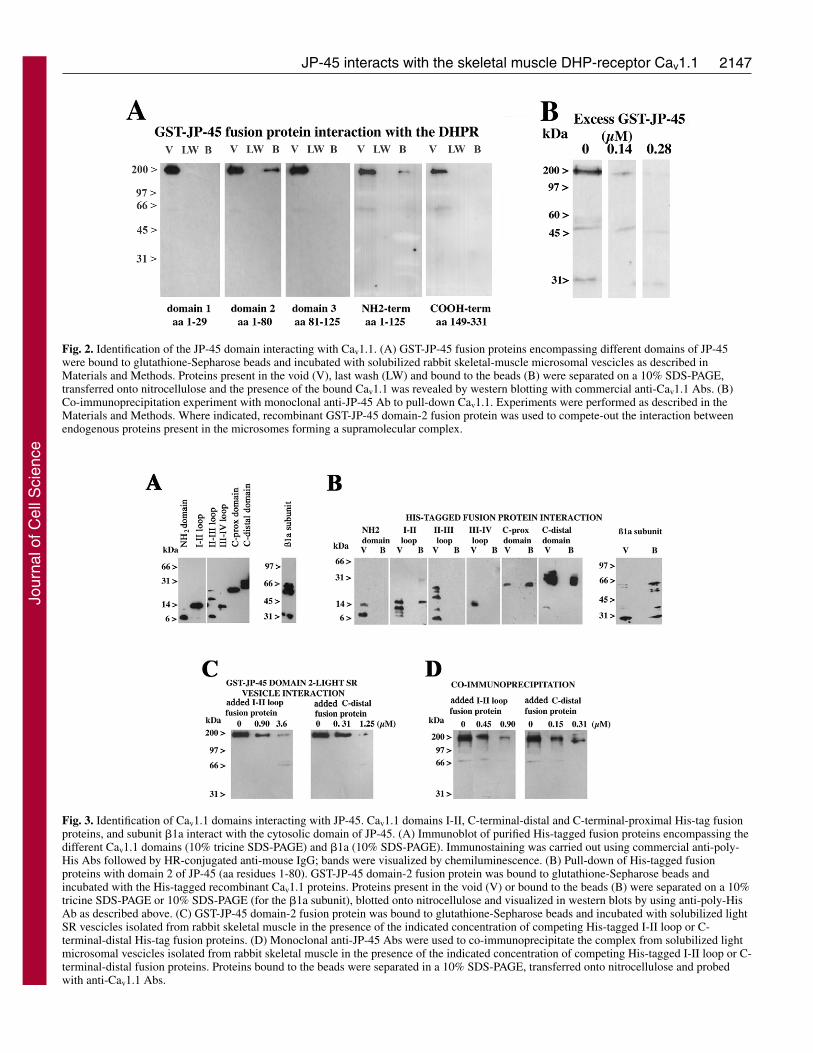
Results and DiscussionIdentification of domains enabling the interaction of JP-45 and Cav1.1In a previous report, we demonstrated that JP-45 interacts withnative Cav1.1 (Anderson et al., 2003). Here, we describe aseries of experiments to identify the domains participating inthis interaction. We prepared recombinant GST-fusion proteinscovering different coding regions of JP-45 (excluding thetransmembrane domain) (see Fig. 1). These proteins were thenbound to glutathione-Sepharose beads and incubated withsolubilized light microsomal vesicles. Fig. 2A shows that thecytoplasmic N-domain of JP-45, more specifically the regionbetween residues 1-80 (domain 2) interacts with native Cav1.1.No interaction was observed between any other JP-45 domainsand Cav1.1. The physiological relevance of the interaction offusion proteins was verified by studying the effect of the GST-JP-45 domain 2 on co-immunoprecipitation of Cav1.1 with amonoclonal anti-JP-45 antibody (Ab). The interaction betweennative JP-45 and Cav1.1 was competed-out by the presence ofsoluble GST-JP-45 domain 2 in the co-immunoprecipitationreaction (Fig. 2B).
Extensive structure-to-function relationship studies on Cav1have helped to define functionally relevant domain boundariesnot only among the subunits making up the supramolecularcomplex, but also within the primary structure of each subunit(Nakai et al., 1998; Grabner et al., 1999; Kugler et al., 2004; VanPetegem et al., 2004). We reasoned that the physiologicalrelevance of the interaction between Cav1.1 and JP-45 couldbe better understood if we first identified JP-45-binding siteswithin the Cav1.1. GST-JP-45 domain-2 fusion protein wasimmobilized on GST-Sepharose and incubated with differentHis-tagged fusion proteins, covering the cytoplasmic domains ofthe Cav1.1 subunit (Tanabe et al., 1987). Whereas some of theHis-tagged fusion proteins display the expected molecular mass,they had in other cases a molecular mass lower than the expectedvalue due to the instability of purified fusion proteins (Fig. 3A,lanes 3 and 7). The protein-protein-interaction experimentsdepicted in Fig. 3B demonstrate that the I-II loop and the C-
Journal of Cell Science 119 (10)
terminal domain of the Cav1.1 as well as the �1a subunit interactwith JP-45, whereas fusion proteins corresponding to the N-terminus – the II-III and III-IV loops of the Cav1.1 subunit – donot interact with GST-JP-45 domain-2 fusion protein. To validatethese results, we performed pull-down experiments using GST-JP-45 domain 2 as bait and solubilized light microsomal vesiclesas ligand, in the presence or absence of increasing concentrationsof either I-II loop or recombinant C-terminal-distal His-tagfusion proteins. Inhibition of Cav1.1-GST-JP-45 interaction wasachieved at 3.6 and 1.25 �M of I-II loop and C-terminal-distalHis-tag fusion proteins, respectively. To further confirm thespecificity of the pull-down assay, we also investigated theseinteractions under native conditions, i.e. by performing co-immunoprecipitation of native JP-45 with Cav1.1 using themonoclonal anti-JP-45 Ab, in the presence or absence ofcompeting I-II loop or recombinant C-terminal-distal His-tagfusion proteins. As shown in Fig. 3D, the presence of thecompeting recombinant fusion proteins substantially diminishedthe interaction between JP-45 and the Cav1.1 domains. Pull-down and co-immunoprecipitation assays demonstrated thatcompetition with one of the Cav1.1 interacting domains (I-II loopor C-terminal-distal His-tag fusion proteins) was sufficient topartially inhibit the interaction. At a first glance it seems unclearhow preventing the Cav1.1–JP-45 interaction through a singledomain reduces total binding to the Cav1.1 to almost zero. Aplausible explanation of the experiments is described in Fig. 3Cand D; addition of excess I-II loop or C-terminal-distal loopoccupies the interacting domain of the N-terminal domain of JP-45. Binding of the 50 aa-long JP-45 binding sites by an excessof the 14 kDa or 30 kDa Cav1.1 loops (I-II and C-terminal,respectively) most probably creates a steric hindrance, wherebythe interaction of alternative domains within the nativesolubilized Cav1.1 is either abolished or substantially weakened.
Immunoprecipitation experiments revealed that JP-45
Fig. 1. Domains of JP-45 used to identify the Cav1.1 binding sites.(A) Schematic representation. (B) 10% SDS-PAGE and CoomassieBrilliant Blue staining of the GST-JP-45 fusion proteins purified byglutathione-Sepharose. The numbers above each lane indicate the aaresidues that were fused in frame in the pGex plasmid to yield theGST-JP-45 fusion protein.
Jour
nal o
f Cel
l Sci
ence
2147JP-45 interacts with the skeletal muscle DHP-receptor Cav1.1
Fig. 2. Identification of the JP-45 domain interacting with Cav1.1. (A) GST-JP-45 fusion proteins encompassing different domains of JP-45were bound to glutathione-Sepharose beads and incubated with solubilized rabbit skeletal-muscle microsomal vescicles as described inMaterials and Methods. Proteins present in the void (V), last wash (LW) and bound to the beads (B) were separated on a 10% SDS-PAGE,transferred onto nitrocellulose and the presence of the bound Cav1.1 was revealed by western blotting with commercial anti-Cav1.1 Abs. (B)Co-immunoprecipitation experiment with monoclonal anti-JP-45 Ab to pull-down Cav1.1. Experiments were performed as described in theMaterials and Methods. Where indicated, recombinant GST-JP-45 domain-2 fusion protein was used to compete-out the interaction betweenendogenous proteins present in the microsomes forming a supramolecular complex.
Fig. 3. Identification of Cav1.1 domains interacting with JP-45. Cav1.1 domains I-II, C-terminal-distal and C-terminal-proximal His-tag fusionproteins, and subunit �1a interact with the cytosolic domain of JP-45. (A) Immunoblot of purified His-tagged fusion proteins encompassing thedifferent Cav1.1 domains (10% tricine SDS-PAGE) and �1a (10% SDS-PAGE). Immunostaining was carried out using commercial anti-poly-His Abs followed by HR-conjugated anti-mouse IgG; bands were visualized by chemiluminescence. (B) Pull-down of His-tagged fusionproteins with domain 2 of JP-45 (aa residues 1-80). GST-JP-45 domain-2 fusion protein was bound to glutathione-Sepharose beads andincubated with the His-tagged recombinant Cav1.1 proteins. Proteins present in the void (V) or bound to the beads (B) were separated on a 10%tricine SDS-PAGE or 10% SDS-PAGE (for the �1a subunit), blotted onto nitrocellulose and visualized in western blots by using anti-poly-HisAb as described above. (C) GST-JP-45 domain-2 fusion protein was bound to glutathione-Sepharose beads and incubated with solubilized lightSR vescicles isolated from rabbit skeletal muscle in the presence of the indicated concentration of competing His-tagged I-II loop or C-terminal-distal His-tag fusion proteins. (D) Monoclonal anti-JP-45 Abs were used to co-immunoprecipitate the complex from solubilized lightmicrosomal vescicles isolated from rabbit skeletal muscle in the presence of the indicated concentration of competing His-tagged I-II loop or C-terminal-distal fusion proteins. Proteins bound to the beads were separated in a 10% SDS-PAGE, transferred onto nitrocellulose and probedwith anti-Cav1.1 Abs.
Jour
nal o
f Cel
l Sci
ence

2148
interacts with the I-II loop of subunit Cav1.1 and with purifiedrecombinant �1a subunit (Fig. 3B). The �1a has been shownto interact strongly with the I-II loop of the �1.1 subunit andto affect its functional expression (Flucher et al., 2000; Chienet al., 1995). In the next set of experiments, we thereforecharacterized in greater detail the latter protein-proteininteraction.
Effect of the �1a subunit on the interaction of JP-45 andCav1.1 We investigated the role of the �1a subunit on the interactionbetween JP-45 and Cav1.1 by pull-down and co-immunoprecipitation assays (Fig. 4). JP-45 domain-2 fusionprotein was immobilized on GST-Sepharose and incubated withthe His-tagged Cav1.1 subunit I-II loop fusion protein in thepresence or absence of increasing concentrations of � 1a fusionprotein. As seen in Fig. 4A the presence of �1a interferes withthe interaction between the I-II loop and JP-45. Pull-down assayswere also performed by using the solubilized light microsomalvesicles as ligand. In the latter case, the association between theGST-JP-45 domain-2 fusion protein and the native Cav1.1subunit was remarkably weaker in the presence of excess �1a-His-tagged fusion protein (Fig. 4B). The interference of theinteraction between JP-45 and the Cav1.1 subunit by excess �1a,was also confirmed by performing co-immunoprecipitationexperiments with anti-JP-45 Abs to pull-down the nativesolubilized complex (Fig. 4C). These results suggest that thepresence of excess �1a disrupts the complex between Cav1.1 andJP-45. We cannot discriminate whether JP-45 interacts with theCav1.1 or �1a, subunit, or whether JP-45 interacts with bothsubunits, thereby forming an oligomeric complex.
The Cav1 �1a subunit has been shown to bind strongly tothe intracellular loop between transmembrane domain I and IIof the Cav1.1, a region composed of 18 aa displaying the motifQQ-E-L-GY-WI-E, conserved in different isoforms (Pragnellet al., 1994). This binding domain is also referred to as �-interacting domain (AID). It is thought that the interactionbetween AID and the �1a subunit is important to determine thestability of this Ca2+ channel on the plasma membrane(Birnbaumer et al., 1998; Flucher et al., 2000; Flucher et al.,2002; Chien et al., 1995; Ahern et al., 2003; Bichet et al.,2000). In addition, the interaction between AID and �1a isregarded to the major structural determinant required for thesurface expression of Cav1.1 (Chien et al., 1995; Beurg et al.,1999; Gregg et al., 1996; Jones et al., 1998). Coexpression ofCav1.1 and �1a subunits has been shown to increase the densityand kinetics of L-type Ca2+ currents, and experimentalevidence suggest that the �1a subunit facilitates gating ofCav1.1 by increasing the coupling between charge movementand pore opening (Sheridan et al., 2003; Sheridan et al., 2004).These effects of the �1a subunit are due to its interaction withthe I-II loop, a domain that also interacts with JP-45. In thenext set of experiments we addressed the question of whetherthe �1a subunit and JP-45 share the AID domain as bindingsite on the I-II loop. To address this issue, we performed pull-down assays with (1) GST-I-II loop fusion protein, (2) with aGST-fusion protein containing AID flanked by 21 and 11residues at the N-terminal and C-terminal end, respectively,and (3) with a synthetic biotinylated-AID peptide. For the firsttwo assays, the GST-fusion proteins were used as bait and theHis-tagged JP-45 domain-2 fusion protein as ligand. Theapparent molecular mass of His-tagged JP-45 domain 2 is
Journal of Cell Science 119 (10)
Fig. 4. Effect of the �1a subunit on theinteraction between JP-45 and Cav1.1.(A) Interaction between GST-JP-45 and His I-II loop in the presence of competing purified�1a subunit. For the fusion-protein–proteininteraction, 0.57 �M of GST-JP-45 domain 2and 1.4 �M I-II loop fusion protein wereincubated in the presence of the indicatedconcentration of His-tagged �1a subunit. His-tagged I-II loop fusion proteins bound toglutathione-Sepharose beads coated with GST-JP-45 domain-2 fusion protein were separatedon a 10% tricine SDS-PAGE and probed withanti-poly-His Ab as described in the legend toFig. 3. (B) Solubilized rabbit skeletal-musclelight SR vesicles were incubated withglutathione-Sepharose beads coated with GST-JP-45 domain-2 fusion protein in the absenceor presence of purified �1a subunit. Proteinspresent in the void (V) and bound to the beads(B), were separated on a 10% SDS-PAGE,blotted onto nitrocellulose and probed withanti-�1.1 subunit Ab. (C) Solubilized rabbitskeletal-muscle light SR vesicles wereincubated with anti-JP-45 Ab followed byincubation with Sepharose-protein G beads inthe absence or presence of competing purified�1a subunit. Proteins present in the void (V)and bound to the beads (B), were separated ona 10% SDS-PAGE, blotted onto nitrocelluloseand probed with anti-Cav1.1 Ab.
Jour
nal o
f Cel
l Sci
ence

2149JP-45 interacts with the skeletal muscle DHP-receptor Cav1.1
larger than the theoretical predicted values (approx. 31 kDa vs14 kDa, respectively). We are confident that the apparent largermolecular mass of the His-tagged JP-45 domain-2 fusionprotein is due its dimerization, because its treatment withDEPC (which destroys the His tag) abolished the binding ofthe anti-His tag Ab (Fig. 5 panel C). Western blot analysis (Fig.5A,B) demonstrates that JP-45 domain 2 interacts directly withthe AID sequence within the I-II loop. The in vitro interactionof the sequence containing AID and JP-45 occurs atmicromolar concentrations of His-tagged JP-45 domain-2fusion protein. Because of such a high concentration it isprobably that, under physiological conditions, the interactionbetween the �1a subunit and AID prevails over that of JP-45and AID. Nevertheless, on the basis of the highly organizedmolecular assembly of the triad membranes, we cannot excludethe possibility that the local concentration of JP-45 in thetriadic gap is sufficient to interfere either with the high-affinityinteraction between AID and the �1a subunit (Pragnell et al.,1994; Sheridan et al., 2003; Strube et al., 1996). If there are noother proteins involved in the interaction with AID, our dataimply that, in the �1a subunit knock-out animal, AID isoccupied mainly by JP-45 and that such an interaction mightaccount, at least in part, for the severe phenotype of the mice(Gregg et al., 1996). Strube et al. have shown that mouseskeletal muscle cells with a null-mutation in the cchb1 gene(encoding the �1a subunit of the Cav1) show a significantdecrease in maximum charge-movement and a shift in thehalf-activation potential towards more negative potentials(Strube et al., 1996).
To investigate the functional effect of JP-45, we altered thestoichiometry of the JP-45–Cav1.1 supramolecular complexeither by JP-45 overexpression or by JP-45 gene silencing indifferentiated C2C12 myotubes, and then examined Cav1.1activity by measuring charge movement.
Effect of JP-45–DsRed2 overexpression on the functionof the Cav1.1To examine the expression of JP45 and the Cav1.1, we carriedout western blot analysis on subcellular fractions isolated fromtransfected cells. Fig. 6A shows a western blot of totalmicrosome fractions from C2C12 cells transfected either withpFP-N3 coupled to red fluorescent protein (DsRed2) (pFP-N3–DsRed2) vector or with JP-45–pFP-N3–DsRed2 plasmid.Cells transfected with the latter plasmid show animmunoreactive band of approximately 45 kDa, whichrepresents endogenous JP-45, and a band of approximately 70kDa, which represents the JP-45-DsRed fusion protein. Toverify whether overexpression of JP-45 affects the averageamount of Cav1.1 subunit, we performed immunoprecipitationexperiments. Total C2C12 lysate of control and JP45-overexpressing cells contain two distinct high-molecular massbands of 175 kDa and 170 kDa that can be attributed to theCav1.1 subunit (Leung et al., 1987). The relative amount ofprotein in the bands did not show a major change (intensityratio of Cav1.1 bands in JP45-overexpressing cells to those ofcells expressing empty vector was 0.971±0.06, mean ± s.d.).C2C12 cells were transfected with JP-45–pFP-N3–DsRed2and the expression of JP-45–DsRed2 fusion protein wasmonitored by fluorescence microscopy to measure directcharge-movement. Fig. 7A shows the maximal chargemovement. Fluorescence relationship for C2C12 cellstransfected with JP-45–pFP-N3–DsRed2 construct (A, n=17)or pFP-N3/DsRed2 vector alone as control (B, n=15). As themagnitude of JP-45–DsRed2 fluorescence increased, the peakcharge-movement significantly decreased (Fig. 7A), aphenomenon that was not observed in the control cellstransfected only with the pFP-3/DsRed2 plasmid (Fig. 7B). Foranalysis of the charge-movement versus membrane-voltagerelationship of cells transfected with either JP-45–pFP-
Fig. 5. JP-45 interacts with the AID domain on the I-II loop of Cav1.1. (A) GST-I-II loop fusion protein or the GST-AID containing domainencompassed within Cav1.1 residues 336-384 were incubated with His-tagged JP-45 domain-2 fusion protein. Pull-down was performed asdescribed in Fig. 3; proteins in the void (V), last wash (LW) or bound to the glutathione resin (B) were separated on a 12.5% SDS-PAGE,transferred onto nitrocellulose and probed with affinity-purified anti-JP-45 Ab. (B) Synthetic biotinylated peptides corresponding to the AIDsequence or an unrelated biotinylated peptide were used to coat neutroavidine beads, which were subsequently incubated with His-JP-45domain 2. Proteins present in the void, last wash or bound to the beads were separated on a 12.5% SDS-PAGE, transferred onto nitrocelluloseand the immunopositive band was visualized using anti-His-tag commercial Abs. (C) A His-tagged fusion protein encompassing domain 2 JP-45 was prepared as described in Materials and Methods. Although the fusion protein migrated slower in SDS-PAGE, its identity was verified bydirect sequencing (not shown) and by immunoblotting with anti-His Ab. Note that treatment of the fusion protein with DTT+DEPC eliminatedits immunoreactivity.
Jour
nal o
f Cel
l Sci
ence

2150
N3–DsRed2 (Fig. 7C) or pFP-N3–DsRed2 plasmid (Fig. 7D),data points were fitted to a Boltzmann equation of the form:
Qon = Qmax / [1 + exp(VQ1/2 – Vm)/K] ,
where Qmax is the maximum charge, Vm is the membranepotential, VQ1/2 is the charge-movement half-activationpotential and K is the steepness of the curve. When cells werepooled for the analysis, Qmax was significantly lower in JP-45/pFP-N3/DsRed2 transfected than in control cells, whereasVQ1/2 was shifted to more negative potentials in the formergroup. The best fitting parameters for Qmax, VQ1/2 and Krecorded in both groups of cells are included in Table 1. Nodifferences in the steepness of the curve were recorded. Tobetter characterize the voltage-dependence of the chargemovement, the group of cells transfected with JP-45–pFP-N3–DsRed2 (Fig. 7A,C) were separated into two subgroups,either exhibiting the five highest or lowest Qmax values (Fig.7E). This approach allowed to identify a more obviousdifference not only in Qmax (~threefold) but also in the halfactivation potential of the charge movement (Table 1). Highexpression levels of JP-45 are associated with a shift of VQ1/2to more negative potentials of ~10 mV (Fig. 7C,D). Nodifferences in the steepness of the curve were observedbetween these two groups of cells (Table 1).
The observation that alteration of peak charge movement istightly linked to the magnitude of JP-45 expression suggeststhat, the stoichiometry of the JP-45/Cav1.1 supramolecular
complex is crucial for the proper function of theCav1.1. If this is so, we reasoned that depletion ofJP-45 in differentiated myotubes would similarlyaffect the charge movement of Cav1.1.
Effect of JP-45 gene silencing on Cav1.1functionThe mRNA encoding JP-45 was much lessabundant in C2C12 myotubes transfected with theplasmid containing JP-45 siRNA than in cellstransfected with the control pSHAG vector (Fig.8A). The presence of residual mRNA for JP-45could either be due to the presence of a smallsubpopulation of cells which had not beentransfected with the JP-45 siRNA construct, or tothe inability of the construct to fully eliminate thetranscript of JP-45. The amount of �-actintranscript did not differ significantly between cellstransfected with the two constructs (Fig. 8A lowerpanel). Western blot analysis was performed toconfirm depletion of JP-45 in differentiatedC2C12 myotubes. Expression of JP-45 in C2C12cells transfected with JP-45 siRNA is lower thanthe detection limit of the anti-JP-45 Abs that were
Journal of Cell Science 119 (10)
Fig. 6. Overexpression of JP-45 in C2C12 cells does not affect theexpression levels of the Cav1.1 subunit. C2C12 cells were transfectedeither with the pFP-N3–DsRed2–JP-45 vector or with pFP-N3–DsRed2 alone as control. (A) Microsomes were prepared fromtransfected differentiated C2C12; 5 �g protein were separated on a10% SDS-PAGE, blotted onto nitrocellulose and probed with anti-JP-45 polyclonal Ab, followed by HR-coupled protein G. Theimmunoreactive band was visualized by chemiluminescence. Notethat control cells show the endogenous JP-45 immunoreactive bandalone, whereas cells transfected with pFP-N3–DsRed2–JP-45 showan additional band of approximately 70 kDa, representing theDsRed–JP-45 fusion protein. (B) Immunoprecipitation and westernblot analysis of Cav1.1 subunit expression in C2C12 cells transfectedwith pFP-N3–DsRed2 or pFP-N3–DsRed2–JP-45. Results arerepresentative of three different transfection experiments.
Fig. 7. Effect of JP-45 overexpression on Cav1.1 charge movement. (A,B) Maximum charge-movement versus fluorescence for C2C12 cells transfected with either pFP-N3–DsRed2 JP-45(A) or pFP-N3/DsRed2 alone as control (B). The lines in A and B represent the linearregression including all data points. (C,D) Charge-movement versus membrane-voltage (Vm)for pFP-N3–DsRed2–JP-45 (C) or pFP-N3–DsRed2 plasmid (D) transfected cells fitted to aBoltzmann equation (see text). The best fitting parameters are included in Table 1. (E) Charge-movement versus Vm for the five highest and lowest Qmax values (E) from JP-45 transfectedcells (C).
Jour
nal o
f Cel
l Sci
ence

2151JP-45 interacts with the skeletal muscle DHP-receptor Cav1.1
raised against the N-terminal-interacting domain of the protein(Fig. 8B). However, we did not see changes in the expressionof the housekeeping gene �-actin. Having established areduction in transcription and expression of JP-45, we nextmeasured Cav1.1 charge-movement. C2C12 cells were co-transfected with pFP-N3–DsRed2 and either the JP-45 siRNApSHAG vector or pSHAG vector. Based on the expression ofthe DsRed2 reporter, C2C12 cells were identified and charge-movement measurements were performed. Fig. 9A shows thatdepletion of JP-45 in C2C12 myotubes is associated with adecrease in Qmax, apparently without affecting VQ1/2 or the
steepness of the curves (Table 2). Fig. 9 also shows charge-movement traces recorded in C2C12 cells transfected withpSHAG (B) or JP-45 siRNA (C). The lower Qmax value in JP-45-depleted C2C12 cells might originate from a decrease in theamount of Cav1.1 in muscle cell membrane. To verify this, wequantified by western blot analysis the relative amount of theCav1.1 in membranes of C2C12 cells that had been transfectedwith JP-45 siRNA or pSHAG. The Cav1.1 shows two distincthigh-molecular mass bands of 175 kDa and 170 kDa (Fig.9D,E) (Leung et al., 1987). Furthermore, in JP-45-depletedC2C12 myotubes, there is a significant reduction of theimmunoreactive band that referes to the Cav1.1 (Fig. 9D,E).Expression of the Cav1.1, measured as the ratio of theimmunopositive band between siRNA-JP-45-transfected andcontrol pSHAG-vector-transfected C2C12 cells, was0.63±0.10 (mean ± s.e.m., n=4). The fractional decrease ofCav1.1 expression is 0.37±0.09 (mean ± s.e.m., n=4) andmatches the decrease in charge movement (fractional decreaseis 0.34) shown in Table 2. These results indicate that JP-45 isimportant for proper insertion of the �1.1 subunit into themembrane of muscle cells.
A number of data have been obtained in the last few yearsconcerning structural determinant(s) necessary for the properfunctional insertion of Cav1.1 into the plasma membrane.Although not all aspects of the functional insertion into themembrane and of the targeting of the Cav1.1 are fullyunderstood, the emerging idea is that cooperation of the AIDdomain in the I-II loop with the C-terminal domain, as well as
Table 1. Best-fitting parameters describing the voltage-dependence of charge movement in JP-45-overexpressing
C2C12 myotubesBest-fitting parameters
Qmax (nC �F–1) VQ1/2 (mV) K
JP-45-transfected cells 6.4±0.3 –9.1±1.2 11.4±1.5(n=17)
Control-transfected cells 8.9±0.3 (**) –5.1±0.7 (**) 10.7±1.1 (n.s.)(vector only) (n=15)
JP-45; max Q-values 10.1±1.0 –3.2±0.03 12.5±0.6(n=5)
JP-45; min Q-values 3.2±0.04 (**) –14.5±0.13 (**) 12.5±0.5 (n.s.)(n=5)
Best-fitting parameters for cells transfected with JP-45 or vector only, andof maximum (Max) and minimum (Min) charge-movement values (Q) for JP-45. **P<0.05; n, number of cells. n.s., not significant. Values are the mean ±s.e.m.
Fig. 8. JP-45 gene silencing in differentiatedC2C12 myotubes. (A) Total RNA wasextracted from transfected and differentiatedC2C12 cells and converted into cDNA. ThecDNA encoding JP-45 and �-actin wasamplified by PCR. Amplified DNA obtainedfrom 50 ng or 100 ng RNA was separated on a7.5% acrylamide gel (JP-45, top panel) or a1% agarose gel (�-actin, bottom panel).(B) Microsomal proteins from transfected anddifferentaiated C2C12 cells were prepared,separated on a 10% SDS-PAGE, blotted ontonitrocellulose and probed with anti-JP-45 Abs(central panel) or commercial anti-�-actinAbs, followed by peroxidase-labelledsecondary Abs. Immunoreactive bands werevisualized by chemiluminescence. Panel onthe right shows blotted proteins stained withPonceau Red. Results are representative ofexperiments carried in three differenttransfection experiments.
Table 2. Best-fitting parameters describing the voltage-dependence of charge movement in C2C12 myotubes
transfected with JP-45 siRNA or pSHAG Best-fitting parameters
Qmax (nC �F–1) VQ1/2 (mV) K
JP-45 siRNA (n=18) 5.8±0.37 –2.1±0.03 12.9±0.6pSHAG vector (n=17) 8.7±0.41(*) –3.2±0.04 13.0±0.7
Values are the mean ± s.e.m.; n, number of cells; *P<0.05.
Jour
nal o
f Cel
l Sci
ence

2152
the tertiary and quaternary structure assembly of the Cav1complex, play a crucial role in the functional expression of theCav1.1 in the cell membrane (Flucher et al., 2002). However,involvement of additional protein-protein interactions betweenother polypeptides of the triad membranehas been implied for the proper functionalinsertion of the Cav1.1. We suggest that JP-45 is one of these additional polypeptidesimportant for the proper assembly of theCav1.1 macromolecular complex into theplasma membrane.
Here, we have shown that JP-45 domain2 interacts with the AID sequence and withthe C-terminal domain of the Cav1.1, twoimportant structural determinants forfunctional expression of the Cav1.1. Inaddition, we have provided clear evidencethat the level of JP-45 expression in C2C12myotubes affects the functional propertiesand expression of Cav1.1. By alteringexpression levels of JP-45 in differentiatedC2C12 myotubes by overexpressing itscDNA or by JP-45 gene silencing, thestoichiometry of the JP-45–Cav1.1supramolecular complex is dramaticallyaltered. It is tempting to speculate thatalteration of the stoichiometry of thecomplex influences the functionalexpression of the Cav1.1 by differentmechanisms. Fig. 10 summarizes aspeculative model, which describes thepotential mode of action of JP-45 on Cav1.1function and underlines the importance of
the correct stoichiometry of JP-45–Cav1.1 on the functionalexpression of Cav1.1. Overexpression of JP-45 does not seemto induce major changes in the expression levels of �1.1,however it could enforce occupation of its binding site of theI-II loop. This occupation of the JP-45 binding site within theI-II loop might occur as consequence of a partial dissociationof the Cav1 �1a subunit or through interaction with the Cav1.1,which does not stably bind �1a subunit (Garcia et al., 2002;Jones et al., 2002). The I-II loop is adjacent to a repeat withinthe �1.1 subunit domain involved in channel gating. It istempting to speculate that the effect of the overexpression andbinding of JP-45 to the binding site within I-II loop is twofold:(1) Interference with the �1a action on gating currents (Strubeet al., 1996, Sheridan et al., 2003). (2) Perturbation of thefunctional role of the adjacent repeat on channel function. Eachoutcome, or the combination of both, would confer a restrictedconformation on the JP-45–Cav1.1 supramolecular complex,
Journal of Cell Science 119 (10)
Fig. 9. JP-45 gene silencing modifies Cav1.1 charge movement.Charge-movement versus membrane-voltage (Vm) recorded inC2C12 cells transfected with JP-45 siRNA (n=18). Control cellswere transfected with pSHAG vector (n=17). (A) Data points,expressed as mean ± s.e.m., were fitted to a Boltzmann equation (seetext). Best fitting parameters are shown in Table 2. (B,C) Charge-movement records in the –30 mV to +30 mV range. Numbers on theleft indicate the membrane potential. Dotted lines represent thebaseline. (D,E) Immunoprecipitation and western blot analysis ofCav1.1 expression in C2C12 cells transfected with siRNA JP-45 andcontrol pSHAG vector. Two assays from a total of four. The Cav1.1expression in D and E decreased by 69% and 28%, respectively. Thelocation of the molecular mass standards in kDa are depicted on theright.
Functional insertion of alpha1.1 into sarcotubular membranes
Wild type
�1a
I II III IV
Targettingsequence
T Tubulemembrane
JP45
RYRJunctional SR
membrane
Inhibited conformation of theJP45/Cav1.1 supramolecular
complex
Overexpression JP-45
I II III IV
RYR
JP45
Low density of alpha 1.1 into skeletal muscle membrane
-RYR
I II III IV
I II III IVRYR
JP-45 knock-down
�1a
Targettingsequence
T Tubulemembrane
Junctional SRmembrane
Fig. 10. Model depicting the potential functional role of JP-45.
Jour
nal o
f Cel
l Sci
ence

2153JP-45 interacts with the skeletal muscle DHP-receptor Cav1.1
leading to a decrease in charge movement. However, asindicated by western blot analysis, depletion of JP-45 affectsthe total amount of Cav1.1 in the cell membrane and, inparallel, also decreases the maximal charge movement. Themechanism leading to the decrease in the amount of Cav1.1 incell membranes of JP-45-depleted cells might have severalexplanations. First, our results show that JP-45 interacts withthe C-terminal domain, a region that has been shown toencompass a sequence involved in membrane targeting. Whenthis sequence does not interact with JP-45, proper membranetargeting of Cav1.1 might be impaired. Second, JP-45 isinvolved in stabilizing the Cav1 complex. The functionalconsequence of both events is a decrease in Qmax, with nochanges in the voltage dependence of the charge movement.
JP-45 is expressed at a very early stage of skeletal-muscledevelopment – its transcript appears in 15-day-old embryos(Anderson et al., 2003) – in a developmental phase in whichER-SR membrane transition is not completed. In immatureskeletal muscle fibres JP-45 might be localized in the ER-SRmembrane network; then, in adult muscle fibres, JP-45 istargeted to the junctional face membrane of the SR. JP-45might be involved in the retention of the Cav1.1 into the ER-SR membrane during the assembly of the Cav1.1 complex atan early stage of skeletal-muscle development. The appearanceof the �1a subunit interferes with the interaction of JP-45 withthe I-II loop of the Cav1.1. Such an event, together with aninteraction of JP-45 and the C-terminal targeting domain,allows the functional expression of the Cav1.1 to the transversetubular membranes.
Materials and MethodsExperimental procedurespGex plasmids, nitrocellulose, rainbow molecular weight markers, glutathione-Sepharose, and [32P]dCTP were from Amersham Biosciences; goat anti-�1 subunitpolyclonal Abs against the skeletal muscle Cav1.1 were from Santa CruzBiotechnology Inc.; isopropyl-�-D-thiogalactoside (IPTG), chemiluminescence kit,restriction enzymes, fugene transfection reagent, peroxidase-conjugated anti-goatAbs and EDTA-free anti-protease cocktail were from Roche Applied Science;protein-G peroxidase, protein assay determination kit and SDS-PAGE proteinstandards were from Bio-Rad; Talon metal affinity resin was from BD Bioscience;tricine, anti-poly-histidine, anti-�-actin Abs and peroxidase-conjugated anti-mouseAbs were from Sigma. Jet PEI transfection reagent was from Polyplus-TransfectionSAS (Illkirch, France). Protein G plus Agarose was from Santa Cruz Biotechnology,Santa Cruz, CA. The pFP-N3/DsRed2 vector was constructed by in-framesubstitution of the sequence encoding EGFP with that of DsRed2 in the pEGFPplasmid (Clonetech). The sequence of the plasmid backbone used for subsequentcloning of JP-45 cDNA was confirmed by sequencing. All other chemicals werereagents of highest grade available.
Production and purification of fusion proteinsPCR-amplified cDNA encoding overlapping sequences of mouse skeletal muscleJP-45 were cloned in-frame into the multiple cloning site of pGex5x-3. PCRamplification conditions and primer sequences were as previously described(Anderson et al., 2003), using the following sets of primers (forward and reverse,respectively): domain 1, encompassing residues 1-29 (5�-AGAATTCTATGAC-TACCAGAGGCCTGG-3� and 5�-AGTCGACGGCTGGTCCCTCCAGAAAT-3�);domain 2, encompassing residues 1-80 (5�-AGAATTCTATGACTACCAGAGG-CCTGG-3� and 5�-AGTCGACTGTGCTCTCCTTGCCCGCTA-3�); domain 3,encompassing residues 81-125 (5�-AGAATTCTGGCAAAGCGGGAACAA-3� and5�-AGTCGACATCTCCCCAGGGCAGGTC-3�); the N-terminus, encompassingresidues 1-125 (5�-AGAATTCTATGACTACCAGAGGCCTGG-3� and 5�-AGTCG-ACATCTCCCCAGGGCAGGTC-3�); the C-terminus, encompassing residues 149-331 (5�-AGAATTCTCGGGACGCAGTGGCT-3� and 5�-AGTCGACGTCACGCC-CCTTCCCTCGCTT-3�). The EcoRI restriction site was added to facilitatesubsequent subcloning. cDNA was amplified in a Perkin Elmer GeneAmp 2400PCR System under the following conditions: 5 minutes at 95°C, followed by 35cycles of 40 seconds annealing at 61°C, 45 seconds extension at 72°C, 30 secondsdenaturation at 92°C and a final elongation step of 4 minutes at 72°C.
The cDNAs encoding different domains of rabbit skeletal muscle Cav1.1 �1.1
subunit (Tanabe et al., 1987) were cloned into the pMR78 expression vectordesigned to express His-tagged proteins. The �1.1 subunit constructs used includethe N-terminus (encompassing residues 1-51), the I-II loop (encompassingresidues 335-432), the II-III loop (encompassing residues 654-797), the III-IVloop (encompassing residues 1059-1118), the proximal C-terminus(encompassing residues 1382-1585), the distal C-terminus (encompassingresidues 1588-1878) and the full-length Cav1.1 �1a subunit. All constructs werechecked by direct sequencing. Plasmids were used to transform E.coli DH5� cellsand fusion-protein production was induced by the addition of 100 �M ofisopropyl-�-D-thiogalactoside (IPTG). Fusion proteins were purified, accordingto the manufacturer’s recommendations, with glutathione-Sepharose for GST-tagged fusion proteins and the Talon metal affinity resins for the His-tagged �1.1subunit fusion proteins. The protein concentration of the purified proteins wasdetermined with the Bio-Rad protein assay kit and bovine serum albumin asstandard (Bradford, 1976). Proteins eluted from the affinity columns wereanalysed by SDS-PAGE or Tricine-SDS-PAGE (Schagger and von Jagow, 1987)and visualized by either Coomassie Brilliant Blue or stained with anti-poly-histidine Abs.
Immunoprecipitation and co-immunoprecipitation experimentsLight microsomal vesicles derived from rabbit skeletal muscle were prepared asdescribed by Saito et al. (Saito et al., 1984) Membranes were solubilized at a finalconcentration of 1 mg/ml, for 30 minutes at room temperature in a buffer composedof 1% CHAPS, 200 mM NaCl, 1 mM dithiothreitol, 50 mM Tris-HCl pH 8.5 towhich the protease inhibitor cocktail was added. Co-immunoprecipitationexperiments of native proteins were performed as previously described with themonoclonal anti-JP-45 Ab (Anderson et al., 2003). To identify the domain(s) of JP-45 interacting with the Cav1.1, CHAPS-solubilized light microsomal vesicles wereincubated for 60 minutes with glutathione-Sepharose beads to which GST-JP-45fusion proteins had been bound. Following low-speed centrifugation, the beads werewashed three times with PBS; bound proteins were eluted using glutathione elutionbuffer, separated on a 10% SDS-PAGE and transferred onto nitrocellulose. Toidentify JP-45-binding domains of Cav1.1, the JP-45 domain-2 fusion proteinencompassing was immobilized on GST-Sepharose beads and incubated for 60minutes with purified His-tagged fusion proteins covering various domains of the�1.1 subunit, including the N-terminal domain, the I-II, II-III, III-IV loops, the C-terminal-distal and C-terminal-proximal domains, and the �1a domain in 10 mMHEPES and 150 mM NaCl plus anti-proteases. Beads were processed as describedabove.
C2C12 cells were washed with PBS, treated with 1% digitonin buffer (1%digitonin, 185 mM KCl, 1.5 mM CaCl2, 10 mM HEPES pH 7.4) on ice for 1 hourand centrifuged at 10,000 g for 10 minutes at 4°C. The lysate (500 �g total protein)was precleared by adding 0.5 �g of mouse IgG together with 20 �l of Protein Gplus Agarose, and incubated for 30 minutes on a rotating device at 4°C. Aftercentrifugation at 1000 g, the supernatant was transferred to a fresh tube on ice.Mouse Cav1.1 �1 subunit primary Ab (IIF7) (kindly provided by Kevin P. Campbell,University of Iowa, Howard Hughes Medical Institute, Iowa City, IA) (Leung et al.,1987) was added and incubated overnight at 4°C. Then 20 �l Protein G plus Agarosewas added to each tube and incubated for 2 hours. After centrifugation, the pelletswere washed with PBS and resuspended in 20 �l of double-strength sample bufferfor 30 minutes at room temperature (Murray and Olendieck, 1997). The proteinswere separated on a 10% SDS-PAGE gel and subsequently transferred onto PVDFmembrane. Non-specific binding was blocked by incubating the membrane in 5%non-fat milk in PBS for 60 minutes at room temperature. Incubation in the primaryAb (1:1000 diluted in blot buffer) was for 2 hours at room temperature after whichproteins were washed three times for 5 minutes with PBS. The membrane wasincubated with anti-mouse IgG conjugated with horseradish peroxidase (HP) for 60minutes, washed, and finally incubated in ECL Reagent and visualized in X-rayfilms. Autoradiograms were scanned and analyzed with KODAK-1D ImageAnalysis Software (Eastman Kodak Company, Rochester, NY).
Polyclonal Ab production and western blot analysisRabbit polyclonal antiserum was generated by immunizing a New Zealand whiterabbit with glutathione-Sepharose purified GST-JP-45 fusion protein encompassingthe cytoplasmic domain (aa 1-125) of JP-45. Serum was tested for the presence ofAb one month after immunization and subsequently the IgG fraction was purifiedby protein A column chromatography. Immunodetection of �1.1 subunit His-taggedfusion proteins was carried out with monoclonal anti-poly-histidine Ab, followedby HP-conjugated anti-mouse IgG; immunodetection of �1.1 subunit and JP-45 wascarried out as described (Anderson et al., 2003). Immunopositive bands werevisualized by chemiluminescence.
Cell culture and transfectionThe mouse C2C12 muscle cell line was obtained from American Type CultureCollection (ATCC, Rockville, MD), cultured in standard conditions and maintainedin growth medium (Dulbecco’s modified Eagle’s medium, DMEM, supplementedwith 20% foetal bovine serum, 100 units/ml penicillin and 100 �g/ml streptomycin).Cells were induced to differentiate by switching the medium to differentiation
Jour
nal o
f Cel
l Sci
ence

2154
medium (DMEM supplemented with 2% horse serum, 100 units/ml penicillin and100 �g/ml streptomycin). For overexpression experiments, C2C12 cells weretransfected with the cDNA encoding JP-45, cloned in-frame into a hybrid pFP-N3–DsRed2 vector. Briefly, 2 days after plating, cells were transfected by adding asolution of plasmid DNA (1 �g) and 3 �l FUGENE6 previously mixed for 30minutes at room temperature, to the tissue culture medium. Electrophysiologicalmeasurements were made on differentiated cells 5-6 days after cell transfection. Todeplete C2C12 of JP-45, we transfected cells with a JP-45 siRNA vector pSHAG.JP-45 RNA interference oligos were designed with siRNA Wizard softwareavailable at InvivoGene webpage. We chose the GCTCAACAAGTGCCTGG-TACTGGCCTCGCTG nucleotides of JP-45 coding sequence. Complementary JP-45 RNAi oligos were synthesised according to the instructions of G. Hannon (ColdSpring Harbor), annealed and ligated downstream U6 RNA Pol III promoter of thepSHAG-1 vector as previously described (Treves et al., 2004). The nucleotidesequence of the JP-45 siRNA construct was verified by sequencing. For transfectionexperiments, C2C12 cells were plated on 12-mm diameter glass coverslips or on100-mm diameter tissue culture dishes and once they reached 50-60% confluencythey were transfected by a combination of CaPO4 and Jet PEI, using a total of 7.5�g of plasmid DNA (coverslips) or 15 �g plasmid DNA (cell culture dishes). Theday after transfection, the medium was changed and the cells were allowed torecover for 24 hours. On day 3, the cells were induced to differentiate by switchingthe medium to differentiation medium (DMEM supplemented with 2% horse serum,100 units/ml penicillin and 100 �g/ml streptomycin) and were transfected again asdescribed above to increase transfection efficiency. The next day freshdifferentiation medium was added and cells were allowed to differentiate for another3 days, after which multinucleated myotubes were clearly visible. Total RNA wasextracted from transfected cells, converted into DNA as previously described(Treves et al., 2004) and reverse transcriptase (RT)-PCR was carried out with JP-45-specific or �-actin-specific primers. Western bloting was done with an Abrecognizing the N-terminal domain of JP-45.
Charge movement and fluorescence recordingsFor charge movement recordings, C2C12 cells were plated on glass coverslips andmounted in a small flow-through Lucite chamber positioned on a microscope stage.Myotubes were continuously perfused with the external solution (see below) usinga push-pull syringe pump (WPI, Saratoga, FL.). Cells were voltage-clamped in thewhole-cell configuration of the patch-clamp (Hamill et al., 1981; Wang et al., 1999)using an Axopatch-200B amplifier (Axon Instruments/Molecular Devices, UnionCity, CA). Micropipettes were pulled from borosilicate glasses (Boralex) using aFlaming Brown micropipette puller (P97, Sutter Instrument Co., Novato, CA) toobtain electrode resistance ranging from 2-4 M�. The composition of the internalsolution (pipette) was (in mM): 140 Cs-aspartate; 5 Mg-aspartate2, 10 Cs2EGTA(ethylene glycol-bis(�-aminoethyl ether)-N,N,N�N�-tetraacetic acid), 10 HEPES,pH was adjusted to 7.4 with CsOH. The high concentration of Mg2+ in the pipettesolution helped to stabilize the preparation for longer periods. The external solutioncontained (in mM): 145 TEA (tetraethylammonium)-Cl, 10 CaCl2, 10 HEPES and0.001 tetrodotoxin. Solution pH was adjusted to 7.4 with TEA.OH. For chargemovement recording, Ca2+ current was blocked by the addition of 0.5 Cd2+ plus 0.3La3+ to the external solution (Hamill et al., 1981; Wang et al., 1999; Wang et al.,2000; Beam and Franzini-Armstrong, 1997).
Whole-cell currents were acquired and filtered at 5 kHz with pClamp 6.04software (Axon). A Digidata 1200 interface (Axon) was used for A-D conversion.Membrane current during a voltage pulse, P, was initially corrected by analoguesubtraction of linear components. The remaining linear components were digitallysubtracted on-line using hyperpolarizing control pulses of one-quarter test pulseamplitude (–P/4 procedure) (Delbono, 1992). The four control pulses were appliedbefore the test pulse. We recorded the charge movement corresponding to gating ofthe L-type Ca2+ channel Cav1.1. To this end, we used a prepulse protocol consistingof a 2-second prepulse to –30 mV and a subsequent 5-millisecond repolarization toa pedestal potential of –50 mV, followed by a 12.5-millisecond depolarization from–50 mV to 50 mV with 10-mV intervals (Adams et al., 1990). The optimal durationof the prepulse defined as the value at which no further immobilization of chargemovement is attained was determined to be 2 seconds, after testing a range ofprepulses from 1-6 seconds (Wang et al., 2000). Intramembrane charge movementswere calculated as the integral of the current in response to depolarizing pulses(charge on, Qon) and were expressed per membrane capacitance (Coulombs perFarad). The complete blockade of the inward Ca2+ current was verified by the Qon
– Qoff linear relationship. Membrane capacitance was calculated as the integral ofthe transient current in response to a brief hyperpolarizing pulse from –80 mV(holding potential) to –100 mV.
To correlate the level of transfection with charge movement, we recordedfluorescence intensity arising from pFP-N3–DsRed2 in JP-45-transfected or pFP-N3–DsRed2-transfected cells with a Radiance 2100K1 laser scanning confocalsystem (Zeiss, Oberkochen, Germany). Fluorescence intensity was acquired in allthe areas of the cell by using a krypton laser at 568 nm and recording the emissionat 640 nm. The acquisition settings, including iris aperture (used at maximum), laserintensity, exposure, and gain were maintained unmodified from cell to cell tostandardize recordings and make the comparison among cells valid. The maximum
fluorescent intensity of the whole cell was analyzed in digitized images andexpressed in arbitrary units (AU).
Data were analyzed using Student’s t-test or analysis of variance (ANOVA). Avalue of P<0.05 was considered significant. Data are expressed as mean ± s.e.m.with the number of observations (n).
This work was supported by grants from the SchweizereischeStiftung für die Erforschung der Muskelkrankheiten, the AssociationFrançaise contre les Myopathies, the European Union (NumberHPRN-CT-2002-00331) and F.I.R.BRBAUO1ERMX, PRIN05, theDepartment of Anaesthesia, University Hospital Basel and by grantsAG18755, AG13934 and AG15820 from the National Institute ofHealth/National Institute on Aging, USA.
ReferencesAdams, B. A., Tanabe, T., Mikami, A., Numa, S. and Beam, K. G. (1990).
Intramembrane charge movement restored in dysgenic skeletal muscle by injection ofdihydropyridine receptor. Nature 346, 569-572.
Ahern, C. A., Sheridan, D. C., Cheng, W., Mortenson, L., Nataaraj, P., Allen, P.,DeWaard, M. and Coronado, R. (2003). Ca2+ current and charge movements inskeletal myotubes promoted by the beta-subunit of the dihydropyridine receptor in theabsence of ryanodine receptor type 1. Biophys. J. 84, 942-959.
Anderson, A. A., Treves, S., Biral, D., Betto, R., Sandona, D., Ronjat, M. and Zorzato,F. (2003). The novel skeletal muscle sarcoplasmic reticulum JP-45 protein. Molecularcloning, tissue distribution, developmental expression, and interaction with alpha 1.1subunit of the voltage-gated calcium channel. J. Biol. Chem. 278, 39987-39992.
Beam, K. G. and Franzini-Armstrong, C. (1997). Functional and structural approachesto the study of excitation-contraction coupling. Methods Cell Biol. 52, 283-306.
Beurg, M., Ahern, C. A., Vallejo, P., Conklin, M. W., Powers, P. A., Gregg, R. G. andCoronado, R. (1999). Involvement of the carboxy-terminus region of thedihydropyridine receptor beta1a subunit in excitation-contraction coupling of skeletalmuscle. Biophys. J. 77, 2953-2967.
Bichet, D., Cornet, V., Geib, S., Carlier, E., Volsen, S., Hoshi, T., Mori, Y. andDeWaard, M. (2000). The I-II loop of the Ca2+ channel alpha1 subunit contains anendoplasmic reticulum retention signal antagonized by the beta subunit. Neuron 25,177-190.
Birnbaumer, L., Qin, N., Olcese, R., Tareilus, E., Platano, D., Costantin, J. andStefani, E. (1998). Structures and functions of calcium channel beta subunits. J.Bioenerg. Biomembr. 30, 357-375.
Bradford, M. M. (1976). A rapid and sensitive method for the quantitation of microgramquantities of protein utilizing the principle of protein-dye binding. Anal. Biochem. 72,248-254.
Catterall, W. A. (1995). Structure and function of voltage-gated ion channels. Annu. Rev.Biochem. 64, 493-531.
Chen, Y., Li, M., Zhang, Y., He, L., Yamada, Y., Fitzmaurice, A., Shen, Y., Zhang,H., Tong, L. and Yang, J. (2004). Structural basis of the alpha1-beta subunitinteraction of voltage-gated Ca2+ channels. Nature 429, 675-680.
Chien, A. J., Zhao, X., Shirokov, R. E., Puri, T. S., Chang, C. F., Sun, D., Rios, E.and Hosey, M. M. (1995). Roles of a membrane-localized beta subunit in the formationand targeting of functional L-type Ca2+ channels. J. Biol. Chem. 270, 30036-30044.
Costello, B., Chadwick, C., Saito, A., Chu, A., Maurer, A. and Fleischer, S. (1986).Characterization of the junctional face membrane from terminal cisternae ofsarcoplasmic reticulum. J. Cell Biol. 103, 741-753.
Delbono, O. (1992). Calcium current activation and charge movement in denervatedmammalian skeletal muscle fibres. J. Physiol. Lond. 451, 187-203.
Endo, M. (1977). Calcium release from sarcoplasmic reticulum. Physiol. Rev. 57, 71-108.Fleischer, S. and Inui, M. (1989). Biochemistry and biophysics of excitation-contraction
coupling. Annu. Rev. Biophys. Biophys. Chem. 18, 333-364.Flucher, B. E., Kasielk, N. and Grabner, M. (2000). The triad targeting signal of the
skeletal muscle calcium channel is localized in the COOH terminus of the alpha(1S)subunit. J. Cell Biol. 151, 467-478.
Flucher, B. E., Weiss, R. G. and Grabner, M. (2002). Cooperation of two-domainCa(2+) channel fragments in triad targeting and restoration of excitation- contractioncoupling in skeletal muscle. Proc. Natl. Acad. Sci. USA 99, 10167-10172.
Franzini-Armstrong, C. (1980). Structure of sarcoplasmic reticulum. Fed. Proc. 39,2403-2409.
Franzini-Armstrong, C. A. and Jorgensen, A. O. (1994). Structure and development ofE-C coupling units in skeletal muscle. Annu. Rev. Physiol. 56, 509-534.
Garcia, R., Carrillo, E., Rebolledo, S., Garcia, M. C. and Sanchez, J. A. (2002). Thebeta1a subunit regulates the functional properties of adult frog and mouse L-type Ca2+channels of skeletal muscle. J. Physiol. 545, 407-419.
Grabner, M., Dirksen, R. T., Suda, N. and Beam, K. G. (1999). The II-III loop of theskeletal muscle dihydropyridine receptor is responsible for the Bi-directional couplingwith the ryanodine receptor. J. Biol. Chem. 274, 21913-21919.
Gregg, R. G., Meissing, A., Strube, C., Beurg, M., Moss, R., Behan, M., Sukhareva,M., Haynes, S., Powell, J. A., Coronado, R. et al. (1996). Absence of the beta subunit(cchb1) of the skeletal muscle dihydropyridine receptor alters expression of the alpha1 subunit and eliminates excitation-contraction coupling. Proc. Natl. Acad. Sci. USA93, 13961-13966.
Hamill, O. P., Marty, A., Neher, E., Sakmann, B. and Sigworth, F. J. (1981). Improved
Journal of Cell Science 119 (10)
Jour
nal o
f Cel
l Sci
ence

2155JP-45 interacts with the skeletal muscle DHP-receptor Cav1.1
patch-clamp techniques for high-resolution current recording from cells and cell-freemembrane patches. Pflugers Arch. 391, 85-100.
Ito, K., Komazaki, S., Sasamoto, K., Yoshida, M., Nishi, M., Kitamura, K. andTakeshima, H. (2001). Deficiency of triad junction and contraction in mutant skeletalmuscle lacking junctophilin type 1. J. Cell Biol. 154, 1059-1067.
Kugler, G., Weiss, R. G., Flucher, B. E. and Grabner, M. (2004). Structuralrequirements of the dihydropyridine receptor alpha1S II-III loop for skeletal-typeexcitation-contraction coupling. J. Biol. Chem. 279, 4721-4728.
Jones, L. P., Wei, A. K. and Yue, D. T. (1998). Mechanism of auxiliary subunitmodulation of neuronal alpha1E calcium channels. J. Gen. Physiol. 112, 125-143.
Jones, S. W. (2002). Calcium channels: when is a subunit not a subunit? J. Physiol. 545,33.
Lacerda, A. E., Kim, H. S., Ruth, P., Perez-Reyes, E., Flockerzi, V., Hofmann, F.,Birnbaumer, L. and Brown, A. M. (1991). Normalization of current kinetics byinteraction between the alpha 1 and beta subunits of the skeletal muscledihydropyridine-sensitive Ca2+ channel. Nature 352, 527-530.
Lamb, G. D. and Stephenson, D. G. (1990). Control of calcium release and the effectof ryanodine in skinned muscle fibres of the toad. J. Physiol. Lond. 423, 519-542.
Leung, A. T., Imagawa, T. and Campbell, K. P. (1987). Structural characterization ofthe 1,4-dihydropyridine receptor of the voltage-dependent Ca2+ channel from rabbitskeletal muscle. Evidence for two distinct high molecular weight subunits. J. Biol.Chem. 262, 7943-7946.
Ma, J. and Pan, Z. (2003). Junctional membrane structure and store operated calciumentry in muscle cells. Front. Biosci. 8, d242-d255.
Meissner, G. (1994). Ryanodine receptor/Ca2+ release channels and their regulation byendogenous effectors. Annu. Rev. Physiol. 56, 485-508.
Murray, B. E. and Ohlendieck, K. (1997). Cross-linking analysis of the ryanodinereceptor and alpha1-dihydropyridine receptor in rabbit skeletal muscle triads. Biochem.J. 324, 689-696.
Nakai, J., Sekiguchi, N., Rando, T. A., Allen, P. D. and Beam, K. G. (1998). Tworegions of the ryanodine receptor involved in coupling with L-type Ca2+ channels. J.Biol. Chem. 273, 13403-13406.
Pragnell, M., De Waard, M., Mori, Y., Tanabe, T., Snutch, T. P. and Campbell, K. P.(1994). Calcium channel beta-subunit binds to a conserved motif in the I-II cytoplasmiclinker of the alpha 1-subunit. Nature 368, 67-70.
Saito, A., Seiler, S., Chu, A. and Fleischer, S. (1984). Preparation and morphology ofsarcoplasmic reticulum terminal cisternae from rabbit skeletal muscle. J. Cell Biol. 99,875-885.
Schagger, H. and von Jagow, G. (1987). Tricine-sodium dodecyl sulfate-polyacrylamidegel electrophoresis for the separation of proteins in the range from 1 to 100 kDa. Anal.Biochem. 166, 368-379.
Sheridan, D. C., Cheng, W., Ahern, C. A., Mortenson, L., Alsammarae, D., Vallejo,P. and Coronado, R. (2003). Truncation of the carboxyl terminus of the
dihydropyridine receptor beta1a subunit promotes Ca2+ dependent excitation-contraction coupling in skeletal myotubes. Biophys. J. 84, 220-237.
Sheridan, D. C., Cheng, W., Carboneau, L., Ahern, C. A. and Coronado, R. (2004).Involvment of a heptad repeat in the carboxyl terminus of the dihydropyridine receptorbeta1a subunit in the mechanism of excitation-contraction coupling in skeletal muscle.Biophys. J. 87, 929-942.
Snutch, T. P. and Reiner, P. B. (1992). Ca2+ channels: diversity of form and function.Curr. Opin. Neurobiol. 2, 247-253.
Strube, C., Beurg, M., Powers, P. A., Gregg, R. G. and Coronado, R. (1996). ReducedCa2+ current, charge movement, and absence of Ca2+ transients in skeletal muscledeficient in dihydropyridine receptor beta 1 subunit. Biophys. J. 71, 2531-2543.
Sutko, J. L. and Airey, J. A. (1996). Ryanodine receptor Ca2+ release channels: doesdiversity in form equal diversity in function? Physiol. Rev. 76, 11027-11071.
Rios, E. and Pizarro, G. (1991). Voltage sensor of excitation-contraction coupling inskeletal muscle Physiol. Rev. 71, 849-908.
Takeshima, H., Komazaki, S., Nishi, M., Iino, M. and Kangawa, K. (2000).Junctophilins: a novel family of junctional membrane complex proteins. Mol. Cell 6,11-22.
Tanabe, T., Takeshima, H., Mikami, A., Flockerzi, V., Takahashi, M., Kangawa, K.,Kojima, M., Matsuo, H., Hirose, T. and Numa, S. (1987). Primary structure ofthe receptor for calcium channel blockers from skeletal muscle. Nature 328, 313-318.
Treves, S., Franzini-Armstrong, C., Moccagatta, L., Arnoult, C., Grasso, C., Schrum,A., Ducreux, S., Zhu, M. X., Mikoshiba, K., Girard, T. et al. (2004). Junctate is akey element in calcium entry induced by activation of InsP3 receptors and/or calciumstore depletion. J. Cell Biol. 166, 537-548.
Tsien, R. W., Ellinor, P. T. and Horne, W. A. (1991). Molecular diversity of voltage-dependent Ca2+ channels. Trends Pharmacol. Sci. 12, 349-354.
Van Petegem, F., Clark, K. A., Chatelain, F. C. and Minor, D. L., Jr (2004). Structureof a complex between a voltage-gated calcium channel beta-subunit and an alpha-subunit domain. Nature 429, 671-675.
Wang, Z.-M., Messi, M. L. and Delbono, O. (1999). Patch-clamp recording of chargemovement, Ca(2+) current, and Ca(2+) transients in adult skeletal muscle fibers.Biophys. J. 77, 2709-2716.
Wang, Z.-M., Messi, M. L. and Delbono, O. (2000). L-Type Ca(2+) channel chargemovement and intracellular Ca(2+) in skeletal muscle fibers from aging mice. Biophys.J. 78, 1947-1954.
Zhang, L., Kelley, J., Schmeisser, G., Kobayashi, Y. M., Jones, L. R. (1997). Complexformation between junctin, triadin, calsequestrin, and the ryanodine receptor. Proteinsof the cardiac junctional sarcoplasmic reticulum membrane. J. Biol. Chem. 272, 23389-23397.
Zorzato, F., Anderson, A. A., Ohlendieck, K., Froemming, G., Guerrini, R. andTreves, S. (2000). Identification of a novel 45 kDa protein (JP-45) from rabbitsarcoplasmic-reticulum junctional-face membrane. Biochem. J. 351, 537-543.
Jour
nal o
f Cel
l Sci
ence


![Shuangjian Guo1, Xiaohui Zhang2, Shengxiang Wang3 arXiv ...arXiv:2002.06017v1 [math.RA] 13 Feb 2020 OnsplitregularHom-Leibniz-Rinehartalgebras Shuangjian Guo1, Xiaohui Zhang2, Shengxiang](https://img.pdfslide.net/doc/110x75/5ffa640993124b7945333343/shuangjian-guo1-xiaohui-zhang2-shengxiang-wang3-arxiv-arxiv200206017v1-mathra.jpg)


![Penn...Zhu Zhaoquan, Hu Zhenlin and Hu Yunfeng Chapter 14 Preliminary Survey of the Social Organization of Rhinopithecus [Rhinopithecus] roxellana in Shennongjia National Natural Reserve,](https://img.pdfslide.net/doc/110x75/6054ae17e1854616ff307cec/penn-zhu-zhaoquan-hu-zhenlin-and-hu-yunfeng-chapter-14-preliminary-survey-of.jpg)