Embed Size (px)
DESCRIPTION
middle
Citation preview

7/21/2019 External and Middle Ear
http://slidepdf.com/reader/full/external-and-middle-ear 1/40
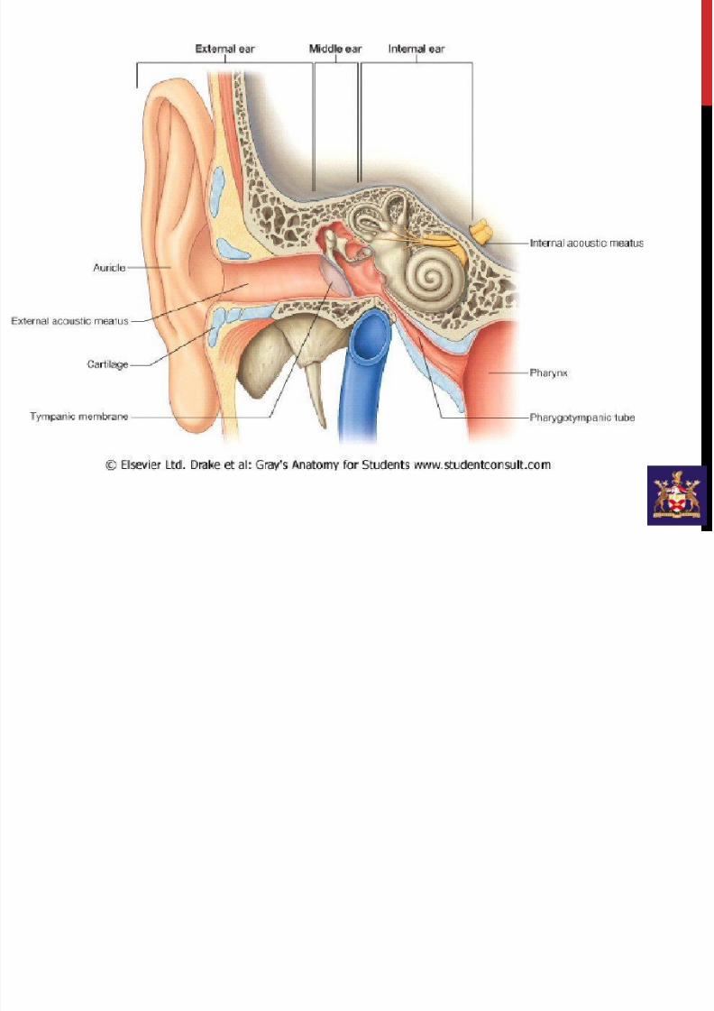
EXTERNAL AND MIDDLE EAR
CLASS: JC3 2013
COURSE: NEUROSCIENCE
CODE: NS 43
LECTURER: DR ROHANA O’CONNELL
DATE: 29/10/13 1600-1700
7/21/2019 External and Middle Ear
http://slidepdf.com/reader/full/external-and-middle-ear 2/40
LEARNING OBJECTIVES
• Structure of external ear
• Tympanic membrane
• Conductive deafness
• Middle ear: ossicles, round and oval
windows
• Tensor tympani and stapedius
• Mastoid air cells
7/21/2019 External and Middle Ear
http://slidepdf.com/reader/full/external-and-middle-ear 3/40
7/21/2019 External and Middle Ear
http://slidepdf.com/reader/full/external-and-middle-ear 4/40
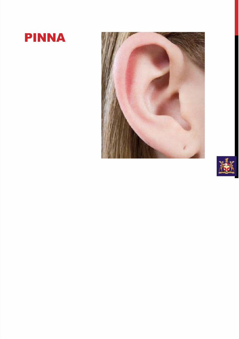
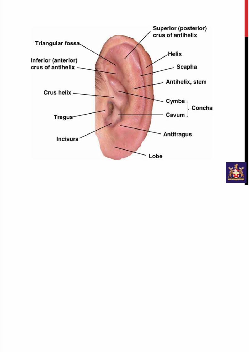
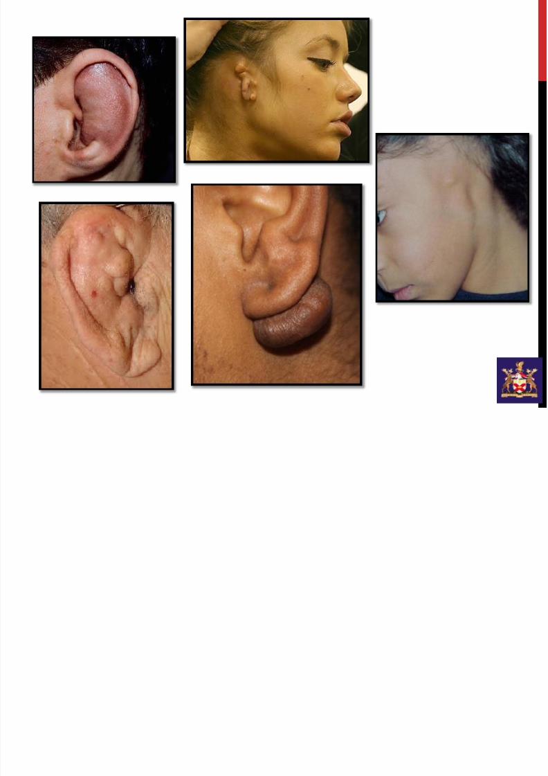
PINNA
7/21/2019 External and Middle Ear
http://slidepdf.com/reader/full/external-and-middle-ear 5/40
7/21/2019 External and Middle Ear
http://slidepdf.com/reader/full/external-and-middle-ear 6/40
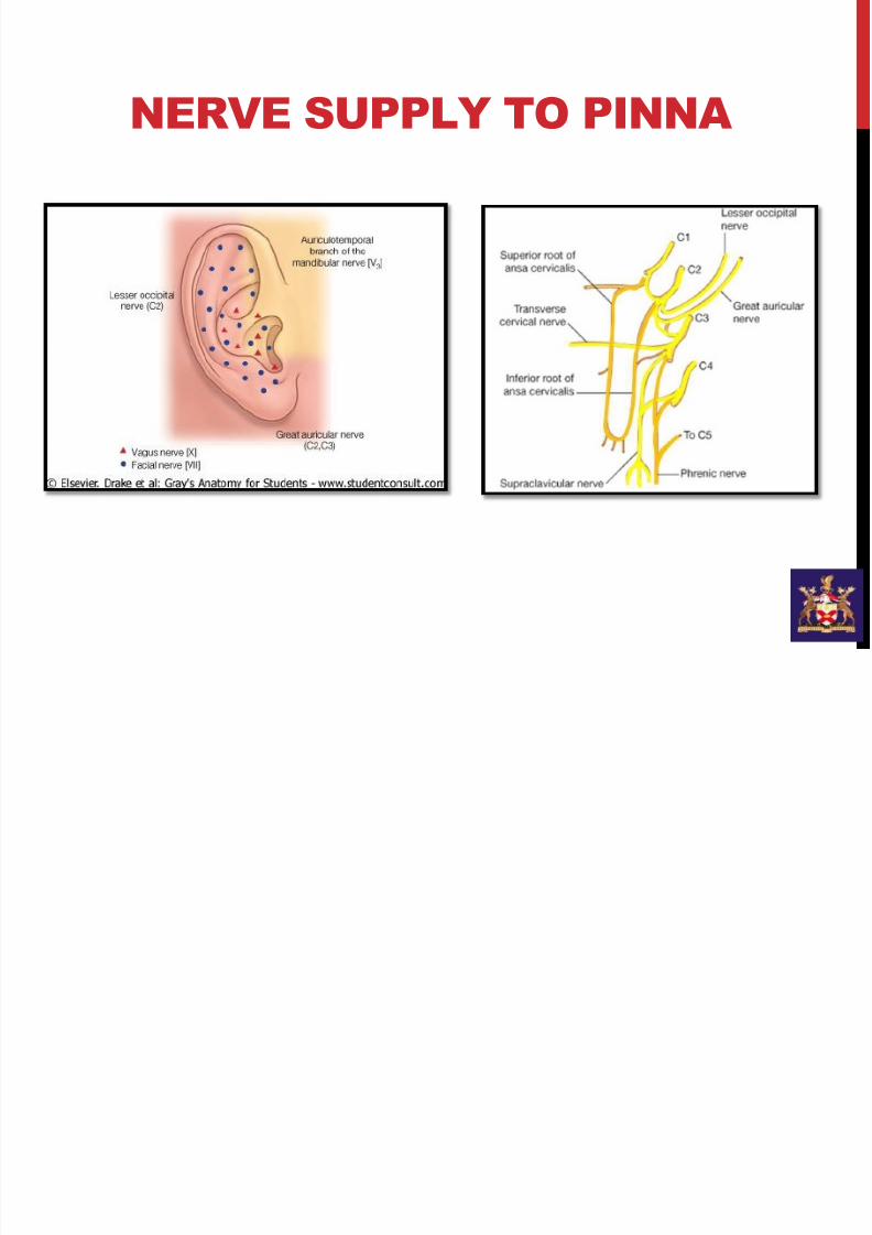
NERVE SUPPLY TO PINNA
7/21/2019 External and Middle Ear
http://slidepdf.com/reader/full/external-and-middle-ear 7/40
7/21/2019 External and Middle Ear
http://slidepdf.com/reader/full/external-and-middle-ear 8/40
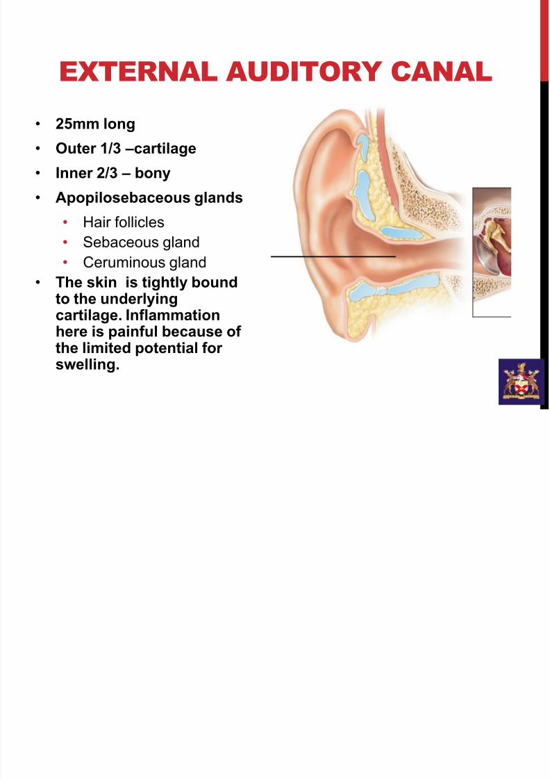
EXTERNAL AUDITORY CANAL
• 25mm long
• Outer 1/3 –cartilage
• Inner 2/3 – bony
• Apopilosebaceous glands
• Hair follicles
• Sebaceous gland
• Ceruminous gland
•The skin is tightly boundto the underlyingcartilage. Inflammationhere is painful because ofthe limited potential forswelling.
7/21/2019 External and Middle Ear
http://slidepdf.com/reader/full/external-and-middle-ear 9/40
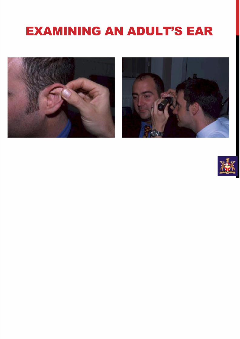
EXAMINING AN ADULT’S EAR
7/21/2019 External and Middle Ear
http://slidepdf.com/reader/full/external-and-middle-ear 10/40
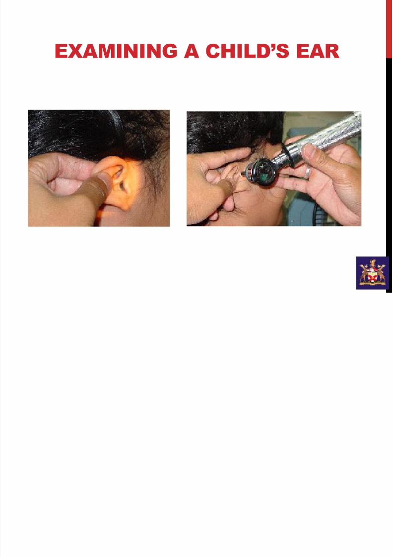
EXAMINING A CHILD’S EAR
7/21/2019 External and Middle Ear
http://slidepdf.com/reader/full/external-and-middle-ear 11/40
7/21/2019 External and Middle Ear
http://slidepdf.com/reader/full/external-and-middle-ear 12/40
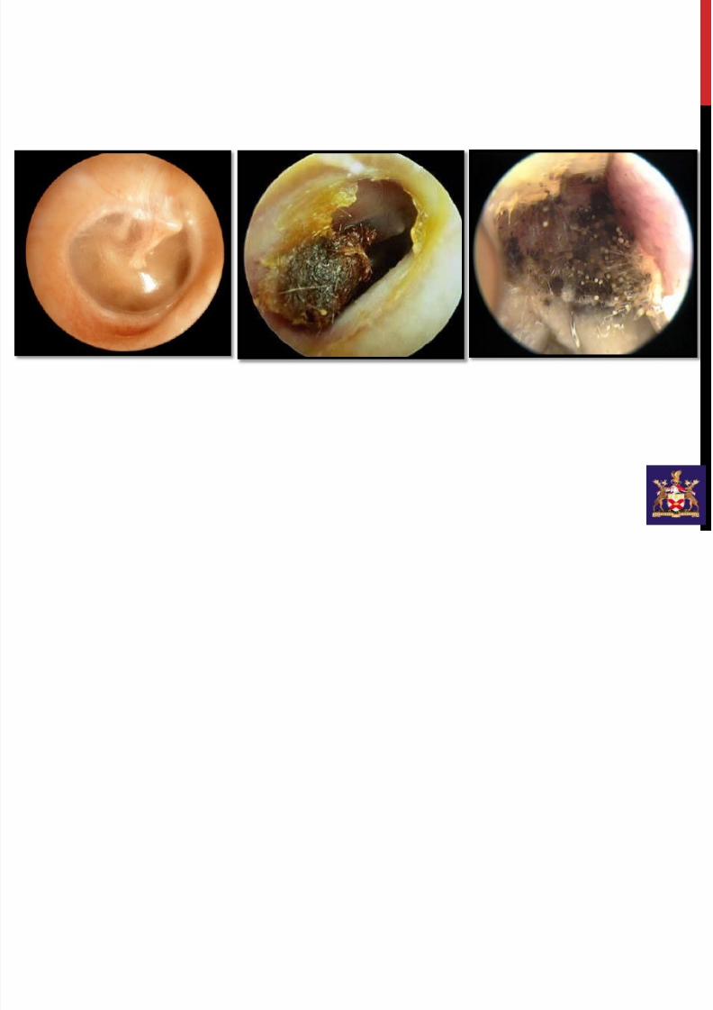
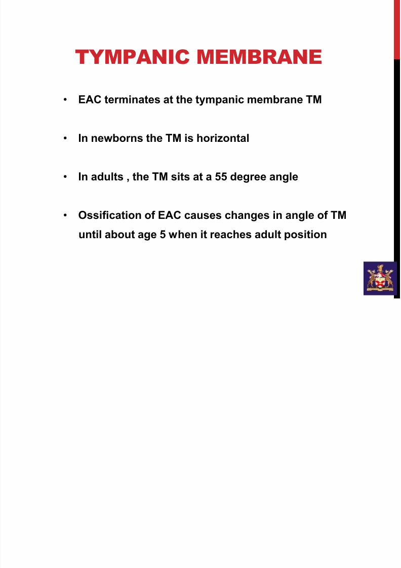
TYMPANIC MEMBRANE
• EAC terminates at the tympanic membrane TM
• In newborns the TM is horizontal
• In adults , the TM sits at a 55 degree angle
• Ossification of EAC causes changes in angle of TM
until about age 5 when it reaches adult position

7/21/2019 External and Middle Ear
http://slidepdf.com/reader/full/external-and-middle-ear 13/40
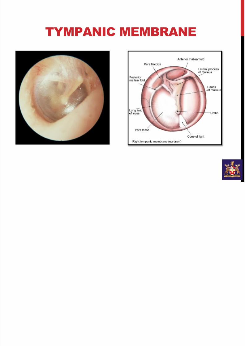
TYMPANIC MEMBRANE
Three Layers of TM
- Ectoderm (cutaneous) - continuous with EAM
- Mesoderm (fibrous) - Radial Fibers
- Concentric Fibers
- Endoderm (mucous) - continuous with TympanicCavity
Pars Tensa contains all three layers.
Pars Flacida DOES NOT contain fibrous layer.
7/21/2019 External and Middle Ear
http://slidepdf.com/reader/full/external-and-middle-ear 14/40
TYMPANIC MEMBRANE

7/21/2019 External and Middle Ear
http://slidepdf.com/reader/full/external-and-middle-ear 15/40
Only epidermal and mucosal layer
7/21/2019 External and Middle Ear
http://slidepdf.com/reader/full/external-and-middle-ear 16/40
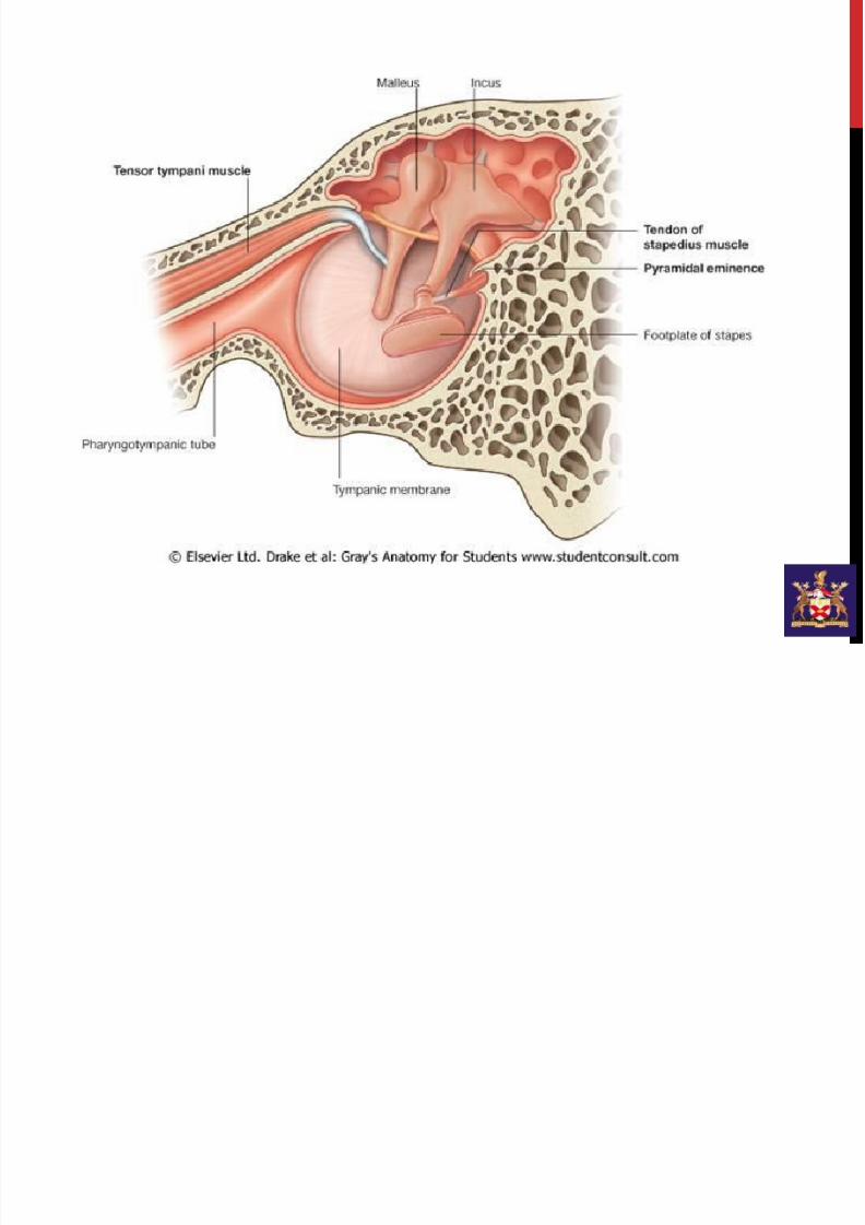
MIDDLE EAR
7/21/2019 External and Middle Ear
http://slidepdf.com/reader/full/external-and-middle-ear 17/40
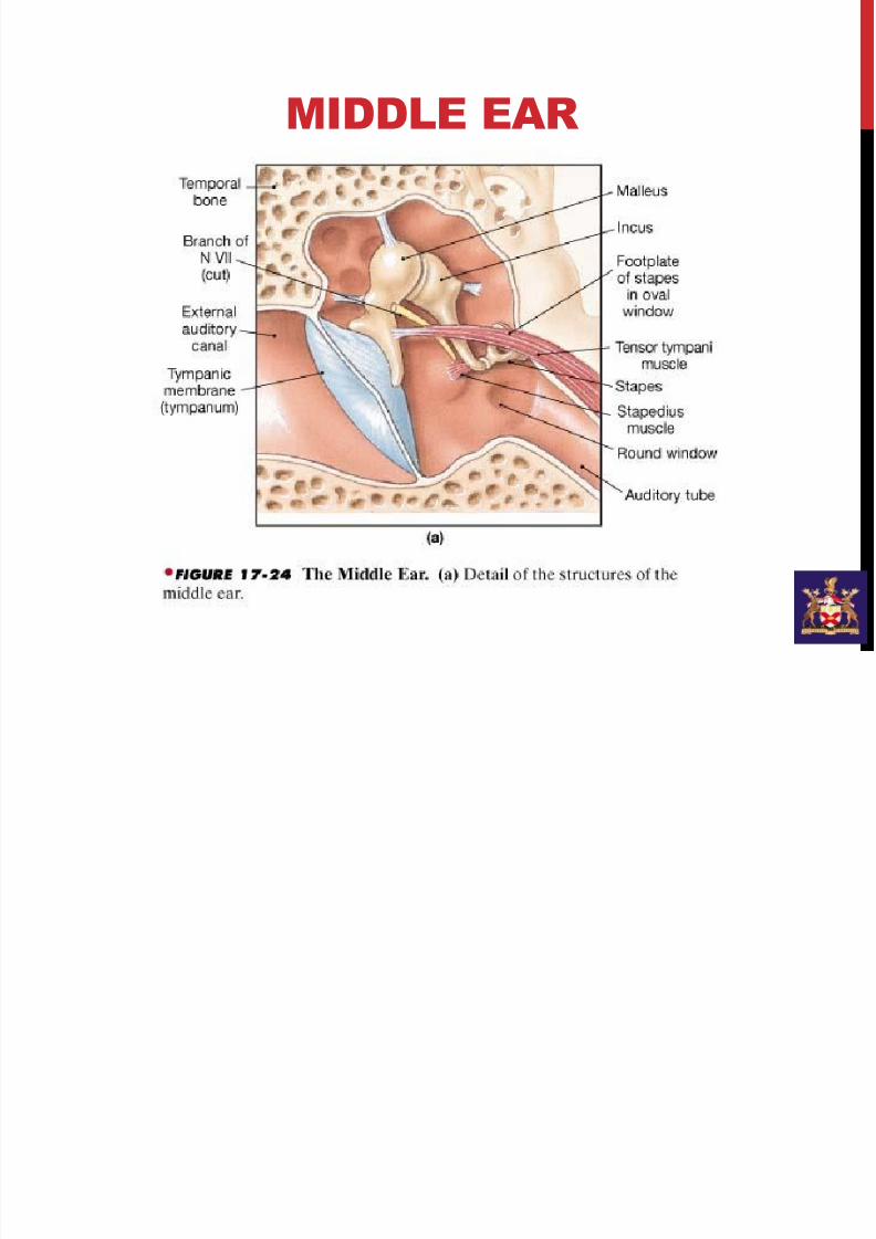
MIDDLE EAR
• Tympanic Cavity
• Ossicles
• Eustachian Tube
• Middle Ear Muscles
• Consists of 4 walls , a ceiling and a floor
7/21/2019 External and Middle Ear
http://slidepdf.com/reader/full/external-and-middle-ear 18/40
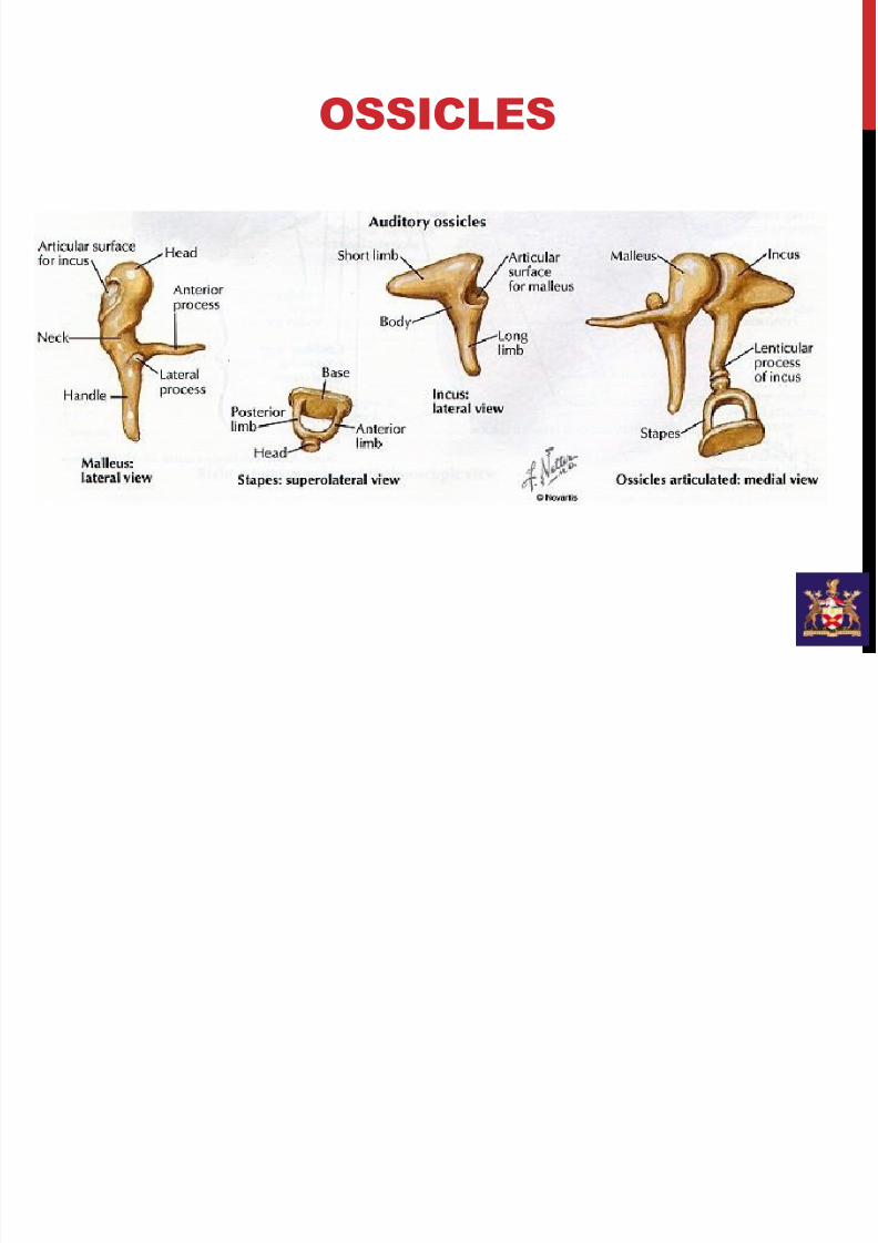
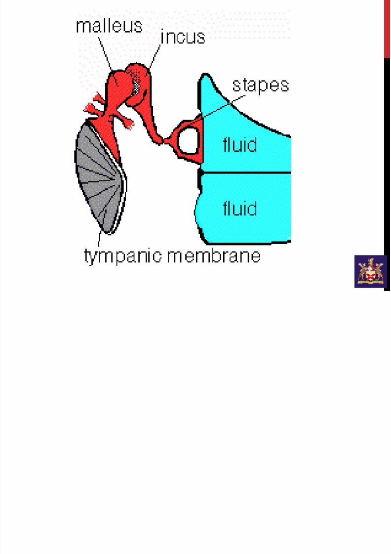
OSSICLES
7/21/2019 External and Middle Ear
http://slidepdf.com/reader/full/external-and-middle-ear 19/40
7/21/2019 External and Middle Ear
http://slidepdf.com/reader/full/external-and-middle-ear 20/40
7/21/2019 External and Middle Ear
http://slidepdf.com/reader/full/external-and-middle-ear 21/40
THE CEILING
Tegmen tympani
• Paper thin.
• Separates the tympanic cavity from the middle cranialfossa which houses the temporal lobe.
• Inflammatory conditions of middle ear can pass through
the petrous-squamosal suture in children directly to the
meninges of temporal lobe of cortex.
7/21/2019 External and Middle Ear
http://slidepdf.com/reader/full/external-and-middle-ear 22/40
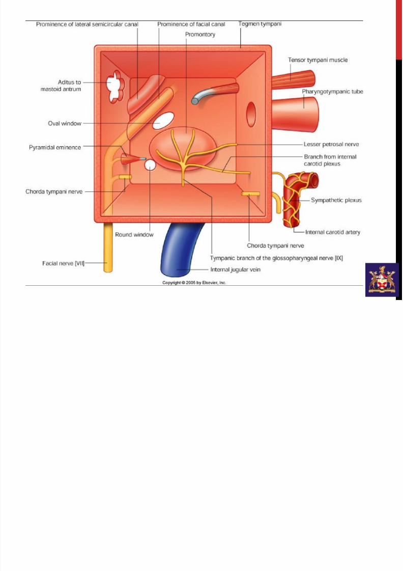
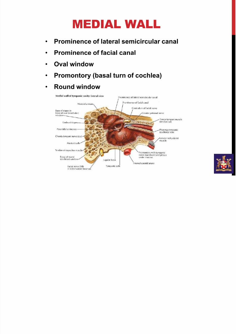
MEDIAL WALL
• Prominence of lateral semicircular canal
• Prominence of facial canal
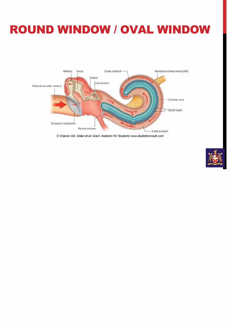
• Oval window
• Promontory (basal turn of cochlea)
• Round window
7/21/2019 External and Middle Ear
http://slidepdf.com/reader/full/external-and-middle-ear 23/40
POSTERIOR WALL
• Mastoid wall
• Aditus ad antrum
• Pyramidal eminence
• Jugular wall and jugular vein
• Carotid wall
• Carotid artery
• Eustachian tube
ANTERIOR WALL
7/21/2019 External and Middle Ear
http://slidepdf.com/reader/full/external-and-middle-ear 24/40
LATERAL WALL
• Membranous wall
• Tympanic membrane
• Jugular wall
• Formed by tympanic plate
THE FLOOR
7/21/2019 External and Middle Ear
http://slidepdf.com/reader/full/external-and-middle-ear 25/40
7/21/2019 External and Middle Ear
http://slidepdf.com/reader/full/external-and-middle-ear 26/40
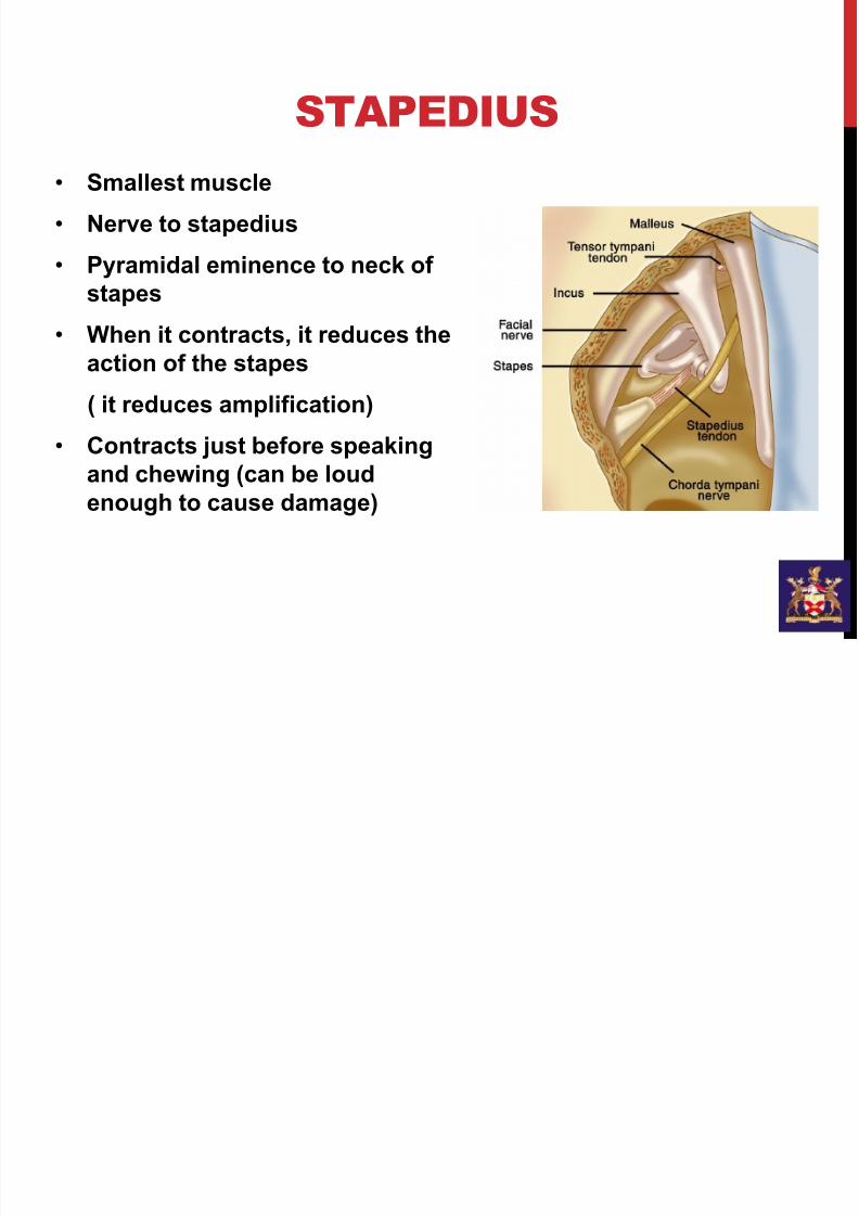
STAPEDIUS
• Smallest muscle
• Nerve to stapedius
• Pyramidal eminence to neck of
stapes• When it contracts, it reduces the
action of the stapes
( it reduces amplification)
• Contracts just before speakingand chewing (can be loud
enough to cause damage)
7/21/2019 External and Middle Ear
http://slidepdf.com/reader/full/external-and-middle-ear 27/40
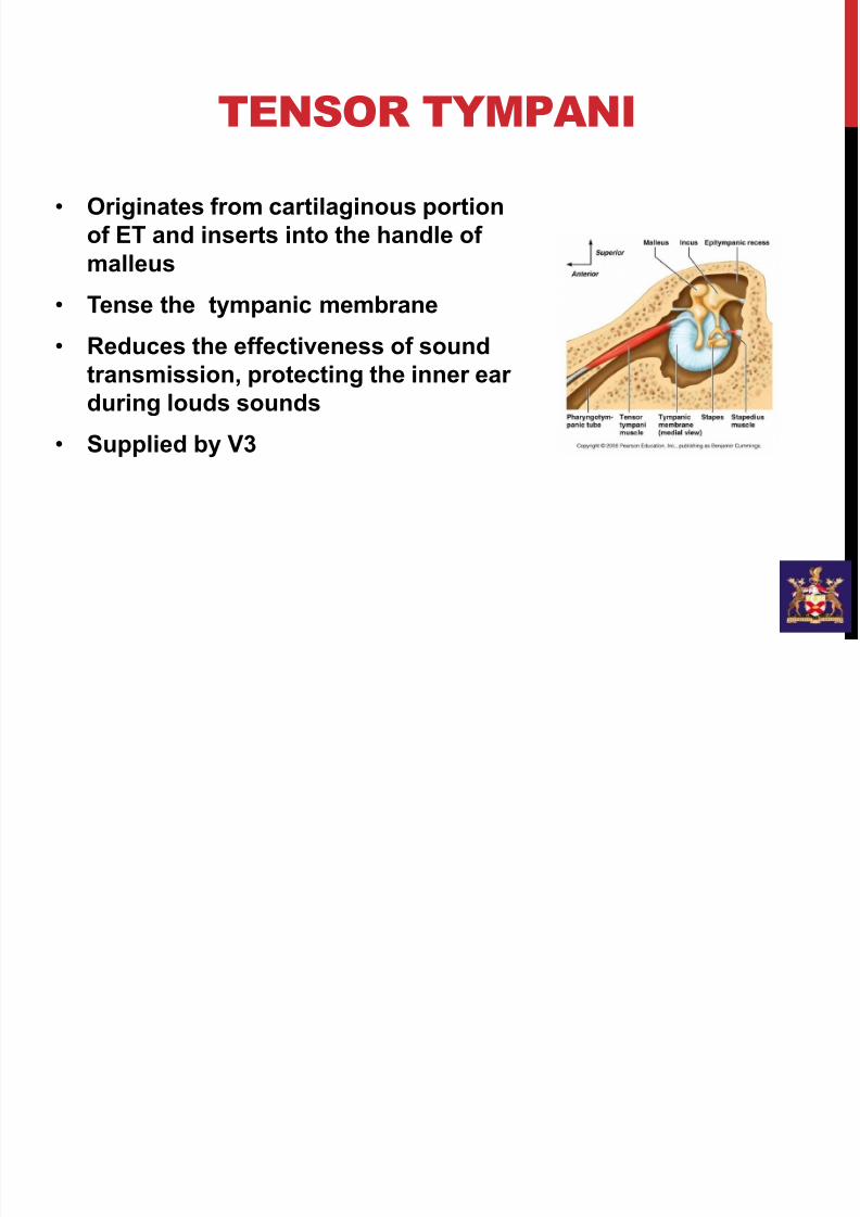
TENSOR TYMPANI
• Originates from cartilaginous portion
of ET and inserts into the handle of
malleus
• Tense the tympanic membrane
• Reduces the effectiveness of sound
transmission, protecting the inner ear
during louds sounds
•Supplied by V3
7/21/2019 External and Middle Ear
http://slidepdf.com/reader/full/external-and-middle-ear 28/40
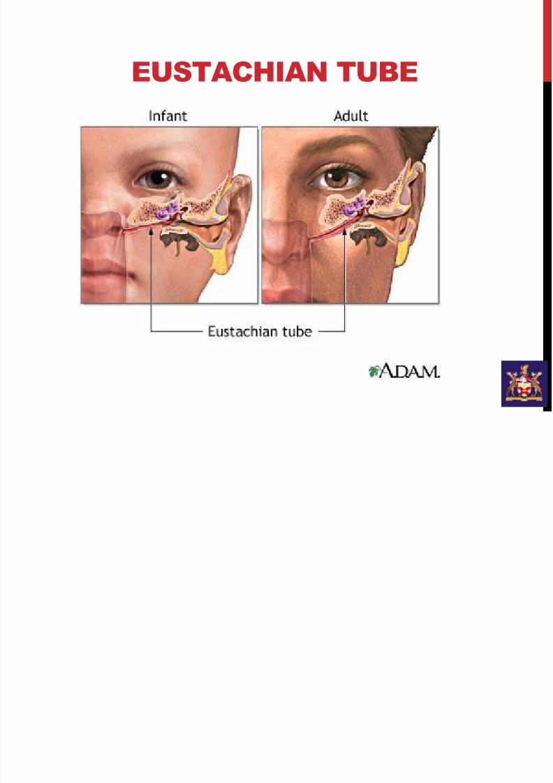
EUSTACHIAN TUBE
7/21/2019 External and Middle Ear
http://slidepdf.com/reader/full/external-and-middle-ear 29/40
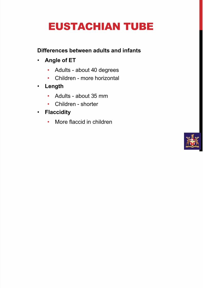
EUSTACHIAN TUBE
Differences between adults and infants
• Angle of ET
• Adults - about 40 degrees
• Children - more horizontal
• Length
• Adults - about 35 mm
• Children - shorter
• Flaccidity
• More flaccid in children
7/21/2019 External and Middle Ear
http://slidepdf.com/reader/full/external-and-middle-ear 30/40
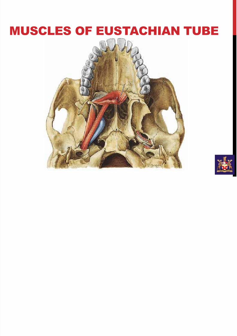
MUSCLES OF EUSTACHIAN TUBE
7/21/2019 External and Middle Ear
http://slidepdf.com/reader/full/external-and-middle-ear 31/40
EUSTACHIAN TUBE
Function:
• Tensor palatini definitely involved in opening ET.
•Levator palatini role is not clear.
• Clearance of middle ear fluid
• Protect middle ear from nasal secretion
• Equalising middle ear pressure

7/21/2019 External and Middle Ear
http://slidepdf.com/reader/full/external-and-middle-ear 32/40
MASTOID ANTRUM
7/21/2019 External and Middle Ear
http://slidepdf.com/reader/full/external-and-middle-ear 33/40
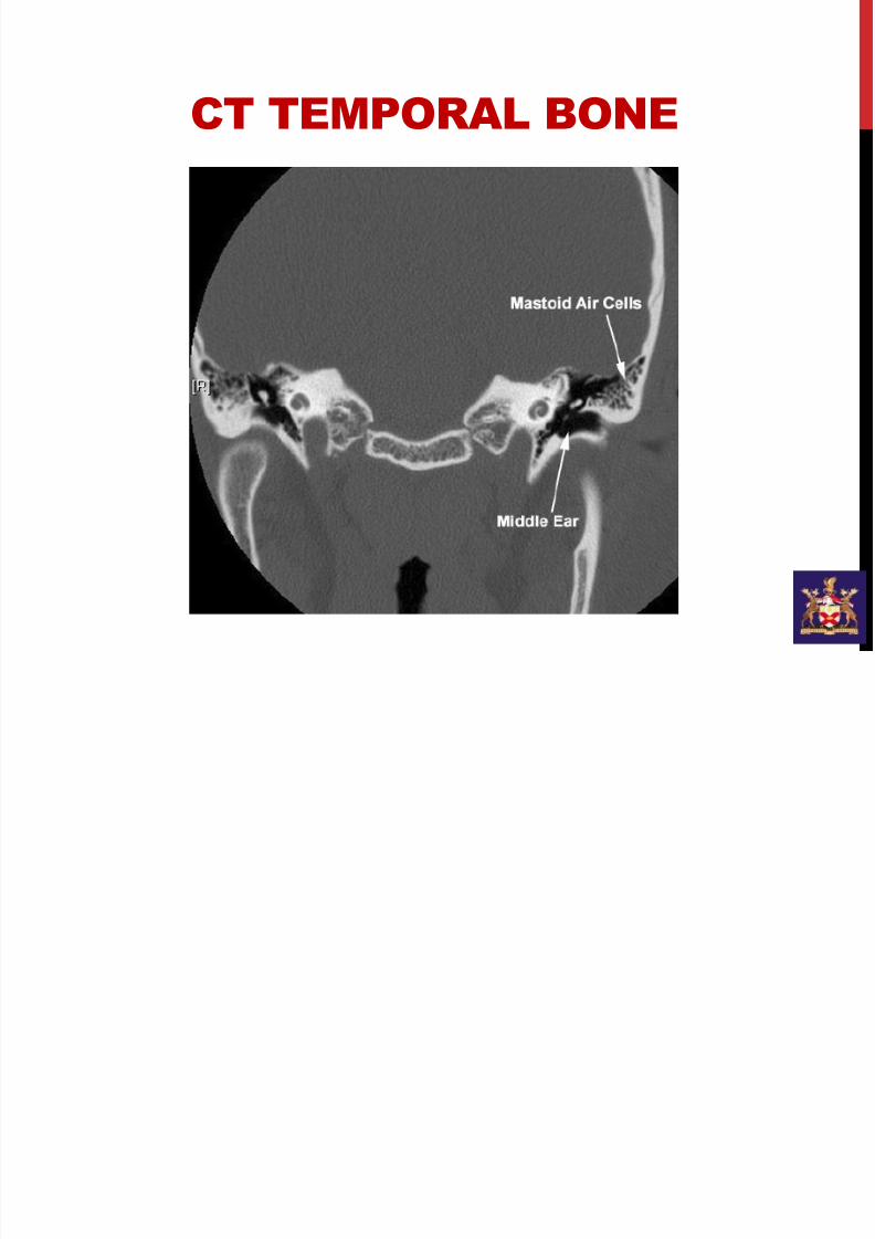
MASTOID AIR CELLS
• Mastoid air cell system served as an reservoir of air and
serves as buffer system to replace air in the middle ear
cavity temporarily in case of Eustachian tube dysfunction.
• The mean volume of air in the mastoid air cell system could
be about 5-8 ml. CT scan evaluation of temporal bone is
considered to be the best modality to assess mastoid air
cell system.
•Patients with poor pneumatization of mastoid air cell systemare more prone to develop adhesive otitis media following
middle ear infections as the normal buffering system of the
mastoid pneumatization is not adequate in them.
7/21/2019 External and Middle Ear
http://slidepdf.com/reader/full/external-and-middle-ear 34/40
CT TEMPORAL BONE
7/21/2019 External and Middle Ear
http://slidepdf.com/reader/full/external-and-middle-ear 35/40
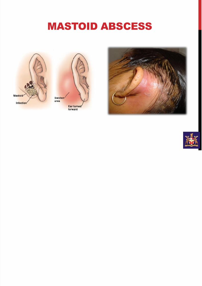
MASTOID ABSCESS
7/21/2019 External and Middle Ear
http://slidepdf.com/reader/full/external-and-middle-ear 36/40
ROUND WINDOW / OVAL WINDOW
7/21/2019 External and Middle Ear
http://slidepdf.com/reader/full/external-and-middle-ear 37/40
7/21/2019 External and Middle Ear
http://slidepdf.com/reader/full/external-and-middle-ear 38/40
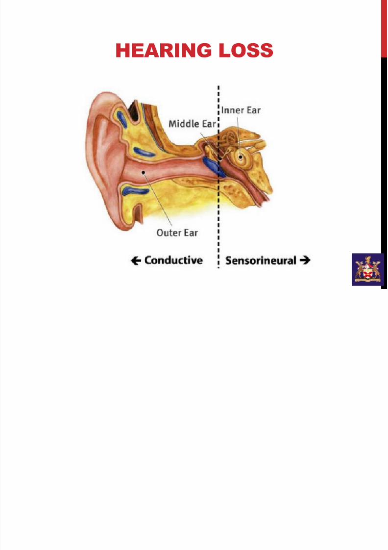
HEARING LOSS
7/21/2019 External and Middle Ear
http://slidepdf.com/reader/full/external-and-middle-ear 39/40
CONDUCTIVE HEARING LOSS
• Is the result of sounds not being able to pass freely to the
inner ear.
•
Usually due to blockage in the outer or middle ear• Ear wax
• Otitis externa
• Stenotic or atretic ear canal
• Perforated tympanic membrane
• Otosclerosis
• Usually can be corrected
7/21/2019 External and Middle Ear
http://slidepdf.com/reader/full/external-and-middle-ear 40/40
SENSORINEURAL
HEARING LOSS
• Sensory, cochlear, neural or inner ear hearing loss.
• Damage to the hair cells within the cochlea or thehearing nerve (or both).
• Causes:
• Presbycusis• Regular and prolonged exposure to loud sounds.
• Ototoxic drugs
• Certain infectious diseases, including Rubella
• Complications at birth
• Injury to the head
• Benign tumours on the auditory nerve
• Genetic predisposition – some people are especiallyprone to hearing loss