Embed Size (px)
Citation preview
Extracellular ATP Functions as an Endogenous ExternalMetabolite Regulating Plant Cell Viability
Stephen Chivasa,a Bongani K. Ndimba,b William J. Simon,c Keith Lindsey,c and Antoni R. Slabasc,1
a Creative Gene Technology, Integrative Cell Biology Laboratory, School of Biological and Biomedical Sciences,
University of Durham, Durham DH1 3LE, United KingdombDepartment of Biotechnology, University of the Western Cape, Bellville, Cape Town, South Africac School of Biological and Biomedical Sciences, University of Durham, Durham DH1 3LE, United Kingdom
ATP is a vital molecule used by living organisms as a universal source of energy required to drive the cogwheels of intra-
cellular biochemical reactions necessary for growth and development. Animal cells release ATP to the extracellular milieu,
where it functions as the primary signaling cue at the epicenter of a diverse range of physiological processes. Although
recent findings revealed that intact plant tissues release ATP as well, there is no clearly defined physiological function of
extracellular ATP in plants. Here, we show that extracellular ATP is essential for maintaining plant cell viability. Its removal
by the cell-impermeant traps glucose–hexokinase and apyrase triggered death in both cell cultures and whole plants. Com-
petitive exclusion of extracellular ATP from its binding sites by treatment with b,g-methyleneadenosine 59-triphosphate,
a nonhydrolyzable analog of ATP, also resulted in death. The death response was observed in Arabidopsis thaliana, maize
(Zea mays), bean (Phaseolus vulgaris), and tobacco (Nicotiana tabacum). Significantly, we discovered that fumonisin
B1 (FB1) treatment of Arabidopsis triggered the depletion of extracellular ATP that preceded cell death and that exogenous
ATP rescues Arabidopsis from FB1-induced death. These observations suggest that extracellular ATP suppresses a de-
fault death pathway in plants and that some forms of pathogen-induced cell death are mediated by the depletion of extra-
cellular ATP.
INTRODUCTION
ATP is a ubiquitous, energy-rich compound in all cells of living
organisms. It is found both within organelles, such as mitochon-
dria and chloroplasts, and in the cytoplasm of higher organisms.
The energy derived from ATP is used to drive a myriad of vital
biochemical reactions that are fundamental to the survival of
cells andwhole organisms. The presence of intracellular ATP has
been recognized since its discovery in living cells in 1929 (Fiske
and Subarrow, 1929). However, cells can secrete ATP to the
extracellular matrix in the absence of cytolysis, an observation
first reported in animal cells in 1959 (Holton, 1959) and only
recently in a plant species, Arabidopsis thaliana (Thomas et al.,
2000).
In animals, nonlytic release of ATP by nonsecretory cells oc-
curs under quiescent conditions (Beigi and Dubyak, 2000;
Schwiebert et al., 2002), after mechanical stimulation (Grierson
andMeldolesi, 1995; Homolya et al., 2000; Yegutkin et al., 2000),
or after treatment with agonists such as bradykinin, acetylcho-
line, and serotonin (Yang et al., 1994). Touch stimulation and
osmotic stress elicit increased ATP release from Arabidopsis
(Jeter et al., 2004), suggesting conservation of ATP release in
response tomechanical stimulation between plants and animals.
Neuronal cells and secretory cells such as mast cells and
pancreatic acinar cells release ATP via regulated exocytosis
(Leitner et al., 1975; Unsworth and Johnson, 1990; Sorensen and
Novak, 2001). The mechanisms by which plant cells and non-
secretory animal cells release ATP are still poorly understood,
but there is evidence for the involvement of ABC transporters
and anion channels (Abraham et al., 1993; Roman et al., 1997;
Thomas et al., 2000; Dutta et al., 2002, 2004).
In animal cells, extracellular ATP is an absolute requirement for
several physiological processes, such as neurotransmission,
platelet aggregation, regulation of blood vessel tone, cell growth
and expansion, differentiation and function of immune cells, and
muscle contraction (Gordon, 1986; Redegeld et al., 1999). In
these cells, extracellular ATP funnels into signaling pathways via
two mechanisms. First, it participates in extracellular phos-
phorylation (Redegeld et al., 1999). For example, extracellular
phosphorylation is an absolute requirement in T cell–mediated
immune responses. Specific abolition of ectophosphorylation,
without affecting intracellular phosphorylation, by the ectopro-
tein kinase inhibitor K-252b is accompanied by a loss of cytolytic
activity of cytotoxic T lymphocytes for target cells (Redegeld
et al., 1997). Second, extracellular ATP participates in signal
transmission via the activation of P2X and P2Y plasma mem-
brane nucleotide binding receptors known as purinoceptors
(Burnstock and Kennedy, 1985). P2X receptors are ligand-gated
ion channels that gate extracellular cations in response to ATP,
1 To whom correspondence should be addressed. E-mail [email protected]; fax 44-191-3341295.The author responsible for distribution of materials integral to thefindings presented in this article in accordance with the policy describedin the Instructions for Authors (www.plantcell.org) is: Antoni R. Slabas([email protected]).Article, publication date, and citation information can be found atwww.plantcell.org/cgi/doi/10.1105/tpc.105.036806.
The Plant Cell, Vol. 17, 3019–3034, November 2005, www.plantcell.orgª 2005 American Society of Plant Biologists
whereas P2Y receptors are G protein–coupled receptors. Some
physiological processes are dependent on the activation of
purinoceptors by extracellular ATP to initiate signaling cascades.
For example, extracellular ATP binds to P2X7 receptors to acti-
vate astrocyteGln release,whichmodulates synaptic activity and
participates in brain intercellular signaling (Duan et al., 2003).
Although extracellular ATP released by intact plant tissues
(Thomas et al., 2000) has no clearly defined function, two lines of
evidence suggest that it may have physiological significance.
The first is based on electrophysiological studies using patch–
clamp techniques. Extracellular ATP depolarizes Arabidopsis
root hair membranes (Lew and Dearnaley, 2000) and triggers an
increase of cytosolic Ca2þ (Demidchik et al., 2003), implicating
a plant homolog of animal purinoceptors; however, plant puri-
noceptors have yet to be identified. This Ca2þ influxmediates the
activation of Arabidopsis gene expression induced by exoge-
nous ATP (Jeter et al., 2004). The second line of evidence is
based on proteomic studies of Arabidopsis extracellular matrix
proteins. Identification of secreted, extracellular phosphopro-
teins in the Arabidopsis extracellular matrix (Chivasa et al., 2002;
Ndimba et al., 2003) suggests the participation of ATP in
extracellular signaling via phosphorylation.
Thus, we initiated studies to examine the role of extracellular
ATP in plants via the specific removal of ATP from the extracel-
lular matrix of cell suspension cultures and intact plants. In this
article, we report the discoveries that (1) a physiological role of
extracellular ATP in plants is to govern cell viability by the
suppression of a default death pathway, and (2) a mechanism
by which fumonisin B1 (FB1) induces death in Arabidopsis is the
depletion of extracellular ATP.
RESULTS
Cell-Impermeant ATP Traps Trigger Death in
Arabidopsis Cell Cultures
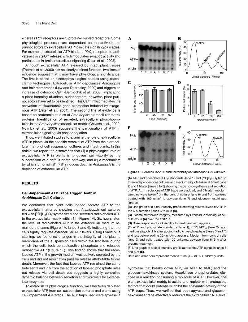
We confirmed that plant cells indeed secrete ATP to the
extracellular matrix by showing that Arabidopsis cell cultures
fed with [32P]H3PO4 synthesized and secreted radiolabeled ATP
to the extracellular matrix within 1 h (Figure 1A). Six hours later,
the level of radiolabeled ATP in the extracellular medium re-
mained the same (Figure 1A, lanes 3 and 6), indicating that the
cells tightly regulate extracellular ATP levels. Using Evans blue
staining, we found no changes in the integrity of the plasma
membrane of the suspension cells within the first hour during
which the cells took up radioactive phosphate and released
radioactive ATP (Figure 1C). This finding shows that the radio-
labeled ATP in the growth medium was actively secreted by the
cells and did not result from passive release attributable to cell
death. Moreover, the fact that labeled ATP remained the same
between 1 and 7 h from the addition of labeled phosphate rules
out release via cell death but suggests a highly controlled
dynamic balance between secretion and hydrolysis by extracel-
lular enzymes.
To establish its physiological function, we selectively depleted
extracellular ATP from cell suspension cultures and plants using
cell-impermeant ATP traps. The ATP traps used were apyrase (a
hydrolase that breaks down ATP, via ADP, to AMP) and the
glucose–hexokinase system. Hexokinase phosphorylates glu-
cose in a reaction consuming a molecule of ATP. However, the
plant extracellular matrix is acidic and replete with proteases,
factors that could potentially inhibit the enzymatic activity of the
ATP traps. Thus, we verified that both apyrase and glucose–
hexokinase traps effectively reduced the extracellular ATP level
Figure 1. Extracellular ATP andCell Viability ofArabidopsisCell Cultures.
(A) ATP and phosphate (PO4) standards (lane 1) and [32P]H3PO4 fed to
three independent cell cultures andmedium aliquots taken at time 0 (lane
2) and 1 h later (lanes 3 to 5) showing the de novo synthesis and secretion
of ATP. At 1 h, solutions of ATP traps were added, and 6 h later, medium
samples were taken from the control culture (lane 6) and from cultures
treated with 100 units/mL apyrase (lane 7) and glucose–hexokinase
(lane 8).
(B) Line graph of a pixel intensity profile showing relative levels of ATP in
the 6-h samples (lanes 6 to 8) in (A).
(C) Plasma membrane integrity, measured by Evans blue staining, of cell
cultures in (A) over the first 1 h.
(D) Dose response of cell viability to treatment with apyrase.
(E) ATP and phosphate standards (lane 1), [32P]H3PO4 (lane 2), and
medium aliquots 1 h after adding radioactive phosphate (lanes 3 and 4)
and just before adding 20 units/mL apyrase. Medium from control cells
(lane 5) and cells treated with 20 units/mL apyrase (lane 6) 6 h after
enzyme treatment.
(F) Line graph of a pixel intensity profile across the ATP bands in lanes 5
and 6 of (E).
Data and error bars represent means 6 SD (n ¼ 3). AU, arbitrary units.
3020 The Plant Cell
in these cell cultures (Figures 1A and 1B). In initial experiments,
we noted that cell death ensued within 24 to 48 h of incubating
Arabidopsis cell cultures with apyrase, as confirmed by propi-
dium iodide and fluorescein diacetate staining (data not shown).
Quantitative estimation of cell death/viability was obtained by
measuring the ability of the cultures to grow subsequent to the
treatment. Cell cultures treated for 72 h were diluted 30-fold with
fresh growth medium to diminish apyrase activity, and the cell
density (packed cell volume) was determined after another 96 h
of growth.
Using this method, we observed a significant dose-dependent
reduction of Arabidopsis cell culture viability in response to
apyrase (Figure 1D). Effects of ATP removal have previously been
confounded with the effects of buffer salts in lyophilized com-
mercial preparations of ATP-degrading enzymes (Redegeld
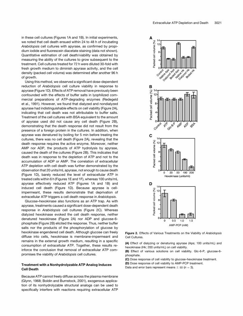
et al., 1991). However, we found that dialyzed and nondialyzed
apyrase had indistinguishable effects on cell viability (Figure 2A),
indicating that cell death was not attributable to buffer salts.
Treatment of the cell cultures with BSA equivalent to the amount
of apyrase used did not cause any cell death (Figure 2B),
demonstrating that the death response did not result from the
presence of a foreign protein in the cultures. In addition, when
apyrase was denatured by boiling for 5 min before treating the
cultures, there was no cell death (Figure 2A), revealing that the
death response requires the active enzyme. Moreover, neither
AMP nor ADP, the products of ATP hydrolysis by apyrase,
caused the death of the cultures (Figure 2B). This indicates that
death was in response to the depletion of ATP and not to the
accumulation of ADP or AMP. The correlation of extracellular
ATP depletion with cell death was further demonstrated by the
observation that 20 units/mL apyrase, not enough to cause death
(Figure 1D), barely reduced the level of extracellular ATP in
treated cells within 6 h (Figures 1E and 1F), whereas 100 units/mL
apyrase effectively reduced ATP (Figures 1A and 1B) and
induced cell death (Figure 1D). Because apyrase is cell-
impermeant, these results demonstrate that deprivation of
extracellular ATP triggers a cell death response in Arabidopsis.
Glucose–hexokinase also functions as an ATP trap. As with
apyrase, treatments caused a significant dose-dependent death
response in Arabidopsis cell cultures (Figure 2C). Whereas
dialyzed hexokinase evoked the cell death response, neither
denatured hexokinase (Figure 2A) nor ADP and glucose-6-
phosphate (Figure 2B) elicited the response. Thus, neither buffer
salts nor the products of the phosphorylation of glucose by
hexokinase engendered cell death. Although glucose can freely
diffuse into cells, hexokinase is membrane-impermeant and
remains in the external growth medium, resulting in a specific
consumption of extracellular ATP. Together, these results re-
inforce the conclusion that removal of extracellular ATP com-
promises the viability of Arabidopsis cell cultures.
Treatment with a Nonhydrolyzable ATP Analog Induces
Cell Death
Because ATP cannot freely diffuse across the plasmamembrane
(Glynn, 1968; Boldin and Burnstock, 2001), exogenous applica-
tion of its nonhydrolyzable structural analogs can be used to
specifically interfere with reactions requiring extracellular ATP
Figure 2. Effects of Various Treatments on the Viability of Arabidopsis
Cell Cultures.
(A) Effect of dialyzing or denaturing apyrase (Aps; 100 units/mL) and
hexokinase (Hk; 200 units/mL) on cell viability.
(B) Effect of various solutions on cell viability. Glc-6-P, glucose-6-
phosphate.
(C) Dose response of cell viability to glucose–hexokinase treatment.
(D) Dose response of cell viability to AMP-PCP treatment.
Data and error bars represent means 6 SD (n ¼ 3).
Extracellular ATP Depletion and Death 3021

hydrolysis (Redegeld et al., 1999). We used bg-methyleneaden-
osine 59-triphosphate (AMP-PCP), a nonhydrolyzable analog of
ATP. Inundation of Arabidopsis cell cultures with exogenous
AMP-PCP was attended by a concentration-dependent de-
crease in cell viability (Figure 2D). This finding confirms that
extracellular ATP plays a role in cell viability because competitive
exclusion from its binding sites by AMP-PCP, as well as its re-
moval by hydrolyzing enzymes, result in cell death.
Extracellular ATP Is Required for the Viability of Intact
Arabidopsis Plants
Extracellular ATP is also required for viability in planta. Treatment
of localized areas of Arabidopsis leaves with apyrase, glucose–
hexokinase, and AMP-PCP resulted in the collapse of tissue
within the treated zone, leading to death between 2 and 4 d after
treatment (Figure 3). Localized treatment was used to show the
contrast with adjacent living tissue. When entire leaves were
treated with the solutions, the whole leaf eventually died (Figure
3). Control leaves treated with BSA, ATP, AMP, ADP, glucose, or
glucose-6-phosphate remained healthy (data not shown). To
demonstrate that death was not the result of internalization of the
ATP traps at the infiltration site caused by mechanical damage,
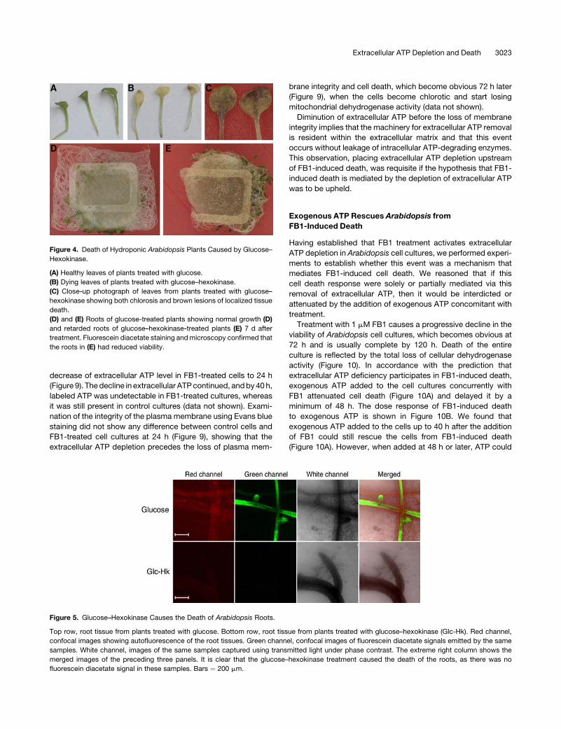
the glucose–hexokinase system applied to the growth medium
of hydroponic Arabidopsis plants caused death of the roots
and some of the shoots (Figures 4 and 5). Similarly, death was
triggered in Arabidopsis plants grown for 1 week on normal
nutrient agar and transferred to nutrient agar supplemented with
AMP-PCP (Figure 6). Control plants transferred to agar supple-
mented with ATP remained viable (Figure 6). Therefore, the cell
death response observed in cell suspension cultures caused by
extracellular ATP deprivation also occurs in whole plants.
Several Plant Species Require Extracellular
ATP for Viability
We investigated the requirement for extracellular ATP in cell
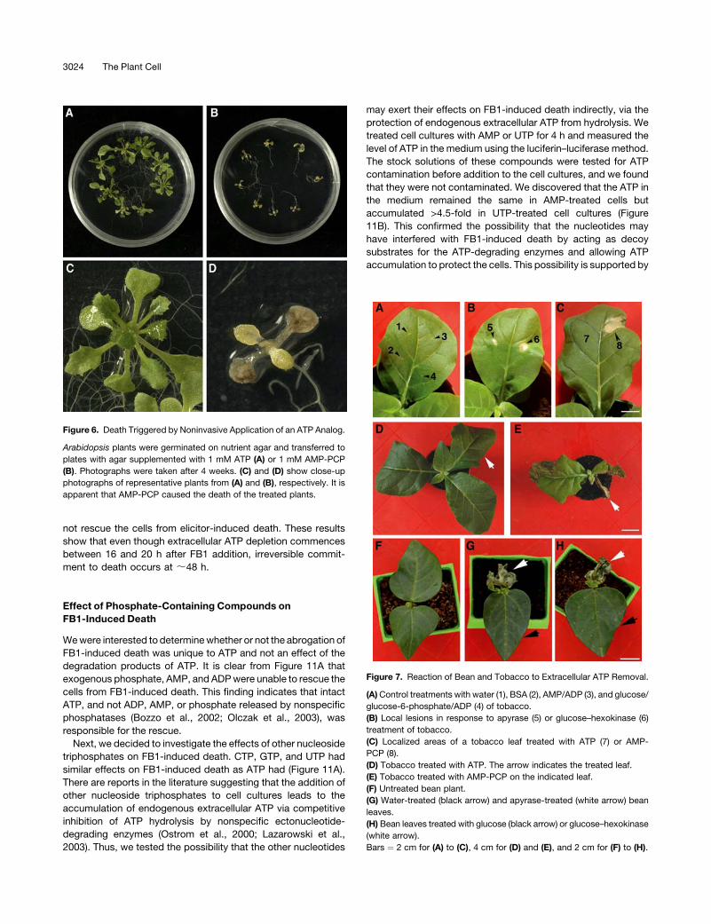
viability across plant species. Tobacco (Nicotiana tabacum)
plants responded to treatment in a similar manner to Arabidop-
sis. Local treatmentwithapyrase,glucose–hexokinase, and1mM
AMP-PCP triggered the development of localized lesions (Figure
7). Treating the entire leaf caused the death of the whole leaf.
However, treatment with a higher concentration (5 mM) of AMP-
PCP had systemic effects: upper leaves that had not received the
primary treatment died as well (Figure 7E). An equivalent con-
centration of ATP did not cause death (Figure 7D). Bean
(Phaseolus vulgaris) plant leaves treated with apyrase or glu-
cose–hexokinase died as well (Figure 7). Subjecting cell cultures
of a monocot, maize (Zea mays), to all three treatments elicited
the death response (Figure 8), indicating that the requirement
of extracellular ATP for viability is not restricted to dicots.
We conclude that extracellular ATP has a physiological role in
plants that is evolutionarily conserved between distantly related
species.
FB1 Triggers Extracellular ATP Depletion That Precedes
Cell Death in Arabidopsis
Having established that the removal of extracellular ATP from
plants or cell suspension cultures triggers death, we wondered
whether some forms of plant cell death invoked during normal
growth and development or during interaction with other organ-
isms might be mediated by the ablation of extracellular ATP. We
decided to investigate the pathogen-induced hypersensitive cell
death, which can be simulated by treating plant tissues or cells
with pathogen-derived elicitors (Levine et al., 1994). To explore
the possibility that diminution of extracellular ATP mediates this
response, we used FB1, a programmed cell death–eliciting
mycotoxin that switches on active plant defenses in Arabidopsis
(Stone et al., 2000).
We wanted to monitor the fate of extracellular ATP in Arabi-
dopsis tissues during FB1 treatment. Thus, we turned to cell
suspension cultures, which offer a simple experimental system in
which to sample the extracellular matrix without damaging cells
and risking contamination with intracellular metabolites. To
achieve this, we used tracer experiments in which cell cultures
were spiked with [32P]ATP and treated with 1 mMFB1 or 0.014%
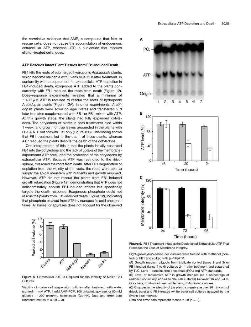
(v/v) methanol as a control. There was a heightened increase in
the hydrolysis of extracellular ATP in FB1-treated cells detect-
able after 16 h of treatment (Figure 9). This resulted in the rapid
Figure 3. Death of Intact Arabidopsis Tissues after Treatment with
Extracellular ATP Traps.
Top row, leaves treated on localized areas that developed localized
lesions. Bottom row, entire leaves treated and beginning to die. These
leaves eventually died except for the controls. Similar results were
obtained when the solutions were infiltrated with a needle or needleless
syringe. Glc-Hk, glucose–hexokinase. Bar ¼ 6 mm.
3022 The Plant Cell
decrease of extracellular ATP level in FB1-treated cells to 24 h
(Figure 9). Thedecline in extracellular ATPcontinued, andby40h,
labeled ATP was undetectable in FB1-treated cultures, whereas
it was still present in control cultures (data not shown). Exami-
nation of the integrity of the plasma membrane using Evans blue
staining did not show any difference between control cells and
FB1-treated cell cultures at 24 h (Figure 9), showing that the
extracellular ATP depletion precedes the loss of plasma mem-
brane integrity and cell death, which become obvious 72 h later
(Figure 9), when the cells become chlorotic and start losing
mitochondrial dehydrogenase activity (data not shown).
Diminution of extracellular ATP before the loss of membrane
integrity implies that the machinery for extracellular ATP removal
is resident within the extracellular matrix and that this event
occurs without leakage of intracellular ATP-degrading enzymes.
This observation, placing extracellular ATP depletion upstream
of FB1-induced death, was requisite if the hypothesis that FB1-
induced death is mediated by the depletion of extracellular ATP
was to be upheld.
Exogenous ATP Rescues Arabidopsis from
FB1-Induced Death
Having established that FB1 treatment activates extracellular
ATP depletion in Arabidopsis cell cultures, we performed experi-
ments to establish whether this event was a mechanism that
mediates FB1-induced cell death. We reasoned that if this
cell death response were solely or partially mediated via this
removal of extracellular ATP, then it would be interdicted or
attenuated by the addition of exogenous ATP concomitant with
treatment.
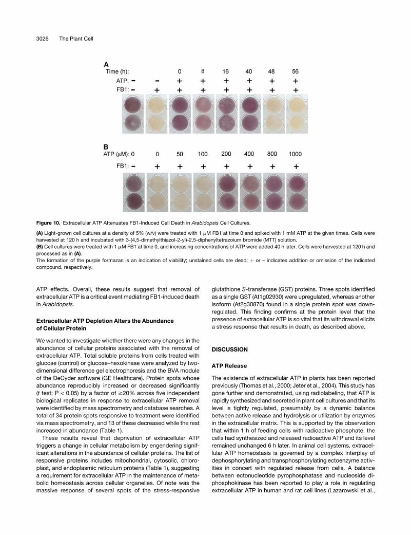
Treatment with 1 mM FB1 causes a progressive decline in the
viability of Arabidopsis cell cultures, which becomes obvious at
72 h and is usually complete by 120 h. Death of the entire
culture is reflected by the total loss of cellular dehydrogenase
activity (Figure 10). In accordance with the prediction that
extracellular ATP deficiency participates in FB1-induced death,
exogenous ATP added to the cell cultures concurrently with
FB1 attenuated cell death (Figure 10A) and delayed it by a
minimum of 48 h. The dose response of FB1-induced death
to exogenous ATP is shown in Figure 10B. We found that
exogenous ATP added to the cells up to 40 h after the addition
of FB1 could still rescue the cells from FB1-induced death
(Figure 10A). However, when added at 48 h or later, ATP could
Figure 5. Glucose–Hexokinase Causes the Death of Arabidopsis Roots.
Top row, root tissue from plants treated with glucose. Bottom row, root tissue from plants treated with glucose–hexokinase (Glc-Hk). Red channel,
confocal images showing autofluorescence of the root tissues. Green channel, confocal images of fluorescein diacetate signals emitted by the same
samples. White channel, images of the same samples captured using transmitted light under phase contrast. The extreme right column shows the
merged images of the preceding three panels. It is clear that the glucose–hexokinase treatment caused the death of the roots, as there was no
fluorescein diacetate signal in these samples. Bars ¼ 200 mm.
Figure 4. Death of Hydroponic Arabidopsis Plants Caused by Glucose–
Hexokinase.
(A) Healthy leaves of plants treated with glucose.
(B) Dying leaves of plants treated with glucose–hexokinase.
(C) Close-up photograph of leaves from plants treated with glucose–
hexokinase showing both chlorosis and brown lesions of localized tissue
death.
(D) and (E) Roots of glucose-treated plants showing normal growth (D)
and retarded roots of glucose–hexokinase-treated plants (E) 7 d after
treatment. Fluorescein diacetate staining andmicroscopy confirmed that
the roots in (E) had reduced viability.
Extracellular ATP Depletion and Death 3023
not rescue the cells from elicitor-induced death. These results
show that even though extracellular ATP depletion commences
between 16 and 20 h after FB1 addition, irreversible commit-
ment to death occurs at ;48 h.
Effect of Phosphate-Containing Compounds on
FB1-Induced Death
Wewere interested to determinewhether or not the abrogation of
FB1-induced death was unique to ATP and not an effect of the
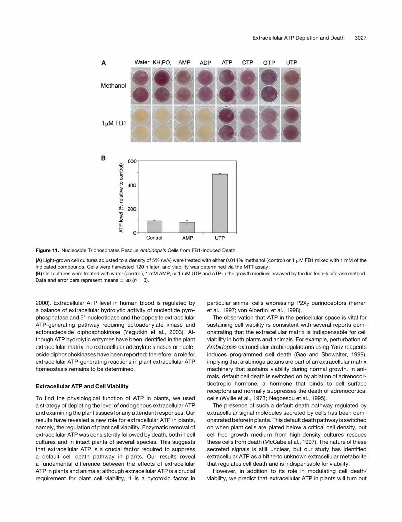
degradation products of ATP. It is clear from Figure 11A that
exogenous phosphate, AMP, and ADPwere unable to rescue the
cells from FB1-induced death. This finding indicates that intact
ATP, and not ADP, AMP, or phosphate released by nonspecific
phosphatases (Bozzo et al., 2002; Olczak et al., 2003), was
responsible for the rescue.
Next, we decided to investigate the effects of other nucleoside
triphosphates on FB1-induced death. CTP, GTP, and UTP had
similar effects on FB1-induced death as ATP had (Figure 11A).
There are reports in the literature suggesting that the addition of
other nucleoside triphosphates to cell cultures leads to the
accumulation of endogenous extracellular ATP via competitive
inhibition of ATP hydrolysis by nonspecific ectonucleotide-
degrading enzymes (Ostrom et al., 2000; Lazarowski et al.,
2003). Thus, we tested the possibility that the other nucleotides
may exert their effects on FB1-induced death indirectly, via the
protection of endogenous extracellular ATP from hydrolysis. We
treated cell cultures with AMP or UTP for 4 h and measured the
level of ATP in the medium using the luciferin–luciferase method.
The stock solutions of these compounds were tested for ATP
contamination before addition to the cell cultures, and we found
that they were not contaminated. We discovered that the ATP in
the medium remained the same in AMP-treated cells but
accumulated >4.5-fold in UTP-treated cell cultures (Figure
11B). This confirmed the possibility that the nucleotides may
have interfered with FB1-induced death by acting as decoy
substrates for the ATP-degrading enzymes and allowing ATP
accumulation to protect the cells. This possibility is supported by
Figure 7. Reaction of Bean and Tobacco to Extracellular ATP Removal.
(A)Control treatments with water (1), BSA (2), AMP/ADP (3), and glucose/
glucose-6-phosphate/ADP (4) of tobacco.
(B) Local lesions in response to apyrase (5) or glucose–hexokinase (6)
treatment of tobacco.
(C) Localized areas of a tobacco leaf treated with ATP (7) or AMP-
PCP (8).
(D) Tobacco treated with ATP. The arrow indicates the treated leaf.
(E) Tobacco treated with AMP-PCP on the indicated leaf.
(F) Untreated bean plant.
(G) Water-treated (black arrow) and apyrase-treated (white arrow) bean
leaves.
(H) Bean leaves treated with glucose (black arrow) or glucose–hexokinase
(white arrow).
Bars ¼ 2 cm for (A) to (C), 4 cm for (D) and (E), and 2 cm for (F) to (H).
Figure 6. Death Triggered by Noninvasive Application of an ATP Analog.
Arabidopsis plants were germinated on nutrient agar and transferred to
plates with agar supplemented with 1 mM ATP (A) or 1 mM AMP-PCP
(B). Photographs were taken after 4 weeks. (C) and (D) show close-up
photographs of representative plants from (A) and (B), respectively. It is
apparent that AMP-PCP caused the death of the treated plants.
3024 The Plant Cell
the correlative evidence that AMP, a compound that fails to
rescue cells, does not cause the accumulation of endogenous
extracellular ATP, whereas UTP, a nucleotide that rescues
elicitor-treated cells, does.
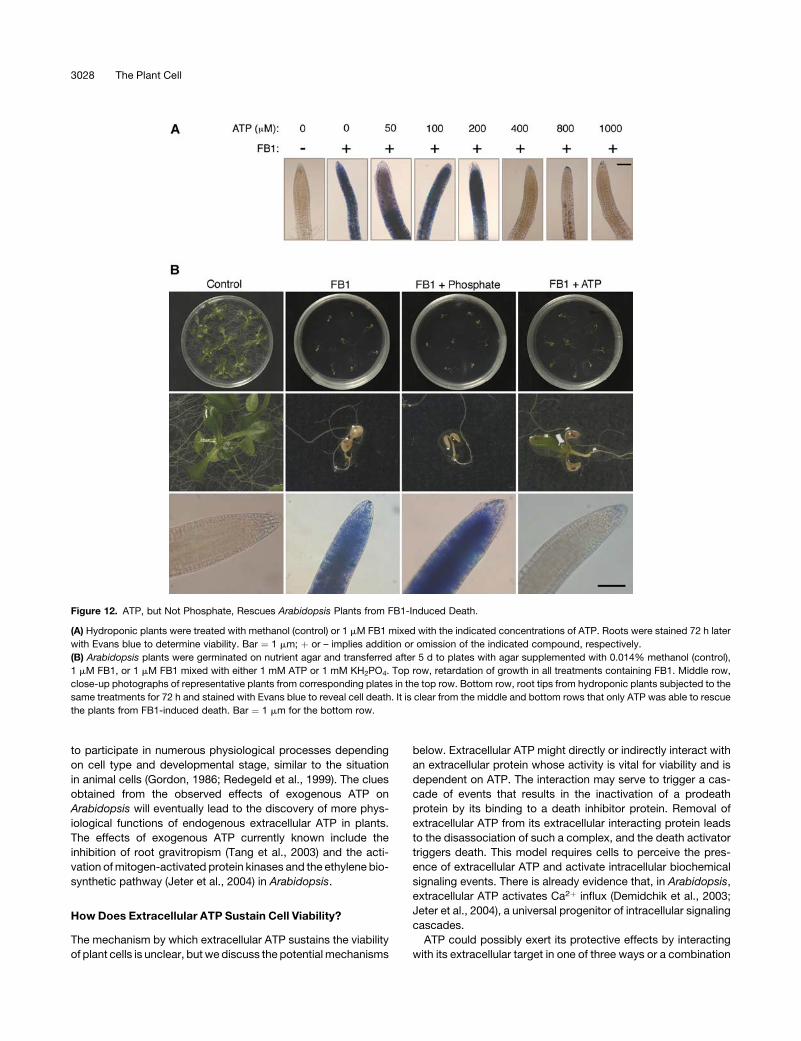
ATP Rescues Intact Plant Tissues from FB1-Induced Death
FB1 kills the roots of submerged hydroponic Arabidopsis plants,
which become stainable with Evans blue 72 h after treatment. In
conformity with a requirement for extracellular ATP depletion in
FB1-induced death, exogenous ATP added to the plants con-
currently with FB1 rescued the roots from death (Figure 12).
Dose–response experiments revealed that a minimum of
;400 mM ATP is required to rescue the roots of hydroponic
Arabidopsis plants (Figure 12A). In other experiments, Arabi-
dopsis plants were sown on agar plates and transferred 5 d
later to plates supplemented with FB1 or FB1 mixed with ATP.
At this growth stage, the plants had fully expanded cotyle-
dons. The cotyledons of plants in both treatments died within
1 week, and growth of true leaves proceeded in the plants with
FB1þ ATP but not with FB1 only (Figure 12B). This finding shows
that FB1 treatment led to the death of these plants, whereas
ATP rescued the plants despite the death of the cotyledons.
One interpretation of this is that the plants initially absorbed
FB1 into the cotyledons and the lack of uptake of themembrane-
impermeant ATP precluded the protection of the cotyledons by
extracellular ATP. Because ATP was restricted to the rhizo-
sphere, it rescued the roots from death. After FB1 degradation or
depletion from the vicinity of the roots, the roots were able to
supply the apical meristem with nutrients and growth resumed.
However, ATP did not rescue the plants from FB1-induced
growth retardation (Figure 12), demonstrating that ATP does not
indiscriminately abolish FB1-induced effects but specifically
targets the death response. Exogenous phosphate could not
rescue the plants from FB1-induced death (Figure 12), indicating
that phosphate cleaved from ATP by nonspecific acid phospha-
tases, ATPases, or apyrases does not account for the observed
Figure 9. FB1 Treatment Induces the Depletion of Extracellular ATP That
Precedes the Loss of Membrane Integrity.
Light-grown Arabidopsis cell cultures were treated with methanol (con-
trol) or FB1 and spiked with [g-32P]ATP.
(A) Growth medium aliquots from triplicate control (lanes 2 and 3) or
FB1-treated (lanes 4 to 6) cultures 24 h after treatment and separated
by TLC. Lane 1 contains free phosphate (PO4) and ATP standards.
(B) Level of radioactive ATP in growth medium (as a percentage of
radioactivity initially added to the cell cultures) between 16 and 24 h.
Gray bars, control cultures; white bars, FB1-treated cultures.
(C) Changes in the integrity of the plasma membrane over 96 h in control
(black bars) and FB1-treated (white bars) cell cultures assayed by the
Evans blue method.
Data and error bars represent means 6 SD (n ¼ 3).
Figure 8. Extracellular ATP Is Required for the Viability of Maize Cell
Cultures.
Viability of maize cell suspension cultures after treatment with water
(control), 1 mM ATP, 1 mM AMP-PCP, 100 units/mL apyrase, or 50 mM
glucose þ 200 units/mL hexokinase (Glc-Hk). Data and error bars
represent means 6 SD (n ¼ 3).
Extracellular ATP Depletion and Death 3025
ATP effects. Overall, these results suggest that removal of
extracellular ATP is a critical event mediating FB1-induced death
in Arabidopsis.
Extracellular ATP Depletion Alters the Abundance
of Cellular Protein
Wewanted to investigate whether there were any changes in the
abundance of cellular proteins associated with the removal of
extracellular ATP. Total soluble proteins from cells treated with
glucose (control) or glucose–hexokinase were analyzed by two-
dimensional difference gel electrophoresis and the BVA module
of the DeCyder software (GE Healthcare). Protein spots whose
abundance reproducibly increased or decreased significantly
(t test; P < 0.05) by a factor of $20% across five independent
biological replicates in response to extracellular ATP removal
were identified by mass spectrometry and database searches. A
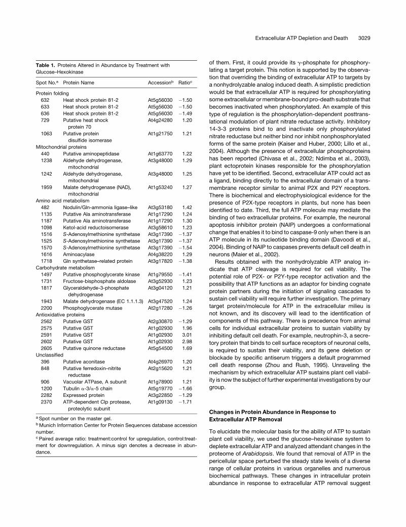
total of 34 protein spots responsive to treatment were identified
via mass spectrometry, and 13 of these decreased while the rest
increased in abundance (Table 1).
These results reveal that deprivation of extracellular ATP
triggers a change in cellular metabolism by engendering signif-
icant alterations in the abundance of cellular proteins. The list of
responsive proteins includes mitochondrial, cytosolic, chloro-
plast, and endoplasmic reticulum proteins (Table 1), suggesting
a requirement for extracellular ATP in the maintenance of meta-
bolic homeostasis across cellular organelles. Of note was the
massive response of several spots of the stress-responsive
glutathione S-transferase (GST) proteins. Three spots identified
as a single GST (At1g02930) were upregulated, whereas another
isoform (At2g30870) found in a single protein spot was down-
regulated. This finding confirms at the protein level that the
presence of extracellular ATP is so vital that its withdrawal elicits
a stress response that results in death, as described above.
DISCUSSION
ATP Release
The existence of extracellular ATP in plants has been reported
previously (Thomas et al., 2000; Jeter et al., 2004). This study has
gone further and demonstrated, using radiolabeling, that ATP is
rapidly synthesized and secreted in plant cell cultures and that its
level is tightly regulated, presumably by a dynamic balance
between active release and hydrolysis or utilization by enzymes
in the extracellular matrix. This is supported by the observation
that within 1 h of feeding cells with radioactive phosphate, the
cells had synthesized and released radioactive ATP and its level
remained unchanged 6 h later. In animal cell systems, extracel-
lular ATP homeostasis is governed by a complex interplay of
dephosphorylating and transphosphorylating ectoenzyme activ-
ities in concert with regulated release from cells. A balance
between ectonucleotide pyrophosphatase and nucleoside di-
phosphokinase has been reported to play a role in regulating
extracellular ATP in human and rat cell lines (Lazarowski et al.,
Figure 10. Extracellular ATP Attenuates FB1-Induced Cell Death in Arabidopsis Cell Cultures.
(A) Light-grown cell cultures at a density of 5% (w/v) were treated with 1 mM FB1 at time 0 and spiked with 1 mM ATP at the given times. Cells were
harvested at 120 h and incubated with 3-(4,5-dimethylthiazol-2-yl)-2,5-diphenyltetrazoium bromide (MTT) solution.
(B) Cell cultures were treated with 1 mM FB1 at time 0, and increasing concentrations of ATP were added 40 h later. Cells were harvested at 120 h and
processed as in (A).
The formation of the purple formazan is an indication of viability; unstained cells are dead; þ or – indicates addition or omission of the indicated
compound, respectively.
3026 The Plant Cell
2000). Extracellular ATP level in human blood is regulated by
a balance of extracellular hydrolytic activity of nucleotide pyro-
phosphatase and 59-nucleotidase and the opposite extracellular
ATP-generating pathway requiring ectoadenylate kinase and
ectonucleoside diphosphokinase (Yegutkin et al., 2003). Al-
though ATP hydrolytic enzymes have been identified in the plant
extracellular matrix, no extracellular adenylate kinases or nucle-
oside diphosphokinases have been reported; therefore, a role for
extracellular ATP-generating reactions in plant extracellular ATP
homeostasis remains to be determined.
Extracellular ATP and Cell Viability
To find the physiological function of ATP in plants, we used
a strategy of depleting the level of endogenous extracellular ATP
and examining the plant tissues for any attendant responses. Our
results have revealed a new role for extracellular ATP in plants,
namely, the regulation of plant cell viability. Enzymatic removal of
extracellular ATPwas consistently followed by death, both in cell
cultures and in intact plants of several species. This suggests
that extracellular ATP is a crucial factor required to suppress
a default cell death pathway in plants. Our results reveal
a fundamental difference between the effects of extracellular
ATP in plants and animals; although extracellular ATP is a crucial
requirement for plant cell viability, it is a cytotoxic factor in
particular animal cells expressing P2X7 purinoceptors (Ferrari
et al., 1997; von Albertini et al., 1998).
The observation that ATP in the pericellular space is vital for
sustaining cell viability is consistent with several reports dem-
onstrating that the extracellular matrix is indispensable for cell
viability in both plants and animals. For example, perturbation of
Arabidopsis extracellular arabinogalactans using Yariv reagents
induces programmed cell death (Gao and Showalter, 1999),
implying that arabinogalactans are part of an extracellular matrix
machinery that sustains viability during normal growth. In ani-
mals, default cell death is switched on by ablation of adrenocor-
ticotropic hormone, a hormone that binds to cell surface
receptors and normally suppresses the death of adrenocortical
cells (Wyllie et al., 1973; Negoescu et al., 1995).
The presence of such a default death pathway regulated by
extracellular signal molecules secreted by cells has been dem-
onstratedbefore inplants. Thisdefault deathpathway is switched
on when plant cells are plated below a critical cell density, but
cell-free growth medium from high-density cultures rescues
these cells from death (McCabe et al., 1997). The nature of these
secreted signals is still unclear, but our study has identified
extracellular ATP as a hitherto unknown extracellular metabolite
that regulates cell death and is indispensable for viability.
However, in addition to its role in modulating cell death/
viability, we predict that extracellular ATP in plants will turn out
Figure 11. Nucleoside Triphosphates Rescue Arabidopsis Cells from FB1-Induced Death.
(A) Light-grown cell cultures adjusted to a density of 5% (w/v) were treated with either 0.014% methanol (control) or 1 mM FB1 mixed with 1 mM of the
indicated compounds. Cells were harvested 120 h later, and viability was determined via the MTT assay.
(B) Cell cultures were treated with water (control), 1 mM AMP, or 1 mM UTP and ATP in the growth medium assayed by the luciferin–luciferase method.
Data and error bars represent means 6 SD (n ¼ 3).
Extracellular ATP Depletion and Death 3027
to participate in numerous physiological processes depending
on cell type and developmental stage, similar to the situation
in animal cells (Gordon, 1986; Redegeld et al., 1999). The clues
obtained from the observed effects of exogenous ATP on
Arabidopsis will eventually lead to the discovery of more phys-
iological functions of endogenous extracellular ATP in plants.
The effects of exogenous ATP currently known include the
inhibition of root gravitropism (Tang et al., 2003) and the acti-
vation ofmitogen-activated protein kinases and the ethylene bio-
synthetic pathway (Jeter et al., 2004) in Arabidopsis.
How Does Extracellular ATP Sustain Cell Viability?
The mechanism by which extracellular ATP sustains the viability
of plant cells is unclear, but we discuss the potential mechanisms
below. Extracellular ATP might directly or indirectly interact with
an extracellular protein whose activity is vital for viability and is
dependent on ATP. The interaction may serve to trigger a cas-
cade of events that results in the inactivation of a prodeath
protein by its binding to a death inhibitor protein. Removal of
extracellular ATP from its extracellular interacting protein leads
to the disassociation of such a complex, and the death activator
triggers death. This model requires cells to perceive the pres-
ence of extracellular ATP and activate intracellular biochemical
signaling events. There is already evidence that, in Arabidopsis,
extracellular ATP activates Ca2þ influx (Demidchik et al., 2003;
Jeter et al., 2004), a universal progenitor of intracellular signaling
cascades.
ATP could possibly exert its protective effects by interacting
with its extracellular target in one of three ways or a combination
Figure 12. ATP, but Not Phosphate, Rescues Arabidopsis Plants from FB1-Induced Death.
(A) Hydroponic plants were treated with methanol (control) or 1 mM FB1 mixed with the indicated concentrations of ATP. Roots were stained 72 h later
with Evans blue to determine viability. Bar ¼ 1 mm; þ or – implies addition or omission of the indicated compound, respectively.
(B) Arabidopsis plants were germinated on nutrient agar and transferred after 5 d to plates with agar supplemented with 0.014% methanol (control),
1 mM FB1, or 1 mM FB1 mixed with either 1 mM ATP or 1 mM KH2PO4. Top row, retardation of growth in all treatments containing FB1. Middle row,
close-up photographs of representative plants from corresponding plates in the top row. Bottom row, root tips from hydroponic plants subjected to the
same treatments for 72 h and stained with Evans blue to reveal cell death. It is clear from the middle and bottom rows that only ATP was able to rescue
the plants from FB1-induced death. Bar ¼ 1 mm for the bottom row.
3028 The Plant Cell
of them. First, it could provide its g-phosphate for phosphory-
lating a target protein. This notion is supported by the observa-
tion that overriding the binding of extracellular ATP to targets by
a nonhydrolyzable analog induced death. A simplistic prediction
would be that extracellular ATP is required for phosphorylating
some extracellular ormembrane-bound pro-death substrate that
becomes inactivated when phosphorylated. An example of this
type of regulation is the phosphorylation-dependent posttrans-
lational modulation of plant nitrate reductase activity. Inhibitory
14-3-3 proteins bind to and inactivate only phosphorylated
nitrate reductase but neither bind nor inhibit nonphosphorylated
forms of the same protein (Kaiser and Huber, 2000; Lillo et al.,
2004). Although the presence of extracellular phosphoproteins
has been reported (Chivasa et al., 2002; Ndimba et al., 2003),
plant ectoprotein kinases responsible for the phosphorylation
have yet to be identified. Second, extracellular ATP could act as
a ligand, binding directly to the extracellular domain of a trans-
membrane receptor similar to animal P2X and P2Y receptors.
There is biochemical and electrophysiological evidence for the
presence of P2X-type receptors in plants, but none has been
identified to date. Third, the full ATP molecule may mediate the
binding of two extracellular proteins. For example, the neuronal
apoptosis inhibitor protein (NAIP) undergoes a conformational
change that enables it to bind to caspase-9 only when there is an
ATP molecule in its nucleotide binding domain (Davoodi et al.,
2004). Binding of NAIP to caspases prevents default cell death in
neurons (Maier et al., 2002).
Results obtained with the nonhydrolyzable ATP analog in-
dicate that ATP cleavage is required for cell viability. The
potential role of P2X- or P2Y-type receptor activation and the
possibility that ATP functions as an adaptor for binding cognate
protein partners during the initiation of signaling cascades to
sustain cell viability will require further investigation. The primary
target protein/molecule for ATP in the extracellular milieu is
not known, and its discovery will lead to the identification of
components of this pathway. There is precedence from animal
cells for individual extracellular proteins to sustain viability by
inhibiting default cell death. For example, neutrophin-3, a secre-
tory protein that binds to cell surface receptors of neuronal cells,
is required to sustain their viability, and its gene deletion or
blockade by specific antiserum triggers a default programmed
cell death response (Zhou and Rush, 1995). Unraveling the
mechanism by which extracellular ATP sustains plant cell viabil-
ity is now the subject of further experimental investigations by our
group.
Changes in Protein Abundance in Response to
Extracellular ATP Removal
To elucidate the molecular basis for the ability of ATP to sustain
plant cell viability, we used the glucose–hexokinase system to
deplete extracellular ATP and analyzed attendant changes in the
proteome of Arabidopsis. We found that removal of ATP in the
pericellular space perturbed the steady state levels of a diverse
range of cellular proteins in various organelles and numerous
biochemical pathways. These changes in intracellular protein
abundance in response to extracellular ATP removal suggest
Table 1. Proteins Altered in Abundance by Treatment with
Glucose–Hexokinase
Spot No.a Protein Name Accessionb Ratioc
Protein folding
632 Heat shock protein 81-2 At5g56030 �1.50
633 Heat shock protein 81-2 At5g56030 �1.50
636 Heat shock protein 81-2 At5g56030 �1.49
729 Putative heat shock
protein 70
At4g24280 1.20
1063 Putative protein
disulfide isomerase
At1g21750 1.21
Mitochondrial proteins
440 Putative aminopeptidase At1g63770 1.22
1238 Aldehyde dehydrogenase,
mitochondrial
At3g48000 1.29
1242 Aldehyde dehydrogenase,
mitochondrial
At3g48000 1.25
1959 Malate dehydrogenase (NAD),
mitochondrial
At1g53240 1.27
Amino acid metabolism
482 Nodulin/Gln-ammonia ligase–like At3g53180 1.42
1135 Putative Ala aminotransferase At1g17290 1.24
1187 Putative Ala aminotransferase At1g17290 1.30
1098 Ketol-acid reductoisomerase At3g58610 1.23
1516 S-Adenosylmethionine synthetase At3g17390 �1.37
1525 S-Adenosylmethionine synthetase At3g17390 �1.37
1570 S-Adenosylmethionine synthetase At3g17390 �1.54
1616 Aminoacylase At4g38220 1.29
1718 Gln synthetase–related protein At3g17820 �1.38
Carbohydrate metabolism
1497 Putative phosphoglycerate kinase At1g79550 �1.41
1731 Fructose-bisphosphate aldolase At3g52930 1.23
1817 Glyceraldehyde-3-phosphate
dehydrogenase
At3g04120 1.21
1943 Malate dehydrogenase (EC 1.1.1.3) At3g47520 1.24
2200 Phosphoglycerate mutase At2g17280 �1.26
Antioxidative proteins
2562 Putative GST At2g30870 �1.29
2575 Putative GST At1g02930 1.96
2591 Putative GST At1g02930 3.01
2602 Putative GST At1g02930 2.98
2605 Putative quinone reductase At5g54500 1.69
Unclassified
396 Putative aconitase At4g26970 1.20
848 Putative ferredoxin–nitrite
reductase
At2g15620 1.21
906 Vacuolar ATPase, A subunit At1g78900 1.21
1200 Tubulin a-3/a-5 chain At5g19770 �1.66
2282 Expressed protein At3g22850 �1.29
2370 ATP-dependent Clp protease,
proteolytic subunit
At1g09130 �1.71
a Spot number on the master gel.bMunich Information Center for Protein Sequences database accession
number.c Paired average ratio: treatment:control for upregulation, control:treat-
ment for downregulation. A minus sign denotes a decrease in abun-
dance.
Extracellular ATP Depletion and Death 3029
that signals initiated by extracellular ATP perception funnel into
numerous intracellular metabolic pathways spanning several
cellular compartments. This reinforces, at the molecular level,
the finding that cells are dependent on extracellular ATP for
viability.
Previous studies of plant programmed cell death responses
have identified changes in the expression of genes involved in
primary metabolism, molecular chaperones, and other enzymes
regulating protein folding, mitochondrial proteins, and antioxi-
dative stress proteins (Desikan et al., 1998; Swidzinski et al.,
2002, 2004). Arabidopsis microarray data available via the
Genevestigator database (http://www.genevestigator.ethz.ch)
and web browser data-mining interface (Zimmermann et al.,
2004) reveal that most of the genes for the proteins identified in
this study are differentially expressed in response to a diverse
range of death-inducing treatments such as herbicides, toxins,
and attack by bacterial and fungal pathogens. This finding
suggests that cell death induced by extracellular ATP depletion
possibly funnels into a shared core death pathway at some
point downstream from the position where ATP deprivation is
crucial. The concept of a shared core deathmachinery intowhich
disparate signaling pathways activated by various pro-death
stimuli feed and converge has been proposed before (McCabe
and Leaver, 2000). This is similar to the sensory and signaling
cascades in the cyanobacterial stress response, in which various
stresses have unique components close to the initial stimulus
sensing but share common components of the signaling cas-
cades (Marin et al., 2003).
The strongest response was observed in GSTs, a family of
stress-responsive proteins whose gene expression can be in-
duced by pathogen attack (Lieberherr et al., 2003) or treatment
with pathogen-derived elicitors (Desikan et al., 1998). Wagner
et al. (2002) reported that salicylic acid suppresses gene ex-
pression of the GST (At2g30870) whose abundance at the
protein level decreased in response to extracellular ATP depri-
vation in this study. Gene expression of the GST protein
(At1g02930) upregulated by ATP removal in our study is also
activated by endogenous signaling molecules such as H2O2,
salicylic acid, ethylene, and jasmonic acid (Wagner et al., 2002).
This suggests that the cell death activated by the removal of
extracellular ATP may potentially be mediated by some of these
signaling molecules. Indeed, it is already known that FB1, which
triggers death via extracellular ATP removal, induces the accu-
mulation of reactive oxygen species inArabidopsis and that FB1-
induced death in the same species requires ethylene, salicylic
acid, and jasmonic acid signaling (Asai et al., 2000).
Depletion of Extracellular ATP Mediates Elicitor-Induced
Cell Death
Cell death is an essential part of plant development that is regu-
lated by a programmed genetic template and affects single cells,
particular cell layers, or entire organs (Buchanan-Wollaston,
1997; Fukuda, 1997; Groover et al., 1997) and is invoked in the
hypersensitive response to pathogen attack (Lam et al., 2001).
Having established that extracellular ATP is vital to sustain cell
viability and that artificially removing it triggers cell death, we
discovered that depletion of extracellular ATP mediates an
elicitor-induced cell death response. FB1, a mycotoxin that
acts as an elicitor in Arabidopsis and triggers defense responses
and cell death, induced a heightened hydrolysis of extracellular
ATP preceding the breach in plasmamembrane integrity and the
onset of death. Crucially, exogenous ATP was able to rescue
plants treated with the elicitor from death. Elicitor-induced cell
death requires an influx of apoplastic Ca2þ into the cell, and its
chelation can block cell death (Levine et al., 1996). However,
the effect of exogenous ATP on FB1-induced death is unlikely
the result of chelation of divalent cations because 1 mM solu-
tions of AMP and ADP, which have a higher chelation capacity
than 200 mM ATP, failed to abolish cell death when 200 mM
ATP blocked it. In cell cultures, prevention of ATP depletion by
adding exogenous ATP concurrently with FB1 or correcting the
deficit by the addition of exogenous ATP several hours later, up
to 40 h, was able to prevent death. When added 48 h after FB1
or later, exogenous ATP was unable to rescue cells from death,
implying that commitment to death occurs ;48 h from FB1
addition.
In cell cultures, it is clear that FB1 upregulates an extracellular
ATP hydrolytic activity after a lag of;16 h. This activity does not
discriminate between different nucleoside triphosphates and
appears not to act on nucleoside monophosphates and diphos-
phates, as shown by the failure of AMP and ADP to block
FB1-induced death, which was abolished by all of the tested
nucleoside triphosphates. The observation that a pathogen
elicitor stimulates extracellular nucleoside triphosphatase activ-
ity is not unprecedented because elicitors have been reported to
activate membrane-bound and cell wall–bound ATPase activity
(Kiba et al., 1995, 1999). The exact mechanism by which FB1
activates extracellular ATP hydrolysis is not yet defined. How-
ever, plant extracellular ATP hydrolysis is performed by ecto-
apyrases (Komoszynski and Wojtczak, 1996), cell wall–bound
ATPases (Kivilaan et al., 1961; Shiraishi et al., 1991), and extra-
cellular purple acid phosphatases (Bozzo et al., 2002; Olczak
et al., 2003). Perhaps FB1 stimulates the activity of a similar
extracellular enzyme, as has been reported for other elicitors. For
example, chitin, a pathogen-derived elicitor known to trigger cell
death (Tada et al., 2001), has been reported to specifically bind
and stimulate the activity of an extracellular apyrase (Etzler et al.,
1999). Recently, a fungal pathogen elicitor was reported to
stimulate a recombinant plant apyrase in vitro (Kawahara et al.,
2003). However, the delay between FB1 addition and the activa-
tion of extracellular ATP hydrolysis might be an indication that
FB1 does not directly activate the enzymes but requires de novo
synthesis of secondary messengers or, alternatively, that syn-
thesis and secretion of the hydrolytic enzymes occur during the
lag period.
Our results allow a rationalization of extracellular ATP, cell
death, and the response of plant cells to somepathogen elicitors.
Elicitors clearly affect cell viability (Levine et al., 1994) and have
been reported to activate enzymes that modulate the level of
extracellular ATP (Kiba et al., 1995; Etzler et al., 1999; Kawahara
et al., 2003). Further studies on a wide range of elicitors and
incompatible host–pathogen combinations are now required to
investigate whether or not hypersensitive cell death in general is
mediated via extracellular ATP depletion.
3030 The Plant Cell
METHODS
Plant Material and Growth Conditions
Cell suspension cultures of Arabidopsis thaliana var erecta were main-
tained as described previously (May and Leaver, 1993). Maize (Zea mays)
cell cultures were maintained by weekly 10-fold dilution in fresh
Murashige and Skoog (MS) growth medium (Murashige and Skoog,
1962) with 2% (w/v) sucrose and 2 mg/L 2,4-D. Suspension cell cultures
of both species were grown either in continuous darkness or with a 16-h
photoperiod at 258C and were used for treatment 3 to 5 d after transfer to
fresh medium. Dark-grown cell cultures were used in all experiments
except for those involving FB1 treatments, in which light-grown
cell cultures were used because FB1-induced cell death in plants is
dependent on light (Asai et al., 2000; Stone et al., 2000). Glucose–
hexokinase treatments were performed with both light- and dark-grown
cell cultures. Tobacco (Nicotiana tabacum), Landsberg erecta and
Columbia ecotypes of Arabidopsis, and bean (Phaseolus vulgaris) plants
for injecting with solutions were grown in soil in a growth cabinet with a
16-h photoperiod at 208Cand 8 h of darkness at 158C.RHwasmaintained
at 60%, and the photon flux density was 250 mmol�m�2�s�1. Plants were
used for treatment 5 to 8 weeks after sowing.
Treatments
Enzymes were dissolved in water and filter-sterilized (0.2 mm). Dialysis
was performed against water using membranes with a molecular mass
cutoff of 8 kD. AMP, ADP, ATP, and AMP-PCP stock solutions in water
were neutralized to pH 7.0 using KOH and filter-sterilized. Whole plants
were treated by infiltration of the apoplast of a small area or entire leaf on
the abaxial surface using a syringe with or without a hypodermic needle.
Concentrations used to treat intact plant tissues were 6 mg/mL BSA,
1 mM AMP, 1 mM ADP, 5 mM ATP, 5 mM AMP-PCP, 50 mM glucose,
1 mM glucose-6-phosphate, 0.5 units/mL apyrase, and 1.85 units/mL
hexokinase. In cell culture experiments, glucose was used at a concen-
tration of 100 mM. Cell cultures were incubated with the appropriate
solutions in a final volume of 1.5 mL and a cell density of 5% (w/v). After
72 h, the cultures were diluted to 50 mL in fresh growth medium. Ninety-
six hours later, 1-mL aliquots were withdrawn and the packed cell volume
was determined as a percentage of the culture volume. A 7 mM stock
solution of FB1 was prepared in absolute methanol, and a working
solution of 50 mM (diluted in water) was used for treatments at a final
concentration of 1 mM, with an equivalent volume of 0.71% methanol
being used to treat controls.
Hydroponic Experiments
Floating Plants
Seeds of Arabidopsis ecotype Columbia were surface-sterilized as
described previously (Topping and Lindsey, 1991) and sown on 7-cm-
diameter, 1-cm-thick polyurethane foam discs soaked in MS medium
(0.22% [w/v] MS salts, 1% [w/v] sucrose, adjusted to pH 5.7 with KOH/
HCl). The discs were placed in sterile phytatrays (Sigma-Aldrich) and
incubated under a 16-h-light/8-h-dark cycle at 228C. One week later,
when the seeds had germinated and the roots had penetrated the foam,
30mL ofMSmediumwas added to the phytatray, which caused the foam
discs to float. The trays were transferred to a shaking platform (25 rpm)
under the same environmental conditions. During the second week, the
roots of the plants had emerged through the other side of the foam and
were submerged in the nutrient-rich medium. Exactly 2 weeks after
sowing, the plants were treated with water (control), 45 mM glucose,
45 mM glucose þ 1 mM glucose-6-phosphate, and 45 mM glucose þ100 units/mL hexokinase.
Submerged Plants
Surface-sterilized Arabidopsis seeds were incubated in 5 mL of MS
medium in 25-mL conical glass flasks on an orbital shaker (120 rpm) at
228C in a 16-h photoperiod regime. Five days later, the plants were
treatedwith 1mMFB1 and 1mMFB1mixedwith either 1mMATP or 1mM
KH2PO4. At 72 h after treatment, the plants were stained with Evans blue
dye as described below.
Agar Plate Assays
Surface-sterilized Arabidopsis seedlings were plated on 0.4% (w/v) agar-
containing MS medium. Five days later, the plants were transferred to
agar plates supplemented with 1 mM FB1 or 1 mM FB1 þ 1 mM ATP. The
plants were photographed 4 weeks later. This experiment was performed
using both the Landsberg erecta and Columbia ecotypes of Arabidopsis.
Microscopy
Roots of Arabidopsis treated with glucose or glucose–hexokinase were
stained with fluorescein diacetate and examined using a LSM 510 Meta
microscope (Carl Zeiss). Autofluorescence was acquired at an excitation
wavelength of 543 nm and an emission long pass of 560 nm. The fluores-
cence of fluorescein diacetate was detected at an excitation wavelength
of 488 nm and an emission band pass between 505 and 530 nm. The
same tissues were also imaged using transmitted light phase contrast,
and all three images were merged.
Cell Death Assay
Determination of cell viability by the MTT assay was performed as
described previously (Watts et al., 1989) with minor modifications. An
aliquot of 400 mL of 5 mg/mL MTT solution in water was incubated with
5mLof cell culture for 4 h. A lawnof cellswasplaced inwells of amicrotiter
plate and scanned to show the color development.
Evans blue assay for the death (and plasmamembrane integrity) of cell
cultures was performed by incubating triplicate 100-mL cell aliquots with
a final 0.0125% (w/v) Evans blue solution for 20 min. Cell death levels in
the treatments were obtained by spectrophotometrically (600 nm) de-
termining the amount of dye taken up by cells and assuming that control
cells were 100% viable and cells boiled for 5 min before staining were 0%
viable. For hydroponic plants, five plants in 5 mL of 0.04% (w/v) Evans
blue solution were vacuum-infiltrated by holding the vacuum for 30 min
and then washed three times with water for 4 h each wash.
ATPMeasurements
De novo synthesis and secretion of ATP were performed by feeding
100 mCi of [32P]H3PO4 to 5 mL of 3-d-old light-grown cell cultures
adjusted to a density of 1% (w/v). Cell-free 170 mL of medium was
withdrawn 1 h later and acidified with 0.6% trichloroacetic acid. Aliquots
of 2 mL were mixed with an equal volume of 50 mM citrate buffer, pH 5.5,
and separated by thin layer chromatography (TLC) on silica gel plateswith
dioxane:ammonium hydroxide:water (6:1:6, v/v/v). To demonstrate the
activity of apyrase and hexokinase, cell cultures labeled with a similar
amount of [32P]H3PO4 for 1 h were treated with 100 units/mL apyrase or
100mMglucoseþ 200 units/mL hexokinase for a further 6 h, andmedium
samples were analyzed by TLC. Apyrase at 20 units/mL was also used in
parallel experiments.
To monitor extracellular ATP during FB1 treatment, 5 mL of light-grown
cell cultures was adjusted to 1% (w/v) density, treated with 0.014% (v/v)
methanol as a control or 1 mM FB1, and spiked with 4.5 mCi [g-32P]ATP
Extracellular ATP Depletion and Death 3031
16 h later. Cell-free medium aliquots were withdrawn periodically and
analyzed by TLC as described above.
In other experiments, medium ATP was measured by the nonradioac-
tive luciferin–luciferase method using the Enlighten ATP assay kit
(Promega). Cell-free 100-mL medium aliquots were mixed with 2 mL of
50% (w/v) trichloroacetic acid containing 0.0005% (w/v) acid blue 147
and stored at �208C. For ATP assay, 5 mL was mixed with 95 mL of
100 mM Tris-acetate buffer, pH 7.8, and the reaction was started by
the addition of 30 mL of luciferin–luciferase followed by a reading delay of
0.3 s and an integration time of 10 s using the Anthos Lucy 1 luminometer
(Labtech International).
Proteomic Analyses
Aliquots of 5 mL each of 3-d-old, dark-grown Arabidopsis cell cultures
were treated with 100 mM glucose (to serve as controls) or a combination
of 100 mM glucose plus 200 units/mL hexokinase. The experiment was
performed using five independent biological replicates. Cells were
harvested 48 h later, and protein was extracted and analyzed by two-
dimensional difference gel electrophoresis as described previously
(Ndimba et al., 2005). Gel images were scanned using the Typhoon
9400 scanner (GE Healthcare), and image analysis was performed using
the DIA and BVA modules of the DeCyder software (GE Healthcare) as
described before (Ndimba et al., 2005). Proteins whose abundance
changed (up or down) by a minimum of 20% were excised from silver-
stained gels, with a total protein loading of 400mg, and identified viamass
spectrometry and database (National Center for Biotechnology Informa-
tion nonredundant database) searches using the methods described by
Chivasa et al. (2002).
Received August 5, 2005; revised August 5, 2005; accepted September
6, 2005; published September 30, 2005.
REFERENCES
Abraham, E., Prat, A., Gerweck, L., Seneveratne, T., Arceci, R.,
Kramer, R., Guidotti, G., and Cantiello, H. (1993). The multidrug
resistance (MDR1) gene product functions as an ATP channel. Proc.
Natl. Acad. Sci. USA 90, 312–316.
Asai, T., Stone, J.M., Heard, J.E., Kovtun, Y., Yorgey, P., Sheen, J.,
and Ausubel, F.M. (2000). Fumonisin B1-induced cell death in Arabi-
dopsis protoplasts requires jasmonate-, ethylene-, and salicylate-
dependent signalling pathways. Plant Cell 12, 1823–1835.
Beigi, R.D., and Dubyak, G.R. (2000). Endotoxin activation of macro-
phages does not induce ATP release and autocrine stimulation of P2
nucleotide receptors. J. Immunol. 165, 7189–7198.
Boldin, P., and Burnstock, G. (2001). Purinergic signalling: ATP re-
lease. Neurochem. Res. 26, 959–969.
Bozzo, G.G., Raghothama, K.G., and Plaxton, W.C. (2002). Purifica-
tion and characterization of two secreted purple acid phosphatase
isozymes from phosphate-starved tomato (Lycopersicon esculentum)
cell cultures. Eur. J. Biochem. (Tokyo) 269, 6278–6286.
Buchanan-Wollaston, V. (1997). The molecular biology of leaf senes-
cence. J. Exp. Bot. 48, 181–199.
Burnstock, G., and Kennedy, C. (1985). Is there a basis for distinguish-
ing two types of P2-purinoceptors? Gen. Pharmacol. 16, 433–440.
Chivasa, S., Ndimba, B.K., Simon, W.J., Robertson, D., Yu, X.-Y.,
Knox, J.P., Bolwell, P., and Slabas, A.R. (2002). Proteomic analysis
of the Arabidopsis thaliana cell wall. Electrophoresis 23, 1754–1765.
Davoodi, J., Lin, L., Kelly, J., Liston, P., and MacKenzie, A.E. (2004).
Neuronal apoptosis-inhibitory protein does not interact with Smac
and requires ATP to bind caspase-9. J. Biol. Chem. 279, 40622–
40628.
Demidchik, V., Nichols, C., Oliynyk, M., Dark, A., Glover, B.J., and
Davies, J.M. (2003). Is ATP a signalling agent in plants? Plant Physiol.
133, 456–461.
Desikan, R., Reynolds, A., Hancock, J.T., and Neill, S.J. (1998).
Harpin and hydrogen peroxide both initiate programmed cell death
but have differential effects on defence gene expression in Arabidop-
sis suspension cultures. Biochem. J. 330, 115–120.
Duan, S., Anderson, C.M., Keung, E.C., Chen, Y., Chen, Y., and
Swanson, R.A. (2003). P2X7 receptor-mediated release of excitatory
amino acids from astrocytes. J. Neurosci. 23, 1320–1328.
Dutta, A.K., Okada, Y., and Sabirov, R.Z. (2002). Regulation of an ATP-
conductive large-conductance anion channel and swelling-induced
ATP release by arachidonic acid. J. Physiol. 542, 803–816.
Dutta, A.K., Sabirov, R.Z., Uramoto, H., and Okada, Y. (2004). Role of
ATP-conductive anion channel in ATP release from neonatal rat
cardiomyocytes in ischaemic or hypoxic conditions. J. Physiol. 559,
799–812.
Etzler, M.E., Kalsi, G., Ewing, N.N., Roberts, N.J., Day, R.B., and
Murphy, J.B. (1999). A nod factor binding lectin with apyrase activity
from legume roots. Proc. Natl. Acad. Sci. USA 96, 5856–5861.
Ferrari, D., Wesselborg, S., Bauer, M.K., and Schulze-Osthoff, K.
(1997). Extracellular ATP activates transcription factor NF-kappaB
through the P2Z purinoceptor by selectively targeting NF-kappaB p65
(RE1A). J. Cell Biol. 139, 1635–1643.
Fiske, C.H., and Subarrow, Y. (1929). Phosphorous compounds of
muscle and liver. Science 70, 381–382.
Fukuda, H. (1997). Tracheary element differentiation. Plant Cell 9,
1147–1156.
Gao, M., and Showalter, A.M. (1999). Yariv reagent treatment induces
programmed cell death in Arabidopsis cell cultures and implicates
arabinogalactan protein involvement. Plant Cell 19, 321–331.
Glynn, I.M. (1968). Membrane adenosine triphosphatase and cation
transport. Br. Med. Bull. 24, 165–169.
Gordon, J.L. (1986). Extracellular ATP: Effects, sources and fate.
Biochem. J. 233, 309–319.
Grierson, J.P., and Meldolesi, J. (1995). Shear stress-induced [Ca2þ]itransients and oscillations in mouse fibroblasts are mediated by
endogenously released ATP. J. Biol. Chem. 270, 4451–4456.
Groover, A., Dewitt, N., Heidel, A., and Jones, A. (1997). Programmed
cell death of plant tracheary elements differentiating in vitro. Proto-
plasma 196, 197–211.
Holton, P. (1959). The liberation of adenosine triphosphate on anti-
dromic stimulation of sensory nerves. J. Physiol. (Lond.) 145,
494–504.
Homolya, L., Steinberg, A.D., and Boucher, R.C. (2000). Cell to cell
communication in response to mechanical stress via bilateral release
of ATP and UTP in polarised epithelia. J. Cell Biol. 150, 1349–1360.
Jeter, C.R., Tang, W., Henaff, E., Butterfield, T., and Roux, S.J.
(2004). Evidence of a novel cell signalling role for extracellular
adenosine triphosphates and diphosphates in Arabidopsis. Plant
Cell 16, 2652–2664.
Kaiser, W.M., and Huber, S.C. (2000). Post-translational regulation of
nitrate reductase: Mechanism, physiological relevance and environ-
mental triggers. J. Exp. Bot. 52, 1981–1989.
Kawahara, T., Toyoda, K., Kiba, A., Miura, A., Ohgawara, T.,
Yamamoto, M., Inagaki, Y., Ichinose, Y., and Shiraishi, T. (2003).
Cloning and characterization of pea apyrases: Involvement of PsAPY1
in response to signal molecules from the pea pathogen Mycosphaer-
ella pinodes. J. Gen. Plant Pathol. 69, 33–38.
Kiba, A., Takeda, T., Kanemitsu, T., Toyoda, K., Ichinose, Y.,
Yamada, T., and Shiraishi, T. (1999). Induction of defence responses
3032 The Plant Cell
by synthetic glycopeptides that have a partial structure of the elicitor
in the spore germination fluid of Mycosphaerella pinodes. Plant Cell
Physiol. 40, 978–985.
Kiba, A., Toyoda, K., Ichinose, Y., Yamada, T., and Shiraishi, T. (1995).
Specific inhibition of cell wall-bound ATPases by fungal suppresser
from Mycosphaerella pinodes. Plant Cell Physiol. 36, 809–817.
Kivilaan, A., Beaman, T.C., and Bandurski, R.S. (1961). Enzymatic
activities associated with cell wall preparations from corn coleoptiles.
Plant Physiol. 36, 605–610.
Komoszynski, M., and Wojtczak, A. (1996). Apyrases (ATP diphos-
phohydrolases, EC 3.6.1.5): Function and relationship to ATPases.
Biochim. Biophys. Acta 1310, 233–241.
Lam, E., Kato, N., and Lawton, M. (2001). Programmed cell death, mito-
chondria and the plant hypersensitive response. Nature 411, 848–853.
Lazarowski, E.R., Boucher, R.C., and Harden, T.K. (2000). Constitutive
release of ATP and evidence for major contribution of ecto-nucleotide
pyrophosphatase and nucleoside diphosphokinase to extracellular
nucleotide concentrations. J. Biol. Chem. 275, 31061–31068.
Lazarowski, E.R., Boucher, R.C., and Harden, T.K. (2003). Mecha-
nisms of release of nucleotides and integration of their actions as
P2X- and P2Y-receptor activating molecules. Mol. Pharmacol. 64,
785–795.
Leitner, J.W., Sussman, K.E., Vatter, A.E., and Schneider, F.H.
(1975). Adenine nucleotides in the secretory granule fraction of rat
islets. Endocrinology 96, 662–677.
Levine, A., Pennell, R.I., Alvarez, M.E., Palmer, R., and Lamb, C.
(1996). Calcium-mediated apoptosis in a plant hypersensitive disease
resistance response. Curr. Biol. 6, 427–437.
Levine, A., Tenhaken, R., Dixon, R., and Lamb, C. (1994). H2O2 from
the oxidative burst orchestrates the plant hypersensitive disease
resistance response. Cell 79, 583–593.
Lew, R.R., and Dearnaley, J.D.W. (2000). Extracellular nucleotide
effects on the electrical properties of growing Arabidopsis thaliana
root hairs. Plant Sci. 153, 1–6.
Lieberherr, D., Wagner, U., Dubuis, P.-H., Metraux, J.-P., and
Mauch, F. (2003). The rapid induction of glutathione S-transferases
AtGSTF2 and AtGSTF6 by avirulent Pseudomonas syringae is the
result of combined salicylic acid and ethylene signalling. Plant Cell
Physiol. 44, 750–757.
Lillo, C., Meyer, C., Lea, U.S., Provan, F., and Oltedal, S. (2004).
Mechanism and importance of post-translational regulation of nitrate
reductase. J. Exp. Bot. 55, 1275–1282.
Maier, J.K., Lahoua, Z., Gendron, N.H., Fetni, R., Johnston, A.,
Davoodi, J., Rasper, D., Roy, S., Slack, R.S., Nicholson, D.W.,
and MacKenzie, A.E. (2002). The neuronal apoptosis inhibitory
protein is a direct inhibitor of caspases 3 and 7. J. Neurosci. 22,
2035–2043.
Marin, K., Suzuki, I., Yamaguchi, K., Ribbeck, K., Yamamoto, H.,
Kanesaki, Y., Hagemann, M., and Murata, N. (2003). Identification
of histidine kinases that act as sensors in the perception of salt stress
in Synechocystis sp. PCC 6803. Proc. Natl. Acad. Sci. USA 100,
9061–9066.
May, M.J., and Leaver, C.J. (1993). Oxidative stimulation of glutathione
synthesis in Arabidopsis thaliana suspension cultures. Plant Physiol.
103, 621–627.
McCabe, P.F., and Leaver, C.J. (2000). Programmed cell death in cell
cultures. Plant Mol. Biol. 44, 359–368.
McCabe, P.F., Levine, A., Meijer, P., Tapon, N.A., and Pennell, R.I.
(1997). A programmed cell death pathway activated in carrot cells
cultured at low cell density. Plant J. 12, 267–280.
Murashige, T., and Skoog, F. (1962). A revised medium for rapid
growth and bioassays with tobacco tissue cultures. Physiol. Plant 15,
473–497.
Ndimba, B.K., Chivasa, S., Hamilton, J.M., Simon, W.J., and Slabas,
A.R. (2003). Proteomic analysis of changes in the extracellular matrix
of Arabidopsis cell suspension cultures induced by fungal elicitors.
Proteomics 3, 1047–1059.
Ndimba, B.K., Chivasa, S., Simon, W.J., and Slabas, A.R. (2005).
Identification of Arabidopsis salt and osmotic stress responsive
proteins using two-dimensional difference gel electrophoresis and
mass spectrometry. Proteomics 5, in press.
Negoescu, A., Labat-Moleur, F., Defaye, G., Mezin, P., Drouet, C.,
Brambilla, E., Chambaz, E.M., and Feige, J.J. (1995). Contribution
of apoptosis to the phenotypic changes of adrenocortical cells in
primary culture. Mol. Cell. Endocrinol. 110, 175–184.
Olczak, M., Morawiecka, B., and Watorek, W. (2003). Plant purple
acid phosphatases—Genes, structures and biological function. Acta
Biochim. Pol. 50, 1245–1256.
Ostrom, R.S., Gregorian, C., and Insel, P.A. (2000). Cellular release of
and response to ATP as key determinants of the set-point of signal
transduction pathways. J. Biol. Chem. 275, 11735–11739.
Redegeld, F., Filippini, A., and Sitkovsky, M. (1991). Comparative
studies of the cytotoxic T-lymphocyte-mediated cytotoxicity and
extracellular ATP-induced cell lysis: Different requirements in extra-
cellular Mg2þ and pH. J. Immunol. 147, 3638–3645.
Redegeld, F.A., Caldwell, C.C., and Sitkovsky, M.V. (1999). Ecto-
protein kinases: Ecto-domain phosphorylation as a novel target
for pharmacological manipulation? Trends Pharmacol. Sci. 20,
453–459.
Redegeld, F.A., Smith, P., Apasov, S., and Sitkovsky, M.V. (1997).
Phosphorylation of T-lymphocyte plasma membrane-associated pro-
teins by ectoprotein kinases: Implications for a possible role for
ectophosphorylation in T-cell effector functions. Biochim. Biophys.
Acta 1328, 151–165.
Roman, R., Wang, Y., Lidofsky, S., Feranchak, A., Lomri, N.,
Scharschmidt, B., and Fitz, J. (1997). Hepatocellular ATP-binding
cassette protein expression enhances ATP release and autocrine
regulation of cell volume. J. Biol. Chem. 272, 21970–21976.
Schwiebert, L.M., Rice, W.C., Kudlow, B.A., Taylor, A.L., and
Schwiebert, E.M. (2002). Extracellular ATP signalling and P2X nucle-
otide receptors in monolayers of primary human vascular endothelial
cells. Am. J. Physiol. 282, C289–C301.
Shiraishi, T., Araki, M., Yoshioka, H., Kobayashi, I., Yamada, T.,
Ichinose, Y., Kunoh, H., and Oku, H. (1991). Inhibition of ATPase
activity in pea plasma membranes in situ by a suppresser from a pea
pathogen,Mycosphaerella pinodes. Plant Cell Physiol. 32, 1067–1075.
Sorensen, C.E., and Novak, I. (2001). Visualization of ATP release in
pancreatic acini in response to cholinergic stimulus. J. Biol. Chem.
276, 32925–32932.
Stone, J.M., Heard, J.E., Asai, T., and Ausubel, F.M. (2000). Simula-
tion of fungal-mediated cell death by fumonisin B1 and selection of
fumonisin B1–resistant (fbr) Arabidopsis mutants. Plant Cell 12, 1811–
1822.
Swidzinski, J.A., Leaver, C.J., and Sweetlove, L.J. (2004). A proteo-
mic analysis of plant programmed cell death. Phytochemistry 65,
1829–1838.
Swidzinski, J.A., Sweetlove, L.J., and Leaver, C.J. (2002). A custom
microarray analysis of gene expression during programmed cell death
in Arabidopsis thaliana. Plant J. 30, 431–446.
Tada, Y., Hata, S., Takata, Y., Nakayashiki, H., Tosa, Y., and
Mayama, S. (2001). Induction and signalling of an apoptotic response
typified by DNA laddering in the defense response of oats to infection
and elicitors. Mol. Plant Microbe Interact. 14, 477–486.
Tang, W., Brady, S.R., Sun, Y., Muday, G.K., and Roux, S.J. (2003).
Extracellular ATP inhibits root gravitropism at concentrations that
inhibit polar auxin transport. Plant Physiol. 131, 147–154.
Extracellular ATP Depletion and Death 3033
Thomas, C., Rajagopal, A., Windsor, B., Dudler, R., Lloyd, A., and
Roux, S.J. (2000). A role for ectophosphatase in xenobiotic resis-
tance. Plant Cell 12, 519–533.
Topping, J.F., and Lindsey, K. (1991). Shoot cultures and root cultures
of tobacco. In Plant Tissue Culture Manual, K. Lindsey, ed (Dordrecht,
The Netherlands: Kluwer Academic Publishers), pp. A4: 1–13.
Unsworth, C.D., and Johnson, R.G. (1990). Acetylcholine and ATP are
coreleased from the electromotor nerve terminals of Narcine brasi-
liensis by an exocytotic mechanism. Proc. Natl. Acad. Sci. USA 87,
553–557.
von Albertini, M., Palmetshofer, A., Kaczmarek, E., Koziak, K.,
Stroka, D., Grey, S.T., Stuhlmeier, K.M., and Robson, S.C. (1998).
Extracellular ATP and ADP activate transcription factor NF-kappa B
and induce endothelial cell apoptosis. Biochem. Biophys. Res.
Commun. 248, 822–829.
Wagner, U., Edwards, R., Dixon, D.P., and Mauch, P. (2002). Probing
the diversity of the Arabidopsis glutathione S-transferase gene family.
Plant Mol. Biol. 49, 515–532.
Watts, M.E., Roberts, I.J., and Woodcock, M.A. (1989). Comparison
of colorimetric and clonogenic assays for hypoxic-specific toxins
with hamster and human cells. Int. J. Radiat. Oncol. Biol. Phys. 16,
939–942.
Wyllie, A.H., Kerr, J.F.R., Macaskill, I.A.M., and Currie, A.R. (1973).
Adrenocortical cell deletion: The role of ACTH. J. Pathol. 111, 85–94.
Yang, S., Cheek, D.J., Westfall, D.P., and Buxton, I.L. (1994).
Purinergic axis in cardiac blood vessels. Agonist-mediated release
of ATP from cardiac endothelial cells. Circ. Res. 74, 401–407.
Yegutkin, G., Bodin, P., and Burnstock, G. (2000). Effect of shear
stress on the release of soluble ecto-enzymes ATPase and 59-nucle-
otidase along with endogenous ATP from vascular endothelial cells.
Br. J. Pharmacol. 129, 921–926.
Yegutkin, G.G., Samburski, S.S., and Jalkanen, S. (2003). Soluble
purine-converting enzymes circulate in human blood and regulate
extracellular ATP level via counteracting pyrophosphatase and phos-
photransfer reactions. FASEB J. 17, 1328–1330.
Zhou, X.F., and Rush, R.A. (1995). Sympathetic neurons in neonatal
rats require endogenous neutrophin-3 for survival. J. Neurosci. 15,
6521–6530.
Zimmermann, P., Hirsch-Hoffmann, M., Hennig, L., and Gruissem,
W. (2004). Genevestigator. Arabidopsis microarray database and
analysis toolbox. Plant Physiol. 136, 2621–2632.
3034 The Plant Cell
DOI 10.1105/tpc.105.036806; originally published online September 30, 2005; 2005;17;3019-3034Plant Cell
Stephen Chivasa, Bongani K. Ndimba, William J. Simon, Keith Lindsey and Antoni R. SlabasViability
Extracellular ATP Functions as an Endogenous External Metabolite Regulating Plant Cell
This information is current as of February 2, 2019
References /content/17/11/3019.full.html#ref-list-1
This article cites 74 articles, 31 of which can be accessed free at:
Permissions https://www.copyright.com/ccc/openurl.do?sid=pd_hw1532298X&issn=1532298X&WT.mc_id=pd_hw1532298X
eTOCs http://www.plantcell.org/cgi/alerts/ctmain
Sign up for eTOCs at:
CiteTrack Alerts http://www.plantcell.org/cgi/alerts/ctmain
Sign up for CiteTrack Alerts at:
Subscription Information http://www.aspb.org/publications/subscriptions.cfm
is available at:Plant Physiology and The Plant CellSubscription Information for
ADVANCING THE SCIENCE OF PLANT BIOLOGY © American Society of Plant Biologists
![University of Groningen Hemopexin activity and extracellular ATP … · 2016. 3. 8. · blood pressure, due to a reduction in nitric oxide (NO) production [27,28]. Knock down of the](https://img.pdfslide.net/doc/110x75/601762796970df23d46b8613/university-of-groningen-hemopexin-activity-and-extracellular-atp-2016-3-8-blood.jpg)


![Extracellular ATP Acts on Jasmonate Signaling to Reinforce ... · Extracellular ATP Acts on Jasmonate Signaling to Reinforce Plant Defense1[OPEN] Diwaker Tripathi,a,2 Tong Zhang,b,3](https://img.pdfslide.net/doc/110x75/5e18d70e3dfa7f511e2fc8ff/extracellular-atp-acts-on-jasmonate-signaling-to-reinforce-extracellular-atp.jpg)