Embed Size (px)
Citation preview
Extramedullary versus intramedullary tibial alignmenttechnique in total knee arthroplasty: A meta-analysisof randomized controlled trialsHuan Bei Zeng,# Xiao Zhou Ying,# Guang Jun Chen, Xia Qing Yang, Duo Duo Lin, Zhi Jie Li,* Hai Xiao Liu*
Second Affiliated Hospital of Wenzhou Medical University, Department of Orthopaedic Surgery, Wenzhou, China.
The aim of this study was to establish whether the use of an extramedullary or intramedullary tibial cuttingguide leads to superior mechanical leg axis and implant positioning. A meta-analysis of six randomizedcontrolled trials including 350 knees was performed. For the mechanical axis, frontal tibial component angleand tibial slope, there were no significant differences in the mean values or the number of outliers (±3o)between the extramedullary and intramedullary groups. A reduced tourniquet time was associated with theintramedullary guide. No significant difference in the complication rate was noted between the two groups.Neither extramedullary nor intramedullary tibial alignment was more accurate in facilitating the tibial cut. Useof an intramedullary guide results in a shorter tourniquet time and exhibits a similar complication rate as theextramedullary guide.
KEYWORDS: Extramedullary; Intramedullary; Total Knee Arthroplasty; Meta-Analysis.
Zeng HB, Ying XZ, Chen GJ, Yang XQ, Lin DD, Li ZJ, et al. Extramedullary versus intramedullary tibial alignment technique in total kneearthroplasty: A meta-analysis of randomized controlled trials. Clinics. 2015;70(10):714-719
Received for publication on May 26, 2015; First review completed on June 30, 2015; Accepted for publication on July 29, 2015
E-mail: [email protected] / [email protected]
*Corresponding authors# Contributed equally to this work
’ INTRODUCTION
Total knee arthroplasty (TKA) is a successful procedure forthe treatment of pain and for restoring physical function inpatients with severe arthritis (1-4). Lower extremity align-ment is one of the paramount factors determining the long-term success of TKA. The survival rate of TKA is increased ifleg alignment is restored within 3o of valgus or varus on themechanical axis (5,6). Malpositioning of the implant can leadto early wear and loosening as well as inferior functionalperformance (7,8), which potentially exposes patients toreduced implant longevity (9-11).Both intramedullary (IM) and extramedullary (EM) techniques
are popular for guiding tibial component alignment in TKA.However, significant debate still exists regarding the optimalalignment guide for the placement of the tibial component. MostTKA systems offer both methods at the choice of the surgeon.Each tibial instrumentation method is reliable, although differentauthors have presented opposing results with respect to whichtype of tibial instrumentation results in better componentalignment (12-14). Some randomized controlled trials (RCTs)
have been published comparing the IM with the EM guide fortibial component alignment in TKA. However, a meta-analysisevaluating the radiographical outcomes between the twoguiding techniques has not been performed.
In this study, we conducted a meta-analysis of pooled datafrom relevant RCTs to evaluate whether an IM or EM tibialguide is more accurate in assuring correct tibial positioning.Moreover, the tourniquet time and complication rate werealso compared between these two techniques.
’ MATERIALS AND METHODS
We conducted a meta-analysis of all English and non-Englisharticles identified from electronic databases including Medline,Embase, Cochrane Library, China National Knowledge Infra-structure, Wan Fang Chinese Periodical and Google. Inaddition, we also manually searched for other relevant studiesincluding those from the reference lists of all included studies.The last search was conducted on September 2, 2014. We usedthe following key words: arthroplasty, replacement, knee, totalknee arthroplasty, randomized, randomised, intramedullaryand extramedullary. These key words were used in combina-tion with the Boolean operators AND or OR. The searchstrategy is presented in Figure 1.
Exclusion criteria and quality criteriaWe included all published RCTs comparing EM guides
with IM guides in patients undergoing primary TKA.DOI: 10.6061/clinics/2015(10)10
Copyright & 2015 CLINICS – This is an Open Access article distributed under theterms of the Creative Commons License (http://creativecommons.org/licenses/by/4.0/) which permits unrestricted use, distribution, and reproduction in anymedium or format, provided the original work is properly cited.
No potential conflict of interest was reported.
714
REVIEW
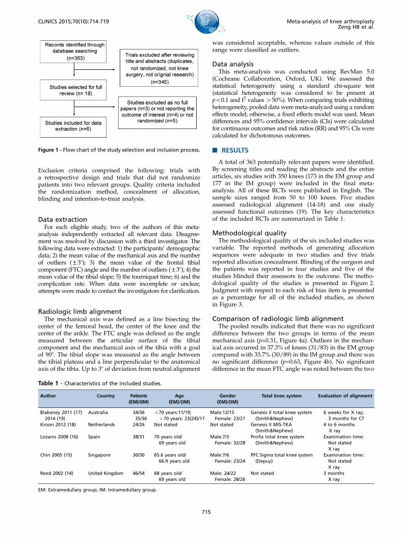
Exclusion criteria comprised the following: trials witha retrospective design and trials that did not randomizepatients into two relevant groups. Quality criteria includedthe randomization method, concealment of allocation,blinding and intention-to-treat analysis.
Data extractionFor each eligible study, two of the authors of this meta-
analysis independently extracted all relevant data. Disagree-ment was resolved by discussion with a third investigator. Thefollowing data were extracted: 1) the participants’ demographicdata; 2) the mean value of the mechanical axis and the numberof outliers (±3o); 3) the mean value of the frontal tibialcomponent (FTC) angle and the number of outliers (±3o); 4) themean value of the tibial slope; 5) the tourniquet time; 6) and thecomplication rate. When data were incomplete or unclear,attempts were made to contact the investigators for clarification.
Radiologic limb alignmentThe mechanical axis was defined as a line bisecting the
center of the femoral head, the center of the knee and thecenter of the ankle. The FTC angle was defined as the anglemeasured between the articular surface of the tibialcomponent and the mechanical axis of the tibia with a goalof 90o. The tibial slope was measured as the angle betweenthe tibial plateau and a line perpendicular to the anatomicalaxis of the tibia. Up to 3o of deviation from neutral alignment
was considered acceptable, whereas values outside of thisrange were classified as outliers.
Data analysisThis meta-analysis was conducted using RevMan 5.0
(Cochrane Collaboration, Oxford, UK). We assessed thestatistical heterogeneity using a standard chi-square test(statistical heterogeneity was considered to be present atpo0.1 and I2 values 450%). When comparing trials exhibitingheterogeneity, pooled data were meta-analyzed using a randomeffects model; otherwise, a fixed effects model was used. Meandifferences and 95% confidence intervals (CIs) were calculatedfor continuous outcomes and risk ratios (RR) and 95% CIs werecalculated for dichotomous outcomes.
’ RESULTS
A total of 363 potentially relevant papers were identified.By screening titles and reading the abstracts and the entirearticles, six studies with 350 knees (173 in the EM group and177 in the IM group) were included in the final meta-analysis. All of these RCTs were published in English. Thesample sizes ranged from 50 to 100 knees. Five studiesassessed radiological alignment (14-18) and one studyassessed functional outcomes (19). The key characteristicsof the included RCTs are summarized in Table 1.
Methodological qualityThe methodological quality of the six included studies was
variable. The reported methods of generating allocationsequences were adequate in two studies and five trialsreported allocation concealment. Blinding of the surgeon andthe patients was reported in four studies and five of thestudies blinded their assessors to the outcome. The metho-dological quality of the studies is presented in Figure 2.Judgment with respect to each risk of bias item is presentedas a percentage for all of the included studies, as shownin Figure 3.
Comparison of radiologic limb alignmentThe pooled results indicated that there was no significant
difference between the two groups in terms of the meanmechanical axis (p=0.31, Figure 4a). Outliers in the mechan-ical axis occurred in 37.3% of knees (31/83) in the EM groupcompared with 33.7% (30/89) in the IM group and there wasno significant difference (p=0.63, Figure 4b). No significantdifference in the mean FTC angle was noted between the two
Figure 1 - Flow chart of the study selection and inclusion process.
Table 1 - Characteristics of the included studies.
Author Country Patients(EM)/(IM)
Age(EM)/(IM)
Gender(EM)/(IM)
Total knee system Evaluation of alignment
Blakeney 2011 (17)2014 (19)
Australia 34/3635/36
o70 years:11/19;470 years: 23(24)/17
Male:12/15Female: 23/21
Genesis II total knee system(Smith&Nephew)
6 weeks for X ray;3 months for CT
Kroon 2012 (18) Netherlands 24/26 Not stated Not stated Genesis II MIS-TKA(Smith&Nephew)
4 to 6 monthsX ray
Lozano 2008 (16) Spain 38/31 70 years old/69 years old
Male:7/3Female: 32/28
Profix total knee system(Smith&Nephew)
Examination time:Not statedX ray
Chin 2005 (15) Singapore 30/30 65.6 years old/66.9 years old
Male:7/6Female: 23/24
PFC Sigma total knee system(Depuy)
Examination time:Not statedX ray
Reed 2002 (14) United Kingdom 46/54 68 years old/69 years old
Male: 24/22Female: 28/26
Not stated 3 monthsX ray
EM: Extramedullary group; IM: Intramedullary group.
715
CLINICS 2015;70(10):714-719 Meta-analysis of knee arthroplastyZeng HB et al.

groups (p=0.60, Figure 4c). The outliers in the FTC angleoccurred in 26.3% of knees (20/76) in the EM groupcompared with 25% (21/84) in the IM group and there wasno significant difference (p=0.89, Figure 4d). There was nosignificant difference between the two groups in terms of themean tibial slope (p=0.42, Figure 4e).
Comparison of the tourniquet time and thecomplication rate
The tourniquet time was shorter in the IM group comparedwith the EM group (p=0.01, Figure 5a). TKA-related complica-tions occurred in 4.2% of knees (3/72) in the EM groupcompared with 3.0% (2/67) in the IM group with no significantdifference (p=0.70, Figure 5b). Blakeney et al. (17) stated thatthere was one case of pulmonary embolism, one deep infectionand one case of knee stiffness in the EM group and one case ofknee stiffness in the IM group. Chin et al. (15) reported that onepatient in the IM group had a mild stroke. The functional scoreswere evaluated, although insufficient data were available forthe meta-analysis. Only one study measured functional kneescores (Oxford knee score) with mean scores of 37.6 in the EMgroup and 36.8 in the IM group.
’ DISCUSSION
Our meta-analysis compared the radiographic outcomesbetween the EM and the IM guiding techniques in patientsundergoing TKA. No significant differences were foundbetween the two groups in terms of the mean values of themechanical axis, the FTC angle or the tibial slope. Moreover,neither the EM nor the IM guiding techniques offer anadvantage over the other method in reducing outliers ofgreater than 3o. The IM guide is associated with a shortertourniquet time and exhibits a similar complication rate asthe EM guide. A comparison of functional outcomes betweenthe two groups could not be performed in this study becauseRCT research on functional outcomes is scarce.
A previous study of British orthopedic surgeons foundthat 75.6% prefer EM and 20.3% prefer IM jigs whendetermining tibial alignment with the remainder using bothor neither (20). The published literature is divided as towhich jig is superior. According to the results of ourliterature review, approximately 52.6% studies argue thatIM and EM guides are equally accurate for tibial alignment
Figure 2 - Methodological quality of the included studies. Thisrisk of bias tool incorporates assessment of randomization(sequence generation and allocation concealment), blinding(participants, personnel and outcome assessors), completenessof outcome data, selection of outcomes reported and othersources of bias. The items were scored with ‘‘yes’’, ‘‘no’’, or‘‘unsure’’.
Figure 3 - Risk of bias. Each risk of bias item is presented as a percentage across all included studies and indicates the proportional levelfor each risk of bias item.
716
Meta-analysis of knee arthroplastyZeng HB et al.
CLINICS 2015;70(10):714-719
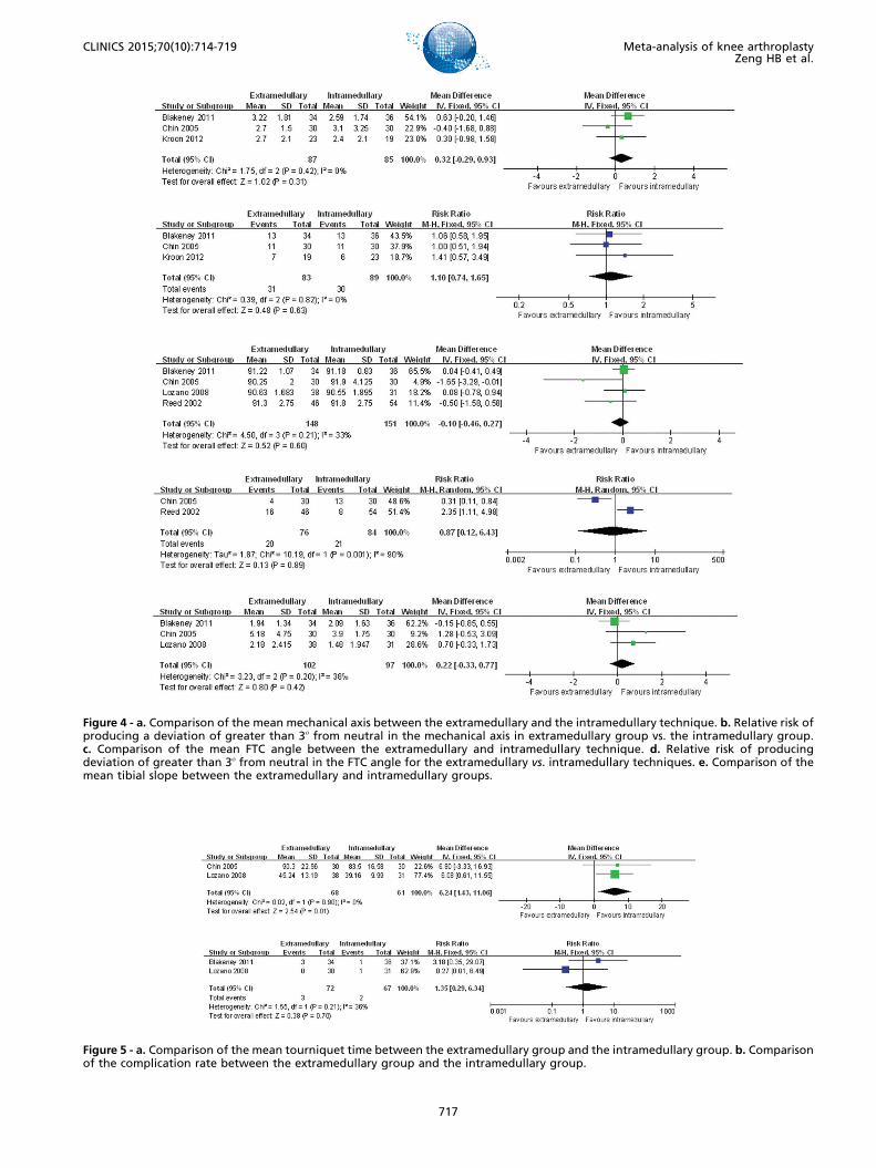
Figure 4 - a. Comparison of the mean mechanical axis between the extramedullary and the intramedullary technique. b. Relative risk ofproducing a deviation of greater than 3o from neutral in the mechanical axis in extramedullary group vs. the intramedullary group.c. Comparison of the mean FTC angle between the extramedullary and intramedullary technique. d. Relative risk of producingdeviation of greater than 3o from neutral in the FTC angle for the extramedullary vs. intramedullary techniques. e. Comparison of themean tibial slope between the extramedullary and intramedullary groups.
Figure 5 - a. Comparison of the mean tourniquet time between the extramedullary group and the intramedullary group. b. Comparisonof the complication rate between the extramedullary group and the intramedullary group.
717
CLINICS 2015;70(10):714-719 Meta-analysis of knee arthroplastyZeng HB et al.
(12,16-18,21-26). Approximately 36.8% suggest that an IMguide is more accurate (13,14,27-30,34) and 10.5% suggestthat an extramedullary guide is more accurate (31,32).However, very few studies included large samples or RCTsfor a comparison of the two methods. Regarding theaccuracy of the tibial cutting, this current meta-analysisstudy suggests that neither EM nor IM tibial alignment ismore accurate than the other approach.Most surgeons prefer to use the extramedullary guide either
because they are more experienced in its use or because of thepossible complications of the IM guide. However, because thecenter of the talus is slightly medial to the midpoint betweenthe malleoli, the surgeon must estimate the location of thecenter of the talus based on these bony landmarks, which maybe obscured by soft tissue in obese patients or by bonyabnormalities (33). For IM guides, the entry point position is akey factor and the ideal entry point position is located on thetibial articular surface corresponding to the proximal con-tinuation of the tibial canal and should be preoperativelydetermined with the help of X-rays (34). Moreover, decom-pression of the medullary contents using suction beforeinstrumenting the canal is also recommended to decreasethe risk of embolizing the medullary contents (35). Therefore,it is important for the surgeon to appreciate the benefits anddeficiencies of each guide and to use whichever method ismost appropriate for each particular case, although both theEM and IM systems allow for satisfactory alignment.Most surgeons have accepted that a postoperative mechan-
ical axis of 0o±3o will result in less pain, betterknee function, faster rehabilitation and improved quality oflife (36-39). Recently, several studies found that a post-operative mechanical axis of 0o±3o did not result in betterlong-term survival of TKA implants compared with a groupof outliers (39-43). In one of the most influential studies,Parratte et al. (40) retrospectively reviewed the data of 398primary TKAs and found that a mechanical axis of 0o±3o didnot improve the rate of survival 15 years postoperatively. Thisresult implies that the accuracy of the mechanical axis likelyprovides limited value with regards to long-term durability. Inaddition, although computer-assisted TKA improves themechanical leg axis and component orientation comparedwith the conventional technique, there is currently no provenclinical benefit of this approach. Therefore, future research ontibial guiding techniques should not only assess radiologicalalignment but also consider functional outcomes.This present meta-analysis has several limitations. First,
only six studies were included and the sample size of theincluded studies was small, which might have affected ourresults. Second, most of the trials focused on short-termradiographic outcomes and only one RCT study evaluatedfunctional outcome. Therefore, we could not perform a validstatistical comparison of the functional outcomes betweenthe two groups. Therefore, further high-quality RCTs withlong-term follow-up should be designed to assess radio-graphic outcomes, knee function and implant survival rate.
’ ACKNOWLEDGMENTS
We thank Hu YZ and Pan XY for their comments and advice.
’ AUTHOR CONTRIBUTIONS
Zeng HB and Liu HX were responsible for the study design and themanuscript preparation. Chen GJ, Yang XQ and Lin DD collected and
analyzed the data. Liu HX and Zeng HB were the principal investigators ofthe study. Li ZJ and Ying XZ were responsible for the study design andmanuscript finalization. All authors read and approved the final manu-script. Zeng HB and Ying XZ contributed equally to this study.
’ REFERENCES
1. Gandhi R, Dhotar H, Razak F, Tso P, Davey JR, Mahomed NN. Predictingthe longer term outcomes of total knee arthroplasty. Knee. 2010;17(1):15-8, http://dx.doi.org/10.1016/j.knee.2009.06.003.
2. Gioe TJ, Sinner P, Mehle S, Ma W, Killeen KK. Excellent survival ofall-polyethylene tibial components in a community joint registry. ClinOrthop Relat Res. 2007;464:88-92.
3. Julin J, Jamsen E, Puolakka T, Konttinen YT, Moilanen T. Younger ageincreases the risk of early prosthesis failure following primary total kneereplacement for osteoarthritis: a follow-up study of 32,019 total kneereplacements in the Finnish arthroplasty register. Acta Orthop. 2010;81(4):413-9, http://dx.doi.org/10.3109/17453674.2010.501747.
4. Spencer SJ, Baird K, Young D, Tait GR. The rotaglide mobile bearing kneearthroplast A 10 to13 year review from an independent centre. Knee.2012;19(1):20-3, http://dx.doi.org/10.1016/j.knee.2010.11.013.
5. Sikorski JM. Alignment in total knee replacement. J Bone Joint Surg Br.2008;90(9):1121-7, http://dx.doi.org/10.1302/0301-620X.90B9.20793.
6. Ensini A, Catani F, Leardini A, Romagnoli M, Giannini S. Alignments andclinical results in conventional and navigated total knee arthroplasty. ClinOrthop Relat Res. 2007;457:156-62.
7. Berger RA, Crossett LS, Jacobs JJ, Rubash HE. Malrotation causing patello-femoral complications after total knee arthroplasty. Clin Orthop Relat Res.1998;356:144-53, http://dx.doi.org/10.1097/00003086-199811000-00021.
8. Jeffery RS, Morris RW, Denham RA. Coronal alignment after total kneereplacement. J Bone Joint Surg Br. 1991;73(5):709-14.
9. Fang DM, Ritter MA, Davis KE. Coronal alignment in total kneearthroplasty: just how important is it? J Arthroplasty. 2009;24(6):39-43,http://dx.doi.org/10.1016/j.arth.2009.04.034.
10. Incavo SJ, Wild JJ, Coughlin KM, Beynnon BD. Early revision for com-ponent malrotation in total knee arthroplasty. Clin Orthop Relat Res.2007;458:131-6.
11. Sharkey PF, Hozack WJ, Rothman RH, Shastri S, Jacoby SM. Why are totalknee arthroplasties failing today? Clin Orthop Relat Res. 2002;(404):7-13,http://dx.doi.org/10.1097/00003086-200211000-00003.
12. Rottman SJ, Dvorkin M, Gold D. Extramedullary versus intramedullarytibial alignment guides for total knee arthroplasty. Orthopedics 2005;28(12):1445-8.
13. Brys DA, Lombardi AV, Mallory TH, Vaughn BK. A comparison ofintramedullary and extramedullary alignment systems for tibial compo-nent placement in total knee arthroplasty. Clin Orthop. 1991;(263):175-9.
14. Reed MR, Bliss W, Sher JL, Emmerson KP, Jones SM, Partington PF.Extramedullary or intramedullary tibial alignment guides: a randomised,prospective trial of radiological alignment. J Bone Joint Surg Br 2002;84(6):858-60, http://dx.doi.org/10.1302/0301-620X.84B6.12702.
15. Chin PL, Yang KY, Yeo SJ, Lo NN. Randomized control trial comparingradiographic total knee arthroplasty implant placement using computernavigation versus conventional technique. J Arthroplasty. 2005;20(5):618-26, http://dx.doi.org/10.1016/j.arth.2005.04.004.
16. Lozano LM, Segur JM, Maculé F, Núñez M, Torner P, Castillo F, et al.Intramedullary versus extramedullary tibial cutting guide in severelyobese patients undergoing total knee replacement: a randomized study of70 patients with body mass index 435 kg/m2. Obes Surg. 2008;18(12):1599-604, http://dx.doi.org/10.1007/s11695-008-9564-1.
17. Blakeney WG, Khan RJK, Wall SJ. Computer-assisted techniques versusconventional guides for component alignment in total knee arthroplasty: arandomized controlled trial. J Bone Joint Surg Am. 2011;93(15):1377-84,http://dx.doi.org/10.2106/JBJS.I.01321.
18. Kroon KE, Houterman S, Janssen RP. Leg alignment and tibial slope afterminimal invasive total knee arthroplasty: a prospective, randomizedradiological study of intramedullary versus extramedullary tibial instru-mentation. The Knee. 2012;19(4):270-4, http://dx.doi.org/10.1016/j.knee.2011.04.007.
19. Blakeney WG, Khan RJK, Palmer JL. Functional outcomes followingtotal knee arthroplasty: A randomised trial comparing computer-assistedsurgery with conventional techniques. The Knee 2014;21(2):364-8,http://dx.doi.org/10.1016/j.knee.2013.04.001.
20. Phillips AM, Goddard NJ, Tomlinson JE. Current techniques in total kneereplacement: results of a national survey. Ann R Coll Surg Engl. 1996;78(6):515-20.
21. Confalonieri N, Manzotti A, Pullen C, Ragone V. Computer-assisted techniqueversus intramedullary and extramedullary alignment systems in total kneereplacement: a radiological comparison. Acta Orthop Belg. 2005;71(6):703-9.
22. Yang SH, Liu TK. Intramedullary versus extramedullary tibial align-ment guides in total knee arthroplasty. J Formos Med Assoc. 1998;97(8):564-8.
718
Meta-analysis of knee arthroplastyZeng HB et al.
CLINICS 2015;70(10):714-719
23. Ishii Y, Ohmori G, Bechtold JE, Gustilo RB. Extramedullary versusintramedullary alignment guides in total knee arthroplasty. Clin OrthopRelat Res. 1995;318:167-75.
24. Tillett ED, Engh GA, Petersen TA. Comparative study of extramedullaryand intramedullary alignment systems in total knee arthroplasty. ClinOrthop Relat Res. 1988;230:176-81.
25. Teter KE, Bregman D, Colwell CW. Accuracy of intramedullary versusextramedullary tibial alignment cutting systems in total knee arthroplasty.Clin Orthop Relat Res. 1995;321:106-10.
26. Mihalko WM, Krackow K. Differences between extramedullary, intra-medullary, and computer-aided surgery tibial alignment techniques fortotal knee arthroplasty. J Knee Surg. 2006;19(1):33-6.
27. Cashman JP, Carty FL, Synnott K, Kenny PJ. Intramedullary versusextramedullary alignment of the tibial component in the Triathlon knee.J Orthop Surg Res. 2011;6:44, http://dx.doi.org/10.1186/1749-799X-6-44.
28. Laskin RS. Intramedullary instrumentation: safer and more accurate thanextramedullary instrumentation. Orthopedics. 2001;24(8):739.
29. Maestro A, Harwin SF, Sandoval MG, Vaquero DH, Murcia A. Influence ofintramedullary versus extramedullary alignment guides on final total kneearthroplasty component position: a radiographic analysis. J Arthroplasty.1998;13(5):552-8, http://dx.doi.org/10.1016/S0883-5403(98)90055-9.
30. Engh GA, Petersen TL. Comparative experience with intramedullary andextramedullary alignment in total knee arthroplasty. J Arthroplasty. 1990;5(1):1-8, http://dx.doi.org/10.1016/S0883-5403(06)80002-1.
31. Jessup DE, Worland RL, Clelland C, Arredondo J. Restoration of limbalignment in total knee arthroplasty: evaluation and methods. J SouthOrthop Assoc. 1997;6(1):37-47.
32. Dennis DA, ChannerM, Susman MH, Stringer EA. Intramedullary versusextramedullary tibial alignment systems in total knee arthroplasty. J Arthro-plasty. 1993;8(1):43-7, http://dx.doi.org/10.1016/S0883-5403(06)80106-3.
33. Siston RA, Daub AC, Giori NJ, Goodman SB, Delp SL. Evaluation of methodsthat locate the center of the ankle for computer-assisted total knee arthro-plasty. Clin Orthop Relat Res. 2005;439:129-35, http://dx.doi.org/10.1097/01.blo.0000170873.88306.56.
34. Karade V, Ravi1 B, Agarwal M. Extramedullary versus intramedullarytibial cutting guides in megaprosthetic total knee replacement. J OrthopSurg Res. 2012;7:33, http://dx.doi.org/10.1186/1749-799X-7-33.
35. Amro RR, Nazarian DG, Norris RB, Kelly MP, Booth RE Jr. Suctioninstrumentation decreases intramedullary pressure, pulmonary embo-lism during total knee arthroplasty. Univ Penn Orthop J. 2001;14:55–59.
36. Czurda T, Fennema P, Baumgartner M, Ritschl P. The associationbetween component malalignment and post-operative pain followingnavigation-assisted total knee arthroplasty: results of a cohort/nestedcase-control study. Knee Surg Sports Traumatol Arthrosc. 2010;18(7):863-9,http://dx.doi.org/10.1007/s00167-009-0990-y.
37. Nicoll D, Rowley DI. Internal rotational error of the tibial component is amajor cause of pain after total knee replacement. J Bone Joint Surg Br.2010;92(9).
38. Choong PF, Dowsey MM, Stoney JD. Does accurate anatomical alignmentresult in better function and quality of life? A prospective randomizedcontrolled trial comparing conventional and computer-assisted total kneearthroplasty. J Arthroplasty. 2009;24(4):560-9, http://dx.doi.org/10.1016/j.arth.2008.02.018.
39. Longstaff LM, Sloan K, Stamp N, Scaddan M, Beaver R. Good alignmentafter total knee arthroplasty leads to faster rehabilitation and betterfunction. J Arthroplasty. 2009;24(4):570-8, http://dx.doi.org/10.1016/j.arth.2008.03.002.
40. Parratte S, Pagnano MW, Trousdale RT, Berry DJ. Effect of postoperativemechanical axis alignment on the fifteen-year survival of modern,cemented total knee replacements. J Bone Joint Surg Am. 2010;92(12):2143-9, http://dx.doi.org/10.2106/JBJS.I.01398.
41. Matziolis G, Krocker D, Weiss U, Tohtz S, Perka C. A prospective, rando-mized study of computer-assisted and conventional total knee arthroplasty.Three-dimensional evaluation of implant alignment and rotation. J Bone JointSurg Am. 2007;89(2):236-43, http://dx.doi.org/10.2106/JBJS.F.00386.
42. Bonner TJ, Eardley WG, Patterson P, Gregg PJ. The effect of post-operativemechanical axis alignment on the survival of primary total knee repla-cements after a follow-up of 15 years. J Bone Joint Surg Br. 2011;93(9):1217-22, http://dx.doi.org/10.1302/0301-620X.93B9.26573.
43. Morgan SS, Bonshahi A, Pradhan N, Gregory A, Gambhir A, Porter ML. Theinfluence of postoperative coronal alignment on revision surgery in totalknee arthroplasty. Int Orthop. 2008 ;32(5):639-42, http://dx.doi.org/10.1007/s00264-007-0391-0.
719
CLINICS 2015;70(10):714-719 Meta-analysis of knee arthroplastyZeng HB et al.


![Intradural-Extramedullary and Intramedullary Spinal ... · [7–9]. In this regard, the spine is the most common site for bony metastases [7]. The incidence of spinal metastases is](https://img.pdfslide.net/doc/110x75/5fcd7bfc64dc771fcc68cd0a/intradural-extramedullary-and-intramedullary-spinal-7a9-in-this-regard.jpg)