Embed Size (px)
Citation preview
2828International Journal of Scientific Study | November 2017 | Vol 5 | Issue 8
Correlation of Posterior Tibial Slope with Metaphysio-diaphyseal Angle in Total Knee Arthroplasty: A Radiological StudyJohney Juneja, Sa Mustafa, Rishi Gupta
Department of Orthopedic Index Medical College
The PTS of various populations has not been completely mapped out at present, but reports suggest a high range from 0° to 18° depending on age, gender, race, genetics, and various diseases and morbidities.[1,2] The PTS of the tibia has been assessed using various methods ranging from direct cadaveric measurements and radiographs to computed tomography (CT) scans and magnetic resonance imaging (MRI).[3]
The patients require an average flexion of 67° for swing phase of gait, 83° for climbing stairs, 90° for descending stairs, and 93° to rise from a seated position.[4,5] In the eastern hemisphere and in the Indian subcontinent, flexion >105° is required for kneeling and squatting during activities of daily living and religious activities.[6]
Among the numerous factors affecting the flexion in the knee, PTS is important, which decides about the sagittal alignment of the tibia.[3] Studies done in the Chinese, Japanese, and Pakistani populations (Asians) have documented a difference in the PTS in their respective
INTRODUCTION
The proximal tibial end is in the form of a plateau with which the femur bone articulates and forms the knee joint. The tibial plateau is not exactly flat but has a slope toward the posterior direction. This posterior inclination of the tibial plateau relative to the longitudinal axis of the tibia is referred to as the posterior tibial slope (PTS).
The PTS plays a very important role in the kinematics and biomechanics of the knee joint. It has been shown that increasing PTS increases the tibial shear force and anterior tibial translation at the knee.[1]
Original Article
AbstractBackground: Posterior tibial slope (PTS) is an important factor affecting post-operative range of motion following total knee arthroplasty (TKA). Metaphysio-diaphyseal angle (MDA) is a new entity defined as the angle between proximal anatomical axis and metaphyseal axis of tibia. This study was undertaken to determine PTS in Indian patients and find its correlation with MDA of tibia. The accuracy of extramedullary jigs and the influence of MDA on the accuracy was also evaluated.
Materials and Methods: Data of 100 consecutive patients undergoing TKA in a single center by a single surgeon were analyzed. Posterior cruciate ligament substituting TKA was done with the same jig to achieve different PTS in different patients. MDA, pre-operative PTS, and post-operative PTS were calculated. The data were analyzed using appropriate statistical analysis.
Results: Mean pre-operative PTS was 11.64° and mean MDA was 23.76° with a strong correlation between them (Pearson’s coefficient 0.72). Extramedullary jigs were accurate in 53% of cases. In remaining 47%, post-operative PTS was less than planned PTS in 30%, and more in 17%. Mean post-operative PTS was 2.54°. In patients with MDA < 20°, post-operative PTS was significantly less (P = 0.0176) compared with those with MDA > 20°.
Conclusions: The study establishes the positive correlation between MDA and PTS in Indians; and that MDA is an independent factor affecting the accuracy of extramedullary jigs in TKA.
Key words: Arthroplasty, Metaphysio-diaphyseal angle, Posterior tibial slope
Access this article online
www.ijss-sn.com
Month of Submission : 09-2017 Month of Peer Review : 10-2017 Month of Acceptance : 10-2017 Month of Publishing : 11-2017
Corresponding Author: Johney Juneja, Department of Orthopedic of Index Medical College. E-mail: [email protected]
Print ISSN: 2321-6379Online ISSN: 2321-595X
DOI: 10.17354/ijss/2017/516

Juneja, et al.: Posterior Tibial Slope with Metaphysio-diaphyseal Angle in Total Knee Arthroplasty
2929 International Journal of Scientific Study | November 2017 | Vol 5 | Issue 8
population from the Caucasians, which is responsible for differences in the range of flexion in these populations.[7-9]
Osteoarthritis leads to decrease in range of motion (ROM), one of the reasons due to altered PTS. Total knee arthroplasty (TKA) is one of the most successful operations, done to achieve improved ROM and pain relief with the attainment of optimum PTS, with an average survival of more than 90% at 15 years.[10,11] In TKA, for the achievement of the desired PTS and thus increased ROM, tibial cut is made with the help of extramedullary jig which is based on the anterior tibial line as a reference.
Metaphysio-diaphyseal angle (MDA) is a new concept, and its role in affecting the TKA is under study and research.
The present study is undertaken to study the PTS in patients undergoing knee joint arthroplasty and to study the role of MDA in achieving desired PTS in TKA using extramedullary jig.
Aims and ObjectivesThe aims and objectives of the present study are as follows:1. To find the mean PTS and MDA in patients planned
for total knee replacement.2. To find a correlation between desired PTS, the MDA,
and post-operative PTS achieved.
REVIEW OF LITERATURE
Whereas the normal PTS has been quoted as 5–10°, racial differences in the PTS have been found. PTS is an important factor affecting post-operative ROM following TKA.
There is a significant correlation between the PTS and the MDA directly.[7] Thus, for achieving the desired PTS, MDA should be considered during the planning of a total knee replacement of knee.
Hofmann et al.[12] found that tibiae cut parallel to the surface exhibited 40% greater load carrying capacity and 70% greater stiffness than paired tibiae cut perpendicular to the long axis.
Bartel et al.[13] suggested that, when the stiffest and strongest cortical bone is removed, the remaining weaker and less stiff cancellous bone stock often is inadequate to support the physiologic loads of the knee.
Chiu et al.[8] studied the posterior slope of the tibial plateau in 25 pairs of Chinese cadaveric tibia and found a good agreement between the PTS measured on radiographs and the actual tibial slope measured on cadaveric tibia.
With radiographic measurement, the posterior slope was 11.5° using intramedullary method, and it was 14.7° using extramedullary method and also concluded that osteoarthritis increases the slope by 2–3°.
Bai et al.[14] noted that anteriorly sloped tibial components led to a tendency to posterior micromotion and thus more wear of implant in TKA.
Turgut et al. and Waelchli et al.[15,16] noted that the posterior slope of the tibial cut affects anteroposterior stability, ROM, and contact pressure within the tibiofemoral joint.
Yoga et al.[17] found that the posterior slope of the tibial plateau is an important feature to preserve during knee replacement. The correct slope aids in the amount of flexion and determines if the knee will be loose on flexion. This study on the posterior tibial plateau slope was based on pre- and post-operative radiographs of 100 consecutive patients who had total knee replacements. The average posterior slope of the tibial plateau was 10.1°. There is a tendency for patients with higher pre-operative posterior tibial plateau slope to have higher post-operative posterior tibial plate slope.
Kettelkamp DB et al.[4] (2010) in a study on 40 males and 19 females in Pakistan found that the PTS was greater in women than men (14.1° vs. 12.5°, P = 0.02) and was greater than the range of 5–10° commonly reported in western literature. They concluded that the greater PTS in Pakistanis suggests that a proximal tibial cut with a greater PTS may reduce the chance of tibial loosening and increase post-operative knee ROM, especially when using posterior cruciate ligament (PCL)-retaining designs.
Yoo et al.[6] quoted that there was no consensus on an ideal anatomical reference to determine the posterior slope of tibia plateau. Posterior slope of the medial tibia plateau was measured with reference to a proposed mechanical axis (MA) and 5 clinically relevant anatomical references in 90 osteoarthritic knees of 66 female patients undergoing TKA. The MA was defined as the line connecting the midpoints of the medial tibia plateau and the tibial plafond, and 5 anatomical references included the anterior cortical line of tibia,anatomical axis of proximal and central tibia, posterior cortical line of proximal tibia, and fibular shaft axis (FSA). The average posterior slope was 10.6° with reference to the MA, and the amount of posterior slope varied widely among the patients and depending on the anatomical reference used to measure. This study indicated that the anatomical reference used to measure the posterior slope should be identified in studies where posterior slope is used to evaluate the sagittal alignment of TKA.

Juneja, et al.: Posterior Tibial Slope with Metaphysio-diaphyseal Angle in Total Knee Arthroplasty
3030International Journal of Scientific Study | November 2017 | Vol 5 | Issue 8
Didia and Jaja[18] performed a radiological study on 212 lateral tibial radiographs of Nigerian population measuring PTS with reference to the anterior tibial cortex and found a mean PTS of 12.3°.
Mohanty et al.[7] in a study of 100 cases concluded a positive correlation between MDA and PTS; and that MDA is an independent factor affecting the accuracy of extramedullary jigs in TKA.
Dargel et al.[19] studied 60 human cadaver knees and concluded that male linear knee joint dimensions were significantly larger when compared with females but the difference was not significant, and hence, there was no need for gender-specific implant design or technique to be employed in TKA.
Bek[20] concluded that, although different extramedullary tibial cutting guides with and without a spike can reproducibly impart a desired PTS in TKA, the spiked guide was user-friendly.
It appears that the MDA determines the MA in the sagittal plane because it brings the center of the knee backward. Therefore, it is susceptible to increase the difference between the anatomical axis obtained from an extramedullary jig (aligned on the diaphysis and the anterior cortical line) and the MA: The greater the MDA, the greater the difference between the diaphyseal axis and the MA in the sagittal plane.
MATERIAL AND METHODS
This study was conducted in the Department of Orthopaedics, Index hospital, Indore, Madhya Pradesh, India. Permission was obtained from the departmental scientific committee and the Institutional Ethical Committee at the beginning of the study.
The study duration was from August 2015 to September 2017. The sample size for the study was of “30 knees” as discussed by the statistician.
The Inclusion Criteria were as Follows1. All patients who underwent primary TKA during the
period of the study, Index hospital, Indore.2. Use of extramedullary spiked jig to cut tibia
intraoperatively.
The Exclusion Criteria were as Follows1. Patients not willing to be a part of the study.2. Patients with complex primary total knee replacement
ROM <50°, angular deformity >10°, FFD >30°, and associated neurovascular disease.
3. Patients with BMI >40.
4. Revision TKA.5. Patients previously operated around knee joint.6. Patients with extra-articular deformity.7. Bone defects requiring build on tibia.
The patients for planned TKA were admitted a few days before surgery. Patients were given informed about the study through the “patient information document” in their own language (Hindi/English). Those who gave a valid written consent were enrolled in the study. Pre-operative assessment as per the pre-decided detailed pro forma was done which will include history taking, examination, investigations, and X-rays lateral and anteroposterior views.
Full weight bearing standard radiographs were taken in both anteroposterior and lateral views not more than 1 week before the date of replacement surgery. Lateral views were taken with both the tibia and femoral condyles overlapping with at least 15 cm of the shaft of tibia visible for calculation of the anatomical axes.
Pre-operative PTS and MDA were calculated as mentioned as follows. All the measurements were carried out manually by the principal investigator under the supervision of the guide and coguide of this study, using radiographs, goniometer, and markers.
Pre-operative PTS [Figure 1] was defined as the angle formed by two lines in the lateral radiograph. The first line was the line perpendicular to the anatomical axis of the proximal tibia proximal tibial anatomical axis (PTAA). The second line formed by joining the most proximal points on the tibial plateau on the lateral radiograph as defined by Massin et al.,[21] avoiding osteophytes.
The PTAA, i.e., the line connecting midpoints of outer cortical diameter at 5 and 15 cm distal to the knee joint (Yoo et al.), is now recommended because it is most parallel to the sagittal MA. Hence, this axis was assumed to be the tibial anatomical axis in our study.
MDAMDA [Figure 1] is angle which is formed between two lines: The first line is the proximal anatomical axis of the tibia which is drawn by connecting midpoints of outer cortical diameter of tibia at 5 and 15 cm distal to the knee joint. The second is the axis of the metaphysis, drawn by defining two points each on anterior and posterior cortices of tibial metaphysis in a lateral film and joining the midpoints. Finally, the angle between these two lines is MDA.
The patients were operated by their respective surgeon using PCL substituting design of implant using spiked extramedullary tibial jig on tibial plateauf [Figures 2 and 3].

Juneja, et al.: Posterior Tibial Slope with Metaphysio-diaphyseal Angle in Total Knee Arthroplasty
3131 International Journal of Scientific Study | November 2017 | Vol 5 | Issue 8
All patients underwent total knee replacement under tourniquet under spinal anesthesia in the supine position
by mid parapatellar approach. After sterile painting and drapping, midline linear anterior incision was made and anteromedial arthrotomy done. Medial release is done, and patella was everted. Menisci were cut and removed. The external jig had an inbuilt slope of 3°.
The jig was aligned by the surgeon as per his judgment to achieve the optimum post-operative PTS. As the general guideline followed in our institute, the desired post-operative PTS was in a range from 0° to 7°. The surgical team was blind for the pre-operative PTS and MDA of the patient calculated for this study.
Primary tibial cut is made with desired PTS, and primary femoral portal is made 1 cm above PCL attachment. Using 5° valgus, primary femoral cut made at 9 mm from distal articular surface. Extension gap is checked and measured. Femur is sized with anterior referencing jig and 3° external rotation marker. Using appropriate size of femoral cutting block, cuts, chamfers, and notch are made. Flexion and extension gap is checked with appropriate spacer. Tibia sizing done and keel is prepared. Flexion, extension, and mid flexion mediolateral stability is checked. Appropriate-sized implant fixation is done with cementing after trial. Closure is done in layers over a suction drain. Sterile compression dressing is applied. Physiotherapy is started at the suitable time.
Post-operative X-rays are recorded at day 2 and alignment of the implant is checked. Suture removal is done on post-operative day 14. The patients were followed in OPD at regular intervals as per routine protocol. X rays were done at each visit and post-operative PTS (post-operative PTS) calculated from lateral radiograph shot at post-operative day 2-week follow-up.
Post-operative PTSIn the post-operative radiograph, the PTS [Figure 5] was calculated by joining two lines. The first line was the line perpendicular to anatomical axis of tibia. The second line is formed by joining the two most proximal points anteriorly and posteriorly along the tibial component of the implant.
Final correlation between PTS, MDA, and post-operative PTS was calculated using appropriate statistical calculations.
ObservationsA total of 32 knees were enrolled in this study of which 2 knees were excluded as one required tibial build up and one knee underwent unicompartmental knee arthroplasty.
The remaining 30 knees belonged to 21 patients. 12 patients had total knee replacement of one knee only, whereas the remaining 9 patients underwent bilateral total knee replacement in Index hospital, Indore.
Figure 1: Calculation of pre-operative posterior tibial slope and metaphysio-diaphyseal angle
Figure 2: Extramedullary tibial cutting jig (Smith and Nephew, Genesis II)
Figure 3: Showing placement of spiked extramedullary tibial jig on tibial plateau
Juneja, et al.: Posterior Tibial Slope with Metaphysio-diaphyseal Angle in Total Knee Arthroplasty
3232International Journal of Scientific Study | November 2017 | Vol 5 | Issue 8
RESULTS
Age DistributionThe average age of the patients was 56 years (range 39–70 years).
Figure 4: Intraoperative alignment of extramedullary jig
Figure 5: Using bone saw on cutting guide to make tibial cut
Table 1: Pre-operative PTS, MDA, and post-operative PTS of case 1Pre-operative PTS 21°MDA 23°Post-operative PTS
15°
PTS: Posterior tibial slope, MDA: Metaphysio‑diaphyseal angle
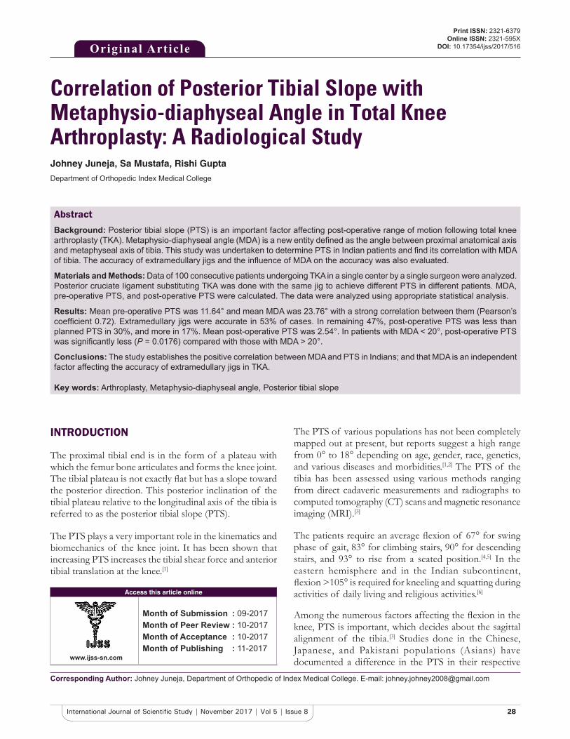
Table 2: Pre-operative PTS, MDA, and post-operative PTS of case 2Pre-operative PTS 14°MDA 27°Post-operative PTS 10°PTS: Posterior tibial slope, MDA: Metaphysio‑diaphyseal angle
Figure 6: Intraoperative alignment of extramedullary jig and anterior view
Figure 7: Calculation of post-operative posterior tibial slope
Figure 8: Pre-operative and post-operative X-rays of case 1 for calculation of angles
Gender Variation66% knees belonged to female patients, whereas remaining 10 knees were of male patients.
Juneja, et al.: Posterior Tibial Slope with Metaphysio-diaphyseal Angle in Total Knee Arthroplasty
3333 International Journal of Scientific Study | November 2017 | Vol 5 | Issue 8
The mean PTS for male knees (n = 10 ) was 13.3° (9°–21° ), and the mean MDA was 19.5° (16°–25°). The
Figure 9: Post-operative clinical pictures of case 1 at post-operative day 2 weeks
Figure 10: Post-operative clinical pictures of case 1 at post-operative day 2 weeks (lateral view)
Figure 11: Pre-operative and post-operative X-rays of Case 2 for calculation of angles
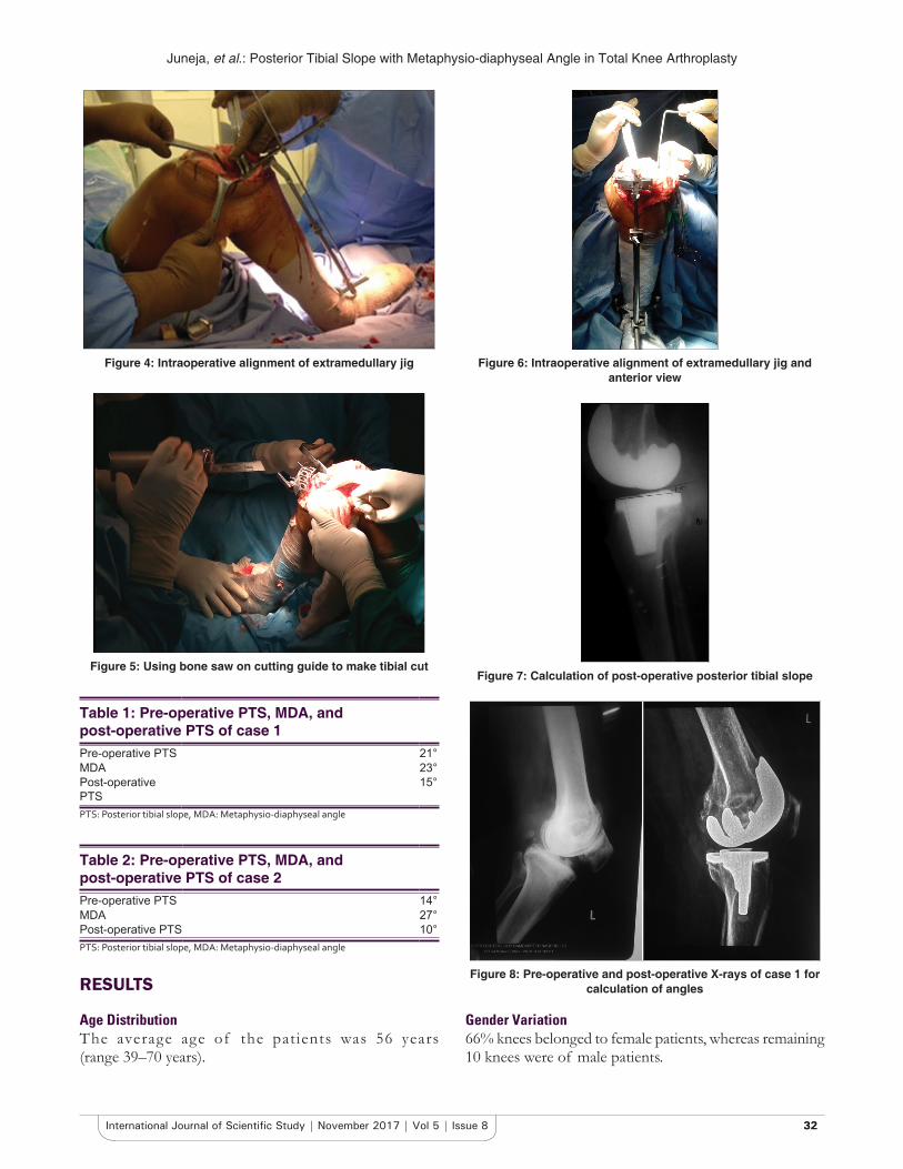
Figure 12: Pre-operative and post-operative X-rays of Case 3 for calculation of angles
Figure 13: Technique of tilting jig to adjust slope of tibial cut by extramedullary jig during total knee arthroplasty
Table 3: Pre-operative PTS, MDA, and post-operative PTS of case 3Pre-operative PTS 14 o
MDA 17 o
Post-operative PTS 6 o
PTS: Posterior tibial slope, MDA: Metaphysio‑diaphyseal angle
Table 4: Gender variation in pre-operative PTS and MDAGender of patient Number Mean pre-operative PTS Mean MDAMale 10 13.3 19.5Female 20 13.45 20.45PTS: Posterior tibial slope, MDA: Metaphysio‑diaphyseal angle
Table 5: Side variation in pre-operative PTS and MDASide of knee Number of cases Mean pre-operative
PTSMean MDA
Right 11 12.54 20.27Left 19 13.89 20.05PTS: Posterior tibial slope, MDA: Metaphysio‑diaphyseal angle

Juneja, et al.: Posterior Tibial Slope with Metaphysio-diaphyseal Angle in Total Knee Arthroplasty
3434International Journal of Scientific Study | November 2017 | Vol 5 | Issue 8
mean pre-operative PTS for female patients was 13.45 (6–19), and the mean MDA was 20.45° (11°–27°).
Laterality63.33% knees were left sided (n = 19), while the reaming were 11 knees were right sided (36.66%).
The mean PTS for right-sided knees (n = 11) was 12.54° (8°–18° ), and the mean MDA was 20.27° (11°–27°). The mean PTS for left-sided knees (n = 19) was 13.89° (6°–21°), and the mean MDA was 20.05° (15°–25°).
Pre-operative PTS and MDAThe mean pre-operative PTS of the study sample was 13.40° with a standard deviation of 3.67°. The mean MDA
Table 6: Pre-operative PTS, MDA, and post-operative PTS of the study sampleVariables Range Mean±SDPre-operative PTS 6–21 13.40±3.67MDA 11–27 20.13±3.19Post-operative PTS 1–17 9.6±3.79PTS: Posterior tibial slope, MDA: Metaphysio‑diaphyseal angle, SD: Standard deviation
Table 7: Mean of pre-operative PTS, MDA, and post-operative PTS of Group A and BVariables Group A Group BNumber of cases 14 16Mean pre-operative PTS 11.06 16.07Mean MDA 17 23.71Mean post-operative PTS 7.86 12.57PTS: Posterior tibial slope, MDA: Metaphysio‑diaphyseal angle
Graph 1: Pie chart showing knees as part of unilateral or bilateral replacement surgery
Graph 2: Age distribution of the study sample
Graph 3: Sex ratio of the study sample
Graph 4: Range and mean of pre-operative posterior tibial slope (PTS), metaphysio-diaphyseal angle, and post-operative
PTS
Graph 5: Various angles arranged in increasing order of metaphysio-diaphyseal angle
Juneja, et al.: Posterior Tibial Slope with Metaphysio-diaphyseal Angle in Total Knee Arthroplasty
3535 International Journal of Scientific Study | November 2017 | Vol 5 | Issue 8
was of the study sample was 20.13° with standard deviation of 3.92°. The mean post-operative PTS was 12.86° with a standard deviation of 5.82°.
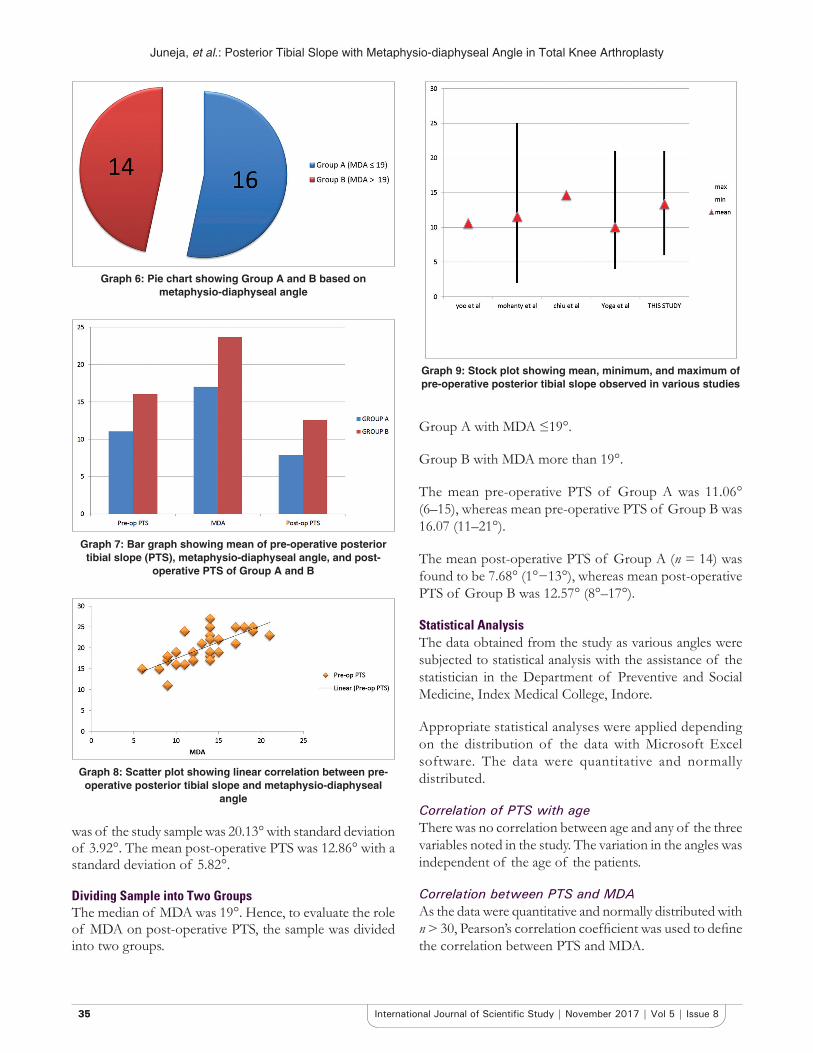
Dividing Sample into Two GroupsThe median of MDA was 19°. Hence, to evaluate the role of MDA on post-operative PTS, the sample was divided into two groups.
Group A with MDA ≤19°.
Group B with MDA more than 19°.
The mean pre-operative PTS of Group A was 11.06° (6–15), whereas mean pre-operative PTS of Group B was 16.07 (11–21°).
The mean post-operative PTS of Group A (n = 14) was found to be 7.68° (1°−13°), whereas mean post-operative PTS of Group B was 12.57° (8°–17°).
Statistical AnalysisThe data obtained from the study as various angles were subjected to statistical analysis with the assistance of the statistician in the Department of Preventive and Social Medicine, Index Medical College, Indore.
Appropriate statistical analyses were applied depending on the distribution of the data with Microsoft Excel software. The data were quantitative and normally distributed.
Correlation of PTS with ageThere was no correlation between age and any of the three variables noted in the study. The variation in the angles was independent of the age of the patients.
Correlation between PTS and MDAAs the data were quantitative and normally distributed with n > 30, Pearson’s correlation coefficient was used to define the correlation between PTS and MDA.
Graph 6: Pie chart showing Group A and B based on metaphysio-diaphyseal angle
Graph 7: Bar graph showing mean of pre-operative posterior tibial slope (PTS), metaphysio-diaphyseal angle, and post-
operative PTS of Group A and B
Graph 8: Scatter plot showing linear correlation between pre-operative posterior tibial slope and metaphysio-diaphyseal
angle
Graph 9: Stock plot showing mean, minimum, and maximum of pre-operative posterior tibial slope observed in various studies
Juneja, et al.: Posterior Tibial Slope with Metaphysio-diaphyseal Angle in Total Knee Arthroplasty
3636International Journal of Scientific Study | November 2017 | Vol 5 | Issue 8
There is a strong positive correlation between pre-operative PTS and MDA with Pearson’s coefficient of r = 0.7364.
Significance of difference of pre-operative PTS and post-operative PTS in two groups of MDAFor comparing two groups defined on the basis of MDA, student’s unpaired t-test was used with an alpha level of 0.05 and beta 0.2, that is, power (1-beta) of 80%.
While comparing pre-operative PTS for Group A and Group B (via unpaired t-test), the t = −5.94479. The P < 0.00001, and hence, the result was satistically significant at P < 0.05. Furthermore, on comparing post-operative PTS for Group A and Group B (via unpaired t-test), the t = 4.33324. The P = 0.000098. The result is significant at P < 0.05.
Hence, the pre-operative PTS as well as post-operative PTS in the group A (MDA ≤19°) was significantly less than Group B (MDA >19°) (Figures 4, 6-13), (Graphs 1-9) and (Tables 1-7).
DISCUSSION
The PTS has been assessed using various methods, ranging from direct cadaveric measurements to CT scans and MRI. However, radiographic calculation is most often used in developing countries due to financial constraints. Even with radiographs, various methods of assessment have been described.
Brazier et al.[13] described the different anatomical axes against which the PTS could be measured. The lines included the tibial proximal anatomical axis (TPAA), tibial shaft anatomical axis (TSAA), posterior tibial cortex (PTC), anterior tibial cortex (ATC), fibular proximal anatomical axis (FPAA), and FSA. They concluded that, among the proximal axes, the TPAA and PTC gave higher reliability. The ideal way to measure the PTS is against the anatomical axis of the tibia Shaft (TSAA). This will require a radiograph of the at least half of the tibia which is usually not taken routinely in clinical practice. The TPAA does not exactly match the MA of the tibia but gives a close correlation.
The normal values for the posterior tibial plateau slope have not been fully mapped out for all populations. However, what is reported show a high variability between races and ranges from 0˚ to 18˚. Whereas, the normal PTS has been quoted as 5-10°; racial differences in the PTS have been found.[6-8,10,11] In this study, we found the pre- operative PTS slope varied from 6 to 21 averaging 13.4°. These values are similar to the values reported in the literature by Mohanty et al.[10] in Indian population (11.64°), Khattak et al.[7] in Pakistani population (12.5° in males and 14.2° in females),
Yoo[9] in the Japanese population (10.6°), and Chiu et al.[8] in the Chinese population (13.1° in osteoarthritic knees and 10.8° in normal knees on radiographs).
Asian population has been found to have an increased PTS as compared with Caucasian population. Moreover, it has been found that the PTS increases with the onset of osteoarthritis.[5,13,14] At the same time, some other authors have reported lower PTSs in the Asian population.[13,22] These variations in the PTS reported in the same population may be due to the difference in the reference axis and the methodology used in the calculation of the PTS.
This study noted NO significant correlation between age of the patients and their knee’s pre-operative PTS. Furthermore, NO significant difference was noted for the side of the body to which the knee belonged.
As was noted in Yoga et al.[17] and a systematic review by Merchant et al.,[23] the slope was nearly equal in males and females in this study. This was in accordance to the study of 60 cadaveric knees by Dargel et al. where they observed that although the male dimensions of knee are larger than their female counterparts, when adjusted for patients’ height and femoral length and on matched pair analysis, there is no significant difference in the morphometric variables in male and female sex. However, Khattak et al.[4] and Didia and Jaja.[18] did find a significant difference in the PTSs between the two sexes.
It was postulated in this study that the MDA might affect the PTS and hence affect the overall sagittal alignment of the proximal tibia. It was also hypothesized that high MDAs could potentially lead surgeons to overestimate the PTS using extramedullary jigs and that it would require a substantial correction depending on the native MDA and PTS.
In this study, a strong Pearson’s coefficient of correlation of 0.737 was found between the pre-operative PTS and MDA which emphasizes the same school of thought. The previous studies have noted that MDA affects the MA in the sagittal plane because it brings the center of the knee backward.
The surgeons desired a post-operative PTS within 0°–7° in all cases. MDA had no effect on the desired PTS, the post-operative PTS of Group A and Group B should have been nearly same, and statistically, no significant difference should have been noted. However, the poster-operative slope of Group A and Group B was 7.86° and 12.57°, respectively, and a significant difference was noted among them (via unpaired t-test). This strongly suggests that, in patients with high MDA (>19° in this study), the achieved PTS is higher than desired PTS.

Juneja, et al.: Posterior Tibial Slope with Metaphysio-diaphyseal Angle in Total Knee Arthroplasty
3737 International Journal of Scientific Study | November 2017 | Vol 5 | Issue 8
It was noted that the MDA affects the accuracy of the extramedullary jig used for achieving the PTS intraoperatively. The variation in the achieved PTS could be explained by the altered proximal tibial anatomy due to different MDA. As per the study by Yoo et al., the anterior cortical line of the tibia can differ from the MA of tibia by as high as 6.2° (mean 3.2, standard deviation 1.3).[9]
Furthermore, it was noted that, for a given PTS, the error between the MA and the anterior cortical line is increased when the MDA increases and vice versa. Therefore, higher MDA makes it susceptible to increase the difference between the anatomical axis obtained from an extramedullary jig (aligned on the diaphysis and the anterior cortical line) and the MA: The greater the MDA, the greater the difference between the diaphyseal axis and the MA in the sagittal plane.[10]
Thus, the extramedullary jig, which is kept parallel to the anterior cortical line, will not compensate for any change in the MDA and hence will lead to a different tibial cut depending on the MDA. The surgeon, who bases himself on the diaphyseal axis, will overestimating the real tibial slope in tibias with a great MDA. This could even lead surgeons to invert the tibial slope in tibias with low native PTS.
Kansara et al. did a clinical study on 61 patients undergoing TKA. The tibial cutting guides accurately achieved the intended posterior slope, but increasing posterior slope from 0° to 5° did not result in a significant increase in the range of movement or Hospital for Special Surgery functional score. In addition, in some cases, attempting a 0° slope gave rise to a negative slope, which was not seen with a 5° intended slope.
Thus, MDA holds potential for a new value to be taken into consideration while planning for sagittal anatomy in TKA.
This study does have limitations. The sample was collected from the patients planned for TKA. Hence, the results of PTS does not apply to the general population. The sample size was small. MRI was not employed in this study, but in an MRI study done by Hashemi et al., the mean tibial slope of the lateral plateau was found to be higher than medial. For simplification, the two plateaus were assumed to be equal. The lateral radiographs do not take into account the slope contributed by the Menisci which will require an MRI. Single observer calculated the angles by manual technique using lateral radiographs and goniometer. Computer-assisted (navigation-assisted TKR) TKR was not available in the institution where the study was conducted. The functional correlation of MDA on
knee arthroplasty could not be correlated as very many other variables were not taken into account due to the limited scope of this study.
Studies have shown that the most accurate method to measure the PTS is CT scan.[12,14] Besides, medial tibial slope and lateral tibial slope vary in a single patient.[7,12,13] However, Chiu et al. found a good agreement between the PTS measured on radiographs and the actual tibial slope measured on the cadaveric tibia.[7] Other anatomical aspects of the knee such as the coronal plane alignment of tibia and its variations with the age, sex, and the onset of osteoarthritis, the mediolateral and anteroposterior dimensions of the tibia, and the correlation between these variables were not considered.
CONCLUSION
In the past few decades, the life expectancy of human beings has increased dramatically. This has caused more population opting for elective total knee replacement to reduce their knee joint-related morbidities.
In this study, we found that females undergoing total knee replacement have nearly the same slope as males. Furthermore, age had no significant correlation with variation in PTS of the patients.
The study highlights the ethnic differences in the PTS. It was noted that the pre-operative PTS in patients undergoing TKA in central India is around 13.4°, which is in accordance with various studies which suggest that PTS in patients of oriental population with osteoarthritic knees is higher than the Caucasian population.
A significant correlation between the pre-operative PTS and the MDA was observed in this study. As PTS and MDA vary in the same direction, there is a substantial risk of error when cutting the tibia based on an extramedullary jig. This error with respect to desired 0–7° of post-operative slope in every patient gets substantial in patients with a higher MDA (Group B in our study).
Although surgeons are able to achieve the desired PTS at the higher extreme of the range of 0–7°, patients with MDA above 19° tend to have post-operative PTS significantly higher than the desired values making them vulnerable to complications. In a patient with a MDA of <19°, planned PTS <3° should be avoided as it risks giving a negative tibial slope or an anterior tibial slope, which is associated with an adverse biomechanics. Moreover, cases with MDA of more than 19° a planned PTS of more than
Juneja, et al.: Posterior Tibial Slope with Metaphysio-diaphyseal Angle in Total Knee Arthroplasty
3838International Journal of Scientific Study | November 2017 | Vol 5 | Issue 8
5° should be avoided. We think a PTS of 3-5° should be safe in all cases.
Thus, MDA should be considered during the planning of a total knee replacement. Necessary modifications (such as pre-operative templating, intraoperative jig angle setting, and using intramedullary jigs) should be done in the current protocols to achieve desired PTS in patients having higher MDA.
This information is important during pre-operative planning for TKA. There seems a need for more research on Indian population to develop better implant and techniques for total knee replacement in the Indian subcontinent.
SUMMARY
Average PTS in Indian patients with osteoarthritic knees is found to be higher than the Caucasian population. The MA in the arthritis patients is shifted further posteriorly as shown by increased PTS in arthritic patients. Of various methods to calculate PTS, lateral radiographs are most commonly used. Post-operative PTS after TKA has been implicated to affect the incidence of complications such as aseptic loosening, knee joint subluxation, tibial dehiscence, and coronal plane joint instability. Most surgeons in North India desire post-operative PTS within the range of 0–7°.
In this study, data of 30 knees undergoing total knee arthrolpasty in the Department of Orthopedics and Traumatology, Index hospital, Indore, India were analyzed. Pre-operative (pre-operative PTS), MDA, and post-operative PTS (post- operative PTS) were recorded. Data were analyzed using Pearson correlation coefficient. Based on MDA, knees were divided into two groups (A - MDA ≤19° and B - MDA >19°). Characteristics of these groups were compared with Unpaired t-tests.
The mean pre-operative PTS was 13.40° with a standard deviation of 3.67°. This was more than the range of 5°–10° found in western populations. The mean MDA was 20.13° with a standard deviation of 3.19°. The mean post-operative PTS was 9.6° with a standard deviation of 3.79°.
There is a strong correlation between pre-operative PTS and MDA with Pearson coefficient of 0.736. Furthermore, the pre-operative PTS as well as post-operative PTS in the group A (MDA ≤19°) was significantly less group B ( MDA >19°).
The knees with higher MDA tend to have higher pre-operative PTS and vice versa. Although surgeons are able to achieve the desired PTS at the higher extreme of the range of 0–7°, patients with MDA above 19° tend to have
post-operative PTS significantly higher than the desired values making them vulnerable to complications. Hence, necessary modifications (such as pre-operative templating, intraoperative extramedullary jig angle setting, and using intramedullary zigs) should be done in the current protocols to achieve desired PTS in patients having higher MDA.
REFERENCES
1. Jiang CC, Yip KM, Liu TK. Posterior slope angle of the medial tibial plateau. J Formos Med Assoc 1994;93:509-12.
2. Matsuda S, Miura H, Nagamine R, Urabe K, Ikenoue T, Okazaki K, et al. Posterior tibial slope in the normal and varus knee. Am J Knee Surg 1999;12:165-8.
3. Muthuuri JM. Determination of posterior tibia slope and slope deterioration with osteoarthritis: A radiological study in an African population. East African Orthop J 2014;8:118.
4. Kettelkamp DB. Gait characteristics of the knee: Normal, abnormal, and post reconstruction. In: American Academy of Orthopaedic Surgeons: Symposium on Reconstructive Surgery of the Knee. St louis: Cv Mosby; 1976. p. 47.
5. Laubenthal KN, Smidt GL, Kettelkamp DB. A quantitative analysis of knee motion during activities of daily living. Phys Ther 1972;52:34-43.
6. Kim JM, Moon MS. Squatting following total knee arthroplasty. Clin Orthop Relat Res 1995;313:177-86.
7. Khattak MJ, Umer M, Davis ET, Habib M, Ahmed M. Lower limb alignment and posteriorr tibia slope in Pakistanis: A radiographic study. J orthop surg 2010;18:22-5.
8. Chiu KY, Zhang SD, Zhang GH. Posterior slope of tibial plateau in Chinese. J Arthroplasty 2000;15:224-7.
9. Yoo JH, Chang CB, Shin KS, Seong SC, Kim TK. Anatomical references to assess the posterior tibial slope in total knee arthroplasty: A comparison of 5 anatomical axes. J Arthroplasty 2008;23:586-92.
10. RodricksDJ,PatilS,PulidoP,ColwellCWJr.Press-fit condylardesigntotal knee arthroplasty. Fourteen to seventeen-year follow-up. J Bone Joint Surg Am 2007;89:89-95.
11. Vessely MB, Whaley AL, Harmsen WS, Schleck CD, Berry DJ. The chitranjan ranawat award: Long-term survivorship and failure modes of 1000 cemented condylar total knee arthroplasties. Clin Orthop Relat Res 2006;452:28-34.
12. Hofmann AA, Bachus KN, Wyatt RW. Effect of the tibial cut on subsidence following total knee arthroplasty. Clin Orthop Relat Res 1991;269:63-9.
13. Bartel DL, Burstein AH, Santavicca EA, Insall JN. Performance of the tibial component in total knee replacement. J Bone Joint Surg Am 1982;64:1026-33.
14. Bai B, Baez J, Testa N, Kummer FJ. Effect of posteriorr cut angle on tibial component loading. J Arthroplasty 2000;15:916-20.
15. Waelchli B, Romero J. Dislocation of the polyethylene inlay due to anterior tibial slope in revision total knee arthroplasty. Knee Surg Sports Traumatol Arthrosc 2001;9:296-8.
16. TurgutA,KayaliC,AğuşH.Radiologicalanalysisofclosedwedgehightibial osteotomy. Eklem Hastalik Cerrahisi 2012;23:82-7.
17. Yoga R, Sivapathasundaram N, Suresh C. Posteriorr slope of the tibia plateau in Malaysian patients undergoing total knee replacement. Malays Orthop J 2009;3:78-80.
18. Didia BC, Jaja BN. Posterior slope of tibial plateau in adult Nigerian subjects. Int J Morphol 2009;27:201-4.
19. Dargel J, Michael JW, Feiser J, Ivo R, Koebke J. Human knee joint anatomy revisited:Morphometryinthelightofsex-specifictotalkneearthroplasty.J Arthroplasty 2011;26:346-53.
20. Bek D. The accuracy of two different extra-medullary tibial cutting guides for posteriorr tibial slope in total knee arthroplasty joint diseases and related surgery. Eklem Hastalik Cerrahisi 2014;25:75-9.
21. Massin P, Gournay A. optimization of the posteriorr condylar offset, tibial slope, and condylar roll-back in total knee arthroplasty. J Arthroplasty 2011;26:124-30.

Juneja, et al.: Posterior Tibial Slope with Metaphysio-diaphyseal Angle in Total Knee Arthroplasty
3939 International Journal of Scientific Study | November 2017 | Vol 5 | Issue 8
22. Yue B, Varadarajan Km, Ai S, Tang T, Rubash HE, Li G. Differences of knee anthropometry between Chinese and white men and women. J Arthroplasty 2011;26:124-30.
How to cite this article: Juneja J, Mustafa S, Gupta R. Correlation of Posterior Tibial Slope with Metaphysio-diaphyseal Angle in Total Knee Arthroplasty: A Radiological Study. Int J Sci Stud 2017;5(8):28-39.
Source of Support: Nil, Conflict of Interest: None declared.
23. Merchant AC, Arendt EA, Dye SF, Fredericson M, Grelsamer RP, Leadbetter WB, et al. The female knee: Anatomic variations and the female-specifictotalkneedesign.ClinOrthopRelatRes2008;466:3059-65.