Embed Size (px)
Citation preview
Eyes
By Orest Kornetsky
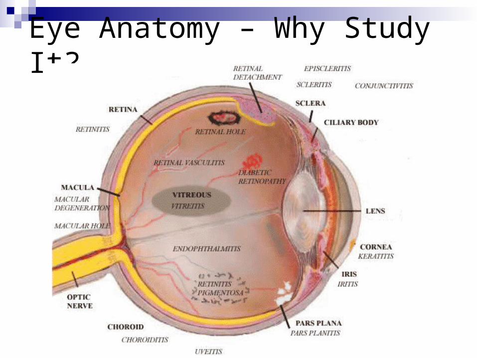
Eye Anatomy – Why Study It?
Why should you care?
Optometrist – Doctor of optometry, 4 year undergrad + 4 year optometry school
Ophthalmologists – Medical doctors In general, optometrists practice primary
and preventive eye care, while ophthalmologists perform eye surgery
What do nurses do?
History
Vision difficulty? Halos around lights – in glaucoma Scotoma – blind spot in visual field – in
glaucoma, optic nerve, and visual pathway disorder
Night blindness – Vit A deficiency, glaucoma,
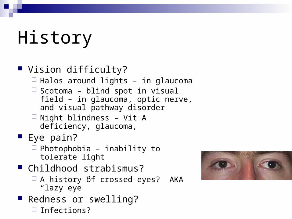
Eye pain? Photophobia – inability to tolerate light
Childhood strabismus? A history of crossed eyes? AKA “lazy eye”
Redness or swelling? Infections?
History cont.
Excessive or lack of tearing? May be due to irritants or obstruction in drainage
Past history of ocular problems? Glaucoma? Family history? Use of glasses or contact lenses? When tested last? Any medications?
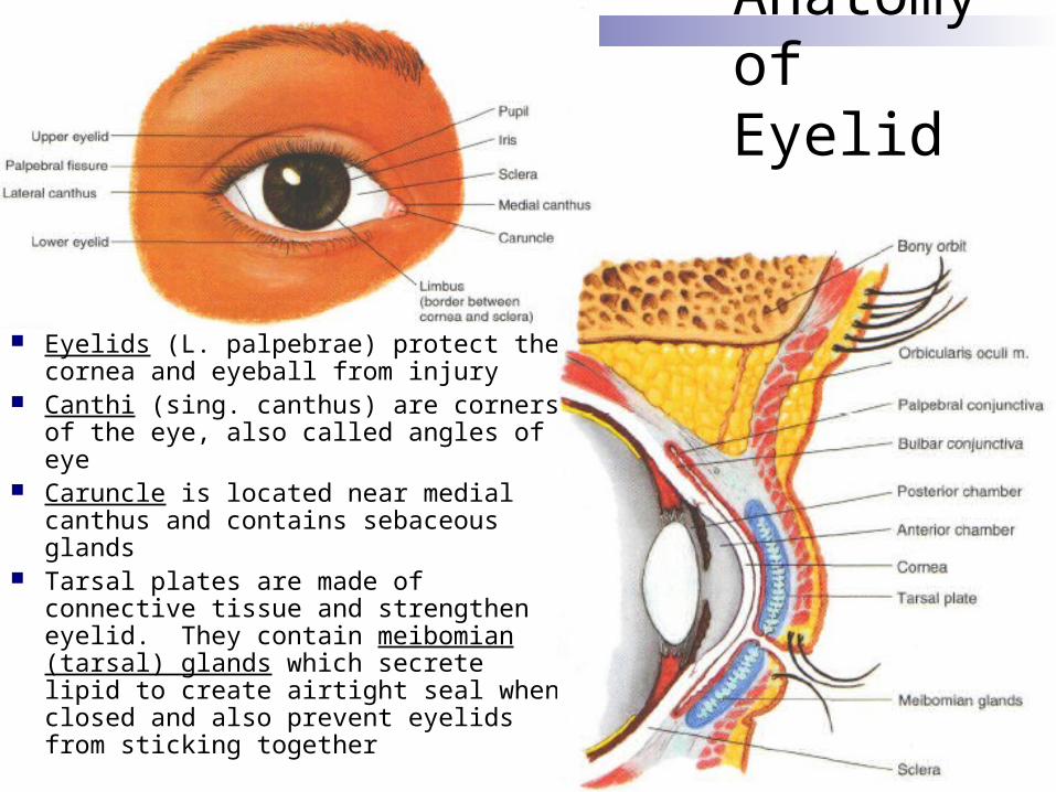
Anatomy of Eyelid
Eyelids (L. palpebrae) protect the cornea and eyeball from injury
Canthi (sing. canthus) are corners of the eye, also called angles of eye
Caruncle is located near medial canthus and contains sebaceous glands
Tarsal plates are made of connective tissue and strengthen eyelid. They contain meibomian (tarsal) glands which secrete lipid to create airtight seal when closed and also prevent eyelids from sticking together
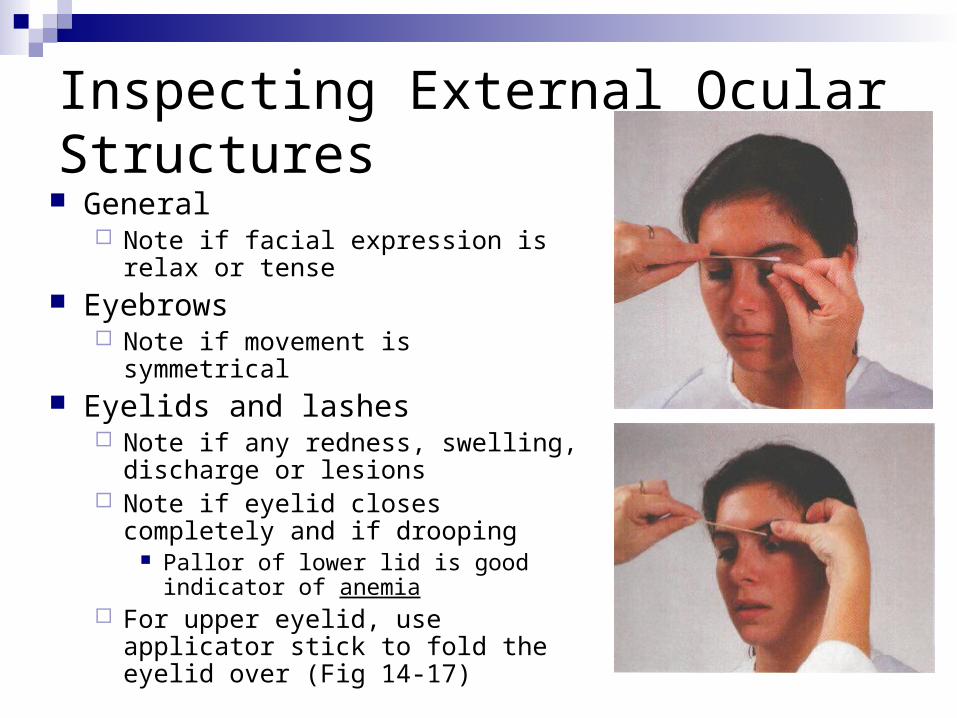
Inspecting External Ocular Structures General
Note if facial expression is relax or tense
Eyebrows Note if movement is symmetrical
Eyelids and lashes Note if any redness, swelling,
discharge or lesions Note if eyelid closes completely and if
drooping Pallor of lower lid is good indicator of
anemia For upper eyelid, use applicator stick
to fold the eyelid over (Fig 14-17)
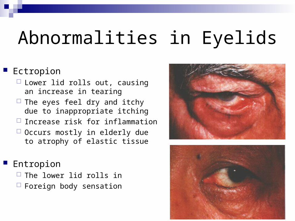
Abnormalities in Eyelids
Ectropion Lower lid rolls out, causing an
increase in tearing The eyes feel dry and itchy due to
inappropriate itching Increase risk for inflammation Occurs mostly in elderly due to
atrophy of elastic tissue
Entropion The lower lid rolls in Foreign body sensation
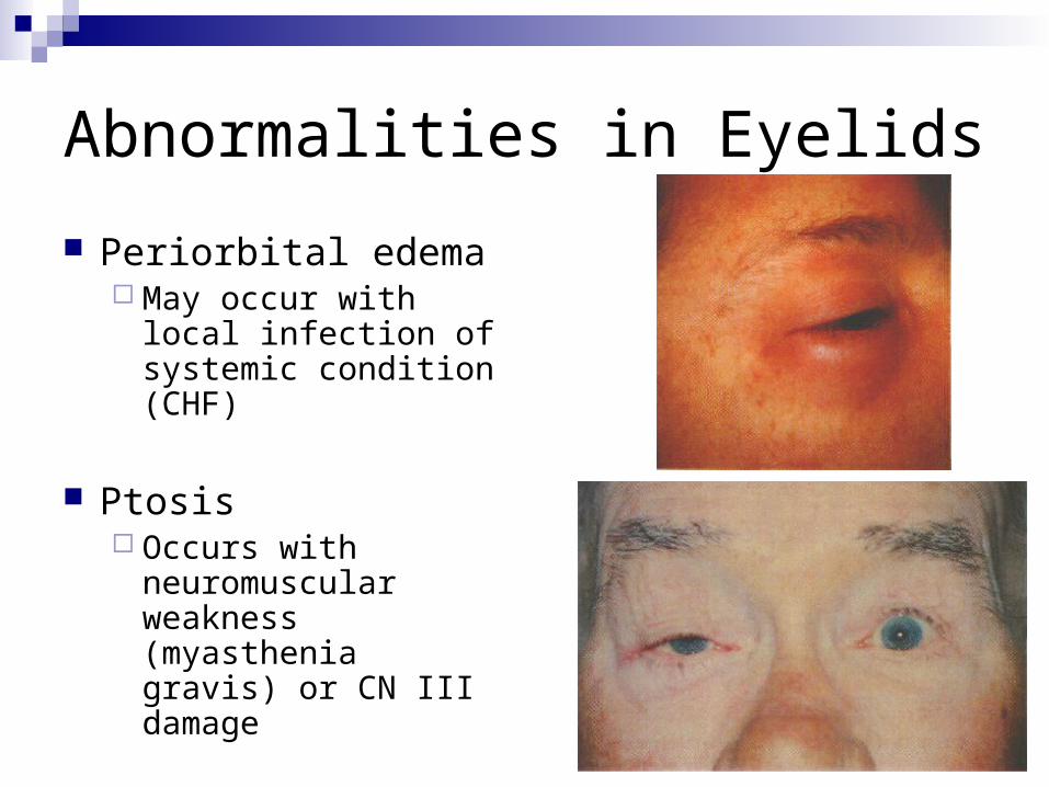
Abnormalities in Eyelids
Periorbital edema May occur with local
infection of systemic condition (CHF)
Ptosis Occurs with
neuromuscular weakness (myasthenia gravis) or CN III damage
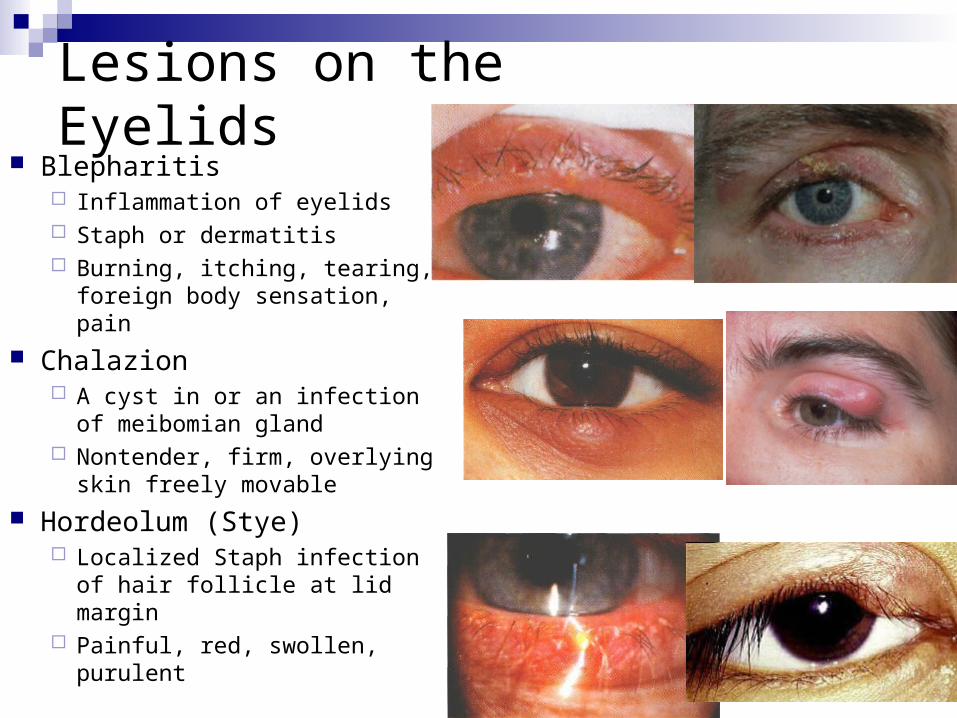
Lesions on the Eyelids
Blepharitis Inflammation of eyelids Staph or dermatitis Burning, itching, tearing,
foreign body sensation, pain
Chalazion A cyst in or an infection of
meibomian gland Nontender, firm, overlying
skin freely movable
Hordeolum (Stye) Localized Staph infection of
hair follicle at lid margin Painful, red, swollen, purulent
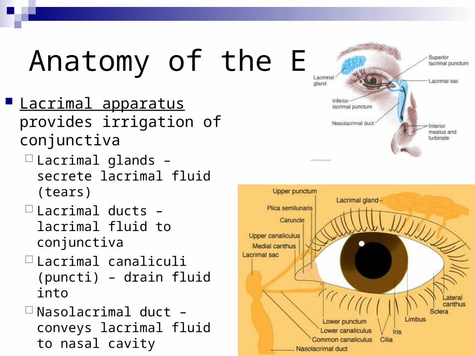
Anatomy of the Eye Lacrimal apparatus
provides irrigation of conjunctiva Lacrimal glands – secrete
lacrimal fluid (tears) Lacrimal ducts – lacrimal
fluid to conjunctiva Lacrimal canaliculi
(puncti) – drain fluid into Nasolacrimal duct –
conveys lacrimal fluid to nasal cavity
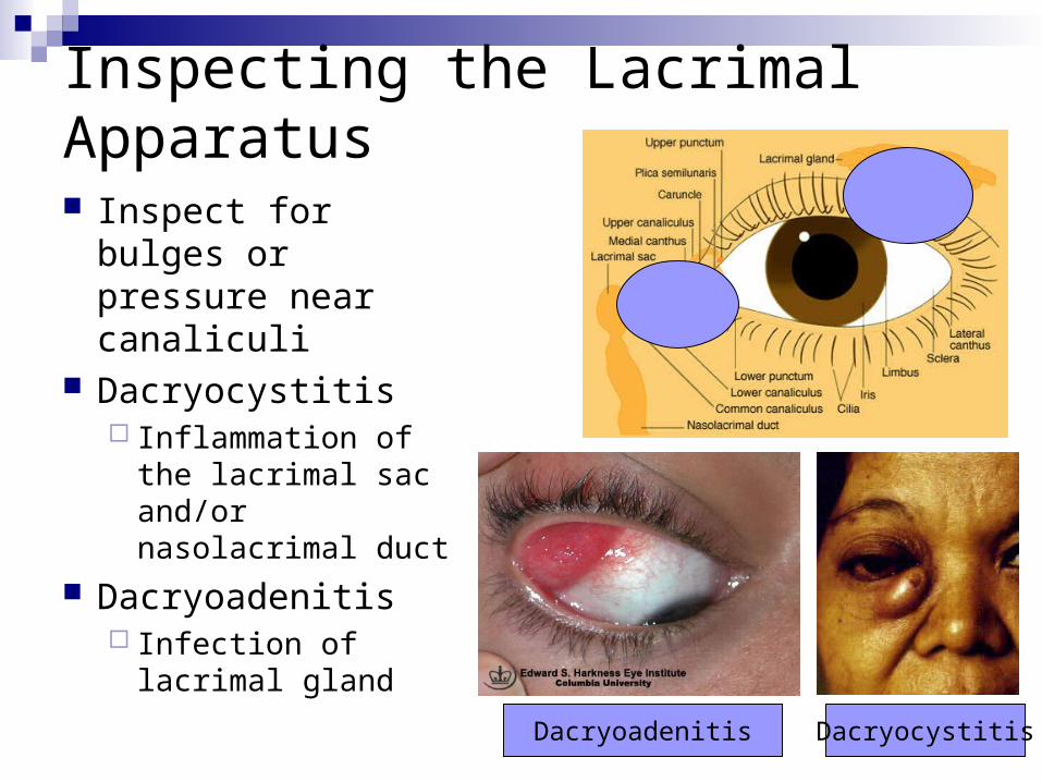
Inspecting the Lacrimal Apparatus
Inspect for bulges or pressure near canaliculi
Dacryocystitis Inflammation of the
lacrimal sac and/or nasolacrimal duct
Dacryoadenitis Infection of lacrimal
gland
DacryocystitisDacryoadenitis
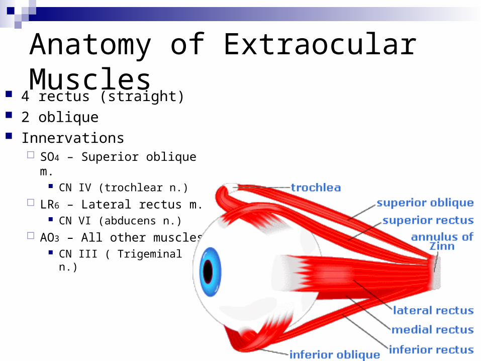
Anatomy of Extraocular Muscles 4 rectus (straight) 2 oblique Innervations
SO4 – Superior oblique m. CN IV (trochlear n.)
LR6 – Lateral rectus m. CN VI (abducens n.)
AO3 – All other muscles CN III ( Trigeminal n.)
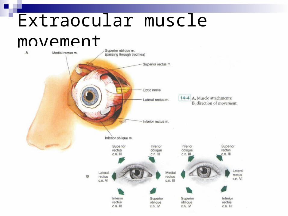
Extraocular muscle movement
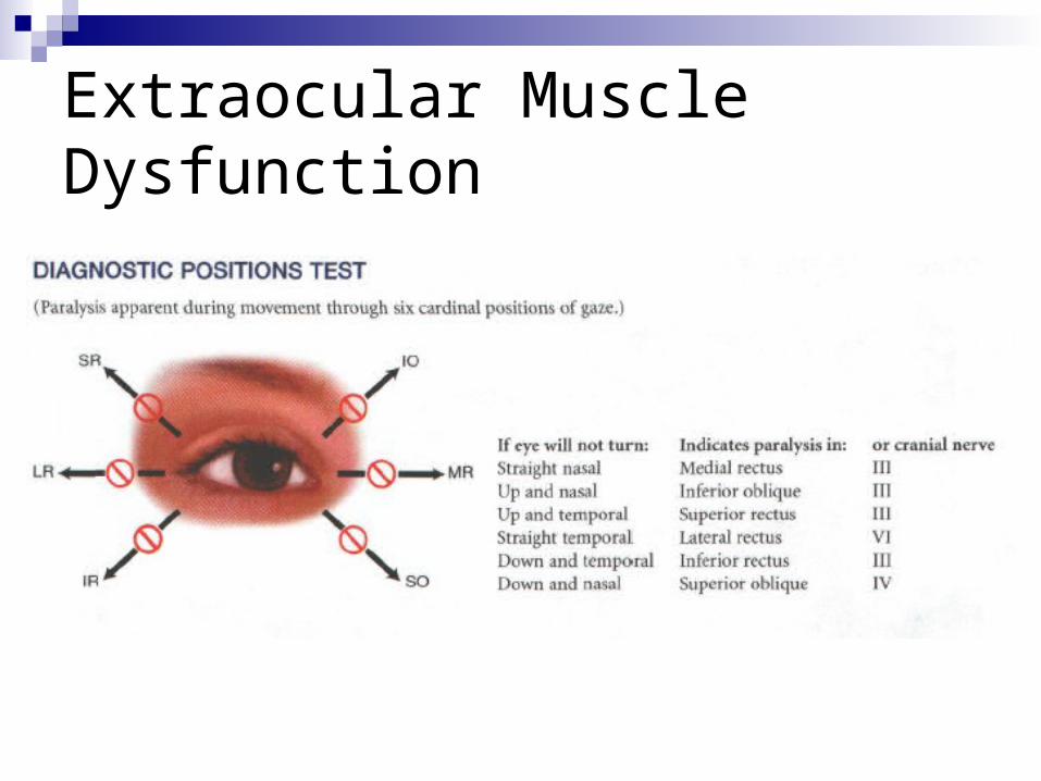
Extraocular Muscle Dysfunction
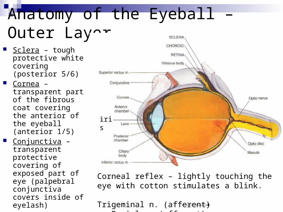
Anatomy of the Eyeball – Outer Layer
Sclera – tough protective white covering (posterior 5/6)
Cornea – transparent part of the fibrous coat covering the anterior of the eyeball (anterior 1/5)
Conjunctiva – transparent protective covering of exposed part of eye (palpebral conjunctiva covers inside of eyelash)
Corneal reflex – lightly touching the eye with cotton stimulates a blink.
Trigeminal n. (afferent) Facial n. (efferent)
iris
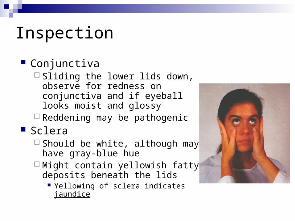
Inspection
Conjunctiva Sliding the lower lids down, observe
for redness on conjunctiva and if eyeball looks moist and glossy
Reddening may be pathogenic Sclera
Should be white, although may have gray-blue hue
Might contain yellowish fatty deposits beneath the lids
Yellowing of sclera indicates jaundice
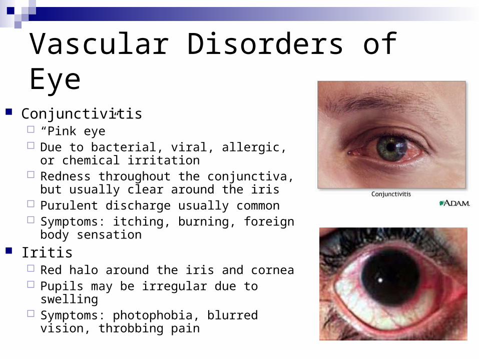
Vascular Disorders of Eye
Conjunctivitis “Pink eye” Due to bacterial, viral, allergic, or chemical
irritation Redness throughout the conjunctiva, but
usually clear around the iris Purulent discharge usually common Symptoms: itching, burning, foreign body
sensation Iritis
Red halo around the iris and cornea Pupils may be irregular due to swelling Symptoms: photophobia, blurred vision,
throbbing pain
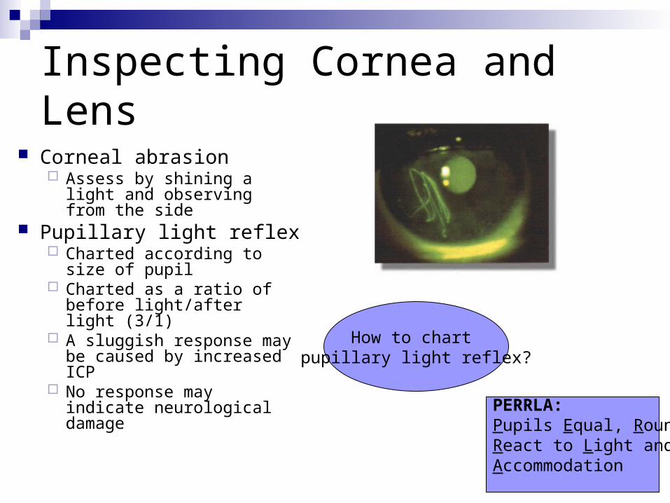
Inspecting Cornea and Lens
Corneal abrasion Assess by shining a light
and observing from the side
Pupillary light reflex Charted according to size
of pupil Charted as a ratio of before
light/after light (3/1) A sluggish response may
be caused by increased ICP
No response may indicate neurological damage
PERRLA:Pupils Equal, Round,React to Light and Accommodation
How to chart pupillary light reflex?
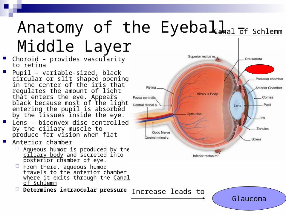
Anatomy of the Eyeball – Middle Layer
Choroid – provides vascularity to retina
Pupil – variable-sized, black circular or slit shaped opening in the center of the iris that regulates the amount of light that enters the eye. Appears black because most of the light entering the pupil is absorbed by the tissues inside the eye.
Lens – biconvex disc controlled by the ciliary muscle to produce far vision when flat
Anterior chamber Aqueous humor is produced by the
ciliary body and secreted into posterior chamber of eye.
From there, aqueous humor travels to the anterior chamber where it exits through the Canal of Schlemm
Determines intraocular pressure
Canal of Schlemm
Increase leads toGlaucoma
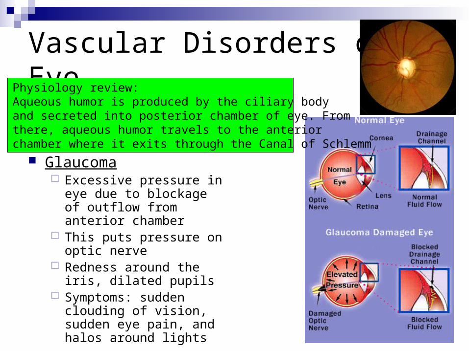
Vascular Disorders of Eye
Glaucoma Excessive pressure in eye
due to blockage of outflow from anterior chamber
This puts pressure on optic nerve
Redness around the iris, dilated pupils
Symptoms: sudden clouding of vision, sudden eye pain, and halos around lights
Physiology review:Aqueous humor is produced by the ciliary bodyand secreted into posterior chamber of eye. From there, aqueous humor travels to the anterior chamber where it exits through the Canal of Schlemm
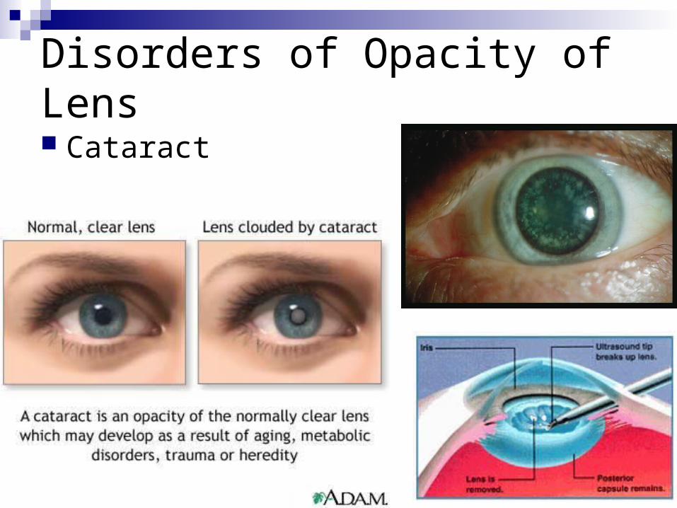
Disorders of Opacity of Lens
Cataract
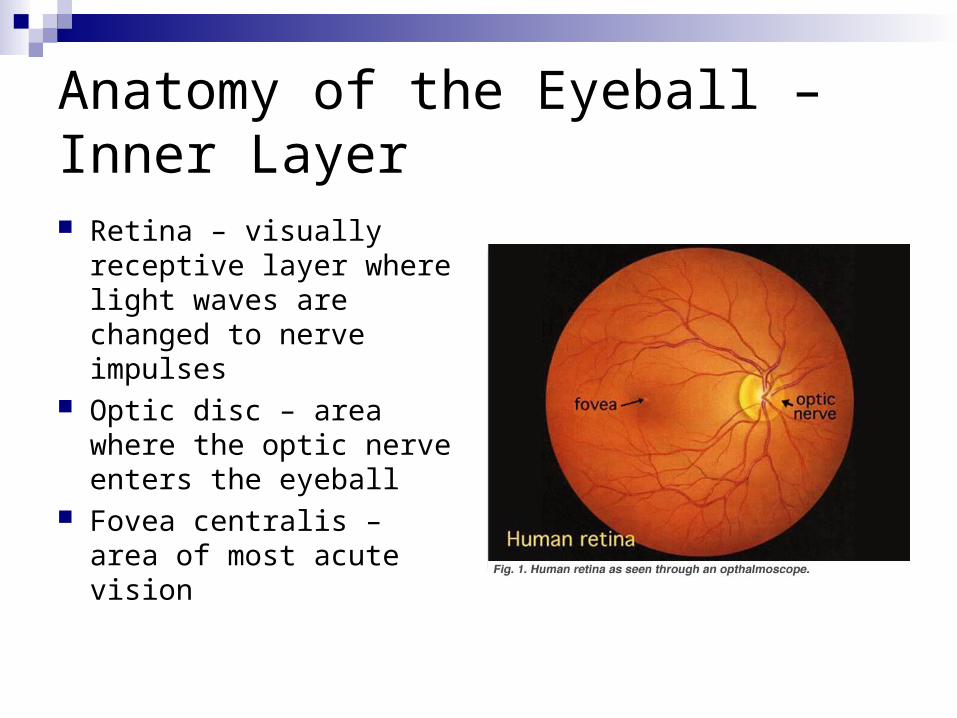
Anatomy of the Eyeball – Inner Layer Retina – visually
receptive layer where light waves are changed to nerve impulses
Optic disc – area where the optic nerve enters the eyeball
Fovea centralis – area of most acute vision
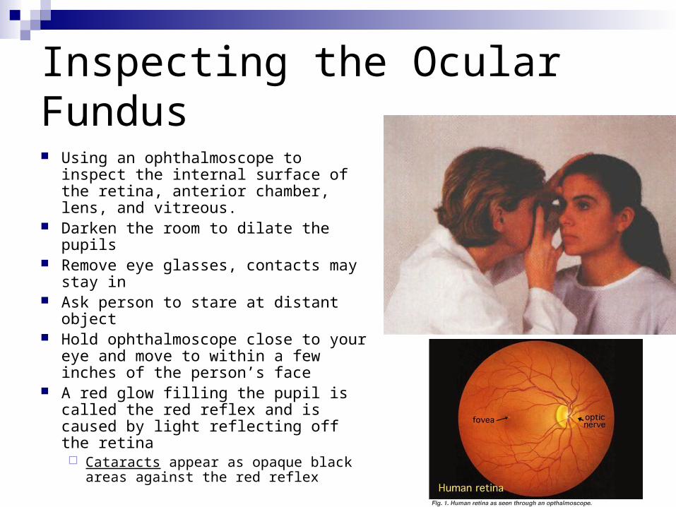
Inspecting the Ocular Fundus
Using an ophthalmoscope to inspect the internal surface of the retina, anterior chamber, lens, and vitreous.
Darken the room to dilate the pupils Remove eye glasses, contacts may
stay in Ask person to stare at distant object Hold ophthalmoscope close to your
eye and move to within a few inches of the person’s face
A red glow filling the pupil is called the red reflex and is caused by light reflecting off the retina Cataracts appear as opaque black
areas against the red reflex
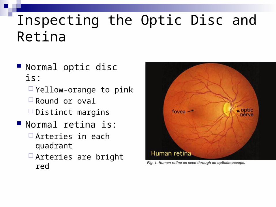
Inspecting the Optic Disc and Retina
Normal optic disc is: Yellow-orange to pink Round or oval Distinct margins
Normal retina is: Arteries in each
quadrant Arteries are bright red
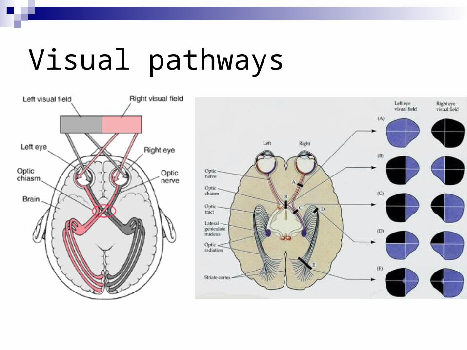
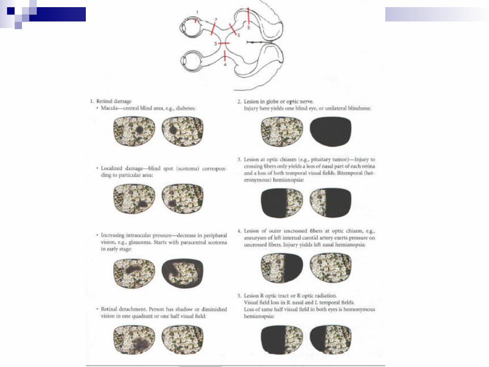
Visual pathways
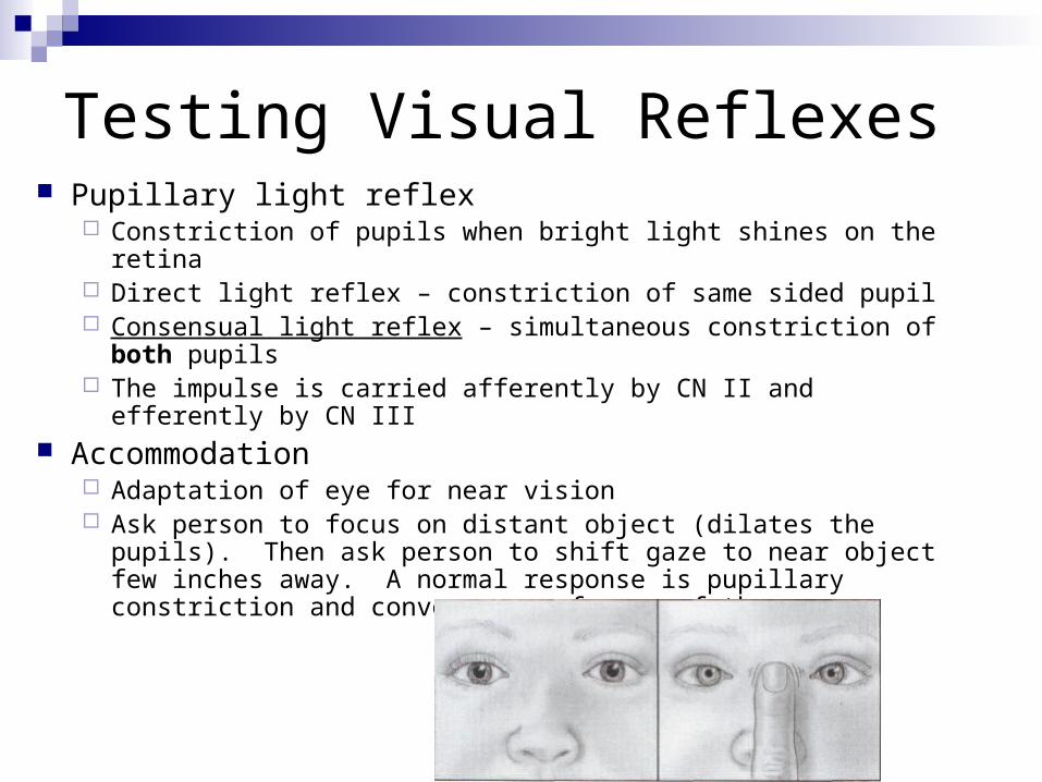
Testing Visual Reflexes Pupillary light reflex
Constriction of pupils when bright light shines on the retina Direct light reflex – constriction of same sided pupil Consensual light reflex – simultaneous constriction of both
pupils The impulse is carried afferently by CN II and efferently by CN III
Accommodation Adaptation of eye for near vision Ask person to focus on distant object (dilates the pupils). Then
ask person to shift gaze to near object few inches away. A normal response is pupillary constriction and convergence of axes of the eyes
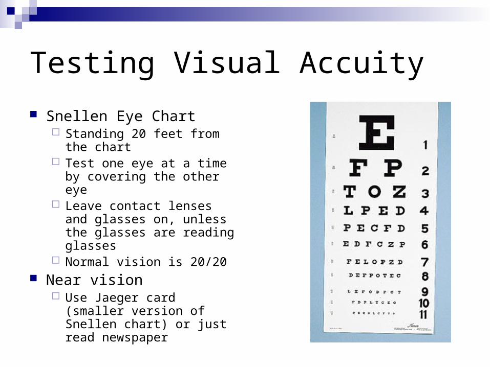
Testing Visual Accuity
Snellen Eye Chart Standing 20 feet from the
chart Test one eye at a time by
covering the other eye Leave contact lenses and
glasses on, unless the glasses are reading glasses
Normal vision is 20/20 Near vision
Use Jaeger card (smaller version of Snellen chart) or just read newspaper
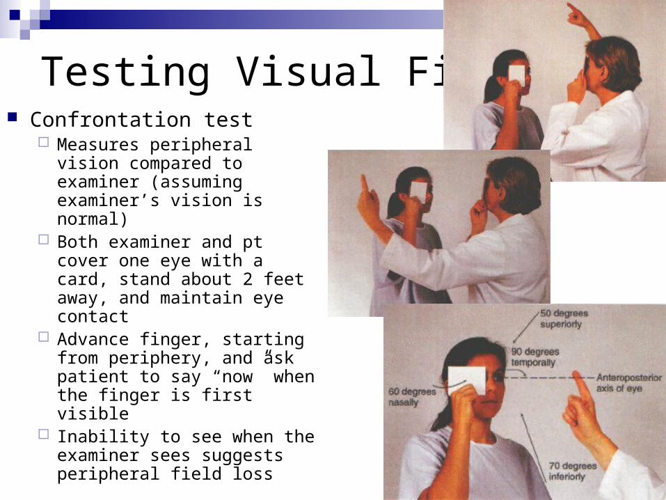
Testing Visual Fields Confrontation test
Measures peripheral vision compared to examiner (assuming examiner’s vision is normal)
Both examiner and pt cover one eye with a card, stand about 2 feet away, and maintain eye contact
Advance finger, starting from periphery, and ask patient to say “now” when the finger is first visible
Inability to see when the examiner sees suggests peripheral field loss
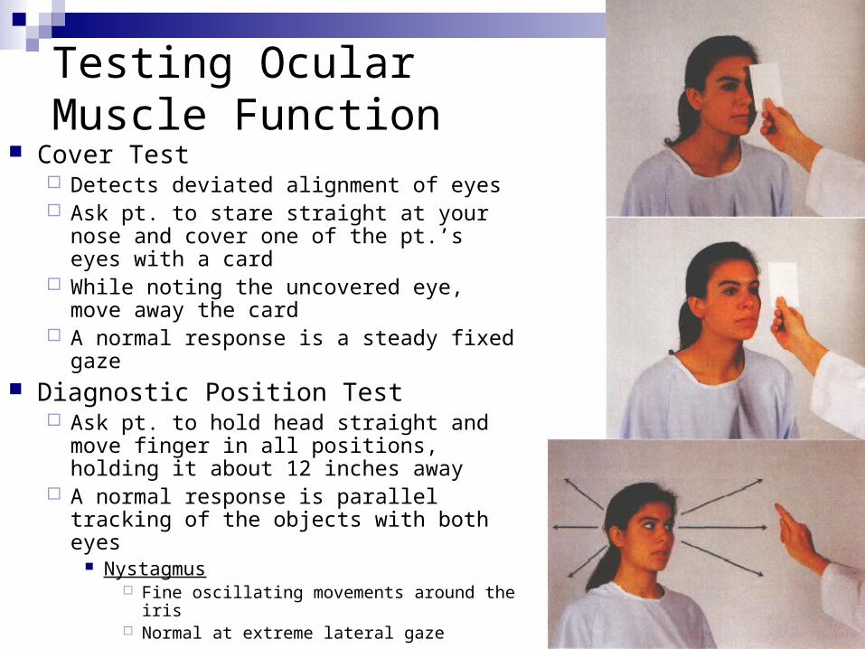
Testing Ocular Muscle Function
Cover Test Detects deviated alignment of eyes Ask pt. to stare straight at your nose and
cover one of the pt.’s eyes with a card While noting the uncovered eye, move
away the card A normal response is a steady fixed gaze
Diagnostic Position Test Ask pt. to hold head straight and move
finger in all positions, holding it about 12 inches away
A normal response is parallel tracking of the objects with both eyes
Nystagmus Fine oscillating movements around the iris Normal at extreme lateral gaze
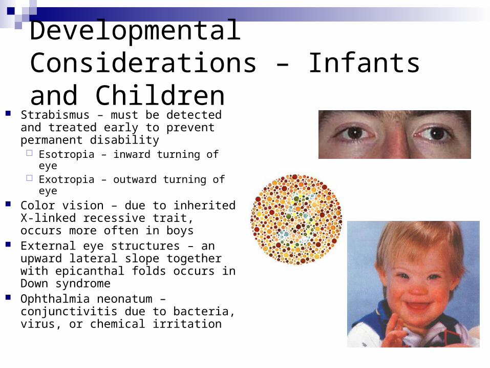
Developmental Considerations – Infants and Children
Strabismus – must be detected and treated early to prevent permanent disability Esotropia – inward turning of eye Exotropia – outward turning of eye
Color vision – due to inherited X-linked recessive trait, occurs more often in boys
External eye structures – an upward lateral slope together with epicanthal folds occurs in Down syndrome
Ophthalmia neonatum – conjunctivitis due to bacteria, virus, or chemical irritation
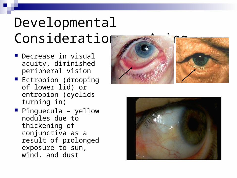
Developmental Considerations – Aging Decrease in visual
acuity, diminished peripheral vision
Ectropion (drooping of lower lid) or entropion (eyelids turning in)
Pinguecula – yellow nodules due to thickening of conjunctiva as a result of prolonged exposure to sun, wind, and dust
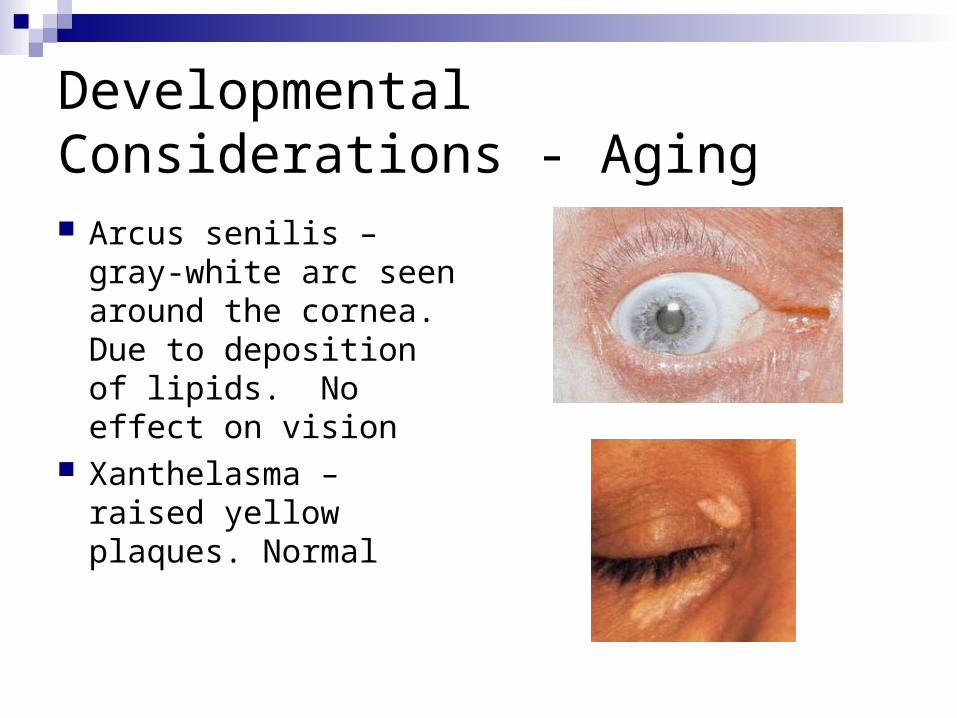
Developmental Considerations - Aging Arcus senilis – gray-
white arc seen around the cornea. Due to deposition of lipids. No effect on vision
Xanthelasma – raised yellow plaques. Normal
THE END
EYE HOPE YOU HAVE A GREAT DAY!!!