Embed Size (px)
DESCRIPTION
Immunology Notes
Citation preview
1
3. Lymphatic System
ImmunologyDavid L. Beck, BS, AM, PhDChapter 1 – Page 9-10,16-24Chapter 8 – Page 291-294 2
Course Summary – Quick Overview Anatomy of the Immune System
Immune Cells Anatomy of Lymphatic Vessels and Tissues
Innate Immunity Adaptive Immunity
MHC, Immunogens Immunoglobulin Development of B Cells and T Cells Dendritic Cells to T Cells T cells to B Cells B Cells
Specialized Lymphocytes Mucosal Immunity Summary
3
3. Lymphatic System and Immunity Overview
Lymphatic SystemVessels and Lymph NodesSpleen and Other Lymphoid TissuesThymus
MALT – Mucosal Immunity (brief look)
4
Anatomy of the lymphatic system.Lymphoid Organs
Primary lymphoid organs: Where immune cells originate and where they mature (bone marrow
and thymus)
Secondary lymphoid organs: Where immune cells reside and become activated. Lymph nodes, spleen, tonsils, Peyer's patch, appendix
Note: Immune cells are concentrated in these organs, but are also spread throughout almost every tissue of the body.
5
Circulation of Fluids in the Body
There are two components to blood: Cellular Components – Red blood cells, White
blood cells, Platelets, etc. Red blood cells exchange gases with the tissue.
Serum – a yellowish fluid that you observe when you get a blister. Serum is what brings the food and immune factors to the tissues.
In healthy circulation serum leaves the vessels, but the cellular component is restricted to the vessel, although some white blood cells can migrate through the vessel wall by diapedesis.
6
Lymphatic System
1. Lymph capillaries2. Lymph vessels (called trunks)3. Elephantiasis4. Lymph Organs and Tissues5. Lymph Node6. Spleen7. Lymph tissues8. Thymus
7
Lymph System
8
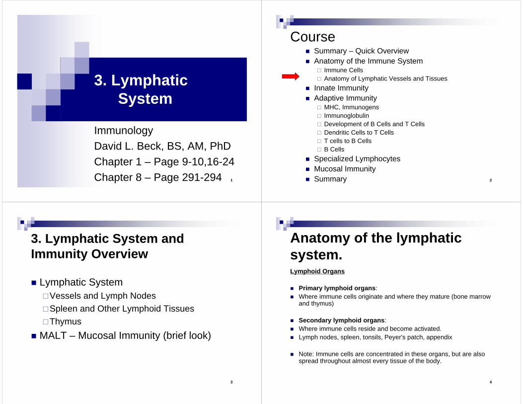
1. Lymph capillaries At the end of a lymphatic capillary there is a very loose-
walled vessel that is closed at the end. This allows for many small gaps between cells allowing for entry of cells, debris, proteins, and fluid to the lymph vessels. The lymphatic capillaries sit in the meshwork of the circulatory system, and travel with the veins. The veins are thinner walled, more fluid comes out of the veins than out of arteries which are thicker walled. This means that the lymphatic vessels travel with the veins to catch the excess fluids coming into the tissue from them. The lymphatic capillaries are anchored in place by filaments that connect to the connective tissue.
Explanation of Next Slide
9
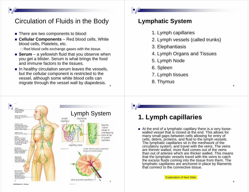
Lymphatic capillarySerum leaks easily from the capillaries. It is picked up again by the lymphatic capillaries which are vessels that have openings at their ends to catch fluids and facilitate entry by white blood cells leaving the tissue.
10
2. Lymph vessels (called trunks)A meshwork of vessels is found in all tissues of the
body except:A. Avascular Tissue - Tissue where no blood
vessels are found (eg. inside the eye ball)B. Central Nervous SystemC. Splenic pulp - although found in the walls of the
spleen, vessels are not found in the pulp of the spleen.
D. Bone Marrow, Bone, Teeth
11
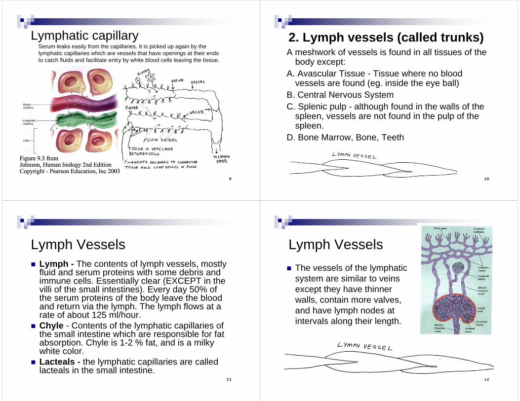
Lymph Vessels Lymph - The contents of lymph vessels, mostly
fluid and serum proteins with some debris and immune cells. Essentially clear (EXCEPT in the villi of the small intestines). Every day 50% of the serum proteins of the body leave the blood and return via the lymph. The lymph flows at a rate of about 125 ml/hour.
Chyle - Contents of the lymphatic capillaries of the small intestine which are responsible for fat absorption. Chyle is 1-2 % fat, and is a milky white color.
Lacteals - the lymphatic capillaries are called lacteals in the small intestine.
12
Lymph Vessels The vessels of the lymphatic
system are similar to veins except they have thinner walls, contain more valves, and have lymph nodes at intervals along their length.
13
Lymph Vessels
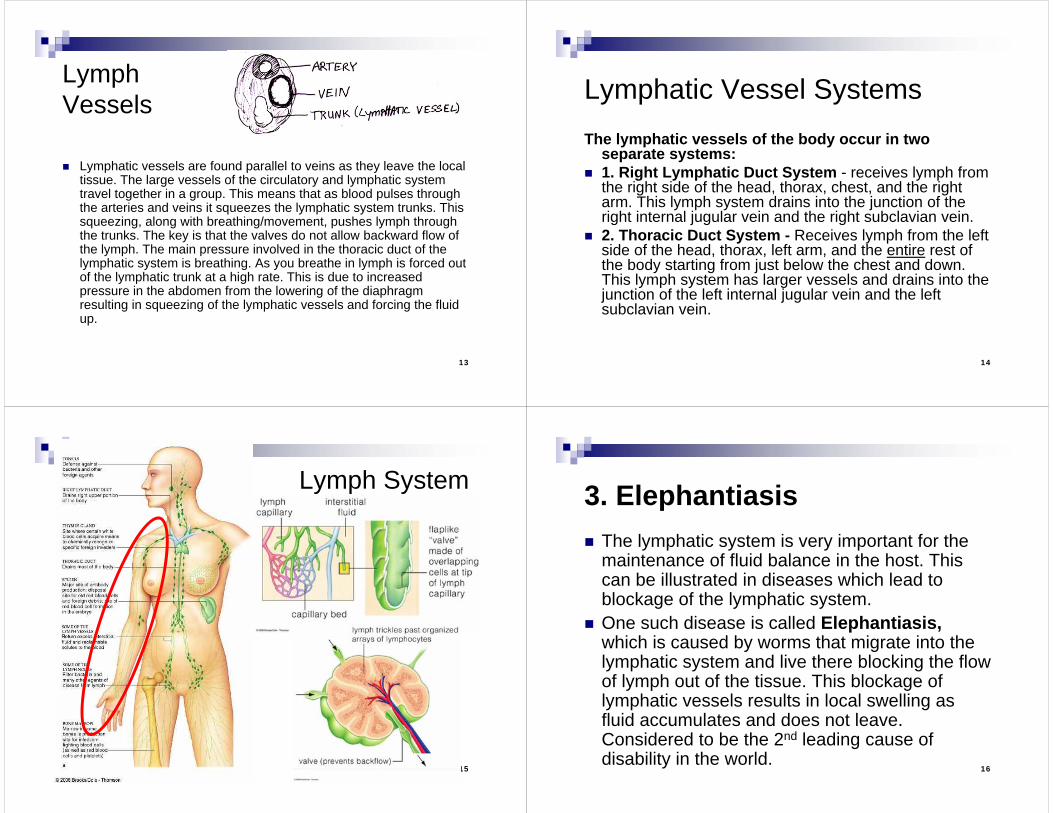
Lymphatic vessels are found parallel to veins as they leave the local tissue. The large vessels of the circulatory and lymphatic system travel together in a group. This means that as blood pulses through the arteries and veins it squeezes the lymphatic system trunks. This squeezing, along with breathing/movement, pushes lymph through the trunks. The key is that the valves do not allow backward flow of the lymph. The main pressure involved in the thoracic duct of the lymphatic system is breathing. As you breathe in lymph is forced out of the lymphatic trunk at a high rate. This is due to increased pressure in the abdomen from the lowering of the diaphragm resulting in squeezing of the lymphatic vessels and forcing the fluid up.
14
Lymphatic Vessel SystemsThe lymphatic vessels of the body occur in two
separate systems: 1. Right Lymphatic Duct System - receives lymph from
the right side of the head, thorax, chest, and the right arm. This lymph system drains into the junction of the right internal jugular vein and the right subclavian vein.
2. Thoracic Duct System - Receives lymph from the left side of the head, thorax, left arm, and the entire rest of the body starting from just below the chest and down. This lymph system has larger vessels and drains into the junction of the left internal jugular vein and the left subclavian vein.
15
Lymph System
16
3. Elephantiasis The lymphatic system is very important for the
maintenance of fluid balance in the host. This can be illustrated in diseases which lead to blockage of the lymphatic system.
One such disease is called Elephantiasis,which is caused by worms that migrate into the lymphatic system and live there blocking the flow of lymph out of the tissue. This blockage of lymphatic vessels results in local swelling as fluid accumulates and does not leave. Considered to be the 2nd leading cause of disability in the world.
17
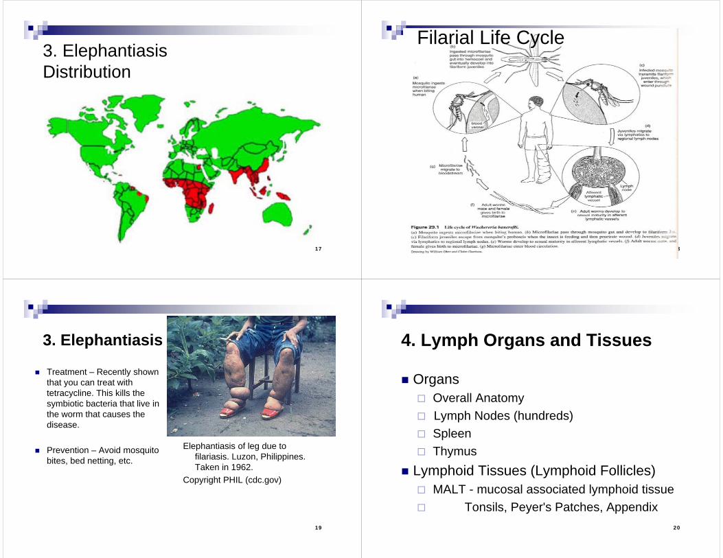
3. ElephantiasisDistribution
18
Filarial Life Cycle
19
3. Elephantiasis
Elephantiasis of leg due to filariasis. Luzon, Philippines. Taken in 1962.
Copyright PHIL (cdc.gov)
Treatment – Recently shown that you can treat with tetracycline. This kills the symbiotic bacteria that live in the worm that causes the disease.
Prevention – Avoid mosquito bites, bed netting, etc.
20
4. Lymph Organs and Tissues
Organs Overall Anatomy Lymph Nodes (hundreds) Spleen Thymus
Lymphoid Tissues (Lymphoid Follicles) MALT - mucosal associated lymphoid tissue Tonsils, Peyer's Patches, Appendix
21
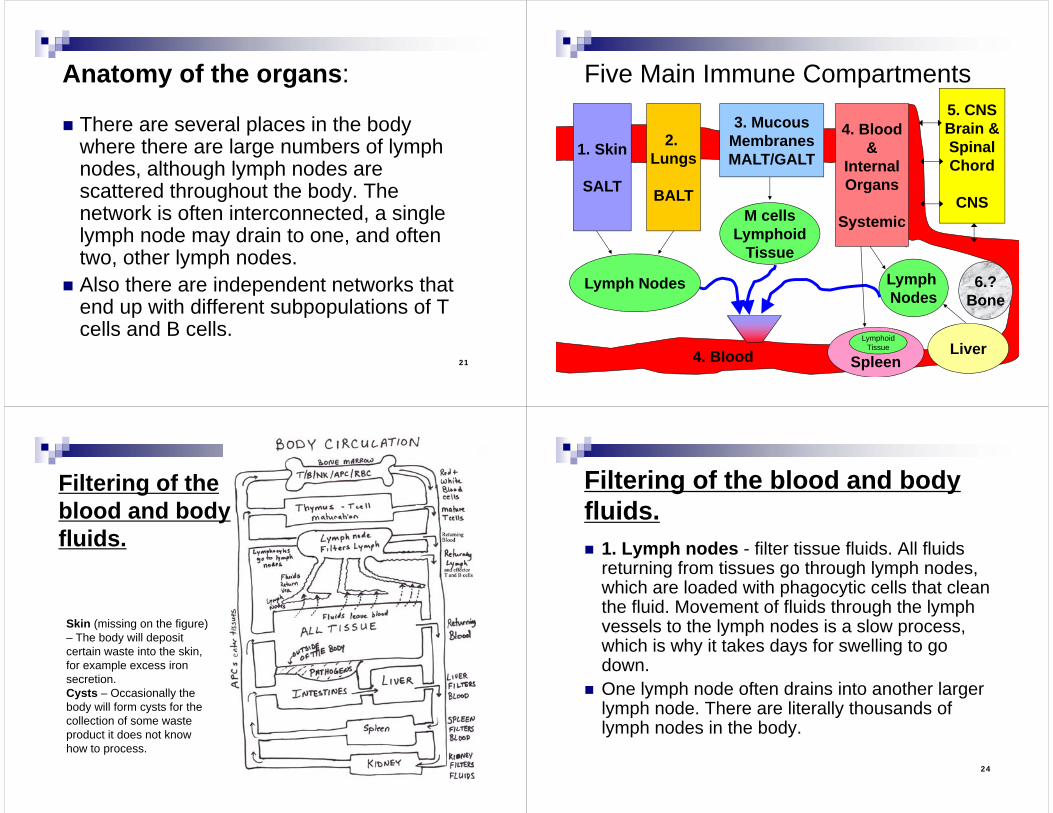
Anatomy of the organs:
There are several places in the body where there are large numbers of lymph nodes, although lymph nodes are scattered throughout the body. The network is often interconnected, a single lymph node may drain to one, and often two, other lymph nodes.
Also there are independent networks that end up with different subpopulations of T cells and B cells.
22
Five Main Immune Compartments
1. Skin
SALT
2. Lungs
BALT
3. MucousMembranesMALT/GALT
5. CNSBrain &SpinalChord
CNSM cells
LymphoidTissue
Lymph Nodes
Spleen
Lymph Nodes
Liver
4. Blood&
InternalOrgans
Systemic
4. Blood
6.?Bone
LymphoidTissue
23
Filtering of the blood and body fluids.
Skin (missing on the figure) – The body will deposit certain waste into the skin, for example excess iron secretion.Cysts – Occasionally the body will form cysts for the collection of some waste product it does not know how to process.
24
Filtering of the blood and body fluids. 1. Lymph nodes - filter tissue fluids. All fluids
returning from tissues go through lymph nodes, which are loaded with phagocytic cells that clean the fluid. Movement of fluids through the lymph vessels to the lymph nodes is a slow process, which is why it takes days for swelling to go down.
One lymph node often drains into another larger lymph node. There are literally thousands of lymph nodes in the body.
25
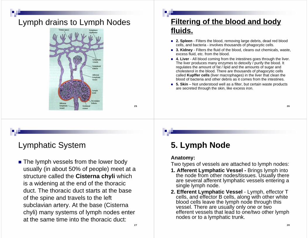
Lymph drains to Lymph Nodes
26
Filtering of the blood and body fluids. 2. Spleen - Filters the blood, removing large debris, dead red blood
cells, and bacteria - involves thousands of phagocytic cells. 3. Kidney - Filters the fluid of the blood, cleans out chemicals, waste,
excess fluid, etc. from the blood. 4. Liver - All blood coming from the intestines goes through the liver.
The liver produces many enzymes to detoxify / purify the blood. It regulates the amount of fat / lipid and the amounts of sugar and cholesterol in the blood. There are thousands of phagocytic cells called Kupffer cells (liver macrophages) in the liver that clean the blood of bacteria and other debris as it comes from the intestines.
5. Skin – Not understood well as a filter, but certain waste products are secreted through the skin, like excess iron.
27
Lymphatic System
The lymph vessels from the lower body usually (in about 50% of people) meet at a structure called the Cisterna chyli which is a widening at the end of the thoracic duct. The thoracic duct starts at the base of the spine and travels to the left subclavian artery. At the base (Cisterna chyli) many systems of lymph nodes enter at the same time into the thoracic duct:
28
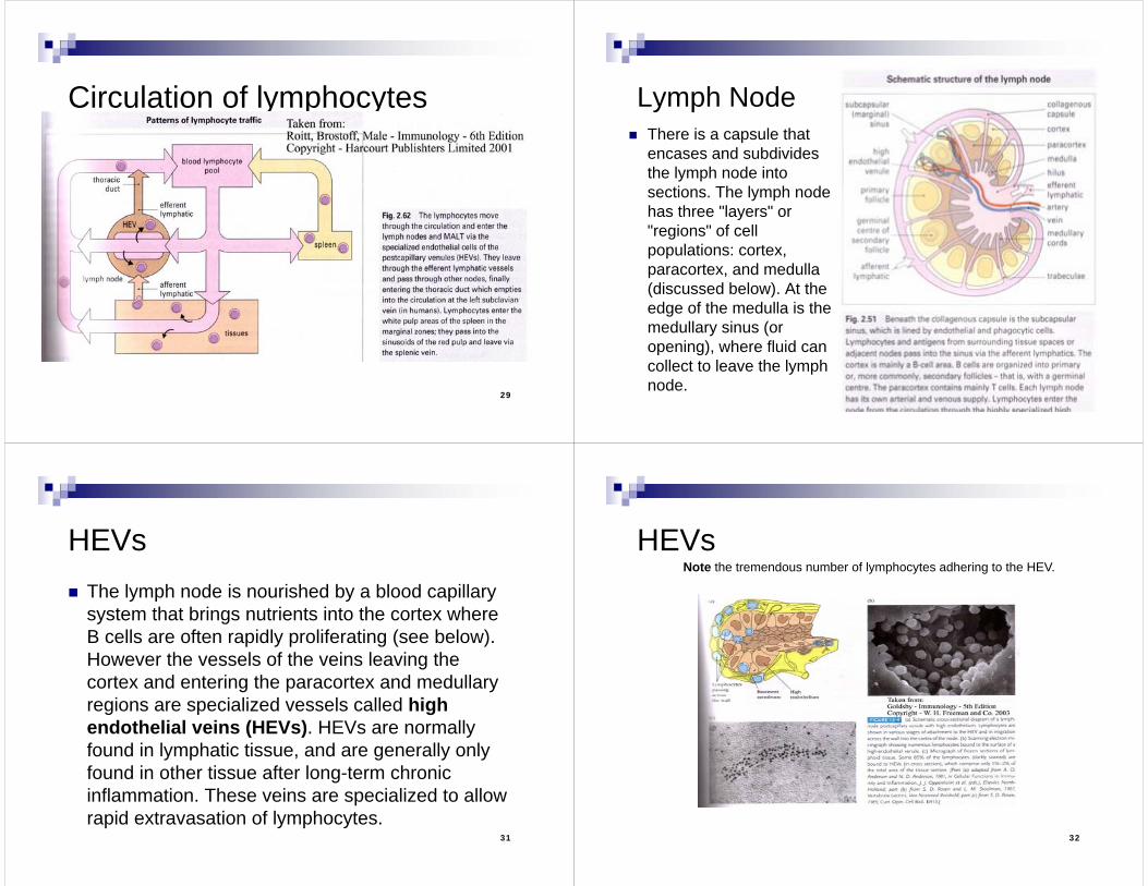
5. Lymph NodeAnatomy:Two types of vessels are attached to lymph nodes:1. Afferent Lymphatic Vessel - Brings lymph into
the node from other nodes/tissues. Usually there are several afferent lymphatic vessels entering a single lymph node.
2. Efferent Lymphatic Vessel - Lymph, effector T cells, and effector B cells, along with other white blood cells leave the lymph node through this vessel. There are usually only one or two efferent vessels that lead to one/two other lymph nodes or to a lymphatic trunk.
29
Circulation of lymphocytes
30
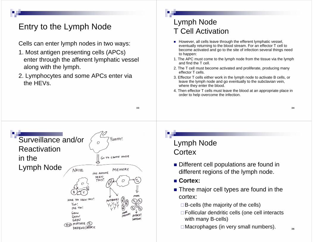
Lymph Node There is a capsule that
encases and subdivides the lymph node into sections. The lymph node has three "layers" or "regions" of cell populations: cortex, paracortex, and medulla (discussed below). At the edge of the medulla is the medullary sinus (or opening), where fluid can collect to leave the lymph node.
31
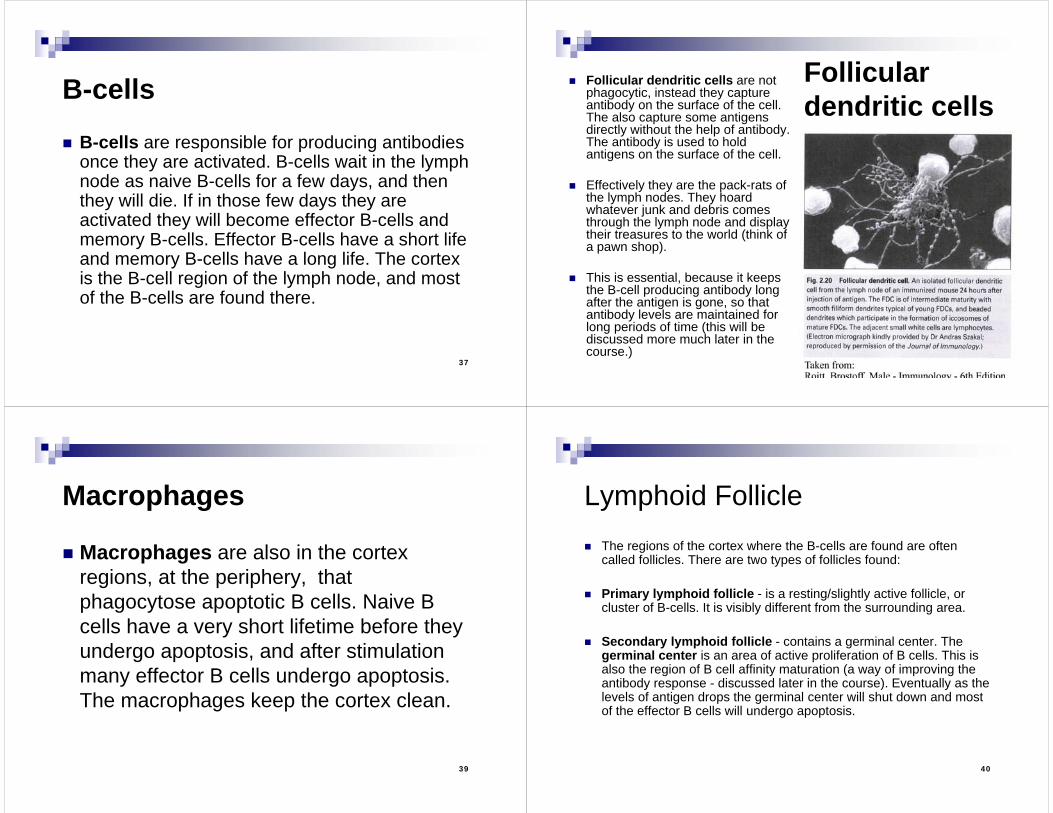
HEVs The lymph node is nourished by a blood capillary
system that brings nutrients into the cortex where B cells are often rapidly proliferating (see below). However the vessels of the veins leaving the cortex and entering the paracortex and medullary regions are specialized vessels called high endothelial veins (HEVs). HEVs are normally found in lymphatic tissue, and are generally only found in other tissue after long-term chronic inflammation. These veins are specialized to allow rapid extravasation of lymphocytes.
32
HEVsNote the tremendous number of lymphocytes adhering to the HEV.
33
Entry to the Lymph Node
Cells can enter lymph nodes in two ways:1. Most antigen presenting cells (APCs)
enter through the afferent lymphatic vessel along with the lymph.
2. Lymphocytes and some APCs enter via the HEVs.
34
Lymph NodeT Cell Activation However, all cells leave through the efferent lymphatic vessel,
eventually returning to the blood stream. For an effector T cell to become activated and go to the site of infection several things need to happen:
1. The APC must come to the lymph node from the tissue via the lymph and find the T cell.
2. The T cell must become activated and proliferate, producing many effector T cells.
3. Effector T cells either work in the lymph node to activate B cells, or leave the lymph node and go eventually to the subclavian vein, where they enter the blood.
4. Then effector T cells must leave the blood at an appropriate place in order to help overcome the infection.
35
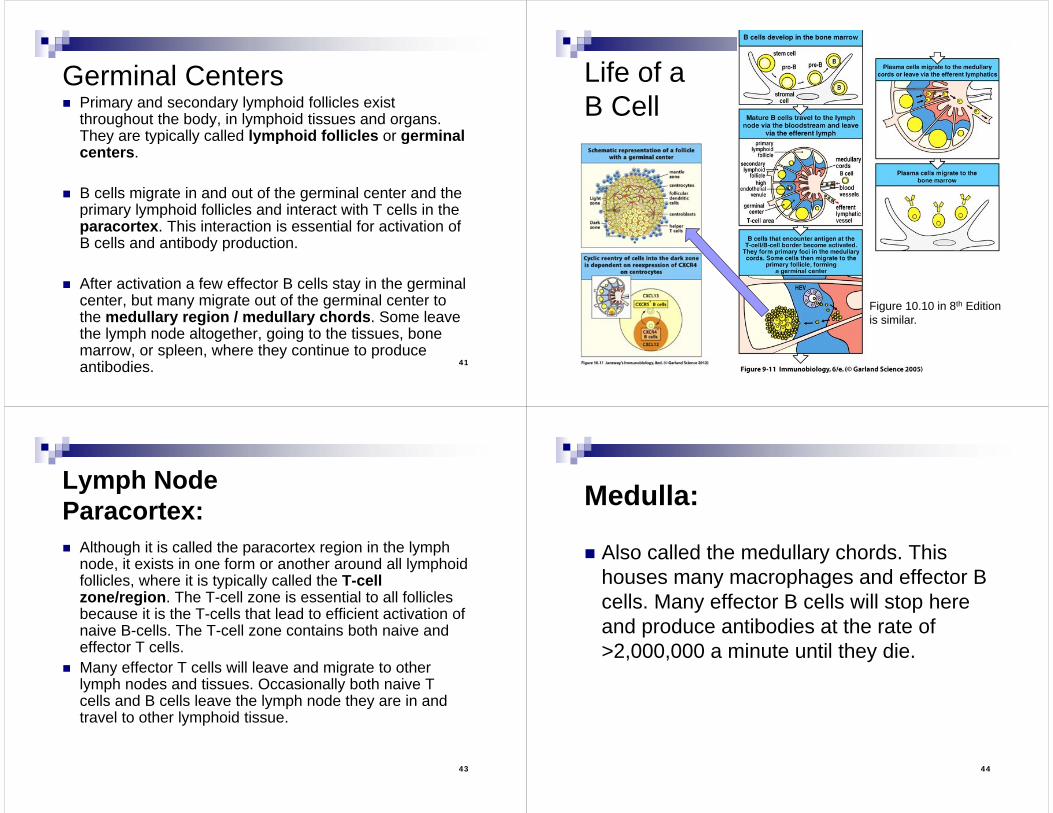
Surveillance and/orReactivationin the Lymph Node
36
Lymph NodeCortex Different cell populations are found in
different regions of the lymph node. Cortex: Three major cell types are found in the
cortex: B-cells (the majority of the cells)Follicular dendritic cells (one cell interacts
with many B-cells)Macrophages (in very small numbers).
37
B-cells
B-cells are responsible for producing antibodies once they are activated. B-cells wait in the lymph node as naive B-cells for a few days, and then they will die. If in those few days they are activated they will become effector B-cells and memory B-cells. Effector B-cells have a short life and memory B-cells have a long life. The cortex is the B-cell region of the lymph node, and most of the B-cells are found there.
38
Follicular dendritic cells
Follicular dendritic cells are not phagocytic, instead they capture antibody on the surface of the cell. The also capture some antigens directly without the help of antibody. The antibody is used to hold antigens on the surface of the cell.
Effectively they are the pack-rats of the lymph nodes. They hoard whatever junk and debris comes through the lymph node and display their treasures to the world (think of a pawn shop).
This is essential, because it keeps the B-cell producing antibody long after the antigen is gone, so that antibody levels are maintained for long periods of time (this will be discussed more much later in the course.)
39
Macrophages
Macrophages are also in the cortex regions, at the periphery, that phagocytose apoptotic B cells. Naive B cells have a very short lifetime before they undergo apoptosis, and after stimulation many effector B cells undergo apoptosis. The macrophages keep the cortex clean.
40
Lymphoid Follicle The regions of the cortex where the B-cells are found are often
called follicles. There are two types of follicles found:
Primary lymphoid follicle - is a resting/slightly active follicle, or cluster of B-cells. It is visibly different from the surrounding area.
Secondary lymphoid follicle - contains a germinal center. The germinal center is an area of active proliferation of B cells. This is also the region of B cell affinity maturation (a way of improving the antibody response - discussed later in the course). Eventually as the levels of antigen drops the germinal center will shut down and most of the effector B cells will undergo apoptosis.
41
Germinal Centers Primary and secondary lymphoid follicles exist
throughout the body, in lymphoid tissues and organs. They are typically called lymphoid follicles or germinal centers.
B cells migrate in and out of the germinal center and the primary lymphoid follicles and interact with T cells in the paracortex. This interaction is essential for activation of B cells and antibody production.
After activation a few effector B cells stay in the germinal center, but many migrate out of the germinal center to the medullary region / medullary chords. Some leave the lymph node altogether, going to the tissues, bone marrow, or spleen, where they continue to produce antibodies. 42
Life of a B Cell
Figure 10.10 in 8th Editionis similar.
43
Lymph NodeParacortex: Although it is called the paracortex region in the lymph
node, it exists in one form or another around all lymphoid follicles, where it is typically called the T-cell zone/region. The T-cell zone is essential to all follicles because it is the T-cells that lead to efficient activation of naive B-cells. The T-cell zone contains both naive and effector T cells.
Many effector T cells will leave and migrate to other lymph nodes and tissues. Occasionally both naive T cells and B cells leave the lymph node they are in and travel to other lymphoid tissue.
44
Medulla:
Also called the medullary chords. This houses many macrophages and effector B cells. Many effector B cells will stop here and produce antibodies at the rate of >2,000,000 a minute until they die.
45
6. Spleen The spleen has three main functions:
1. Houses aged red blood cells, about 30% of the body's red blood cells, as reserves. It removes old red blood cells and platelets by phagocytosis. This area is called the Red Pulp.
2. Houses lymphoid follicles. These lymphoid follicles are primarily responsible for responses to blood infections. About 25% of the body's lymphoid cells are in the spleen, and as such it is a major site of serum (IgG and IgM) antibody production. This area is called the White Pulp.
3. Stores platelets. Additionally, in the fetus the spleen is one of the sites of
hematopoesis.
46
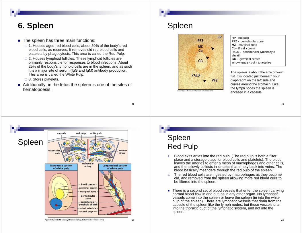
SpleenRP - red pulpPFZ - perifollicular zoneMZ - marginal zoneCo - B cell coronaPALS - periarteriolar lymphocyte sheathGC - germinal centerarrowheads - point to arteries
The spleen is about the size of your fist. It is located just beneath your diaphragm on the left side and curves around the stomach. Like the lymph nodes the spleen is encased in a capsule.
47
Spleen
48
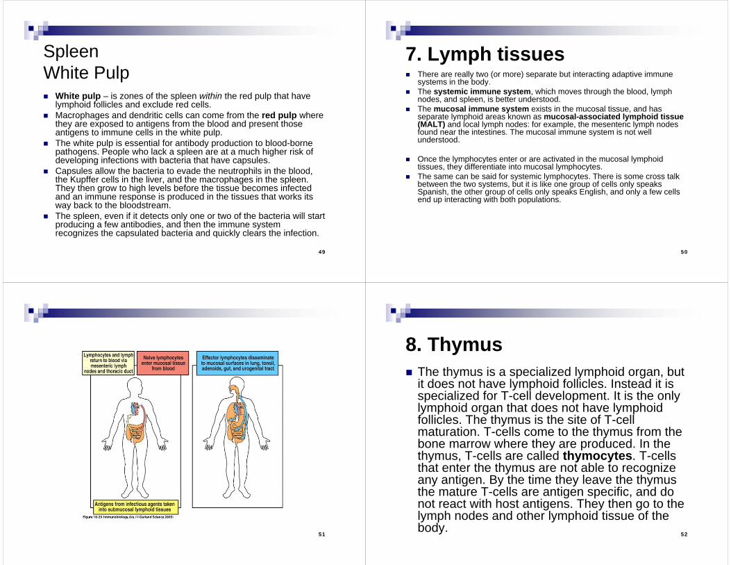
SpleenRed Pulp1. Blood exits arties into the red pulp. (The red pulp is both a filter
place and a storage place for blood cells and platelets). The blood leaves the arteries to enter a mesh of macrophages and other cells, and then slowly collects in sinuses that empty back into veins. The blood basically meanders through the red pulp of the spleen.
2. The red blood cells are ingested by macrophages as they become old, and removed from the spleen allowing more red blood cells to be filtered into the spleen.
There is a second set of blood vessels that enter the spleen carrying normal blood flow in and out, as in any other organ. No lymphatic vessels come into the spleen or leave the spleen (ie into the white pulp of the spleen). There are lymphatic vessels that drain from the capsule of the spleen like the lymph nodes, but those vessels drain into the thoracic duct of the lymphatic system, and not into the spleen.
49
SpleenWhite Pulp White pulp – is zones of the spleen within the red pulp that have
lymphoid follicles and exclude red cells. Macrophages and dendritic cells can come from the red pulp where
they are exposed to antigens from the blood and present those antigens to immune cells in the white pulp.
The white pulp is essential for antibody production to blood-borne pathogens. People who lack a spleen are at a much higher risk of developing infections with bacteria that have capsules.
Capsules allow the bacteria to evade the neutrophils in the blood, the Kupffer cells in the liver, and the macrophages in the spleen. They then grow to high levels before the tissue becomes infected and an immune response is produced in the tissues that works its way back to the bloodstream.
The spleen, even if it detects only one or two of the bacteria will start producing a few antibodies, and then the immune system recognizes the capsulated bacteria and quickly clears the infection.
50
7. Lymph tissues There are really two (or more) separate but interacting adaptive immune
systems in the body. The systemic immune system, which moves through the blood, lymph
nodes, and spleen, is better understood. The mucosal immune system exists in the mucosal tissue, and has
separate lymphoid areas known as mucosal-associated lymphoid tissue (MALT) and local lymph nodes: for example, the mesenteric lymph nodes found near the intestines. The mucosal immune system is not well understood.
Once the lymphocytes enter or are activated in the mucosal lymphoid tissues, they differentiate into mucosal lymphocytes.
The same can be said for systemic lymphocytes. There is some cross talk between the two systems, but it is like one group of cells only speaks Spanish, the other group of cells only speaks English, and only a few cells end up interacting with both populations.
51 52
8. Thymus The thymus is a specialized lymphoid organ, but
it does not have lymphoid follicles. Instead it is specialized for T-cell development. It is the only lymphoid organ that does not have lymphoid follicles. The thymus is the site of T-cell maturation. T-cells come to the thymus from the bone marrow where they are produced. In the thymus, T-cells are called thymocytes. T-cells that enter the thymus are not able to recognize any antigen. By the time they leave the thymus the mature T-cells are antigen specific, and do not react with host antigens. They then go to the lymph nodes and other lymphoid tissue of the body.
53
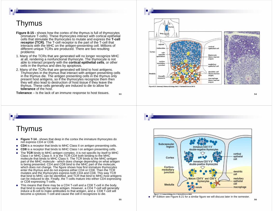
ThymusFigure 8-15 - shows how the cortex of the thymus is full of thymocytes
(immature T-cells). These thymocytes interact with cortical epithelial cells that stimulate the thymocytes to mutate and express the T-cell receptor (TCR). The T-cell receptor is the part of the T-cell that interacts with the MHC on the antigen-presenting cell. Millions of different unique TCRs are produced. There are two resulting problems:
1. Many of the TCRs that are generated will no longer recognize MHC at all, rendering a nonfunctional thymocyte. The thymocyte is not able to interact properly with the cortical epithelial cells, or other cells in the thymus and dies by apoptosis.
2. Many of the TCRs that are generated will bind to host antigens. Thymocytes in the thymus that interact with antigen presenting cells in the thymus die. The antigen presenting cells in the thymus only present host antigens, so if the thymocytes recognize them then they will also lead to destruction of host tissue if they leave the thymus. These cells generally are induced to die to allow for tolerance of the host.
Tolerance – Is the lack of an immune response to host tissues. 54
55
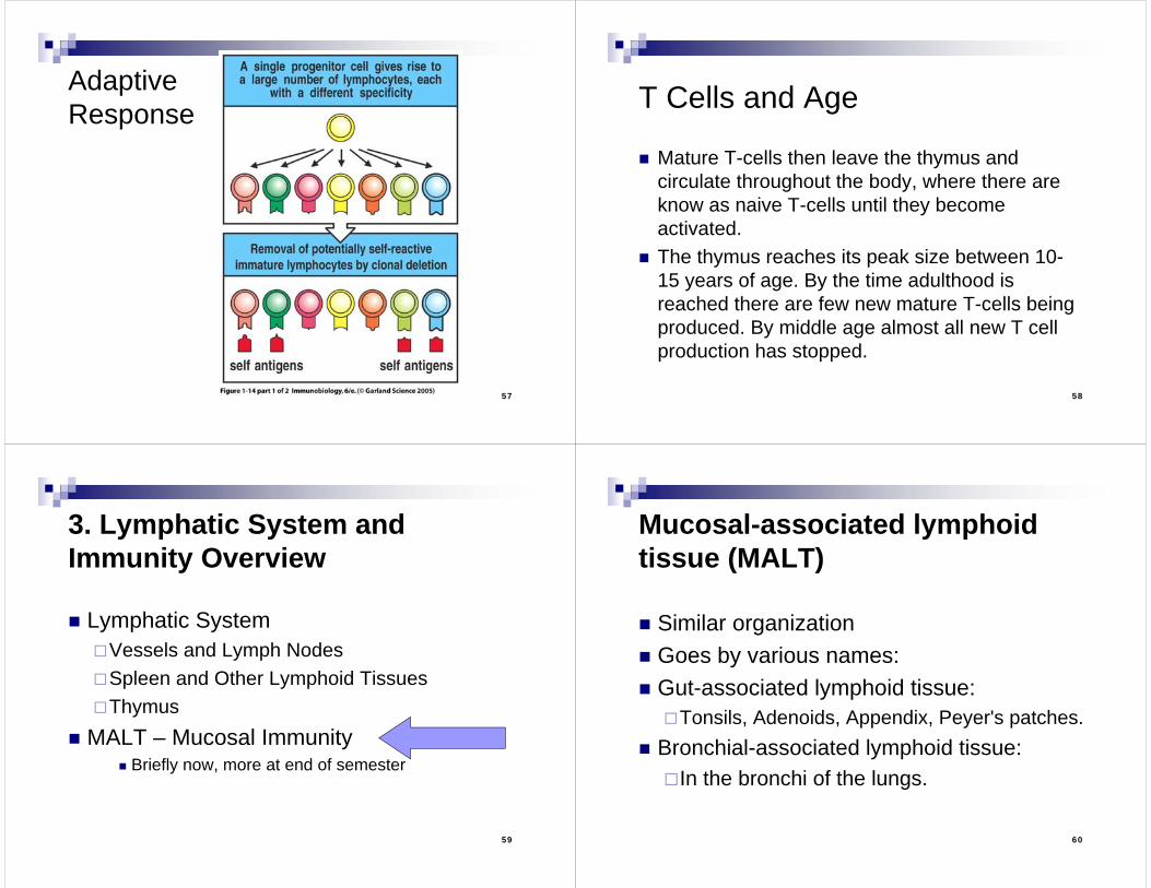
Thymus Figure 7-14 - shows that deep in the cortex the immature thymocytes do
not express CD4 or CD8. CD4 is a receptor that binds to MHC Class II on antigen presenting cells. CD8 is a receptor that binds to MHC Class I on antigen presenting cells. The TCR binds to MHC-antigen complex, it is not specific by itself to MHC
Class I or MHC Class II. It is the TCR-CD4 both binding to the MHC molecule that binds to MHC Class II. The TCR binds to the MHC-antigen part of the MHC molecule - which does change depending on what antigen is being presented. CD4 and CD8 bind to the MHC part of the molecule, which does not change. The figure shows how first immature thymocytes enter the thymus and do not express either CD4 or CD8. Then the TCR mutates and the thymocytes express both CD4 and CD8. This way TCR that bind to MHC can be identified, and TCR that bind to MHC-host antigens can be induced to die. Finally, the T-cells mature into either CD4 expressing or CD8 expressing T-cells.
This means that there may be a CD4 T-cell and a CD8 T-cell in the body that bind to exactly the same antigen. However, a CD4 T-cell will generally induce a B-cell to make antibodies to that antigen, and a CD8 T-cell will become a cytotoxic T-cell and cause the cell it recognizes to die.
56 8th Edition see Figure 8.21 for a similar figure we will discuss later in the semester.
57
AdaptiveResponse
58
T Cells and Age
Mature T-cells then leave the thymus and circulate throughout the body, where there are know as naive T-cells until they become activated.
The thymus reaches its peak size between 10-15 years of age. By the time adulthood is reached there are few new mature T-cells being produced. By middle age almost all new T cell production has stopped.
59
3. Lymphatic System and Immunity Overview
Lymphatic SystemVessels and Lymph NodesSpleen and Other Lymphoid TissuesThymus
MALT – Mucosal Immunity Briefly now, more at end of semester
60
Mucosal-associated lymphoid tissue (MALT)
Similar organization Goes by various names: Gut-associated lymphoid tissue:Tonsils, Adenoids, Appendix, Peyer's patches.
Bronchial-associated lymphoid tissue:In the bronchi of the lungs.
61
Mucosal-associated lymphoid tissue (MALT) The mucosal immune system exists in the mucosal tissue, and
has separate lymphoid areas known as mucosal-associated lymphoid tissue (MALT) and local lymph nodes: for example, the mesenteric lymph nodes found near the intestines. The mucosal immune system is not well understood.
The mucosal immune system is constantly stimulated and somehow decides not to make antibodies to carrots, but to make antibodies to Salmonella. How it tells a carrot from Salmonella is not always clear. Immune responses that are generated in one mucosal tissue, for instance in the adenoids at the back of the nose, will rapidly show up in ALL mucosal tissues (the gut, reproductive tract, the trachea) but will not show up, except at very low levels, in the systemic immune system (blood or body lymph nodes). This means that once the lymphocytes enter or are activated in the mucosal lymphoid tissues, they differentiate into mucosal lymphocytes.
62
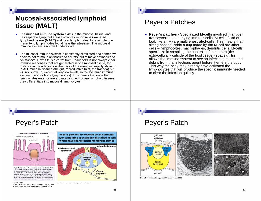
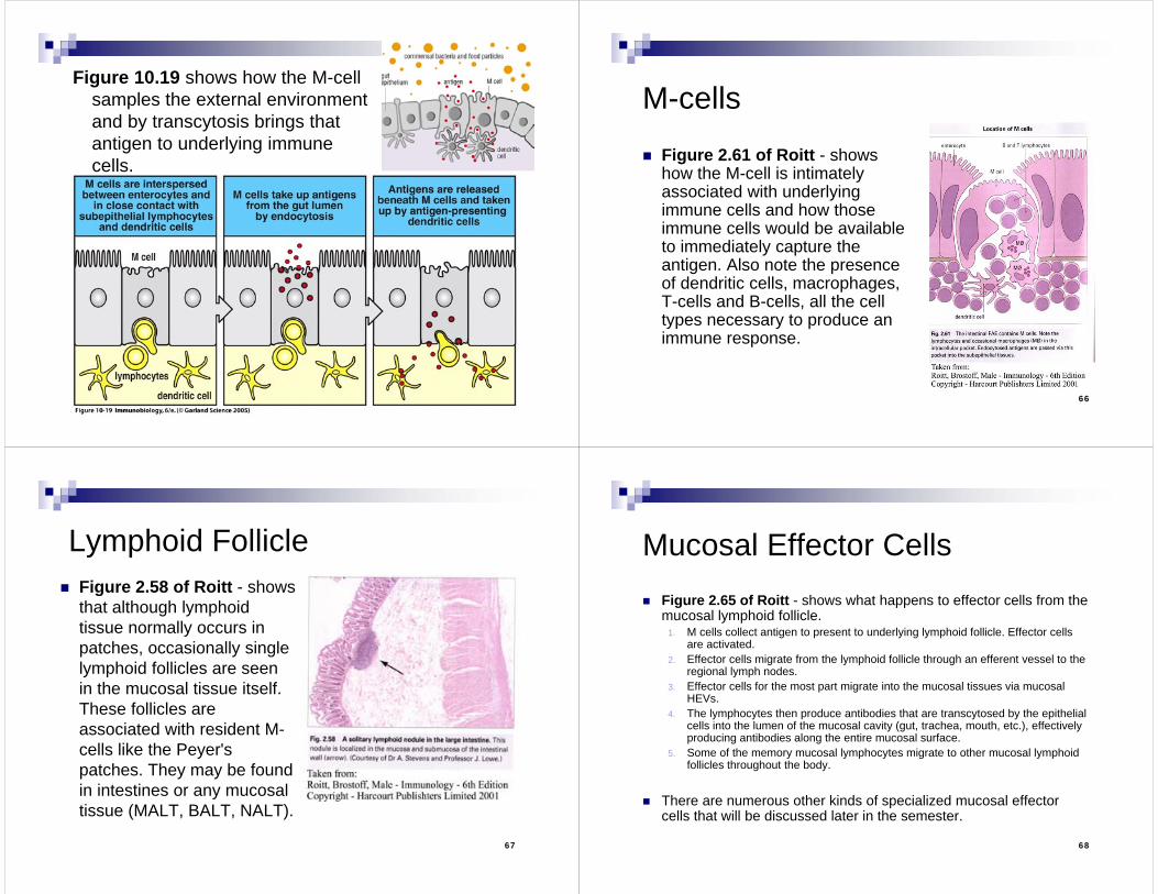
Peyer’s Patches Peyer’s patches - Specialized M-cells involved in antigen
transcytosis to underlying immune cells. M-cells (kind of look like an M) are multifenestrated-cells. This means that sitting nestled inside a cup made by the M-cell are other cells – lymphocytes, macrophages, dendritic cells. M-cells specialize in sampling the contents of the lumen (the extracellular - outside of the host tissue - space). This allows the immune system to see an infectious agent, and debris from that infectious agent before it enters the body. This way the body may already have activated the lymphocytes that will produce the specific immunity needed to clear the infection quickly.
63
Peyer’s Patch
64
Peyer’s Patch
65
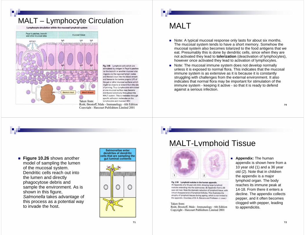
Figure 10.19 shows how the M-cell samples the external environment and by transcytosis brings that antigen to underlying immune cells.
66
M-cells
Figure 2.61 of Roitt - shows how the M-cell is intimately associated with underlying immune cells and how those immune cells would be available to immediately capture the antigen. Also note the presence of dendritic cells, macrophages, T-cells and B-cells, all the cell types necessary to produce an immune response.
67
Lymphoid Follicle Figure 2.58 of Roitt - shows
that although lymphoid tissue normally occurs in patches, occasionally single lymphoid follicles are seen in the mucosal tissue itself. These follicles are associated with resident M-cells like the Peyer's patches. They may be found in intestines or any mucosal tissue (MALT, BALT, NALT).
68
Mucosal Effector Cells Figure 2.65 of Roitt - shows what happens to effector cells from the
mucosal lymphoid follicle. 1. M cells collect antigen to present to underlying lymphoid follicle. Effector cells
are activated.2. Effector cells migrate from the lymphoid follicle through an efferent vessel to the
regional lymph nodes. 3. Effector cells for the most part migrate into the mucosal tissues via mucosal
HEVs. 4. The lymphocytes then produce antibodies that are transcytosed by the epithelial
cells into the lumen of the mucosal cavity (gut, trachea, mouth, etc.), effectively producing antibodies along the entire mucosal surface.
5. Some of the memory mucosal lymphocytes migrate to other mucosal lymphoid follicles throughout the body.
There are numerous other kinds of specialized mucosal effector cells that will be discussed later in the semester.
69
MALT – Lymphocyte Circulation
70
MALT Note: A typical mucosal response only lasts for about six months.
The mucosal system tends to have a short memory. Somehow the mucosal system also becomes tolarized to the food antigens that we eat. Presumably this is done by dendritic cells, since when they are not activated they lead to tolerization (deactivation of lymphocytes), however once activated they lead to activation of lymphocytes.
Note: The mucosal immune system does not develop normally unless it is exposed to normal flora. This indicates that the mucosal immune system is as extensive as it is because it is constantly struggling with challenges from the external environment. It also indicates that normal flora may be important for stimulation of the immune system - keeping it active - so that it is ready to defend against a serious infection.
71
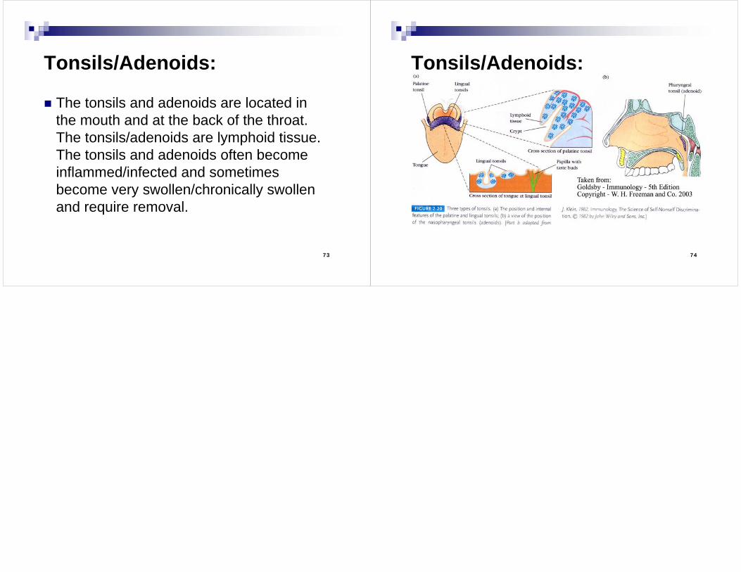
Figure 10.26 shows another model of sampling the lumen of the mucosal system. Dendritic cells reach out into the lumen and directly phagocytose debris and sample the environment. As is shown in this figure, Salmonella takes advantage of this process as a potential way to invade the host.
72
MALT-Lymphoid Tissue Appendix: The human
appendix is shown here from a 10 year old (1) and a 36 year old (2). Note that in children the appendix is a major lymphoid organ. The body reaches its immune peak at 14-18. From there it enters a decline. The appendix collects pepper, and it often becomes clogged with pepper, leading to appendicitis.
73
Tonsils/Adenoids:
The tonsils and adenoids are located in the mouth and at the back of the throat. The tonsils/adenoids are lymphoid tissue. The tonsils and adenoids often become inflammed/infected and sometimes become very swollen/chronically swollen and require removal.
74
Tonsils/Adenoids: