Embed Size (px)
Citation preview
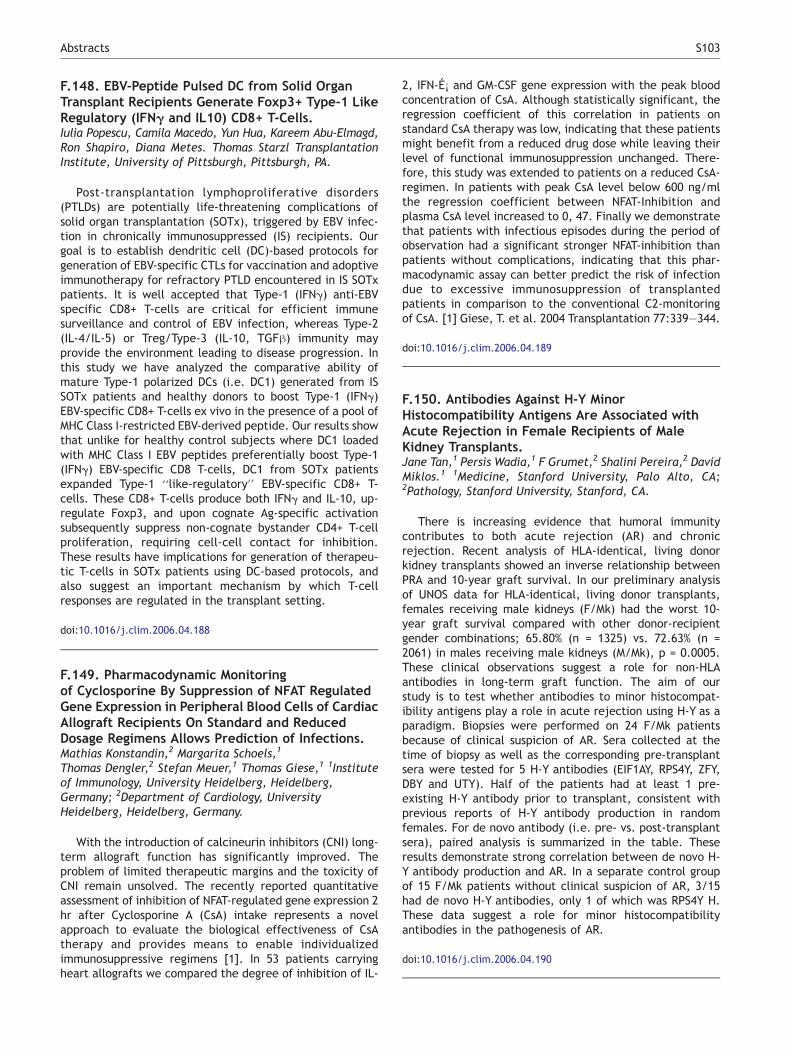
F.148. EBV-Peptide Pulsed DC from Solid OrganTransplant Recipients Generate Foxp3+ Type-1 LikeRegulatory (IFN; and IL10) CD8+ T-Cells.Iulia Popescu, Camila Macedo, Yun Hua, Kareem Abu-Elmagd,Ron Shapiro, Diana Metes. Thomas Starzl TransplantationInstitute, University of Pittsburgh, Pittsburgh, PA.
Post-transplantation lymphoproliferative disorders(PTLDs) are potentially life-threatening complications ofsolid organ transplantation (SOTx), triggered by EBV infec-tion in chronically immunosuppressed (IS) recipients. Ourgoal is to establish dendritic cell (DC)-based protocols forgeneration of EBV-specific CTLs for vaccination and adoptiveimmunotherapy for refractory PTLD encountered in IS SOTxpatients. It is well accepted that Type-1 (IFNg) anti-EBVspecific CD8+ T-cells are critical for efficient immunesurveillance and control of EBV infection, whereas Type-2(IL-4/IL-5) or Treg/Type-3 (IL-10, TGFh) immunity mayprovide the environment leading to disease progression. Inthis study we have analyzed the comparative ability ofmature Type-1 polarized DCs (i.e. DC1) generated from ISSOTx patients and healthy donors to boost Type-1 (IFNg)EBV-specific CD8+ T-cells ex vivo in the presence of a pool ofMHC Class I-restricted EBV-derived peptide. Our results showthat unlike for healthy control subjects where DC1 loadedwith MHC Class I EBV peptides preferentially boost Type-1(IFNg) EBV-specific CD8 T-cells, DC1 from SOTx patientsexpanded Type-1 ‘‘like-regulatory’’ EBV-specific CD8+ T-cells. These CD8+ T-cells produce both IFNg and IL-10, up-regulate Foxp3, and upon cognate Ag-specific activationsubsequently suppress non-cognate bystander CD4+ T-cellproliferation, requiring cell-cell contact for inhibition.These results have implications for generation of therapeu-tic T-cells in SOTx patients using DC-based protocols, andalso suggest an important mechanism by which T-cellresponses are regulated in the transplant setting.
doi:10.1016/j.clim.2006.04.188
F.149. Pharmacodynamic Monitoringof Cyclosporine By Suppression of NFAT RegulatedGene Expression in Peripheral Blood Cells of CardiacAllograft Recipients On Standard and ReducedDosage Regimens Allows Prediction of Infections.Mathias Konstandin,2 Margarita Schoels,1
Thomas Dengler,2 Stefan Meuer,1 Thomas Giese,1 1Instituteof Immunology, University Heidelberg, Heidelberg,Germany; 2Department of Cardiology, UniversityHeidelberg, Heidelberg, Germany.
With the introduction of calcineurin inhibitors (CNI) long-term allograft function has significantly improved. Theproblem of limited therapeutic margins and the toxicity ofCNI remain unsolved. The recently reported quantitativeassessment of inhibition of NFAT-regulated gene expression 2hr after Cyclosporine A (CsA) intake represents a novelapproach to evaluate the biological effectiveness of CsAtherapy and provides means to enable individualizedimmunosuppressive regimens [1]. In 53 patients carryingheart allografts we compared the degree of inhibition of IL-
2, IFN-ER and GM-CSF gene expression with the peak bloodconcentration of CsA. Although statistically significant, theregression coefficient of this correlation in patients onstandard CsA therapy was low, indicating that these patientsmight benefit from a reduced drug dose while leaving theirlevel of functional immunosuppression unchanged. There-fore, this study was extended to patients on a reduced CsA-regimen. In patients with peak CsA level below 600 ng/mlthe regression coefficient between NFAT-Inhibition andplasma CsA level increased to 0, 47. Finally we demonstratethat patients with infectious episodes during the period ofobservation had a significant stronger NFAT-inhibition thanpatients without complications, indicating that this phar-macodynamic assay can better predict the risk of infectiondue to excessive immunosuppression of transplantedpatients in comparison to the conventional C2-monitoringof CsA. [1] Giese, T. et al. 2004 Transplantation 77:339—344.
doi:10.1016/j.clim.2006.04.189
F.150. Antibodies Against H-Y MinorHistocompatibility Antigens Are Associated withAcute Rejection in Female Recipients of MaleKidney Transplants.Jane Tan,1 Persis Wadia,1 F Grumet,2 Shalini Pereira,2 DavidMiklos.1 1Medicine, Stanford University, Palo Alto, CA;2Pathology, Stanford University, Stanford, CA.
There is increasing evidence that humoral immunitycontributes to both acute rejection (AR) and chronicrejection. Recent analysis of HLA-identical, living donorkidney transplants showed an inverse relationship betweenPRA and 10-year graft survival. In our preliminary analysisof UNOS data for HLA-identical, living donor transplants,females receiving male kidneys (F/Mk) had the worst 10-year graft survival compared with other donor-recipientgender combinations; 65.80% (n = 1325) vs. 72.63% (n =2061) in males receiving male kidneys (M/Mk), p = 0.0005.These clinical observations suggest a role for non-HLAantibodies in long-term graft function. The aim of ourstudy is to test whether antibodies to minor histocompat-ibility antigens play a role in acute rejection using H-Y as aparadigm. Biopsies were performed on 24 F/Mk patientsbecause of clinical suspicion of AR. Sera collected at thetime of biopsy as well as the corresponding pre-transplantsera were tested for 5 H-Y antibodies (EIF1AY, RPS4Y, ZFY,DBY and UTY). Half of the patients had at least 1 pre-existing H-Y antibody prior to transplant, consistent withprevious reports of H-Y antibody production in randomfemales. For de novo antibody (i.e. pre- vs. post-transplantsera), paired analysis is summarized in the table. Theseresults demonstrate strong correlation between de novo H-Y antibody production and AR. In a separate control groupof 15 F/Mk patients without clinical suspicion of AR, 3/15had de novo H-Y antibodies, only 1 of which was RPS4Y H.These data suggest a role for minor histocompatibilityantibodies in the pathogenesis of AR.
doi:10.1016/j.clim.2006.04.190
Abstracts S103


![Topic (7): Antibodies and Antigens - Doctor 2016...antigens [Ags] (the other two are T-cell receptor [TCR] and major histocompatibility complex [MHC]) {Figure 1}. Antibodies have a](https://img.pdfslide.net/doc/110x75/5f0a54a97e708231d42b2066/topic-7-antibodies-and-antigens-doctor-2016-antigens-ags-the-other-two.jpg)