Embed Size (px)
Citation preview
Facts About
Rare Muscular Dystrophies(Congenital, Distal, Emery-Dreifuss and Oculopharyngeal)
Updated December 2009
2 CMD, DD, EDMD and OPMD • ©2011 MDA
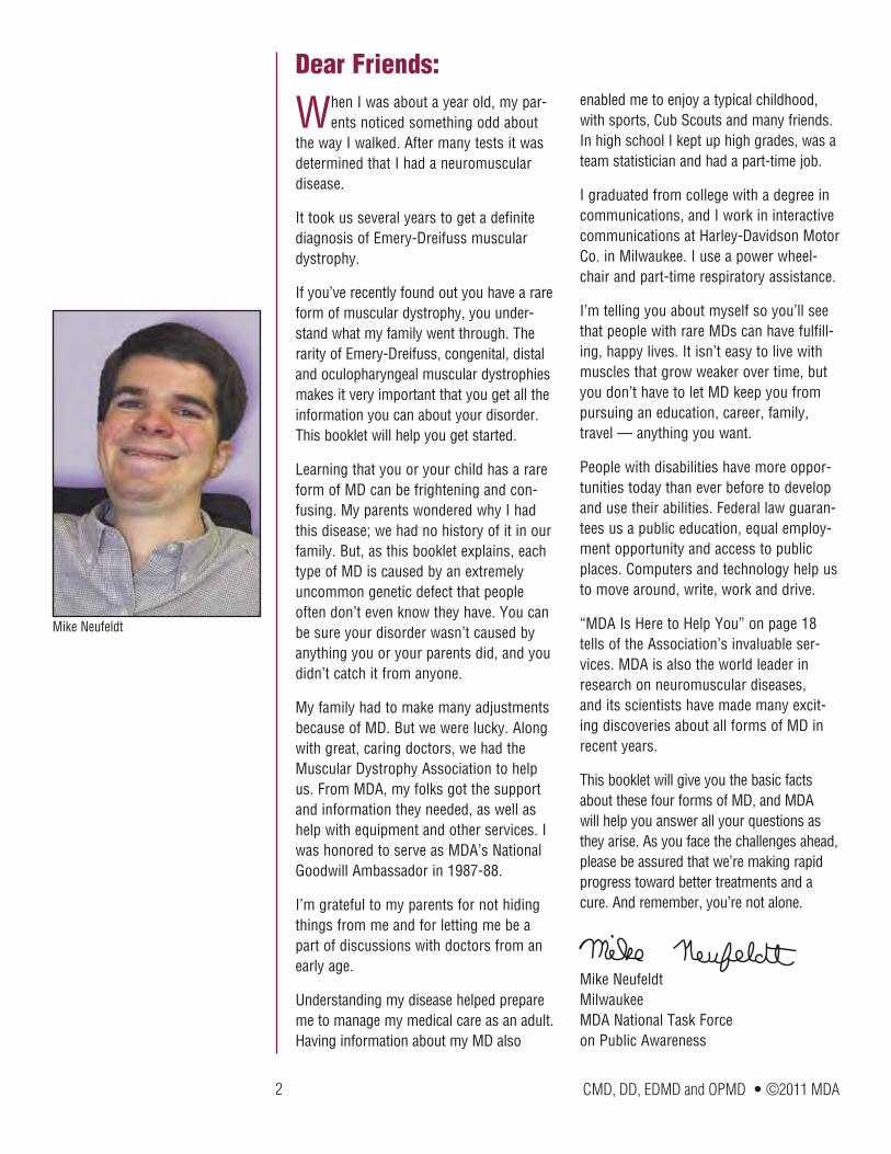
Dear Friends:
When I was about a year old, my par-ents noticed something odd about
the way I walked. After many tests it was determined that I had a neuromuscular disease.
It took us several years to get a definite diagnosis of Emery-Dreifuss muscular dystrophy.
If you’ve recently found out you have a rare form of muscular dystrophy, you under-stand what my family went through. The rarity of Emery-Dreifuss, congenital, distal and oculopharyngeal muscular dystrophies makes it very important that you get all the information you can about your disorder. This booklet will help you get started.
Learning that you or your child has a rare form of MD can be frightening and con-fusing. My parents wondered why I had this disease; we had no history of it in our family. But, as this booklet explains, each type of MD is caused by an extremely uncommon genetic defect that people often don’t even know they have. You can be sure your disorder wasn’t caused by anything you or your parents did, and you didn’t catch it from anyone.
My family had to make many adjustments because of MD. But we were lucky. Along with great, caring doctors, we had the Muscular Dystrophy Association to help us. From MDA, my folks got the support and information they needed, as well as help with equipment and other services. I was honored to serve as MDA’s National Goodwill Ambassador in 1987-88.
I’m grateful to my parents for not hiding things from me and for letting me be a part of discussions with doctors from an early age.
Understanding my disease helped prepare me to manage my medical care as an adult. Having information about my MD also
enabled me to enjoy a typical childhood, with sports, Cub Scouts and many friends. In high school I kept up high grades, was a team statistician and had a part-time job.
I graduated from college with a degree in communications, and I work in interactive communications at Harley-Davidson Motor Co. in Milwaukee. I use a power wheel-chair and part-time respiratory assistance.
I’m telling you about myself so you’ll see that people with rare MDs can have fulfill-ing, happy lives. It isn’t easy to live with muscles that grow weaker over time, but you don’t have to let MD keep you from pursuing an education, career, family, travel — anything you want.
People with disabilities have more oppor-tunities today than ever before to develop and use their abilities. Federal law guaran-tees us a public education, equal employ-ment opportunity and access to public places. Computers and technology help us to move around, write, work and drive.
“MDA Is Here to Help You” on page 18 tells of the Association’s invaluable ser-vices. MDA is also the world leader in research on neuromuscular diseases, and its scientists have made many excit-ing discoveries about all forms of MD in recent years.
This booklet will give you the basic facts about these four forms of MD, and MDA will help you answer all your questions as they arise. As you face the challenges ahead, please be assured that we’re making rapid progress toward better treatments and a cure. And remember, you’re not alone.
Mike Neufeldt Milwaukee MDA National Task Force on Public Awareness
Mike Neufeldt
3 CMD, DD, EDMD and OPMD • ©2011 MDA
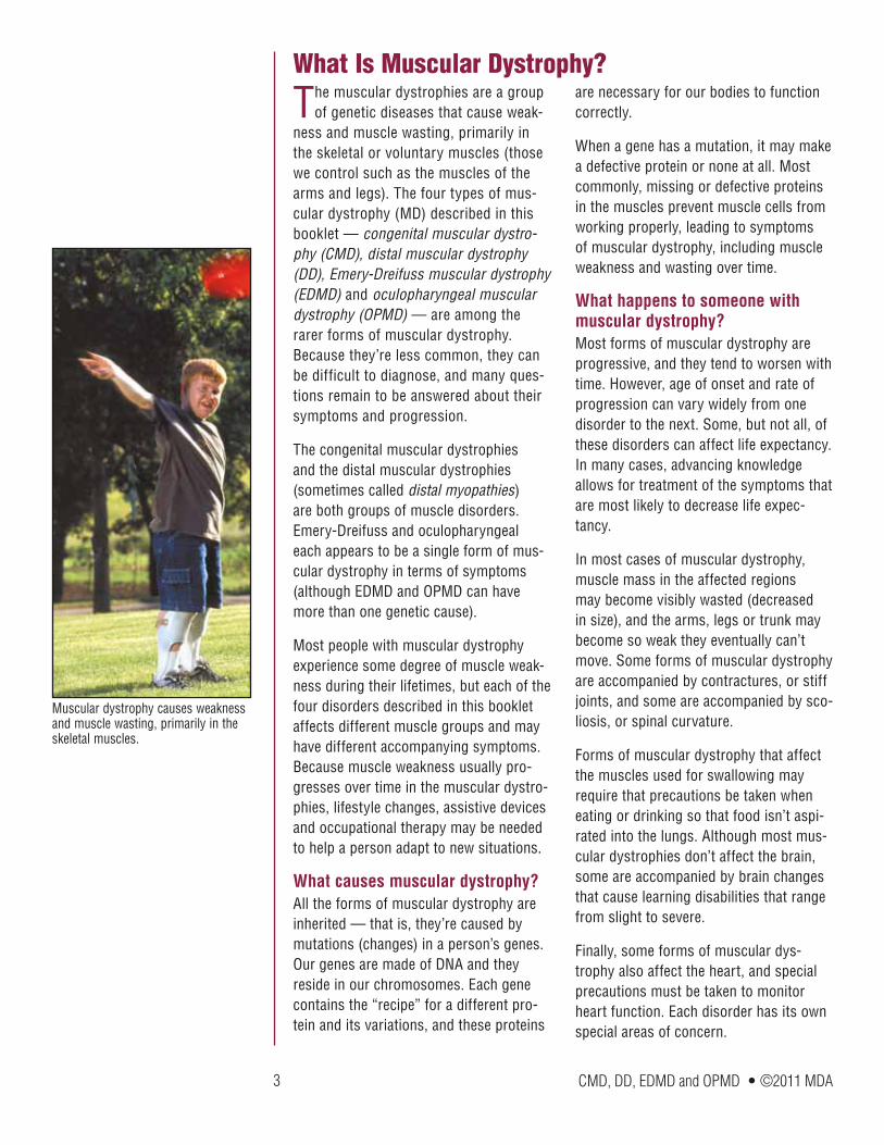
The muscular dystrophies are a group of genetic diseases that cause weak-
ness and muscle wasting, primarily in the skeletal or voluntary muscles (those we control such as the muscles of the arms and legs). The four types of mus-cular dystrophy (MD) described in this booklet — congenital muscular dystro-phy (CMD), distal muscular dystrophy (DD), Emery-Dreifuss muscular dystrophy (EDMD) and oculopharyngeal muscular dystrophy (OPMD) — are among the rarer forms of muscular dystrophy. Because they’re less common, they can be difficult to diagnose, and many ques-tions remain to be answered about their symptoms and progression.
The congenital muscular dystrophies and the distal muscular dystrophies (sometimes called distal myopathies) are both groups of muscle disorders. Emery-Dreifuss and oculopharyngeal each appears to be a single form of mus-cular dystrophy in terms of symptoms (although EDMD and OPMD can have more than one genetic cause).
Most people with muscular dystrophy experience some degree of muscle weak-ness during their lifetimes, but each of the four disorders described in this booklet affects different muscle groups and may have different accompanying symptoms. Because muscle weakness usually pro-gresses over time in the muscular dystro-phies, lifestyle changes, assistive devices and occupational therapy may be needed to help a person adapt to new situations.
What causes muscular dystrophy?All the forms of muscular dystrophy are inherited — that is, they’re caused by mutations (changes) in a person’s genes. Our genes are made of DNA and they reside in our chromosomes. Each gene contains the “recipe” for a different pro-tein and its variations, and these proteins
are necessary for our bodies to function correctly.
When a gene has a mutation, it may make a defective protein or none at all. Most commonly, missing or defective proteins in the muscles prevent muscle cells from working properly, leading to symptoms of muscular dystrophy, including muscle weakness and wasting over time.
What happens to someone with muscular dystrophy?Most forms of muscular dystrophy are progressive, and they tend to worsen with time. However, age of onset and rate of progression can vary widely from one disorder to the next. Some, but not all, of these disorders can affect life expectancy. In many cases, advancing knowledge allows for treatment of the symptoms that are most likely to decrease life expec-tancy.
In most cases of muscular dystrophy, muscle mass in the affected regions may become visibly wasted (decreased in size), and the arms, legs or trunk may become so weak they eventually can’t move. Some forms of muscular dystrophy are accompanied by contractures, or stiff joints, and some are accompanied by sco-liosis, or spinal curvature.
Forms of muscular dystrophy that affect the muscles used for swallowing may require that precautions be taken when eating or drinking so that food isn’t aspi-rated into the lungs. Although most mus-cular dystrophies don’t affect the brain, some are accompanied by brain changes that cause learning disabilities that range from slight to severe.
Finally, some forms of muscular dys-trophy also affect the heart, and special precautions must be taken to monitor heart function. Each disorder has its own special areas of concern.
What Is Muscular Dystrophy?
Muscular dystrophy causes weakness and muscle wasting, primarily in the skeletal muscles.
4 CMD, DD, EDMD and OPMD • ©2011 MDA
What tests are used to diagnose muscular dystrophy? In diagnosing any form of muscular dys-trophy, a doctor usually begins by taking a patient and family history and perform-ing a physical examination. Much can be learned from these, including the pattern of weakness. The history and physical go a long way toward making the diagnosis, even before any complicated diagnostic tests are done.
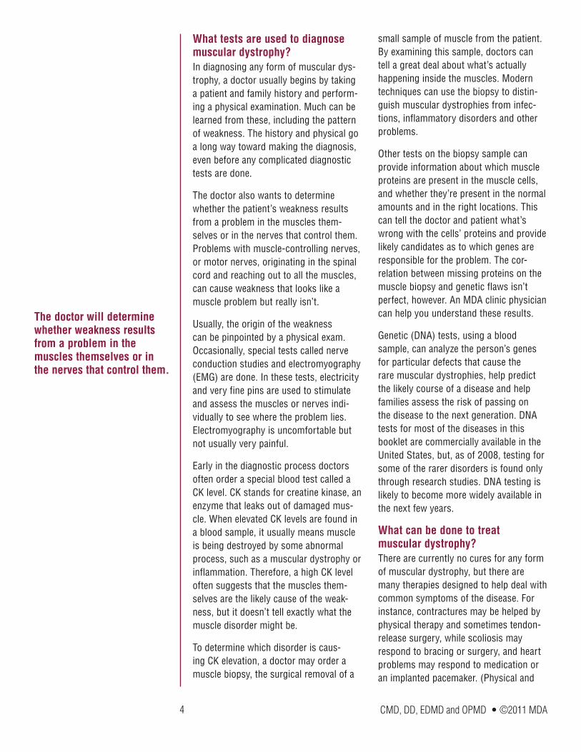
The doctor also wants to determine whether the patient’s weakness results from a problem in the muscles them-selves or in the nerves that control them. Problems with muscle-controlling nerves, or motor nerves, originating in the spinal cord and reaching out to all the muscles, can cause weakness that looks like a muscle problem but really isn’t.
Usually, the origin of the weakness can be pinpointed by a physical exam. Occasionally, special tests called nerve conduction studies and electromyography (EMG) are done. In these tests, electricity and very fine pins are used to stimulate and assess the muscles or nerves indi-vidually to see where the problem lies. Electromyography is uncomfortable but not usually very painful.
Early in the diagnostic process doctors often order a special blood test called a CK level. CK stands for creatine kinase, an enzyme that leaks out of damaged mus-cle. When elevated CK levels are found in a blood sample, it usually means muscle is being destroyed by some abnormal process, such as a muscular dystrophy or inflammation. Therefore, a high CK level often suggests that the muscles them-selves are the likely cause of the weak-ness, but it doesn’t tell exactly what the muscle disorder might be.
To determine which disorder is caus-ing CK elevation, a doctor may order a muscle biopsy, the surgical removal of a
small sample of muscle from the patient. By examining this sample, doctors can tell a great deal about what’s actually happening inside the muscles. Modern techniques can use the biopsy to distin-guish muscular dystrophies from infec-tions, inflammatory disorders and other problems.
Other tests on the biopsy sample can provide information about which muscle proteins are present in the muscle cells, and whether they’re present in the normal amounts and in the right locations. This can tell the doctor and patient what’s wrong with the cells’ proteins and provide likely candidates as to which genes are responsible for the problem. The cor-relation between missing proteins on the muscle biopsy and genetic flaws isn’t perfect, however. An MDA clinic physician can help you understand these results.
Genetic (DNA) tests, using a blood sample, can analyze the person’s genes for particular defects that cause the rare muscular dystrophies, help predict the likely course of a disease and help families assess the risk of passing on the disease to the next generation. DNA tests for most of the diseases in this booklet are commercially available in the United States, but, as of 2008, testing for some of the rarer disorders is found only through research studies. DNA testing is likely to become more widely available in the next few years.
What can be done to treat muscular dystrophy?There are currently no cures for any form of muscular dystrophy, but there are many therapies designed to help deal with common symptoms of the disease. For instance, contractures may be helped by physical therapy and sometimes tendon-release surgery, while scoliosis may respond to bracing or surgery, and heart problems may respond to medication or an implanted pacemaker. (Physical and
The doctor will determine whether weakness results from a problem in the muscles themselves or in the nerves that control them.
5 CMD, DD, EDMD and OPMD • ©2011 MDA
occupational therapies can be arranged through your child’s school or through your MDA clinic.)
Many people with these forms of mus-cular dystrophy live very full lives. Your MDA clinic director will help you plan the best strategy for coping with your or your child’s specific needs.
Congenital muscular dystrophy (CMD)The term congenital muscular dystrophy (CMD) is actually the name for a group of muscular dystrophies that are united by the fact that muscle weakness begins in infancy or very early childhood (typically before age 2). Congenital diseases are those in which the symptoms are present at or soon after birth.
A diagnosis of CMD can be confusing because for many years the term was used as a “catchall” name to describe conditions that looked like other muscular dystrophies, but started much earlier or followed different patterns of inheritance. In recent years doctors have agreed that
there are several categories of “true” CMD, caused by specific gene mutations, and they’re distinct from other muscular dystrophies. It’s possible that some peo-ple who received diagnoses of CMD many years ago may actually have some other known form of muscular dystrophy with an unusually early onset.
Although children with CMD can have dif-ferent associated symptoms, degrees of severity and rates of progression, most exhibit some progressive muscle weak-ness. This weakness, usually first identi-fied as hypotonia, or lack of muscle tone, can make an infant seem “floppy.” Later, infants and toddlers may be slow to meet motor milestones such as rolling over, sit-ting up or walking, or may not meet some milestones at all.
Some of the rarer forms of CMD are also accompanied by significant learning dis-abilities or mental retardation.
What causes CMD?The CMDs are caused by genetic defects that affect important muscle proteins. Most forms of CMD are inherited in an
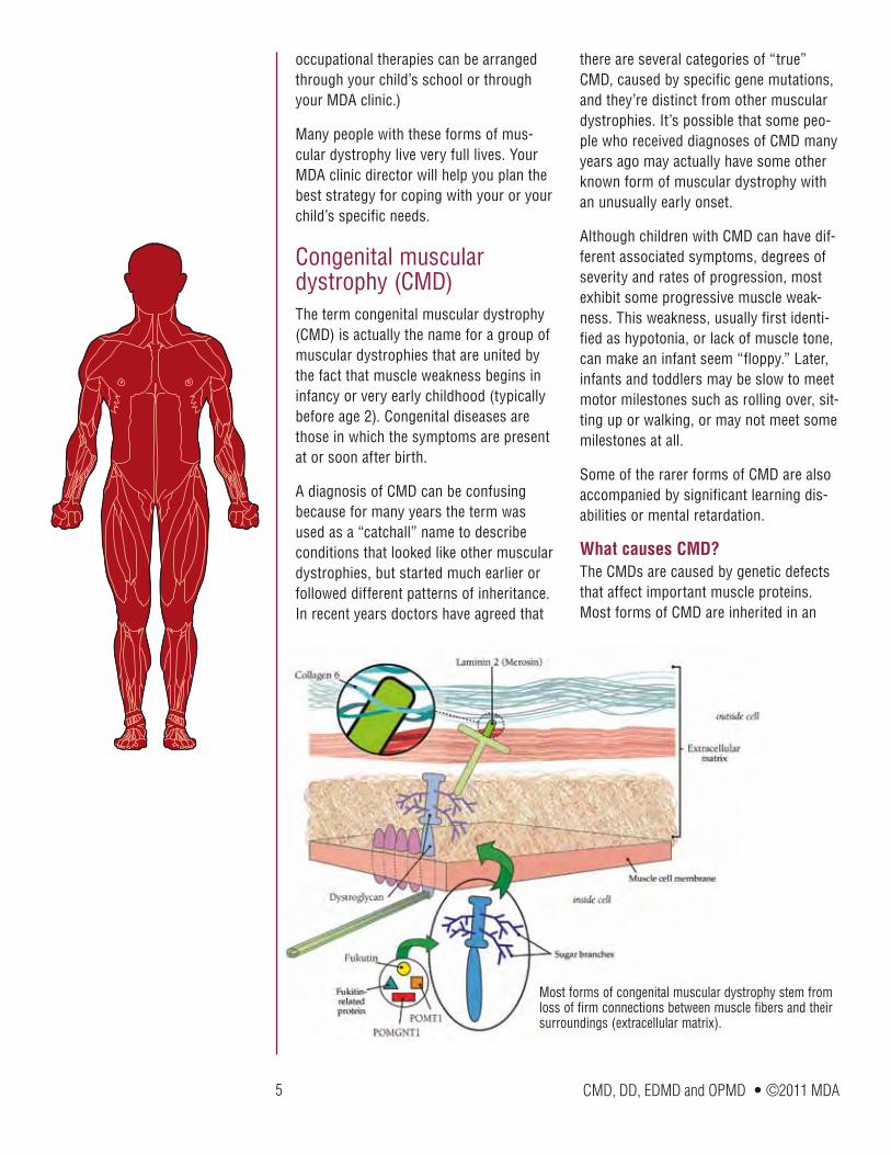
Most forms of congenital muscular dystrophy stem from loss of firm connections between muscle fibers and their surroundings (extracellular matrix).
6 CMD, DD, EDMD and OPMD • ©2011 MDA
autosomal recessive pattern, but at least one form appears to follow a dominant pattern of inheritance. (For more about inheritance patterns, see “Does It Run in the Family?” on page 16.)
It isn’t known why the CMDs cause muscle weakness earlier than other types of muscular dystrophy. One possibility is that the muscle proteins affected in CMD are required early in the development of an infant’s muscle, while muscle proteins linked to other muscular dystrophies don’t become important until the muscles begin to get a lot of use as a child grows.
It’s important to note that just because the muscle weakness in CMD starts earlier, CMD isn’t automatically more severe than other forms of muscular dystrophy. The degree and rate of progression of muscle weakness varies with different forms of CMD and from one child to the next.
In the mid-1990s, researchers found that a deficiency of a protein then called mero-sin and now more often called laminin 2 was the underlying cause of at least some cases of CMD. Merosin normally anchors muscle cells to a structure that encases them (like the skin on a hot dog) called the basal lamina.
Doctors began to classify CMD as either “merosin-deficient” (this page) or “mero-sin-positive.” The gene for merosin is on chromosome 6.
Then, in 1998, researchers identified mutations in the gene for integrin as another cause of CMD (see page 7). Integrin, which surrounds and supports each muscle fiber, connects laminin 2 with proteins inside the cells.
As the 20th century ended, researchers began to suspect that Ullrich’s disease, now known as Ullrich CMD (see page 7), was caused by a lack of collagen 6, a ropelike protein located in the area where laminin 2 is found.
Collagen 6, which helps support the mus-cle fiber, probably affects muscle cells via its connection to laminin 2. Laminin 2, in turn, connects to muscle cells via either of two other proteins: integrin or dystro-glycan.
Dystroglycan links the outer surface of muscle cells with structures outside them via branches, made of sugar molecules, that protrude from its surface and stick to laminin.
The branch structure explains why muta-tions in diverse genes all appear to cause CMD. Each of these proteins contributes in a different way to the process of “sugar coating” (glycosylating) dystroglycan. Several forms of CMD that affect not only the muscles but the eyes and brain — Fukuyama CMD (seen mainly in Japan), muscle-eye-brain disease and Walker-Warburg syndrome (see page 7) — arise from defects in these glycosylation pro-teins.
The illustration on page 5 shows the physical relationships among these pro-teins.
In 2001, researchers identified muta-tions in the gene for selenoprotein N1 as a cause of CMD with a rigid spine (see page 7) and sometimes frozen joints at the elbows, hips, ankles or knees. This protein is believed to play a role in muscle development early in life and doesn’t appear to be part of the linked protein cluster shown below.
For more about the causes of specific CMDs and their inheritance patterns, see “Classification of CMDs” on page 15.
What are the types of CMD?
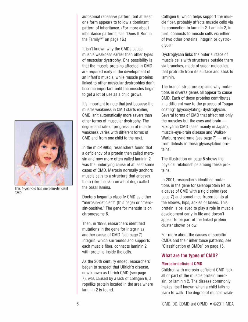
Merosin-deficient CMDChildren with merosin-deficient CMD lack all or part of the muscle protein mero-sin, or laminin 2. The disease commonly makes itself known when a child fails to learn to walk. The degree of muscle weak-
This 4-year-old has merosin-deficient CMD.
7 CMD, DD, EDMD and OPMD • ©2011 MDA
ness can range from severe (never walk-ing) to mild (walking at 2 to 3 years), and depends on how much merosin protein a child has.
This form of CMD progresses very slowly or, in some cases, not at all. Special problems include contractures, difficulty breathing and seizures (in 20 percent of cases). Intelligence is usually normal, but learning disabilities have been docu-mented.
A distinctive diagnostic feature of this type of CMD is found by magnetic reso-nance imaging (MRI). These brain images show changes in the white matter, which consists of nerve fibers that carry mes-sages from the brain to the spinal cord. Despite the appearance on the MRI, those with merosin-negative CMD have few signs of brain impairment in everyday life.
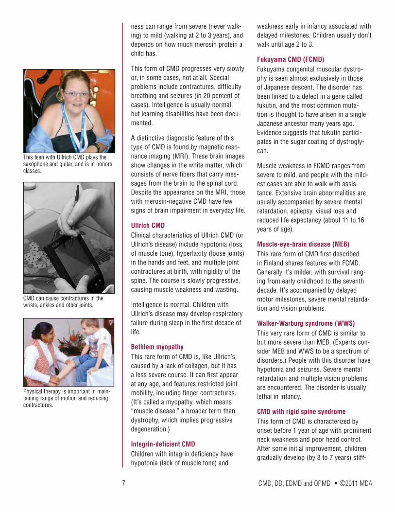
Ullrich CMDClinical characteristics of Ullrich CMD (or Ullrich’s disease) include hypotonia (loss of muscle tone), hyperlaxity (loose joints) in the hands and feet, and multiple joint contractures at birth, with rigidity of the spine. The course is slowly progressive, causing muscle weakness and wasting.
Intelligence is normal. Children with Ullrich’s disease may develop respiratory failure during sleep in the first decade of life.
Bethlem myopathyThis rare form of CMD is, like Ullrich’s, caused by a lack of collagen, but it has a less severe course. It can first appear at any age, and features restricted joint mobility, including finger contractures. (It’s called a myopathy, which means “muscle disease,” a broader term than dystrophy, which implies progressive degeneration.)
Integrin-deficient CMDChildren with integrin deficiency have hypotonia (lack of muscle tone) and
weakness early in infancy associated with delayed milestones. Children usually don’t walk until age 2 to 3.
Fukuyama CMD (FCMD)Fukuyama congenital muscular dystro-phy is seen almost exclusively in those of Japanese descent. The disorder has been linked to a defect in a gene called fukutin, and the most common muta-tion is thought to have arisen in a single Japanese ancestor many years ago. Evidence suggests that fukutin partici-pates in the sugar coating of dystrogly-can.
Muscle weakness in FCMD ranges from severe to mild, and people with the mild-est cases are able to walk with assis-tance. Extensive brain abnormalities are usually accompanied by severe mental retardation, epilepsy, visual loss and reduced life expectancy (about 11 to 16 years of age).
Muscle-eye-brain disease (MEB)This rare form of CMD first described in Finland shares features with FCMD. Generally it’s milder, with survival rang-ing from early childhood to the seventh decade. It’s accompanied by delayed motor milestones, severe mental retarda-tion and vision problems.
Walker-Warburg syndrome (WWS)This very rare form of CMD is similar to but more severe than MEB. (Experts con-sider MEB and WWS to be a spectrum of disorders.) People with this disorder have hypotonia and seizures. Severe mental retardation and multiple vision problems are encountered. The disorder is usually lethal in infancy.
CMD with rigid spine syndromeThis form of CMD is characterized by onset before 1 year of age with prominent neck weakness and poor head control. After some initial improvement, children gradually develop (by 3 to 7 years) stiff-
This teen with Ullrich CMD plays the saxophone and guitar, and is in honors classes.
CMD can cause contractures in the wrists, ankles and other joints.
Physical therapy is important in main-taining range of motion and reducing contractures.
8 CMD, DD, EDMD and OPMD • ©2011 MDA
ness or rigidity of the spine. To a lesser extent contractures of limb muscles are seen.
By the teens, the respiratory muscles are affected, while limb muscle strength is less affected. Intellectual function is normal.
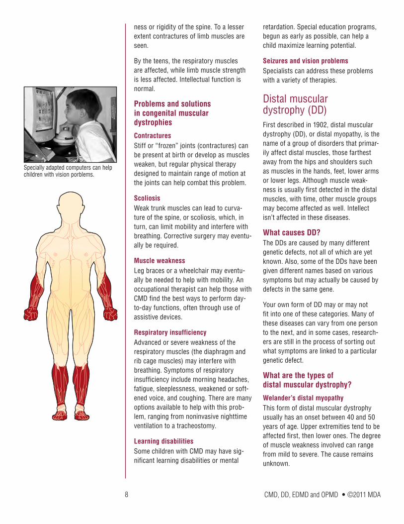
Problems and solutions in congenital muscular dystrophies
ContracturesStiff or “frozen” joints (contractures) can be present at birth or develop as muscles weaken, but regular physical therapy designed to maintain range of motion at the joints can help combat this problem.
ScoliosisWeak trunk muscles can lead to curva-ture of the spine, or scoliosis, which, in turn, can limit mobility and interfere with breathing. Corrective surgery may eventu-ally be required.
Muscle weaknessLeg braces or a wheelchair may eventu-ally be needed to help with mobility. An occupational therapist can help those with CMD find the best ways to perform day-to-day functions, often through use of assistive devices.
Respiratory insufficiencyAdvanced or severe weakness of the respiratory muscles (the diaphragm and rib cage muscles) may interfere with breathing. Symptoms of respiratory insufficiency include morning headaches, fatigue, sleeplessness, weakened or soft-ened voice, and coughing. There are many options available to help with this prob-lem, ranging from noninvasive nighttime ventilation to a tracheostomy.
Learning disabilitiesSome children with CMD may have sig-nificant learning disabilities or mental
retardation. Special education programs, begun as early as possible, can help a child maximize learning potential.
Seizures and vision problemsSpecialists can address these problems with a variety of therapies.
Distal muscular dystrophy (DD)First described in 1902, distal muscular dystrophy (DD), or distal myopathy, is the name of a group of disorders that primar-ily affect distal muscles, those farthest away from the hips and shoulders such as muscles in the hands, feet, lower arms or lower legs. Although muscle weak-ness is usually first detected in the distal muscles, with time, other muscle groups may become affected as well. Intellect isn’t affected in these diseases.
What causes DD?The DDs are caused by many different genetic defects, not all of which are yet known. Also, some of the DDs have been given different names based on various symptoms but may actually be caused by defects in the same gene.
Your own form of DD may or may not fit into one of these categories. Many of these diseases can vary from one person to the next, and in some cases, research-ers are still in the process of sorting out what symptoms are linked to a particular genetic defect.
What are the types of distal muscular dystrophy?
Welander’s distal myopathyThis form of distal muscular dystrophy usually has an onset between 40 and 50 years of age. Upper extremities tend to be affected first, then lower ones. The degree of muscle weakness involved can range from mild to severe. The cause remains unknown.
Specially adapted computers can help children with vision porblems.
9 CMD, DD, EDMD and OPMD • ©2011 MDA
Finnish (tibial) distal myopathyFinnish muscular dystrophy features weakness starting after age 40 in the lower extremities (particularly the mus-cles over the tibia, a bone in the lower leg) and progressing slowly to the upper extremities and trunk muscles. Cardiac problems can be a feature. This distal myopathy results from mutations in the protein titin, which plays a role in muscle fiber structure and force generation.
Finnish muscular dystrophy (also called tibial MD) can be severe or benign, and typically affects only people of Finnish descent. Those with only one defective gene experience mild weakness of the tibial leg muscles (front of the calf) some-time after age 40. Those with two defec-tive genes have progressive weakness starting in childhood and may lose the ability to walk by age 30.
Miyoshi distal myopathyThis disorder involves weakness that begins in the lower extremities, especially in the calf muscles. It can progress to other muscles as well. Symptoms usually begin between 15 and 30 years of age.
The genetic defects that cause Miyoshi myopathy are in the gene for the dysferlin protein.
Defects in the dysferlin gene also can cause limb-girdle muscular dystrophy 2B, which results in muscle weakness in and around the hips and shoulders. People with the same genetic defect in their dys-ferlin genes can have either disease, and it isn’t known what determines which pat-tern of symptoms a person gets.
Nonaka distal myopathyUsually found in families of Japanese descent, this DD has symptoms that begin between ages 20 and 40. The ante-rior lower leg muscles (those in the front of the leg) are typically affected first, but the disease may progress to affect upper
arm and leg muscles and neck muscles. The quadriceps muscles (in the thigh) tend to remain strong.
The disease is caused by defects in the GNE gene, the same gene that underlies one form of hereditary inclusion-body myositis (HIBM2). (This condition also is called inclusion-body myopathy.)
The GNE protein that comes from this gene modifies compounds on cell surfac-es in a way that’s needed for cells to sig-nal each other and adhere to each other.
Gowers-Laing distal myopathyThis disorder has its onset from child-hood to 25 years of age. Weakness is first seen in the leg and neck muscles, and progresses slowly to include upper leg muscles, hands and more neck muscles.
Gowers-Laing distal myopathy results from mutations in the MYH7 gene, which instructs for myosin heavy chain 7, a pro-tein that participates in muscle contrac-tion.
Hereditary inclusion-body myositis (myopathy) type 1 (HIBM1)HIBM1 usually begins between the ages of 25 and 40, first affecting the muscles that lift the front of the foot and the thigh muscles. Other muscles can be affected later. Under the microscope, muscle cells show inclusion bodies, which are abnormal clumps of cellular material; and vacuoles, which are cellular bubbles. The cause is unknown.
Distal myopathy with vocal cord and pharyngeal weakness This disorder has been linked to chromo-some 5 in the same region as the gene that’s defective in limb-girdle MD type 1A. Symptoms first appear between about 35 and 60 years of age and include weakness of the hands, legs or voice. Difficulty in swallowing may be a feature.
Distal muscular dystrophy refers to a group of disorders that primarily affects the muscles farthest away from the hips and shoulders.
10 CMD, DD, EDMD and OPMD • ©2011 MDA
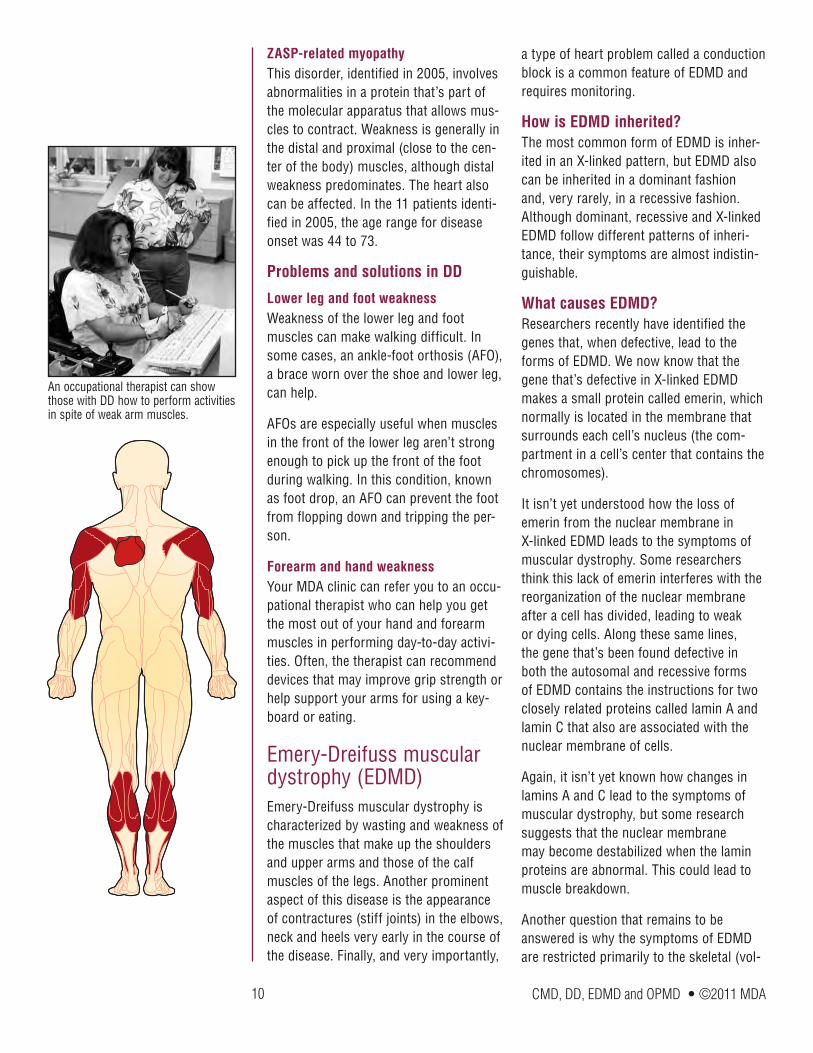
ZASP-related myopathyThis disorder, identified in 2005, involves abnormalities in a protein that’s part of the molecular apparatus that allows mus-cles to contract. Weakness is generally in the distal and proximal (close to the cen-ter of the body) muscles, although distal weakness predominates. The heart also can be affected. In the 11 patients identi-fied in 2005, the age range for disease onset was 44 to 73.
Problems and solutions in DD
Lower leg and foot weaknessWeakness of the lower leg and foot muscles can make walking difficult. In some cases, an ankle-foot orthosis (AFO), a brace worn over the shoe and lower leg, can help.
AFOs are especially useful when muscles in the front of the lower leg aren’t strong enough to pick up the front of the foot during walking. In this condition, known as foot drop, an AFO can prevent the foot from flopping down and tripping the per-son.
Forearm and hand weaknessYour MDA clinic can refer you to an occu-pational therapist who can help you get the most out of your hand and forearm muscles in performing day-to-day activi-ties. Often, the therapist can recommend devices that may improve grip strength or help support your arms for using a key-board or eating.
Emery-Dreifuss muscular dystrophy (EDMD)Emery-Dreifuss muscular dystrophy is characterized by wasting and weakness of the muscles that make up the shoulders and upper arms and those of the calf muscles of the legs. Another prominent aspect of this disease is the appearance of contractures (stiff joints) in the elbows, neck and heels very early in the course of the disease. Finally, and very importantly,
a type of heart problem called a conduction block is a common feature of EDMD and requires monitoring.
How is EDMD inherited?The most common form of EDMD is inher-ited in an X-linked pattern, but EDMD also can be inherited in a dominant fashion and, very rarely, in a recessive fashion. Although dominant, recessive and X-linked EDMD follow different patterns of inheri-tance, their symptoms are almost indistin-guishable.
What causes EDMD?Researchers recently have identified the genes that, when defective, lead to the forms of EDMD. We now know that the gene that’s defective in X-linked EDMD makes a small protein called emerin, which normally is located in the membrane that surrounds each cell’s nucleus (the com-partment in a cell’s center that contains the chromosomes).
It isn’t yet understood how the loss of emerin from the nuclear membrane in X-linked EDMD leads to the symptoms of muscular dystrophy. Some researchers think this lack of emerin interferes with the reorganization of the nuclear membrane after a cell has divided, leading to weak or dying cells. Along these same lines, the gene that’s been found defective in both the autosomal and recessive forms of EDMD contains the instructions for two closely related proteins called lamin A and lamin C that also are associated with the nuclear membrane of cells.
Again, it isn’t yet known how changes in lamins A and C lead to the symptoms of muscular dystrophy, but some research suggests that the nuclear membrane may become destabilized when the lamin proteins are abnormal. This could lead to muscle breakdown.
Another question that remains to be answered is why the symptoms of EDMD are restricted primarily to the skeletal (vol-
An occupational therapist can show those with DD how to perform activities in spite of weak arm muscles.
11 CMD, DD, EDMD and OPMD • ©2011 MDA
untary) muscles and heart muscle, given that the emerin and lamin proteins are found in most tissues of the body.
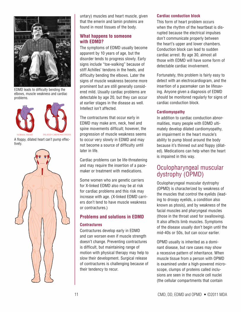
What happens to someone with EDMD?The symptoms of EDMD usually become apparent by 10 years of age, but the disorder tends to progress slowly. Early signs include “toe-walking” because of stiff Achilles’ tendons in the heels, and difficulty bending the elbows. Later the signs of muscle weakness become more prominent but are still generally consid-ered mild. Usually cardiac problems are detectable by age 20, but they can occur at earlier stages in the disease as well. Intellect isn’t affected.
The contractures that occur early in EDMD may make arm, neck, heel and spine movements difficult; however, the progression of muscle weakness seems to occur very slowly in EDMD and may not become a source of difficulty until later in life.
Cardiac problems can be life-threatening and may require the insertion of a pace-maker or treatment with medications.
Some women who are genetic carriers for X-linked EDMD also may be at risk for cardiac problems and this risk may increase with age. (X-linked EDMD carri-ers don’t tend to have muscle weakness or contractures.)
Problems and solutions in EDMD
ContracturesContractures develop early in EDMD and can worsen even if muscle strength doesn’t change. Preventing contractures is difficult, but maintaining range of motion with physical therapy may help to slow their development. Surgical release of contractures is challenging because of their tendency to recur.
Cardiac conduction blockThis form of heart problem occurs when the rhythm of the heartbeat is dis-rupted because the electrical impulses don’t communicate properly between the heart’s upper and lower chambers. Conduction block can lead to sudden cardiac arrest. By age 30, almost all those with EDMD will have some form of detectable cardiac involvement.
Fortunately, this problem is fairly easy to detect with an electrocardiogram, and the insertion of a pacemaker can be lifesav-ing. Anyone given a diagnosis of EDMD should be monitored regularly for signs of cardiac conduction block.
CardiomyopathyIn addition to cardiac conduction abnor-malities, many people with EDMD ulti-mately develop dilated cardiomyopathy, an impairment in the heart muscle’s ability to pump blood around the body because it’s thinned out and floppy (dilat-ed). Medications can help when the heart is impaired in this way.
Oculopharyngeal muscular dystrophy (OPMD)Oculopharyngeal muscular dystrophy (OPMD) is characterized by weakness of the muscles that control the eyelids (lead-ing to droopy eyelids, a condition also known as ptosis), and by weakness of the facial muscles and pharyngeal muscles (those in the throat used for swallowing). It also affects limb muscles. Symptoms of the disease usually don’t begin until the mid-40s or 50s, but can occur earlier.
OPMD usually is inherited as a domi-nant disease, but rare cases may show a recessive pattern of inheritance. When muscle tissue from a person with OPMD is examined under a high-powered micro-scope, clumps of proteins called inclu-sions are seen in the muscle cell nuclei (the cellular compartments that contain
EDMD leads to difficulty bending the elbows, muscle weakness and cardiac problems.
NORMAL HEART DILATED CARDIOMYOPATHY
A floppy, dilated heart can’t pump effec-tively.
12 CMD, DD, EDMD and OPMD • ©2011 MDA
the chromosomes), and bubblelike struc-tures (vacuoles) appear in the muscle cells.
The disease is most common in French-Canadian families or families of French-Canadian descent. There’s also a high inci-dence of OPMD among Hispanic residents of northern New Mexico.
OPMD also can affect people who aren’t of French-Canadian or Hispanic background.
What causes OPMD?The gene that’s defective in OPMD was dis-covered in 1998. It carries instructions for a polyadenylate binding protein (PABPN1) that’s normally present in the cell nucleus. Researchers suspect that in OPMD, the presence of extra amino acids in the pro-tein made from a defective PABPN1 gene causes the PABPN1 protein to clump together in the muscle cell nuclei, perhaps interfering with cell function. The disease often can be diagnosed with a DNA test for the most common PABPN1 mutation.
What happens to someone with OPMD?A person with OPMD may first notice drooping eyelids that gradually lead to tip-ping the head backward to see properly. Alternatively, some people might first notice that they tend to choke frequently
and may have other problems related to difficulty swallowing (called dysphagia). Most people eventually develop some degree of both ptosis and dysphagia.
Eventual weakness of the muscles in the face and limbs is common. For instance, many people with OPMD report problems with kneeling, bending, squatting, walking and climbing stairs. Double vision and a “breathy” quality of the voice also may occur.
Currently, there’s no cure for OPMD, but many people with the disease find that treating symptoms as they occur is ben-eficial.
Problems and solutions in OPMD
DysphagiaDifficulty swallowing, or dysphagia, can cause a person to aspirate food or liquid into the lungs, which in turn may lead to a serious problem called aspiration pneu-monia. If you find that you’re choking fre-quently while eating or drinking, you may need to have your swallowing abilities evaluated by a professional. There are a number of techniques that may help treat dysphagia, ranging from holding the head in different positions to changing the con-sistency of foods and liquids. Commercial
airway
esophagus
vocal cords
larynx
When swallowing muscles don’t function normally, food and liquids can be routed to the airway and lungs instead of to the esophagus and stomach.
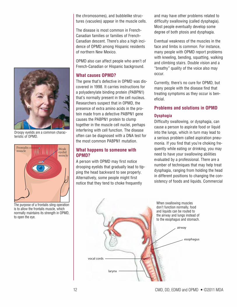
Droopy eyelids are a common charac-teristic of OPMD.
Weak eyelid muscle
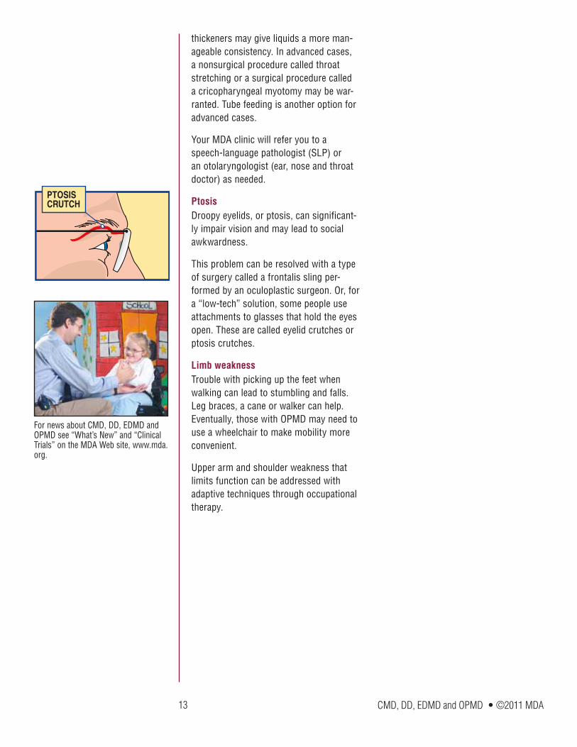
Frontalis muscle
The purpose of a frontalis sling operation is to allow the frontalis muscle, which normally maintains its strength in OPMD, to open the eye.
13 CMD, DD, EDMD and OPMD • ©2011 MDA
thickeners may give liquids a more man-ageable consistency. In advanced cases, a nonsurgical procedure called throat stretching or a surgical procedure called a cricopharyngeal myotomy may be war-ranted. Tube feeding is another option for advanced cases.
Your MDA clinic will refer you to a speech-language pathologist (SLP) or an otolaryngologist (ear, nose and throat doctor) as needed.
PtosisDroopy eyelids, or ptosis, can significant-ly impair vision and may lead to social awkwardness.
This problem can be resolved with a type of surgery called a frontalis sling per-formed by an oculoplastic surgeon. Or, for a “low-tech” solution, some people use attachments to glasses that hold the eyes open. These are called eyelid crutches or ptosis crutches.
Limb weaknessTrouble with picking up the feet when walking can lead to stumbling and falls. Leg braces, a cane or walker can help. Eventually, those with OPMD may need to use a wheelchair to make mobility more convenient.
Upper arm and shoulder weakness that limits function can be addressed with adaptive techniques through occupational therapy.
For news about CMD, DD, EDMD and OPMD see “What’s New” and “Clinical Trials” on the MDA Web site, www.mda.org.
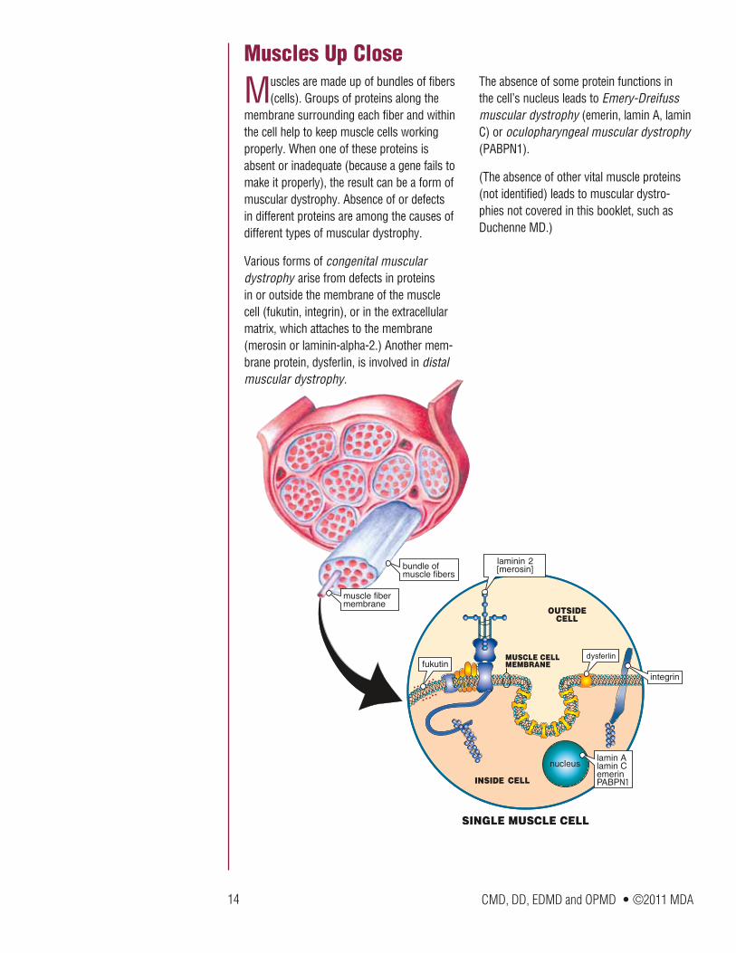
PTOSISCRUTCH
14 CMD, DD, EDMD and OPMD • ©2011 MDA
Muscles are made up of bundles of fibers (cells). Groups of proteins along the
membrane surrounding each fiber and within the cell help to keep muscle cells working properly. When one of these proteins is absent or inadequate (because a gene fails to make it properly), the result can be a form of muscular dystrophy. Absence of or defects in different proteins are among the causes of different types of muscular dystrophy.
Various forms of congenital muscular dystrophy arise from defects in proteins in or outside the membrane of the muscle cell (fukutin, integrin), or in the extracellular matrix, which attaches to the membrane (merosin or laminin-alpha-2.) Another mem-brane protein, dysferlin, is involved in distal muscular dystrophy.
The absence of some protein functions in the cell’s nucleus leads to Emery-Dreifuss muscular dystrophy (emerin, lamin A, lamin C) or oculopharyngeal muscular dystrophy (PABPN1).
(The absence of other vital muscle proteins (not identified) leads to muscular dystro-phies not covered in this booklet, such as Duchenne MD.)
Muscles Up Close
CMD, DD, EDMD and OPMD • ©2011 MDA
Type of CMD Cause Inheritance patternWelander’s distal myopathy abnormalities in
chromosome 2 genedominant
Finnish (tibial) distal myopathy titin abnormalities dominant
Miyoshi distal myopathy dysferlin abnormalities recessive
Nonaka distal myopathy; also called hereditary inclusion-body myositis type 2 (HIBM2)
GNE abnormalities recessive
Gowers-Laing distal myopathy MYH7 abnormalities dominant
Hereditary inclusion-body myositis type 1 (HIBM1)
unknown dominant
Distal myopathy with vocal cord and pharyngeal weakness
abnormalities in chromosome 5 gene
dominant
ZASP-related myopathy ZASP abnormalities dominant
15
Classification of Congenital Muscular Dystrophies
Classification of Distal Muscular Dystrophies
Type of CMD Cause Inheritance patternMerosin-deficient CMD
lack of merosin (laminin 2) or other defect leading to merosin deficiency
Chromosome 6 gene. Other genes
Ullrich CMD abnormalities in collagen 6 Chromosome 2 or 21 genes, reces-sive or dominant
Bethlem myopathy abnormalities in collagen 6 Chromosome 2 or 21 genes, dominant
Integrin-deficient CMD
lack of integrin alpha 7 Chromosome 12 gene, recessive
Fukuyama CMD (FCMD)
lack of fukutin Chromosome 9 gene, recessive
Muscle-eye-brain disease (MEB)
lack of POMGnT1, fukutin or fukutin-related protein
Chromosome 1, 9 or 19 genes, recessive
Walker-Warburg syndrome (WWS)
lack of POMT1, POMT2, fukutin or fukutin-related protein
Chromosome 9, 14 or 19 genes, recessive
CMD with rigid spine syndrome
lack of selenoprotein N1 Chromosome 1 gene, recessive
See illustration, page 5.
Source: Washington University Neuromuscular Home Page, May 2006
16 CMD, DD, EDMD and OPMD • ©2011 MDA
On being told they have a genetic dis-order such as muscular dystrophy,
bewildered patients often ask, “But it doesn’t run in the family, so how could it be genetic?”
Muscular dystrophy can run in a family, even if only one person in the biological family has it. This is because of the ways in which genetic diseases are inherited.
Each form of muscular dystrophy fol-lows one of three patterns of inheritance: recessive, dominant or X-linked. In brief, if a disease is recessive, two copies of the defective gene (one from each parent) are required to produce the disease. Each parent would be a carrier of the gene flaw, but wouldn’t usually have the disease.
If a disease is dominant, then only one copy of the genetic defect is needed to cause the disease. Anyone with the gene flaw will have disease symptoms and can pass the disorder to children.
If a disease is X-linked, it’s passed from mother to son, while daughters can be carriers but don’t generally get the dis-ease.
Many times MD appears to have occurred “out of the blue,” but in reality, one or both parents may be carriers, silently har-boring the genetic mutation (a flaw in the gene). Many parents have no idea they’re carriers of a disease until they have a child who has the disease.
In rare cases, muscular dystrophy actually can occur “out of the blue” when a new mutation appears with a baby’s concep-tion, though neither parent carries the gene flaw. These are called spontaneous mutations, and, after they occur, they can be passed on to the next generation, thereby introducing the gene for a specific MD into the family.
The risk of passing on a form of muscular dystrophy to your children depends on many circumstances, including exactly which type of MD has been diagnosed. A good way to find out more about these risks is to talk to your MDA clinic physi-cian or ask to see the genetic counselor at the MDA clinic. You also can see MDA’s pamphlet “Facts About Genetics and Neuromuscular Diseases.”
Does It Run in the Family?
Many times MD appears to have occurred “out of the blue,” but in reality, one or both parents may be carriers.
17 CMD, DD, EDMD and OPMD • ©2011 MDA
The MDA website is constantly updated with the latest information about the
neuromuscular diseases in its program. See the latest research news at www.mda.org.
Ever since 1986, when MDA-supported scientists identified the gene for Duchenne muscular dystrophy, they have forged ahead to isolate and characterize genes involved in almost all the neuromuscular disorders covered in MDA’s program. These include many of the gene defects responsible for the congenital and distal muscular dystrophies, Emery-Dreifuss MD and oculopharyngeal MD. These discover-ies have enabled scientists to understand variations among different forms of the diseases and have helped doctors to pro-vide more accurate diagnosis.
Now that this essential first step is almost accomplished in its entirety, MDA is exploring ways to correct muscle prob-lems caused by the different gene defects. Areas of especially active research include:
• Genetherapy: a mechanism for supplementing defective genes with healthy genes in the tissues affected by neuromuscular disease;
• Genesilencing: turning off genetic instructions that cause the production of toxic proteins; and
• Celltherapy: transplanting new muscle cells, using stem cells or immature muscle cells from a donor or geneti-cally corrected cells from the patient’s own body.
At the same time, other MDA-supported scientists are studying ways to preserve muscle despite the presence of a degen-erative disease. As of late 2009, some scientists are concentrating on preserving muscle by interfering with a protein called myostatin, a natural inhibitor of muscle growth. Others are studying the biochemi-cal signals that favor muscle repair, main-tenance and regeneration, with the aim of improving those functions.
MDA’s Search for Treatments and Cures
18 CMD, DD, EDMD and OPMD • ©2011 MDA
The Muscular Dystrophy Association offers a vast array of services to help
you and your family deal with CMD, DD, EDMD or OPMD. The staff at your local MDA office is there to assist you in many ways. The Association’s services include:
• nationwide network of clinics staffed by top neuromuscular disease specialists
• MDA summer camps for kids with neu-romuscular diseases
• help with locating durable medical equipment through its national equip-ment program
• financial assistance with repairs or modifications to all types of durable medical equipment
• annual occupational, physical, respira-tory or speech therapy consultations
• annual flu shots
• support groups for those affected, spouses, parents or other caregivers
• online support services through the e-community myMDA and through myMuscleTeam, a program that helps recruit and coordinate in-home help
MDA’s public health education program helps you stay abreast of research news, medical findings and disability information through magazines, publications, edu-cational speakers, seminars, videos and newsletters.
MDA’s website at www.mda.org contains thousands of pages of valuable informa-tion, including disease specifics, research findings, clinical trials and past magazine articles.
Everyone registered with MDA automati-cally receives Quest, MDA’s award-win-ning quarterly magazine. Quest publishes detailed articles about research findings, medical and day-to-day care, helpful products and devices, social and family issues, and much more. Other MDA pub-lications can be found at www.mda.org/publications; many booklets are available in Spanish. Ask your local office for “MDA Services for the Individual, Family and Community” and for help with obtaining copies of other publications.
If you have any questions about CMD, DD, EDMD or OPMD, someone at MDA will help you find the answer. To reach your local MDA office, call (800) 572-1717.
MDA Is Here to Help You
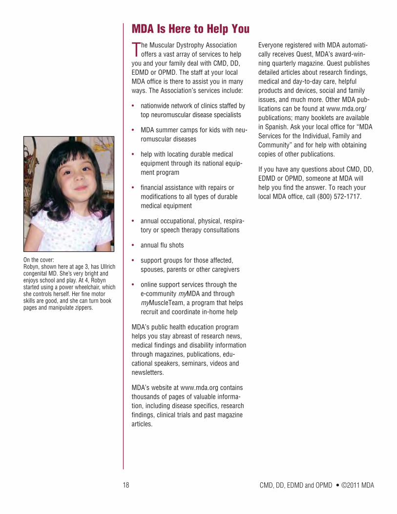
On the cover: Robyn, shown here at age 3, has Ullrich congenital MD. She’s very bright and enjoys school and play. At 4, Robyn started using a power wheelchair, which she controls herself. Her fine motor skills are good, and she can turn book pages and manipulate zippers.
19 CMD, DD, EDMD and OPMD • ©2011 MDA
The Muscular Dystrophy Association fights neuromuscular diseases through
an unparalleled worldwide research effort. The following diseases are included in MDA’s program:
Muscular DystrophiesMyotonic dystrophy (Steinert disease) Duchenne muscular dystrophy Becker muscular dystrophy Limb-girdle muscular dystrophy Facioscapulohumeral muscular dystrophy Congenital muscular dystrophy Oculopharyngeal muscular dystrophy Distal muscular dystrophy Emery-Dreifuss muscular dystrophy
Motor Neuron DiseasesAmyotrophic lateral sclerosis (ALS) Infantile progressive spinal muscular atrophy (Type 1, Werdnig-Hoffmann disease) Intermediate spinal muscular atrophy (Type 2) Juvenile spinal muscular atrophy (Type 3, Kugelberg-Welander disease) Adult spinal muscular atrophy (Type 4) Spinal-bulbar muscular atrophy (Kennedy disease)
Inflammatory MyopathiesPolymyositis Dermatomyositis Inclusion-body myositis
Diseases of Neuromuscular JunctionMyasthenia gravis Lambert-Eaton (myasthenic) syndrome Congenital myasthenic syndromes
Diseases of Peripheral NerveCharcot-Marie-Tooth disease Friedreich’s ataxia Dejerine-Sottas disease
Metabolic Diseases of MusclePhosphorylase deficiency (McArdle disease) Acid maltase deficiency (Pompe disease) Phosphofructokinase deficiency (Tarui disease) Debrancher enzyme deficiency (Cori or Forbes disease) Mitochondrial myopathy Carnitine deficiency Carnitine palmityl transferase deficiency Phosphoglycerate kinase deficiency Phosphoglycerate mutase deficiency Lactate dehydrogenase deficiency Myoadenylate deaminase deficiency
Myopathies Due to Endocrine AbnormalitiesHyperthyroid myopathy Hypothyroid myopathy
Other MyopathiesMyotonia congenita Paramyotonia congenita Central core disease Nemaline myopathy Myotubular myopathy Periodic paralysis
MDA’s Purpose and Programs
mda.org • (800) 572-1717
©2009, 2011, Muscular Dystrophy Association Inc.
P-214W 7/11
MDA’s website, mda.org, is constantly updated with the latest research news and information about the diseases in its program. Follow MDA on Facebook, Twitter and YouTube.