Embed Size (px)
Citation preview
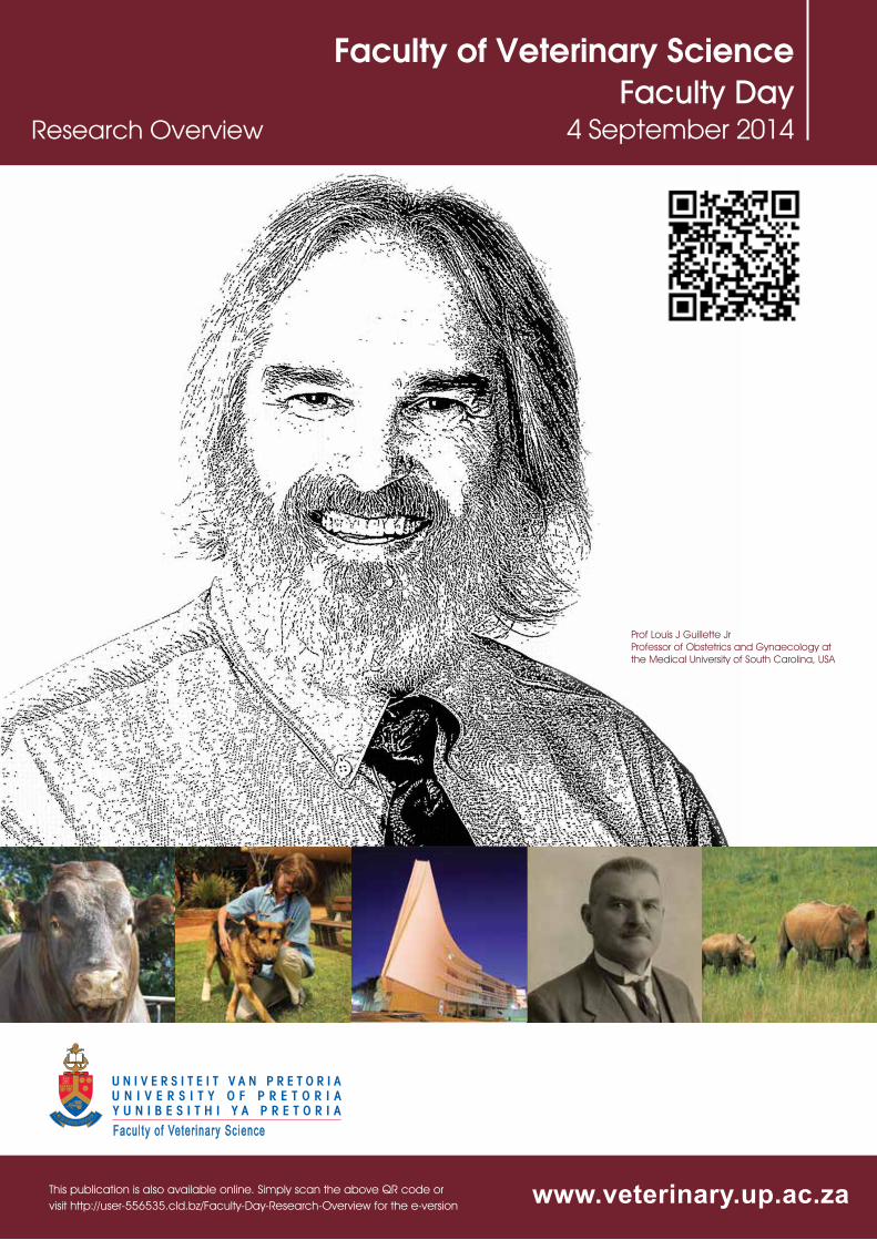
Prof Louis J Guillette Jr Professor of Obstetrics and Gynaecology at the Medical University of South Carolina, USA
Faculty of Veterinary ScienceFaculty Day
4 September 2014
www.veterinary.up.ac.za
Research Overview
Facu l ty o f Ve te r inary Sc ience
This publication is also available online. Simply scan the above QR code or visit http://user-556535.cld.bz/Faculty-Day-Research-Overview for the e-version
i
Faculty Day of the amalgamated Faculty of Veterinary Science reflects a proud tradition, which had been nurtured by the original faculties of Veterinary Science of both Medunsa and the University of Pretoria, of showcasing the research activities of staff and students on a special, dedicated occasion.
Since the inception of the Faculty of Veterinary Science at Medunsa in the early 1980s, the staff, and later students, were involved in the activities of the “Academic Day”, which was aimed at highlighting the research activities of the University, as well as exposing young researchers to a conference environment. The Faculty of Veterinary Science of the University of Pretoria at Onderstepoort followed this trend shortly thereafter and the first “Faculty Day”, which focused on the research activities of the faculty, was held on 5 September 1984, sponsored by the then Dean, Prof JMW le Roux. The combined research skills of the two original institutions are today reflected in the proceedings of the Faculty Day held each year in the spring at the Onderstepoort Campus.
Brief history of Faculty Day
Sponsorships
The Faculty of Veterinary Science wishes to express its sincere thanks to the following sponsors for their very generous contribution in support of the 2014 Faculty Day.
University of Pretoria Faculty of Veterinary Science - Faculty Day 4 September 2014
ii
Faculty Day
Faculty of Veterinary ScienceUniversity of Pretoria
4 September 2014
iii Contents/Programme
07:30 – 07:55 Registration and Coffee
Master of Ceremonies: Prof Neil Duncan
08:00 – 08:25 Welcoming and Opening Address - Prof Darrell Abernethy, Dean of the Faculty of Veterinary Science08:25 – 09:40 First Session
SESSION CHAIRPERSON: Dr Leith Meyer
1 Piscivory does not cause pansteatitis (yellow fat disease) in Oreochromis mossambicus from an African sub-tropical reservoir 7
J Dabrowski2 Comparison of electrical stunning with manual capture in farmed Nile crocodiles (Crocodylus
niloticus) by monitoring stress-related physiological parameters 8 S Pfitzer3 Tremors in the white rhinoceros (Ceratotherium simum) during chemical immobilization 9 SS de Lange4 Clinical anatomy of the cloaca and spinal venous sinus of the Nile crocodile 10 JG Myburgh5 Treatmentofradiusandtibiafracturesinwildantelopeutilizingexternalfixation 11 FJ Venter
09:45 – 10:40 Sir Arnold Theiler Memorial Lecture: Prof Louis J. Guillette Jr Predisposition for health or disease: The ‘new’ genetics of environmental health
10:45 – 11:10 Faculty Awards11:10 – 11:55 Tea12:00 – 13:00 Second Session
SESSION CHAIRPERSON: Dr Johan Marais
6 Catastrophic distal forelimb musculoskeletal injuries associated with racetracks in Gauteng, South Africa from 1998-2012 12
KE Spargo7 A comparison between juvenile pubic symphysiodesis and juvenile pubic symphysectomy:
a one-year follow-up 13 FJ Venter8 Effect of intravenous lidocaine on the minimum infusion rate of alfaxalone in goats 14 PS Ndawana9 Comparison of anaesthetic induction and recovery characteristics of diazepam-ketamine
combination to propofol alone; in healthy dogs undergoing orchidectomy 15 JP Ferreira
13:00 – 14:00 Lunch14:00 – 15:15 Third Session
SESSION CHAIRPERSON: Prof Moritz van Vuuren
10 Molecular characterization of vaccine candidates from Anaplasma marginale strains in South Africa 16
P Hove
University of Pretoria Faculty of Veterinary Science - Faculty Day 4 September 2014
iv
11 Molecular detection of Anaplasma marginale and Anaplasma subspecies centrale in cattle in South Africa 17
ME Chaisi12 SensitivityandspecificityofrRT-PCR,histopathology,andimmunohistochemistryforthe
detectionofRiftValleyfevervirusinnaturally-infectedcattleandsheep 18 L Odendaal13 Molecular detection of an Anaplasma sp strain in domestic dogs in Mnisi, South Africa 19 AO Kolo 14 IdentificationofPeste-Des-PetitsRuminantsVirus(PPRV)AsianlineageIVinNigeriaand
co-circulationwithPPRVlineageII 20 TY Woma
15:15 – 15:30 Break15:30 – 16:45 Fourth Session
SESSION CHAIRPERSON: Dr Sarah Clift
15 Cytotoxicity of diplodiatoxin, dipmatol and diplonine, metabolites synthesized by Stenocarpella maydis 21
MG Masango16 InvestigationoftheinflammatoryimmuneresponseindogsnaturallyinfectedwithBabesia
rossi,usingflowcytometry 22 Y Rautenbach17 Dynamicsofanowned,free-roamingdogpopulation:implicationsforrabiescontrol 23 DL Knobel18 TheMnisiCommunityProgramme2009-2013:Anoverviewofthefirstfiveyearsofthe
Programme, its relevance to the Faculty, and its future vision 24 J van Rooyen19 Developingamultiplecriteriadecisionanalysistooltoassessthecontroloffoot-and-mouth
disease in South Africa 25 LC Roberts
16:45 – 17:00 Break17:00 – 18:15 Fifth Session
SESSION CHAIRPERSON: Prof Kobus Eloff
20 Leaf extracts of selected Anacardiaceae trees had excellent antimycobacterial activity and contained several antimycobacterial compounds 26
PN Kabongo21 An overview of the pathology, epidemiology, and ecological physiology of infections of a
novel Mycobacterium species, M. mungi, in its only known host, the banded mongoose 27 PN Laver22 Screening of banded mongooses (Mungos mungo) in the Kruger National Park for
mycobacterial infection 28 AC Brüns23 DistributionofBacillus anthracis genotypes in Kruger National Park in South Africa 29 MB Ledwaba24 An Investigation into infection by Intracellular parasites and Bacillus anthracis in blood
smears in the Kruger National Park in 2010 30 A Hassim
18:15 Cocktail
v
POSTER PRESENTATIONS
P1 Antimicrobial activity of berries, leaves, barks and roots of the edible plant Grewia flava against four enteric pathogens 31
MS Lamola, FS Botha, C Van Wyk
P2 Blowflies as potential vectors of Bacillus anthracis in the Kruger National Park 32
L Basson, EH Dekker, H van Heerder, W Beyer, J Rossouw
P3 Characterization of Bacillus anthracis using Multiple Locus Variable Number of Tandem Repeat Analyses (MLVA) for the typical African laboratory 33
A Hassim, Y Hauck, J Rossouw, G Vergnaud and H van Heerden
P4 Determining adrenocortical activity as a measure of stress in male giraffes (Giraffa camelopardis) 34
TE Wolf, ASW Tordiffe, A Ganswindt
P5 Eleven-year antibiotic resistance profiles of Staphylococcus aureus in dairy herds across southern Africa 35
T.J van der Schans, J Karzis, IM Petzer
P6 Epidemiology of bluetongue virus in Mnisi, Mpumalanga 36
J Steyn, GJ Venter, P Coetzee, EH Venter
P7 Exposure of lions to classical rabies virus and Mokola virus in provincial and private game reserves in Mpumalanga province 37
SL Kejelepula, M van Vuuren, B Reininghaus and CT Sabeta
P8 Generation of white rhinoceros (Ceratotherium simum) IFN-g specific recombinant chicken antibodies and there use in the rhinoceros IFN-g assay for diagnosis of Mycobacterium bovis infection. 38
D Morar-Leather, J Godfroid, E Tijhaar, V Rutten, J Fehrsen
P9 Immunogenicity and protective efficacy of the Sterne 34F2 live spore anthrax vaccine in goats 39
O. Ndumnego, S. Koehler, J. Crafford, W. Beyer, H. van Heerden
P10 Investigating the possible presence of Theileria parva carrier cattle in Mnisi area 40
CN Choopa, D Geysen, D Knobel, MC Oosthuizen, NE Collins
P11 Methicillin resistance in Staphylococci isolated from milk samples of South African dairy cows 41
R Badenhorst, J Karzis, IM Petzer
P12 Model to test the protection against anthrax in goats through correlation of passive protection test in mice 42
PH Phaswana, OC Ndumnego, H van Heerden
P13 Molecular Characterisation of Peste Des Petits Ruminants Viruses of Sheep and Goats in Nigeria 43
S Mantip, M Van Vuuren, M Quan, D Shamaki
P14 Molecular detection of Rickettsia africae and Rickettsia felis from ticks and fleas collected from domestic dogs in Mnisi, South Africa 44
AO Kolo, KP Sibeko-Matjila, DL Knobel and PT Matjila
University of Pretoria Faculty of Veterinary Science - Faculty Day 4 September 2014
vi
P15 Two novel species of non-tuberculous mycobacteria (NTM) revealed by multiple gene sequence characterisation 45
Gcebe N, Jenkins A, Rutten V, Michel A.
P16 Preliminary screening of some South African Rubiaceae species showing promising antimycobacterial activity 46
AO Aro, JN Eloff, LJ McGaw
P17 Prevalence of enterobacteriaceae in retail eggs in South Africa 47
AR Jambalang, FS Botha, EM Buys
P18 Protective effects of South African plants against mutagenicity of aflatoxin B1 48
Nkala BA, Botha CJ, Elgorashi EE
P19 Quantitative anti-anthrax IgG ELISA correlates with the anthrax toxin neutralization assay in goats 49
OC Ndumnego, J Crafford, W Beyer, H van Heerden
P20 Reproductive activity pattern and its endocrine correlates in the African lesser bushbaby, Galago moholi 50
J Scheun, NC Bennett, Julia Nowack, A Ganswindt
P21 Screening of South African plants for activities against salmonellosis 51
ZP Mahlangu, E Madoroba, F Botha, EE Elgorashi
P22 Serological evidence of camel exposure to Peste-des-petits ruminants virus (PPRV) in Nigeria 52
TY Woma, DJU Kalla, PS Ekong, DG Bwala, D Bailey, D Shamaki, A Diallo, M Quan
P23 The antioxidant activity and total phenolic contents of nine tree extracts with high activity against Escherichia coli 53
IL Elisha, JP Dzoyem, FS Botha, JN Eloff
P24 The enigmatic bill tip organ of the ostrich and emu 54
MR Crole, JT Soley
P25 The potential role of recombinant mycobacterial antigens of non-tuberculous mycobacteria and Mycobacteria tuberculosis complex in the diagnosis of tuberculosis in cattle 55
Jenkins AO, Gormley E, Gcebe N, Conan A, Michel AL, Rutten VPMG
P26 The power of poo – Non-invasive measures of reproduction and stress in wildlife 56
A Ganswindt, TE Wolf
P27 The Sapotaceae as a source of antitubercular metabolites and isolation of antimycobacterial pentacyclic triterpenes from Sideroxylon inerme 57
LJ McGaw, MD Awouafack, BM Sakong, TJ Makhafola, OO Udom, TM Hlokwe, E Madoroba, JN Eloff
1 Message from the Dean
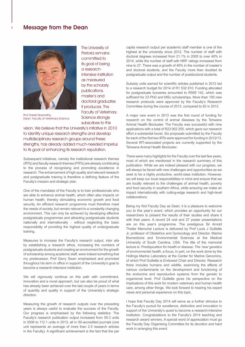
The University of Pretoria remains committed to its goal of being a research-intensive institution as measured by the scholarly publications, master’s and doctoral graduates it produces. The Faculty of Veterinary Science strongly subscribes to this
vision. We believe that the University’s initiative in 2010 to identify unique research strengths and develop multidisciplinary research groups around these strengths, has already added much-needed impetus to its goal of enhancing its research reputation.
Subsequent initiatives, namely the institutional research themes (IRTs) and faculty research themes (FRTs) are already contributing to the process of recognising and promoting excellence in research. The enhancement of high-quality and relevant research and postgraduate training is therefore a defining feature of the Faculty’s mission and strategic plan.
One of the mandates of the Faculty is to train professionals who are able to enhance animal health, which often also impacts on human health, thereby stimulating economic growth and food security. An efficient research programme must therefore meet the needs of society, but remain relevant to a constantly changing environment. This can only be achieved by developing effective postgraduate programmes and attracting postgraduate students nationally and internationally – without neglecting the basic responsibility of providing the highest quality of undergraduate training.
Measures to increase the Faculty’s research output, inter alia by establishing a research ethos, increasing the numbers of postgraduate students and creating an environment for the growth of scholarship among academic staff, were indeed something that my predecessor, Prof Gerry Swan emphasised and promoted throughout his term in office in support of the University’s goal to become a research-intensive institution.
We will vigorously continue on this path with commitment, innovation and a novel approach, but can also be proud of what has already been achieved over the last couple of years in terms of quantity and quality in support of the University’s strategic direction.
Measuring the growth of research outputs over the preceding years is always useful to evaluate the success of the Faculty. Our progress is emphasised by the following statistics: The Faculty’s research publication output increased from 55.3 units in 2006 to 112.1 units in 2013, all in ISI-accredited journals (one unit represents an average of more than 2.5 research articles in the Faculty). A significant achievement is the fact that the per
Prof Darrell Abernethy, Dean: Faculty of Veterinary Science
capita research output per academic staff member is one of the highest at the university since 2012. The number of staff with doctoral degrees increased from 21.1% in 2005 to over 40% in 2014, while the number of staff with NRF ratings increased from nine to 27. There was a growth of 49% in the number of master’s and doctoral students, and the Faculty more than doubled its postgraduate output and the number of postdoctoral students.
Subsidy units earned for scientific articles published in 2012 led to a research budget for 2014 of R1 532 810. Funding allocated for postgraduate bursaries amounted to R569 142, which was sufficient for 23 PhD and MSc scholarships. More than 100 new research protocols were approved by the Faculty’s Research Committee during the course of 2013, compared to 82 in 2012.
A major new event in 2013 was the first round of funding for research on the control of animal diseases by the Tshwane Animal Health Biocluster. The Faculty was successful with nine applications with a total of R23 902 255, which gave our research effort a substantial boost. Six proposals submitted by the Faculty for each of the first two IRTs were approved for funding in 2012/13. Several IRT-associated projects are currently supported by the Tshwane Animal Health Biocluster.
There were many highlights for the Faculty over the last few years, most of which are mentioned in the research summary of this publication. While we are indeed pleased with our progress, we will always be faced with new challenges and opportunities as we seek to be a highly productive, world-class institution. However, we will keep our local responsibilities in mind and ensure that we are locally relevant to the challenges of animal health, poverty and food security in southern Africa, while ensuring we make an impact internationally with cutting-edge research and high-level collaborations.
Being my first Faculty Day as Dean, it is a pleasure to welcome you to this year’s event, which provides an opportunity for our researchers to present the results of their studies and share it with their peers. A record 24 oral and 27 poster presentations are on this year’s programme. The prestigious Sir Arnold Theiler Memorial Lecture is delivered by Prof Louis J Guillette Jr, professor of Obstetrics and Gynecology and Director, Marine Biomedicine and Environmental Sciences at the Medical University of South Carolina, USA. The title of the memorial lecture is: Predisposition for health or disease: The ‘new’ genetics of environmental health, a focus, in part, on the work done by the Hollings Marine Laboratory at the Center for Marine Genomics, of which Prof Guillette is Endowed Chair and Director. Research there includes humans and wildlife, examining the effects of various contaminants on the development and functioning of the endocrine and reproductive systems from the genetic to organismal level. Prof Guillette gives his perspective on the implications of this work for modern veterinary and human health care, among other things. We look forward to hearing his expert views and personal experience on this topic.
I hope that Faculty Day 2014 will serve as a further stimulus to the Faculty’s pursuit for excellence, distinction and innovation in support of the University’s quest to become a research-intensive institution. Congratulations to the Faculty’s 2014 teaching and research award winners. A special word of appreciation must go the Faculty Day Organising Committee for its devotion and hard work in arranging this event.
University of Pretoria Faculty of Veterinary Science - Faculty Day 4 September 2014
2Curriculum Vitae: Professor Louis J Guillette Jr
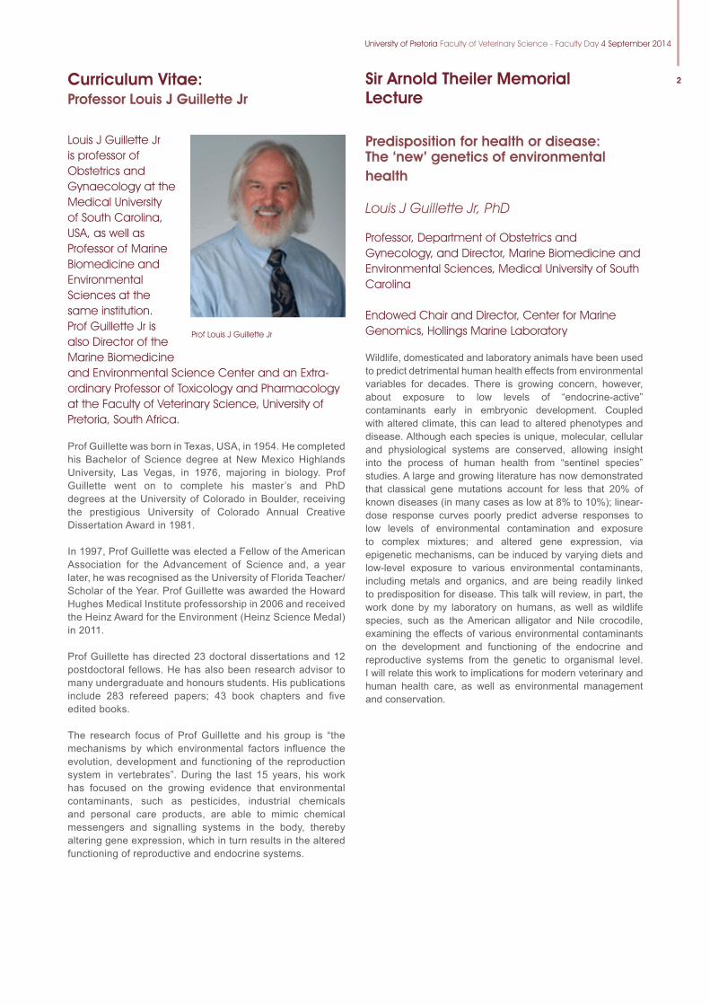
Louis J Guillette Jr is professor of Obstetrics and Gynaecology at the Medical University of South Carolina, USA, as well as Professor of Marine Biomedicine and Environmental Sciences at the same institution. Prof Guillette Jr is also Director of the Marine Biomedicine and Environmental Science Center and an Extra-ordinary Professor of Toxicology and Pharmacology at the Faculty of Veterinary Science, University of Pretoria, South Africa.
Prof Guillette was born in Texas, USA, in 1954. He completed his Bachelor of Science degree at New Mexico Highlands University, Las Vegas, in 1976, majoring in biology. Prof Guillette went on to complete his master’s and PhD degrees at the University of Colorado in Boulder, receiving the prestigious University of Colorado Annual Creative Dissertation Award in 1981.
In 1997, Prof Guillette was elected a Fellow of the American Association for the Advancement of Science and, a year later, he was recognised as the University of Florida Teacher/Scholar of the Year. Prof Guillette was awarded the Howard Hughes Medical Institute professorship in 2006 and received the Heinz Award for the Environment (Heinz Science Medal) in 2011.
Prof Guillette has directed 23 doctoral dissertations and 12 postdoctoral fellows. He has also been research advisor to many undergraduate and honours students. His publications include 283 refereed papers; 43 book chapters and five edited books.
The research focus of Prof Guillette and his group is “the mechanisms by which environmental factors influence the evolution, development and functioning of the reproduction system in vertebrates”. During the last 15 years, his work has focused on the growing evidence that environmental contaminants, such as pesticides, industrial chemicals and personal care products, are able to mimic chemical messengers and signalling systems in the body, thereby altering gene expression, which in turn results in the altered functioning of reproductive and endocrine systems.
Prof Louis J Guillette Jr
Sir Arnold Theiler MemorialLecture
Predisposition for health or disease: The ‘new’ genetics of environmental health
Louis J Guillette Jr, PhD
Professor, Department of Obstetrics and Gynecology, and Director, Marine Biomedicine and Environmental Sciences, Medical University of South Carolina
Endowed Chair and Director, Center for Marine Genomics, Hollings Marine Laboratory
Wildlife, domesticated and laboratory animals have been used to predict detrimental human health effects from environmental variables for decades. There is growing concern, however, about exposure to low levels of “endocrine-active” contaminants early in embryonic development. Coupled with altered climate, this can lead to altered phenotypes and disease. Although each species is unique, molecular, cellular and physiological systems are conserved, allowing insight into the process of human health from “sentinel species” studies. A large and growing literature has now demonstrated that classical gene mutations account for less that 20% of known diseases (in many cases as low at 8% to 10%); linear- dose response curves poorly predict adverse responses to low levels of environmental contamination and exposure to complex mixtures; and altered gene expression, via epigenetic mechanisms, can be induced by varying diets and low-level exposure to various environmental contaminants, including metals and organics, and are being readily linked to predisposition for disease. This talk will review, in part, the work done by my laboratory on humans, as well as wildlife species, such as the American alligator and Nile crocodile, examining the effects of various environmental contaminants on the development and functioning of the endocrine and reproductive systems from the genetic to organismal level. I will relate this work to implications for modern veterinary and human health care, as well as environmental management and conservation.
3
1984: T Gutsche “Theiler – His Personal Significance Today.”
1985: Prof HPA De Boom “Vlammende Fakkels, Ou Bene, Ivoortorings en Rooi Vlae.”
1986: Prof BC Jansen “Theiler Gedenklesing.”
1987: Opening of the Sir Arnold Theiler Building - No Lecture
1988: Dr RD Bigalke “Important Research Requirements for future Animal Production-Orientated Research with Particular Reference to Veterinary Science”
1989: Dr R Swanepoel “The Joy of Research.”
1990: Dr A Schutte “The Impact of controlled Breeding on the Cattle Industry in Southern Africa”
1991: Prof DM Joubert “Sir Arnold Theilergedenklesing – Theiler en die Fakulteit Veeartsenykunde”
1992: Dr CM Cameron “The Environment – Whose Responsibility?”
1993: Opening of the Onderstepoort Veterinary Academic Hospital – No Lecture
1994: Dr W Plowright “Rinderpest and Cell-Culture Revolution”
1995: Prof WL Jenkins *
1996: Prof PV Tobias “Premature Discoveries in Science.”
1997: Prof DL Block “Our Universe: Accident of Design?”
1998: Prof TW Naudé “A Stroll Through the Wondrous Garden of South African Toxicology”
1999: * *
2000: Dr DW Verwoerd “The Molecular Revolution in biology and its Influence on Veterinary Science.”
2001: Prof H Huismans “Molecular Biology and its Impact on the study and Control of Viral Diseases such as Bluetongue and African Horse Sickness.”
2002: Prof I Horak “The Joy of Research”
2003: Prof WFO Marasas “Fumonisins: Historical Perspective and Future Objectives”
2004: Dr RA Kock “Wildlife Domestic Animal Disease Interface – Hard or Soft Edge?”
2005: Prof SS van den Berg: “The Past, Present and Future of the Clinical Departments in the Faculty of Veterinary Science.”
2006: Dr BD Perry “The Global Poverty Reduction Agenda: What are the Implications for Animal Health Research and Development?”
2007: Prof dr AWCA Cornelissen “What makes an excellent Faculty of Veterinary Medicine?”
2008: Dr G Brückner “New challenges for the veterinary profession in global animal disease control and the trade in animals and animal products.”
2009: Prof P Doherty “Adventures in Infection and Immunity.”
2010: Dr R Moerane “The Role of the Veterinary Profession in the Current Developmental Agenda in South Africa.”
2011: World Veterinary Congress in SA – no Faculty Day
2012: Prof NJ MacLachlan “Emerging viral diseases; the example of bluetongue, from Theiler to climate change”
2013 Prof MC Horzinek “A personal journey through coronavirus evolution”
** We do apologise that the above list is not complete. It will be appreciated if anyone who has access to some of the missing information, contacts either Dr Paul van Dam ([email protected] or 012 529 8203) or Mr Chris van Blerk ([email protected] or 012 529 8436)
Sir Arnold Theiler Memorial Lectures
University of Pretoria Faculty of Veterinary Science - Faculty Day 4 September 2014
4Research Summary: 2012–2014
One of the objectives in the Faculty’s mission and strategic plan over the last few years was and still is to enhance high-quality and relevant research and postgraduate training. Not only does this require increasing research outputs in terms of both quantity and quality in support of the University’s goal to become a research-intensive institution, but also that these are locally relevant and keep pace with worldwide trends.
One of the mandates of the Faculty is to train professionals who are able to protect animal health, which often also impacts on human health, thereby stimulating economic growth and food security. An efficient research programme must therefore meet the needs of society, but remain relevant to a constantly changing environment. This can only be achieved by developing effective postgraduate programmes and focusing on attracting more postgraduate students nationally and internationally. To meet these requirements, the Faculty operates within the following research focus areas:
Molecular studies on infectious and parasitic diseases of animals: A research focus utilising biotechnology for the development of improved diagnostic techniques and vaccines for animal diseases and for the study of their pathogenesis.
Phytomedicine and ethno-veterinary medicine: An established multidisciplinary and collaborative research programme focusing on the development of extracts from plants with antimicrobial or anti-parasitic activity purposes.
Wildlife and environmental health: This is an inclusive research focus with contributions from all five departments of the Faculty, including studies on tuberculosis in buffalo, immune-contraception in elephants, theileriosis in roan and sable, toxicity of non-steroidal anti-inflammatories in vultures and endocrine disruptors in the environment.
Veterinary aspects of food safety and food security: This is an established research focus, which includes, inter alia, programmes in veterinary public health, community development, epidemiology, and risk assessment and poultry health.
Equine and companion animal health and welfare: The focus on infectious and other diseases of horses and other companion animals, with an important impact on trade and sports medicine (the racing industry) or on the welfare and management of these animals.
Research output and growthThe year 2013 marked the end of a very successful year for the Faculty in support of strategic direction of the University of Pretoria. Measuring the growth of the research outputs over the preceding years is always useful to evaluate the success of the Faculty.
The Faculty’s growth and progress are confirmed by the following figures: The number of staff with doctoral degrees increased from 21.1% in 2005 to over 40% in 2014, while the number of staff with NRF ratings increased from nine to 27. There was a growth of 49% in the number of master’s and doctoral students, and the Faculty more than doubled its postgraduate output and the number of postdoctoral students. Furthermore, the Faculty’s research publication output
increased from 55.3 units in 2006 to 112.1 units in 2013, all in ISI-accredited journals (one unit represents an average of more than 2.5 research articles in the Faculty). A significant achievement is the fact that the per capita research output per academic staff member is one of the highest at the University since 2012.
Subsidy units earned for scientific articles published in 2012 led to a research budget for 2014 of R1 532 810. Funding allocated for postgraduate bursaries amounted to R569 142, which was sufficient for 23 PhD and MSc scholarships. More than 100 new research protocols were approved by the Faculty’s Research Committee during 2013, compared to 82 in 2012. Since 2011, two new research chairs were established: the Chair in Primary Animal Health Care (PAHC) and the Chair in Poultry Health and Production.
A major new event in 2013 was the first round of funding for research on the control of animal diseases by the Tshwane Animal Health Biocluster. The Faculty was successful with nine applications for a total of R23 902 255, which gave our research effort a substantial boost.
Highlights: 2012–2014
University honours exceptional academic achievers and recent NRF-rated researchersOne of the University’s key drivers is its commitment to delivering quality research outputs. The achievement of the Faculty’s staff members not only underline this commitment, but also subscribe to the notion of the Faculty to make research a primary thrust, aiming to stimulate and focus research on unique problems that will give the Faculty a leading edge. The Faculty excelled at a gala dinner on 23 April 2013, celebrating the University’s exceptional academic achievers and recent NRF-rated researchers. The highlight of the evening was the presentation of certificates to the award winners. Dr Dayo Fasina received an Exceptional Young Researcher Award. Prof Pete Irons, Prof Bruce Gummow, Prof Johan Nöthling, Prof Brighton Dzikiti and Dr Kgomotso Sibeko received NRF ratings for the period 2013 to 2018. The event clearly showed the excellent progress made by the Faculty in building its research capacity in recent years.
Institutional Research Themes (IRTs)
An important initiative during 2012 was the implementation by the University of selected Institutional Research Themes (IRTs). These themes were selected on the basis of existing strengths of the University and their potential to stimulate interfaculty and international collaboration as a method to stimulate research. Five themes were initially approved for special funding, and the Faculty actively collaborates in three of these: Animal and Zoonotic Diseases, Genomics, and Food,
5 Research Summary: 2012–2014 (continued)
Nutrition and Wellbeing. Six proposals submitted by the Faculty for the first two IRTs were approved for funding in 2012/13. Several IRT-associated projects are currently supported by the Tshwane Animal Health Biocluster.
Tshwane Animal Health BioclusterThe final signing of a Memorandum of Agreement for the establishment of the Tshwane Animal Health Biocluster between government’s Technology Innovation Agency (TIA) and the Agricultural Research Council (ARC), Council for Scientific and Industrial Research (CSIR), Onderstepoort Biological Products (OBP), National Research Foundation (NRF) and the University of Pretoria took place in 2012. The purpose of the agreement is to stimulate collaboration between these institutions in the development of commercially viable technologies for the control of animal diseases of major social and economic importance for South Africa and the southern African region. In the first round of applications for funding in 2013, the Faculty was successful with nine proposals for a total of almost R24 million.
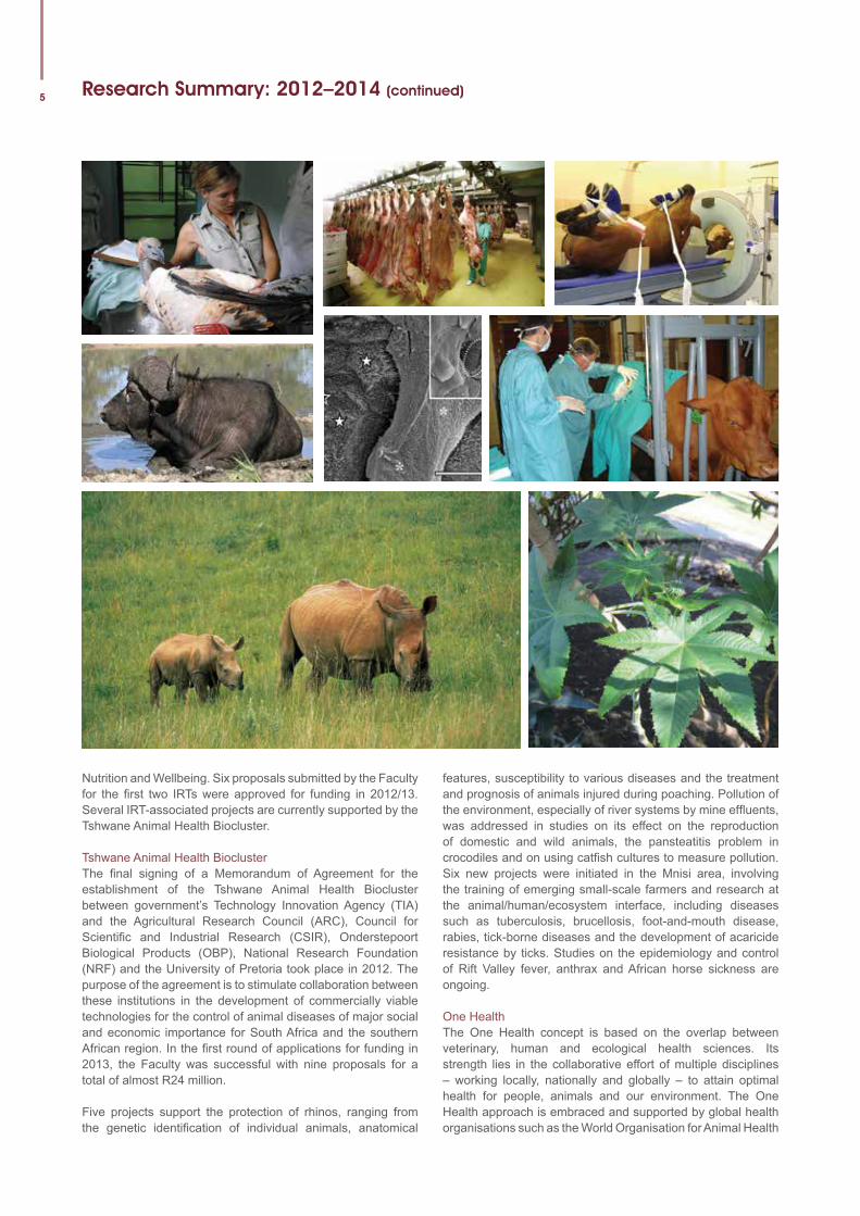
Five projects support the protection of rhinos, ranging from the genetic identification of individual animals, anatomical
features, susceptibility to various diseases and the treatment and prognosis of animals injured during poaching. Pollution of the environment, especially of river systems by mine effluents, was addressed in studies on its effect on the reproduction of domestic and wild animals, the pansteatitis problem in crocodiles and on using catfish cultures to measure pollution. Six new projects were initiated in the Mnisi area, involving the training of emerging small-scale farmers and research at the animal/human/ecosystem interface, including diseases such as tuberculosis, brucellosis, foot-and-mouth disease, rabies, tick-borne diseases and the development of acaricide resistance by ticks. Studies on the epidemiology and control of Rift Valley fever, anthrax and African horse sickness are ongoing.
One HealthThe One Health concept is based on the overlap between veterinary, human and ecological health sciences. Its strength lies in the collaborative effort of multiple disciplines – working locally, nationally and globally – to attain optimal health for people, animals and our environment. The One Health approach is embraced and supported by global health organisations such as the World Organisation for Animal Health
University of Pretoria Faculty of Veterinary Science - Faculty Day 4 September 2014
6Research Summary: 2012–2014 (continued)
(OIE), the United Nations’ Food and Agriculture Organisation (FAO) and the World Health Organisation (WHO).
UP does not only have the only faculty of veterinary science in the country, but is the only tertiary institution with a full set of faculties that would allow the national and regional development of the One Health concept within the Southern African Development Community (SADC), with a focus on animal/human/ecosystem health. The University’s One Health Platform consists of two components: an institutional platform supporting research, teaching and learning, and continuing professional development in One Health, and a field-based platform at the Hans Hoheisen Wildlife Research Station. The combined capacity and infrastructure of the institutional and field-based platforms provide unique opportunities for One Health training and research.
The Faculty’s first One Health summer school took place from 18 to 25 August 2013. This was followed by a second summer school from 13 to 26 July 2014. Selected under- and postgraduate students from University of California, Davis, Iowa State University, the Royal Veterinary College, Utrecht University, Southern African Centre for Infectious Diseases Surveillance (SACIDS), Research Platform – Production and Conservation in Partnership (RP-PCP), the University of Zimbabwe and the University of Pretoria attended a two- week programme. During their stay, the students were exposed to different One Health environments at the human/livestock/wildlife/ecosystem interface. The students were joined by facilitators from each institution who share their knowledge and expertise in the context of One Health.
The purpose of the summer school is to provide students from diverse backgrounds with the opportunity to develop and apply their leadership, communication, team-building, analytical and critical thinking skills to assess and respond to global health problems arising at the human/animal/ecosystem interface, and to design, implement and evaluate practical, cost-effective and sustainable solutions in collaboration with local and regional stakeholders and global partners. The focus is on the Great Limpopo Transfrontier Conservation Area (GLTFCA) as it is one of the most important health interfaces in the region.
Research Chair in Poultry Health and Production The Poultry Disease Management Agency (PDMA) and the Poultry Research Chair in the Faculty were officially launched in March 2013 in an exciting collaborative partnership with the Southern African Poultry Association (SAPA). Already operational since August 2012, the partnership aims, among other things, to conduct research on poultry diseases that have an impact on our economy. The first incumbent of this Chair, Prof Celia Abolnik, is conducting research projects in conjunction with the PDMA, the government and other relevant stakeholders. The recent upgrading in 2014 of the Poultry Biosafety Level 3 (BSL 3) laboratory, funded by the SAPA, will enable poultry research at the Faculty on notifiable poultry diseases in particular. Prof Abolnik has already sourced more than R8 million to support postgraduate student training to improve diagnostic tools and produce research outputs of a high international standard.
Postgraduate student symposiumAfter the first successful postgraduate student symposium at the Faculty in 2009, a similar successful event was hosted in September 2012, in collaboration with the Institute of Tropical Medicine, Antwerpen, Belgium. It highlighted the importance of research and cooperation and was attended by 92 delegates from 16 countries. Topics related to One Health were addressed and postgraduate students from the SADC region presented their research to their peers, supervisors and invited scientists with international standing. A third symposium is planned for 2015.
14th International Conference of the Association of Institutions for Tropical Veterinary Medicine (AITVM)The AITVM conference was presented for the first time in South Africa from 26 to 29 August 2013. It was jointly organised by the Department of Veterinary Tropical Diseases and the Institute of Tropical Medicine, Antwerpen, Belgium. The AITVM has 22 member institutions worldwide and organises an international conference every three years. Approximately 160 delegates from 38 countries attended the conference. Prof Koos Coetzer, current Deputy Dean: Research, Postgraduate Studies and Internationalisation of the Faculty and President of the AITVM for six years until 2013, presented the successful bid to host the conference in South Africa during the previous conference in Bangkok, Thailand, in 2010.
Faculty Day 2013 and research awardsThe annual Faculty Day in 2013 provided an opportunity for our researchers to showcase the research activities in the Faculty to colleagues and peers, and was well attended by staff members, visitors and sponsor companies alike. The prestigious Sir Arnold Theiler Memorial Lecture was delivered by Prof Marian C Horzinek, Professor Emeritus and former Director of the Graduate School of Animal Health at the Utrecht University, Holland. The title was “A personal journey through coronavirus evolution”. His thought-provoking and interesting lecture fittingly illustrated the dedication of our Faculty to international collaboration with experts all over the world.
Excellence in research performance was again recognised at the event by the identification of the Faculty’s Top 10 researchers and the allocation of the following research awards:
Researcher of the Year:Prof Vinny Naidoo (UPBRC)Young Researcher of the Year:Dr Rhoda Leask (Department of Production Animal Studies)
The following nine top researchers (the top 10 names are published and qualify for the Dean’s lunch):Prof Peter ThompsonProf John SoleyProf Estelle VenterProf Anita MichelProf Banie PenzhornProf Andre GanswindtProf Robert KirbergerProf Geoff FosgateProf Moritz van Vuuren
7 Research Programme: Oral Presentations
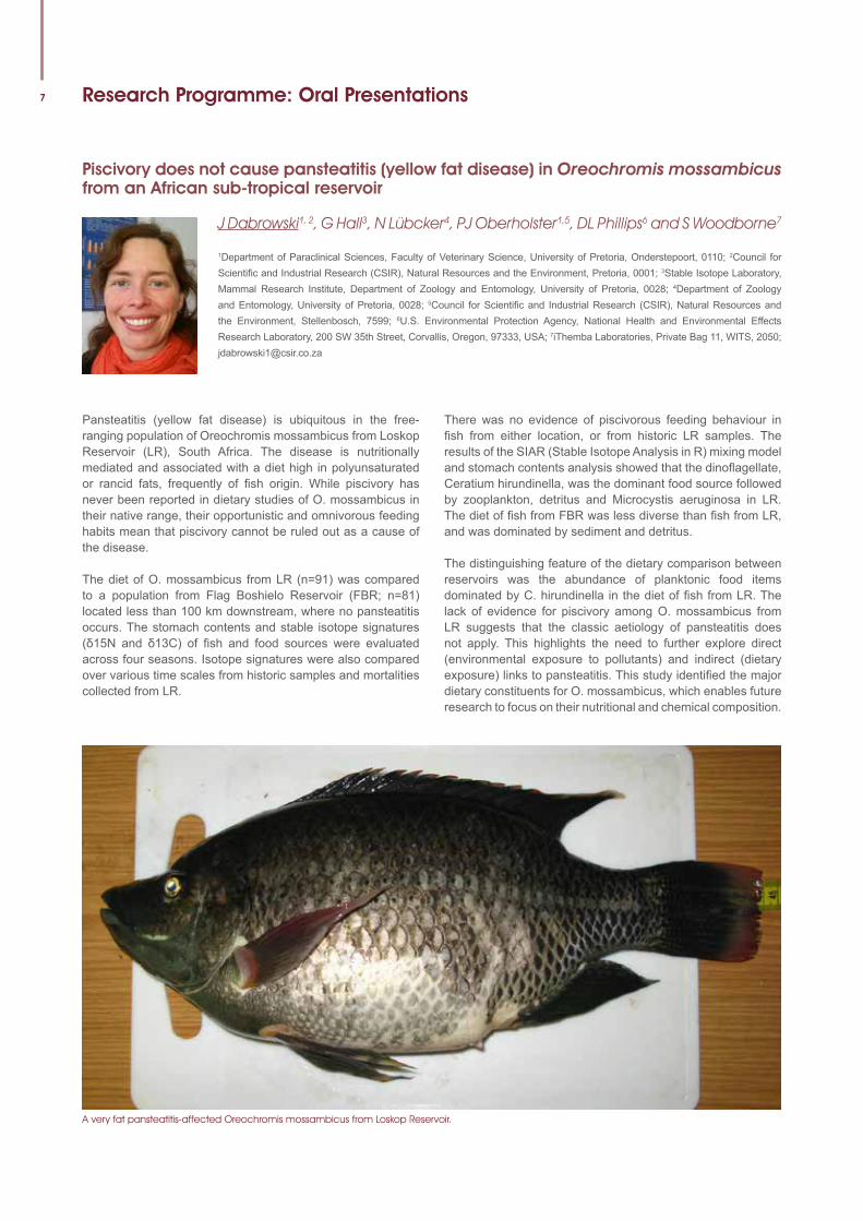
Piscivory does not cause pansteatitis (yellow fat disease) in Oreochromis mossambicus from an African sub-tropical reservoir
J Dabrowski1, 2, G Hall3, N Lübcker4, PJ Oberholster1,5, DL Phillips6 and S Woodborne7
1Department of Paraclinical Sciences, Faculty of Veterinary Science, University of Pretoria, Onderstepoort, 0110; 2Council for Scientific and Industrial Research (CSIR), Natural Resources and the Environment, Pretoria, 0001; 3Stable Isotope Laboratory, Mammal Research Institute, Department of Zoology and Entomology, University of Pretoria, 0028; 4Department of Zoology and Entomology, University of Pretoria, 0028; 5Council for Scientific and Industrial Research (CSIR), Natural Resources and the Environment, Stellenbosch, 7599; 6U.S. Environmental Protection Agency, National Health and Environmental Effects Research Laboratory, 200 SW 35th Street, Corvallis, Oregon, 97333, USA; 7iThemba Laboratories, Private Bag 11, WITS, 2050; [email protected]
Pansteatitis (yellow fat disease) is ubiquitous in the free-ranging population of Oreochromis mossambicus from Loskop Reservoir (LR), South Africa. The disease is nutritionally mediated and associated with a diet high in polyunsaturated or rancid fats, frequently of fish origin. While piscivory has never been reported in dietary studies of O. mossambicus in their native range, their opportunistic and omnivorous feeding habits mean that piscivory cannot be ruled out as a cause of the disease.
The diet of O. mossambicus from LR (n=91) was compared to a population from Flag Boshielo Reservoir (FBR; n=81) located less than 100 km downstream, where no pansteatitis occurs. The stomach contents and stable isotope signatures (δ15N and δ13C) of fish and food sources were evaluated across four seasons. Isotope signatures were also compared over various time scales from historic samples and mortalities collected from LR.
There was no evidence of piscivorous feeding behaviour in fish from either location, or from historic LR samples. The results of the SIAR (Stable Isotope Analysis in R) mixing model and stomach contents analysis showed that the dinoflagellate, Ceratium hirundinella, was the dominant food source followed by zooplankton, detritus and Microcystis aeruginosa in LR. The diet of fish from FBR was less diverse than fish from LR, and was dominated by sediment and detritus.
The distinguishing feature of the dietary comparison between reservoirs was the abundance of planktonic food items dominated by C. hirundinella in the diet of fish from LR. The lack of evidence for piscivory among O. mossambicus from LR suggests that the classic aetiology of pansteatitis does not apply. This highlights the need to further explore direct (environmental exposure to pollutants) and indirect (dietary exposure) links to pansteatitis. This study identified the major dietary constituents for O. mossambicus, which enables future research to focus on their nutritional and chemical composition.
A very fat pansteatitis-affected Oreochromis mossambicus from Loskop Reservoir.
University of Pretoria Faculty of Veterinary Science - Faculty Day 4 September 2014
8
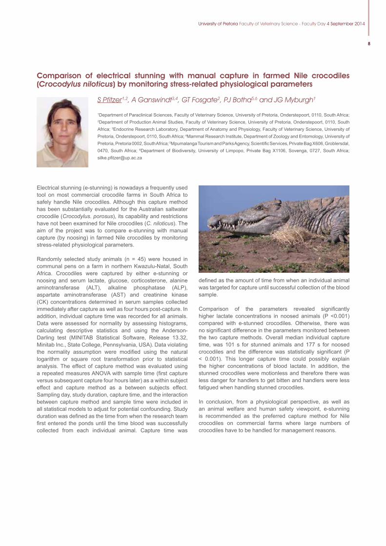
Comparison of electrical stunning with manual capture in farmed Nile crocodiles (Crocodylus niloticus) by monitoring stress-related physiological parameters
S Pfitzer1,2, A Ganswindt3,4, GT Fosgate2, PJ Botha5,6 and JG Myburgh1
1Department of Paraclinical Sciences, Faculty of Veterinary Science, University of Pretoria, Onderstepoort, 0110, South Africa; 2Department of Production Animal Studies, Faculty of Veterinary Science, University of Pretoria, Onderstepoort, 0110, South Africa; 3Endocrine Research Laboratory, Department of Anatomy and Physiology, Faculty of Veterinary Science, University of Pretoria, Onderstepoort, 0110, South Africa; 4Mammal Research Institute, Department of Zoology and Entomology, University of Pretoria, Pretoria 0002, South Africa; 5Mpumalanga Tourism and Parks Agency, Scientific Services, Private Bag X606, Groblersdal, 0470, South Africa; 6Department of Biodiversity, University of Limpopo, Private Bag X1106, Sovenga, 0727, South Africa; [email protected]
Electrical stunning (e-stunning) is nowadays a frequently used tool on most commercial crocodile farms in South Africa to safely handle Nile crocodiles. Although this capture method has been substantially evaluated for the Australian saltwater crocodile (Crocodylus. porosus), its capability and restrictions have not been examined for Nile crocodiles (C. niloticus). The aim of the project was to compare e-stunning with manual capture (by noosing) in farmed Nile crocodiles by monitoring stress-related physiological parameters.
Randomly selected study animals (n = 45) were housed in communal pens on a farm in northern Kwazulu-Natal, South Africa. Crocodiles were captured by either e-stunning or noosing and serum lactate, glucose, corticosterone, alanine aminotransferase (ALT), alkaline phosphatase (ALP), aspartate aminotransferase (AST) and creatinine kinase (CK) concentrations determined in serum samples collected immediately after capture as well as four hours post-capture. In addition, individual capture time was recorded for all animals. Data were assessed for normality by assessing histograms, calculating descriptive statistics and using the Anderson-Darling test (MINITAB Statistical Software, Release 13.32, Minitab Inc., State College, Pennsylvania, USA). Data violating the normality assumption were modified using the natural logarithm or square root transformation prior to statistical analysis. The effect of capture method was evaluated using a repeated measures ANOVA with sample time (first capture versus subsequent capture four hours later) as a within subject effect and capture method as a between subjects effect. Sampling day, study duration, capture time, and the interaction between capture method and sample time were included in all statistical models to adjust for potential confounding. Study duration was defined as the time from when the research team first entered the ponds until the time blood was successfully collected from each individual animal. Capture time was
defined as the amount of time from when an individual animal was targeted for capture until successful collection of the blood sample.
Comparison of the parameters revealed significantly higher lactate concentrations in noosed animals (P <0.001) compared with e-stunned crocodiles. Otherwise, there was no significant difference in the parameters monitored between the two capture methods. Overall median individual capture time, was 101 s for stunned animals and 177 s for noosed crocodiles and the difference was statistically significant (P < 0.001). This longer capture time could possibly explain the higher concentrations of blood lactate. In addition, the stunned crocodiles were motionless and therefore there was less danger for handlers to get bitten and handlers were less fatigued when handling stunned crocodiles.
In conclusion, from a physiological perspective, as well as an animal welfare and human safety viewpoint, e-stunning is recommended as the preferred capture method for Nile crocodiles on commercial farms where large numbers of crocodiles have to be handled for management reasons.
9
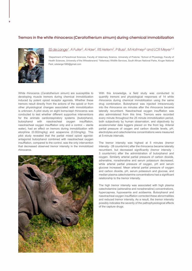
Tremors in the white rhinoceros (Ceratotherium simum) during chemical immobilization
SS de Lange1, A Fuller2, A Haw2, RS Hetem2, P Buss3, M Hofmeyr3 and LCR Meyer1,2
1Department of Paraclinical Sciences, Faculty of Veterinary Science, University of Pretoria; 2School of Physiology, Faculty of Health Sciences, University of the Witwatersrand; 3Veterinary Wildlife Services, South African National Parks, Kruger National Park; [email protected]
White rhinoceros (Ceratotherium simum) are susceptible to developing muscle tremors during chemical immobilization induced by potent opioid receptor agonists. Whether these tremors result directly from the actions of the opioid or from other physiological changes associated with immobilization is unknown. A pilot study on eight boma-kept rhinoceros was conducted to test whether different supportive interventions for the animals cardiorespiratory systems (butorphanol, butorphanol with nasotracheal oxygen insufflation, nasotracheal oxygen insufflation only and a control – sterile water), had an effect on tremors during immobilization with etorphine (0.002mg/kg) and azaperone (0.03mg/kg). This pilot study revealed that the partial mixed opioid agonist-antagonist butorphanol combined with nasotracheal oxygen insufflation, compared to the control, was the only intervention that decreased observed tremor intensity in the immobilized rhinoceros.
With this knowledge, a field study was conducted to quantify tremors and physiological responses of 14 white rhinoceros during chemical immobilization using the same drug combination. Butorphanol was injected intravenously into the rhinoceros six minutes after the rhinoceros became laterally recumbent. Nasotracheal oxygen insufflation was also administered from this time. Tremors were recorded every minute throughout the 25 minute immobilization period, both subjectively by human observation, and objectively by accelerometer data loggers placed on the front leg. Arterial partial pressure of oxygen and carbon dioxide levels, pH, electrolytes and catecholamine concentrations were measured at 5-minute intervals.
The tremor intensity was highest at 5 minutes (tremor intensity - 28 counts/min) after the rhinoceros became laterally recumbent, but decreased significantly (tremor intensity - 3 counts/min) after the administration of butorphanol and oxygen. Similarly arterial partial pressure of carbon dioxide, adrenaline, noradrenaline and serum potassium decreased, while arterial partial pressure of oxygen, pH and serum glucose increased. Mean arterial partial pressure of oxygen and carbon dioxide, pH, serum potassium and glucose, and median plasma catecholamine concentrations had a significant relationship to the tremor intensity.
The high tremor intensity was associated with high plasma catecholamine (adrenaline and noradrenaline) concentrations, hypercapnea, hypoxeamia and acidaemia. Butorphanol and nasotracheal oxygen insufflation corrected these abnormalities and reduced tremor intensity. As a result, the tremor intensity possibly indicates the severity of the pathophysiological effects of the capture drugs.
University of Pretoria Faculty of Veterinary Science - Faculty Day 4 September 2014
10
Clinical anatomy of the cloaca and spinal venous sinus of the Nile crocodile
JG Myburgh1, JT Soley2, JCA Steyl1, RM Kirberger3, FW Huchzermeyer1 and LJ Guillette Jr1,4
1Department of Paraclinical Sciences, Faculty of Veterinary Science, University of Pretoria, Onderstepoort, 0110, South Africa; 2Department of Anatomy and Physiology, Faculty of Veterinary Science, University of Pretoria, Onderstepoort, 0110, South Africa; 3Department of Companion Animal Clinical Studies, Faculty of Veterinary Science, University of Pretoria, Onderstepoort, 0110, South Africa; 4Department of Obstetrics and Gynecology, Marine Biomedicine and Environmental Sciences Center, Medical University of South Carolina, Charleston, 29425, South Carolina, United States of America;[email protected]
Although techniques for the collection of blood and urine from crocodilians are well-established, the clinical anatomy of the spinal vein (blood) and cloaca (urine) in the Nile crocodile has not been investigated. Blood is usually collected from the post-occipital spinal venous sinus, while urine is obtained from the cloaca with an ordinary dog urinary catheter. To promote safe and efficient sample collection a thorough knowledge of the anatomy of the collection route is necessary.
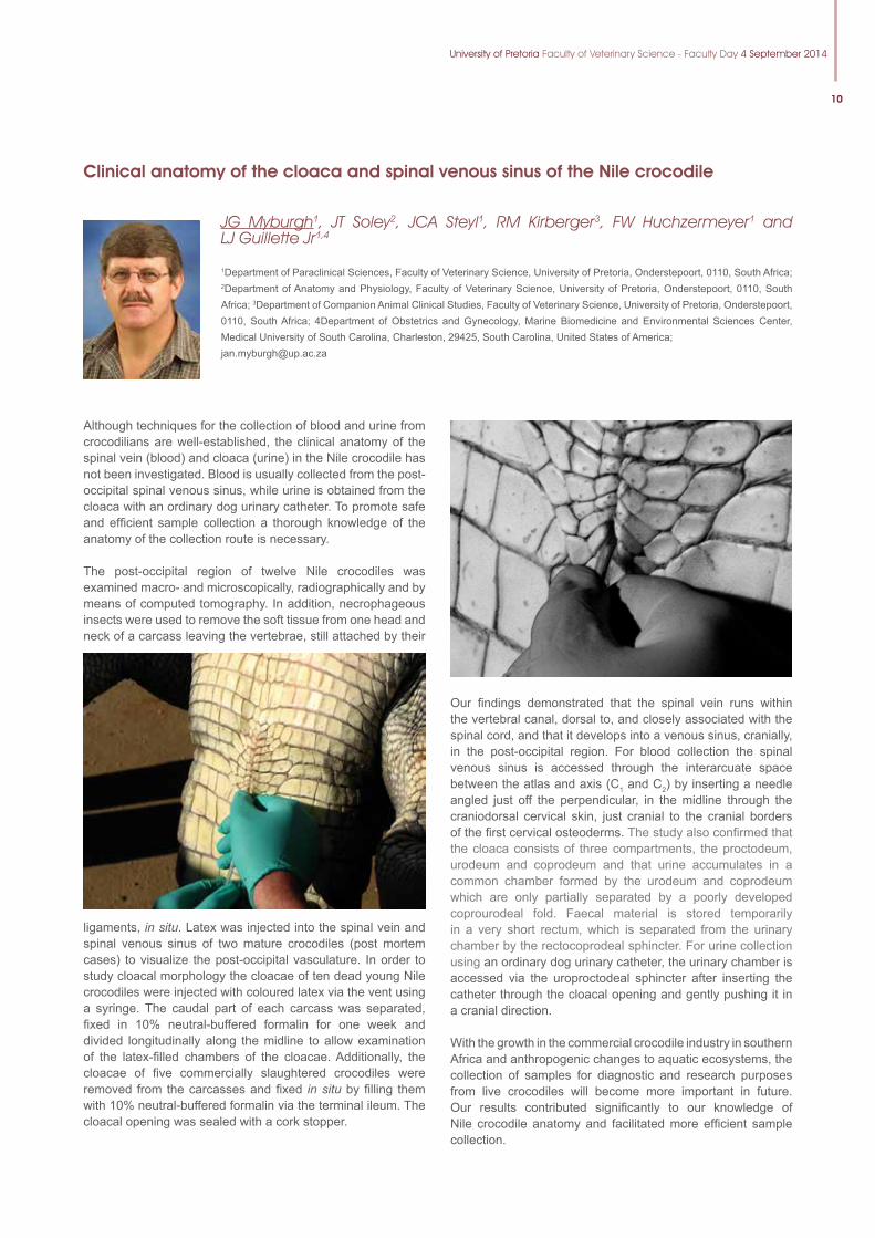
The post-occipital region of twelve Nile crocodiles was examined macro- and microscopically, radiographically and by means of computed tomography. In addition, necrophageous insects were used to remove the soft tissue from one head and neck of a carcass leaving the vertebrae, still attached by their
ligaments, in situ. Latex was injected into the spinal vein and spinal venous sinus of two mature crocodiles (post mortem cases) to visualize the post-occipital vasculature. In order to study cloacal morphology the cloacae of ten dead young Nile crocodiles were injected with coloured latex via the vent using a syringe. The caudal part of each carcass was separated, fixed in 10% neutral-buffered formalin for one week and divided longitudinally along the midline to allow examination of the latex-filled chambers of the cloacae. Additionally, the cloacae of five commercially slaughtered crocodiles were removed from the carcasses and fixed in situ by filling them with 10% neutral-buffered formalin via the terminal ileum. The cloacal opening was sealed with a cork stopper.
Our findings demonstrated that the spinal vein runs within the vertebral canal, dorsal to, and closely associated with the spinal cord, and that it develops into a venous sinus, cranially, in the post-occipital region. For blood collection the spinal venous sinus is accessed through the interarcuate space between the atlas and axis (C1 and C2) by inserting a needle angled just off the perpendicular, in the midline through the craniodorsal cervical skin, just cranial to the cranial borders of the first cervical osteoderms. The study also confirmed that the cloaca consists of three compartments, the proctodeum, urodeum and coprodeum and that urine accumulates in a common chamber formed by the urodeum and coprodeum which are only partially separated by a poorly developed coprourodeal fold. Faecal material is stored temporarily in a very short rectum, which is separated from the urinary chamber by the rectocoprodeal sphincter. For urine collection using an ordinary dog urinary catheter, the urinary chamber is accessed via the uroproctodeal sphincter after inserting the catheter through the cloacal opening and gently pushing it in a cranial direction.
With the growth in the commercial crocodile industry in southern Africa and anthropogenic changes to aquatic ecosystems, the collection of samples for diagnostic and research purposes from live crocodiles will become more important in future. Our results contributed significantly to our knowledge of Nile crocodile anatomy and facilitated more efficient sample collection.
11
Treatment of radius and tibia fractures in wild antelope utilizing external fixation
FJ Venter, BG Nevill, A Mahne, G Zeiler, GF Stegman and GL Coetzee
Department of Companion Animal Studies, Onderstepoort Veterinary Faculty, University of Pretoria, South Africa [email protected]
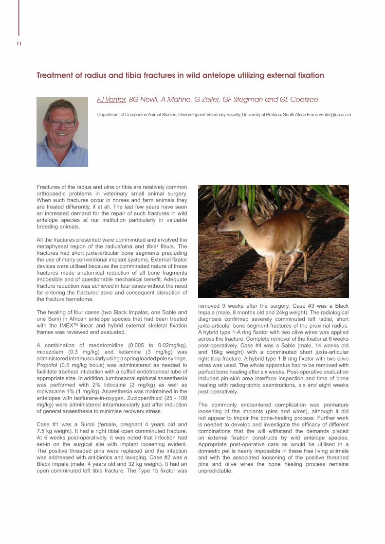
Fractures of the radius and ulna or tibia are relatively common orthopaedic problems in veterinary small animal surgery. When such fractures occur in horses and farm animals they are treated differently, if at all. The last few years have seen an increased demand for the repair of such fractures in wild antelope species at our institution particularly in valuable breeding animals.
All the fractures presented were comminuted and involved the metaphyseal region of the radius/ulna and tibia/ fibula. The fractures had short juxta-articular bone segments precluding the use of many conventional implant systems. External fixator devices were utilised because the comminuted nature of these fractures made anatomical reduction of all bone fragments impossible and of questionable mechanical benefit. Adequate fracture reduction was achieved in four cases without the need for entering the fractured zone and consequent disruption of the fracture hematoma.
The healing of four cases (two Black Impalas, one Sable and one Suni) in African antelope species that had been treated with the IMEXTM linear and hybrid external skeletal fixation frames was reviewed and evaluated.
A combination of medetomidine (0.005 to 0.02mg/kg), midazolam (0.3 mg/kg) and ketamine (3 mg/kg) was administered intramuscularly using a spring loaded pole syringe. Propofol (0.5 mg/kg bolus) was administered as needed to facilitate tracheal intubation with a cuffed endotracheal tube of appropriate size. In addition, lumbosacral epidural anaesthesia was performed with 2% lidocaine (2 mg/kg) as well as ropivacaine 1% (1 mg/kg). Anaesthesia was maintained in the antelopes with isoflurane-in-oxygen. Zuclopenthixol (25 - 100 mg/kg) were administered intramuscularly just after induction of general anaesthesia to minimise recovery stress.
Case #1 was a Sunni (female, pregnant 4 years old and 7.5 kg weight). It had a right tibial open comminuted fracture. At 6 weeks post-operatively, it was noted that infection had set-in on the surgical site with implant loosening evident. The positive threaded pins were replaced and the infection was addressed with antibiotics and lavaging. Case #2 was a Black Impala (male, 4 years old and 32 kg weight). It had an open comminuted left tibia fracture. The Type 1b fixator was
removed 9 weeks after the surgery. Case #3 was a Black Impala (male, 8 months old and 24kg weight). The radiological diagnosis confirmed severely comminuted left radial, short juxta-articular bone segment fractures of the proximal radius. A hybrid type 1-A ring fixator with two olive wires was applied across the fracture. Complete removal of the fixator at 6 weeks post-operatively. Case #4 was a Sable (male, 14 weeks old and 16kg weight) with a comminuted short juxta-articular right tibia fracture. A hybrid type 1-B ring fixator with two olive wires was used. The whole apparatus had to be removed with perfect bone healing after six weeks. Post-operative evaluation included pin-skin area interface inspection and time of bone healing with radiographic examinations, six and eight weeks post-operatively.
The commonly encountered complication was premature loosening of the implants (pins and wires), although it did not appear to impair the bone-healing process. Further work is needed to develop and investigate the efficacy of different combinations that the will withstand the demands placed on external fixation constructs by wild antelope species. Appropriate post-operative care as would be utilised in a domestic pet is nearly impossible in these free living animals and with the associated loosening of the positive threaded pins and olive wires the bone healing process remains unpredictable.
University of Pretoria Faculty of Veterinary Science - Faculty Day 4 September 2014
12
Catastrophic distal forelimb musculoskeletal injuries associated with racetracks in Gauteng, South Africa from 1998-2012
KE Spargo1, L Rubio-Martinez2, I Cilliers3, DP Wheeler4, A Guthrie5, L Fletcher6 and A Carstens1
1Section Diagnostic Imaging, Department of Companion Animal Clinical Studies, Faculty of Veterinary Science, University of Pretoria, Onderstepoort 0110; 2Equine division, University of Liverpool, Leahurst Campus, Chester High Road, CH64 7TE, Neston, UK; 3Section Equine Medicine and Surgery, Department of Companion Animal Clinical Studies, Faculty of Veterinary Science, University of Pretoria, Onderstepoort 0110; 4National Horseracing Authority of Southern Africa, Turf Club Street, Turffontein, Johannesburg, SA; 5Equine Research Centre, Faculty of Veterinary Science, University of Pretoria, Onderstepoort 0110; 6Department of Statistics, Faculty of Economic and Management Sciences, University of Pretoria; [email protected]
Catastrophic musculoskeletal limb injuries (CMIs) on the racetrack result in the immediate end of a racehorse’s career. Numerous studies on the incidence rates and factors infl uencing injuries and fatalities have been reported in most of the major horse racing countries around the world, however, limited published data on the incidence rates and factors of CMIs exist from South Africa. The factors which show potential for increasing the risk of injury include age, gender, track surface type and condition, distance of race, number of previous starts and time interval between races.
This study was conducted to describe and report on the incidence and types of CMIs of the distal forelimb leading to immediate euthanasia in racing Thoroughbreds on Gauteng racetracks and also to identify their associated risk factors.
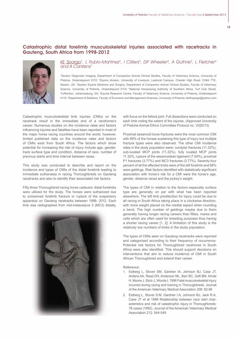
Fifty-three Thoroughbred racing horse cadavers’ distal forelimbs were utilized for the study. The horses were euthanized due to unresolved forelimb fracture or rupture of the suspensory apparatus on Gauteng racetracks between 1998- 2012. Each limb was radiographed from mid-metacarpus 3 (MC3) distally,
with focus on the fetlock joint. Full dissections were conducted on each limb noting the extent of the injuries. (Approved University of Pretoria Animal Ethics Committee Protocol no. V020/13)
Proximal sesamoid bone fractures were the most common CMI with 89% of the horses sustaining this type of injury but multiple fracture types were also observed . The other CMI incidence rates in the study population were: condylar fractures (11.32%), sub-luxated MCP joints (11,32%), fully luxated MCP joints 11.32%, rupture of the sesamoidean ligament (7.54%), proximal P1 fractures (3.77%) and MC3 fractures (3.77%). Seventy-four percent of all the affected limbs were of the left forelimb and 68% were geldings. Risk factors identifi ed with statistically signifi cant association with horse’s risk for a CMI were the horse’s age, gender, distance raced and the jockey’s weight.
The types of CMI in relation to the factors especially surface type are generally on par with what has been reported elsewhere. The left limb predilection for injury could be due to all racing in South Africa taking place in a clockwise direction, with more weight placed on the medial aspect when rounding a bend. The high number of geldings maybe due to them generally having longer racing careers than fi llies, mares and colts which are often used for breeding purposes thus having a shorter racing career [1, 2]. A limitation of this study is the relatively low numbers of limbs in the study population.
The types of CMIs seen on Gauteng racetracks were reported and categorised according to their frequency of occurrence. Potential risk factors for Thoroughbred racehorse in South Africa were also identifi ed. This should support decisions on interventions that aim to reduce incidence of CMI in South African Thoroughbred and extend their career.
Reference: 1. Estberg L, Stover SM, Gardner IA, Johnson BJ, Case JT,
Ardans AA, Read DH, Anderson ML, Barr BC, Daft BM, Kinde H, Moore J, Stolz J, Woods L 1996 Fatal musculoskeletal injury incurred during racing and training in Thoroughbreds. Journal of the American Veterinary Medical Association 208: 92-96
2. Estberg L, Stover S.M, Gardner I.A, Johnson BJ, Jack R.A, Case JT et al 1998 Relationship between race start char-acteristics and risk of catastrophic injury in Thoroughbreds: 78 cases (1992). Journal of the American Veterinary Medical Association 212: 544-549
13
A comparison between juvenile pubic symphysiodesis and juvenile pubic symphysectomy: a one-year follow-up
FJ Venter1, BG Nevill1 and G L Coetzee1
1Small Animal Surgery, Department of Companion Animal Clinical Studies, Faculty of Veterinary Science, University of Pretoria, South Africa.
Hip dysplasia (HD) is one of the most common orthopaedic diseases of large-breed dogs. In affected dogs, joint laxity, joint incongruence, and secondary osteoarthritis can lead to crippling pain. Current topics on hip surgery involve the technique of juvenile symphysiodesis (JPS), a technique performed to improve the congruence of growing coxofemoral joints in young dogs. Although alternative procedures do exist, we proposes a modified technique termed juvenile pubic symphysectomy (JPSec).
Radiographic and computed tomography (CT) measurements were made in the study group of 21 immature puppies aged 16 weeks. The following measurements were used for the determination of HD: Subluxation index (SI) with the Flückiger method, Norberg angles5, lateral center-edge angles, dorsal acetabular rim angles and acetabular ventro version angles (VV). Joint laxity was determined subjectively by using the Barden’s lift technique and the Ortolani sign. The HD positive puppies were randomly selected for the surgical techniques used in this study. The study consisted of three groups: Group 1 = HD free (non-surgical) control group (N=7), Group 2 HD at risk positive dogs treated with the JPS technique using electro cautery (N=7) and Group 3 the JPSec technique was performed on the pre-determined hip dysplastic dogs (N=7). The pelvic ventroversion angles were assessed pre-and immediately post-op, at 16 weeks, and with follow-up assessments at 20, 24 and 54 weeks of age in all the groups.
The JPSec technique resulted in immediate change in the acetabular ventro version angles by ±5º post surgically (24-48h) due to rotation of both acetabulae (ventroversion).
The JPSec appeared to provide greater and more dorsal acetabular covering (DARA) after 24 and 54 weeks than JPS. The lateral center edge angles also improved on follow-up assessments of both the techniques. There was a noticeable change in ventroversion angle improvement, more so with the JPSec than with JPS procedure and as a result, also better dorsal coxofemoral coverage by the dorsal acetabular rim with decreased hip joint laxity. JPSec and JPS did not have any significant effect on the sacral width or sacral conformation.
The pubic symphysectomy procedure is a simple surgical technique that yields good results without the need for specialized equipment. The age of the dogs is an important factor. It is believed that the benefits of removing the pubic symphysis growth plate at 16 weeks of age would be optimal. The difference between the two techniques lies in the severity of the HD of the dog. It was found that the JPS yielded the best results with a Norberg angle between 95º and 102º, whereas JPSec gave the best results between 85º and 95º degrees. The JPSec technique requires fixation of the pubic ramii and symphysis with orthopedic wire, these implants will serve as an indicator of a previous corrective surgical procedure when such an animal is later presented for HD evaluation.
Dog food was sponsored by Nestlé Purina South Africa, Protocol No.: V059/08
University of Pretoria Faculty of Veterinary Science - Faculty Day 4 September 2014
14
Effect of intravenous lidocaine on the minimum infusion rate of alfaxalone in goats
PS Ndawana1, TB Dzikiti1, G Zeiler1, FS Stegmann1 and LN Dzikiti2
1Department of Companion Animal Clinical Studies, University of Pretoria, Private Bag X04, Onderstepoort, 0110, South Africa; 2School of Health Systems and Public Health, University of Pretoria, Private Bag X323, Pretoria, 0001, South Africa; [email protected]
The technique of using drugs administered solely by the intravenous route for induction and maintenance of general anaesthesia, Total Intravenous Anaesthesia (TIVA), is gaining popularity in small animal and equine anaesthesia. The relatively new neuroactive steroid, alfaxalone, is commonly used as an intravenous agent to induce and maintain general anaesthesia in the dog and cat. Lidocaine has recently been used as an adjunct to inhalation anaesthesia to reduce anaesthetic requirements in dogs, horses and goats with minimal cardiovascular adverse effects. However, there is currently no known information on alfaxalone TIVA requirements in goats together with the effects of intravenous lidocaine.
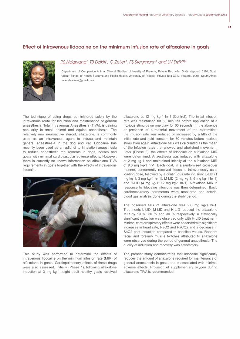
This study was performed to determine the effects of intravenous lidocaine on the minimum infusion rate (MIR) of alfaxalone in goats. Cardiopulmonary effects of these drugs were also assessed. Initially (Phase 1), following alfaxalone induction at 3 mg kg-1, eight adult healthy goats received
alfaxalone at 12 mg kg-1 hr-1 (Control). The initial infusion rate was maintained for 30 minutes before application of a noxious stimulus on one claw for 60 seconds. In the absence or presence of purposeful movement of the extremities, the infusion rate was reduced or increased by a fifth of the initial rate and held constant for 30 minutes before noxious stimulation again. Alfaxalone MIR was calculated as the mean of the infusion rates that allowed and abolished movement. Later (Phase 2), the effects of lidocaine on alfaxalone MIR were determined. Anaesthesia was induced with alfaxalone at 2 mg kg-1 and maintained initially at the alfaxalone MIR of 9.6 mg kg-1 hr-1. Each goat, in a randomised crossover manner, concurrently received lidocaine intravenously as a loading dose, followed by a continuous rate infusion: L-LID (1 mg kg-1; 3 mg kg-1 hr-1), M-LID (2 mg kg-1; 6 mg kg-1 hr-1) and H-LID (4 mg kg-1; 12 mg kg-1 hr-1). Alfaxalone MIR in response to lidocaine infusions was then determined. Basic cardiorespiratory parameters were monitored and arterial blood gas analysis done during the study period.
The observed MIR of alfaxalone was 9.6 mg kg-1 hr-1. Treatments L-LID, M-LID and H-LID reduced the alfaxalone MIR by 10 %, 30 % and 30 % respectively. A statistically significant reduction was observed only with H-LID treatment. Minimal cardiorespiratory effects were observed with significant increases in heart rate, PaO2 and PaCO2 and a decrease in SaO2 post induction compared to baseline values. Random facial and forelimb muscle twitches attributed to alfaxalone were observed during the period of general anaesthesia. The quality of induction and recovery was satisfactory.
The present study demonstrates that lidocaine significantly reduces the amount of alfaxalone required for maintenance of general anaesthesia in goats and is associated with minimal adverse effects. Provision of supplementary oxygen during alfaxalone TIVA is recommended.
15
Comparison of anaesthetic induction and recovery characteristics of diazepam-ketamine combination to propofol alone; in healthy dogs undergoing orchidectomy
JP Ferreira1, R Buck1, B Nevill1, GE Zelier1, B Gummow2, TB Dzikiti1 and L Bester1
1Onderstepoort Veterinary Academic hospital, University of Pretoria, Gauteng, 0110; 2School of Veterinary and Biomedical Sciences, James Cook University, Australia; [email protected]
Diazepam and ketamine combination provides an alternative to propofol for induction of anaesthesia in healthy and hypotensive dogs. There is however no consensus between practitioners regarding the quality of induction and recovery from diazepam-ketamine anaesthesia, and as such questioning its practicality in the clinical setting. The purpose of the study was to compare anaesthetic induction and recovery characteristics of diazepam-ketamine combination to propofol alone in dogs undergoing elective orchidectomyin a prospective, randomised clinical trial incorporating 36 healthy adult male dogs of various breeds with a mean age of 26.2±13.4 months and weight of 5.5±2.3kg.
After demeanour scoring (simple descriptive scale; SDS); the dogs were sedated with morphine (0.3 mg/kg) and acepromazine (0.02 mg/kg) intramuscularly. Forty minutes after administration a premedication score (SDS) was allocated. Immediately after premedication had been scored, general anaesthesia was induced with either a combination of diazepam and ketamine (D/K) or propofol (P) intravenously. Initial induction doses of 0.375, 0.5 and 2 mg/kg, for diazepam and ketamine or propofol respectively were administered to facilitate endotracheal intubation. Anaesthesia was maintained with isoflurane. Scores for the quality of induction, intubation and degree of myoclonus were allocated (SDS). Orchidectomy was performed in a standard way by a single experienced surgeon. Recovery from anaesthesia was scored (SDS). Times to extubation and standing were recorded. Data were analysed for statistically significant differences (P<0.05) using the t-test for parametric data and the Wilcoxon Mann-Whitney test for non-parametric data. The Kappa Reliability and Kendall Tau tests were used to assess the degree of agreement between the scorers for the scored characteristics.
There were no statistically significant differences between groups in age, weight, cage rest score, premedication score and duration of maintenance of anaesthesia. Group P was associated with a poorer quality of induction (P=0.014) and more pronounced myoclonus (P=0.003); but had better quality of recovery (P=0.000002) and shorter recovery times (P=0.035) compared to group D/K.
It was concluded that propofol is associated with poorer anaesthetic induction characteristics, but better and quicker recovery from anaesthesia compared to diazepam-ketamine in male dogs premedicated with morphine and acepromazine.
University of Pretoria Faculty of Veterinary Science - Faculty Day 4 September 2014
16
Molecular characterization of vaccine candidates from Anaplasma marginale strains in South Africa
P Hove1,2, KA Brayton3, MC Oosthuizen1, SM Mtshali4, ME Chaisi1 and NE Collins1
1Department of Veterinary Tropical Diseases, Faculty of Veterinary Sciences, University of Pretoria, P. Bag X04, Onderstepoort, 0110, Pretoria, South Africa; 2Agricultural Research Council, Onderstepoort Veterinary Institute, Biotechnology Platform, 0110, Onderstepoort, Pretoria, South Africa; 3Department of Veterinary Microbiology and Pathology, Paul G. Allen School for Global Animal Health, Washington State University, Pullman, WA 99164-7040. USA; 4Research and Scientific Services Department, National Zoological Gardens of South Africa, P O Box 754, Pretoria 0001, South Africa; [email protected]
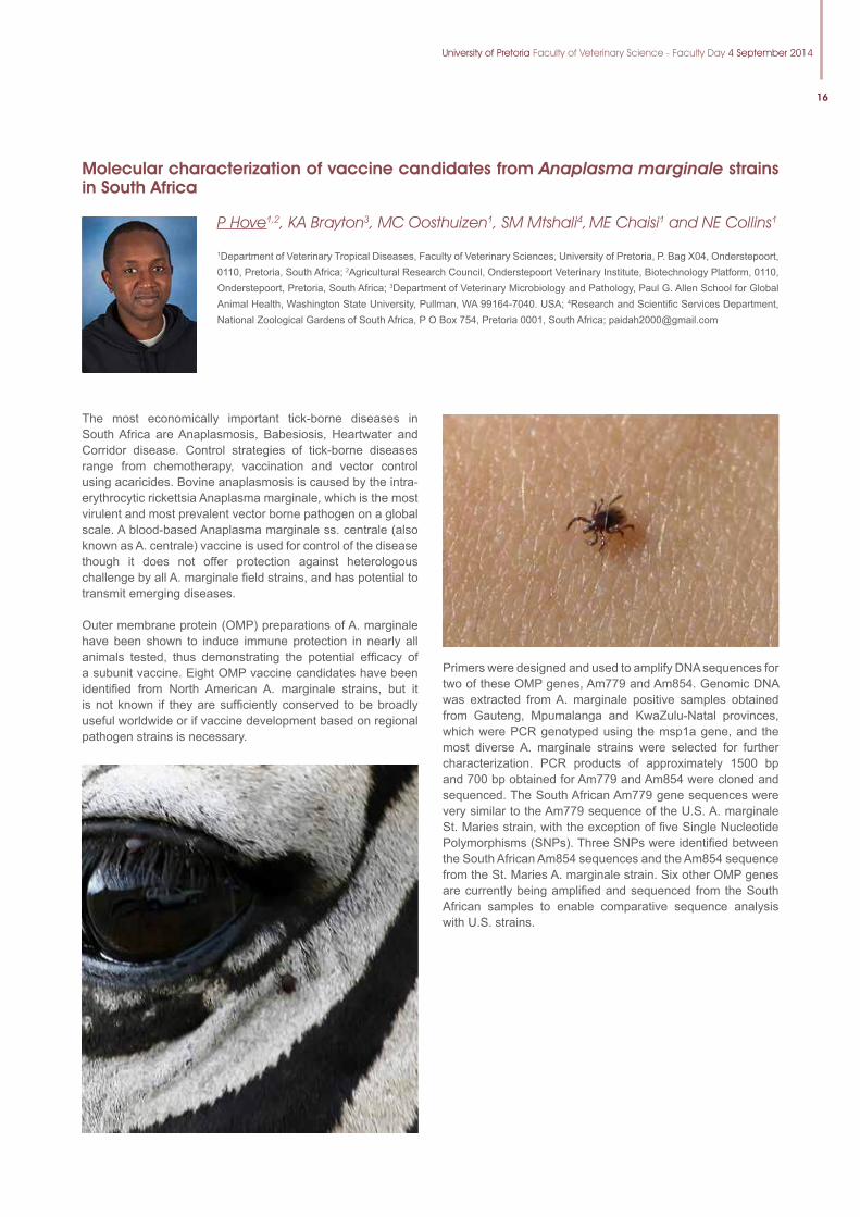
The most economically important tick-borne diseases in South Africa are Anaplasmosis, Babesiosis, Heartwater and Corridor disease. Control strategies of tick-borne diseases range from chemotherapy, vaccination and vector control using acaricides. Bovine anaplasmosis is caused by the intra-erythrocytic rickettsia Anaplasma marginale, which is the most virulent and most prevalent vector borne pathogen on a global scale. A blood-based Anaplasma marginale ss. centrale (also known as A. centrale) vaccine is used for control of the disease though it does not offer protection against heterologous challenge by all A. marginale field strains, and has potential to transmit emerging diseases.
Outer membrane protein (OMP) preparations of A. marginale have been shown to induce immune protection in nearly all animals tested, thus demonstrating the potential efficacy of a subunit vaccine. Eight OMP vaccine candidates have been identified from North American A. marginale strains, but it is not known if they are sufficiently conserved to be broadly useful worldwide or if vaccine development based on regional pathogen strains is necessary.
Primers were designed and used to amplify DNA sequences for two of these OMP genes, Am779 and Am854. Genomic DNA was extracted from A. marginale positive samples obtained from Gauteng, Mpumalanga and KwaZulu-Natal provinces, which were PCR genotyped using the msp1a gene, and the most diverse A. marginale strains were selected for further characterization. PCR products of approximately 1500 bp and 700 bp obtained for Am779 and Am854 were cloned and sequenced. The South African Am779 gene sequences were very similar to the Am779 sequence of the U.S. A. marginale St. Maries strain, with the exception of five Single Nucleotide Polymorphisms (SNPs). Three SNPs were identified between the South African Am854 sequences and the Am854 sequence from the St. Maries A. marginale strain. Six other OMP genes are currently being amplified and sequenced from the South African samples to enable comparative sequence analysis with U.S. strains.
17
Molecular detection of Anaplasma marginale and Anaplasma subspecies centrale in cattle in South Africa
ME Chaisi1,2, P Hove1, C Choopa1, MC Oosthuizen1 and NE Collins1
1Department of Veterinary Tropical Diseases, Faculty of Veterinary Science, University of Pretoria, Onderstepoort, South Africa, 2Department of Biology, National University of Lesotho, Faculty of Science and Technology, Roma 180, Lesotho;[email protected]
Bovine anaplasmosis is a potentially fatal tick-borne disease of cattle caused by the intra-erythrocytic rickettsia, Anaplasma marginale. It is widely distributed around the world. In South Africa, it is endemic in most of the cattle-farming areas. A. marginale subsp. centrale causes a milder form of anaplasmosis in cattle and has been used as a vaccine in many parts of the world. Conventional PCR has been used for the detection and differentiation of A. marginale and other tick-borne pathogens from hosts and vectors using different genetic markers. More recently, quantitative real-time polymerase chain reaction (qPCR) has been used for the detection of these organisms in infected hosts and vectors. qPCR has a number of advantages over the conventional PCR: a) it is more sensitive and specifi c in the detection of organisms, b) the reaction can be monitored in real-time, and c) detection and quantifi cation occur in the same reaction during the cycling process therefore eliminating the need for post-PCR analysis and reducing the risk of contamination.
We assessed the qPCR assays of Carelli et al. (2007) and Decaro et al. (2008) respectively, for the detection of A. marginale and simultaneous detection of A. marginale and A. marginale subsp. centrale in 390 cattle samples originating from different areas in South Africa. In 231 of these samples, the A. marginale results were compared to those of the Reverse Line Blot (RLB) hybridization assay. The RLB assay can simultaneously detect all known Theileria and Babesia spp. in infected organisms using species-specifi c probes.
When using the duplex real-time PCR assay, A. marginale and A. marginale subsp. centrale were detected in 69% and 16%
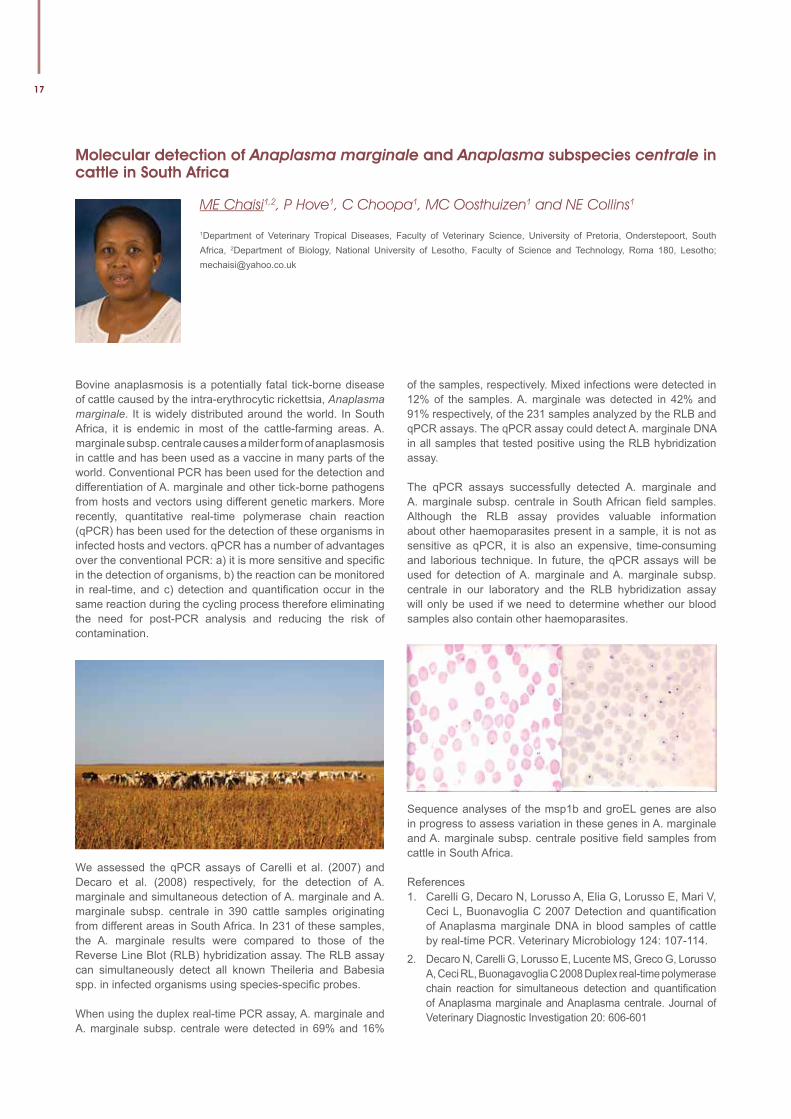
of the samples, respectively. Mixed infections were detected in 12% of the samples. A. marginale was detected in 42% and 91% respectively, of the 231 samples analyzed by the RLB and qPCR assays. The qPCR assay could detect A. marginale DNA in all samples that tested positive using the RLB hybridization assay.
The qPCR assays successfully detected A. marginale and A. marginale subsp. centrale in South African fi eld samples. Although the RLB assay provides valuable information about other haemoparasites present in a sample, it is not as sensitive as qPCR, it is also an expensive, time-consuming and laborious technique. In future, the qPCR assays will be used for detection of A. marginale and A. marginale subsp. centrale in our laboratory and the RLB hybridization assay will only be used if we need to determine whether our blood samples also contain other haemoparasites.
Sequence analyses of the msp1b and groEL genes are also in progress to assess variation in these genes in A. marginale and A. marginale subsp. centrale positive fi eld samples from cattle in South Africa.
References1. Carelli G, Decaro N, Lorusso A, Elia G, Lorusso E, Mari V,
Ceci L, Buonavoglia C 2007 Detection and quantifi cation of Anaplasma marginale DNA in blood samples of cattle by real-time PCR. Veterinary Microbiology 124: 107-114.
2. Decaro N, Carelli G, Lorusso E, Lucente MS, Greco G, Lorusso A, Ceci RL, Buonagavoglia C 2008 Duplex real-time polymerase chain reaction for simultaneous detection and quantifi cation of Anaplasma marginale and Anaplasma centrale. Journal of Veterinary Diagnostic Investigation 20: 606-601
University of Pretoria Faculty of Veterinary Science - Faculty Day 4 September 2014
18
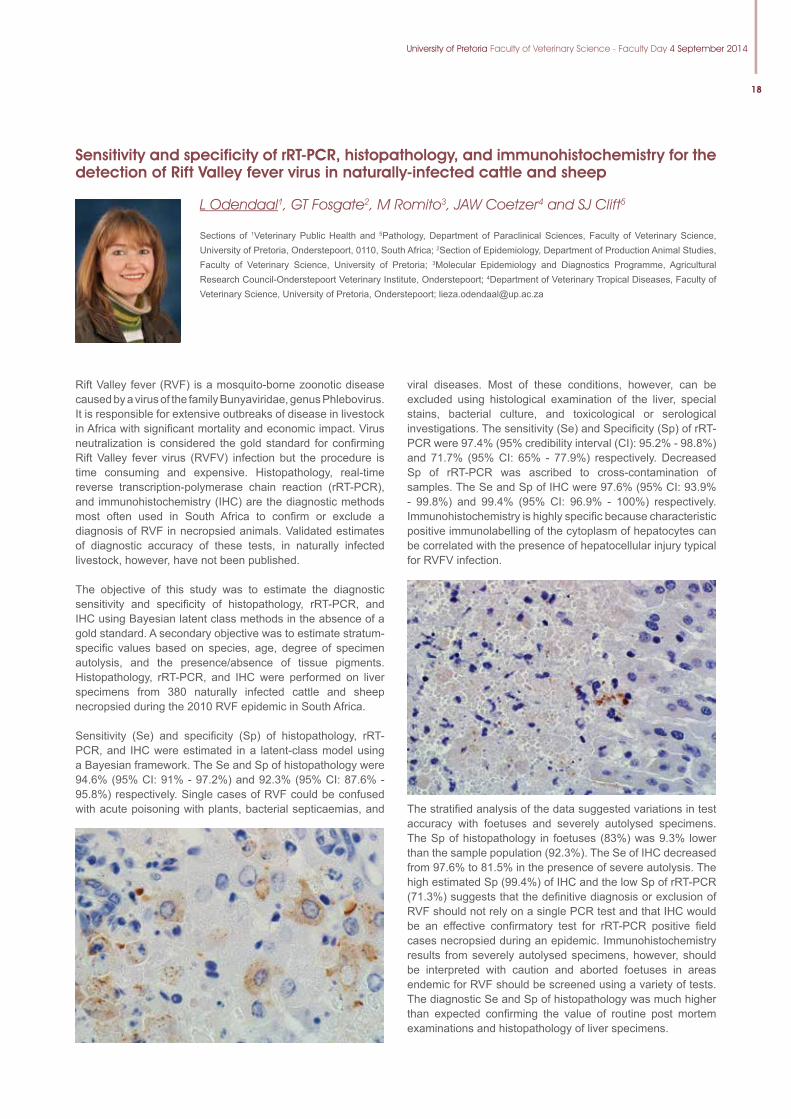
Sensitivity and specificity of rRT-PCR, histopathology, and immunohistochemistry for the detection of Rift Valley fever virus in naturally-infected cattle and sheep
L Odendaal1, GT Fosgate2, M Romito3, JAW Coetzer4 and SJ Clift5
Sections of 1Veterinary Public Health and 5Pathology, Department of Paraclinical Sciences, Faculty of Veterinary Science, University of Pretoria, Onderstepoort, 0110, South Africa; 2Section of Epidemiology, Department of Production Animal Studies, Faculty of Veterinary Science, University of Pretoria; 3Molecular Epidemiology and Diagnostics Programme, Agricultural Research Council-Onderstepoort Veterinary Institute, Onderstepoort; 4Department of Veterinary Tropical Diseases, Faculty of Veterinary Science, University of Pretoria, Onderstepoort; [email protected]
Rift Valley fever (RVF) is a mosquito-borne zoonotic disease caused by a virus of the family Bunyaviridae, genus Phlebovirus. It is responsible for extensive outbreaks of disease in livestock in Africa with signifi cant mortality and economic impact. Virus neutralization is considered the gold standard for confi rming Rift Valley fever virus (RVFV) infection but the procedure is time consuming and expensive. Histopathology, real-time reverse transcription-polymerase chain reaction (rRT-PCR), and immunohistochemistry (IHC) are the diagnostic methods most often used in South Africa to confi rm or exclude a diagnosis of RVF in necropsied animals. Validated estimates of diagnostic accuracy of these tests, in naturally infected livestock, however, have not been published.
The objective of this study was to estimate the diagnostic sensitivity and specifi city of histopathology, rRT-PCR, and IHC using Bayesian latent class methods in the absence of a gold standard. A secondary objective was to estimate stratum-specifi c values based on species, age, degree of specimen autolysis, and the presence/absence of tissue pigments. Histopathology, rRT-PCR, and IHC were performed on liver specimens from 380 naturally infected cattle and sheep necropsied during the 2010 RVF epidemic in South Africa.
Sensitivity (Se) and specifi city (Sp) of histopathology, rRT-PCR, and IHC were estimated in a latent-class model using a Bayesian framework. The Se and Sp of histopathology were 94.6% (95% CI: 91% - 97.2%) and 92.3% (95% CI: 87.6% - 95.8%) respectively. Single cases of RVF could be confused with acute poisoning with plants, bacterial septicaemias, and
viral diseases. Most of these conditions, however, can be excluded using histological examination of the liver, special stains, bacterial culture, and toxicological or serological investigations. The sensitivity (Se) and Specifi city (Sp) of rRT-PCR were 97.4% (95% credibility interval (CI): 95.2% - 98.8%) and 71.7% (95% CI: 65% - 77.9%) respectively. Decreased Sp of rRT-PCR was ascribed to cross-contamination of samples. The Se and Sp of IHC were 97.6% (95% CI: 93.9% - 99.8%) and 99.4% (95% CI: 96.9% - 100%) respectively. Immunohistochemistry is highly specifi c because characteristic positive immunolabelling of the cytoplasm of hepatocytes can be correlated with the presence of hepatocellular injury typical for RVFV infection.
The stratifi ed analysis of the data suggested variations in test accuracy with foetuses and severely autolysed specimens. The Sp of histopathology in foetuses (83%) was 9.3% lower than the sample population (92.3%). The Se of IHC decreased from 97.6% to 81.5% in the presence of severe autolysis. The high estimated Sp (99.4%) of IHC and the low Sp of rRT-PCR (71.3%) suggests that the defi nitive diagnosis or exclusion of RVF should not rely on a single PCR test and that IHC would be an effective confi rmatory test for rRT-PCR positive fi eld cases necropsied during an epidemic. Immunohistochemistry results from severely autolysed specimens, however, should be interpreted with caution and aborted foetuses in areas endemic for RVF should be screened using a variety of tests. The diagnostic Se and Sp of histopathology was much higher than expected confi rming the value of routine post mortem examinations and histopathology of liver specimens.
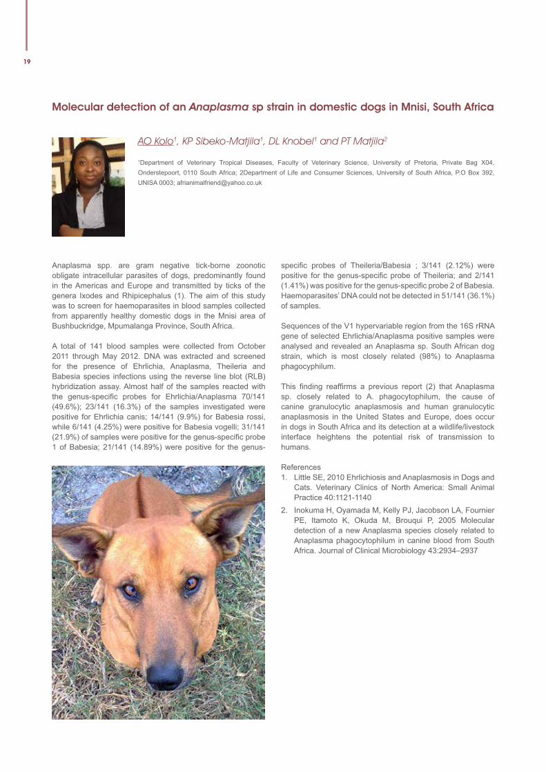
19
Molecular detection of an Anaplasma sp strain in domestic dogs in Mnisi, South Africa
AO Kolo1, KP Sibeko-Matjila1, DL Knobel1 and PT Matjila2
1Department of Veterinary Tropical Diseases, Faculty of Veterinary Science, University of Pretoria, Private Bag X04, Onderstepoort, 0110 South Africa; 2Department of Life and Consumer Sciences, University of South Africa, P.O Box 392, UNISA 0003; [email protected]
Anaplasma spp. are gram negative tick-borne zoonotic obligate intracellular parasites of dogs, predominantly found in the Americas and Europe and transmitted by ticks of the genera Ixodes and Rhipicephalus (1). The aim of this study was to screen for haemoparasites in blood samples collected from apparently healthy domestic dogs in the Mnisi area of Bushbuckridge, Mpumalanga Province, South Africa.
A total of 141 blood samples were collected from October 2011 through May 2012. DNA was extracted and screened for the presence of Ehrlichia, Anaplasma, Theileria and Babesia species infections using the reverse line blot (RLB) hybridization assay. Almost half of the samples reacted with the genus-specific probes for Ehrlichia/Anaplasma 70/141 (49.6%); 23/141 (16.3%) of the samples investigated were positive for Ehrlichia canis; 14/141 (9.9%) for Babesia rossi, while 6/141 (4.25%) were positive for Babesia vogelli; 31/141 (21.9%) of samples were positive for the genus-specific probe 1 of Babesia; 21/141 (14.89%) were positive for the genus-
specific probes of Theileria/Babesia ; 3/141 (2.12%) were positive for the genus-specific probe of Theileria; and 2/141 (1.41%) was positive for the genus-specific probe 2 of Babesia. Haemoparasites’ DNA could not be detected in 51/141 (36.1%) of samples.
Sequences of the V1 hypervariable region from the 16S rRNA gene of selected Ehrlichia/Anaplasma positive samples were analysed and revealed an Anaplasma sp. South African dog strain, which is most closely related (98%) to Anaplasma phagocyphilum.
This finding reaffirms a previous report (2) that Anaplasma sp. closely related to A. phagocytophilum, the cause of canine granulocytic anaplasmosis and human granulocytic anaplasmosis in the United States and Europe, does occur in dogs in South Africa and its detection at a wildlife/livestock interface heightens the potential risk of transmission to humans.
References1. Little SE, 2010 Ehrlichiosis and Anaplasmosis in Dogs and
Cats. Veterinary Clinics of North America: Small Animal Practice 40:1121-1140
2. Inokuma H, Oyamada M, Kelly PJ, Jacobson LA, Fournier PE, Itamoto K, Okuda M, Brouqui P, 2005 Molecular detection of a new Anaplasma species closely related to Anaplasma phagocytophilum in canine blood from South Africa. Journal of Clinical Microbiology 43:2934–2937
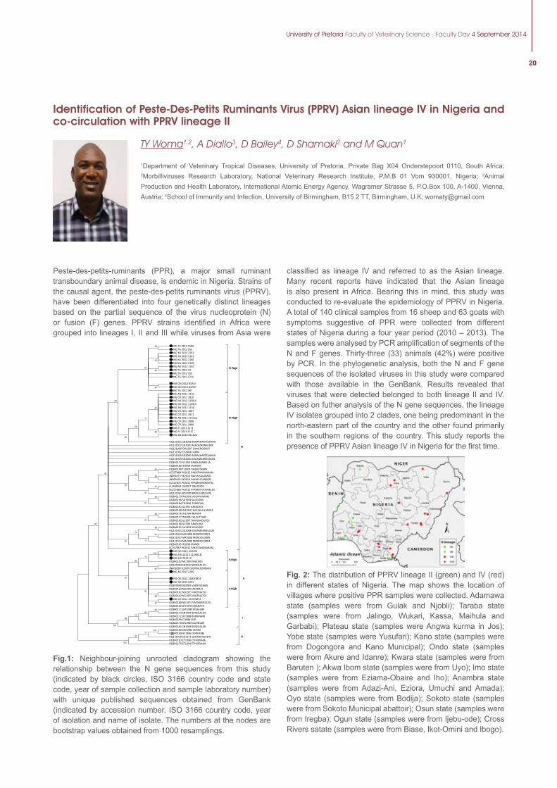
University of Pretoria Faculty of Veterinary Science - Faculty Day 4 September 2014
20
Identification of Peste-Des-Petits Ruminants Virus (PPRV) Asian lineage IV in Nigeria and co-circulation with PPRV lineage II
TY Woma1,2, A Diallo3, D Bailey4, D Shamaki2 and M Quan1
1Department of Veterinary Tropical Diseases, University of Pretoria, Private Bag X04 Onderstepoort 0110, South Africa; 2Morbilliviruses Research Laboratory, National Veterinary Research Institute, P.M.B 01 Vom 930001, Nigeria; 3Animal Production and Health Laboratory, International Atomic Energy Agency, Wagramer Strasse 5, P.O.Box 100, A-1400, Vienna, Austria; 4School of Immunity and Infection, University of Birmingham, B15 2 TT, Birmingham, U.K; [email protected]
Peste-des-petits-ruminants (PPR), a major small ruminant transboundary animal disease, is endemic in Nigeria. Strains of the causal agent, the peste-des-petits ruminants virus (PPRV), have been differentiated into four genetically distinct lineages based on the partial sequence of the virus nucleoprotein (N) or fusion (F) genes. PPRV strains identified in Africa were grouped into lineages I, II and III while viruses from Asia were
classified as lineage IV and referred to as the Asian lineage. Many recent reports have indicated that the Asian lineage is also present in Africa. Bearing this in mind, this study was conducted to re-evaluate the epidemiology of PPRV in Nigeria. A total of 140 clinical samples from 16 sheep and 63 goats with symptoms suggestive of PPR were collected from different states of Nigeria during a four year period (2010 – 2013). The samples were analysed by PCR amplification of segments of the N and F genes. Thirty-three (33) animals (42%) were positive by PCR. In the phylogenetic analysis, both the N and F gene sequences of the isolated viruses in this study were compared with those available in the GenBank. Results revealed that viruses that were detected belonged to both lineage II and IV. Based on futher analysis of the N gene sequences, the lineage IV isolates grouped into 2 clades, one being predominant in the north-eastern part of the country and the other found primarily in the southern regions of the country. This study reports the presence of PPRV Asian lineage IV in Nigeria for the first time.
2
NG TA 2012 250A NG TA 2012 253 NG YO 2013 2161 NG YO 2013 2162 NG KA 2012 2160 NG KA 2012 2159 NG AD 2012 2167 NG TA 2012 23 NG TA 2012 355 NG TA 2012 17LV NG ON 2012 832LV NG ON 2012 832KD NG TA 2012 367
IV-NigA
NG AN 2011 2152 NG CR 2011 1810 NG IM 2012 2150LG NG IM 2012 2149LV NG AN 2011 2154 NG CR 2011 1807 NG CR 2011 1811 NG AN 2011 2151LG NG CR 2011 1808 NG CR 2011 1806 NG PL 2013 2171 NG PL 2013 2170 NG AD 2010 NJ13LN HQ131921 SD2008 SOBAKHARTOUMKH
IV-NigB
HQ131917 SD2000 ALAZAZABNSUD00 HQ131960 CM1997 CAMEROON97 HQ131962 CF2004 CAR04 HQ131920 SD2000 SOBAKHARTOUMKH HQ131929 SD2000 EDDAMARNSUD001 DQ840173 IL1993 ISRAELARABELLA DQ840186 IR1998 IRAN983 DQ840198 TJ2004 TAJIKISTAN04 KC207868 PK2012 PAKISTANOKARAM JN009673 PK2010 PAKFAISALABAD2 JN009674 PK2010 PAKMULTAN2010 KC207875 PK2012 PPRMM69SHEEP20 EU340363 CN2007 TIBET0702 KC207883 PK2012 PPRMM77CAMEL20 HQ131961 BD2009 BANGLADESH09 DQ840179 IN1994 SASSAYANA942 DQ840196 SA1999 SAUDI999 DQ840184 TR1996 TURKEY96 DQ840181 IL1995 ISREAL953 DQ840180 IN1994 THATAGULLAN941 DQ840176 IN1994 INDIA94 DQ840177 IN1995 CALCUTTA95 DQ840182 IL1995 TAMILNADU952 DQ840188 IL1998 ISRAEL982 DQ840195 SA1999 SAUDI997 HQ131931 SD2008 EDDAMARNSUD08 HQ131923 MA2008 MOROCCO081 HQ131927 MA2008 MOROCCO089 HQ131924 MA2008 MOROCCO082 DQ840185 IR1998 IRAN98 KC207867 PK2012 PAKISTANOKARAM
IV
NG OG 2011 31GYM NG KW 2012 1111MSLN NG KW 2012 LG
DQ840192 ML1999 MALI991 HQ131963 SD2010 SENEGAL10 JN602080 SL2009 SIERRALEONEKAB
II-NigA
NG AK 2013 2205 NG OS 2011 110GYM14 NG SO 2013 2201
DQ837640 002005 VACRUSSIA05 DQ840163 NG1976 ACCRA76 DQ840161 NG1975 NIGERIA752 DQ840162 NG1975 NIGERIA753
NG OY 2011 127GYM31 DQ840160 NG1975 VACNIGERIA751 DQ840166 GH1978 GHANA78
II-NigB
II
DQ840171 GW1989 BISSAU89 DQ840174 SN1994 SENEGAL94 DQ840172 BF1988 BURKINA88 DQ840199 CI1989 CI89 DQ840170 GN1988 GUINEA88 DQ840165 SN1968 SENEGAL68
I
DQ840168 OM1983 IBIR83 DQ840169 AE1986 DORCAS86 HQ131918 SD1971 GEDARIFKSUD71 DQ840183 ET1996 ETHIOPIA96 DQ840175 ET1994 ETHIOPIA94
III
67
100 70
95
84
68 97
80
97
96
95
65
87
89
87
87
77
68
56
90
87 96
78
59
67 69
70
66 81
50 64
86 61
78
74
67
88
85
100 71
84 100
100 98
71 86
Fig.1: Neighbour-joining unrooted cladogram showing the relationship between the N gene sequences from this study (indicated by black circles, ISO 3166 country code and state code, year of sample collection and sample laboratory number) with unique published sequences obtained from GenBank (indicated by accession number, ISO 3166 country code, year of isolation and name of isolate. The numbers at the nodes are bootstrap values obtained from 1000 resamplings.
Fig. 2: The distribution of PPRV lineage II (green) and IV (red) in different states of Nigeria. The map shows the location of villages where positive PPR samples were collected. Adamawa state (samples were from Gulak and Njobli); Taraba state (samples were from Jalingo, Wukari, Kassa, Maihula and Garbabi); Plateau state (samples were Angwa kurma in Jos); Yobe state (samples were Yusufari); Kano state (samples were from Dogongora and Kano Municipal); Ondo state (samples were from Akure and Idanre); Kwara state (samples were from Baruten ); Akwa Ibom state (samples were from Uyo); Imo state (samples were from Eziama-Obaire and Iho); Anambra state (samples were from Adazi-Ani, Eziora, Umuchi and Amada); Oyo state (samples were from Bodija); Sokoto state (samples were from Sokoto Municipal abattoir); Osun state (samples were from Iregba); Ogun state (samples were from Ijebu-ode); Cross Rivers satate (samples were from Biase, Ikot-Omini and Ibogo).
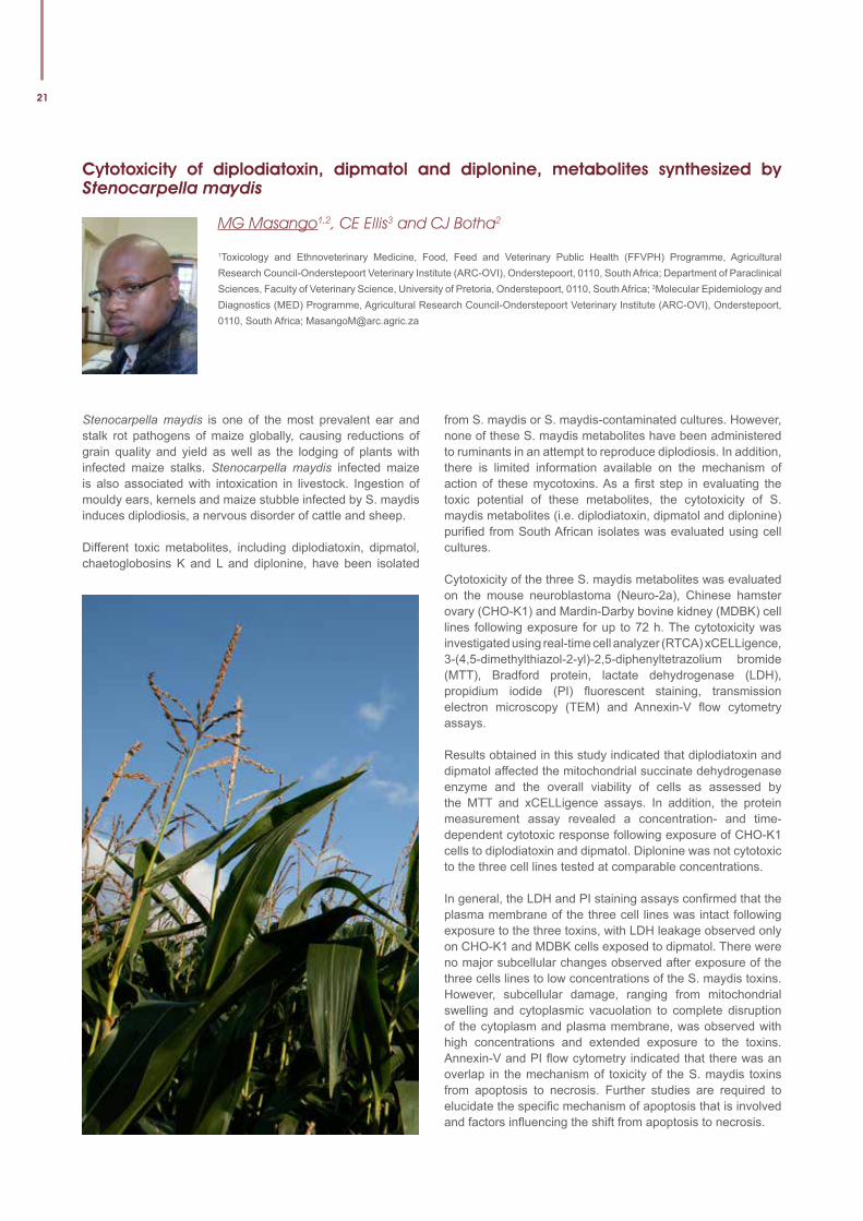
21
Cytotoxicity of diplodiatoxin, dipmatol and diplonine, metabolites synthesized by Stenocarpella maydis
MG Masango1,2, CE Ellis3 and CJ Botha2
1Toxicology and Ethnoveterinary Medicine, Food, Feed and Veterinary Public Health (FFVPH) Programme, Agricultural Research Council-Onderstepoort Veterinary Institute (ARC-OVI), Onderstepoort, 0110, South Africa; Department of Paraclinical Sciences, Faculty of Veterinary Science, University of Pretoria, Onderstepoort, 0110, South Africa; 3Molecular Epidemiology and Diagnostics (MED) Programme, Agricultural Research Council-Onderstepoort Veterinary Institute (ARC-OVI), Onderstepoort, 0110, South Africa; [email protected]
Stenocarpella maydis is one of the most prevalent ear and stalk rot pathogens of maize globally, causing reductions of grain quality and yield as well as the lodging of plants with infected maize stalks. Stenocarpella maydis infected maize is also associated with intoxication in livestock. Ingestion of mouldy ears, kernels and maize stubble infected by S. maydis induces diplodiosis, a nervous disorder of cattle and sheep.
Different toxic metabolites, including diplodiatoxin, dipmatol, chaetoglobosins K and L and diplonine, have been isolated
from S. maydis or S. maydis-contaminated cultures. However, none of these S. maydis metabolites have been administered to ruminants in an attempt to reproduce diplodiosis. In addition, there is limited information available on the mechanism of action of these mycotoxins. As a first step in evaluating the toxic potential of these metabolites, the cytotoxicity of S. maydis metabolites (i.e. diplodiatoxin, dipmatol and diplonine) purified from South African isolates was evaluated using cell cultures.
Cytotoxicity of the three S. maydis metabolites was evaluated on the mouse neuroblastoma (Neuro-2a), Chinese hamster ovary (CHO-K1) and Mardin-Darby bovine kidney (MDBK) cell lines following exposure for up to 72 h. The cytotoxicity was investigated using real-time cell analyzer (RTCA) xCELLigence, 3-(4,5-dimethylthiazol-2-yl)-2,5-diphenyltetrazolium bromide (MTT), Bradford protein, lactate dehydrogenase (LDH), propidium iodide (PI) fluorescent staining, transmission electron microscopy (TEM) and Annexin-V flow cytometry assays.
Results obtained in this study indicated that diplodiatoxin and dipmatol affected the mitochondrial succinate dehydrogenase enzyme and the overall viability of cells as assessed by the MTT and xCELLigence assays. In addition, the protein measurement assay revealed a concentration- and time-dependent cytotoxic response following exposure of CHO-K1 cells to diplodiatoxin and dipmatol. Diplonine was not cytotoxic to the three cell lines tested at comparable concentrations.
In general, the LDH and PI staining assays confirmed that the plasma membrane of the three cell lines was intact following exposure to the three toxins, with LDH leakage observed only on CHO-K1 and MDBK cells exposed to dipmatol. There were no major subcellular changes observed after exposure of the three cells lines to low concentrations of the S. maydis toxins. However, subcellular damage, ranging from mitochondrial swelling and cytoplasmic vacuolation to complete disruption of the cytoplasm and plasma membrane, was observed with high concentrations and extended exposure to the toxins. Annexin-V and PI flow cytometry indicated that there was an overlap in the mechanism of toxicity of the S. maydis toxins from apoptosis to necrosis. Further studies are required to elucidate the specific mechanism of apoptosis that is involved and factors influencing the shift from apoptosis to necrosis.
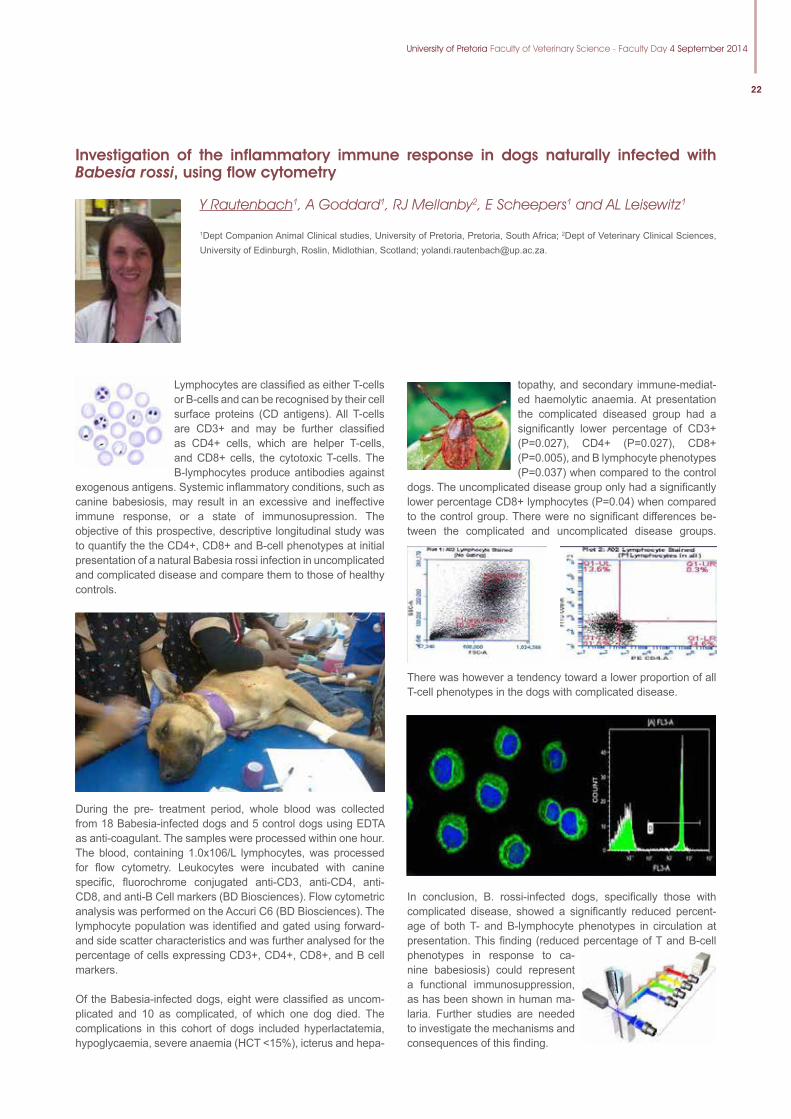
University of Pretoria Faculty of Veterinary Science - Faculty Day 4 September 2014
22
Investigation of the inflammatory immune response in dogs naturally infected with Babesia rossi, using flow cytometry
Y Rautenbach1, A Goddard1, RJ Mellanby2, E Scheepers1 and AL Leisewitz1
1Dept Companion Animal Clinical studies, University of Pretoria, Pretoria, South Africa; 2Dept of Veterinary Clinical Sciences, University of Edinburgh, Roslin, Midlothian, Scotland; [email protected].
Lymphocytes are classified as either T-cells or B-cells and can be recognised by their cell surface proteins (CD antigens). All T-cells are CD3+ and may be further classified as CD4+ cells, which are helper T-cells, and CD8+ cells, the cytotoxic T-cells. The B-lymphocytes produce antibodies against
exogenous antigens. Systemic inflammatory conditions, such as canine babesiosis, may result in an excessive and ineffective immune response, or a state of immunosupression. The objective of this prospective, descriptive longitudinal study was to quantify the the CD4+, CD8+ and B-cell phenotypes at initial presentation of a natural Babesia rossi infection in uncomplicated and complicated disease and compare them to those of healthy controls.
During the pre- treatment period, whole blood was collected from 18 Babesia-infected dogs and 5 control dogs using EDTA as anti-coagulant. The samples were processed within one hour. The blood, containing 1.0x10⁶/L lymphocytes, was processed for flow cytometry. Leukocytes were incubated with canine specific, fluorochrome conjugated anti-CD3, anti-CD4, anti-CD8, and anti-B Cell markers (BD Biosciences). Flow cytometric analysis was performed on the Accuri C6 (BD Biosciences). The lymphocyte population was identified and gated using forward- and side scatter characteristics and was further analysed for the percentage of cells expressing CD3+, CD4+, CD8+, and B cell markers.
Of the Babesia-infected dogs, eight were classified as uncom-plicated and 10 as complicated, of which one dog died. The complications in this cohort of dogs included hyperlactatemia, hypoglycaemia, severe anaemia (HCT <15%), icterus and hepa-
topathy, and secondary immune-mediat-ed haemolytic anaemia. At presentation the complicated diseased group had a significantly lower percentage of CD3+ (P=0.027), CD4+ (P=0.027), CD8+ (P=0.005), and B lymphocyte phenotypes (P=0.037) when compared to the control
dogs. The uncomplicated disease group only had a significantly lower percentage CD8+ lymphocytes (P=0.04) when compared to the control group. There were no significant differences be-tween the complicated and uncomplicated disease groups.
There was however a tendency toward a lower proportion of all T-cell phenotypes in the dogs with complicated disease.
In conclusion, B. rossi-infected dogs, specifically those with complicated disease, showed a significantly reduced percent-age of both T- and B-lymphocyte phenotypes in circulation at presentation. This finding (reduced percentage of T and B-cell phenotypes in response to ca-nine babesiosis) could represent a functional immunosuppression, as has been shown in human ma-laria. Further studies are needed to investigate the mechanisms and consequences of this finding.
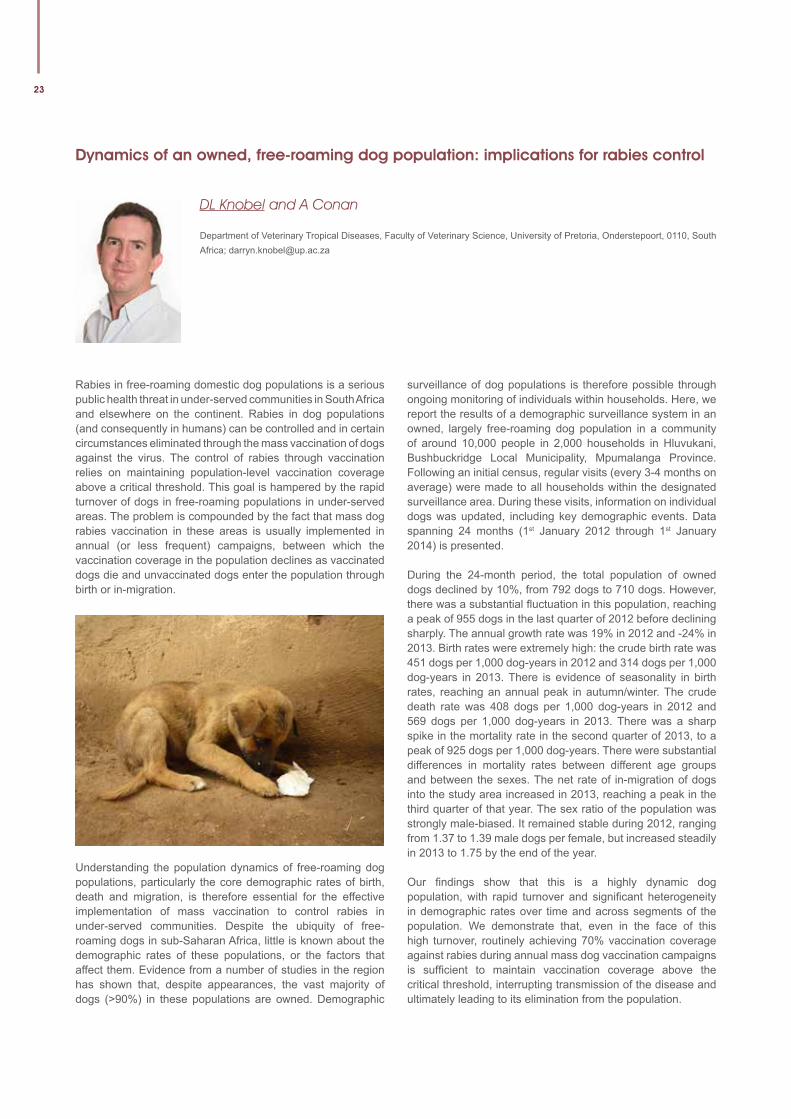
23
Dynamics of an owned, free-roaming dog population: implications for rabies control
DL Knobel and A Conan
Department of Veterinary Tropical Diseases, Faculty of Veterinary Science, University of Pretoria, Onderstepoort, 0110, South Africa; [email protected]
Rabies in free-roaming domestic dog populations is a serious public health threat in under-served communities in South Africa and elsewhere on the continent. Rabies in dog populations (and consequently in humans) can be controlled and in certain circumstances eliminated through the mass vaccination of dogs against the virus. The control of rabies through vaccination relies on maintaining population-level vaccination coverage above a critical threshold. This goal is hampered by the rapid turnover of dogs in free-roaming populations in under-served areas. The problem is compounded by the fact that mass dog rabies vaccination in these areas is usually implemented in annual (or less frequent) campaigns, between which the vaccination coverage in the population declines as vaccinated dogs die and unvaccinated dogs enter the population through birth or in-migration.
Understanding the population dynamics of free-roaming dog populations, particularly the core demographic rates of birth, death and migration, is therefore essential for the effective implementation of mass vaccination to control rabies in under-served communities. Despite the ubiquity of free-roaming dogs in sub-Saharan Africa, little is known about the demographic rates of these populations, or the factors that affect them. Evidence from a number of studies in the region has shown that, despite appearances, the vast majority of dogs (>90%) in these populations are owned. Demographic
surveillance of dog populations is therefore possible through ongoing monitoring of individuals within households. Here, we report the results of a demographic surveillance system in an owned, largely free-roaming dog population in a community of around 10,000 people in 2,000 households in Hluvukani, Bushbuckridge Local Municipality, Mpumalanga Province. Following an initial census, regular visits (every 3-4 months on average) were made to all households within the designated surveillance area. During these visits, information on individual dogs was updated, including key demographic events. Data spanning 24 months (1st January 2012 through 1st January 2014) is presented.
During the 24-month period, the total population of owned dogs declined by 10%, from 792 dogs to 710 dogs. However, there was a substantial fluctuation in this population, reaching a peak of 955 dogs in the last quarter of 2012 before declining sharply. The annual growth rate was 19% in 2012 and -24% in 2013. Birth rates were extremely high: the crude birth rate was 451 dogs per 1,000 dog-years in 2012 and 314 dogs per 1,000 dog-years in 2013. There is evidence of seasonality in birth rates, reaching an annual peak in autumn/winter. The crude death rate was 408 dogs per 1,000 dog-years in 2012 and 569 dogs per 1,000 dog-years in 2013. There was a sharp spike in the mortality rate in the second quarter of 2013, to a peak of 925 dogs per 1,000 dog-years. There were substantial differences in mortality rates between different age groups and between the sexes. The net rate of in-migration of dogs into the study area increased in 2013, reaching a peak in the third quarter of that year. The sex ratio of the population was strongly male-biased. It remained stable during 2012, ranging from 1.37 to 1.39 male dogs per female, but increased steadily in 2013 to 1.75 by the end of the year.
Our findings show that this is a highly dynamic dog population, with rapid turnover and significant heterogeneity in demographic rates over time and across segments of the population. We demonstrate that, even in the face of this high turnover, routinely achieving 70% vaccination coverage against rabies during annual mass dog vaccination campaigns is sufficient to maintain vaccination coverage above the critical threshold, interrupting transmission of the disease and ultimately leading to its elimination from the population.
University of Pretoria Faculty of Veterinary Science - Faculty Day 4 September 2014
24
The Mnisi Community Programme 2009-2013: An overview of the first five years of the Programme, its relevance to the Faculty, and its future vision
J van Rooyen1 and DL Knobel1
1Department of Veterinary Tropical Diseases, Faculty of Veterinary Science, University of Pretoria, Onderstepoort, 0110, South Africa; [email protected]
The Mnisi community is situated on communal land in the Bushbuckridge Local Municipality at the wildlife-livestock interface outside the Orpen area of the Kruger National Park. The Mnisi Community Programme was initiated in 2008 with the first research activities commencing in 2009. The Programme is a partnership between the Faculty of Veterinary Science and the Mnisi Traditional Authority. Through a platform consisting of this Programme, the Hluvukani Animal Clinic, and the Hans Hoheisen Wildlife Research Station, post- and undergraduate students from various Faculties within the University and elsewherework together to address pertinent health-related questions posed by the ever-more intimate interface between humans; wild and domesticated animals; and the environment they all share. It is now more than five years since the first postgraduate project commenced in the study area. Here, we share a synopsis of this unique Programme, highlighting its momentum, relevance,and a vision for its future.
By the end of 2013,62 individual research projects had been registered within the Programme. The number of projects registered per year hasincreased progressively over the five-year period from six projects listed in 2009to 22 registered in 2013. Most of these projects were conducted by postgraduate students registered at the Faculty of Veterinary Science. Forty percent of the projects were active with fieldwork and data collectionby the end of 2013 whilst 19% had been concluded. The rest (41%) were in various stages of either planning or completion. Of the registered projects 13% were PhD projects, 19% research MSc’s, 11% web-based MSc’s, and approximately 31% Masters projects conducted by Utrecht University students in collaboration with the Faculty of Veterinary Science during their graduate years in veterinary medicine. Most projects relate to aspects of animal health management and services(34%); disease ecology, emergence, and control (26%); and zoonoses (15%).
Over and above the significant contribution through research, the Programme is playing a leading role in many aspects of conservation, land use planning, social upliftment and sustainable rural development in the area. It fulfils a
crucialroleinvarious structures of the Kruger to Canyons Biosphere Reserve’s extended network of organisations. Through these networks and associated collaborations it was able to appoint eleven previously unemployed community members as research assistants in 2013 with the prospect of employing six more in 2014. By mobilising Faculty resources and collaborations it has trained more than 150 livestock farmers in Primary Animal Health Care in 2012.
In future, the Programme wishes to expand its current strategy to channel focussed research through longitudinal research platforms, such as the health and demographic surveillance systems for domestic animals, and the Herding for Health programme that will embed innovative rural and social development strategies within its core research and training activities. We believe this Programme will become a model that demonstrate the reality and importance of multi-institutional collaborations between industry, society, government, and scientists that could impact communities positively whilst achieving our objective of becoming an internationally recognised academic institution with an undisputable local relevance.
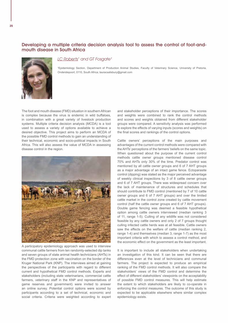
25
Developing a multiple criteria decision analysis tool to assess the control of foot-and-mouth disease in South Africa
LC Roberts1 and GT Fosgate¹
¹Epidemiology Section, Department of Production Animal Studies, Faculty of Veterinary Science, University of Pretoria, Onderstepoort, 0110, South Africa; [email protected]
The foot and mouth disease (FMD) situation in southern African is complex because the virus is endemic in wild buffaloes, in combination with a great variety of livestock production systems. Multiple criteria decision analysis (MCDA) is a tool used to assess a variety of options available to achieve a desired objective. This project aims to perform an MCDA of the possible FMD control methods to gain an understanding of their technical, economic and socio-political impacts in South Africa. This will also assess the value of MCDA in assessing disease control in the region.
A participatory epidemiology approach was used to interview communal cattle farmers from ten randomly-selected dip tanks and seven groups of state animal health technicians (AHTs) in the FMD protection zone with vaccination on the border of the Kruger National Park (KNP). The interviews aimed at gaining the perspectives of the participants with regard to different current and hypothetical FMD control methods. Experts and stakeholders (including state veterinarians, commercial cattle farmers, veterinary staff in the KNP and representatives of game reserves and government) were invited to answer an online survey. Potential control options were scored by participants according to a set of technical, economic and social criteria. Criteria were weighted according to expert
and stakeholder perceptions of their importance. The scores and weights were combined to rank the control methods and scores and weights obtained from different stakeholder groups were compared. A sensitivity analysis was performed to explore the effects of varying inputs (scores and weights) on the final scores and rankings of the control options.
Cattle owners’ perceptions of the main purposes and advantages of the current control methods were compared with the AHTs’ perceptions of the farmers’ beliefs on the same topic. When questioned about the purpose of the current control methods cattle owner groups mentioned disease control 70% and AHTs only 30% of the time. Predator control was mentioned by all cattle owner groups and 6 of 7 AHT groups as a major advantage of an intact game fence. Ectoparasite control (dipping) was stated as the major perceived advantage of weekly clinical inspections by 3 of 8 cattle owner groups and 6 of 7 AHT groups. There was widespread concern over the lack of maintenance of structures and schedules that should contribute to FMD control (mentioned by 7 of 10 cattle owner groups and 6 of 7 AHT groups) and over the limited cattle market in the control zone created by cattle movement control (half the cattle owner groups and 6 of 7 AHT groups). Double game fencing was deemed a feasible hypothetical option among cattle owners interviewed (median ranking 3 of 11, range 1-5). Culling of any wildlife was not considered feasible by any cattle owners and only 2 of 7 groups thought culling infected cattle herds was at all feasible. Cattle owners saw the effects on the welfare of cattle (median ranking 2, range 1-4) and themselves (median 3, range 1-7) as the most important criteria with which to assess a control method, and the economic effect on the government as the least important.
It is important to include all stakeholders when undertaking an investigation of this kind. It can be seen that there are differences even at the level of technicians and communal farmers. The project is expected to produce an empirical ranking of the FMD control methods. It will also compare the stakeholders’ views of the FMD control and determine the effect of different stakeholders’ viewpoints on the acceptability of possible FMD control measures. This will help estimate the extent to which stakeholders are likely to co-operate in enforcing the control measures. The outcome of this study is expected to be applicable elsewhere where similar complex epidemiology exists.
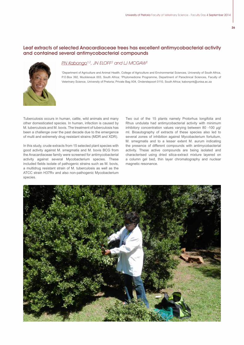
University of Pretoria Faculty of Veterinary Science - Faculty Day 4 September 2014
26
Leaf extracts of selected Anacardiaceae trees has excellent antimycobacterial activity and contained several antimycobacterial compounds
PN Kabongo1,2, JN ELOFF2 and LJ MCGAW2
1Department of Agriculture and Animal Health, College of Agriculture and Environmental Sciences, University of South Africa, P.O.Box 392, Muckleneuk 003, South Africa; 2Phytomedicine Programme, Department of Paraclinical Sciences, Faculty of Veterinary Science, University of Pretoria; Private Bag X04, Onderstepoort 0110, South Africa; [email protected]
Tuberculosis occurs in human, cattle, wild animals and many other domesticated species. In human, infection is caused by M. tuberculosis and M. bovis. The treatment of tuberculosis has been a challenge over the past decade due to the emergence of multi and extremely drug resistant strains (MDR and XDR).
In this study, crude extracts from 15 selected plant species with good activity against M. smegmatis and M. bovis BCG from the Anacardiaceae family were screened for antimycobacterial activity against several Mycobacterium species. These included fields isolate of pathogenic strains such as M. bovis, a multidrug resistant strain of M. tuberculosis as well as the ATCC strain H37Rv and also non-pathogenic Mycobacterium species.
Two out of the 15 plants namely Protorhus longifolia and Rhus undulata had antimycobacterial activity with minimum inhibitory concentration values varying between 80 -100 µg/ml. Bioautography of extracts of these species also led to several zones of inhibition against Mycobacterium fortuitum, M. smegmatis and to a lesser extent M. aurum indicating the presence of different compounds with antimycobacterial activity. These active compounds are being isolated and characterised using dried silica-extract mixture layered on a column gel bed, thin layer chromatography and nuclear magnetic resonance.
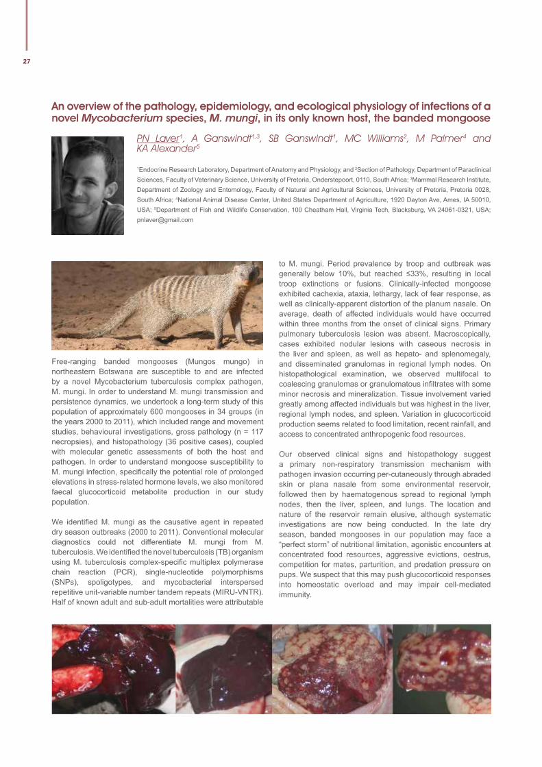
27
An overview of the pathology, epidemiology, and ecological physiology of infections of a novel Mycobacterium species, M. mungi, in its only known host, the banded mongoose
PN Laver1, A Ganswindt1,3, SB Ganswindt1, MC Williams2, M Palmer4 and KA Alexander5
1Endocrine Research Laboratory, Department of Anatomy and Physiology, and 2Section of Pathology, Department of Paraclinical Sciences, Faculty of Veterinary Science, University of Pretoria, Onderstepoort, 0110, South Africa; 3Mammal Research Institute, Department of Zoology and Entomology, Faculty of Natural and Agricultural Sciences, University of Pretoria, Pretoria 0028, South Africa; 4National Animal Disease Center, United States Department of Agriculture, 1920 Dayton Ave, Ames, IA 50010, USA; 5Department of Fish and Wildlife Conservation, 100 Cheatham Hall, Virginia Tech, Blacksburg, VA 24061-0321, USA; [email protected]
Free-ranging banded mongooses (Mungos mungo) in northeastern Botswana are susceptible to and are infected by a novel Mycobacterium tuberculosis complex pathogen, M. mungi. In order to understand M. mungi transmission and persistence dynamics, we undertook a long-term study of this population of approximately 600 mongooses in 34 groups (in the years 2000 to 2011), which included range and movement studies, behavioural investigations, gross pathology (n = 117 necropsies), and histopathology (36 positive cases), coupled with molecular genetic assessments of both the host and pathogen. In order to understand mongoose susceptibility to M. mungi infection, specifi cally the potential role of prolonged elevations in stress-related hormone levels, we also monitored faecal glucocorticoid metabolite production in our study population.
We identifi ed M. mungi as the causative agent in repeated dry season outbreaks (2000 to 2011). Conventional molecular diagnostics could not differentiate M. mungi from M. tuberculosis. We identifi ed the novel tuberculosis (TB) organism using M. tuberculosis complex-specifi c multiplex polymerase chain reaction (PCR), single-nucleotide polymorphisms (SNPs), spoligotypes, and mycobacterial interspersed repetitive unit-variable number tandem repeats (MIRU-VNTR). Half of known adult and sub-adult mortalities were attributable
to M. mungi. Period prevalence by troop and outbreak was generally below 10%, but reached ≤33%, resulting in local troop extinctions or fusions. Clinically-infected mongoose exhibited cachexia, ataxia, lethargy, lack of fear response, as well as clinically-apparent distortion of the planum nasale. On average, death of affected individuals would have occurred within three months from the onset of clinical signs. Primary pulmonary tuberculosis lesion was absent. Macroscopically, cases exhibited nodular lesions with caseous necrosis in the liver and spleen, as well as hepato- and splenomegaly, and disseminated granulomas in regional lymph nodes. On histopathological examination, we observed multifocal to coalescing granulomas or granulomatous infi ltrates with some minor necrosis and mineralization. Tissue involvement varied greatly among affected individuals but was highest in the liver, regional lymph nodes, and spleen. Variation in glucocorticoid production seems related to food limitation, recent rainfall, and access to concentrated anthropogenic food resources.
Our observed clinical signs and histopathology suggest a primary non-respiratory transmission mechanism with pathogen invasion occurring per-cutaneously through abraded skin or plana nasale from some environmental reservoir, followed then by haematogenous spread to regional lymph nodes, then the liver, spleen, and lungs. The location and nature of the reservoir remain elusive, although systematic investigations are now being conducted. In the late dry season, banded mongooses in our population may face a “perfect storm” of nutritional limitation, agonistic encounters at concentrated food resources, aggressive evictions, oestrus, competition for mates, parturition, and predation pressure on pups. We suspect that this may push glucocorticoid responses into homeostatic overload and may impair cell-mediated immunity.
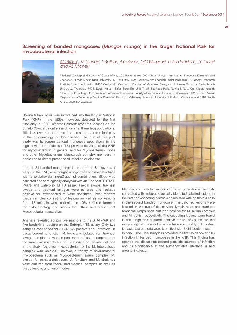
University of Pretoria Faculty of Veterinary Science - Faculty Day 4 September 2014
28
Screening of banded mongooses (Mungos mungo) in the Kruger National Park for mycobacterial infection
AC Brüns1, M Tanner2, L Botha3, A O’Brien4, MC Williams5, P Van Helden3, J Clarke4 and AL Michel6
1National Zoological Gardens of South Africa, 232 Boom street, 0001 South Africa; 2Institute for Infectious Diseases and Zoonoses, Ludwig-Maximilians-University LMU, 80539 Munich, Germany and Friedrich Löffler Institute (FLI), Federal Research Institute for Animal Health, 17493 Greifswald, Germany; 3Division of Molecular Biology and Human Genetics, Stellenbosch University, Tygerberg 7505, South Africa; 4Enfer Scientific, Unit T, M7 Business Park, Newhall, Naas,Co. Kildare,Ireland; 5Section of Pathology, Department of Paraclinical Sciences, Faculty of Veterinary Science, Onderstepoort 0110, South Africa; 6Department of Veterinary Tropical Diseases, Faculty of Veterinary Science, University of Pretoria, Onderstepoort 0110, South Africa; [email protected]
Bovine tuberculosis was introduced into the Kruger National Park (KNP) in the 1950s, however, detected for the first time only in 1990. Whereas current research focuses on the buffalo (Syncerus caffer) and lion (Panthera leo) populations, little is known about the role that small predators might play in the epidemiology of this disease. The aim of this pilot study was to screen banded mongoose populations in the high bovine tuberculosis (bTB) prevalence zone of the KNP for mycobacterium in general and for Mycobacterium bovis and other Mycobacterium tuberculosis complex members in particular, to detect presence of infection or disease.
In total, 81 banded mongooses in and around Skukuza staff village in the KNP, were caught in cage traps and anaesthetized with a cyclohexylamine/α2-agonist combination. Blood was collected and serologically analyzed with an ElephantTB STAT-PAK® and EnferplexTM TB assay. Faecal swabs, tracheal swabs and tracheal lavages were cultured and isolates positive for mycobacterium were speciated. Post mortem tissue samples consisting of lesions as well as non-lesions from 12 animals were collected in 10% buffered formalin for histopathology and frozen for culture and subsequent Mycobacterium speciation.
Analysis revealed six positive reactors to the STAT-PAK and five borderline reactors on the Enferplex TB assay. Only two samples overlapped for STAT-PAK positive and Enferplex TB assay borderline reaction. M. bovis was isolated from tracheal lavage samples as well as post mortem tissue samples from the same two animals but not from any other animal included in the study. No other mycobacterium of the M. tuberculosis complex was isolated. However, a variety of environmental mycobacteria such as Mycobacterium avium complex, M. simiae, M. parascrofulaceum, M. fortuitum and M. chelonae were cultured from faecal and tracheal samples as well as tissue lesions and lymph nodes.
Macroscopic nodular lesions of the aforementioned animals correlated with histopathologically identified calcified lesions in the first and caseating necrosis associated with epitheloid cells in the second banded mongoose. The calcified lesions were located in the superficial cervical lymph node and tracheo-bronchial lymph node culturing positive for M. avium complex and M. bovis, respectively. The caseating lesions were found in the lungs and cultured positive for M. bovis, as did the morphological unremarkable tracheo-bronchial lymph nodes. No acid fast bacteria were identified with Ziehl Neelsen stain.In conclusion, this study has provided the first evidence of bTB infection in banded mongooses in the KNP. This finding has opened the discussion around possible sources of infection and its significance at the human/wildlife interface in and around Skukuza.
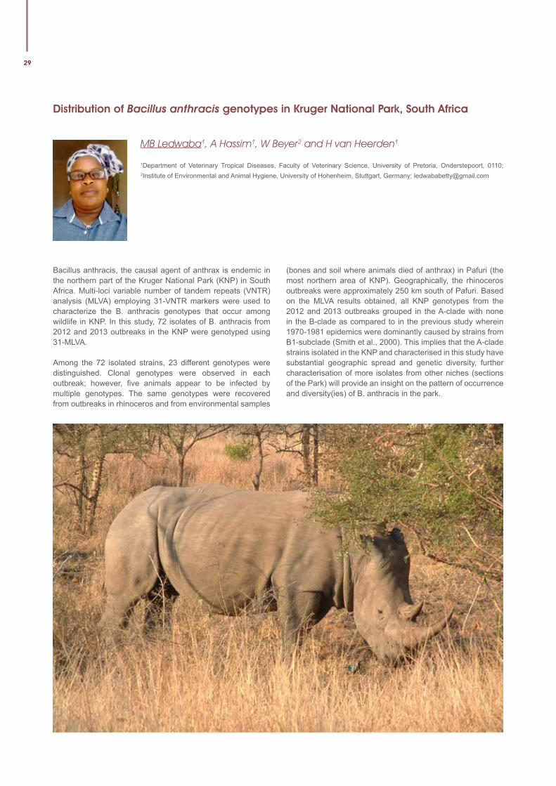
29
Distribution of Bacillus anthracis genotypes in Kruger National Park, South Africa
MB Ledwaba1, A Hassim1, W Beyer2 and H van Heerden1
1Department of Veterinary Tropical Diseases, Faculty of Veterinary Science, University of Pretoria, Onderstepoort, 0110; 2Institute of Environmental and Animal Hygiene, University of Hohenheim, Stuttgart, Germany; [email protected]
Bacillus anthracis, the causal agent of anthrax is endemic in the northern part of the Kruger National Park (KNP) in South Africa. Multi-loci variable number of tandem repeats (VNTR) analysis (MLVA) employing 31-VNTR markers were used to characterize the B. anthracis genotypes that occur among wildlife in KNP. In this study, 72 isolates of B. anthracis from 2012 and 2013 outbreaks in the KNP were genotyped using 31-MLVA.
Among the 72 isolated strains, 23 different genotypes were distinguished. Clonal genotypes were observed in each outbreak; however, five animals appear to be infected by multiple genotypes. The same genotypes were recovered from outbreaks in rhinoceros and from environmental samples
(bones and soil where animals died of anthrax) in Pafuri (the most northern area of KNP). Geographically, the rhinoceros outbreaks were approximately 250 km south of Pafuri. Based on the MLVA results obtained, all KNP genotypes from the 2012 and 2013 outbreaks grouped in the A-clade with none in the B-clade as compared to in the previous study wherein 1970-1981 epidemics were dominantly caused by strains from B1-subclade (Smith et al., 2000). This implies that the A-clade strains isolated in the KNP and characterised in this study have substantial geographic spread and genetic diversity, further characterisation of more isolates from other niches (sections of the Park) will provide an insight on the pattern of occurrence and diversity(ies) of B. anthracis in the park.
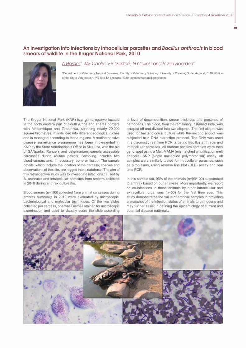
University of Pretoria Faculty of Veterinary Science - Faculty Day 4 September 2014
30
An Investigation into infections by intracellular parasites and Bacillus anthracis in blood smears of wildlife in the Kruger National Park, 2010
A Hassim1, ME Chaisi1, EH Dekker2, N Collins1 and H van Heerden1
1Department of Veterinary Tropical Diseases, Faculty of Veterinary Science, University of Pretoria, Onderstepoort, 0110; 2Offi ce of the State Veterinarian, PO Box 12 Skukuza, 1350; [email protected]
The Kruger National Park (K NP) is a game reserve located in the north eastern part of South Africa and shares borders with Mozambique and Zimbabwe, spanning nearly 20,000 square kilometres. It is divided into different ecological niches and is managed according to these regions. A routine passive disease surveillance programme has been implemented in KNP by the State Veterinarian’s Offi ce in Skukuza, with the aid of SANparks. Rangers and veterinarians sample accessible carcasses during routine patrols. Sampling includes two blood smears and, if necessary, bone or tissue. The sample details, which include the location of the carcass, species and observations of the site, are logged into a database. The aim of this retrospective study was to investigate infections caused by B. anthracis and intracellular parasites from smears collected in 2010 during anthrax outbreaks.
Blood smears (n=100) collected from animal carcasses during anthrax outbreaks in 2010 were evaluated by microscopic, bacteriological and molecular techniques. Of the two slides collected per carcass, one was Giemsa stained for microscopic examination and used to visually score the slide according
to level of decomposition, smear thickness and presence of pathogens. The blood, from the remaining unstained slide, was scraped off and divided into two aliquots. The fi rst aliquot was used for bacteriological culture while the second aliquot was subjected to a DNA extraction protocol. The DNA was used in a diagnostic real time PCR targeting Bacillus anthracis and intracellular parasites. All anthrax positive samples were then genotyped using a Melt-MAMA (mismatched amplifi cation melt analysis) SNP (single nucleotide polymorphism) assay. All samples were similarly tested for intracellular parasites, such as piroplasms, using reverse line blot (RLB) assay and real time PCR.
In this sample set, 96% of the animals (n=96/100) succumbed to anthrax based on our analyses. More importantly, we report on co-infections in these animals by other intracellular and extracellular organisms (n=50) for the fi rst time ever. This study demonstrates the value of archival samples in providing a snapshot of the infection status of animals to pathogens and may further assist in defi ning the epidemiology of current and potential disease outbreaks.
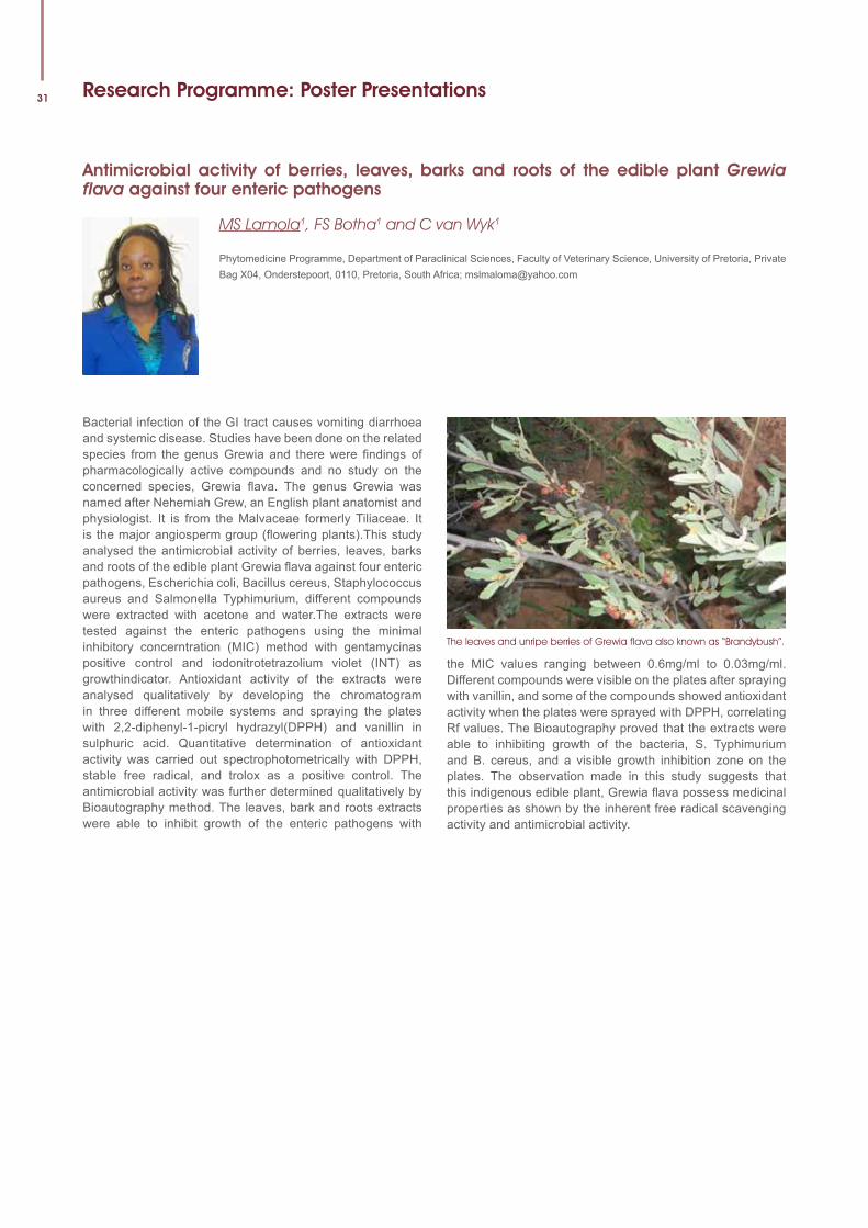
31
the MIC values ranging between 0.6mg/ml to 0.03mg/ml. Different compounds were visible on the plates after spraying with vanillin, and some of the compounds showed antioxidant activity when the plates were sprayed with DPPH, correlating Rf values. The Bioautography proved that the extracts were able to inhibiting growth of the bacteria, S. Typhimurium and B. cereus, and a visible growth inhibition zone on the plates. The observation made in this study suggests that this indigenous edible plant, Grewia flava possess medicinal properties as shown by the inherent free radical scavenging activity and antimicrobial activity.
Research Programme: Poster Presentations
Antimicrobial activity of berries, leaves, barks and roots of the edible plant Grewia flava against four enteric pathogens
MS Lamola1, FS Botha1 and C van Wyk1
Phytomedicine Programme, Department of Paraclinical Sciences, Faculty of Veterinary Science, University of Pretoria, Private Bag X04, Onderstepoort, 0110, Pretoria, South Africa; [email protected]
Bacterial infection of the GI tract causes vomiting diarrhoea and systemic disease. Studies have been done on the related species from the genus Grewia and there were findings of pharmacologically active compounds and no study on the concerned species, Grewia flava. The genus Grewia was named after Nehemiah Grew, an English plant anatomist and physiologist. It is from the Malvaceae formerly Tiliaceae. It is the major angiosperm group (flowering plants).This study analysed the antimicrobial activity of berries, leaves, barks and roots of the edible plant Grewia flava against four enteric pathogens, Escherichia coli, Bacillus cereus, Staphylococcus aureus and Salmonella Typhimurium, different compounds were extracted with acetone and water.The extracts were tested against the enteric pathogens using the minimal inhibitory concerntration (MIC) method with gentamycinas positive control and iodonitrotetrazolium violet (INT) as growthindicator. Antioxidant activity of the extracts were analysed qualitatively by developing the chromatogram in three different mobile systems and spraying the plates with 2,2-diphenyl-1-picryl hydrazyl(DPPH) and vanillin in sulphuric acid. Quantitative determination of antioxidant activity was carried out spectrophotometrically with DPPH, stable free radical, and trolox as a positive control. The antimicrobial activity was further determined qualitatively by Bioautography method. The leaves, bark and roots extracts were able to inhibit growth of the enteric pathogens with
The leaves and unripe berries of Grewia flava also known as “Brandybush”.
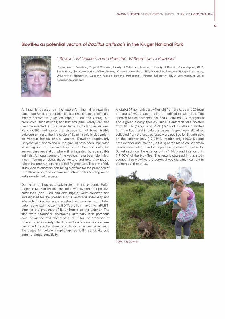
University of Pretoria Faculty of Veterinary Science - Faculty Day 4 September 2014
32
Blowflies as potential vectors of Bacillus anthracis in the Kruger National Park
L Basson1, EH Dekker2, H van Heerder1, W Beyer3 and J Rossouw4
1Department of Veterinary Tropical Diseases, Faculty of Veterinary Science, University of Pretoria, Onderstepoort, 0110, South Africa, 2State Veterinarians Office, Skukuza, Kruger National Park, 1350, 3Head of the Molecular Biological Laboratory, University of Hohenheim, Germany, 4Special Bacterial Pathogens Reference Laboratory, NICD, Johannesburg, 2131; [email protected]
Anthrax is caused by the spore-forming, Gram-positive bacterium Bacillus anthracis. It’s a zoonotic disease affecting mainly herbivores (such as impala, kudu and zebra), but carnivores (such as lions) and humans (albeit rarely) can also become infected. Anthrax is endemic to the Kruger National Park (KNP) and since the disease is not transmissible between animals, the life cycle of B. anthracis is dependent on various factors and/or vectors. Blowflies (particularly Chrysomya albiceps and C. marginalis) have been implicated in aiding in the dissemination of the bacteria onto the surrounding vegetation where it is ingested by susceptible animals. Although some of the vectors have been identified, most information about these vectors and how they play a role in the anthrax life cycle is still fragmentary. The aim of this study was to examine non-biting blowflies for the presence of B. anthracis on their exterior and interior after feeding on an anthrax-infected carcass.
During an anthrax outbreak in 2014 in the endemic Pafuri region in KNP, blowflies associated with two anthrax-positive carcasses (one kudu and one impala) were collected and investigated for the presence of B. anthracis externally and internally. Blowflies were washed with saline and plated onto polymyxin-lysozyme-EDTA-thallium acetate (PLET) agar for the presence of B. anthracis on the exterior. The flies were thereafter disinfected externally with peracetic acid, squashed and plated onto PLET for the presence of B. anthracis interiorly. Bacillus anthracis identification was confirmed by sub-culture onto blood agar and examining the plates for colony morphology, penicillin sensitivity and gamma-phage sensitivity.
A total of 57 non-biting blowflies (29 from the kudu and 28 from the impala) were caught using a modified malaise trap. The species of flies collected included C. albiceps, C. marginalis and a green blowfly species. Bacillus anthracis was isolated from 65.5% (19/29) and 25% (7/28) of blowflies collected from the kudu and impala carcasses, respectively. Blowflies collected from the kudu carcass were positive for B. anthracis on the exterior only (17.24%), interior only (10.34%) and both exterior and interior (37.93%) of the blowflies. Whereas blowflies collected from the impala carcass were positive for B. anthracis on the exterior only (7.14%) and interior only (17.86%) of the blowflies. The results obtained in this study suggest that blowflies are potential vectors which can aid in the spread of anthrax.
Collecting blowflies.
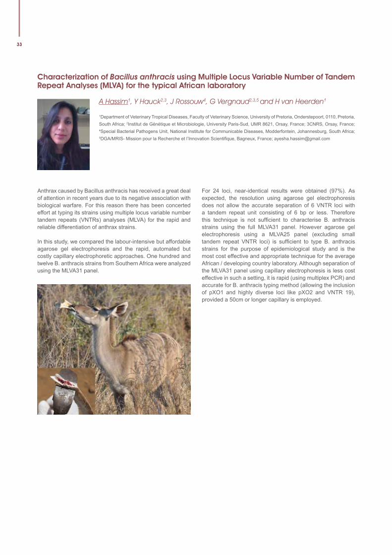
33
Characterization of Bacillus anthracis using Multiple Locus Variable Number of Tandem Repeat Analyses (MLVA) for the typical African laboratory
A Hassim1, Y Hauck2,3, J Rossouw4, G Vergnaud2,3,5 and H van Heerden1
1Department of Veterinary Tropical Diseases, Faculty of Veterinary Science, University of Pretoria, Onderstepoort, 0110, Pretoria, South Africa; 2Institut de Génétique et Microbiologie, University Paris-Sud, UMR 8621, Orsay, France; 3CNRS, Orsay, France; 4Special Bacterial Pathogens Unit, National Institute for Communicable Diseases, Modderfontein, Johannesburg, South Africa; 5DGA/MRIS- Mission pour la Recherche et l’Innovation Scientifi que, Bagneux, France; [email protected]
Anthrax caused by Bacillus anthracis has received a great deal of attention in recent years due to its negative association with biological warfare. For this reason there has been concerted effort at typing its strains using multiple locus variable number tandem repeats (VNTRs) analyses (MLVA) for the rapid and reliable differentiation of anthrax strains.
In this study, we compared the labour-intensive but affordable agarose gel electrophoresis and the rapid, automated but costly capillary electrophoretic approaches. One hundred and twelve B. anthracis strains from Southern Africa were analyzed using the MLVA31 panel.
For 24 loci, near-identical results were obtained (97%). As expected, the resolution using agarose gel electrophoresis does not allow the accurate separation of 6 VNTR loci with a tandem repeat unit consisting of 6 bp or less. Therefore this technique is not suffi cient to characterise B. anthracis strains using the full MLVA31 panel. However agarose gel electrophoresis using a MLVA25 panel (excluding small tandem repeat VNTR loci) is suffi cient to type B. anthracis strains for the purpose of epidemiological study and is the most cost effective and appropriate technique for the average African / developing country laboratory. Although separation of the MLVA31 panel using capillary electrophoresis is less cost effective in such a setting, it is rapid (using multiplex PCR) and accurate for B. anthracis typing method (allowing the inclusion of pXO1 and highly diverse loci like pXO2 and VNTR 19), provided a 50cm or longer capillary is employed.
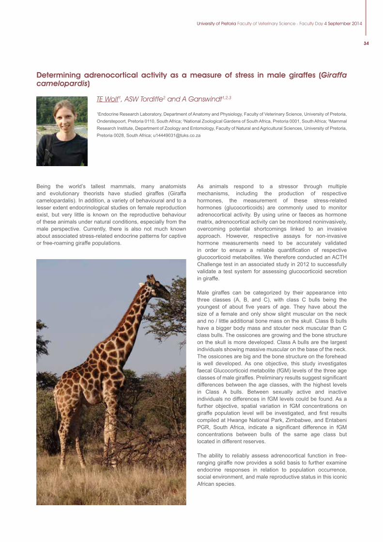
University of Pretoria Faculty of Veterinary Science - Faculty Day 4 September 2014
34
Determining adrenocortical activity as a measure of stress in male giraffes (Giraffa camelopardis)
TE Wolf1, ASW Tordiffe2 and A Ganswindt1,2,3
1Endocrine Research Laboratory, Department of Anatomy and Physiology, Faculty of Veterinary Science, University of Pretoria, Onderstepoort, Pretoria 0110, South Africa; 2National Zoological Gardens of South Africa, Pretoria 0001, South Africa; 3Mammal Research Institute, Department of Zoology and Entomology, Faculty of Natural and Agricultural Sciences, University of Pretoria, Pretoria 0028, South Africa; [email protected]
Being the world’s tallest mammals, many anatomists and evolutionary theorists have studied giraffes (Giraffa camelopardalis). In addition, a variety of behavioural and to a lesser extent endocrinological studies on female reproduction exist, but very little is known on the reproductive behaviour of these animals under natural conditions, especially from the male perspective. Currently, there is also not much known about associated stress-related endocrine patterns for captive or free-roaming giraffe populations.
As animals respond to a stressor through multiple mechanisms, including the production of respective hormones, the measurement of these stress-related hormones (glucocorticoids) are commonly used to monitor adrenocortical activity. By using urine or faeces as hormone matrix, adrenocortical activity can be monitored noninvasively, overcoming potential shortcomings linked to an invasive approach. However, respective assays for non-invasive hormone measurements need to be accurately validated in order to ensure a reliable quantification of respective glucocorticoid metabolites. We therefore conducted an ACTH Challenge test in an associated study in 2012 to successfully validate a test system for assessing glucocorticoid secretion in giraffe.
Male giraffes can be categorized by their appearance into three classes (A, B, and C), with class C bulls being the youngest of about five years of age. They have about the size of a female and only show slight muscular on the neck and no / little additional bone mass on the skull. Class B bulls have a bigger body mass and stouter neck muscular than C class bulls. The ossicones are growing and the bone structure on the skull is more developed. Class A bulls are the largest individuals showing massive muscular on the base of the neck. The ossicones are big and the bone structure on the forehead is well developed. As one objective, this study investigates faecal Glucocorticoid metabolite (fGM) levels of the three age classes of male giraffes. Preliminary results suggest significant differences between the age classes, with the highest levels in Class A bulls. Between sexually active and inactive individuals no differences in fGM levels could be found. As a further objective, spatial variation in fGM concentrations on giraffe population level will be investigated, and first results compiled at Hwange National Park, Zimbabwe, and Entabeni PGR, South Africa, indicate a significant difference in fGM concentrations between bulls of the same age class but located in different reserves.
The ability to reliably assess adrenocortical function in free-ranging giraffe now provides a solid basis to further examine endocrine responses in relation to population occurrence, social environment, and male reproductive status in this iconic African species.
35
Eleven-year antibiotic resistance profiles of Staphylococcus aureus in dairy herds across southern Africa
T.J van der Schans1, J Karzis1 and IM Petzer1
1Department of Production Animal Studies, Section Udder Health, Faculty of Veterinary Science, University of Pretoria, Private Bag X04, Onderstepoort, 0110 South Africa; [email protected]
Staphylococcus aureus (STA) susceptibility and patterns of intermediate and total resistance in bovine milk areof important international concern and a global threat (WHO 2014). STA is one of the primary causes of mastitis which in turn is one of the major causes of loss in productivity. This research describes the results of eleven years of surveillance for resistance of STA to nine commonly used antibiotics in South Africa and neighboring countries. Data were sufficient to investigate both seasonal and regional trends.
There was a significant decrease in the overall proportion of total resistance of STA to antibiotics over the study period. The exception was an increasing trend towards intermediate resistance to tylosin (TY). There were significant increasing differences (P<0.0001) for the proportions of STA susceptible,
intermediately resistant and totally resistant to penicillin G (PEN) in this study from 2003 compared to those in 2010, as well as for proportions of STA susceptible, intermediately resistant to TY (P<0.0001) and for proportions of STA totally resistant to amoxycillin (AML) (P<0.0001). Seasonal differences in STA resistance were small, but an increase in intermediate resistance was found to be present during summer and autumn. Regional differences were found in STA isolates from Kwazulu Natal (KZN) which showed the least intermediate resistance against most antibiotics, except for AML and PEN. STA isolates from North West (NW) had the least intermediate antibiotic resistance against AML, and from Namibia against PEN.
This study indicated an overall decline of STA isolates from milk of dairy cows from southern Africa with total and intermediate resistance against 9 commonly used antibiotics, contrary to European findings (Pitkälä et al., 2004; Tenhagen et al., 2006). However, the intermediate resistance to PEN and TY and total resistance to AML and PEN of STA differed significantly in proportion, compared to almost all STA which were susceptible between 2003 and 2010. During the same time period the susceptibility of STA to these antibiotics remained almost stable.
References1. Pitkälä A, Haver i M, Pyörälä S, Myllys V, Honkanen-Buzalski
T 2004 Bovine mastitis in Finland 2001-Prevalence, distribution of bacteria, and antimicrobial resistance. Journal of Dairy Science 87(8):2433-2441
2. Tenhagen BA, Köster G, Wallmann J, Heuwieser W 2006 Prevalence of mastitis pathogens and their resistance against antimicrobial agents in dairy cows in Brandenburg, Germany. Journal of Dairy Science 89(7):2542-2551
Antibiotic Resistant(n)(%) Intermediate(n)(%) Susceptible(n)(%) Total n
Amoxycillin 428 (47.4%) 157 (17.4%) 318 (35.2%) 903
Ampicillin 1220 (52.1%) 328 (14.0%) 793 (33.9%) 2341
Cefuroxine 2121 (84.7%) 80 (3.2%) 302 (12.1%) 2503
Cephalexin 1333 (76.9%) 92 (5.3%) 309 (17.8%) 1734
Clindamycin 1382 (57,4%) 227 (9.4%) 798 (33.2%) 2407
Cloxacillin 1683 (70.0%) 249 (10.4%) 473 (19.6%) 2405
Oxy-tetracycline 1669 (67.7%) 295 (12.0%) 500 (20.3%) 2464
Penicillin G 1007 (48.6%) 414 (20.0%) 649 (31.4%) 2070
Tylosin 563 (32.9%) 139 (8.1%) 1011 (59.0%) 1713
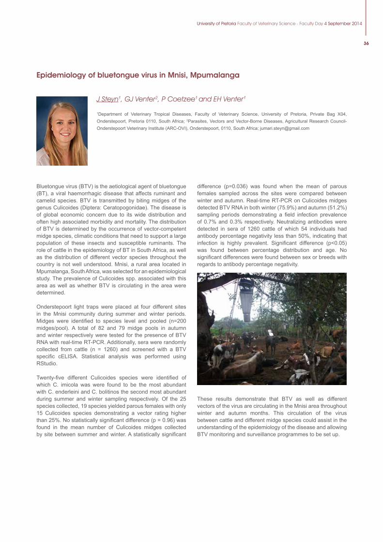
University of Pretoria Faculty of Veterinary Science - Faculty Day 4 September 2014
36
Epidemiology of bluetongue virus in Mnisi, Mpumalanga
J Steyn1, GJ Venter2, P Coetzee1 and EH Venter1
1Department of Veterinary Tropical Diseases, Faculty of Veterinary Science, University of Pretoria, Private Bag X04, Onderstepoort, Pretoria 0110, South Africa; 2Parasites, Vectors and Vector-Borne Diseases, Agricultural Research Council-Onderstepoort Veterinary Institute (ARC-OVI), Onderstepoort, 0110, South Africa; [email protected]
Bluetongue virus (BTV) is the aetiological agent of bluetongue (BT), a viral haemorrhagic disease that affects ruminant and camelid species. BTV is transmitted by biting midges of the genus Culicoides (Diptera: Ceratopogonidae). The disease is of global economic concern due to its wide distribution and often high associated morbidity and mortality. The distribution of BTV is determined by the occurrence of vector-competent midge species, climatic conditions that need to support a large population of these insects and susceptible ruminants. The role of cattle in the epidemiology of BT in South Africa, as well as the distribution of different vector species throughout the country is not well understood. Mnisi, a rural area located in Mpumalanga, South Africa, was selected for an epidemiological study. The prevalence of Culicoides spp. associated with this area as well as whether BTV is circulating in the area were determined.
Onderstepoort light traps were placed at four different sites in the Mnisi community during summer and winter periods. Midges were identified to species level and pooled (n=200 midges/pool). A total of 82 and 79 midge pools in autumn and winter respectively were tested for the presence of BTV RNA with real-time RT-PCR. Additionally, sera were randomly collected from cattle (n = 1260) and screened with a BTV specific cELISA. Statistical analysis was performed using RStudio.
Twenty-five different Culicoides species were identified of which C. imicola was were found to be the most abundant with C. enderleini and C. bolitinos the second most abundant during summer and winter sampling respectively. Of the 25 species collected, 19 species yielded parous females with only 15 Culicoides species demonstrating a vector rating higher than 25%. No statistically significant difference (p = 0.96) was found in the mean number of Culicoides midges collected by site between summer and winter. A statistically significant
difference (p=0.036) was found when the mean of parous females sampled across the sites were compared between winter and autumn. Real-time RT-PCR on Culicoides midges detected BTV RNA in both winter (75.9%) and autumn (51.2%) sampling periods demonstrating a field infection prevalence of 0.7% and 0.3% respectively. Neutralizing antibodies were detected in sera of 1260 cattle of which 54 individuals had antibody percentage negativity less than 50%, indicating that infection is highly prevalent. Significant difference (p<0.05) was found between percentage distribution and age. No significant differences were found between sex or breeds with regards to antibody percentage negativity.
These results demonstrate that BTV as well as different vectors of the virus are circulating in the Mnisi area throughout winter and autumn months. This circulation of the virus between cattle and different midge species could assist in the understanding of the epidemiology of the disease and allowing BTV monitoring and surveillance programmes to be set up.
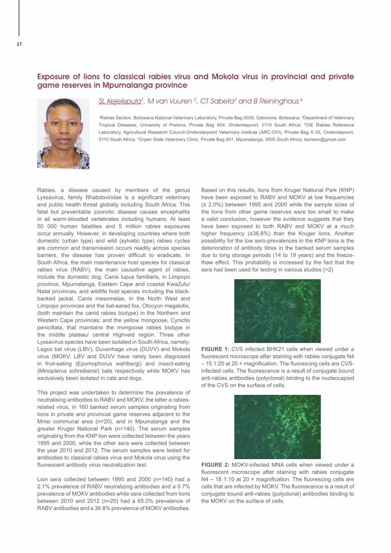
37
Exposure of lions to classical rabies virus and Mokola virus in provincial and private game reserves in Mpumalanga province
SL Kejelepula1, M van Vuuren 2, CT Sabeta3 and B Reininghaus 4
1Rabies Section, Botswana National Veterinary Laboratory, Private Bag 0035, Gaborone, Botswana; 2Department of Veterinary Tropical Diseases, University of Pretoria, Private Bag X04, Onderstepoort, 0110 South Africa; 3OIE Rabies Reference Laboratory, Agricultural Research Council-Ondersterpoort Veterinary Institute (ARC-OVI), Private Bag X 05, Onderstepoort, 0110 South Africa; 4Orpen State Veterinary Clinic, Private Bag 001, Mpumalanga, 0000 South Africa; [email protected]
Rabies, a disease caused by members of the genus Lyssavirus, family Rhabdoviridae is a significant veterinary and public health threat globally including South Africa. This fatal but preventable zoonotic disease causes encephalitis in all warm-blooded vertebrates including humans. At least 50 000 human fatalities and 5 million rabies exposures occur annually. However, in developing countries where both domestic (urban type) and wild (sylvatic type) rabies cycles are common and transmission occurs readily across species barriers, the disease has proven difficult to eradicate. In South Africa, the main maintenance host species for classical rabies virus (RABV), the main causative agent of rabies, include the domestic dog, Canis lupus familiaris, in Limpopo province, Mpumalanga, Eastern Cape and coastal KwaZulu/Natal provinces, and wildlife host species including the black-backed jackal, Canis mesomelas, in the North West and Limpopo provinces and the bat-eared fox, Otocyon megalotis, (both maintain the canid rabies biotype) in the Northern and Western Cape provinces; and the yellow mongoose, Cynictis penicillata, that maintains the mongoose rabies biotype in the middle plateau/ central Highveld region. Three other Lyssavirus species have been isolated in South Africa, namely: Lagos bat virus (LBV), Duvenhage virus (DUVV) and Mokola virus (MOKV. LBV and DUVV have rarely been diagnosed in fruit-eating (Epomophorus wahlbergi) and insect-eating (Minopterus schreibersii) bats respectively while MOKV has exclusively been isolated in cats and dogs.
This project was undertaken to determine the prevalence of neutralising antibodies to RABV and MOKV, the latter a rabies-related virus, in 160 banked serum samples originating from lions in private and provincial game reserves adjacent to the Mnisi communal area (n=20), and in Mpumalanga and the greater Kruger National Park (n=140). The serum samples originating from the KNP lion were collected between the years 1995 and 2000, while the other sera were collected between the year 2010 and 2012. The serum samples were tested for antibodies to classical rabies virus and Mokola virus using the fluorescent antibody virus neutralization test.
Lion sera collected between 1995 and 2000 (n=140) had a 2.1% prevalence of RABV neutralizing antibodies and a 0.7% prevalence of MOKV antibodies while sera collected from lions between 2010 and 2012 (n=20) had a 65.0% prevalence of RABV antibodies and a 36.8% prevalence of MOKV antibodies.
Based on this results, lions from Kruger National Park (KNP) have been exposed to RABV and MOKV at low frequencies (≤ 2.0%) between 1995 and 2000 while the sample sizes of the lions from other game reserves were too small to make a valid conclusion, however the evidence suggests that they have been exposed to both RABV and MOKV at a much higher frequency (≥36.8%) than the Kruger lions. Another possibility for the low sero-prevalences in the KNP lions is the deterioration of antibody titres in the banked serum samples due to long storage periods (14 to 19 years) and the freeze-thaw effect. This probability is increased by the fact that the sera had been used for testing in various studies (>2)
FIGURE1:CVS infected BHK21 cells when viewed under a fluorescent microscope after staining with rabies conjugate N4 – 15 1:20 at 20 × magnification. The fluorescing cells are CVS-infected cells. The fluorescence is a result of conjugate bound anti-rabies antibodies (polyclonal) binding to the nucleocapsid of the CVS on the surface of cells.
FIGURE2: MOKV-infected MNA cells when viewed under a fluorescent microscope after staining with rabies conjugate N4 – 18 1:10 at 20 × magnification. The fluorescing cells are cells that are infected by MOKV. The fluorescence is a result of conjugate bound anti-rabies (polyclonal) antibodies binding to the MOKV on the surface of cells.
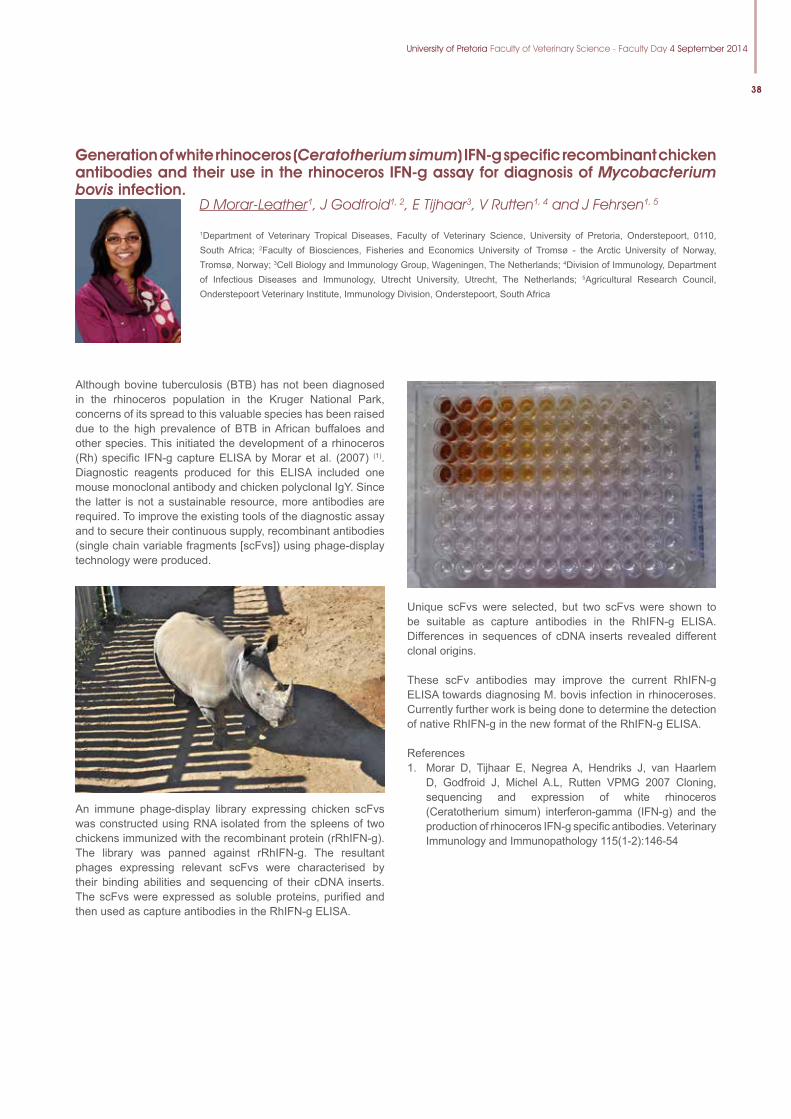
University of Pretoria Faculty of Veterinary Science - Faculty Day 4 September 2014
38
Generation of white rhinoceros (Ceratotherium simum) IFN-g specific recombinant chicken antibodies and their use in the rhinoceros IFN-g assay for diagnosis of Mycobacterium bovis infection.
D Morar-Leather1, J Godfroid1, 2, E Tijhaar3, V Rutten1, 4 and J Fehrsen1, 5
1Department of Veterinary Tropical Diseases, Faculty of Veterinary Science, University of Pretoria, Onderstepoort, 0110, South Africa; 2Faculty of Biosciences, Fisheries and Economics University of Tromsø - the Arctic University of Norway, Tromsø, Norway; 3Cell Biology and Immunology Group, Wageningen, The Netherlands; 4Division of Immunology, Department of Infectious Diseases and Immunology, Utrecht University, Utrecht, The Netherlands; 5Agricultural Research Council, Onderstepoort Veterinary Institute, Immunology Division, Onderstepoort, South Africa
Although bovine tuberculosis (BTB) has not been diagnosed in the rhinoceros population in the Kruger National Park, concerns of its spread to this valuable species has been raised due to the high prevalence of BTB in African buffaloes and other species. This initiated the development of a rhinoceros (Rh) specific IFN-g capture ELISA by Morar et al. (2007) (1). Diagnostic reagents produced for this ELISA included one mouse monoclonal antibody and chicken polyclonal IgY. Since the latter is not a sustainable resource, more antibodies are required. To improve the existing tools of the diagnostic assay and to secure their continuous supply, recombinant antibodies (single chain variable fragments [scFvs]) using phage-display technology were produced.
An immune phage-display library expressing chicken scFvs was constructed using RNA isolated from the spleens of two chickens immunized with the recombinant protein (rRhIFN-g). The library was panned against rRhIFN-g. The resultant phages expressing relevant scFvs were characterised by their binding abilities and sequencing of their cDNA inserts. The scFvs were expressed as soluble proteins, purified and then used as capture antibodies in the RhIFN-g ELISA.
Unique scFvs were selected, but two scFvs were shown to be suitable as capture antibodies in the RhIFN-g ELISA. Differences in sequences of cDNA inserts revealed different clonal origins.
These scFv antibodies may improve the current RhIFN-g ELISA towards diagnosing M. bovis infection in rhinoceroses. Currently further work is being done to determine the detection of native RhIFN-g in the new format of the RhIFN-g ELISA.
References1. Morar D, Tijhaar E, Negrea A, Hendriks J, van Haarlem
D, Godfroid J, Michel A.L, Rutten VPMG 2007 Cloning, sequencing and expression of white rhinoceros (Ceratotherium simum) interferon-gamma (IFN-g) and the production of rhinoceros IFN-g specific antibodies. Veterinary Immunology and Immunopathology 115(1-2):146-54
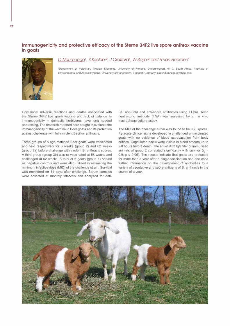
39
Immunogenicity and protective efficacy of the Sterne 34F2 live spore anthrax vaccine in goats
O Ndumnego1, S Koehler2, J Crafford1, W Beyer2 and H van Heerden1
1Department of Veterinary Tropical Diseases, University of Pretoria, Onderstepoort, 0110, South Africa; 2Institute of Environmental and Animal Hygiene, University of Hohenheim, Stuttgart, Germany; [email protected]
Occasional adverse reactions and deaths associated with the Sterne 34F2 live spore vaccine and lack of data on its immunogenicity in domestic herbivores have long needed addressing. The research reported here sought to evaluate the immunogenicity of the vaccine in Boer goats and its protection against challenge with fully virulent Bacillus anthracis.
Three groups of 5 age-matched Boer goats were vaccinated and held respectively for 6 weeks (group 2) and 62 weeks (group 3a) before challenge with virulent B. anthracis spores. A third group (group 3b) was re-vaccinated at 58 weeks and challenged at 62 weeks. A total of 6 goats (group 1) served as negative controls and were also utilized in estimating the minimum infective dose (MID) of the challenge strain. Survival was monitored for 14 days after challenge. Serum samples were collected at monthly intervals and analyzed for anti-
PA, anti-BclA and anti-spore antibodies using ELISA. Toxin neutralizing antibody (TNA) was assessed by an in vitro macrophage culture assay.
The MID of the challenge strain was found to be <36 spores. Peracute clinical signs developed in challenged unvaccinated goats with no evidence of blood extravasation from body orifices. Capsulated bacilli were visible in blood smears up to 2.8 hours before death. The anti-rPA83 IgG titer of immunised animals of group 2 correlated significantly with survival (rs = 0.9; p ≤ 0,05). The results indicate that goats are protected for more than a year after a single vaccination and disclosed further information on the development of antibodies to a variety of vegetative and spore antigens of B. anthracis in the course of a year.
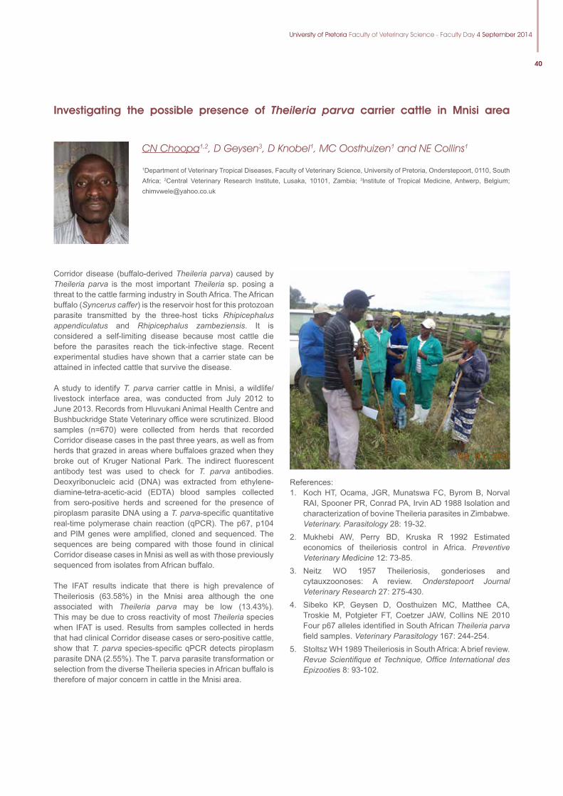
University of Pretoria Faculty of Veterinary Science - Faculty Day 4 September 2014
40
Investigating the possible presence of Theileria parva carrier cattle in Mnisi area
CN Choopa1,2, D Geysen3, D Knobel1, MC Oosthuizen1 and NE Collins1
1Department of Veterinary Tropical Diseases, Faculty of Veterinary Science, University of Pretoria, Onderstepoort, 0110, South Africa; 2Central Veterinary Research Institute, Lusaka, 10101, Zambia; 3Institute of Tropical Medicine, Antwerp, Belgium; [email protected]
Corridor disease (buffalo-derived Theileria parva) caused by Theileria parva is the most important Theileria sp. posing a threat to the cattle farming industry in South Africa. The African buffalo (Syncerus caffer) is the reservoir host for this protozoan parasite transmitted by the three-host ticks Rhipicephalus appendiculatus and Rhipicephalus zambeziensis. It is considered a self-limiting disease because most cattle die before the parasites reach the tick-infective stage. Recent experimental studies have shown that a carrier state can be attained in infected cattle that survive the disease.
A study to identify T. parva carrier cattle in Mnisi, a wildlife/livestock interface area, was conducted from July 2012 to June 2013. Records from Hluvukani Animal Health Centre and Bushbuckridge State Veterinary office were scrutinized. Blood samples (n=670) were collected from herds that recorded Corridor disease cases in the past three years, as well as from herds that grazed in areas where buffaloes grazed when they broke out of Kruger National Park. The indirect fluorescent antibody test was used to check for T. parva antibodies. Deoxyribonucleic acid (DNA) was extracted from ethylene-diamine-tetra-acetic-acid (EDTA) blood samples collected from sero-positive herds and screened for the presence of piroplasm parasite DNA using a T. parva-specific quantitative real-time polymerase chain reaction (qPCR). The p67, p104 and PIM genes were amplified, cloned and sequenced. The sequences are being compared with those found in clinical Corridor disease cases in Mnisi as well as with those previously sequenced from isolates from African buffalo.
The IFAT results indicate that there is high prevalence of Theileriosis (63.58%) in the Mnisi area although the one associated with Theileria parva may be low (13.43%). This may be due to cross reactivity of most Theileria species when IFAT is used. Results from samples collected in herds that had clinical Corridor disease cases or sero-positive cattle, show that T. parva species-specific qPCR detects piroplasm parasite DNA (2.55%). The T. parva parasite transformation or selection from the diverse Theileria species in African buffalo is therefore of major concern in cattle in the Mnisi area.
References: 1. Koch HT, Ocama, JGR, Munatswa FC, Byrom B, Norval
RAI, Spooner PR, Conrad PA, Irvin AD 1988 Isolation and characterization of bovine Theileria parasites in Zimbabwe. Veterinary. Parasitology 28: 19-32.
2. Mukhebi AW, Perry BD, Kruska R 1992 Estimated economics of theileriosis control in Africa. Preventive Veterinary Medicine 12: 73-85.
3. Neitz WO 1957 Theileriosis, gonderioses and cytauxzoonoses: A review. Onderstepoort Journal Veterinary Research 27: 275-430.
4. Sibeko KP, Geysen D, Oosthuizen MC, Matthee CA, Troskie M, Potgieter FT, Coetzer JAW, Collins NE 2010 Four p67 alleles identified in South African Theileria parva field samples. Veterinary Parasitology 167: 244-254.
5. Stoltsz WH 1989 Theileriosis in South Africa: A brief review. Revue Scientifique et Technique, Office International des Epizooties 8: 93-102.

41
Methicillin resistance in Staphylococci isolated from milk samples of South African dairy cows
R Badenhorst1, J Karzis1 and IM Petzer1
1Department of Production Animal Studies, Section Udder Health, Faculty of Veterinary Science, University of Pretoria, Private Bag X04, Onderstepoort, 0110 South Africa; [email protected]
Methicillin-resistant Staphylococcus aureus (MRSA), a criti-cally important human pathogen, is becoming a concern in veterinary medicine. Asymptomatic colonisation of MRSA has been described in numerous countries with large variations in prevalence. Little is known about the presence of MRSA in both humans and farm animals in South Africa. The aim of the study was to determine the presence of MRSA in stored iso-lates from southern African dairy cows collected in 1981 and 2012, as well as to evaluate their degree of resistance to other antimicrobial groups.
Forty six freeze dried Staphylococcus aureus (STA) isolates collected during1981; and 560 STA isolates from cow milk samples of 20 commercial dairy herds collected during 2012 were tested using cefoxitin discs for primary MRSA diagnosis.. MRSA isolates were further tested for resistance to routinely used antibiotics: beta-lactams (penicillin G (PEN), cloxacillin (OB), ampicillin (AMP), amoxicillin (AML), cephalexin (CL), cefuroxime (CXM), cephalonium (CNM), oxy-tetracycline (OT) and tylosin (TY). These susceptibility test results were compared to 313 routine STA susceptibility tests performed during routine work in 2012 (excluding cefoxitin testing).
MRSA was found to be present in 4.17% and 3.78% of the 1981 and 2012 STA isolates, respectively. However, there were 27.03% MRSA isolates with multidrug resistance and 24.32% MRSA resistant to one antimicrobial of the randomly selected South African dairy herds (2012). MRSA isolates showed higher resistance than STA to beta-lactams, OT (P=0.104) and macrolides, although not significant. The greatest resistance of MRSA was seen for PEN (19.51%) (P=0.340), AML (21.95%) and AMP (17.07%) (P=0.190). Multiple antimicrobial resistance to 3 antimicrobial groups (macrolides, beta-lactams and OT) was seen with 6.22% of MRSA and 1.60% of STA isolates.
The prevalence of MRSA within 20 positive STA herds was 3.78%. Significant differences (P<0.001) were present between MRSA and STA in % pan-susceptibility and resistance to one or more antimicrobial groups.
MRSA was present in South African commercial dairy herds in 1981 and is still present in 2012.
ReferenceClinical and Laboratory Standards Institute (CLSI). Methods for Antimicrobial Susceptibility Testing of Anaerobic Bacteria. Approved Standard, Eighth Edition, 32(3), CLSI, Wayne PA, January 2012.
Table: Trends in % MRSA according to veterinary breakpoints with Cefoxitin disks (CLSI 2012).
Isolates testedZone diameters in mmResistant Sensitive<21 mm >22 mm
Staphylococcus aureus1981 95.83% (46) 4.17% (2)
Staphylococcus aureus2012 96.22% (560) 4.44% (22)
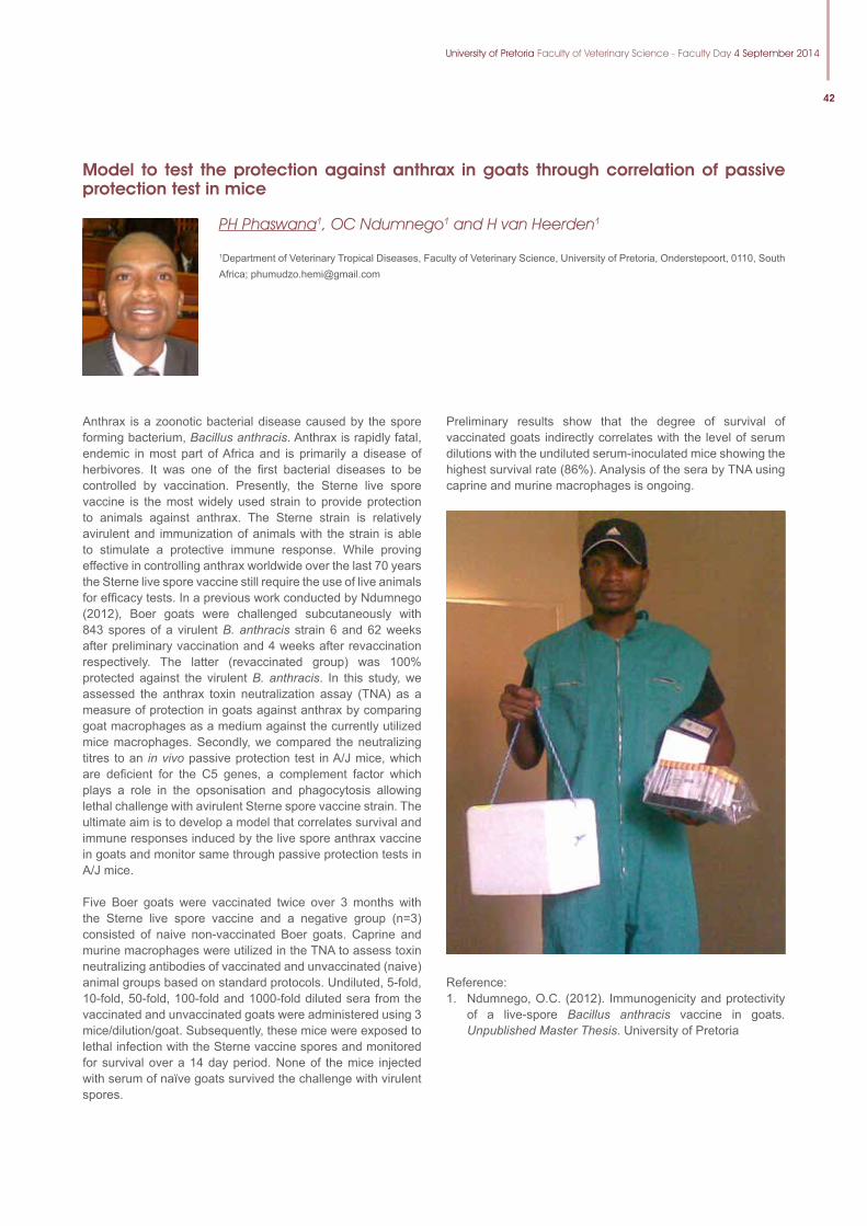
University of Pretoria Faculty of Veterinary Science - Faculty Day 4 September 2014
42
Model to test the protection against anthrax in goats through correlation of passive protection test in mice
PH Phaswana1, OC Ndumnego1 and H van Heerden1
1Department of Veterinary Tropical Diseases, Faculty of Veterinary Science, University of Pretoria, Onderstepoort, 0110, South Africa; [email protected]
Anthrax is a zoonotic bacterial disease caused by the spore forming bacterium, Bacillus anthracis. Anthrax is rapidly fatal, endemic in most part of Africa and is primarily a disease of herbivores. It was one of the first bacterial diseases to be controlled by vaccination. Presently, the Sterne live spore vaccine is the most widely used strain to provide protection to animals against anthrax. The Sterne strain is relatively avirulent and immunization of animals with the strain is able to stimulate a protective immune response. While proving effective in controlling anthrax worldwide over the last 70 years the Sterne live spore vaccine still require the use of live animals for efficacy tests. In a previous work conducted by Ndumnego (2012), Boer goats were challenged subcutaneously with 843 spores of a virulent B. anthracis strain 6 and 62 weeks after preliminary vaccination and 4 weeks after revaccination respectively. The latter (revaccinated group) was 100% protected against the virulent B. anthracis. In this study, we assessed the anthrax toxin neutralization assay (TNA) as a measure of protection in goats against anthrax by comparing goat macrophages as a medium against the currently utilized mice macrophages. Secondly, we compared the neutralizing titres to an in vivo passive protection test in A/J mice, which are deficient for the C5 genes, a complement factor which plays a role in the opsonisation and phagocytosis allowing lethal challenge with avirulent Sterne spore vaccine strain. The ultimate aim is to develop a model that correlates survival and immune responses induced by the live spore anthrax vaccine in goats and monitor same through passive protection tests in A/J mice.
Five Boer goats were vaccinated twice over 3 months with the Sterne live spore vaccine and a negative group (n=3) consisted of naive non-vaccinated Boer goats. Caprine and murine macrophages were utilized in the TNA to assess toxin neutralizing antibodies of vaccinated and unvaccinated (naive) animal groups based on standard protocols. Undiluted, 5-fold, 10-fold, 50-fold, 100-fold and 1000-fold diluted sera from the vaccinated and unvaccinated goats were administered using 3 mice/dilution/goat. Subsequently, these mice were exposed to lethal infection with the Sterne vaccine spores and monitored for survival over a 14 day period. None of the mice injected with serum of naïve goats survived the challenge with virulent spores.
Preliminary results show that the degree of survival of vaccinated goats indirectly correlates with the level of serum dilutions with the undiluted serum-inoculated mice showing the highest survival rate (86%). Analysis of the sera by TNA using caprine and murine macrophages is ongoing.
Reference:1. Ndumnego, O.C. (2012). Immunogenicity and protectivity
of a live-spore Bacillus anthracis vaccine in goats. Unpublished Master Thesis. University of Pretoria
43
Molecular Characterisation of Peste Des Petits Ruminants Viruses of Sheep and Goats in Nigeria
S Mantip1,2, M Van Vuuren1, M Quan1 and D Shamaki2
1Department of Veterinary Tropical Diseases. Faculty of Veterinary Science. University of Pretoria, Onderstepoort, 0110, South Africa; 2Viral Research Division. National Veterinary Research Institute, PMB 0001, Vom, Plateau State, Nigeria; [email protected]
Peste des petits ruminants virus (PPRV) belongs to the family Paramyxoviridae and genus morbillivirus. It is a highly contagious, fatal and economically important viral disease of small ruminants that is still endemic and militate against the production of sheep and goats in Nigeria. It is a notifiable disease according to the World Organization for Animal Health (Office International des Epizooties). In this study, a molecular analysis of PPRV from sheep and goats from recent outbreaks across the different regions of Nigeria was carried out. The aim was to describe the viral strains and the movement of the virus within the country compared to other endemic areas of the world. This was carried out through tissue and swab samples collected from sheep and goats in various agro-ecological zones of Nigeria. The evolution and relationship of earlier PPRV strains/isolates and those circulating and causing recent outbreaks was determined by sequencing of the nucleoprotein (N)-gene.
Twenty tissue and swab samples from apparently healthy and sick sheep and goats were collected randomly from each of three states of each of the six agro-ecological zones visited. A total of 360 samples were collected. A total of 35 samples of 360 (9.7 %) tested positive by RT-PCR, of which 25 were from oculo-nasal swabs and 10 were from tissue samples.
Phylogenetic analysis was carried out using the N-gene sequences of the PPRV amplicons. Alignment of the sequences and related sequences from GenBank and neighbour-joining phylogenetic analysis using PAUP identified four different lineages, i.e. lineages I to IV. Interestingly, the Nigerian strains described in this study grouped in two separate major lineages i.e. lineages II and IV. Strains from Sokoto, Oyo, Plateau and Ondo states grouped according to the historical distribution of PPRV together with the Nigerian 75/1 strain of lineage II, while other strains from Sokoto, Oyo, Plateau, Akwa-Ibom, Adamawa, Kaduna, Lagos, Bauchi, Niger and Kano states grouped together with the East-African and Asian strains of lineage IV. This finding suggests that both lineages II and IV strains of PPRVs are circulating presently in Nigeria, contrary to an earlier publication which indicated that only strains of lineage II were circulating in the country.
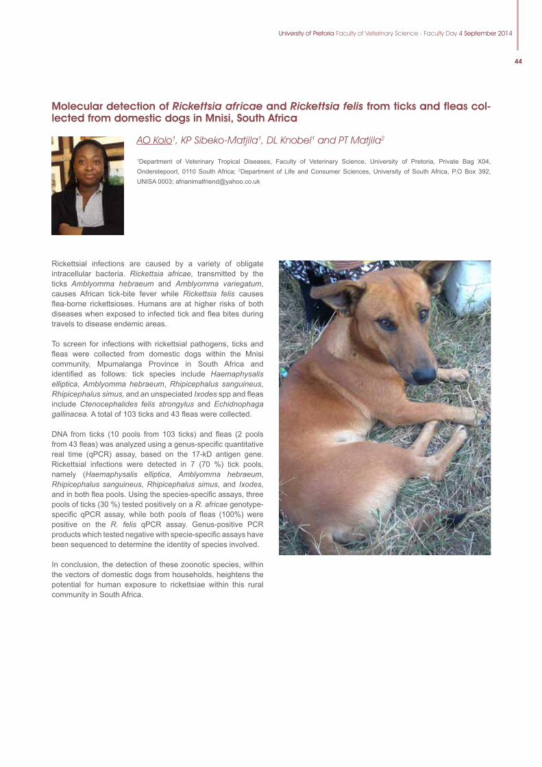
University of Pretoria Faculty of Veterinary Science - Faculty Day 4 September 2014
44
Rickettsial infections are caused by a variety of obligate intracellular bacteria. Rickettsia africae, transmitted by the ticks Amblyomma hebraeum and Amblyomma variegatum, causes African tick-bite fever while Rickettsia felis causes flea-borne rickettsioses. Humans are at higher risks of both diseases when exposed to infected tick and flea bites during travels to disease endemic areas.
To screen for infections with rickettsial pathogens, ticks and fleas were collected from domestic dogs within the Mnisi community, Mpumalanga Province in South Africa and identified as follows: tick species include Haemaphysalis elliptica, Amblyomma hebraeum, Rhipicephalus sanguineus, Rhipicephalus simus, and an unspeciated Ixodes spp and fleas include Ctenocephalides felis strongylus and Echidnophaga gallinacea. A total of 103 ticks and 43 fleas were collected.
DNA from ticks (10 pools from 103 ticks) and fleas (2 pools from 43 fleas) was analyzed using a genus-specific quantitative real time (qPCR) assay, based on the 17-kD antigen gene. Rickettsial infections were detected in 7 (70 %) tick pools, namely (Haemaphysalis elliptica, Amblyomma hebraeum, Rhipicephalus sanguineus, Rhipicephalus simus, and Ixodes, and in both flea pools. Using the species-specific assays, three pools of ticks (30 %) tested positively on a R. africae genotype-specific qPCR assay, while both pools of fleas (100%) were positive on the R. felis qPCR assay. Genus-positive PCR products which tested negative with specie-specific assays have been sequenced to determine the identity of species involved.
In conclusion, the detection of these zoonotic species, within the vectors of domestic dogs from households, heightens the potential for human exposure to rickettsiae within this rural community in South Africa.
Molecular detection of Rickettsia africae and Rickettsia felis from ticks and fleas col-lected from domestic dogs in Mnisi, South Africa
AO Kolo1, KP Sibeko-Matjila1, DL Knobel1 and PT Matjila2
1Department of Veterinary Tropical Diseases, Faculty of Veterinary Science, University of Pretoria, Private Bag X04, Onderstepoort, 0110 South Africa; 2Department of Life and Consumer Sciences, University of South Africa, P.O Box 392, UNISA 0003; [email protected]
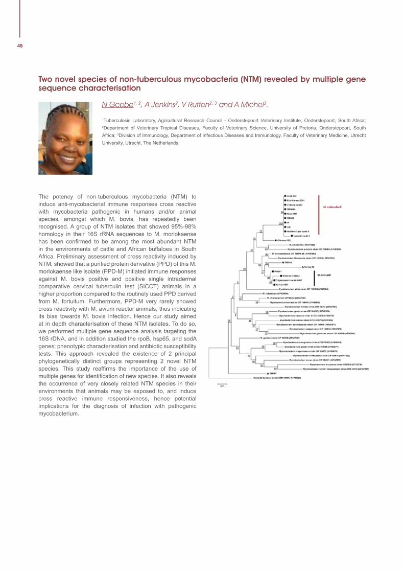
45
Two novel species of non-tuberculous mycobacteria (NTM) revealed by multiple gene sequence characterisation
N Gcebe1, 2, A Jenkins2, V Rutten2, 3 and A Michel2.
1Tuberculosis Laboratory, Agricultural Research Council - Onderstepoort Veterinary Institute, Onderstepoort, South Africa; 2Department of Veterinary Tropical Diseases, Faculty of Veterinary Science, University of Pretoria, Onderstepoort, South Africa; 3Division of Immunology, Department of Infectious Diseases and Immunology, Faculty of Veterinary Medicine, Utrecht University, Utrecht, The Netherlands.
The potency of non-tuberculous mycobacteria (NTM) to induce anti-mycobacterial immune responses cross reactive with mycobacteria pathogenic in humans and/or animal species, amongst which M. bovis, has repeatedly been recognised. A group of NTM isolates that showed 95%-98% homology in their 16S rRNA sequences to M. moriokaense has been confirmed to be among the most abundant NTM in the environments of cattle and African buffaloes in South Africa. Preliminary assessment of cross reactivity induced by NTM, showed that a purified protein derivative (PPD) of this M. moriokaense like isolate (PPD-M) initiated immune responses against M. bovis positive and positive single intradermal comparative cervical tuberculin test (SICCT) animals in a higher proportion compared to the routinely used PPD derived from M. fortuitum. Furthermore, PPD-M very rarely showed cross reactivity with M. avium reactor animals, thus indicating its bias towards M. bovis infection. Hence our study aimed at in depth characterisation of these NTM isolates. To do so, we performed multiple gene sequence analysis targeting the 16S rDNA, and in addition studied the rpoB, hsp65, and sodA genes; phenotypic characterisation and antibiotic susceptibility tests. This approach revealed the existence of 2 principal phylogenetically distinct groups representing 2 novel NTM species. This study reaffirms the importance of the use of multiple genes for identification of new species. It also reveals the occurrence of very closely related NTM species in their environments that animals may be exposed to, and induce cross reactive immune responsiveness, hence potential implications for the diagnosis of infection with pathogenic mycobacterium.
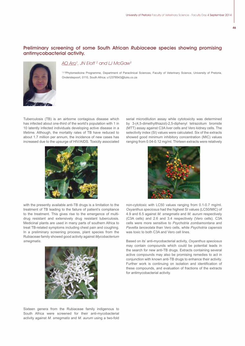
University of Pretoria Faculty of Veterinary Science - Faculty Day 4 September 2014
46
Preliminary screening of some South African Rubiaceae species showing promising antimycobacterial activity.
AO Aro1, JN Eloff 2 and LJ McGaw3
1,2,3Phytomedicine Programme, Department of Paraclinical Sciences, Faculty of Veterinary Science, University of Pretoria, Onderstepoort, 0110, South Africa; [email protected]
Tuberculosis (TB) is an airborne contagious disease which has infected about one-third of the world’s population with 1 in 10 latently infected individuals developing active disease in a lifetime. Although, the mortality rates of TB have reduced to about 1.7 million per annum, the incidence of new cases has increased due to the upsurge of HIV/AIDS. Toxicity associated
with the presently available anti-TB drugs is a limitation to the treatment of TB leading to the failure of patient’s compliance to the treatment. This gives rise to the emergence of multi-drug resistant and extensively drug resistant tuberculosis. Medicinal plants are used in many parts of southern Africa to treat TB-related symptoms including chest pain and coughing. In a preliminary screening process, plant species from the Rubiaceae family showed good activity against Mycobacterium smegmatis.
Sixteen genera from the Rubiaceae family indigenous to South Africa were screened for their anti-mycobacterial activity against M. smegmatis and M. aurum using a two-fold
serial microdilution assay while cytotoxicity was determined by 3-(4,5-dimethylthiazol)-2,5-diphenyl tetrazolium bromide (MTT) assay against C3A liver cells and Vero kidney cells. The selectivity index (SI) values were calculated. Six of the extracts showed good minimum inhibitory concentration (MIC) values ranging from 0.04-0.12 mg/ml. Thirteen extracts were relatively
non-cytotoxic with LC50 values ranging from 0.1-0.7 mg/ml. Oxyanthus speciosus had the highest SI values (LC50/MIC) of 4.9 and 6.5 against M. smegmatis and M. aurum respectively (C3A cells) and 2.6 and 3.4 respectively (Vero cells). C3A cells were more sensitive to Psychotria zombamontana and Pavetta lanceolata than Vero cells, while Psychotria capensis was toxic to both C3A and Vero cell lines.
Based on its’ anti-mycobacterial activity, Oxyanthus speciosus may contain compounds which could be potential leads in the search for new anti-TB drugs. Extracts containing several active compounds may also be promising remedies to act in conjunction with known anti-TB drugs to enhance their activity. Further work is continuing on isolation and identifi cation of these compounds, and evaluation of fractions of the extracts for antimycobacterial activity.
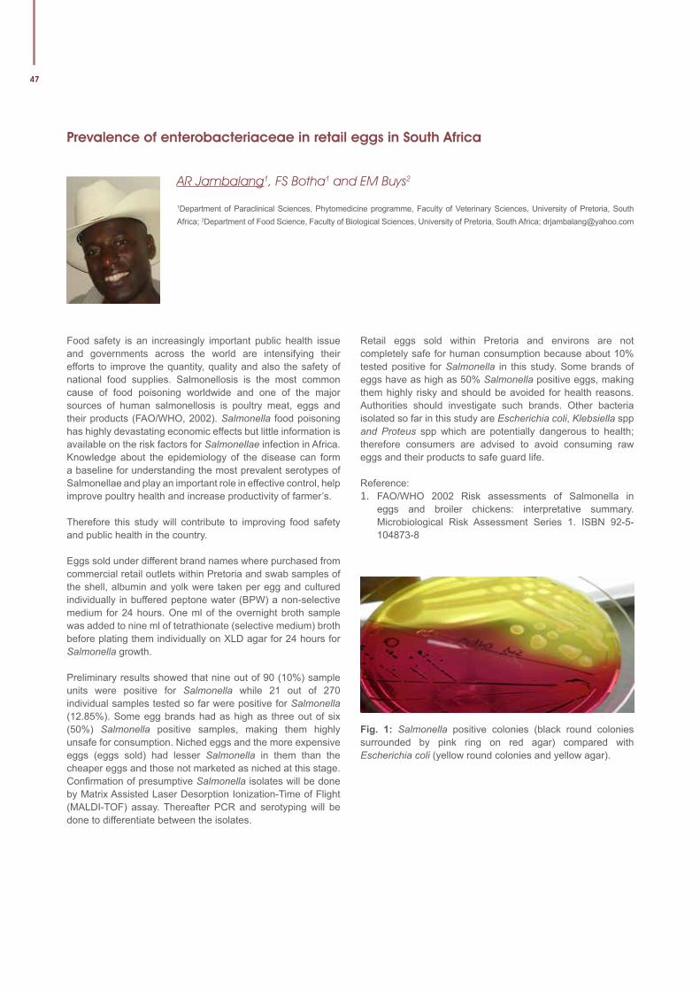
47
Prevalence of enterobacteriaceae in retail eggs in South Africa
AR Jambalang1, FS Botha1 and EM Buys2
1Department of Paraclinical Sciences, Phytomedicine programme, Faculty of Veterinary Sciences, University of Pretoria, South Africa; 2Department of Food Science, Faculty of Biological Sciences, University of Pretoria, South Africa; [email protected]
Food safety is an increasingly important public health issue and governments across the world are intensifying their efforts to improve the quantity, quality and also the safety of national food supplies. Salmonellosis is the most common cause of food poisoning worldwide and one of the major sources of human salmonellosis is poultry meat, eggs and their products (FAO/WHO, 2002). Salmonella food poisoning has highly devastating economic effects but little information is available on the risk factors for Salmonellae infection in Africa. Knowledge about the epidemiology of the disease can form a baseline for understanding the most prevalent serotypes of Salmonellae and play an important role in effective control, help improve poultry health and increase productivity of farmer’s.
Therefore this study will contribute to improving food safety and public health in the country.
Eggs sold under different brand names where purchased from commercial retail outlets within Pretoria and swab samples of the shell, albumin and yolk were taken per egg and cultured individually in buffered peptone water (BPW) a non-selective medium for 24 hours. One ml of the overnight broth sample was added to nine ml of tetrathionate (selective medium) broth before plating them individually on XLD agar for 24 hours for Salmonella growth. Preliminary results showed that nine out of 90 (10%) sample units were positive for Salmonella while 21 out of 270 individual samples tested so far were positive for Salmonella (12.85%). Some egg brands had as high as three out of six (50%) Salmonella positive samples, making them highly unsafe for consumption. Niched eggs and the more expensive eggs (eggs sold) had lesser Salmonella in them than the cheaper eggs and those not marketed as niched at this stage. Confirmation of presumptive Salmonella isolates will be done by Matrix Assisted Laser Desorption Ionization-Time of Flight (MALDI-TOF) assay. Thereafter PCR and serotyping will be done to differentiate between the isolates.
Retail eggs sold within Pretoria and environs are not completely safe for human consumption because about 10% tested positive for Salmonella in this study. Some brands of eggs have as high as 50% Salmonella positive eggs, making them highly risky and should be avoided for health reasons. Authorities should investigate such brands. Other bacteria isolated so far in this study are Escherichia coli, Klebsiella spp and Proteus spp which are potentially dangerous to health; therefore consumers are advised to avoid consuming raw eggs and their products to safe guard life.
Reference: 1. FAO/WHO 2002 Risk assessments of Salmonella in
eggs and broiler chickens: interpretative summary. Microbiological Risk Assessment Series 1. ISBN 92-5-104873-8
Fig. 1: Salmonella positive colonies (black round colonies surrounded by pink ring on red agar) compared with Escherichia coli (yellow round colonies and yellow agar).
University of Pretoria Faculty of Veterinary Science - Faculty Day 4 September 2014
48
Protective effects of South African plants against mutagenicity of aflatoxin B1
BA Nkala1,2, CJ Botha2 and EE Elgorashi1,2
1Toxicology Section, ARC-Onderstepoort Veterinary Institute, Private Bag X05, 0110, RSA; 2Department of Paraclinical Sciences, Faculty of Veterinary Science, University of Pretoria, Private Bag X04,Onderstedpoort, 0110, RSA; [email protected]
Aflatoxin B1(AFB1) is the most carcinogenic, hepatotoxic, directly and indirectly-acting mutagen; which poses serious health risk for both livestock and humans [1,2]. Naturally occurring plant compounds inhibit the mutagenic effect of AFB1 against Salmonella strains [3,4]. Essentially, plant materials with protective effect against mutagenic of Aflatoxin B1 will serves as promising anticancer properties.
The aim of this study was to identify plants with a potential chemo-protective effect against the genotoxicity of AFB1 which are endemic to SA.
The antimutagenic effect of 42 plant extracts was evaluated using Ames test (TA98, TA100 & TA102) with or without metabolic activation [5] and micronucleus test [6] using C3A human liver cell line [7] against mutagenicity of AFB1. Ames test is widely used bacterial mutagenicity test, which is used to assess the mutagenic potential of chemical compounds. Micronucleus is a test used to screen for potential genotoxic compounds.
None of the extracts had genotoxic effect in Ames test, with or without metabolic activation. 13 extracts possessed some degree of antimutagenic effect against mutagenicity of AFB1. However, extracts of Acacia polyacantha (bark) and Protea nitida (bark) exhibited strong antimutagenic effects against mutagenicity of AFB1 in both Ames and micronucleus assays.
Extracts of Acacia polyacantha (bark) and Protea nitida (bark) are promising chemo-preventative agents against mutagenicity of AFB1.
Acknowledgments: The study was supported financially by the National Research Foundation (NRF) and Department of Science and Technology (DST), South Africa.
References: 1. Loarca-Pinã, G., Kuzmicky, P.A., de Mejía, E.G., Kado,
N.Y. (1998). “Inhibitory effects of ellagic acid on the direct-acting mutagenicity of aflatoxin B1 in the Salmonella microsuspension assay.” Mutation Research (398): 183–187.
2. Riley, R. T. (1998). Mechanistic interactions of mycotoxins: theoretical consideration. Mycotoxins in Agriculture and Food Safety. Marcel Dekker. K. K. In: Sinha, Bhatanagar, D.(Eds.),. Basel, New York: 227–254.
3. Horn, R. C., Vargas, V.M.F. (2003). “Antimutagenic activity of extracts of natural substances in the Salmonella/microsome assay.” Mutagenesis 18(2): 113-118.
4. Nogueira, M. E. I., Passoni, M.H., Biso, F.I., Longo, M.C., Cardoso, C.R.P., dos Santos, L.C., Varanda, E.A. (2006). “Investigation of genotoxic and antigenotoxic activities of Melampodium divaricatum in Salmonella typhimurium.” Toxicology in Vitro (20): 361–366.
5. Vershaeve, L., Kestens, V., Taylor, J.L.S., Elgorashi, E.E., Maes, A., Van Puyvelde, L., Kimpe, D., van Staden, J. (2004). “Investigation of the antimutagenic effects of selected South African medicinal plant extracts.” Toxicology in Vitro (18): 29-35.
6. Fenech, M., Morley, A. (1985). “Measurement of micronuclei in lymphocytes.” Mutation Research 147(29-36).
7. Baudoin, R., Griscom, L., Prot, J.M., Legallais, C., Leclerc, E. (2011). “Behaviour of HepG2/C3A cell cultures in a microfluidic bioreactor.” Biochemical Engineering Journal 53: 172-181.
49
Quantitative anti-anthrax IgG ELISA correlates with the anthrax toxin neutralization assay in goats
OC Ndumnego1, J Crafford1, W Beyer2 and H van Heerden1
1Department of Veterinary Tropical Diseases, Faculty of Veterinary Science, University of Pretoria, Onderstepoort, South Africa; 2Institute of Environmental and Animal Hygiene, University of Hohenheim, Stuttgart, Germany; [email protected]
Presently, little data exist on the level and duration of anti-protective antigen (PA) IgG in vaccinated livestock. Various adaptation of enzyme-linked immunosorbent assays (ELISAs) have been developed in studies to assess immune response following vaccination, albeit mostly in laboratory rodent models. The quantitative anti-anthrax IgG ELISA in this study describes a method of enumerating the concentration of anti-PA specific IgG present in sera of immunized goats, with the aid of an affinity-purified caprine polyclonal anti-anthrax PA-83 IgG standard. This was compared with the anthrax toxin neutralization assay (TNA) which measures a functional subset of toxin neutralizing anti-PA IgG.
The measured concentrations obtained in the standard curve correlated with the known concentration at each dilution. Percentage recovery of the standard concentrations ranged from 89 to 98% (lower and upper asymptote respectively). Mean correlation coefficient (r2) of the standard curve was
0.998. Evaluation of the intra-assay coefficient of variation showed ranges of 0.23-16.90% and 0.40-12.46% for days 28 and 140 sera samples respectively, following vaccination. The mean inter-assay coefficient of variation for triplicate samples repeated on 5 different days was 18.53 and 12.17% for days 28 and 140 sera samples respectively. Spearman’s rank correlation of log-transformed IgG concentrations and TNA titres showed strong positive correlation (rs = 0.942; P = 0.01).
This study provides evidence that an indirect ELISA can be used for the quantification of anti-anthrax PA IgG in goats with the added advantage of using single dilutions to save time and resources. The use of such related immunoassays can serve as potential adjuncts to potency tests for Sterne and other vaccine types under development in ruminant species. This is the first report on the correlation of polyclonal anti-anthrax PA83 antibody with the TNA in goats.
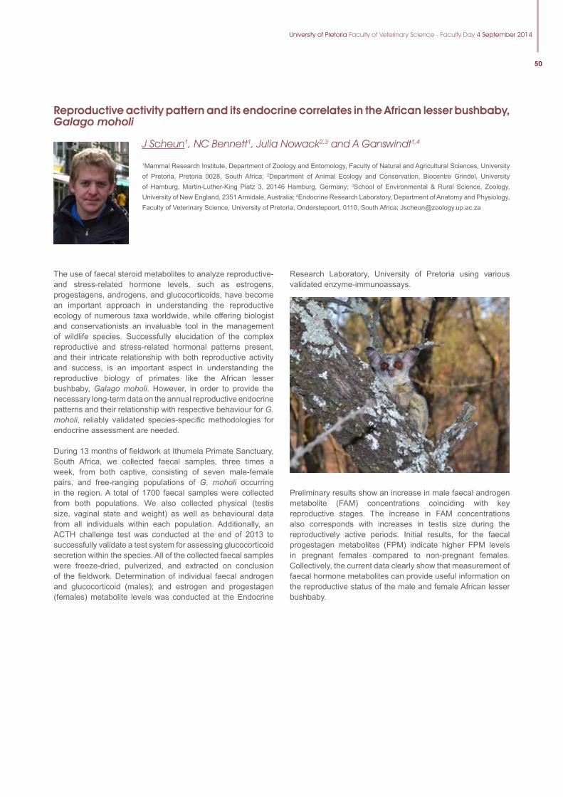
University of Pretoria Faculty of Veterinary Science - Faculty Day 4 September 2014
50
Reproductive activity pattern and its endocrine correlates in the African lesser bushbaby, Galago moholi
J Scheun1, NC Bennett1, Julia Nowack2,3 and A Ganswindt1,4
1Mammal Research Institute, Department of Zoology and Entomology, Faculty of Natural and Agricultural Sciences, University of Pretoria, Pretoria 0028, South Africa; 2Department of Animal Ecology and Conservation, Biocentre Grindel, University of Hamburg, Martin-Luther-King Platz 3, 20146 Hamburg, Germany; 3School of Environmental & Rural Science, Zoology, University of New England, 2351 Armidale, Australia; 4Endocrine Research Laboratory, Department of Anatomy and Physiology, Faculty of Veterinary Science, University of Pretoria, Onderstepoort, 0110, South Africa; [email protected]
The use of faecal steroid metabolites to analyze reproductive- and stress-related hormone levels, such as estrogens, progestagens, androgens, and glucocorticoids, have become an important approach in understanding the reproductive ecology of numerous taxa worldwide, while offering biologist and conservationists an invaluable tool in the management of wildlife species. Successfully elucidation of the complex reproductive and stress-related hormonal patterns present, and their intricate relationship with both reproductive activity and success, is an important aspect in understanding the reproductive biology of primates like the African lesser bushbaby, Galago moholi. However, in order to provide the necessary long-term data on the annual reproductive endocrine patterns and their relationship with respective behaviour for G. moholi, reliably validated species-specific methodologies for endocrine assessment are needed.
During 13 months of fieldwork at Ithumela Primate Sanctuary, South Africa, we collected faecal samples, three times a week, from both captive, consisting of seven male-female pairs, and free-ranging populations of G. moholi occurring in the region. A total of 1700 faecal samples were collected from both populations. We also collected physical (testis size, vaginal state and weight) as well as behavioural data from all individuals within each population. Additionally, an ACTH challenge test was conducted at the end of 2013 to successfully validate a test system for assessing glucocorticoid secretion within the species. All of the collected faecal samples were freeze-dried, pulverized, and extracted on conclusion of the fieldwork. Determination of individual faecal androgen and glucocorticoid (males); and estrogen and progestagen (females) metabolite levels was conducted at the Endocrine
Research Laboratory, University of Pretoria using various validated enzyme-immunoassays.
Preliminary results show an increase in male faecal androgen metabolite (FAM) concentrations coinciding with key reproductive stages. The increase in FAM concentrations also corresponds with increases in testis size during the reproductively active periods. Initial results, for the faecal progestagen metabolites (FPM) indicate higher FPM levels in pregnant females compared to non-pregnant females. Collectively, the current data clearly show that measurement of faecal hormone metabolites can provide useful information on the reproductive status of the male and female African lesser bushbaby.
51
Screening of South African plants for activities against salmonellosis
ZP Mahlangu1,3, E Madoroba2, F Botha3 and EE Elgorashi1,3
1Toxicology section, Agricultural Research Council – Onderstepoort Veterinary Institute, Private Bag X05, Onderstepoort, 0110, South Africa; 2Bacteriology section, Agricultural Research Council – Onderstepoort Veterinary Institute, Private Bag X05, Onderstepoort, 0110, South Africa; 3Department of Paraclinical Sciences, Faculty of Veterinary Science, University of Pretoria, Onderstepoort, 0110, South Africa; [email protected]
Salmonellosis, caused by the members of the genus Salmonella, is responsible for high morbidity and mortality rates in both humans and animals worldwide. Salmonellosis commonly manifests as enteric and systemic disease in animals, with the severity of infection observed among young, old, and pregnant animals. Enteric disease is often self-limiting whereas systemic disease requires treatment with antimicrobial agents. Increasing prevalence of antimicrobial resistant and multi drug resistant strains of non-typhoidal Salmonella pose a threat in the management and treatment of systemic infection caused by Salmonella. Thus this study was carried out to explore alternative antimicrobials such as plant-based compounds to offer a solution to the challenge of antimicrobial resistance.
Powdered plant material of 70 plant species, used for the treatment of diarrhea, was extracted with acetone, water, dichloromethane, and methanol. The crude plant extracts were screened for antimicrobial activity against Salmonellatyphimurium, S. enteritidis, S. dublin, and S. gallinarumusing microplate dilution method. Plant species with good antimicrobial activity were screened for cytotoxic
effects using MTT cell proliferation assay on human hepatocellular carcinoma (C3A) and Bovine kidney cells (MDBK). Potential genotoxic effects were evaluated using Ames assay (tester strains TA100 and AT98).
The minimal inhibitory concentration (MIC) of all tested extracts ranged between >2 and 0.125 mg/mL All tested extracts had minimal inhibitory concentrations (MIC) ranged between >2 and 0.125 mg/mL, while minimal bactericidal concentrations (MBC) ranged between >2 and 0.25 mg/mL. The methanol leaf extract of Albizia gummifera had the lowest MIC and MBC against all the tested strains.Cell viability against A. gummifera leaf extracts, measured as IC50 for C3A and MDBK cells was 15.70 and 9.37 µg/mL respectively. The number of reverted colonies for both TA 98 and TA 100 showed no indication of mutagenic potential.
A.gummifera with good MIC and MBC against all tested non-typhoidal strains of Salmonella; low toxicity; and no indication of genotoxic effect, can be used as an alternative treatment of salmonellosis caused by the non-typhoidal strains of Salmonella.
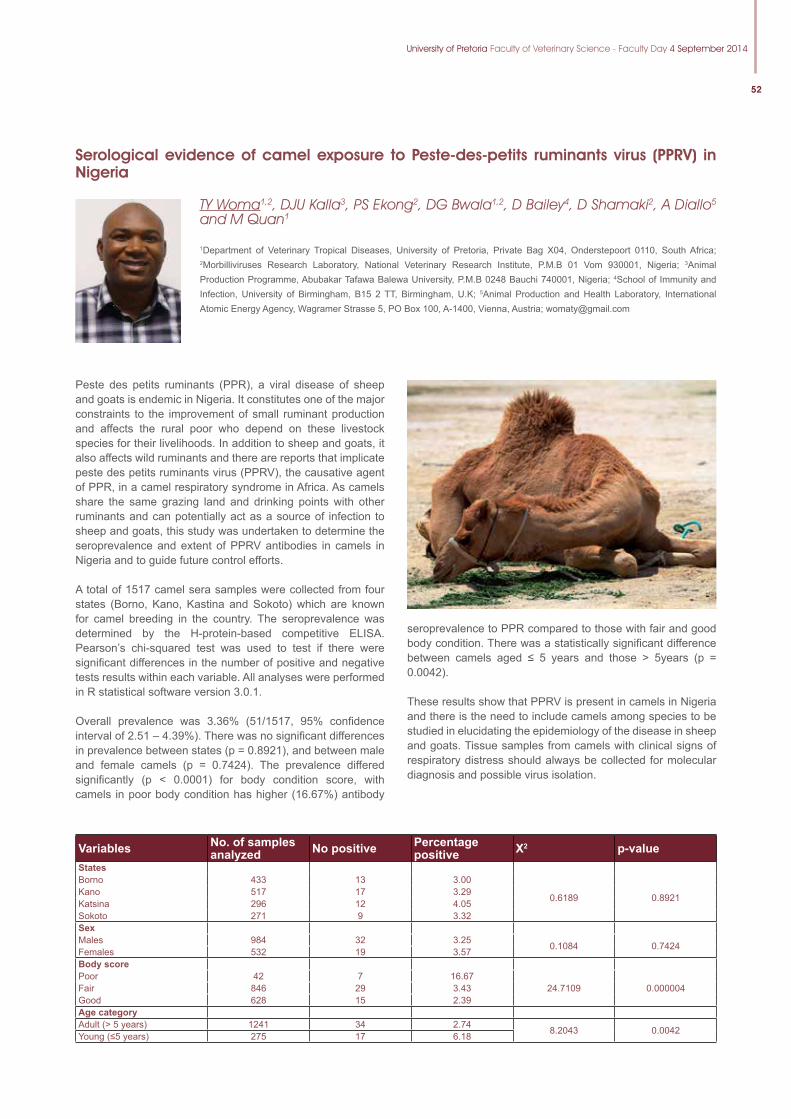
University of Pretoria Faculty of Veterinary Science - Faculty Day 4 September 2014
52
seroprevalence to PPR compared to those with fair and good body condition. There was a statistically significant difference between camels aged ≤ 5 years and those > 5years (p = 0.0042).
These results show that PPRV is present in camels in Nigeria and there is the need to include camels among species to be studied in elucidating the epidemiology of the disease in sheep and goats. Tissue samples from camels with clinical signs of respiratory distress should always be collected for molecular diagnosis and possible virus isolation.
Serological evidence of camel exposure to Peste-des-petits ruminants virus (PPRV) in Nigeria
TY Woma1,2, DJU Kalla3, PS Ekong2, DG Bwala1,2, D Bailey4, D Shamaki2, A Diallo5 and M Quan1
1Department of Veterinary Tropical Diseases, University of Pretoria, Private Bag X04, Onderstepoort 0110, South Africa; 2Morbilliviruses Research Laboratory, National Veterinary Research Institute, P.M.B 01 Vom 930001, Nigeria; 3Animal Production Programme, Abubakar Tafawa Balewa University, P.M.B 0248 Bauchi 740001, Nigeria; 4School of Immunity and Infection, University of Birmingham, B15 2 TT, Birmingham, U.K; 5Animal Production and Health Laboratory, International Atomic Energy Agency, Wagramer Strasse 5, PO Box 100, A-1400, Vienna, Austria; [email protected]
Peste des petits ruminants (PPR), a viral disease of sheep and goats is endemic in Nigeria. It constitutes one of the major constraints to the improvement of small ruminant production and affects the rural poor who depend on these livestock species for their livelihoods. In addition to sheep and goats, it also affects wild ruminants and there are reports that implicate peste des petits ruminants virus (PPRV), the causative agent of PPR, in a camel respiratory syndrome in Africa. As camels share the same grazing land and drinking points with other ruminants and can potentially act as a source of infection to sheep and goats, this study was undertaken to determine the seroprevalence and extent of PPRV antibodies in camels in Nigeria and to guide future control efforts.
A total of 1517 camel sera samples were collected from four states (Borno, Kano, Kastina and Sokoto) which are known for camel breeding in the country. The seroprevalence was determined by the H-protein-based competitive ELISA. Pearson’s chi-squared test was used to test if there were significant differences in the number of positive and negative tests results within each variable. All analyses were performed in R statistical software version 3.0.1.
Overall prevalence was 3.36% (51/1517, 95% confidence interval of 2.51 – 4.39%). There was no significant differences in prevalence between states (p = 0.8921), and between male and female camels (p = 0.7424). The prevalence differed significantly (p < 0.0001) for body condition score, with camels in poor body condition has higher (16.67%) antibody
Variables No. of samples analyzed No positive Percentage
positive X2 p-valueStatesBorno 433 13 3.00
0.6189 0.8921Kano 517 17 3.29Katsina 296 12 4.05Sokoto 271 9 3.32SexMales 984 32 3.25 0.1084 0.7424Females 532 19 3.57Body scorePoor 42 7 16.67
24.7109 0.000004Fair 846 29 3.43Good 628 15 2.39Age categoryAdult (> 5 years) 1241 34 2.74 8.2043 0.0042Young (≤5 years) 275 17 6.18
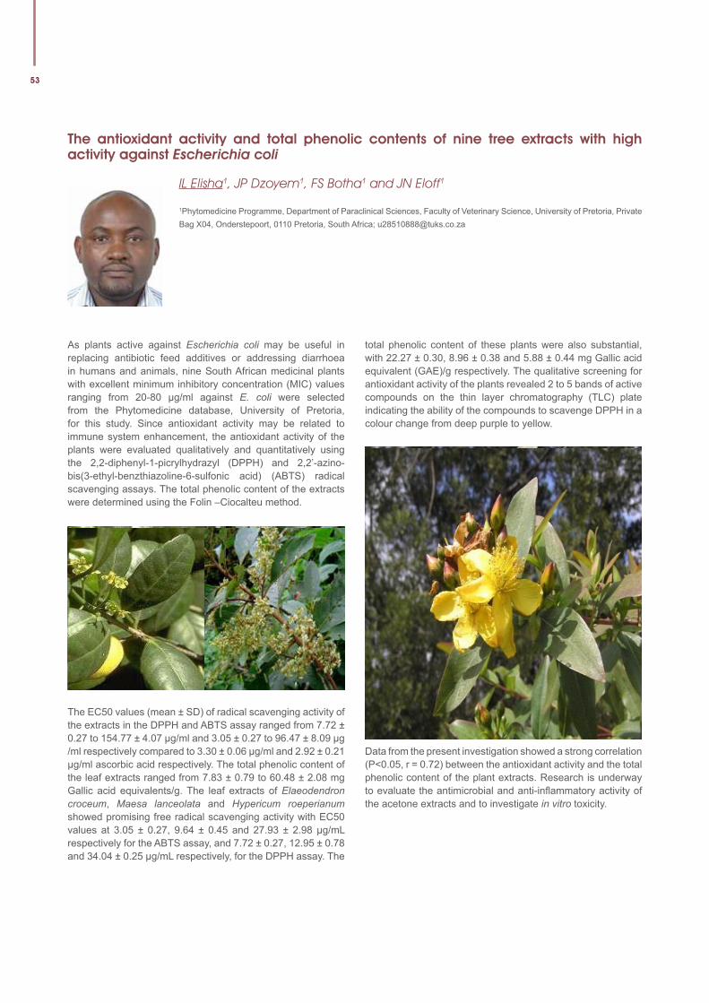
53
The antioxidant activity and total phenolic contents of nine tree extracts with high activity against Escherichia coli
IL Elisha1, JP Dzoyem1, FS Botha1 and JN Eloff1
1Phytomedicine Programme, Department of Paraclinical Sciences, Faculty of Veterinary Science, University of Pretoria, Private Bag X04, Onderstepoort, 0110 Pretoria, South Africa; [email protected]
As plants active against Escherichia coli may be useful in replacing antibiotic feed additives or addressing diarrhoea in humans and animals, nine South African medicinal plants with excellent minimum inhibitory concentration (MIC) values ranging from 20-80 µg/ml against E. coli were selected from the Phytomedicine database, University of Pretoria, for this study. Since antioxidant activity may be related to immune system enhancement, the antioxidant activity of the plants were evaluated qualitatively and quantitatively using the 2,2-diphenyl-1-picrylhydrazyl (DPPH) and 2,2’-azino-bis(3-ethyl-benzthiazoline-6-sulfonic acid) (ABTS) radical scavenging assays. The total phenolic content of the extracts were determined using the Folin –Ciocalteu method.
The EC50 values (mean ± SD) of radical scavenging activity of the extracts in the DPPH and ABTS assay ranged from 7.72 ± 0.27 to 154.77 ± 4.07 µg/ml and 3.05 ± 0.27 to 96.47 ± 8.09 µg /ml respectively compared to 3.30 ± 0.06 µg/ml and 2.92 ± 0.21 µg/ml ascorbic acid respectively. The total phenolic content of the leaf extracts ranged from 7.83 ± 0.79 to 60.48 ± 2.08 mg Gallic acid equivalents/g. The leaf extracts of Elaeodendron croceum, Maesa lanceolata and Hypericum roeperianum showed promising free radical scavenging activity with EC50 values at 3.05 ± 0.27, 9.64 ± 0.45 and 27.93 ± 2.98 µg/mL respectively for the ABTS assay, and 7.72 ± 0.27, 12.95 ± 0.78 and 34.04 ± 0.25 µg/mL respectively, for the DPPH assay. The
total phenolic content of these plants were also substantial, with 22.27 ± 0.30, 8.96 ± 0.38 and 5.88 ± 0.44 mg Gallic acid equivalent (GAE)/g respectively. The qualitative screening for antioxidant activity of the plants revealed 2 to 5 bands of active compounds on the thin layer chromatography (TLC) plate indicating the ability of the compounds to scavenge DPPH in a colour change from deep purple to yellow.
Data from the present investigation showed a strong correlation (P<0.05, r = 0.72) between the antioxidant activity and the total phenolic content of the plant extracts. Research is underway to evaluate the antimicrobial and anti-inflammatory activity of the acetone extracts and to investigate in vitro toxicity.
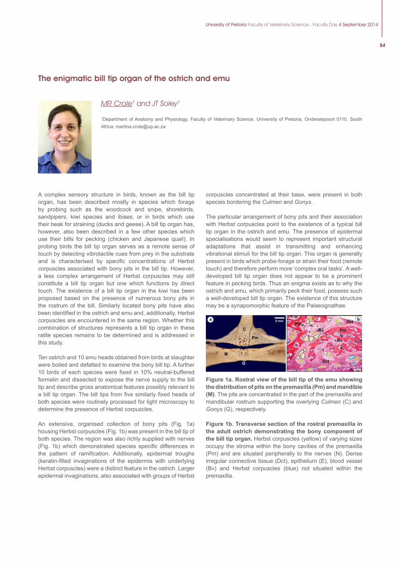
University of Pretoria Faculty of Veterinary Science - Faculty Day 4 September 2014
54
The enigmatic bill tip organ of the ostrich and emu
MR Crole1 and JT Soley1
1Department of Anatomy and Physiology, Faculty of Veterinary Science, University of Pretoria, Onderstepoort 0110, South Africa; [email protected]
A complex sensory structure in birds, known as the bill tip organ, has been described mostly in species which forage by probing such as the woodcock and snipe, shorebirds, sandpipers, kiwi species and ibises, or in birds which use their beak for straining (ducks and geese). A bill tip organ has, however, also been described in a few other species which use their bills for pecking (chicken and Japanese quail). In probing birds the bill tip organ serves as a remote sense of touch by detecting vibrotactile cues from prey in the substrate and is characterised by specific concentrations of Herbst corpuscles associated with bony pits in the bill tip. However, a less complex arrangement of Herbst corpuscles may still constitute a bill tip organ but one which functions by direct touch. The existence of a bill tip organ in the kiwi has been proposed based on the presence of numerous bony pits in the rostrum of the bill. Similarly located bony pits have also been identified in the ostrich and emu and, additionally, Herbst corpuscles are encountered in the same region. Whether this combination of structures represents a bill tip organ in these ratite species remains to be determined and is addressed in this study.
Ten ostrich and 10 emu heads obtained from birds at slaughter were boiled and defatted to examine the bony bill tip. A further 10 birds of each species were fixed in 10% neutral-buffered formalin and dissected to expose the nerve supply to the bill tip and describe gross anatomical features possibly relevant to a bill tip organ. The bill tips from five similarly fixed heads of both species were routinely processed for light microscopy to determine the presence of Herbst corpuscles.
An extensive, organised collection of bony pits (Fig. 1a) housing Herbst corpuscles (Fig. 1b) was present in the bill tip of both species. The region was also richly supplied with nerves (Fig. 1b) which demonstrated species specific differences in the pattern of ramification. Additionally, epidermal troughs (keratin-filled invaginations of the epidermis with underlying Herbst corpuscles) were a distinct feature in the ostrich. Larger epidermal invaginations, also associated with groups of Herbst
corpuscles concentrated at their base, were present in both species bordering the Culmen and Gonys.
The particular arrangement of bony pits and their association with Herbst corpuscles point to the existence of a typical bill tip organ in the ostrich and emu. The presence of epidermal specialisations would seem to represent important structural adaptations that assist in transmitting and enhancing vibrational stimuli for the bill tip organ. This organ is generally present in birds which probe-forage or strain their food (remote touch) and therefore perform more ‘complex oral tasks’. A well-developed bill tip organ does not appear to be a prominent feature in pecking birds. Thus an enigma exists as to why the ostrich and emu, which primarily peck their food, possess such a well-developed bill tip organ. The existence of this structure may be a synapomorphic feature of the Palaeognathae.
Figure1a.Rostralviewofthebilltipoftheemushowingthe distribution of pits on the premaxilla (Pm) and mandible (M). The pits are concentrated in the part of the premaxilla and mandibular rostrum supporting the overlying Culmen (C) and Gonys (G), respectively.
Figure 1b. Transverse section of the rostral premaxilla in the adult ostrich demonstrating the bony component of the bill tip organ. Herbst corpuscles (yellow) of varying sizes occupy the stroma within the bony cavities of the premaxilla (Pm) and are situated peripherally to the nerves (N). Dense irregular connective tissue (Dct), epithelium (E), blood vessel (Bv) and Herbst corpuscles (blue) not situated within the premaxilla.
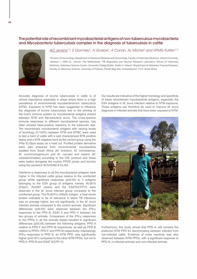
55
The potential role of recombinant mycobacterial antigens of non-tuberculous mycobacteria and Mycobacteria tuberculosis complex in the diagnosis of tuberculosis in cattle
AO Jenkins1,3, E Gormley2, N Gcebe3, A Conan, AL Michel3 and VPMG Rutten1,3
1Division of Immunology, Department of Infectious Diseases and Immunology, Faculty of Veterinary Medicine, Utrecht University, Yalelaan 1, 3584 CL, Utrecht, The Netherlands; 2TB diagnostics and Vaccine Research Laboratory, School Of Veterinary Medicine, Veterinary Science Centre, University College Dublin, Dublin 4, Ireland; 3Department of Veterinary Tropical Diseases, Faculty of Veterinary Science, University of Pretoria, Private Bag X04, Onderstepoort, 0110, South Africa.
Accurate diagnosis of bovine tuberculosis in cattle is of utmost importance especially in areas where there is a high prevalence of environmental mycobacteria/non tuberculous (NTM). Exposure to NTM has been suggested to influence the diagnosis of bovine tuberculosis due to the priming of the host’s immune system by mycobacterial antigens shared between NTM and Mycobacteria bovis. The cross-reactive immune responses to different mycobacterial species, has often showed false-positive reactions to the tuberculin test. Ten recombinant mycobacterial antigens with varying levels of homology (0-100%) between NTM and MTBC were used to test a herd of cattle with a well characterized BTB positive status and a BTB negative herd as the control group using the IFNy ELISpot assay as a read out. Purified protein derivative were also prepared from environmental mycobacteria isolated from South Africa (M. fortuitum, M. moriokaense, M. nonchromogenicum and M. vaccae) and Ireland (M. vanbalenii/vallee) according to the OIE protocol and these were tested alongside the routine PPDS (avian and bovine) using the sandwich BOVIGAM ® ELISA.
Interferon-y responses to all the mycobacterial antigens were higher in the infected cattle group relative to the uninfected group whilst significant responses (p≤0.05) to 3 antigens belonging to the ESX group of antigens namely, Rv3615 (ESpC), Rv0287 (esxG) and the ESAT6/CFP10 were observed in the M. bovis infected group compared to the uninfected group. The Rv2031c (HSpX) antigen, a heat shock protein indicated to be of relevance in latent TB infections was on average higher, but not significantly, in the M. bovis infected animals compared to the control animals. Significant differences (p≤0.05) were observed between the IFN-y responses to the PPD B, ESAT 6 and PPD V between the two groups of animals. Comparison of the IFN-y responses to the PPDs in all the animals tested resulted in significant differences (p≤0.05) between the following antigens; PPD A relative to PPD F and PPD M respectively, as well as PPD B relative to PPDV, PPD F and PPD M respectively. Interestingly IFN-y responses to PPD N, an NTM PPD, was significantly higher (p≤0.001) compared to the other NTM PPDs, but not to PPD A, PPD B and ESAT 6/CFP-10.
Our results are indicative of the higher homology and specificity of these recombinant mycobacterial antigens, especially the ESX antigens in M. bovis infection relative to NTM exposure. These antigens can therefore be used to improve M. bovis diagnosis in infected animals that have been exposed to NTM.
Furthermore, this study shows that PPD A, still remains the preferred NTM PPD for discriminating between infected from non-infected cattle. Evidence of cross reactivity was also observed between NTM PPDs, with a significant response to PPD N, in infected animals and non-infected animals.
University of Pretoria Faculty of Veterinary Science - Faculty Day 4 September 2014
56
The power of poo – Non-invasive measures of reproduction and stress in wildlife
A Ganswindt1,2,3 and TE Wolf1
1Endocrine Research Laboratory, Department of Anatomy and Physiology, Faculty of Veterinary Science, University of Pretoria, Onderstepoort 0110, South Africa; 2Mammal Research Institute, Department of Zoology and Entomology, Faculty of Natural and Agricultural Sciences, University of Pretoria, Pretoria 0028, South Africa; 3National Zoological Gardens of South Africa, Pretoria, 0001 South Africa; [email protected]
Behavioural endocrinology is the study of the endocrine system in relation to behaviour. The involved hormones are chemical messengers secreted by specific glands, travelling through the blood system to target sites where they unfold their effect, i.e. regulating the physiology and behaviour of an individual. It is a bidirectional interaction as hormones can affect behaviour, and behaviour can influence hormone concentrations. Therefore hormone analysis is an ideal tool for monitoring reproductive function and responses to stressors in mammals, birds, reptiles, amphibians, fish, and even invertebrates. Although hormones can be measured in various biological matrices, non-invasive methods (particularly the use of faeces as hormone matrix) have gained popularity over the past three decades as a more practical approach for assessing ovarian, testicular and, more recently, adrenocortical activity in especially intractable free-roaming animals. However, respective assays need to be carefully validated in terms of applicability for the species-specific hormone matrix of interest to ensure a reliable quantification of respective hormone concentrations. With a reliable test system in place, key management issues can be addressed in captive populations; like the avoidance of stress (and of situations/procedures likely to cause it), as well as optimizing breeding efforts either naturally or by assisted/artificial means. On the other hand, a reliable non-invasive
approach, like hormone measurement from faeces, allows field researchers to link the endocrine status of animals to behaviour or other life-history traits without interfering with the natural behaviour of the animals due to capture and restraint for invasive sampling.
Using vivid examples for monitoring male and female reproductive activity (e.g. Musth related changes in androgen concentrations in Asian elephants or progestagens pregnancy profiles in White rhinoceros) and the level of stress experienced by animals (e.g. elevated glucocorticoid levels in physically injured African elephants or individually housed African buffalo), this presentation will highlight the value and inter-disciplinary approach of behavioural endocrine research.
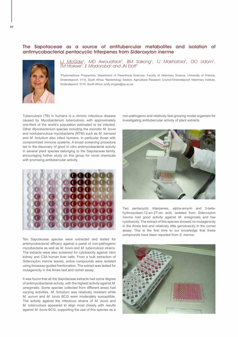
57
The Sapotaceae as a source of antitubercular metabolites and isolation of antimycobacterial pentacyclic triterpenes from Sideroxylon inerme
LJ McGaw1, MD Awouafack1, BM Sakong1, TJ Makhafola1, OO Udom1, TM Hlokwe2, E Madoroba2 and JN Eloff1
1Phytomedicine Programme, Department of Paraclinical Sciences, Faculty of Veterinary Science, University of Pretoria, Onderstepoort, 0110, South Africa; 2Bacteriology Section, Agricultural Research Council-Onderstepoort Veterinary Institute, Onderstepoort, 0110, South Africa; [email protected]
Tuberculosis (TB) in humans is a chronic infectious disease caused by Mycobacterium tuberculosis, with approximately one-third of the world’s population estimated to be infected. Other Mycobacterium species including the zoonotic M. bovis and nontuberculous mycobacteria (NTM) such as M. kansasii and M. fortuitum also infect humans, in particular those with compromised immune systems. A broad screening procedure led to the discovery of good in vitro antimycobacterial activity in several plant species belonging to the Sapotaceae family, encouraging further study on this group for novel chemicals with promising antitubercular activity.
Ten Sapotaceae species were extracted and tested for antimycobacterial efficacy against a panel of non-pathogenic mycobacteria as well as M. bovis and M. tuberculosis strains. The extracts were also screened for cytotoxicity against Vero kidney and C3A human liver cells. From a bulk extraction of Sideroxylon inerme leaves, active compounds were isolated using bioassay-guided fractionation. The extract was tested for mutagenicity in the Ames test and comet assay.
It was found that all the Sapotaceae extracts had some degree of antimycobacterial activity, with the highest activity against M. smegmatis. Some species collected from different areas had varying activities. M. fortuitum was relatively resistant while M. aurum and M. bovis BCG were moderately susceptible. The activity against the infectious strains of M. bovis and M. tuberculosis appeared to align most closely with results against M. bovis BCG, supporting the use of this species as a
non-pathogenic and relatively fast-growing model organism for investigating antitubercular activity of plant extracts.
Two pentacyclic triterpenes, alpha-amyrin and 3-beta-hydroxyolean-12-en-27-oic acid, isolated from Sideroxylon inerme had good activity against M. smegmatis and low cytotoxicity. The extract of this species showed no mutagenicity in the Ames test and relatively little genotoxicity in the comet assay. This is the first time to our knowledge that these compounds have been reported from S. inerme.
University of Pretoria Faculty of Veterinary Science - Faculty Day 4 September 2014
58Scientific, peer-refereed articles, published in 2013, of which a member or student of the Faculty of Veterinary Science, University of Pretoria, was an author. (alphabetical according to the name of the first author)
1. Abernethy DA, Upton P, Higgins I, McGrath G, Goodchild A, Rolfe S, Broughan J, Downs S, Clifton-Hadley, R, Menzies F, de la Rua-Domenech R, Blissitt M, Duigan A, More S (2013) Bovine tuberculosis trends in the UK and the Republic of Ireland, 1995-2010. Veterinary Record 172:1-15
2. Abolnik C, Fehrsen J, Olivier A, Van Wyngaardt W, Fosgate GT, Ellis C (2013) Serological investigation of highly pathogenic avian influenza H5N2 in ostriches (Struthio camelus). Avian Pathology 42:206-214
3. Abu Samra N, Jori F, Xiao L, Rikhotso O, Thompson PN (2013) Molecular characterization of Cryptosporidium species at the wildlife/livestock interface of the Kruger National Park, South Africa. Comparative Immunology, Microbiology and Infectious Diseases 36:295-302
4. Abu Samra N, Thompson PN, Jori F, Frean J, Poonsamy B, du Plessis D, Mogoye B, Xiao L (2013) Genetic characterization of Cryptosporidium spp. in diarrhoeic children from four provinces in South Africa. Zoonoses and Public Health 60:1-6
5. Adakal H, Biguezoton A, Zoungrana S, Courtin F, De Clercq E, Madder M (2013) Alarming spread of the Asian cattle tick Rhipicephalus microplus in West Africa—another three countries are affected: Burkina Faso, Mali and Togo. Experimental and Applied Acarology 61:383-386
6. Adam Y, Cecchi G, Kgori P, Marcotty TLBM, Mahama C, Abavana M, Anderson B, Paone M, Mattioli R, Bouyer J (2013) The Sequential Aerosol Technique: A Major Component in an Integrated Strategy of Intervention against Riverine Tsetse in Ghana. PLoS Neglected Tropical Diseases 7:1-9
7. Adamu M, Naidoo V, Eloff JN (2013) Efficacy and toxicity of thirteen plant leaf acetone extracts used in ethnoveterinary medicine in South Africa on egg hatching and larval development of Haemonchus contortus. BMC Veterinary Research 9:online
8. Ahmed AS, McGaw LJ, Eloff JN (2013) Evaluation of pharmacological activities, cytotoxicity and phenolic composition of four Maytenus species used in southern African traditional medicine to treat intestinal infections and diarrhoeal diseases. BMC Complementary and Alternative Medicine 13:online
9. Ajibola A, Chamunorwa JP, Erlwanger K (2013) Comparative effect of cane syrup and natural honey on abdominal viscera of growing male and female rats. Indian Journal of Experimental Biology 51:303-312
10. Ali S, Hamed A, Soltan M, Hegazy U, Elgorashi EE, El-Garf I, Hussein A (2013) In-vitro evaluation of selected Egyptian traditional herbal medicines for treatment of alzheimer disease. BMC Complementary and Alternative Medicine 13:online
11. Angkawanish T, Morar D, van Kooten P, Bontekoning I, Schreuder J, Maas M, Wajjwalku W, Sirimalaisuwan A, Michel AL, Tijhaar E, Rutten VPM (2013) The Elephant Interferon Gamma Assay: A Contribution to Diagnosis of Tuberculosis in Elephants. Transboundary and Emerging Diseases 60:53-59
12. Apanaskevich D, Horak IG, Mulumba-Mfumu L (2013) A new species of Rhipicephalus (Acari: Ixodidae), a parasite of red river hogs and domestic pigs in the Democratic Republic of Congo. Journal of Medical Entomology 50:479-484
13. Apanaskevich D, Walker J, Heyne H, Bezuidenhout J, Horak IG (2013) First description of the immature stages and redescription of the adults of Cosmiomma hippopotamensis (Acari: Ixodidae) with notes on its bionomics. Journal of Medical Entomology 50:709-722
14. Aranzamendi C, de Bruin A, Kuiper R, Boog C, van Eden W, Rutten VPM, Pinelli E (2013) Protection against allergic airway inflammation during the chronic and acute phases of Trichinella spiralis infection. Clinical and Experimental Allergy 43:103-115
15. Arnold K, Boxall A, Brown A, Cuthbert R, Gaw S, Hutchinson T, Jobling S, Madden J, Metcalfe C, Naidoo V, Shore R, Smits J, Taggart M, Thompson H (2013) Assessing the exposure risk and impacts of pharmaceuticals in the environment of individuals and ecosystems. Biology Letters 9:1-4
16. Augustyn W, Du Plooy W, Botha B, van Wilpe E (2013) Infestation of Mangifera indica by the mango gall fly, Procontarinia matteiana, (Kieffer & Cecconi)(Diptera: Cecidomyiidae). African Entomology 21:79-88
17. Awouafack MD, McGaw LJ, Gottfried S, Mbouangouere R, Tane P, Spiteller M, Eloff JN (2013) Antimicrobial activity and cytotoxicity of the ethanol extract, fractions and eight compounds isolated from Eriosema robustum (Fabaceae). BMC Complementary and Alternative Medicine 13:online
18. Awouafack MD, Tane P, Eloff JN (2013) Two new antioxidant flavones from the twigs of Eriosema robustum (Fabaceae). Phytochemistry Letters 6:62-66
19. Bai Y, Malania L, Castillo D, Moran D, Boonmar S, Chanlun A, Suksawat F, Maruyama S, Knobel DL, Kosoy M (2013) Global Distribution of Bartonella Infections in Domestic Bovine and Characterization of Bartonella bovis Strains Using Multi-Locus Sequence Typing. PLoS ONE 8:1-8
20. Bakhoum M, Fall M, Fall A, Bellis G, Gottlieb Y, Labuschagne K, Venter GJ, Diop M, Mall I, Seck M, Allène X, Diarra M, Gardès L, Bouyer J, Balenghien T, , (2013) First Record of Culicoides oxystoma Kieffer and Diversity of Species within the Schultzei Group of Culicoides Latreille (Diptera: Ceratopogonidae) Biting Midges in Senegal. PLoS ONE 8:1-8
21. Bath GF, Leask R, Pettey KP, Coetzee DJ (2013) Abortions in sheep associated with Arcobacter skirrowii infection. Journal of the South African Veterinary Association 84:1-4
22. Bhattarai B, Fosgate GT, Osterstock J, Fossler C, Park S, Roussel A (2013) Comparison of calf weaning weight and associated economic variables between beef cows with and without serum antibodies against or isolation from feces of Mycobacterium avium subsp paratuberculosis. Journal of the American Veterinary Medical Association 243:1609-1615
23. Bhattarai B, Fosgate GT, Osterstock J, Fossler C, Park S, Roussel A (2013) Perceptions of veterinarians in bovine practice and producers with beef cow-calf operations enrolled in the US Voluntary Bovine Johne’s Disease Control Program concerning economic losses associated with Johne’s disease. Preventive Veterinary Medicine 112:330-337
24. Blunt C, Jansen van Vuuren M, Picard JA (2013) Antimicrobial susceptibility profiles of Staphylococcus intermedius isolates from clinical cases of canine pyoderma in South Africa. Journal of the South African Veterinary Association 84:1-6
25. Bocchino C, Burroughs REJ (2013) Synergies across the natural resources management fields in Southern Africa: Disaster Risk Reduction and One Health. Jamba: Journal of Disaster Risk Studies 5:1-10
26. Borchers A, Wilkins P, Marsh P, Axon J, Read J, Castagnetti C, Pantaleon L, Clark C, Qura’n L, Belgrave R, Schwarzwald C, Levy M, Bedenice D, Saulez MN, Boston R (2013) Sequential L-lactate concentration in hospitalised equine neonates: A prospective multicentre study. Equine Veterinary Journal 45:2-7
27. Boshra H, Truong T, Nfon C, Gerdts V, Tikoo S, Babiuk L, Kara P, Mather A, Wallace DB, Babiuk S (2013) Capripoxvirus-vectored vaccines against livestock diseases in Africa. Antiviral Research 98:217-227
28. Bosman A, Oosthuizen MC, Venter EH, Steyl JCA, Gous T, Penzhorn BL (2013) Babesia lengau associated with cerebral and haemolytic babesiosis in two domestic cats. Parasites & Vectors 6:1-6
29. Bostrom B, Xenoulis P, Newman S, Pool R, Fosgate GT, Steiner J (2013) Chronic pancreatitis in dogs: A retrospective study of clinical, clinicopathological findings in 61 cases. Veterinary Journal 195: 73-79
5930. Botha CJ (2013) Krimpsiekte in South Africa: Historical perspectives.
Journal of the South African Veterinary Association 84:online
31. Botha CJ, Coetser H, Schultz A, Labuschagne L, van der Merwe D (2013) Spatial variation of epoxyscillirosidine concentrations in Moraea pallida (yellow tulp) in South Africa. Onderstepoort Journal of Veterinary Research 80:online
32. Brettschneider H, Voster J, Lane E, van Wilpe E, Biden P, Dalton D, Kotze A (2013) Molecular detection of Yaba monkey tumour virus from a vervet monkey. Journal of the South African Veterinary Association 84:1-5
33. Breytenbach A, Lourens A, Marsh S (2013) The role of the Jotello F. Soga Library in the digital preservation of South African veterinary history. Journal of the South African Veterinary Association 84:1-7
34. Bronsvoort B, Thumbi S, Poole E, Kiara H, Auguet O, Handel I, Jennings A, Van Wyk (Conradie) I, Ndila M, Toye P, Hanotte O, Coetzer JAW, Woolhouse M (2013) Design and descriptive epidemiology of the Infectious Diseases of East African Livestock (IDEAL) project, a longitudinal calf cohort study in western Kenya. BMC Veterinary Research 9:1-22
35. Cangi N, Horak IG, Apanaskevich D, Matthee S, Neves L, Estrada-Peña A, Matthee C (2013) The Infl uence of Interspecifi c Competition and Host Preference on the Phylogeography of Two African Ixodid Tick Species. PLoS ONE 8:1-12
36. Carstens A, Kirberger RM, Dahlberg L, Prozesky L, Fletcher L, Lammentausta E (2013) Validation of delayed gadolinium-enhanced magnetic resonance imaging of cartilage and T2 mapping for quantifying distal metacarpus/metatarsus cartilage thickness in thoroughbred racehorses. Veterinary Radiology & Ultrasound 54:139-148
37. Carstens A, Kirberger RM, Velleman M, Dahlberg L, Fletcher L, Lammentausta E (2013) Feasibility for mapping cartilage T1 relaxation times in the distal metacarpus3/metatarsus3 of thoroughbred racehorses using delayed gadolinium-enhanced magnetic resonance imaging of cartilage (dGemric): normal cadaver study. Veterinary Radiology & Ultrasound 54:365-372
38. Cassel N, Carstens A, Becker P (2013) The comparison of bolus tracking and test bolus techniques for computed tomography thoracic angiography in healthy beagles. Journal of the South African Veterinary Association 84:1-9
39. Cenci-Goga B, Ciampelli A, Sechi P, Veronesi F, Moretta I, Cambiotti V, Thompson PN (2013) Seroprevalence and risk factors for Toxoplasma gondii in sheep in Grosseto district, Tuscany, Italy. BMC Veterinary Research 9:1-8
40. Chaisi ME, Collins NE, Potgieter F, Oosthuizen MC (2013) Sequence variation identifi ed in the 18S rRNA gene of Theileria mutans and Theileria velifera from the African buffalo (Syncerus caffer). Veterinary Parasitology 191:132-137
41. Chaisi ME, Janssens M, Vermeiren L, Oosthuizen MC, Collins NE, Geysen D (2013) Evaluation of a Real-Time PCR Test for the Detection and Discrimination of Theileria Species in the African Buffalo (Syncerus caffer). PLoS ONE 8:1-12
42. Chaka H, Goutard F, Gil P, Abolnik C, Servan de Almeida R, Bisschop SPR, Thompson PN (2013) Serological and molecular investigation of Newcastle disease in household chicken fl ocks and associated markets in Eastern Shewa zone, Ethiopia. Tropical Animal Health and Production 45:705-714
43. Chaka H, Goutard F, Roger F, Bisschop SPR, Thompson PN (2013) Household-level risk factors for Newcastle disease seropositivity and incidence of Newcastle disease virus exposure in backyard chicken fl ocks in Eastern Shewa zone, Ethiopia. Preventive Veterinary Medicine 109:312-320
44. Chapwanya A, Meade K, Doherty M, Callanan J, O’Farrelly C (2013) Endometrial epithelial cells are potent producers of tracheal antimicrobial peptide and serum amyloid A3 gene expression in response to E.coli stimulation. Veterinary Immunology and Immunopathology 151:157-162
45. Chitanga S, Namangala B, De Deken R, Marcotty TLBM (2013) Shifting from wild to domestic hosts: The effect on the transmission of Trypanosoma congolense to tsetse fl ies. Acta Tropica 125:32-36
46. Christensen M, Langhorn R, Goddard A, Andreasen E, Moldal E, Tvarijonaviciute A, Kirpenteijn J, Jakobsen S, Persson F, Kjelgaard-Hansen M (2013) Canine Serum Amyloid A (SAA) Measured by Automated Latex Agglutination Turbidimetry Is Useful for Routine Sensitve and Specifi c Detection of Systemic Infl ammation in a General Clinical Setting. Journal of Veterinary Medical Science 75:459-466
47. Claerebout E, Losson B, Cochez C, Casaert S, Dalemans A, De Cat A, Madder M, Saegerman C, Heyman P, Lempereur L (2013) Ticks and associated pathogens collected from dogs and cats in Belgium. Parasites & Vectors 6:1-9
48. Codron J, Kirkman K, Duffy K, Sponheimer M, Lee-Thorp J, Ganswindt A, Clauss M, Codron D (2013) Stable isotope turnover and variability in tail hairs of captive and free-ranging African elephants (Loxodonta africana) reveal dietary niche differences within populations. Canadian Journal of Zoology 91:124-134
49. Coetzee P, Stokstad M, Myrmel M, Mutowembwa P, Loken T, Venter EH, Jansen van Vuuren M (2013) Transplacental infection in goats experimentally infected with a European strain of bluetongue virus serotype 8. Veterinary Journal 197:335-341
50. Côté E, Schwarz L, Sithole F (2013) Thoracic radiographic fi ndings for dogs with cardiac tamponade attributable to pericardial effusion. Journal of the American Veterinary Medical Association 243:232-235
51. Crafford JE, Lourens CW, Gardner I, MacLachlan N, Guthrie AJ (2013) Passive transfer and rate of decay of maternal antibody against African horse sickness virus in South African Thoroughbred foals. Equine Veterinary Journal 45:604-607
52. Crole MR, Soley JT, Clift SJ (2013) Incidental Mycobacterium-induced granulomatous infl ammation of the follicular pharyngeal tonsils in a South African farmed ostrich (Struthio camelus). Journal of the South African Veterinary Association 84:1-9
53. Dabrowski J, Oberholster PJ, Dabrowshi J, Le Brasseur J, Gieskes J (2013) Chemical characteristics and limnology of Loskop Dam on the Olifants river (South Africa), in light of recent fi sh and crocodile mortalities. Water SA 39:675-686
54. De Garine-Wichatitsky M, Caron A, Kock R, Tschopp R, Munyeme M, Hofmeyr M, Michel AL (2013) A review of bovine tuberculosis at the wildlife-livestock-human interface in sub-Saharan Africa. Epidemiology and Infection 141:1342-1356
Elanco Helpline: 0861 777 735REGISTRATION HOLDER: ELANCO ANIMAL HEALTHA division of Eli Lilly (SA)(Pty)Ltd. (Co. Reg. No.: 1957/000371/07)Private Bag X119, BRYANSTON, 2021, Republic of South AfricaTel.: (012) 657-6200 Fax: (012) 657-6216
COMFORTIS® CHEWABLE TABLETS 140/270/560/810/1620mg Reg. No’s. G4011/G4012/G4013/G4014/G4015 Act 36/19471. Elanco study T9C060330, data on file. 2. Elanco study T9C010313, data on file. 3. Elanco study T9CAL0339, data on file. 4. Elanco studies T9CAL0401, T9CAL0403, data on file.Elanco and Comfortis are trademarks owned or licensed by Eli Lilly and Company, its subsidiaries or affiliates. ZACACCMF00007 ©2013 Elanco, a division of Eli Lilly and Company Limited
Fast-actingmonth-long flea protectionin a chewable tablet
Elanco_Comfortis A7_03.indd 1 2014/08/11 1:47 PM
University of Pretoria Faculty of Veterinary Science - Faculty Day 4 September 2014
6055. De Ruijter-Villani M, van Boxtel P, Stout TAE (2013) Fibroblast growth
factor-2 expression in the preimplantation equine conceptus and endometrium of pregnant and cyclic mares. Theriogenology 80:979-989
56. Delsink A, Kirkpatrick J, van Altena J, Bertschinger HJ, Ferreira S, Slotow R (2013) Lack of spatial and behavioral responses to immunocontraception application in African elephants (Loxodonta africana). Journal of Zoo and Wildlife Medicine 44:52-74
57. Dogonyaro BB, Bosman A, Sibeko-Matjila KP, Venter EH, Jansen van Vuuren M (2013) Genetic analysis of the VP2-encoding gene of canine parvovirus strains from Africa. Veterinary Microbiology 165:460-465
58. Donnellan C, Page PC, Nurton JP, van den Berg JS, Guthrie AJ (2013) Comparison of glycopyrrolate and atropine in ameliorating the adverse effects of imidocarb dipropionate in horses. Equine Veterinary Journal 45:625-629
59. Doving Osvik R, Sperstad S, Breines E, Hareide E, Godfroid JXL, Zhou Z, Ren P, Geoghegan C, Holzapfel W, Ringo E (2013) Bacterial diversity of aMasi, a South African fermented milk product, determined by clone library and denaturing gradient gel electrophoresis analysis. African Journal of Microbiology Research 7:4146-4158
60. Du Plessis L, Soley JT (2013) A novel transient structure with phylogenetic implications found in ratite spermatids. Evolutionary Biology 13:1-6
61. Du Toit CJL, Meissner HH, van Niekerk WA (2013) Direct greenhouse gas emissions of the game industry in South Africa. South African Journal of Animal Science 43:376-393
62. Du Toit CJL, Meissner HH, van Niekerk WA (2013) Direct methane and nitrous oxide emissions of South African dairy and beef cattle. South African Journal of Animal Science 43:320-339
63. Du Toit CJL, van Niekerk WA, Meissner HH (2013) Direct greenhouse gas emissions of the South African small stock sectors. South African Journal of Animal Science 43:340-361
64. Du Toit CJL, van Niekerk WA, Meissner HH (2013) Direct methane and nitrous oxide emissions of monogastric livestock in South Africa. South African Journal of Animal Science 43:362-375
65. Dubey J, van Wilpe E, Blignaut DJC, Schares G, Williams JH (2013) Development of early tissue cysts and associated pathology of Besnoitia besnoiti in a naturally infected bull (Bos taurus) from South Africa. Journal of Parasitology 99:459-466
66. Dzikiti TB (2013) Intravenous anaesthesia in goats: A review. Journal of the South African Veterinary Association 84:1-8
67. Elisha I, Elgorashi EE, Hussein A, Duncan G, Eloff JN (2013) Acetylcholinesterase inhibitory effects of the bulb of Ammocharis coranica (Amaryllidaceae) and its active constituent lycorine. South African Journal of Botany 85:44-47
68. Esposito G, Medina V, Schneider A, Gilbert R, Butler W (2013) Effect of dietary conjugated linoleic acid (CLA) on the metabolism and reproduction of dairy cows. South African Journal of Animal Science 43:S33-S37
69. Estrada-Pena A, Farkas R, Jaenson T, Koenen F, Madder M, Pascucci L, Salman M, Tarres-Call J, Jongejan F (2013) Association of environmental traits with the geographic ranges of ticks (Acari: Ixodidae) of medical and veterinary importance in the western Palearctic. A digital data set. Experimental and Applied Acarology 59:351-366
70. Fafetine J, Domingos A, Antunes S, Esteves A, Paweska J, Coetzer JAW, Rutten VPM, Neves L (2013) Generation and Characterization of Monoclonal Antibodies Against Rift Valley Fever Virus Nucleoprotein. Transboundary and Emerging Diseases 60:24-30
71. Fafetine J, Neves L, Thompson PN, Paweska J, Rutten VPM, Coetzer JAW (2013) Serological Evidence of Rift Valley Fever Virus Circulation in Sheep and Goats in Zambézia Province, Mozambique. PLoS Neglected Tropical Diseases 7:1-8
72. Fasina FO, Connell D, Talabi O, Lazarus DD, Adeleke G, Olusanya T, Hernandez J (2013) Foot-and-mouth disease virus strains and examination of exposure factors associated with seropositivity of cattle herds in Nigeria during 2007-2009. Preventive Veterinary Medicine 109:334-342
73. Fasina FO, Olaokun OO, Oladipo O, Fasina M, Makinde A, Heath L, Bastos DS (2013) Phytochemical analysis and in-vitro anti-African swine fever virus activity of extracts and fractions of Ancistrocladus uncinatus, Hutch and Dalziel (Ancistrocladaceae). BMC Veterinary Research 9:1-11
74. Fernandez-Pinero J, Gallardo C, Elizalde M, Robles A, Gomez C, Bishop R, Heath L, Couacy-Hymman E, Fasina FO, Pelayo V, Soler A, Arias M (2013) Molecular diagnosis of African Swine Fever by a new real-time PCR using universal probe library. Transboundary and Emerging Diseases 60:48-58
75. Fosgate GT, Petzer I, Karzis J (2013) Sensitivity and specificity of a hand-held milk electrical conductivity meter compared to the California mastitis test for mastitis in dairy cattle. Veterinary Journal 196:98-102
76. Fourie J, Crafford D, Horak IG, Stanneck D (2013) Prophylactic Treatment of Flea-Infested Dogs with an Imidacloprid/Flumethrin Collar (Seresto®, Bayer) to Preempt Infection with Dipylidium caninum. Parasitology Research 112:33-46
77. Fourie J, Liebenberg J, Nyangiwe N, Austin C, Horak IG, Bhushan C (2013) The effects of a pour-on formulation of fluazuron 2.5% and flumethrin 1% on populations of Rhipicephalus decoloratus and Rhipicephalus microplus both on and off bovine (Bosmara Breed) hosts. Parasitology Research 112:67-79
78. Fourie J, Luus H, Stanneck D, Jongejan F (2013) The efficacy of Advantix® to prevent transmission of Ehrlichia canis to dogs by Rhipicephalus sanguineus ticks. Parasite 20:1-8
79. Fourie J, Olagnier C, Beugnet F, Luus H, Jongejan F (2013) Prevention of transmission of Ehrlichia canis by Rhipicephalus sanguineus ticks to dogs treated with a combination of fipronil, amitraz and (S)-methoprene (CERTIFECT® ). Veterinary Parasitology 193:223-228
80. Fourie J, Stanneck D, Jongejan F (2013) Prevention of transmission of Babesia canis by Dermacentor reticulatus ticks to dogs treated with an imidacloprid/flumethrin collar. Veterinary Parasitology 192:273-278
81. Fourie J, Stanneck D, Luus H, Beugnet F, Wijnveld M, Jongejan F (2013) Transmission of Ehrlichia canis by Rhipicephalus sanguineus ticks feeding on dogs and on artificial membranes. Veterinary Parasitology 197:595-603
82. Gallardo C, Soler A, Nieto R, Carrascosa A, De Mia G, Bishop R, Martins C, Fasina FO, Couacy-Hymman E, Heath L, Pelayo V, Martin E, Simon A, Martin R, Arias M, , , (2013) Comparative evaluation of novel African swine fever virus (ASF) antibody detection techniques derived from specific ASF viral genotypes with the OIE internationally prescribed serological tests. Veterinary Microbiology 162:32-43
83. Gcebe N, Rutten VPM, Gey van Pittius N, Michel AL (2013) Prevalence and Distribution of Non-Tuberculous Mycobacteria (NTM) in Cattle, African Buffaloes (Syncerus caffer) and their Environments in South Africa. Transboundary and Emerging Diseases 60:74-84
84. Genthe B, Le Roux W, Schachtschneider K, Oberholster PJ, Aneck-Hahn NH, Chamier J (2013) Health risk implications from simultaneous exposure to multiple environmental contaminants. Ecotoxicology and Environmental Safety 93:171-179
85. Ghosal R, Ganswindt A, Seshagiri P, Sukumar R (2013) Endocrine correlates of musth in free-ranging Asian elephants (Elephas maximus) determined by non-invasive faecal steroid hormone metabolite measurements. PLoS ONE 8:1-5
86. Goddard A, Wiinberg B, Schoeman JP, Kristensen A, Kjelgaard-Hansen M (2013) Mortality in virulent canine babesiosis is associated with a consumptive coagulopathy. Veterinary Journal 196:213-217
87. Godfroid JXL, Dahouk S, Pappas G, Roth F, Matope G, Muma J, Marcotty TLBM, Pfeiffer D, Skjerve E (2013) A “One Health” surveillance and control of brucellosis in developing countries: Moving away from improvisation. Comparative Immunology, Microbiology and Infectious Diseases 36:241-248
88. Godfroid JXL, Garin-Bastuji B, Saegerman C, Blasco J (2013) Brucellosis in terrestrial wildlife. Revue Scientifique et Technique - Office International des Epizooties 32:27-42
89. Gordon S, Bolwell C, Rogers C, Guthrie AJ, Magunda F, Hove P (2013) Descriptive epidemiology of African horse sickness in Zimbabwe. Onderstepoort Journal of Veterinary Research 80:1-5
6190. Grewar J, Weyer CT, Guthrie AJ, Koen P, Davey S, Quan M, Visser D,
Russouw E, Bührmann G (2013) The 2011 outbreak of African horse sickness in the African horse sickness controlled area in South Africa. Journal of the South African Veterinary Association 84:1-7
91. Guthrie AJ, Maclachlan NJ, Joone C, Lourens CW, Weyer CT, Quan M, Monyai MS, Gardner I (2013) Diagnostic accuracy of a duplex real-time reverse transcription quantitative PCR assay for detection of African horse sickness virus. Journal of Virology 189:30-35
92. Halliday J, Knobel DL, Allan K, Bronsvoort B, Handel I, Agwanda B, Cutler S, Olack B, Ahmed A, Hartskeerl R, Njenga M, Cleaveland S, Breiman R (2013) Urban Leptospirosis in Africa: A Cross-Sectional Survey of Leptospira Infection in Rodents in the Kibera Urban Settlement, Nairobi, Kenya. American Journal of Tropical Medicine and Hygiene 89:1095-1102
93. Harper CK, Vermeulen G, Clarke AB, De Wet J, Guthrie AJ (2013) Extraction of nuclear DNA from rhinoceros horn and characterization of DNA profiling systems for white (Ceratotherium simum) and black (Diceros bicornis) rhinoceros. Forensic Science International - Genetics 7:428-433
94. Hartman MJ, Groenewald HB, Kirberger RM (2013) Morphology of the female reproductive organs of the African lion (Panthera leo). Acta Zoologica 94:437-446
95. Hartman MJ, Monnet E, Kirberger RM, Venter LJ, Bester L, Schulman ML, Serfontein T, Pansegrouw (Fourie) (Fourie), Schoeman JP (2013) Laparoscopic Sterilization of the African Lioness (Panthera leo). Veterinary Surgery 42:559-564
96. Hayashida K, Abe T, Weir W, Nakao R, Ito K, Kajino K, Suzuki Y, Jongejan F, Geysen D, Sugimoto C (2013) Whole-Genome Sequencing of Theileria parva Strains Provides Insight into Parasite Migration and Diversification in the African Continent. DNA Research 20:209-220
97. Hergert M, Nel LH (2013) Dog Bite Histories and Response to Incidents in Canine Rabies-Enzootic KwaZulu-Natal, South Africa. PLoS Neglected Tropical Diseases 7:online
98. Hettlich B, Allen M, Pascetta D, Fosgate GT, Litsky A (2013) Biomechanical Comparison Between Bicortical Pin and Monocortical Screw/Polymethylmethacrylate Constructs in the Cadaveric Canine Cervical Vertebral Column. Veterinary Surgery 42:693-700
99. Hlokwe T, van Helden P, Michel AL (2013) Evaluation of the Discriminatory Power of Variable Number of Tandem Repeat Typing of Mycobacterium bovis Isolates from Southern Africa. Transboundary and Emerging Diseases 60:111-120
100. Horak IG, Apanaskevich D, Kariuki E (2013) A new species of Rhipicephalus (Acari: Ixodidae), a parasite of giraffes in Kenya. Journal of Medical Entomology 50:685-690
101. Hossain F, Hossain M, Kabir M, Fasina FO (2013) Effectiveness of combined treatment using Spirulina and vitamin A against chronic arsenicosis in rats. African Journal of Pharmacy and Pharmacology 7:1260-1266
102. Hrovat A, Schoeman JP, De Laat B, Meyer E, Smets P, Goddard A, Nagel SS, Daminet S (2013) Evaluation of snake envenomation-induced renal dysfunction in dogs using early urinary biomarkers of nephrotoxicity. Veterinary Journal 198:239-244
103. Huchzermeyer FW, Groenewald HB, Myburgh JG, Steyl JCA, Crole MR (2013) Osteoarthropathy of unknown aetiology in the long bones of farmed and wild Nile crocodiles (Crocodylus niloticus). Journal of the South African Veterinary Association 84:1-5
104. Huchzermeyer KDA, Osthoff G, Hugo A, Govender D (2013) Comparison of the lipid properties of healthy and pansteatitis-affected African sharptooth catfish, Clarias gariepinus (Burchell), and the role of diet in pansteatitis outbreaks in the Olifants River in the Kruger National Park, South Africa. Journal of Fish Diseases 36:1-13
105. Hughes SB, Quan M, Guthrie AJ, Schulman ML (2013) Development of real-time reverse transcription polymerase chain reaction assays to quantify insulin-like growth factor receptor and insulin receptor expression in equine tissue. Onderstepoort Journal of Veterinary Research 80:1-7
106. Imrat P, Suthanmapinanth P, Saikhun K, Mahasawangkul S, Sostaric E, Sombutputorn P, Jansittiwate S, Thongtip N, Pinyopummin A, Colenbrander B, Holt W, Stout TAE (2013) Effect of pre-freeze semen quality, extender and cryoprotectant on the post-thaw quality of Asian elephant (Elephas maximus indicus) semen. Cryobiology 66:52-59
107. Jansen van Rijssen FW, Morris EJ, Eloff JN (2013) A critical scientific review on South African governance of genetically modified organisms (GMOs). African Journal of Biotechnology 12:5010-5021
108. Jansen van Rijssen FW, Morris EJ, Eloff JN (2013) Food Safety: Importance of Composition for Assessing Genetically Modified Cassava (Manihot esculenta Crantz). Journal of Agricultural and Food Chemistry 61:8333-8339
109. Jiang J, Maina A, Knobel DL, Cleaveland S, Laudisoit A, Wanburu K, Ogola E, Parola P, Breiman R, Njenga M, Richards A (2013) Molecular detection of Rickettsia felis and Candidatus Rickettsia asemboesis in fleas from human habits, Asembo, Kenya. Vector-borne and Zoonotic Diseases 13:550-558
110. Jori F, Mokospasetso M, Etter E, Munstermann S, Newman S, Michel AL (2013) Preliminary Assessment of Bovine Tuberculosis at the Livestock/Wildlife Interface in two Protected Areas of Northern Botswana. Transboundary and Emerging Diseases 60:28-36
111. Jori F, Vial L, Penrith ML, Perez-Sanchez R, Etter E, Albina E, Michaud V, Roger F (2013) Review of the sylvatic cycle of African swine fever in sub-Saharan Africa and the Indian ocean. Virus Research 173:212-227
112. Journee S, de Ruijter-Villani M, Hendriks W, Stout TAE (2013) Efficacy of transvaginal ultrasound-guided twin reduction in the mare by embryonic or fetal stabbing compared with yolk sac or allantoic fluid aspiration. Theriogenology 80:346-349
113. Kalume M, Saegerman C, Mbahikyavo D, Makumyavir A, Marcotty TLBM, Madder M, Caron Y, Lempereur L, Losson B (2013) Identification of hard ticks (Acari: Ixodidae) and seroprevalence to Theileria parva in cattle raised in North Kivu Province, Democratic Republic of Congo. Parasitology Research 112:789-797
114. Karama M, Gyles C (2013) Virulence Profiling of Shiga Toxin-producing Escherichia coli 0111: NM Isolates from Cattle. Applied and Environmental Microbiology 79:4164-4165
115. Katale B, Mbugi E, Karimuribo E, Keyyu J, Kendall S, Kibiki G, Godfrey-Faussett P, Michel AL, Kazwala R, van Helden P, Matee M (2013) Prevalence and risk factors for infection of bovine tuberculosis in indigenous cattle in the Serengeti ecosystem, Tanzania. BMC Veterinary Research 9:1-11
116. Kimaro WH, Madekurozwa MN, Groenewald HB (2013) Histomorphometrical and ultrastructural study of the effects of carbendazim on the magnum of the Japanese quail (Coturnix coturnix japonixa). Onderstepoort Journal of Veterinary Research 80:1-8
117. Kirberger RM, Clift SJ, van Wilpe E, Dvir E (2013) Spirocerca lupi-associated vertebral changes: A radiologic-pathologic study. Veterinary Parasitology 195:87-94
118. Kirberger RM, Stander N, Cassel N, Pazzi P, Mukorera V, Christie JC, Carstens A, Dvir E (2013) Computed tomographic and radiographic characteristics of aortic lesions in 42 dogs with spirocercosis. Veterinary Radiology & Ultrasound 54:212-222
119. Kitshoff AM, de Rooster H, Ferreira S, Burger NDL, Steenkamp G (2013) The comparative biomechanics of the reinforced interdental crossover and the Stout loop composite splints for mandibular fracture repair in dogs. Veterinary and Comparative Orthopaedics and Traumatology 6:461-468
120. Kitshoff AM, de Rooster H, Ferreira S, Steenkamp G (2013) A retrospective study of 109 dogs with mandibular fractures. Veterinary and Comparative Orthopaedics and Traumatology 26:1-5
121. Knobel DL, Maina A, Cutler S, Ogola E, Feiken D, Jungae M, Halliday J, Richards A, Breiman R, Cleaveland S, Njenga M (2013) Coxiella burnetii in humans, domestic ruminants, and ticks in rural Western Kenya. American Journal of Tropical Medicine and Hygiene 88:513-518
122. LaGrange L, Govender D, Mukaratirwa S (2013) The occurrence of Trichinella zimbabwensis in naturally infected wild crocodiles (Crocodylus niloticus) from the Kruger National Park, South Africa. Journal of Helminthology 87:91-96
123. Lane E, Huchzermeyer FW, Govender D, Bengis R, Buss P, Hofmeyer MS, Myburgh JG, Steyl JCA, Pienaar D, Kotze A (2013) Pansteatitis of unknown etiology associated with large-scale nile crocodile (Crocodylus niloticus) mortality in Kruger National Park, South Africa: Pathologic findings. Journal of Zoo and Wildlife Medicine 44:899-910
University of Pretoria Faculty of Veterinary Science - Faculty Day 4 September 2014
62124. Leask R, Blignaut DJC, Grobler MJ (2013) Corynebacterium
pseudotuberculosis associated with otitis media-interna in goats. Journal of the South African Veterinary Association 84:1-3
125. Leask R, Botha AM, Bath GF (2013) Schmallenberg virus - Is it present in South Africa?. Journal of the South African Veterinary Association 84:1-4
126. Leask R, Pettey KP, Bath GF (2013) An afebrile heartwater-like syndrome in goats. Journal of the South African Veterinary Association 84:1-4
127. Leask R, van Wyk JA, Thompson PN, Bath GF (2013) The effect of application of the FAMACHA© system on selected production parameters in sheep. Small Ruminant Research 110:1-8
128. Liebenberg CE, Goddard A, Wiinberg B, Kjelgaard-Hansen M, van der Merwe L, Thompson PN, Matjila PT, Schoeman JP (2013) Hemostatic Abnormalities in Uncomplicated Babesiosis (Babesia rossi) in Dogs. Journal of Veterinary Internal Medicine 27:150-156
129. Lim CK, Kirberger RM, Lane E, Elliott DL (2013) Computed tomography imaging of a leopard tortoise (Geochelone pardalis pardalis) with confi rmed pulmonary fi brosis: a case report. Acta Veterinaria Scandinavica 55:1-6
130. Lim CK, Saulez MN, Viljoen A, Carstens A (2013) Basilar skull fracture in a Thoroughbred colt: Radiography or computed tomography?. Journal of the South African Veterinary Association 84:1-6
131. Lindsey PA, Barnes J, Nyirenda V, Pumfrett B, Tambling C, Taylor WA, t’Sas Rolfes M (2013) The Zambian wildlife ranching industry: scale, associated benefi ts, and limitations affecting its development. PLoS ONE 8:1-16
132. Looringh van Beeck F, Leegwater P, Herrmann T, Broere F, Rutten VPM, Willemse T, van Rhijn I (2013) Tandem repeats modify the structure of the canine CD1D gene. Animal Genetics 44:325-355
133. Lubinga JC, Tupperainen E, Stoltsz WH, Ebersohn K, Coetzer JAW, Venter EH (2013) Detection of lumpy skin disease virus in saliva of ticks fed on lumpy skin disease virus-infected cattle. Experimental and Applied Acarology 61:129-138
134. Ludwig C, Wachter B, Silinski-Mehr S, Ganswindt A, Bertschinger HJ, Hofer H, Dehnhard M (2013) Characterisation and validation of an enzyme-immunoassay for the non-invasive assessment of faecal glucocorticoid metabolites in cheetahs (Acinonyx jubatus). General and Comparative Endocrinology 180:15-23
135. Maas M, Keet DF, Nielen M (2013) Hematologic and serum chemistry reference intervals for free-ranging lions (Panthera leo). Research In Veterinary Science 95:266-268
136. Maas M, Michel AL, Rutten VPM (2013) Facts and dilemmas in diagnosis of tuberculosis in wildlife. Comparative Immunology, Microbiology and Infectious Diseases 36:269-285
137. Mabeta PL (2013) Decreased secretion of vascular endothelial growth factor is associated with increased apoptosis in vascular tumor derived endothelial cells. Journal of Physiology and Pharmacology 64:473-477
138. Madekurozwa MN (2013) An Immunohistochemical Study of the Oviduct in the Domestic Fowl (Gallus domesticus). Anatomia Histologia Embryologia 42:48-56
139. Madekurozwa MN (2013) Post-hatch changes in the immunoexpression of desmin, smooth muscle actin and vimentin in the testicuar capsule and interstitial tissue of the Japanese quail (Coturnix coturnix japonica). Anatomia Histologia Embryologia 42:369-378
140. Maggi R, Mascarelli P, Havenga L, Naidoo V, Breitschwerdt E (2013) Co-infection with Anaplasma platys, Bartonella henselae and Candidatus Mycoplasma haematoparvum in a veterinarian. Parasites & Vectors 6:1-10
141. Magwedere K, Sithole F, Hoffman L, Hemberger Y, Dziva F (2013) Investigating the contributing factors to postmortem pH changes in springbok, eland, red hartebeest and kudu edible offal. Journal of the South African Veterinary Association 84:online
142. Mahlo SM, McGaw LJ, Eloff JN (2013) Antifungal activity and cytotoxicity of isolated compounds from leaves of Breonadia salicina. Journal of Ethnopharmacology 148:909-913
143. Mai HM, Irons PC, Kabir J, Thompson PN (2013) Herd-level risk factors for Campylobacter fetus infection, Brucella seropositivity and within-herd seroprevalence of brucellosis in cattle in northern Nigeria. Preventive Veterinary Medicine 111:256-267
144. Mai HM, Irons PC, Kabir J, Thompson PN (2013) Prevalence of bovine genital campylobacteriosis and trichomonosis of bulls in northern Nigeria. Acta Veterinaria Scandinavica 55:1-9
145. Makungu MLJ, Du Plessis W, Barrows M, Koeppel K, Groenewald HB (2013) Polycystic kidneys in the Red panda (Ailurus fulgens). Journal of Zoo and Wildlife Medicine 44:777-780
146. Marais HJ, Hendrickson D, Stetter M, Zuba J, Penning M, Siegal-Willott J, Hardy C (2013) Laparoscopic vasectomy in African Savannah Elephant (Loxodonta africana); Surgical technique and results. Journal of Zoo and Wildlife Medicine 44:S18-S20
147. Marcotty TLBM, Thys E, Conrad P, Godfroid JXL, Craig P, Zinsstag J, Meheus F, Boukarya A, Badé M, Sahibi H, Filali H, Hendrickx S, Pissang C, Van Herp M, van der Roost D, , , , , , , (2013) Intersectoral collaboration between the medical and veterinary professions in low-resource societies: The role of research and training institutions. Comparative Immunology, Microbiology and Infectious Diseases 36:233-239
148. Maree FF, Blignaut B, de Beer TAP, Rieder E (2013) Analysis of SAT Type Foot-And-Mouth Disease Virus Capsid Proteins and the Identifi cation of Putative Amino Acid Residues Affecting Virus Stability. PLoS ONE 8:1-12
149. Marneweck DG, Ganswindt A, Rhodes S, Bellem A, Bryant J, Wielebnowski N, Dalerum F (2013) Reproductive endocrinology of zoo-housed aardwolves. Acta Theriologica 58:223-232
150. Masevhe NA, Awouafack MD, Ahmed AS, McGaw LJ, Eloff JN (2013) Clerodendrumic Acid, A new Triterpenoid from Clerodendrum glabrum (Verbanaceae), and antimicrobial activities of fractions and constituents. Helvetica Chimica Acta 96:1693-1703
151. Mbizeni S, Potgieter F, Troskie C, Mans BJ, Penzhorn BL, Latif AAI (2013) Field and laboratory studies on Corridor disease (Theileria parva infection) in cattle population at the livestock/game interface of uPhongolo-Mkuze area, South Africa. Ticks and Tick-borne Diseases 4:227-234
152. Mcclure V, Dvir E (2013) Adipsia and hypernatraemia in a 6-month-old Staffordshire bull terrier. Journal of the South African Veterinary Association 84:1-5
The moment today‘s technology inspires the scientists of tomorrow.This is the moment we work for.
www.zeiss.com/microscopy
// FASCINATION MADE BY CARL ZEISS
63153. Mcclure V, van Schoor M, Thompson PN, Kjelgaard-Hansen
M, Goddard A (2013) Evaluation of the use of serum C-reactive protein concentration to predict outcome in puppies infected with canine parvovirus. Journal of the American Veterinary Medical Association 243:361-366
154. Mcconnell EJ, Rioja Garcia E, Bester L, Sanz MG, Fosgate GT, Saulez MN (2013) Use of near-infrared spectroscopy to identify trends in regional cerebral oxygen saturation in horses. Equine Veterinary Journal 45:470-475
155. Mccrindle CME, Siegmund-Schultze M, Heeb A, Zárate A, Ramrajh S (2013) Improving food security and safety through use of edible by-products from wild game. Environment, Development and Sustainability 15:1245-1257
156. McGaw LJ, Bagla VP, Steenkamp P, Fouche G, Olivier J, Eloff JN, Myer M (2013) Antifungal and antibacterial activity and chemical composition of polar and non-polar extracts of Athrixia phylicoides determined using bioautography and HPLC. BMC Complementary and Alternative Medicine 13:356-358
157. Meissner HH, Scholtz M, Engelbrecht F (2013) Sustainability of the South African livestock sector towards 2050 Part 2: Challenges, changes and required implementations. South African Journal of Animal Science 43:298-319
158. Meissner HH, Scholtz M, Palmer A (2013) Sustainability of the South African livestock sector towards 2050 Part 1: Worth and impact of the sector. South African Journal of Animal Science 43:282-297
159. Mellanby R, Ogden R, Clements D, French A, Gow A, Powell R, Corcoran B, Schoeman JP, Summers K (2013) Population structure and genetic heterogeneity in popular dog breeds in the UK. Veterinary Journal 196:92-97
160. Metras R, Baguelin M, Edmunds W, Thompson PN, Kemp A, Pfeiffer D, Collins L, White R (2013) Transmission Potential of Rift Valley Fever Virus over the Course of the 2010 Epidemic in South Africa. Emerging Infectious Diseases 19:916-924
161. Meulenbroeks C, van der Meide N, Ziass D, Sloet van Oldruitenborgh- M, van der Lugt J, Smak J, Rutten VPM, Willemse T (2013) Seasonal differences in cytokine expression in the skin of Shetland ponies suffering from insect bite hypersensitivity. Veterinary Immunology and Immunopathology 151:147-156
162. Michel AL, Hlokwe MT, Espie I, van Zijll Langhout M, Koeppel K, Lane E (2013) Mycobacterium tuberculosis at the Human/Wildlife Interface in a High TB Burden Country. Transboundary and Emerging Diseases 60:46-52
163. Mitchell G, Roberts DG, van Sittert SJ, Skinner JD (2013) Growth patterns and masses of the heads and necks of male and female giraffes. Journal of Zoology 290:49-57
164. Mitchell G, Roberts DG, van Sittert SJ, Skinner JD (2013) Orbit orientation and eye morphometrics in giraffes (Giraffa camelopardalis). African Zoology 48:333-339
165. Mokgotho M, Gololo S, Masoko P, Mdee L, Mbazima V, Shai L, Bagla V, Eloff JN, Mampuru L (2013) Isolation and Chemical Structure Characterisation of a Compound with Antioxidant activity from the roots of Senna italica. Evidence-based Complementary and Alternative Medicine 2013:online
166. Mokoka A, McGaw LJ, Mdee LK, Bagla VP, Iwalewa O, Eloff JN (2013) Antimicrobial activity and cytotoxicity of triterpenes isolated from leaves of Maytenus undata (Celastraceae). BMC Complementary and Alternative Medicine 13:online
167. Mokoka Abednego, McGaw LJ, Mdee LK, Bagla VP, Iwalewa O, Eloff JN (2013) Antimicrobial activity and cytotoxicity of triterpenes isolated from leaves of Maytenus undata (Celastraceae). BMC Complementary and Alternative Medicine 13:online
168. Morar D, Schreuder J, Meny M, van Kooten P, Tijhaar E, Michel AL, Rutten VPM (2013) Towards Establishing a Rhinoceros-Specific Interferon-Gamma (IFN-y) Assay for Diagnosis of Tuberculosis. Transboundary and Emerging Diseases 60:60-66
169. Moyo S, McCrindle R, Mokgalaka N, Myburgh JG, Mujuru M (2013) Source apportionment of polycyclic aromatic hydrocarbons in sediments from polluted rivers. Pure and Applied Chemistry 85:2175-2196
170. Mukandiwa L, Ahmed AS, Eloff JN, Naidoo V (2013) Isolation of seselin from Clausena anisata (Rutaceae) leaves and its effects on the feeding and development of Lucilia cuprina larvae may explain its use in ethnoveterinary medicine. Journal of Ethnopharmacology 150:886-891
171. Mukorera V, Kirberger RM, Mabeta PL, Dvir E (2013) Vascular endothelial growth factor concentrations in dogs with spirocercosis. Journal of Veterinary Internal Medicine 27:1642-1645
172. Murray G, Woolhouse M, Tapio M, Mbole-Kariuki M, Sonstegard T, Thumbi S, Jennings A, Van Wyk (Conradie) I, Chase-Topping M, Kiara H, Toye P, Coetzer JAW, Bronsvoort B, Hanotte O (2013) Genetic susceptibility to infectious disease in East African Shorthorn Zebu: a genome-wide analysis of the effect of heterozygosity and exotic introgression. BMC Evolutionary Biology 13:1-8
173. Mwape KE, Phiri I, Praet N, Speybroeck N, Muma J, Dorny P, Gabriel S (2013) The Incidence of Human Cysticercosis in a Rural Community of Eastern Zambia. PLoS Neglected Tropical Diseases 7:1-7
174. Nagel SS, Williams JH, Schoeman JP (2013) Fatal disseminated toxoplasmosis in an immunocompetent cat. Journal of the South African Veterinary Association 84:1-6
175. Nakao R, Abe T, Nijhof A, Yamamoto S, Jongejan F, Ikemura T, Sugimoto C (2013) A novel approach, based on BLSOMs (Batch Learning Self-Organizing Maps), to the microbiome analysis of ticks. ISME Journal 2013:1-13
176. Ndumnego OC, Crafford JE, Beyer W, van Heerden H (2013) Quantitative anti-PA IgG ELISA; assessment and comparability with the anthrax toxin neutralization assay in goats. BMC Veterinary Research 9:1-7
177. Nyangiwe N, Harrison AJ, Horak IG (2013) Displacement of Rhipicephalus decoloratus by Rhipicephalus microplus (Acari: Ixodidae) in the Eastern Cape Province, South Africa. Experimental and Applied Acarology 61:371-382
178. Nyangiwe N, Matthee C, Horak IG, Matthee S (2013) First record of the pantropical blue tick Rhipicephalus microplus in Namibia. Experimental and Applied Acarology 61:503-507
179. Oberholster PJ, Dabrowski J, Botha A (2013) Using modified multiple phosphorus sensitivity indices for mitigation and management of phosphorus loads on a catchment level. Fundamental and Applied Limnology 182: 1-16
GLOBAL LEADER AND
TRUSTED BY VETERINARIANS
AND FAMILY PHYSICIANS
FOR ALMOST 100 YEARS
Contact Darren on 011 463 8140 or [email protected] for student prices
University of Pretoria Faculty of Veterinary Science - Faculty Day 4 September 2014
64180. Oberholster PJ, Genthe B, Hobbs P, Cheng P, de klerk A, Botha A
(2013) An ecotoxicological screening tool to prioritise acid mine drainage impacted streams for future restoration. Environmental Pollution 176:1-10
181. Obsomer V, Wirtgen M, Linden A, Claerebout E, Heyman P, Heylen D, Madder M, Maris J, Lebrun M, Tack W, Lempereur L, Hance T, Van Impe G (2013) Spatial disaggregation of tick occurrence and ecology at a local scale as a preliminary step for spatial surveillance of tick-borne diseases: general framework and health implications in Belgium. Parasites & Vectors 6:1-19
182. Olaokun OO, McGaw LJ, Eloff JN, Naidoo V (2013) Evaluation of the inhibition of carbohydrate hydrolysing enzymes, antioxidant activity and polyphenolic content of extracts of ten African Ficus species (Moraceae) used traditionally to treat diabetes. BMC Complementary and Alternative Medicine 13:1-10
183. Patterson B, Barr J, Fosgate GT, Berghoff N, Steiner J, Suchodolski J, Black D (2013) Homocysteine in dogs with systemic inflammatory response syndrome. Journal of Small Animal Practice 54:620-624
184. Pazzi P, Hartman MJ, Schoeman JP (2013) Congenital pyloric mucosal fold resulting in an antral valve outflow obstruction in a bull terrier. Journal of Small Animal Practice 54:160-163
185. Pazzi P, TOMPKINS SC, Kirberger RM (2013) Canine spirocercosis-associated extraskeletal osteosarcoma with central nervous system metastasis. Journal of the South African Veterinary Association 84:1-4
186. Penrith ML (2013) History of ‘swine fever’ in Southern Africa. Journal of the South African Veterinary Association 84:1-6
187. Penrith ML, Vosloo W, Jori F, Bastos DS (2013) African swine fever virus eradication in Africa. Virus Research 173:228-246
188. Petzer I, Karzis J, Lesosky MR, Watermeyer JC, Badenhorst R (2013) Host adapted intramammary infections in pregnant heifers which were co-housed and reared on fresh milk as calves. BMC Veterinary Research 9:1-6
189. Petzer I, Karzis J, Meyer IA, van der Schans TJ (2013) A cost-benefit model comparing the California Milk Cell Test and Milk Electrical Resistance Test. Onderstepoort Journal of Veterinary Research 80:1-6
190. Phipps WL, Willis S, Wolter K, Naidoo V (2013) Foraging Ranges of Immature African White-Backed Vultures (Gyps africanus) and Their Use of Protected Areas in Southern Africa. PLoS ONE 8:1-11
191. Pienaar NJ, Thompson PN (2013) Temporal and spatial history of Rift Valley fever in South Africa: 1950 to 2011. Onderstepoort Journal of Veterinary Research 80:1-13
192. Praet N, Verweij J, Mwape KE, Phiri I, Muma J, Zulu G, van Lieshout L, Rodriguez-Hidalgo R, Benitez-Ortiz W, Dorny P, Gabriel S (2013) Bayesian modelling to estimate the test characteristics of coprology, coproantigen ELISA and a novel real-time PCR for the diagnosis of taeniasis. Tropical Medicine & International Health 18:608-614
193. Putra A, Hampson K, Girardi J, Hiby E, Knobel DL, Mardiana I, Townsend S, Scott-Orr H (2013) Responses to a rabies epidemic, Bali, Indonesia, 2008-2011. Emerging Infectious Diseases 19:648-651
194. Rosa CT, Schoeman JP, Berry J, Mellanby R, Dvir E (2013) Hypovitaminosis D in Dogs with Spirocercosis. Journal of Veterinary Internal Medicine 27:1159-1164
195. Rubio Martinez LM, Carstens A (2013) Medullary decompression of the radius as treatment for lameness in a horse. Veterinary and Comparative Orthopaedics and Traumatology 4:311-317
196. Rubio Martinez LM, Rioja Garcia E, Shakespeare AS (2013) Surgical stabilization of shoulder luxation in a pot-bellied pig. Journal of the American Veterinary Medical Association 242:807-811
197. Santema W, Rutten VPM, Segers R, Poot J, Hensen S, Heesterbeek H, Koets A (2013) Postexposure Subunit Vaccination against Chronic Enteric Mycobacterial Infection in a Natural Host. Infection and Immunity 81:1990-1995
198. Saulez MN, Viljoen A, Kafka UCM, Rubio Martinez LM, van Wilpe E, Steyl JCA (2013) The use of nuclear imaging for a mixed C cell microfollicular carcinoma of the thyroid gland in a mature horse. Equine Veterinary Education 25:118-125
199. Saunders A, Gordon S, Rector M, DeMaster A, Jackson N, Clubb F, Fosgate GT, Miller M (2013) Bradyarrhythmias and Pacemaker Therapy in Dogs with Chagas Disease. Journal of Veterinary Internal Medicine 27:890-894
200. Schoeman JP, Goddard A, Leisewitz AL (2013) Biomarkers in canine parvovirus enteritis. New Zealand Veterinary Journal 61:217-222
201. Schulman ML, Botha AE, Muenscher-Ganswindt S, Annandale CH, Guthrie AJ, Bertschinger HJ (2013) Reversibility of the effects of GnRH-vaccination used to suppress reproductive function in mares. Equine Veterinary Journal 45:111-113
202. Schulman ML, Kass P, Becker A, van der Merwe B (2013) A predictive model for reproductive performance following abortion in Thoroughbred mares. Veterinary Record 172:1-6
203. Schulman ML, May CE, Keys B, Guthrie AJ (2013) Contagious equine metritis: Artificial reproduction changes the epidemiologic paradigm. Veterinary Microbiology 167:2-8
204. Sechi P, Paolotto P, Mccrindle CME, Cenci-Goga B (2013) Seroepidemiological study of Johne’s-disease in diary cattle in Umbria, Italy. Italian Journal of Animal Science 12:196-199
205. Seeber PA, Duncan P, Fritz H, Ganswindt A (2013) Androgen changes and flexible rutting behaviour in male giraffes. Biology Letters 9:1-4
206. Selala MC, Botha A, de klerk A, de Klerk L, Myburgh JG, Blettler M, Oberholster PJ (2013) Effects of vegetable oil pollution on aquatic macroinvertebrate assemblage in a freshwater wetland in its use as a remediation tool. Water, Air and Soil Pollution 224:online
207. Selala MC, Oberholster PJ, Surridge AKJ, de klerk A, Botha A (2013) Responses of selected biota after biostimulation of a vegetable oil spill in the Con Joubert Bird Sanctuary wetland: A pilot study. African Journal of Biotechnology 12:385-399
208. Servan de Almeida R, Hammoumi S, Gil P, Briand F, Molia S, Gaidet N, Cappelle J, Chevalier V, Balanca G, Traore A, Grillet C, Maminiaina O, Guendouz S, Dakouo M, Thompson PN, , , , , , , , (2013) New Avian Paramyxoviruses Type I Strains Identified in Africa Provide New Outsomes for Phylogeny Reconstruction and Genotype Classification. PLoS ONE 8:1-16
209. Sipma K, Cornillie P, Saulez MN, Stout TAE, Voorhout G, Back W (2013) Phenotypic Characteristics of Hydrocephalus in Stillborn Friesian Foals. Veterinary Pathology 50:1037-1042
210. Solomon P, Bisschop S, Joannis T, Shittu I, Meseko C, Sulaiman L, Gado D, Oladokun A, Olawuyi K, Abolnik C (2013) Phylogenetic analysis of Newcastle disease viruses isolated from asymptomatic guinea fowls (Numida meleagris) and Muscovy ducks (Cariana moscata) in Nigeria. Tropical Animal Health and Production 45:53-57
211. Sow A, Ganaba R, Percoma L, Sidibé I, Bengaly Z, Adam Y, Koné P, Sawadogo G, Van Den Abbeele J, Marcotty TLBM, Delespaux V (2013) Baseline survey of animal trypanosomosis in the region of the Boucle du Mouhoun, Burkina Faso. Research In Veterinary Science 94:573-578
212. Stansfield FJ, Nothling JO, Allen W (2013) The progression of small-follicle reserves in the ovaries of wild African elephants (Loxodonta africana) from puberty to reproductive senescence. Reproduction Fertility and Development 25:1165-1173
213. Steckler D, Nothling JO, Harper CK (2013) Prediction of the optimal time for insemination using frozen-thawed semen in a multi-sire insemination trial in bitches. Animal Reproduction Science 142:191-197
214. Sung Cho Y, Hu L, Hou H, Lee H, Xu J, Kwon S, OH S, Kim H, Jho S, Kim S, Shin Y, Kim B, Kim H, Kim C, Harper CK, , , , , , , , , , , , , , , , , , , , , , , , , , , , , , , , , , , , , , , , , , , (2013) The tiger genome and comparative analysis with lion and snow leopard genomes. Nature Communications 4:1-7
215. Tack W, Madder M, Baeten L, Vanhellemont M, Verheyen K (2013) Shrub clearing adversely affects the abundance of Ixodes ricinus ticks. Experimental and Applied Acarology 60:411-420
216. Taylor WA, Skinner JD, Boomker JDF (2013) Nematodes of the small intestine of African buffaloes, Syncerus caffer, in the Kruger National Park, South Africa. Onderstepoort Journal of Veterinary Research 80:1-4
65217. Thomson GR, Penrith ML, Atkinson M, Atkinson S, Cassidy D, Osofsky
S (2013) Balancing Livestock Production and Wildlife Conservation in and around Southern Africa’s Transfrontier Conservation Areas. Transboundary and Emerging Diseases 60:492-506
218. Thomson GR, Penrith ML, Atkinson M, Thalwitzer S, Mancuso A, Atkinson S, Osofsky S (2013) International Trade Standards for Commodities and Products Derived from Animals: The Need for a System that Integrates Food Safety and Animal Disease Risk Management. Transboundary and Emerging Diseases 60:507-515
219. Thumbi S, Bronsvoort B, Kiara H, Toye P, Poole E, Ndila M, Van Wyk (Conradie) I, Jennings A, Handel I, Coetzer JAW, Steyl JCA, Hanotte O, Woolhouse M (2013) Mortality in East African shorthorn zebu cattle under one year: predictors of infectious-disease mortality. BMC Veterinary Research 9:1-13
220. Thumbi S, Bronsvoort B, Poole E, Kiara H, Toye P, Ndila M, Van Wyk (Conradie) I, Jennings A, Handel I, Coetzer JAW, Hanotte O, Woolhouse M (2013) Parasite co-infections show synergistic and antagonistic interactions on growth performance of East African zebu cattle under one year. Parasitology 140:1789-1798
221. Tordiffe ASW, Cassel N, Lane E, Reyers F (2013) Multiple myeloma in a captive lion (Panthera leo). Journal of the South African Veterinary Association 84:1-5
222. Townsend S, Sumantra I, Pudjiatmoko P, Bagus G, Brum E, Cleaveland S, Crafter S, Dewi A, Dharma D, Dushoff J, Girardi J, Gunata I, Hiby E, Kalalo C, Knobel DL, , , , , , , , , (2013) Designing Programs for Eliminating Canine Rabies from Islands: Bali, Indonesia as a Case Study. PLoS Neglected Tropical Diseases 7:2372-2383
223. Toye P, Batten C, Kiara H, Henstock M, Edwards L, Thumbi S, Poole E, Handel I, Bronsvoort B, Hanotte O, Coetzer JAW, Woolhouse M, Oura C (2013) Bluetongue and Epizootic Haemorrhagic Disease virus in local breeds of cattle in Kenya. Research In Veterinary Science 94:769-773
224. Toye P, Handel I, Gray J, Kiara H, Thumbi S, Jennings A, Van Wyk (Conradie) I, Ndila M, Hanotte O, Coetzer JAW, Woolhouse M, Bronsvoort M (2013) Maternal antibody uptake, duration and influence on survival and growth rate in a cohort of indigenous calves in a smallholder farming system in western Kenya. Veterinary Immunology and Immunopathology 155:129-134
225. Tupperainen E, Lubinga JC, Stoltsz WH, Troskie M, Carpenter S, Coetzer JAW, Venter EH, Oura C (2013) Evidence of vertical transmission of lumpy skin disease virus in Rhipicephalus decoloratus ticks. Ticks and Tick-borne Diseases 4:329-333
226. Tupperainen E, Lubinga JC, Stoltsz WH, Troskie M, Carpenter S, Coetzer JAW, Venter EH, Oura C (2013) Mechanical transmission of lumpy skin disease virus by Rhicephalus appendiculatus male ticks. Epidemiology and Infection 141:425-430
227. Ularamu HG, Sibeko-Matjila KP, Bosman A, Venter EH, Jansen van Vuuren M (2013) Genetic characterization of bovine viral diarrhoea (BVD) viruses: confirmation of the presence of BVD genotype 2 in Africa. Archives of Virology 158:155-163
228. Van der Goot AC, Dalerum F, Ganswindt A, Martin GB, Millar RP, Paris MCJ (2013) Faecal progestagen profiles in wild southern white rhinoceros (Ceratotherium simum). African Zoology 48:143-151
229. Van Sittert S, Drew T, Kotze J, Strydom T, Weyer CT, Guthrie AJ (2013) Occurrence of African horse sickness in a domestic dog without apparent ingestion of horse meat. Journal of the South African Veterinary Association 84:1-5
230. Van Sonsbeek G, van der Kolk J, van Leeuwen J, Everts H, Marais HJ, Schaftenaar W (2013) Effect of calcium and cholecalciferol supplementation on several parameters of calcium status in plasma and urine of captive Asian (Elephas maximus) and African elephants (Loxodonta africana). Journal of Zoo and Wildlife Medicine 44:529-540
231. Van Wie E, Fosgate GT, Mankin J, Jeffery N, Kerwin S, Levine G, Greatting H, Chen A, Barker A, Levine J (2013) Prospectively Recorded versus Medical Record-Derived Spinal Cord Injury Scores in Dogs with Intervertebral Disk Herniation. Journal of Veterinary Internal Medicine 27:1273-1277
232. Van Wyk JA, Mayhew E (2013) Morphological identification of parasitic nematode infective larvae of small ruminants and cattle: A practical lab guide. Onderstepoort Journal of Veterinary Research 80:1-14
233. Vanderstichel R, Dohoo I, Sanchez J, Sithole F, Keefe G, Stryhn H (2013) Predicting the effect of anthelmintic treatment on milk production of dairy cattle in Canada using an Ostertagia ostertagi ELISA from individual milk samples. Preventive Veterinary Medicine 111:63-75
234. Venter GJ, Labuschagne K, Boikanyo S, Morey L (2013) The suitability of the Triple trap for the collection of South African livestock-associated Culicoides species. Journal of the South African Veterinary Association 84:1-5
235. Vrieling M, Santema W, Vordermeier M, Rutten VPM, Koets A (2013) Hsp70 vaccination-induced primary immune responses in efferent lymph of the draining lymph node. Vaccine 31:4720-4727
236. Wangdi C, Picard J, Tan R, Condon F, Dowling B, Gummow B (2013) Equine leptospirosis in tropical Northern Queensland. Australian Veterinary Journal 91:190-197
237. Watermeyer R, Putterhill J, Boomker JDF, Kuzmin Y, Junker K (2013) Redescription of Setaria graberi Shoho in Troncy, Graber & Thal, 1976 (Nematoda: Filarioidea) based on specimens from Redunca arundinum (Bovidae) in South Africa. Parasite - Journal de la Societe Francaise de Parasitologie 20:1-8
238. Weyer CT, Quan M, Joone C, Lourens CW, MacLachlan N, Guthrie AJ (2013) African horse sickness in naturally infected, immunised horses. Equine Veterinary Journal 45:117-119
239. Wilson W, Romito M, Jasperson D, Weingartl H, Binepal Y, Maluleke M, Wallace DB, Jansen van Vuuren P, Paweska J (2013) Development of a Rift Valley fever real-time RT-PCR assay that can detect all three genome segments. Journal of Virological Methods 193:426-431
240. Zambelli B, Clift SJ, Gerber D, Schoeman JP (2013) Hypercalcaemic multicentric lymphoma in a dog presenting as clitoromegaly. Journal of the South African Veterinary Association 84:1-8
241. Zeiler GE, Rioja E, Boucher C, Tordiffe ASW (2013) Anaesthetic management of two Bengal tiger (Panthera tigris tigris) cubs for fracture repair. Journal of the South African Veterinary Association 84:1-5
242. Zeiler GE, van der Zwan H, Oosthuizen MC (2013) Genetic testing of canine degenerative myelopathy in the South African Boxer dog population. Journal of the South African Veterinary Association 84:1-5
243. Zheng X, O’Connor J, Huchzermeyer FW, Wang X, Wang Y, Wang M, Zhou Z (2013) Preservation of ovarian follicles reveals early evolution of avian reproductive behaviour. Nature 495:507-511

University of Pretoria Faculty of Veterinary Science - Faculty Day 4 September 2014
66
Registration Holder: Bayer (Pty) Ltd., Animal Health Division.Co. Reg. No. 1968/011192/07. 27 Wrench Road, Isando, 1601, South Africa.Tel: + 27 11 921 5573. Fax: + 27 11 921 5762. www.bayeranimalhealth.co.zaSeresto dog and cat collar Reg.No. G4026 (Act 36/1947) contains imidacloprid 100 mg/g and flumethrin 45 mg/g.® is the registered trademark of Bayer AG, Germany.
L.ZA.06.2014.0139
Tick, flea and lice* FREE FOR UP TO 8 MONTHS
* for dogs only
VETERINARIAN
ASK YOUR
ASK YOUR
Protection from ticks, fleas and lice* has never been so effective, for so long.
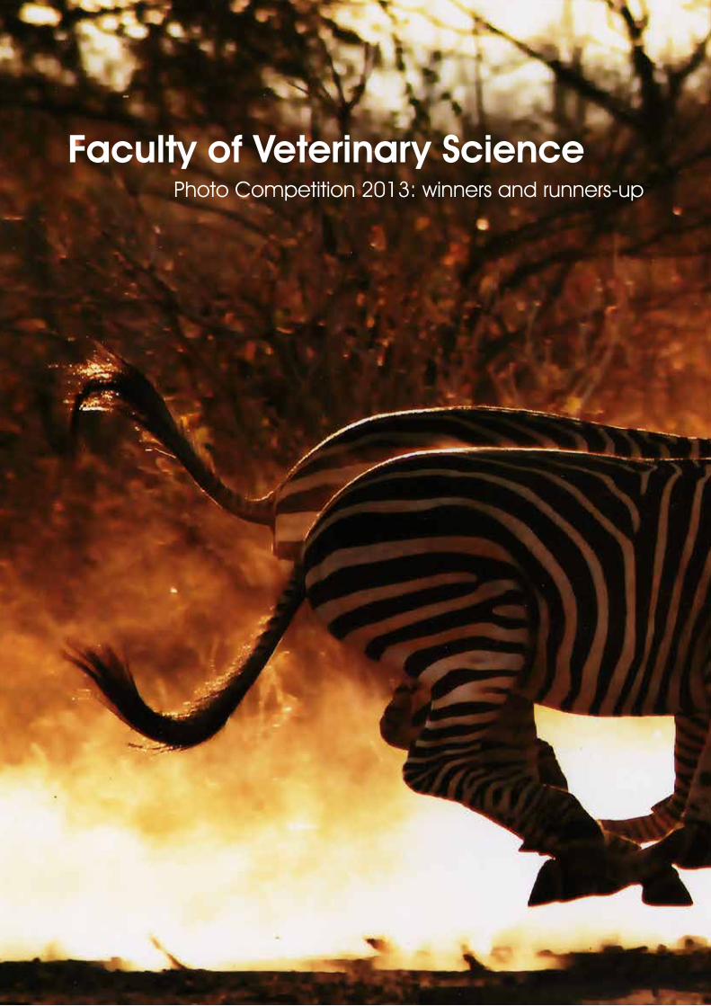
67
Faculty of Veterinary SciencePhoto Competition 2013: winners and runners-up
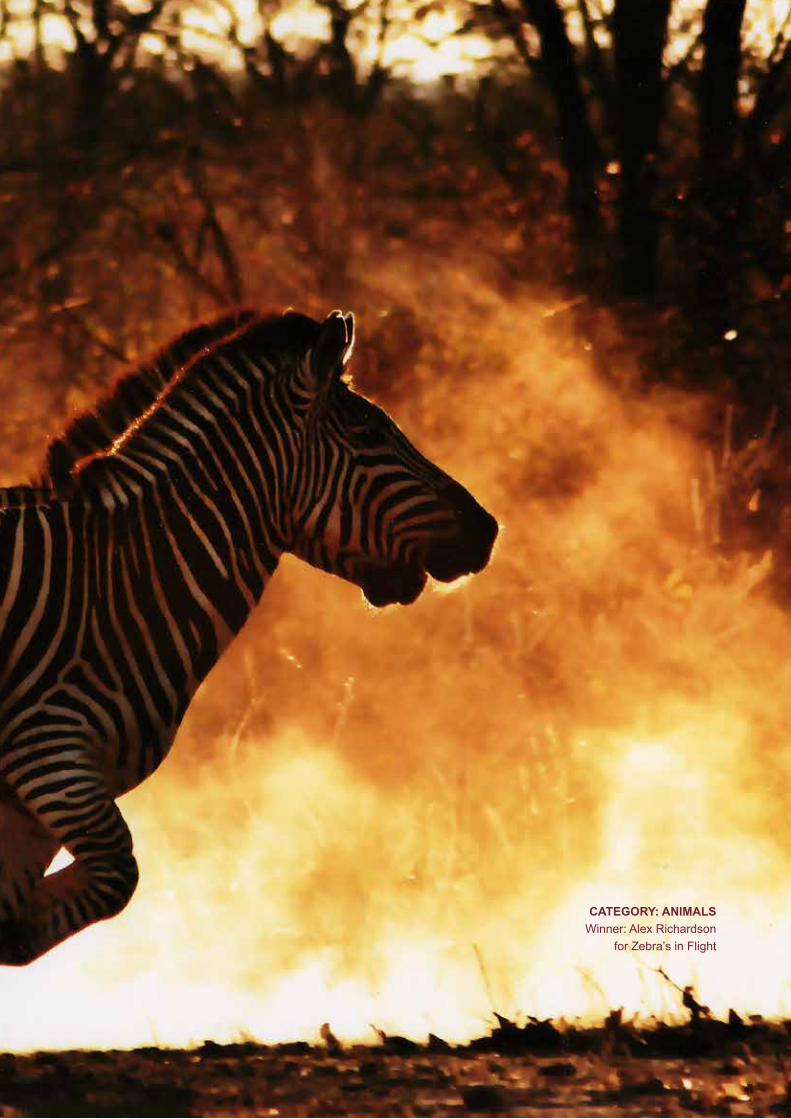
University of Pretoria Faculty of Veterinary Science - Faculty Day 4 September 2014
68
CATEGORY:ANIMALSWinner: Alex Richardson
for Zebra’s in Flight
69
CATEGORY:ANIMALSRunner-up: Roy Wakefield for
Morning Plunge
CATEGORY:STUDENT/LIFERunner-up: June Williams for Path
CATEGORY:STUDENT/LIFEWinner: Lana Botha for “Waar kom jy vandaan?”

71CATEGORY:NATUREWinner: Roy Wakefield for Antelope Island, Kariba

University of Pretoria Faculty of Veterinary Science - Faculty Day 4 September 2014
72
73CATEGORY:NATURERunner-up: Chris Neethling for “Sononder by Bitterpan”
University of Pretoria Faculty of Veterinary Science - Faculty Day 4 September 2014
74
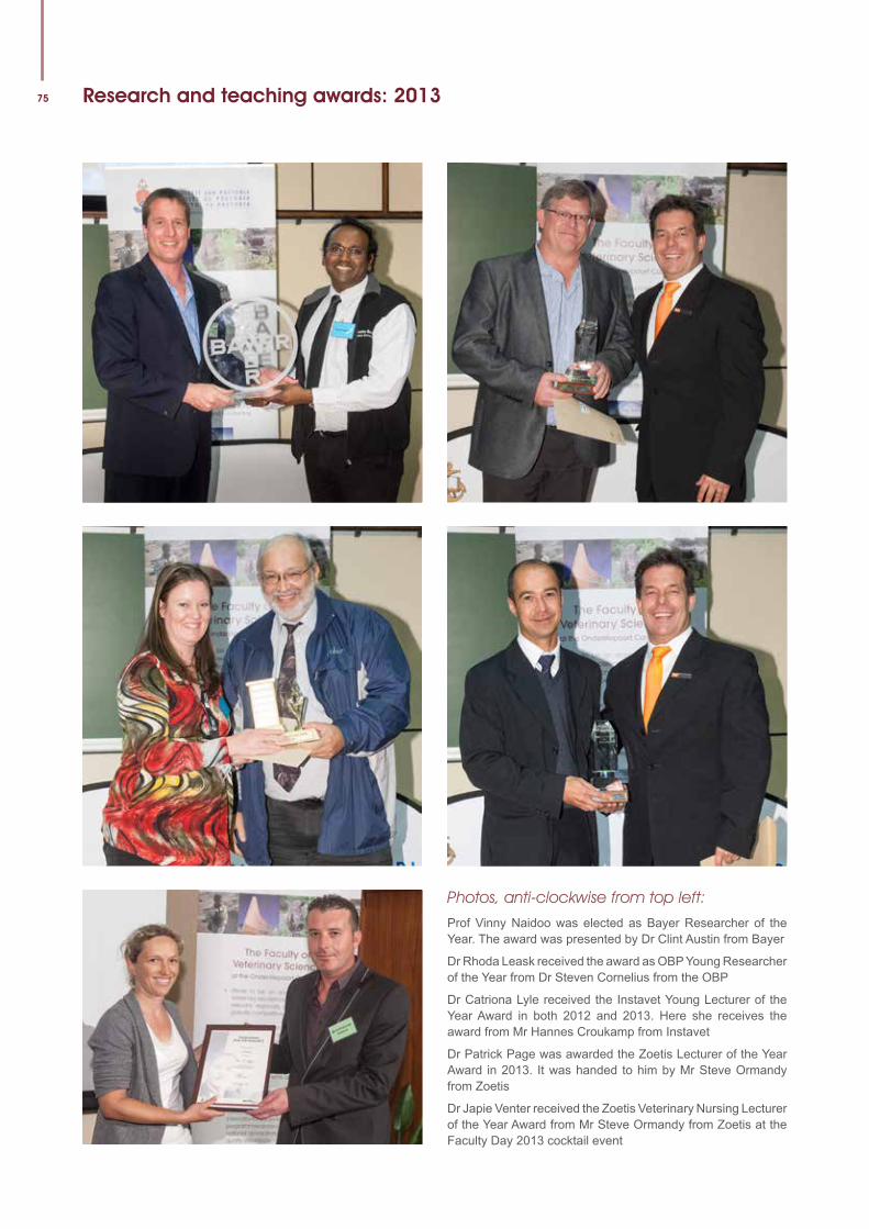
75 Research and teaching awards: 2013
Photos, anti-clockwise from top left:
Prof Vinny Naidoo was elected as Bayer Researcher of the Year. The award was presented by Dr Clint Austin from Bayer
Dr Rhoda Leask received the award as OBP Young Researcher of the Year from Dr Steven Cornelius from the OBP
Dr Catriona Lyle received the Instavet Young Lecturer of the Year Award in both 2012 and 2013. Here she receives the award from Mr Hannes Croukamp from Instavet
Dr Patrick Page was awarded the Zoetis Lecturer of the Year Award in 2013. It was handed to him by Mr Steve Ormandy from Zoetis
Dr Japie Venter received the Zoetis Veterinary Nursing Lecturer of the Year Award from Mr Steve Ormandy from Zoetis at the Faculty Day 2013 cocktail event
University of Pretoria Faculty of Veterinary Science - Faculty Day 4 September 2014
76Faculty Day 2014: Committees
Organising CommitteeDr Paul van Dam (Chairperson)Mr Chris van Blerk (Publications and Marketing)Sr Sarah Johnson (Exhibitors and Sponsors)Ms Niesje Tromp (Administration)Ms Rita Zeeman (Administration)
Master of CeremoniesProf Neil Duncan
Scientific CommitteeProf Vinny NaidooProf Anita MichelDr Lillian MukandiwaProf Andrew LeisewitzDr Dayo FasinaProf Andre Ganswindt
AdjudicatorsOral and Posters:Prof Pete IronsProf Amelia GoddardDr Dietmar HolmDr Musafiri KaramaDr June WilliamsDr Jannie CraffordDr Mohamed Sirdar
Parallel Student ProgrammeDr Paul van Dam
Photo competitionMs Reinette LudikeMs Ingrid de GoedeMs Armina KoebergMs Daleen Anderson
Layout and DesignJanine Smit Editorial Services
EditorChris van Blerk
Published by the Faculty of Veterinary Science, University of PretoriaHead: Marketing and CommunicationE-mail: [email protected]
UniversiteitvanPretoria•UniversityofPretoria•YunibesithiyaPretoriaPretoria 0002 Suid-Afrika • South Africa • Afrika BorwaTel: +27 (0) 12 420 3111 • Faks/Fax: +27 (0) 12 420 4530
www.veterinary.up.ac.za
Facu l ty o f Ve te r inary Sc ience