Embed Size (px)
Citation preview
Ann. rheum. Dis. (1971), 30, 359
Felty's syndromeA clinical and pathological survey of 21 patients and theirresponse to treatment*
C. G. BARNES, A. L. TURNBULL, AND B. VERNON-ROBERTSThe London Hospital
In the 36 years since Felty (1924) described a clinicalsyndrome of 'chronic arthritis in the adult, associatedwith splenomegaly and leucopenia', a number ofindividual cases and a few series of patients havebeen described, but some fundamental questionsremain unanswered.With isolated exceptions (Denko and Zumpft,
1962), most authors agree that Felty's syndrome isnot merely a variant of systemic lupus erythematosus(SLE) or a coincidence of arthritis with someunassociated disease. However, there is littleunanimity as to its aetiology and pathology, itsrelationship to the various manifestations ofrheumatoid disease, the range of clinical and hae-matological manifestations that may occur, and,most important for the individual patient, how thedisease should be treated.
Material and methods
SELECTION OF PATIENTS
The diagnosis was accepted when the patient presentedwith features of 'classical or definite' rheumatoidarthritis (RA) (Ropes, Bennett, Cobb, Jacox, and Jessar,1959), splenic enlargement without other demonstrablecause, and leucopenia with a total white cell count ofless than 3,500 per cu. mm. and a neutrophil count of2,000 per cu. mm. or less.
IMMUNOLOGYLatex tests were performed by a modification of themethod of Singer and Plotz (1956). Waaler-Rose testsand subsequently differential sheep cell agglutinationtitres were performed by modifications of the methodof Ball (1950).
Tests for antinuclear factor (ANF), white cell ANF,and salivary gland antibody were performed by immuno-fluorescence (Roitt and Doniach, 1966).Serum white cell antibodies were sought by the comple-
ment-fixation technique of van Rood, van Leeuwen, andEernisse (1959). Platelet antibodies were sought by
agglutination, indirect antiglobulin consumption, andcomplement fixation, by the methods of van de Wiel,van de Wiel-Dorfmeyer, and van Loghem (1961) andAster, Cooper, and Singer (1964).
HAEMATOLOGYRoutine haematological measurements were performed bystandard techniques (Dacie and Lewis, 1968). For thedetermination of their survival, red cells from thepatient were labelled with 56Cr by the method of Mollisonand Veall (1955). The first (100 per cent. activity) bloodsample was drawn after initial equilibration of labelledcells in the spleen as determined by counting in vivo overthe organ. The normal range in our laboratory for 50per cent. disappearance of activity is 23 to 29 days.The accumulation of excess counts over the liver andspleen was calculated by the method of Hughes Jonesand Szur (1957); in normal subjects the excess counts at50 per cent. disappearance of activity in the blood do notexced 250 for the spleen and 150 for the liver.
HISTOLOGY OF THE SPLEEN
51. sections of paraffin-embedded formol-fixed materialfrom the available spleens from patients with Felty'ssyndrome ('Felty spleens') were stained with haemato-xylin and eosin, Gomori's reticulin, elastic Van Gieson,Perls' iron stain, and congo red.For comparison, sections were also prepared from 22
spleens weighing less than 300 g. from patients with RAcoming to autopsy ('RA control spleens') The exclusionof control spleens weighing more than 300 g. was neces-sary, since in many of these spleens the presence ofamyloid, infection, or malignancy obscured other struc-tural changes.
Results
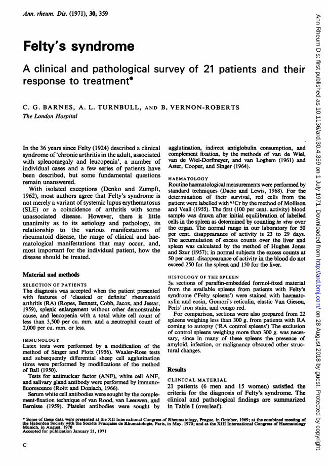
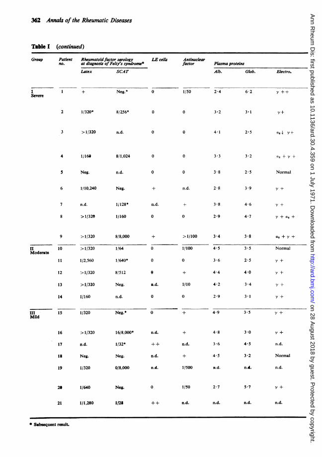
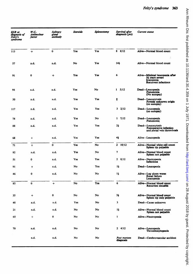
CLINICAL MATERIAL21 patients (6 men and 15 women) satisfied thecriteria for the diagnosis of Felty's syndrome. Theclinical and pathological findings are summarizedin Table I (oveileaf).
* Some of these data were presented at the XII International Congress of Rheumatology, Prague, in October, 1969; at the combined meeting ofthe Heberden Society with the Soci6t6 Fran9aise de Rhumatologie, Paris, in May, 1970; and at the XIII International Congress of HaematologyMunich, in August, 1970Accepted for publication January 21, 1971
C
on 28 August 2018 by guest. P
rotected by copyright.http://ard.bm
j.com/
Ann R
heum D
is: first published as 10.1136/ard.30.4.359 on 1 July 1971. Dow
nloaded from
360 Annals of the Rheumatic Diseases
Table I Clinical andpathological findings in 21 patients with Felty's syndrome (continued on pp. 362-363)
Group Patient Sex Age at Rheumatoid disease Relative precedenceno. onset of of splenic enlargement
RA Duration of Articular Nodules Sjbgren's Other and leucopenia(yrs) RA before involvement syndrome rheumatoid
Felty's at onset of manifestationssyndrome Felty's syndrome(yrs)
I 1 F 25 27 Quiescent 0 + - Leucopenia 2 yrsSevere
2 F 38 Mildly active 0 0
3 M 52 10 Moderatelyactive
0 + _ Leucopenia 4 mths
4 M 60 7
5 M 53 7
6 F 32 15
7 M 44 1
8 F 46 6
9 F 38 6
II 10 F 35 7Moderate
11 F 28 34
12 F 59 14
13 F 30 39
14 F 40 3
III 15 F 26 8Mild
16
17
18
19
F 35 14
M 46 5
M 38 6
F 50 14
20 F 59 17
21 F 26 ?
Active
Severe'burnt out'
Quiescent
Active
Active
Moderatelyactive
Quiescent
Active
Quiescent
Severe'burnt out'
Active
Quiescent
Severe'burnt out'
Quiescent
Mildly active
Quiescent
Quiescent
Severe'burnt out'
+ 0 Neuropathy Simultaneous
0 _ _
+ + Neuropathy Leucopenia 7 mths
0 Raynaud's Simultaneous
+ Raynaud's Simultaneous
+
+
Arteritis Splenic enlargement3 mths
0 - Leucopenia 3 mths
Carpal tunnel Leucopenia 4 yrsArteritis
+ Carpal tunnelArteritis
+ + Leucopenia 4 mths
+ 0 Polymyositis Simultaneous
+ + - Simultaneous
+
+ Scieritis
Arteritis Splenic enlargement2 yrs
-_
Splenic enlargement3 yrs
+ + NeuropathyArteritis
n.d. = not done
Simultaneous
on 28 August 2018 by guest. P
rotected by copyright.http://ard.bm
j.com/
Ann R
heum D
is: first published as 10.1136/ard.30.4.359 on 1 July 1971. Dow
nloaded from
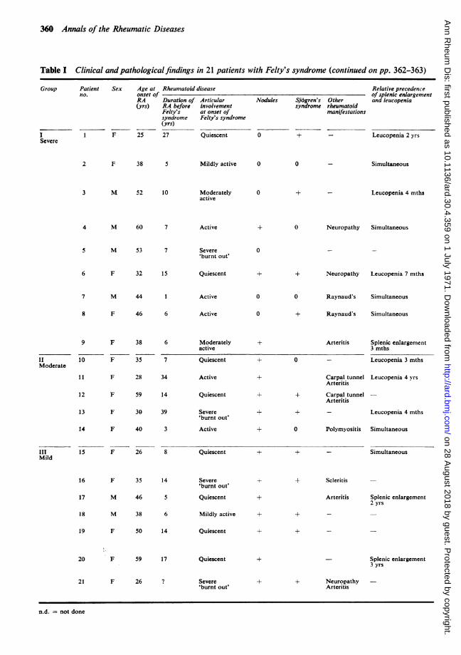
Felty's syndrome 361
Spken Blood studies (lowest values) Highest lymphocyte Lowest Highest Red cellmaximum count(per platelet count retic- survival (days)size (cm.) Hb White cell Neutrophils cu. mm.) (x 10') ulocyte (l'Cr half life)weight (g.) (g. per cent.) count (per before count
(per cu. mm.) spknectomy (?4)
cu. mm.)
Pneumonia Tip 9*5BronchitisUrineVulva
Bronchitis 6 8*8Mastoiditis 714UrinePharyngitis
Bronchitis 3 12 5Pneumonia 435FurunculosisBuccal andperineal ulcers
Oral moniliasis 2*5 9*6PneumoniaBronchitis
Carbuncle 6 6 0Pyrexia 1,572unknown origin
Mouth ulcers 14 7 0Pneumonia 2,150Urine
Pharyngitis 3 11P8Pneumonia 390
Diarrhoea 2 9.1Vomiting 560Pyrexiaunknown origin
Pneumonia 5 10-41,248
None Tip 8*8
Urine 3 9*8
Bronchitis 2 9.1428
Leg ulcers 5 7.5810
None 1 9*0
2,200 90 3,650
700 0 644
1,200
1,000
1,200
1,000
2,400
1,000
1,000
900
650
2,100
700
2,000
38 4,600
75 2,340
530 1,125
128 1,450
0
170
360
230
26
132
335
300
4,640
1,260
1,760
1,020
1,740
2,938
400
2,130
n.d. 2-1 nd.
120 1-7 n.d.
162 5S0 n.d.
114 9 0 12
25 18 9
82
254
47
38
368
108
202
142
440
4 18
n.d. n.d.
7 20
3-5 n.d.
<2 23
1 30
<2 26
3-4 16
2-6 >30
Sinusitis 4 10-7Conjunctivitis 685UrineFurunculosis
Urine 3 10-2
Pneumonia 2 10-8Tuberculosis 1,319
None 2 13-0
Leg ulcers 5 9.5(by x rayandscanning)
Urine 2 9*0PneumoniaSacral ulcers
None Not 9*9palpable588
1,500
2,400
2,100
2,000
1,700
1,700
725 950
576
1,200
620
715
3,528
800
1,386
1,615
504 2,610
3,000 2,100 2,000
207 3-5 20
114
n.d.
252
268
44
<2 >30
n.d. n.d.
<2 29
<2 25
1 n.d.
n.d. n.d. n.d.
Infections
on 28 August 2018 by guest. P
rotected by copyright.http://ard.bm
j.com/
Ann R
heum D
is: first published as 10.1136/ard.30.4.359 on 1 July 1971. Dow
nloaded from
362 Annals of the Rheumatic Diseases
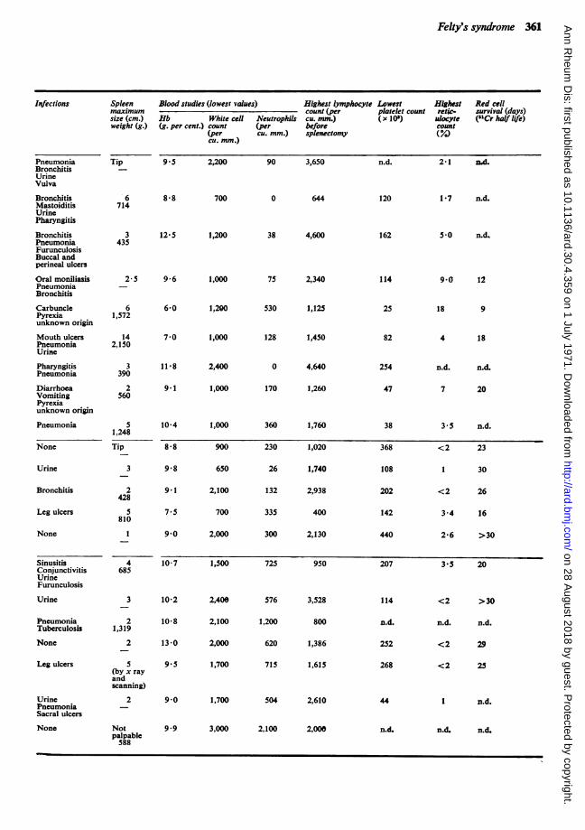
Table I (continued)
Group Patient Rheumatoidfactor serology LE cells Antinuclearno. at diagnosls ofFelty's syndrome factor Plasma proteins
Latex SCAT
I 1 + Neg.* 0 1/50Severe
2 1/320*
3 >1/320
4 1/160
5 Neg.
6 1/10,240
n.d.
>1/320
7
8
8/256*
n.d.
8/1,024
n.d.
Neg.
1/128*
1/160
0
0
0
0 0
n.d.
n.d. +
0 0
9 > 1/320 8/8,000 + > 1/100
II 10 > 1/320 1/64 0 1/100Moderato
11 112,560 1/640* 0 0
12 > 1/320 8/512 8 +
13 >1/320 Neg. a.d. 1/10
14 11160 n.d. 0 0
m 15 1/320 Neg.* 0 +Mild
16
17
IS
19
> 1/320
n.d.
Neg.
1/320
16/8,000*
1/32*
Neg.
0/8,000
n.d.
++
n.d.
n.d.
n.d.
+
1/500
Alb. Glob. Electro.
2-4 6-2 v ++
3 2 3-1 +
4-1 2-5 at vY+
3 3 3-2 a2 + y +
3 8 2- 5 Normal
2-8 39 v+
3-8 4-6 v +
2-9 4-7 y + at +
3-4 3-8 at + v +
4.5
3 -6
4-4
4-2
2-9
4.9
4-8
3 -6
4 5
m.d.
3 5
2-5
4 0
3 -4
3-1
Normal
Y +
+
y +
Y +
3-5 7 +
3 0
4.5
3-2
n.d.
+
n.d.
Normal
n.d.
Neg. 0 1150 2-7 57 +
21 1/1,280 1/28 + + n.d. n.d. n.d. n.d.
s Subsequent result.
20 1/640
on 28 August 2018 by guest. P
rotected by copyright.http://ard.bm
j.com/
Ann R
heum D
is: first published as 10.1136/ard.30.4.359 on 1 July 1971. Dow
nloaded from
Felty's syndrome 363
ESR at W.C. Salivarydiagnosis of Antinuclear glandFelty's factor antibodysyndrome
Staold Spenectomy Survival after Current statusdiagnosis (yrs)
113 + 0 Yes Yes 8 8/12 Alive-Normal blood count
57 n.d. n.d. No Yes 141 Alive-Normal blood count
91 +
98 n.d.
50 n.d.
117 n.d.
78
88
68
71
82
51
91
46
n.d.
n.d.
n.d.
0
0
n.d.
n.d.
n.d.
n.d.
n.d.
n.d.
0
n.d.
n.d.
n.d.
n.d.
Yes
Yes
Yes
Yes
Yes
Yes
Yes
Yes
Yes
Yes
No
No
85 0 + No
Yes
No
Yes
Yes
No
Yes
Yes
No
No
Yes
Yes
No
Yes
6
1 5/12
*
3 2/12
1 7/12
2*
41
2 10/12
7
2 8/12
1*
1i
6
Aliv-Minimal leucopenia after5* yea sevreleucopeniaRecurrent infections
Dead-LeucopeniaPneumonia(No autopsy)
Dead-LeucocytosisPyrenia unknown origin(no autopsy)
Dead-Leucopenia(no autopsy)
Dead-LeucopeniaPneumonia
Dead-LeucocytosisPostoperative infectionand portal vein thrombosi
Alive-Leucopenia
Alive-Normal wkite cel countSpleen tip palpable
Alive-Normal blood countSpleen not palpable
Alive-NeutropeniaInfections
Dead-Leucopenia
Alive-Leg ulcers worseRenal failureLeucopema
Alive-Normal blood countRecurrent sinusit
n.d.
n.d.
70 n.d.
0
n.d.
n.d.
0
n.d.
n.d. n.d.
No
Yes
No
No
No
No
No
No
No
No
No No
3*
3
1*
2 4/12
Post mortemdiapos
Alive-Normal blood countSpleen tip only palpable
Dead-Cause unknown
Alive-Normal blood countSpleen not palpable
Alive-Neutropenia
Alive-LeucopeniaThrombocytopenia
Dead-Cerebrovascular accident
35
48
21
65
on 28 August 2018 by guest. P
rotected by copyright.http://ard.bm
j.com/
Ann R
heum D
is: first published as 10.1136/ard.30.4.359 on 1 July 1971. Dow
nloaded from
364 Annals of the Rheumatic Diseases
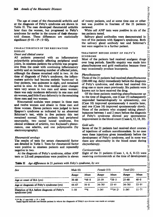
The age at onset of the rheumatoid arthritis andat the diagnosis of Felty's syndrome are shown inTable II. The men developed rheumatoid arthritislater than the women, but progressed to Felty'ssyndrome far earlier in the course of their rheuma-toid disease. These differences are statisticallysignificant (001<P<0 02).
CHARACTERISTICS OF THE RHEUMATOIDDISEASEOnset and clinical courseAll patients presented with an inflammatorypolyarthritis principally affecting peripheral smalljoints. In nineteen patients the arthritis was progres-sive from the onset with continuing inflammatoryactivity and an increasing number of affected joints,although the disease remained mild in two. At thetime of diagnosis of Felty's syndrome, the inflam-matory activity had become entirely 'burnt-out' infour patients, was quiescent in eight, and remainedactive in nine. At this time residual joint changeswere very severe in two men and seven women;there was only moderate deformity in one man andsixwomen, and little ifany deformity in theremainingthree men and two women.Rheumatoid nodules were present in three men
and twelve women and absent in three men andthree women. Eleven patients were judged to haveSj0gren's syndrome as indicated by an abnormalSchirmer's test, while in five patients this testremained normal. Three patients had peripheralneuropathy, two carpal tunnel syndrome, fiveclinical evidence of arteritis, two Raynaud's pheno-menon, one scleritis, and one polymyositis (byelectromyography).
Rheumatoid serologyThe results of tests for serum rheumatoid factorare detailed in Table I. Tests for rheumatoid factorwere positive in nineteen patients and repeatedlynegative in two.At the diagnosis of Felty's syndrome, either ANF
tests or LE-cell preparations were positive in eleven
of twenty patients, and at some time one or othertest was positive in fourteen of the 21 patientsreviewed.White cell ANF tests were positive in six of the
ten patients tested.Salivary gland antibodies were demonstrated in
two of five patients with Sj0gren's syndrome. Boththe salivary gland antibody test and Schirmer'stest were negative in a further patient.
TREATMENT BEFORE ONSET OF FELTY'SSYNDROMEMost of the patients had received analgesic drugsover long periods. Specific enquiry was made intophenylbutazone and gold medication because thesemay occasionally produce leucopenia.
PhenylbutazoneThree of the 21 patients had received phenylbutazone(200400 mg. daily) immediately before the diagnosisof Felty's syndrome, and eleven had received thisdrug one or more years previously. Six patients wereknown not to have received the drug.Of the three patients reoeiving phenylbutazone up
to the time of diagnosis, one (Case 15) subsequentlyresponded haematologically to splenectomy, one(Case 10) improved spontaneously 6 months later,and one (Case 16) improved spontaneously slowly.None of the patients who stopped taking phenyl-butazone between 1 and 2 years before the diagnosisof Felty's syndrome showed any spontaneousimprovement in the blood count (Cases 6, 12, 18, 20).
Gold saltsSeven of the 21 patients had received short coursesof injections of sodium aurothiomalate. In no casewere these injections given immediately before thedevelopment of Felty's syndrome, and in none wasthere any abnormality in the blood count duringchrysotherapy.
CorticosteroidsEight of the 21 patients (Cases 1, 4, 6, 8-12) werereceiving corticosteroids at the time of development
Table II Age differences in 21 patients with Felty's syndrome, by sex
Sex Male (6) Female (15) Total (21)
Range Mean Range Mean Range Mean
Age at onset of RA (yrs) 30-60 *48-8 25-59 *38.5 25-60 40-0
Age at diagnosis of Felty's syndrome (yrs) 44 67 54-8 34-76t 53-4t 34-76t 53-9t
Duration of RA before diagnosis of Felty's 1-10 **6 3-34t **14.2t 1-34t 11 7tsyndrome (yrs)
*/* By It' test 0.01 < P < 0-02Those figures exclude one female patient in whom the diagnosis of Felty's syn drome was made at autopsy
on 28 August 2018 by guest. P
rotected by copyright.http://ard.bm
j.com/
Ann R
heum D
is: first published as 10.1136/ard.30.4.359 on 1 July 1971. Dow
nloaded from
Felty's syndrome 365
of Felty's syndrome, in a dosage of 5-15 mg. pred-nisolone (or its equivalent) daily.
FEATURES OF FELTY' S SYNDROMEOnsetIn ten patients, routine examination revealed eitherleucopenia or splenic enlargement in the absenceof relevant symptoms. Six patients presented withinfection, two with haemolytic anaemia, and twowith skin ulceration. One patient was diagnosedat autopsy.The time relationship between the detection of
splenic enlargement and the development of neu-tropenia was variable. The priority of leucopenia(7 patients: up to 4 years) or splenic enlargement(3 patients: up to 3 years) was not related to anyother feature of the illness (Table I). As in otherseries (Collier and Brush, 1966; Ruderman, Miller,and Pinals, 1968) there was no correlation betweenthe size of the spleen and the degree ofhaematologicalabnormality.On review of the clinical features of their illness
before treatment the patients appeared to fall intothree groups:
1. SEVERE The nine patients (5 women; 4 men) in thisgroup had neutrophil counts frequently below 500 percu. mm. and a variety of infections. Four patients alsohad haemolytic anaemia (Cases 4-6, 8). Seven patientswere seriously ill from the time of recognition of leuco-penia and splenic enlargement; in the other two theonset of severe symptoms was delayed for 2 years.
II. MODERATE Five patients (all women) had only mildclinical manifestation despite neutropenia as severe asthose in Group I. One patient had moderate haemolyticanaemia (Case 13).
HI. MILD In seven patients (5 women; 2 men) lesssevere neutropenia (500-2,000 per cu. mm.) was associatedwith only moderate or mild symptoms. In two of these(Cases 15, 16) there had been moderately severe infections,but in both they antedated the neutropenia by some years.One elderly patient (Case 20) had recurrent urinaryinfections and bronchopneumonia.
The clinical and serological features of thearthritis, and the relationship between the onset ofleucopenia and clinical enlargement of the spleendid not differ in the three groups (Table I).
Splenic enlargementThe spleen was palpable in nineteen of the 21 patients.In one grossly obese patient the spleen could not bepalpated with certainty, but was shown to beconsiderably enlarged by plain x ray and by scinti-scanning after the administration of heated 51Cr-labelled red cells. In the last severely crippledpatient, an enlarged spleen (588 g.) was discoveredonly at autopsy.
Lymph nodesConspicuously enlarged lymph nodes were presentin four patients. Biopsy of an enlarged node in twopatients, and post mortem examination in a third,revealed only benign follicular hyperplasia of thetype associated with rheumatoid arthritis (Motulsky,Weinberg, Saphir, and Rosenberg, 1952).
Weight lossIn five ofeleven patients there was a loss ofmore than7 lb. in the preceding 6 months.
Skin pigmentationSome mild general increase in skin pigmentation waspresent in three patients.
UlcerationUlceration of the legs was present in four patientswithout clinical evidence of varicose veins, of theperineum in one, of the mouth and genitalia in onepatient each, and of both the latter sites in a furtherpatient.
InfectionsThese occurred in 14 of the 21 patients (Table I).
PurpuraFour patients developed bruising or purpura. Threewere thrombocytopenic, two of whom were receivingcorticosteroids. The fourth had a normal plateletcount and was not receiving corticosteroids.
LiverSevere haematemesis in one patient (Case 9) suggestedthe presence of portal hypertension and this wasconfirmed at laparotomy. Percutaneous (2) andoperative hepatic biopsies showed some portalfibrosis without the changes characteristic ofcirrhosis.
Clinical features of liver disease were not seen inany other patient. Flocculation tests were oftenabnormal in the presence of hyperglobulinaemia.A mild unexplained increase in serum alkalinephosphatase was noted in one, patient (Case 2).
HAEMATOLOGYPeripheral bloodRed cells (Fig. 1, overleaf). All patients wereanaemic (haemoglobin less than 13-5 g./100 ml. inmen and less than 12 g./100 ml. in women) but thiswas severe (less than 8 g./100 ml.) in only one manand two women. The mean corpuscular haemo-globin concentration was less than 30 per cent. infive of the 21 patients, in only three of whom was theblood film considered to be hypochromic. The redcells were normochromic in all other patients,including six in whom polychromasia was present;two showed macrocytosis (Cases 5 and 8).
on 28 August 2018 by guest. P
rotected by copyright.http://ard.bm
j.com/
Ann R
heum D
is: first published as 10.1136/ard.30.4.359 on 1 July 1971. Dow
nloaded from
366 Annals of the Rheumatic Diseases
q9/wl20
15 -
10-
5.
0
...
lowestHb
5000.
4500*
4000*
3500'3000
2500
2000-
1500'
1000
500a
0
0
s.
W.B.C.
S
lowestNP.
*0
0
0
0
3I
T00
lymphs
10/Vmm!400
350
300
250
200
150
100
50
0 .
0
0
0*
00
0
0
lb0
owestplat.
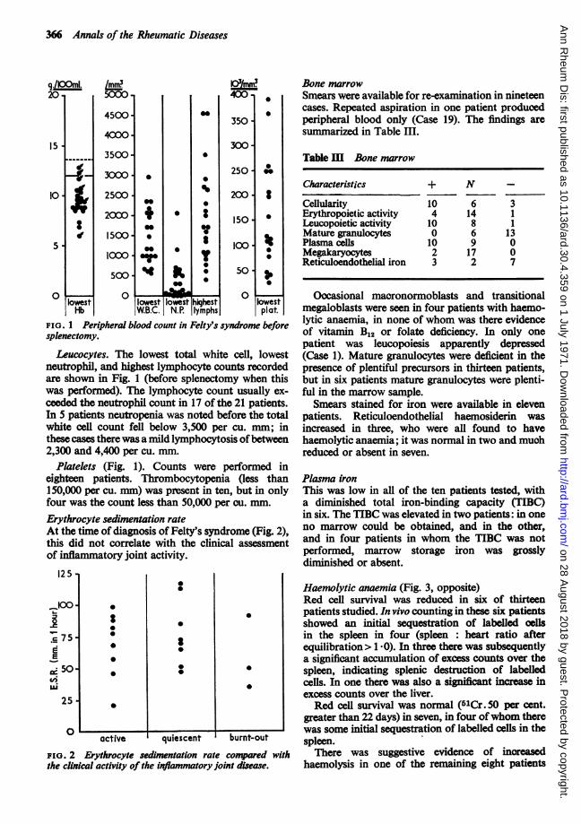
FIG. 1 Peripheral blood count in Felty's syndrome beforesplenectomy.
Leucocytes. The lowest total white cell, lowestneutrophil, and highest lymphocyte counts recordedare shown in Fig. 1 (before splenectomy when thiswas performed). The lymphocyte count usually ex-ceeded the neutrophil count in 17 of the 21 patients.In 5 patients neutropenia was noted before the totalwhite cell count fell below 3,500 per cu. mm; inthese cases there was a mild lymphocytosis ofbetween2,300 and 4,400 per cu. mm.
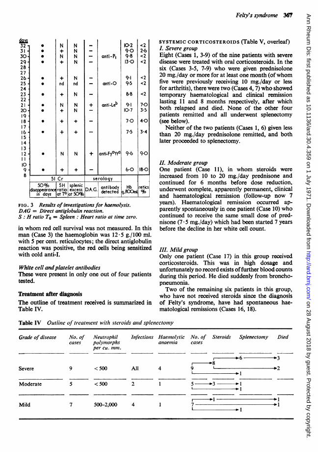
Platelets (Fig. 1). Counts were performed ineighteen patients. Thrombocytopenia (less than150,000 per cu. mm) was present in ten, but in onlyfour was the count less than 50,000 per cu. mm.Erythrocyte sedimentation rateAt the time of diagnosis of Felty's syndrome (Fig. 2),this did not correlate with the clinical assessmentof inflammatory joint activity.
125-
L00-o0
' 75-
E
(t 50
25
0
0
0
0
0
active
00
0
a0
quiescent
FIG. 2 Erythrocyte sedimentation ratethe clinical activity of the inflamnmatory joii
Bone marrowSmears were available for re-examination in nineteencases. Repeated aspiration in one patient producedperipheral blood only (Case 19). The findings aresummarized in Table III.
Table HI Bone marrow
Characteristics + N -
Cellularity 10 6 3Erythropoietic activity 4 14 1Leucopoietic activity 10 8 1Mature granulocytes 0 6 13Plasma cells 10 9 0Megakaryocytes 2 17 0Reticuloendothelial iron 3 2 7
Oocasional maoronormoblasts and transitionalmegaloblasts were seen in four patients with haemo-lytic anaemia, in none of whom was there evidenceof vitamin B12 or folate deficiency. In only onepatient was leucopoiesis apparently depressed(Case 1). Mature granulocytes were deficient in thepresence of plentiful precursors in thirteen patients,but in six patients mature granulocytes were plenti-ful in the marrow sample.
Smears stained for iron were available in elevenpatients. Reticuloendothelial haemosiderin wasincreased in three, who were all found to havehaemolytic anaemia; it was normal in two and muchreduced or absent in seven.
Plasma ironThis was low in all of the ten patients tested, witha diminished total iron-binding capacity (TIBC)in six. The TIBC was elevated in two patients: in oneno marrow could be obtained, and in the other,and in four patients in whom the TIBC was notperformed, marrow storage iron was grosslydiminished or absent.
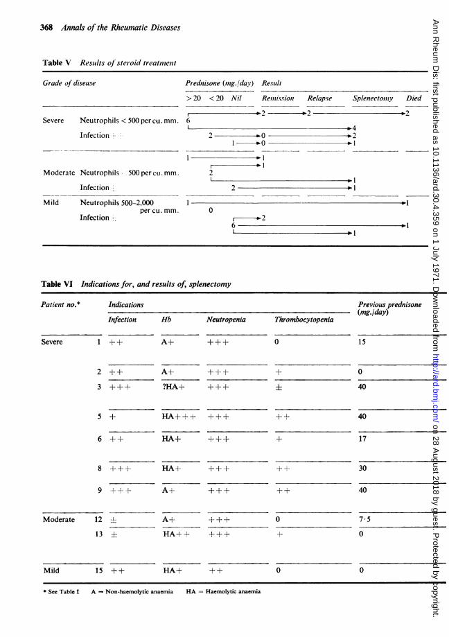
Haemolytic anaemia (Fig. 3, opposite)Red cell survival was reduced in six of thirteen
* patients studied. In vivo counting in these six patientsshowed an initial sequestration of labelled cellsin the spleen in four (spleen : heart ratio afterequilibration> 1 -0). In three there was subsequentlya significant accumulation of excess counts over the
* spleen, indicating splenic destruction of labelledcells. In one there was also a significant increase inexcess counts over the liver.Red cell survival was normal (5lCr. 50 per cent.
greater than 22 days) in seven, in four ofwhom therewas some initial sequestration of labelled cells in the
burnt-out spleen.conpared with There was suggestive evidence of increasednt disease. haemolysis in one of the remaining eight patients
__
Ic
on 28 August 2018 by guest. P
rotected by copyright.http://ard.bm
j.com/
Ann R
heum D
is: first published as 10.1136/ard.30.4.359 on 1 July 1971. Dow
nloaded from
Felty's syndrome 367
323130292827262524232221201918171615141312l1098
S000
0
0
0
0
0
0
0
0
N
N
nd
N
N
+
NNNN
Nnd
N
NN
N
+
+I
anti - PI
anti-C
anti-Leb
anti-FyaYta
10-2909-813-0
9-19.5
8*8
9-110-7
7-0
7.5
9'6
b 0
<22.6<2<2
<2<2
<2
7-03.54-0
3-4
9*0
18-0I.
51 Cr serology
50I o S:H splenicIin days at TO at 50% deet .lOOn
FIG. 3 Results of investigations for haemolysis.DAG = Direct antiglobulin reaction.S: H ratio To = Spleen: Heart ratio at time zero.
in whom red cell survival was not measured. In thisman (Case 3) the haemoglobin was 12-5 g./100 ml.with 5 per cent. reticulocytes; the direct antiglobulinreaction was positive, the red cells being sensitizedwith cold anti-I.
White cell andplatelet antibodiesThese were present in only one out of four patientstested.
Treatment after diagnosisThe outline of treatment received is summarized inTable IV.
SYSTEMIC CORTICOSTEROIDS (Table V, overleaf)L Severe groupEight (Cases 1, 3-9) of the nine patients with severedisease were treated with oral corticosteroids. In thesix (Cases 3-5, 7-9) who were given prednisolone20 mg./day or more for at least one month (ofwhomfive were previously receiving 10 mg./day or lessfor arthritis), there were two (Cases 4, 7) who showedtemporary haematological and clinical remissionlasting 11 and 8 months respectively, after whichboth relapsed and died. None of the other fourpatients remitted and all underwent splenectomy(see below).
Neither of the two patients (Cases 1, 6) given lessthan 20 mg./day prednisolone remitted, and bothlater proceeded to splenectomy.
IL Moderate groupOne patient (Case 11), in whom steroids wereincreased from 10 to 20 mg./day prednisone andcontinued for 6 months before dose reduction,underwent complete, apparently permanent, clinicaland haematological remission (follow-up now 7years). Haematological remission occurred ap-parently spontaneously in one patient (Case 10) whocontinued to receive the same small dose of pred-nisone (7 * 5 mg./day) which had been started 7 yearsbefore the decline in her white cell count.
III. Mild groupOnly one patient (Case 17) in this group receivedcorticosteroids. This was in high dosage andunfortunately no record exists offurther blood countsduring this period. He died suddenly from broncho-pneumonia.Two of the remaining six patients in this group,
who have not received steroids since the diagnosisof Felty's syndrome, have had spontaneous hae-matological remissions (Cases 16, 18).
Table IV Outline of treatnient with steroids anid splenectomy
Grade of disease
Severe
No. of Neutrophil Infections Haetnolytic No. of Steroids Splenectomy Diedcases polymorphs anaemia cases
per cll. mmn.
--6 - 3
9 < 500 All 4 9 p2:1
Moderate 5 < 500 2 1 5 o3 0 1
Mild 7 500-2,000 4 1 7 II :1
on 28 August 2018 by guest. P
rotected by copyright.http://ard.bm
j.com/
Ann R
heum D
is: first published as 10.1136/ard.30.4.359 on 1 July 1971. Dow
nloaded from
368 Annals of the Rheumatic Diseases
Table V Resiths of steroid treatment
Grade of disease
Severe Neutrophils < 500 per cu. mm.
Infection -'
Moderate Neutrophils 500 per cu. mm.
Infection
Mild Neutrophils 500-2,000per cu. mm.
Infection o
Prednisone (mg./day) Result
> 20 < 20 Nil Remission Relapse Splenectomy Died
6
2 0- t21 o--
2
2-1
o
6I
Table VI Indications for, and results of, splenectomy
Indications
Infection Hb Neutropenia Thrombocytopenia
Previous prednisone(mg./day)
Severe 1 ++ A+ +++ 0
2
3
++ A+
?HA+
5 + HA+++ +++
6 ++ HA+ +++
++ 40
+ 17
8 +++ HA+ +++ ++ 30
9 +++ A+ +++
0A+
HA++
Mild 15 ++ HA+ ++
* See Table I A - Non-haemolytic anaemia HA = Haemolytic anaemia
Patient no.*
15
+
+
0
40
Moderate 12 4-
13 ±
++ 40
7 5
0
0 0
* 1.1
on 28 August 2018 by guest. P
rotected by copyright.http://ard.bm
j.com/
Ann R
heum D
is: first published as 10.1136/ard.30.4.359 on 1 July 1971. Dow
nloaded from
Felty's syndrome 369
Thus, in the whole series, remissions were seen inthree of eight patients given at least 20 mg./dayprednisone for a minimum of one month, but onlyone of these did not relapse. Of those given less than20 mg./day prednisone, only one patient, receiving7 - 5 mg./day, underwent partial remission.
There were also two spontaneous haematologicalremissions in the nine patients not treated withcorticosteroids, in one of whom the spleen becameimpalpable.
SURGICAL TREATMENT-SPLENECTOMYThe indications for operation and its effect in the tenpatients submitted to splenectomy are shown inTable VI.The immediate good effect of splenectomy seen
in eight of the ten patients was maintained in onlythree patients, but one ofthem (Case 5) died 4 monthsafter operation, probably from infection (no autopsy),without any deterioration in his white cell count.The other two patients (Cases 2, 15) have continuedto have a completely normal blood picture 14* and6 years respectively after splenectomy. A complete
and spontaneous remission eventually occurred inone of the five patients who relapsed (Case 1). Threeother patients died: one shortly after operation, one15 months after operation of unknown cause(no autopsy), and one 15 months after operation inrenal failure.
Histopathological changes
(1) SPLEENThe histopathological findings are shown in TableVII (overleaf). None of the spleens showed involve-ment by primary or secondary malignant disease, orevidence of a specific infection, or perivascularfibrosis of the 'onion-skin' type. Amyloid materialwas not seen in any Felty spleen.
White pulpThe walls of the follicular arteries in all the Feltyspleens were thickened by a structureless hyalinematerial lying immediately under the endothelium.In five spleens this change was marked with obliter-ation of the lumen of some vessels. This was not
Immediate responseImmediate response(0-28 days after operation)
Clinical Haematological
Good Leucocytosis
Good
Improved
Improved
Good
Died 13 daysportal thrombosis
Transientporto-systemicencephalopathy
Good
No change
Good
Normal
Normal
LeucocytosisRemission of haemolysis
Normal
Survivalafteroperation(yrs)
Alive 8*
Alive 14*Alive 5*
Died 4 mths
Died 1I
Late response
Clinical Infectionsand ulcers
Good 0
Active RA
Improved
Died?infection
ImprovedSuddendeath
0
+
Haematological
Relapse at 3 mthsSpontaneous and completeremission at 3i years
Normal
Severe relapse at 3 mthsSpontaneous improvement5 yrs
+ + + Leucocytosis
+ Relapse at 3 mths
Leucocytosis Died 13 days
Immediate improvementRelapse within 28 days
Normal
Stabilized haemoglobinPersistent leucopenia
Normal
Alive 10 mths Improved 0
Alive 21Died 15 mths
Alive 6
Poor
Poor untildeath inrenalfailure
Improved
++
Moderate neutropenia
Severe neutropenia
Neutropenia
+ Normal
on 28 August 2018 by guest. P
rotected by copyright.http://ard.bm
j.com/
Ann R
heum D
is: first published as 10.1136/ard.30.4.359 on 1 July 1971. Dow
nloaded from
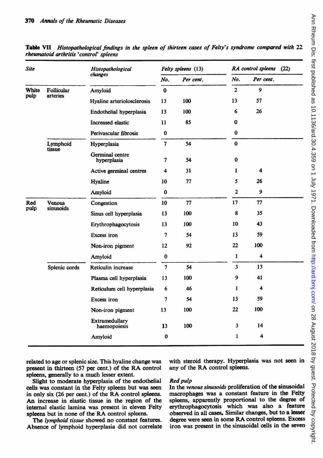
370 Annals of the Rheumatic Diseases
Table VII Histopathological findings in the spleen of thirteen cases of Felty's syndrome compared with 22rheumatoid arthritis 'control' spleens
Site Histopathologicalchanges
White Follicularpulp arteries
Lymphoidtissue
Red Venouspulp sinusoids
Splenic cords
Amyloid
Hyaline arteriolosclerosis
Endothelial hyperplasia
Increased elastic
Perivascular fibrosis
Hyperplasia
Germinal centrehyperplasia
Active germinal centres
Hyaline
Amyloid
Congestion
Sinus cell hyperplasia
Erythrophagocytosis
Excess iron
Non-iron pigment
Amyloid
Reticulin increase
Plasma cell hyperplasia 13
Reticulum cell hyperplasia 6
Excess iron
Non-iron pigrnent
Extramedullaryhaemopoiesis
Amyloid
Felty spleens (13)
No. Per cent.
0
13 100
13 100
11 85
0
7 54
7 54
4 31
10 77
0
10 77
13 100
13 100
7 54
12 92
0
7 54
100
46
7 54
13 100
13 100
0
RA control spleens (22)
No. Per cent.
2 9
13 57
6 26
0
0
0
0
1 4
5 26
2 9
17 77
8 35
10 43
13 59
22 100
1 4
3 13
9 41
1 4
13 59
22 100
3 14
1 4
related to age or splenic size. This hyaline change waspresent in thirteen (57 per cent.) of the RA controlspleens, generally to a much lesser extent.
Slight to moderate hyperplasia of the endothelialcells was constant in the Felty spleens but was seenin only six (26 per cent.) of the RA control spleens.An increase in elastic tissue in the region of theinternal elastic lamina was present in eleven Feltyspleens but in none of the RA control spleens.The lymphoid tissue showed no constant features.
Absence of lymphoid hyperplasia did not correlate
with steroid therapy. Hyperplasia was not seen inany of the RA control spleens.
RedpulpIn the venous sinusoids proliferation of the sinusoidalmacrophages was a constant feature in the Feltyspleens, apparently proportional to the degree oferythrophagocytosis which was also a featureobserved in all cases. Similar changes, but to a lesserdegree were seen in some RA control spleens. Excessiron was present in the sinusoidal cells in the seven
on 28 August 2018 by guest. P
rotected by copyright.http://ard.bm
j.com/
Ann R
heum D
is: first published as 10.1136/ard.30.4.359 on 1 July 1971. Dow
nloaded from

Felty's syndrome 371
Felty spleens in which erythrophagocytosis was mostmarked. Excess iron was also seen in a similarproportion of the RA control spleens. In all but oneof the Felty spleens, and in all the RA controlspleens, varying amounts of birefringent particulatematerial were present in the cytoplasm of thesinusoidal macrophages. This had the characteristicsof formalin pigment, since it disappeared afterexposure to picric acid and did not stain for iron.After treatment with picric acid the cytoplasm ofthese cells was seen to contain ill-defined colourlessinclusions. This birefringent particulate material wasnot found in spleens from 25 patients withoutrheumatoid disease.
In the splenic cords increased reticulin formationand reticulum cell hyperplasia were found moreoften in the Felty spleens than in the RA controlspleens. In all Felty spleens plasma cells wereincreased and there were scattered foci of mega-karyocytes and myeloid cells indicating extra-medullary haemopoiesis. In only three RA controlspleens was there any extra-medullary haemopoiesis.
(2) LIVERHistological examination of the liver in the five caseswhich came to autopsy (Nos 4, 7, 8, 17, and 21) andin the one patient who had liver biopsies (No. 9)revealed no evidence of cirrhosis or amyloidosis.
Discussion
Some authors have doubted the specific entity ofFelty's syndrome. In this series there was no evidenceof another cause of splenic enlargement, e.g.lymphoma or cirrhosis, and no patient diagnosed asFelty's syndrome had later to be excluded because ofthe discovery of another disease or of splenicamyloidosis. We have seen one patient with sero-negative psoriatic arthropathy and leucopenia,whose splenic enlargement was found at operationto be due to lymphosarooma, but we have beenunable to find a single case of 'idiopathic' spleno-megaly and leucopenia in degenerative joint disease,nor has splenectomy been performed in any suchpatient in The London Hospital over the last 12years. The clinical features of the patients underreview provide further evidence for the existence of aspecific syndrome, and the pathological materialfrom splenectomies and autopsies supports thisview.While leucopenia is one of the accepted criteria for
the diagnosis, five ofour patients showed neutropeniawith a normal total white count in the early stages oftheir haematological illness, and therefore it wouldbe preferable to specify neutropenia in the de-finition of the syndrome. While Felty's syndrome hasbeen considered to develop mainly in the elderly
(de Gruchy, 1965), one-third of our patients wereunder 60 years. The men were significantly older thanthe women at the onset of arthritis, but developedFelty's syndrome far earlier in the course of theirillness. This was not related to any obvious sexdifferences in the character of their rheumatoiddisease.Rheumatoid nodules and Sj0gren's syndrome were
both common in our patients, but other nonarticularmanifestations of rheumatoid disease were notunduly frequent. No patient showed a skin rash,inflammation of serous surfaces or myocarditis, orany other features suggestive of SLE. One patienthad moderate renal failure, but the renal biopsyshowed chronic glomerulonephritis.As in other series (Ruderman and others, 1968),
most ofour patients had positive tests for rheumatoidfactor, often in high titre. Two patients had negativelatex tests but were considered to have undoubtedrheumatoid arthritis because in each the jointinvolvement was typical with erosive radiologicalchanges. Biopsy of a nodule from the elbow of oneshowed the classical histology of a rheumatoidnodule, and in the other splenic histology did notshow any features suggestive of SLE. Such patientshave previously been reported (de Gruchy andLangley, 1961).The finding of ANF and/or LE cells in two-thirds
of our patients does not necessitate a diagnosis ofSLE since about 25 per cent. of patients with rheu-matoid arthritis have positive ANF tests, particularlythose with severe systemic disease (Willkens andDecker, 1963). The absence of histological featuresof SLE in the thirteen spleens examined makes itextremely unlikely that Felty's syndrome is amanifestation of this disease as suggested by Denkoand Zumpft (1962).The nonarticular clinical features in our patients
were much the same as previously described (deGruchy, 1965; Ruderman and others, 1968).Infections may be directly related to neutropenia.Other clinical manifestations, such as leg ulcerationand lymph node enlargement, cannot be attributeddirectly to the haematological abnormality. Theseshould probably be regarded not as features ofFelty's syndrome but as manifestations of severerheumatoid disease, since they all occur in rheumatoidpatients without splenic enlargement or neutropenia(Willkens and Decker, 1963).Every patient was anaemic. Haemolysis appeared
to be the major factor in the three patients wvithsevere anaemia, and was also found in three others.In two of these six shortened red cell survival wasprobably due to sensitization by immune antibodies(positive direct antiglobulin test). Destruction ofred cells by the spleen appeared to be the cause inthree, in whom together with the sixth patient the
on 28 August 2018 by guest. P
rotected by copyright.http://ard.bm
j.com/
Ann R
heum D
is: first published as 10.1136/ard.30.4.359 on 1 July 1971. Dow
nloaded from
372 Annals of the Rheumatic Diseases
51Cr study also revealed significant pooling (seques-tration) of red oells in the organ. However, poolingby itself cannot account for haemolysis in thissixth patient because it was also found in fourpatients with a normal red cell survival. Whilehaemolytic anaemia in Felty's syndrome has beenattributed to the direct action of the large spleen(Hume, Dagg, Fraser, and Goldberg, 1964) this doesnot seem to be the only mechanism.The mechanism of anaemia in the remaining
patients was less certain, many showing disturbanceof iron metabolism as is frequently seen in RA.An increase in plasma volume is commonly assoc-iated with enlargement of the spleen and probablycontributes to anaemia in patients with Felty'ssyndrome (Blendis, Ansell, Lloyd-Jones, Hamilton,and Williams, 1970a). In most patients the neutrophilcount was lower than lymphocytes. Absolutelymphocytosis is claimed only to occur after sple-nectomy (de Gruchy, 1965), but five of our patientshad, early in the course of their disease, sufficientlymphocytes to keep the total white cell countwithin normal limits in the presence of neutropenia.The variability of the manifestations of the
syndrome make it difficult to assess prognosis inindividual patients and the value of the treatmentgiven. This may explain the conflicting reports ofthe effects of corticosteroids (Pengelly, 1966;Ruderman and others, 1968) and of splenectomy(Collier and Brush, 1966; Green and Fromke, 1966;Ruderman and others, 1968). To make a more validassessment we have divided our patients into threegroups according to the severity of their disease.As in other series the benefit of corticosteroid
treatment was variable and generally unspectacular.One moderately affected patient appeared to havebeen cured by prednisone. The results in thispatient and in one reported by Pengelly (1966)indicate that corticosteroid therapy may occasionallybe successful. On the other hand the same dosageof prednisone produced only partial improvementin two out of six severely affected patients. Corti-costeroids in small dosage did not have any significantbeneficial effect on the haematological abnormality.The rarity of dramatic benefit from steroid therapy
in our series agrees with previous reports (Collierand Brush, 1966; Ruderman and others, 1968) andmust be considered in the light of the spontaneousremissions in this series seen in two patients, albeitmildly affected, who never received corticosteroidsand in one on a small constant dose.The initial haematological response to splenectomy
was as good in this series as has previously beendescribed (de Gruchy, 1965; Collier and Brush, 1966;Ruderman and others, 1968), and was associatedwith comparable clinical improvement.
There was less complete correlation between
clinical and haematological status in the monthsafter splenectomy. Only one patient achieved acomplete permanent remission. Three patientsmaintained normal blood counts for more than3 months but all continued to have infections whichproved fatal in one. The blood count relapsed in fourpatients within 3 months of operation but only oneof these has since had severe infections.
This experience is very similar to that of Rudermanand others(1968) but is much less favourable than hasbeen suggested in some previous reports in which thefollow-up has been shorter (Green and Fromke,1966; Hahn, Mayne, and Kiely, 1963). Only three ofthe ten splenectomized patients in this series stillhave fully normal blood counts, 14i, 8j, and 6 yearssince operation respectively, and one of these wentthrough a haematological relapse lasting 3 yearsfrom which she recovered spontaneously. However,even those who relapsed haematologically appearto have had a better chance of survival than before.Thus we believe that splenectomy remains thetreatment of choice for patients with very severeinfection.The second indication for splenectomy is severe
anaemia due either to sequestration of red cells andhaemodilution (Blendis and others, 1970a) or tohaemolysis when this is not oontrolled by smalldoses of steroids, and particularly when the spleenis the site of red cell destruction.The third definite indication for splenectomy is
severe haemorrhage associated with thrombocyto-penia (Cudkowicz, 1956). If, as in Case No. 9,haemorrhage is due to portal hypertension, thismust be treated on its own merits.The lack of correlation between infection and the
blood count before and after operation in theirpatients led Ruderman and others (1968) to concludethat leucopenia alone was not an indication forsplenectomy. Only one of our patients had asplenectomy for leucopenia without serious infection.The relapse in her blood count 4 months later andthe development of recurrent chest infections 1Iyears after operation support this conclusion.Furthermore, in our own cases, the similar lack ofcorrelation between the level of the neutrophilcount and the severity of infection (as indicated bydifferences in patients in groups I and II), and thelack of correlation between infection and the whitecell count after splenectomy, all confirm the beliefthat neutropenia alone does not warrant splenectomy.The pathological findings in the thirteen spleens
from patients with Felty's syndrome, when comparedwith those from rheumatoid arthritis without Felty'ssyndrome, give further evidence of a specificpathological entity.The severity of the changes in the follicular
arteries could not be correlated with the age of the
on 28 August 2018 by guest. P
rotected by copyright.http://ard.bm
j.com/
Ann R
heum D
is: first published as 10.1136/ard.30.4.359 on 1 July 1971. Dow
nloaded from
Felty's syndrome 373
patient nor with the degree of splenic enlargement.In the absence of essential hypertension, thesechanges with endothelial hyperplasia and reticulinincrease suggest that some degree of portal hyper-tension is a constant feature in Felty's syndrome(Ellman, Cudkowicz, and Elwood, 1955).The lymphoid follicles showed no constant
changes, but the germinal follicles were generallyinactive and hyaline material was present in themajority. This is in direct contrast to the view thatstriking hyperplasia of germinal centres in enlargedfollicles is a characteristic feature of the spleen inFelty's syndrome (Gardner, 1965).
Sinus cell hyperplasia accompanied by erythro-phagocytosis was present in all the Felty spleens.Those in which erythrophagocytosis was mostmarked also displayed an excess of iron in macro-phages and reticulum cells. These findings clearlyindicate that excessive blood destruction within thespleen is a constant feature in Felty's syndrome,although this is not always sufficient to causesignificant shortening of red cell survival. Whetherneutrophil polymorphs are similarly destroyed isuncertain. Amyloid was not found in any of thespecimens.These consistent pathological abnormalities do
not adequately explain the role of the spleen inFelty's syndrome. The presence of portal hyper-tension does not necessarily imply that this was thecause of the enlargement of the spleen, since somerise in portal blood pressure may be related to theincreased blood flow characteristic of many types ofsplenic enlargement (Williams, Parsonson, Somers,and Hamilton, 1966; Blendis, Banks, Ramboer, andWilliams, 1970b).Although an improvement in white cell count
usually follows splenectomy, the frequency ofsubsequent relapse and occasional spontaneousremission later suggest that hypersplenism is onlyone factor in pathogenesis, probably not the mostimportant. The lack of correlation between splenicsize and the severity of the haematologioal abnor-mality is further evidence for this view. Our findingsdo not, however, indicate what other mechanism isprimarily responsible.
It is never possible fully to exclude the effect ofdrugs on the bone marrow. The chief reasons fordiscounting their influence are the usual rapidrecovery of bone marrow activity following thediscontinuation of the drug (de Gruchy, 1965),the absence of sustained splenic enlargement in adrug-induced neutropenia and the usual good earlyresponse to splenectomy in patients with Felty'ssyndrome. In the individual case it is always import-ant to exclude other causes of bone marrow dys-function, particularly deficiences of vitamin B12 or
folic acid, and paroxysmal nocturnal haemo-globinuria which may present as mild haemolysiswith leucopenia and thrombocytopenia withoutany macroscopic blood pigment in the urine. Theseconditions did not acoount for the haematologicalabnormalities in the cases reviewed.
Summary21 patients with Felty's syndrome have been reviewed.The six men were significantly older than the fifteenwomen at the onset of arthritis but developed thesyndrome sooner. Nonarticular rheumatoid featureswere common, Sj0gren's syndrome being found ineleven of sixteen patients tested. Sera of all but twocontained rheumatoid factor, and LE cell pre-parations and/or ANF tests were positive in fourteenpatients. Absolute neutropenia was characteristicbut its degree did not fully correlate with the severityof infections. All patients were anaemic, red cellsurvival being reduced in six of thirteen patientsstudied with excessive splenic destruction in three.
Haematological remission occurred in two patientsspontaneously and in one of two on a small constantdose of prednisone. Of eight patients given 20 mg.prednisone per day or more, four had transientpartial improvement and one apparent clinical cure.
Ten patients underwent splenectomy ofwhom twoappear to have been cured. Two patients eventuaUyachieved normal blood counts after early relapses.
Eight patients have died including four followingsplenectomy.
The following histological features were found inall thirteen spleens examined:
(i) Hyaline change and endothelial hyperplasia offollicular arteries;
(ii) Sinuscellhyperplasiawitherythrophagocytosis;(iii) Plasma cell hyperplasia and extramedullary
haemopoiesis in the cords;(iv) A birefringent particulate material in maco-
phages and reticulum oells.
It is a pleasure to thank the physicians who have allowedus to study their patients and in particular Dr. MichaelMason and Dr. H. L. F. Currey who also gave us muchhelp and advice, the staff of the Clinical Laboratories forthe routine haematological and immunological investi-gations, Dr. W. S. Jenkins of the Blood TransfusionLaboratory, Brentwood, for the tests for white cell andplatelet antibodies, and Miss Olive Sporton for devotedsecretarial assistance.
on 28 August 2018 by guest. P
rotected by copyright.http://ard.bm
j.com/
Ann R
heum D
is: first published as 10.1136/ard.30.4.359 on 1 July 1971. Dow
nloaded from
374 Annals of the Rheumatic Diseases
ReferencesAsTER, R. H., CooPER, H. E., AND SINGER, C. L. (1964) J. Lab. clin. Med., 63, 161 (Simplified complement fixation
test for the detection of platelet antibodies in human serum).BALL, J. (1950) Lancet, 2, 520 (Serum factor in rheumatoid arthritis agglutinating sensitized sheep red cells).BLENDIS, L. M., ANSELL, I. D., LLOYD-JONES, K., HAmILToN, E., AND WILLIAMS, R. (1970a) Brit. med. J., 1, 131
(Liver in Felty's syndrome).-, BANKS, D. C., RAMBoER, C., AND WILLIAMS, R. (1970b) Clin. Sci., 38, 73 (Spleen blood flow and splanchnic
haemodynamics in blood dyscrasia and other splenomegalies).COLLiER, R. L., AND BRUSH, B. E. (1966) Amer. J. Surg., 112, 869 (Hematologic disorder in Felty's syndrome.
Prolonged benefits of splenectomy).CUDKOWICZ, L. (1956) Rheumatism, 12, 37 (Felty's syndrome).DACE, J. V., AND LEwiS, S. M. (1968) 'Practical Haematology', 4th ed. Churchill, London.DE GRUCHY, G. C. (1965) Geriatrics, 20, 219 (Diagnosis and treatment of Felty's syndrome).- AND LANGLEY, G. R. (1961) Austr. Ann. Med., 10, 292 (Felty's syndrome).
DENKO, C. W., AND ZUMPFT, C. W. (1962) Arthr. and Rheum., 5, 478 (Chronic arthritis with splenomegaly andleukopenia).
ELLMAN, P., CUDKOWICZ, L., AND ELWOOD, J. S. (1955) Ann. rheum. Dis., 14, 84 (Therapy of 'Felty's syndrome').FELTY, A. R. (1924) Bull. Johns Hopk. Hosp., 35, 16 (Chronic arthritis in the adult, associated with splenomegaly
and leucopenia. A report of 5 cases of an unusual clinical syndrome).GARDNER, D. L. (1965) 'Pathology of the Connective Tissue Diseases,' p. 84. Arnold, London.GREEN, R. A., AND FROMKE, V. L. (1966) Ann. intern. Med., 64, 1265 (Splenectomy in Felty's syndrome).HAHN, R. G., MAYNE, J. G., AND KiELY, J. M. (1963) Arthr. and Rheum., 6, 275 (Abstract: Clinical and
haematologic study of Felty's syndrome).HUGHES JoNEs, N. C., AND SzuR, L. (1957) Brit. J. Haemat., 3, 320 (Determination of the sites of red-cell
destruction using 56Cr-labelled cells).HUME, R., DAGG, J. H., FRASER, T. N., AND GOLDBERG, A. (1964) Ann. rheum. Dis., 23, 267 (Anaemia of Felty's
syndrome).MOLLISON, P. L., AND VEALL, N. (1955) Brit. J. Haemat, 1, 62 (The use of the isotope 51Cr as a label for red cells).MorULSKY, A. G., WEINBERG, S., SAPHIR, O., AND ROsENBERG, E. (1952) Arch. intern. Med., 90, 660 (Lymph
nodes in rheunatoid arthritis).PENGELLY, C. D. R. (1966) Brit. med. J., 2, 986 (Felty's syndrome. Good response to adrenocorticosteroids.
Possible mechanism of anaemia).Rorrr, I. M., AND DONIACH, D. (1966) 'Methods used at the W.H.O. Reference Laboratory for Autoimmune
Serology of the Middlesex Hospital Medical School'. London, W.1.ROOD, J.3. VAN, LEEUWEN, A. VAN, AND EERNIssE, J. G. (1959) Vox. sang., n.s. 4, 427 (Leucocyte antibodies in
sera of pregnant women).ROPES, M. W., BENNETr, G. A., COBB, S., JACOX, R., AND JESSAR, R. A. (1959) Ann. rheum. Dis., 18, 49 (Revision
of diagnostic criteria for rheumatoid arthritis).RUDERMAN, M., MILLER, L. M., AND PINALS, R. S. (1968) Arthr. and rheum., 11, 377 (Clinical and serologic
observations on 27 patients with Felty's syndrome).SINGER, J. M., AND PLOTZ, C. M. (1956) Amer. J. Med., 21, 888 (The latex fixation test. 1. Application to the
serologic diagnosis of rheumatoid arthritis).WEIL, T. W. M. VAN DE, WIEL-DORMEYER, H. VAN DE, AND LOGHEM, J. J. VAN (1961) VOx Sang. (Basel), n.s. 6,641
(Studies on platelet antibodies in man).WILLAMS, R., PAoNsoeN; A., Semw,s, Am ANILJDN, P. J. S. (1966) Lancet, 1, 329 (Portal hypertension in
idiopathic tropical splenomegaly).WILLKENS, R. F., AND DECKR, J. L. (1963) Arthr. and Rheum., 6, 720 (Rheunatoid arthritis with serologic
evidence suggesting systemic lupus erythematosus: clinical, serologic and chromatographic studies).
on 28 August 2018 by guest. P
rotected by copyright.http://ard.bm
j.com/
Ann R
heum D
is: first published as 10.1136/ard.30.4.359 on 1 July 1971. Dow
nloaded from