Embed Size (px)
Citation preview

Female Genital System Basic Robbins Chapter 18, pages 681-704
Big Robbins Chapter 22 M.E. Bauman, MD
Slides for review: 22, 34, 37, 94, 104, 153, 163, 181, 192, 198, 223 and normal uterus slide. One online clinical case: #12 A woman with vaginal bleeding Note: The material presented below generally follows the outline of Chapter 18 of Basic Robbins. Occasionally the order of the material presented below differs from that of the text; additional material not in the text is also presented.
Vulva
Labium minus/labia majora Vulvitis
Contact irritant dermatitis Contact allergic dermatitis Infectious dermatitis Bartholin gland duct obstruction Non-neoplastic Epithelial Disorders Lichen Sclerosus et atrophicus (figure 18-1) Leukoplakia Lichen simplex chronicus (squamous epithelial hyperplasia) (figure 18-1) Tumors Condyloma (condylomas/condylomata) (figure 18-2) Condylomata lata Condylomata acuminata Vulvar carcinomas
Female reproductive page 2
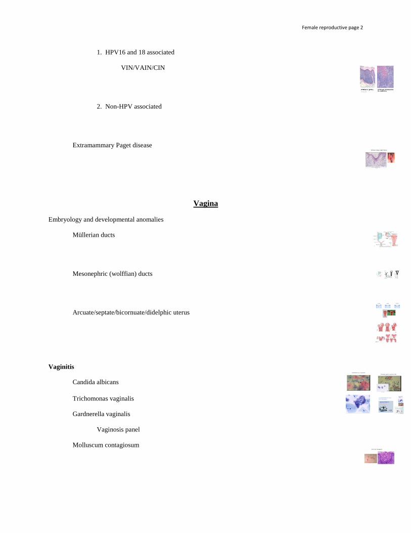
1. HPV16 and 18 associated VIN/VAIN/CIN 2. Non-HPV associated Extramammary Paget disease
Vagina Embryology and developmental anomalies Müllerian ducts Mesonephric (wolffian) ducts Arcuate/septate/bicornuate/didelphic uterus Vaginitis Candida albicans Trichomonas vaginalis Gardnerella vaginalis Vaginosis panel Molluscum contagiosum

Female reproductive page 3
Malignant neoplasms Squamous cell carcinoma Clear cell carcinoma Vaginal adenosis Sarcoma botryoides (embryonal rhabdomyosarcoma)
Cervix Cervicitis Chlamydia trachomatis Neisseria gonorrhea Ureaplasma urealyticum HSV-2 Neoplasia of the cervix Transformation zone (figure 18-4) Risk factors for CIN HPV Vaccines Low risk and high risk HPV (Figure 18-5) Pap tests

Female reproductive page 4
CIN/LSIL/HSIL (Figure 18-6, 18-7) Colposcopy/LEEP Cervical carcinoma
Body of Uterus (Corpus uteri)
Uterus/uteri Endometritis Infection: N. gonorrhea, C. trachomatis, mycobacteria Retained products of conception Intrauterine device Complications Adenomyosis Complications Endometriosis Possible etiologies (figure 18-9) Complications

Female reproductive page 5
Histology of the endometrial cycle
Menorrhagia Metrorrhagia Oligomenorrhea/Amenorrhea Menopause/postmenopause Abnormal uterine bleeding

Female reproductive page 6
AUB secondary to organic uterine lesions AUB secondary to functional etiologies (= dysfunctional uterine bleeding) Proliferative lesions of the endometrium and myometrium Endometrial hyperplasia Endometrial polyps Leiomyoma (leiomyomas, leiomyomata) Leiomyosarcoma STUMP Endometrial carcinoma Type I Type II

Female reproductive page 7
Malignant Mixed Müllerian Tumor (Triple MT)
Fallopian Tubes Gabriello Fallopio Salpinx Salpingitis Tubo-ovarian abscess Pelvic inflammatory disease (PID)
Ovaries
Gonadotropic hormones from the hypothalamus cause the anterior lobe of the pituitary gland (pars distalis) to release follicle-stimulating hormone (FSH) and luteinizing hormone (LH). These hormones stimulate the development of ovarian follicles, which in turn secrete estrogen. Rising estrogen levels trigger a surge of LH. This results in ovulation and the development of the corpus luteum, which begins to secrete progesterone and estrogen. Progesterone builds up the endometrial wall in preparation for implantation (pregnancy). Progesterone also inhibits LH, so if no pregnancy occurs, the corpus luteum causes its own demise. If a pregnancy occurs, the placenta produces human chorionic gonadotropin (hCG) to maintain the corpus luteum and pregnancy.
Cochard. Netter's Atlas of Human Embryology: Updated Edition. Saunders, 062012. <vbk:978-1-4557-3977-6#outline(2.3)> Follicular and luteal cysts Corpus luteum/corpora lutea Corpus albicans/corpora albicantia Polycystic ovarian disease (PCOD)

Female reproductive page 8
Torsion of ovary Tumors of the ovary 1. 2. 3. 4. 1. Surface epithelial tumors Serous Cystadenoma Mucinous Cystadenofibroma Endometrioid Tumor of low malignant potential Brenner/transitional/urothelial Cystadenocarcinoma Carcinoma Serous tumors Psammoma bodies Low malignant potential Mucinous tumors Pseudomyxoma peritonei Omental “caking”

Female reproductive page 9
Endometrioid tumors Brenner/transitional/urothelial tumors 2. Germ cell tumors Teratomas Benign (mature) cystic teratoma/dermoid cyst Malignant (immature) teratoma Specialized teratoma, e.g. struma ovarii Dysgerminomas Yolk sac (endodermal sinus) tumor Schiller-Duval body α-fetoprotein Choriocarcinoma Syncytioblasts and cytotrophoblasts β-hCG 3. Sex cord-stromal tumors Derivation Granulosa cell tumor (granulosa-theca cell tumor) Juvenile granulosa cell tumor Call-Exner bodies Fibroma-thecoma

Female reproductive page 10
Meigs syndrome Sertoli-Leydig cell tumor (androblastoma) 4. Metastatic tumors to the ovary Müllerian origin Extra-Müllerian origin Krukenberg tumor
Diseases of Pregnancy Placentation

Female reproductive page 11
Single umbilical artery Umbilical knot Umbilical cord prolapse
Nuchal cord Amniotic band syndrome Placental infarction Placental abruption Placenta accreta/increta/percreta Succenturiate lobe Velamentous insertion of cord Placenta previa

Female reproductive page 12
Twin gestations Monozygotic/dizygotic Monoamnionic Monochorionic (MoMo) Diamnionic Monochorionic (DiMo) Diamnionic Dichorionic (DiDi), fused or separate Monochorionic = Twin-twin transfusion syndrome Placental infections Ascending (transcervical) infections PPROM, PROM Chorioamnionitis Funisitis Hematogenous (transplacental) infections T O R C H Ectopic pregnancy
Female reproductive page 13
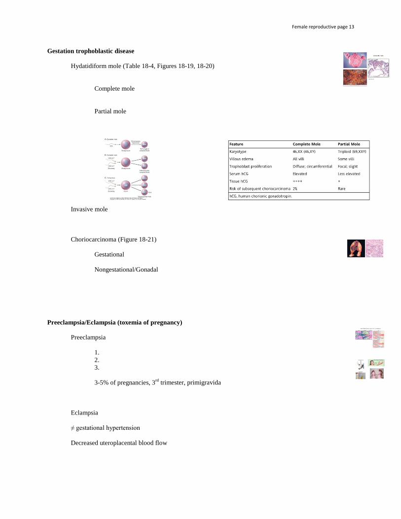
Gestation trophoblastic disease Hydatidiform mole (Table 18-4, Figures 18-19, 18-20) Complete mole Partial mole
Invasive mole Choriocarcinoma (Figure 18-21) Gestational Nongestational/Gonadal Preeclampsia/Eclampsia (toxemia of pregnancy) Preeclampsia 1. 2. 3. 3-5% of pregnancies, 3rd trimester, primigravida Eclampsia ≠ gestational hypertension Decreased uteroplacental blood flow

Female reproductive page 14
Altered circulating endothelial factors Placenta Maternal endothelial dysfunction End organ failure HELLP in 10% of eclamptic patients H EL LP Rx
End of Handout