Embed Size (px)
Citation preview
Fertilization and Embryogeny in Ephedra trifurcaAuthor(s): W. J. G. LandSource: Botanical Gazette, Vol. 44, No. 4 (Oct., 1907), pp. 273-292Published by: The University of Chicago PressStable URL: http://www.jstor.org/stable/2467425 .
Accessed: 15/05/2014 10:40
Your use of the JSTOR archive indicates your acceptance of the Terms & Conditions of Use, available at .http://www.jstor.org/page/info/about/policies/terms.jsp
.JSTOR is a not-for-profit service that helps scholars, researchers, and students discover, use, and build upon a wide range ofcontent in a trusted digital archive. We use information technology and tools to increase productivity and facilitate new formsof scholarship. For more information about JSTOR, please contact [email protected].
.
The University of Chicago Press is collaborating with JSTOR to digitize, preserve and extend access toBotanical Gazette.
http://www.jstor.org
This content downloaded from 194.29.185.134 on Thu, 15 May 2014 10:40:25 AMAll use subject to JSTOR Terms and Conditions
FERTILIZATION AND EMBRYOGENY IN EPHEDRA TRIFURCA'
CONTRIBUTIONS FROM THE HULL BOTANICAL LABORATORY I02
W. J. G. LAND
(WvITH PLATES XX-XXII)
Little attention has been given to the fertilization and embryogeny of Ephedra, the only accounts being those of STRASBURGER and JACCARD. In i872 STRASBURGER,2 studying E. altissima, described and figured unfertilized eggs and four stages of the embryo immediately before and after formation of cotyledons. In I876 he described a series of stages in E. altissima from the central cell of the archegonium to the formation of suspensors. He noted the penetration of the pollen tube, which, he says, reaches the egg through a canal formed by the breaking-down of the neck cells of the archegonium. Fertili- zation was not observed, but several stages of the 'roembryo were noted. Usually three to eight proembryos are formed and arranged in a row down the middle of the egg. He calls attention to what he considers an abnormal condition, in which the cytoplasm of the egg is pierced through by irregular tortuous channels supported through- out their length by kinoplasmic fibers. These channels extend around and finally inclose a portion of the egg cytoplasm together with a single nucleus. He thinks these channels result from the activities of this single nucleus which is finally inclosed. The breaking-out of the suspensors from the archegonia was noted. His third account,4 published in I879, deals at some length with the embryogeny of E. campylopoda, but adds little to the results obtained from E. altissina.
In i894 JACCARD,5 studying E. helvetica, observed the pollen tube I Read before the Botanical Society of America, December i906; abstract pub-
lished in Science N. S. 25: 282, 283. I907.
2 STRASBURGER, E., Die Coniferen und die Gnetaceen. i8722.
3 STRASBURGER, E., Ueber Zellbildung und Zellteilung. i876. 4 STRASBURGER, E., Die Angiospermen und die Gymnospermen. 1879. 5 JACCARD, P., Recherches embryologiques sur lEphedra helvetica. Inaugural
Dissertation. Lausanne. I894.
273] [Botanical Gazette, vol. 44
This content downloaded from 194.29.185.134 on Thu, 15 May 2014 10:40:25 AMAll use subject to JSTOR Terms and Conditions
274 BOTANICAL GAZETTE [OCTOBER
entering the archegonium, one male nucleus in contact with the egg nucleus, and the second male nucleus, the stalk cell, and the tube nu- cleus lying in the micropylar end of the cgg. He also studied pollen tubes grown in five per cent. gelatin in sterilized pear juice. He noted the disorganization of the jacket cells and describes a fusion of the nuclei of the jacket cells with one another and their subsequent migra- tion into the cytoplasm of the egg. He also observed a large number of small nuclei in the egg, the nature and origin of which he was not able to determine. He further states that a suspensor ("prosuspen- seur" or "embryonalschlauch ") is not formed. The elongation of the basal cells of the embryo to form a secondary suspensor (" le columelle") is noted.
The present study of the fertilization and embryogeny of E. trifurca is the continuation of the paper6 which dealt with spermatogenesis and oogenesis in the same species. Material for the study was collected at Mesilla, New Mexico, chiefly in April I904. Much of the material collected in I903 was unfit for a study of fertilization stages because of the ravages of a parasitic insect which attacked the ovules about the time of pollination. Embryos having been observed about April 20, I903, it was hoped that by beginning to make collec- tions as early as April i in the following season and continuing them at intervals of two days a fairly complete series could be obtained. Collecting was discontinued April 20, I904, at which time the majority of embryos had attained their maximum size. The season of I904
was unprecedentedly hot and dry and in consequence development was much accelerated, so much so in fact that embryos with large secondary suspensors were found in the material collected April i.
Not all of the material collected at the earliest date had been pollinated. In I905 staminate strobili sent from the Carnegie Desert Laboratory, Tucson, Arizona, made possible the final study of the male gameto phyte.
POLLEN TUBES AND FERTILIZATION
Pollen grains were placed in a ten per cent. solution of saccharose on a slide in a moist chamber. By evaporation the solution became considerably denser toward the close of the experiment. Shortly
6 LAND, W. J. G., Spermatogenesis and oogenesis in Ephedra trifurca. BOT.
GAZETTE 38: i-i8. pis. I-5. I904.
This content downloaded from 194.29.185.134 on Thu, 15 May 2014 10:40:25 AMAll use subject to JSTOR Terms and Conditions
1907] LAND-IEPHEDRA TRIFURCA 275
after being placed in the sugar solution, the fluted exine is ruptured by a longitudinal split extending along one side of the grain from end to end, and the intine with its contents is completely freed. The two prothallial cells soon disappear and the cytoplasm of the prothallial end becomes very vacuolate. The body cell next divides, the spindle being parallel with the long axis of the pollen grain (fig. i). At the same time the tube nucleus and the stalk cell, which lie at opposite ends of the grain, each move through ninety degrees and lie side by side, or occasionally they may lie as shown in fig. 2. The tube nucleus comes to rest against the wall, assumes a lenticular shape, and soon afterward the pollen tube is put out from this point of contact. Appear- ances seem to indicate that the pollen tube is produced at the point of separation of the tetrads, but this could not be definitely determined because of the symmetry of the pollen grain and the possibility of a slight rotation of the extruded mass after escaping from the exine.
The male nuclei are of equal volume and are truly elliptical. One of them almost invariably passes in advance of the stalk cell, which takes a position in advance of the rearmost male nucleus (fig. 3). Measure- ments of male nuclei show them to be exactly of like dimensions; neither is there any optical difference. Their relative position in the pollen tube is the only evidence, if evidence it be, of any inequality. There is little doubt that the male nucleus nearest the end of the tube will succeed in fusing with the egg nucleus because of its favorable position. Within ten hours from the time the pollen grains were placed in the sugar solution the pollen tubes were sufficiently long to reach the egg. After twenty-four hours the tubes ceased to elongate, at which time they were three to four times longer than is neces- sary to penetrate to the egg. The stalk nucleus is usually about half the diameter of the male nucleus. The tube nucleus is variable in form, as is common among tube nuclei of gymnosperms.
As has been shown in the study of spermatogenesis and oogenesis, the pollen grains come to rest on the bottom of the pollen chamber in contact with the gametophyte, in the vicinity of or on the necks of the archegonia. Occasionally pollen grains rest on the sides of the pollen chamber as well as on the bottom, but by far the greater number rest on the bottom, and in consequence the pollen tubes are put out directly into the archegonia. Since all material studied was fixed
This content downloaded from 194.29.185.134 on Thu, 15 May 2014 10:40:25 AMAll use subject to JSTOR Terms and Conditions
276 BOTANICAL GAZETTE [OCTOBER
at least four days after pollination, and fertilization is possible within ten hours after pollination, it has been very difficult to secure fertili- zation and early proembryonal stages. Only those ovules which have been incompletely pollinated by having the pollen lodged for a time in the long tubular micropyle, and later dislodged and reaching the pollen chamber, can by any possibility show fertilization. From the nature of things such a delay in pollination must be very rare, although the ovules are peculiarly fitted to have such delays occur. Among over 2800 ovules sectioned at the time when fertilization was expected, only two (figs. 4 and 5) showed early stages.
The pollen tube forces its way between the neck cells of the arche- gonium, rarely destroying them in its passage. Only in one or two instances were the lower neck cells destroyed. In fig. 4 the lower and apparently double nucleus shows the all but complete fusion of the sperm and the egg nuclei. The small nucleus lying immediately above the fusing nuclei is the ventral nucleus; the one immediately above it is the second male nucleus, which in this instance has passed deeper into the egg cytoplasm than usual. Fig. 5 shows the fusion nucleus with fragments of the ventral nucleus lying above it. The second male nucleus, with the stalk nucleus and tube nucleus lying immediately below it, is seen in the upper end of the egg. The stalk nucleus and the tube nucleus soon disappear. The second male nucleus does not ordinarily penetrate deeper into the egg than is shown in fig. 5, but undergoes its final changes here. The dense mass of cytoplasm surrounding the fusion nucleus and extending downward through the egg is well shown in fgs. 4 and 5. This mass first appears as a densely staining spherical body in the upper third of the central cell, and disappears with the formation of proembryos.
EMBRYOGENY
The fusion nucleus gives rise to eight free nuclei, more or less unequal in size, and three to five of the largest ones surround them- selves by a wall and produce embryos. The free nuclei are either arranged in a row down the middle of the egg, or more frequently are scattered through it. The lower ones in general successfully produce embryos, although a few instances were noted in which the lower ones did not function further than the one-celled stage, the micropylar ones
This content downloaded from 194.29.185.134 on Thu, 15 May 2014 10:40:25 AMAll use subject to JSTOR Terms and Conditions
1907] LAND-EPHEDRA TRIFURCA 277
alone being successful. Nearly all of the largest vacuoles in the egg disappear shortly after fertilization, and the cytoplasm becomes more and more homogeneous as free nuclear division progresses. The pro- embryonal nuclei usually become so dense that they can be seen with difficulty in the cytoplasm.
Delicate cytoplasmic radiations, similar to those figured in the unfertilized egg, stream out from each nucleus and end in a denser ring of cytoplasm. This delicately radiating cytoplasm becomes more and more vacuolate, finally taking on the appearance shown in figs. 6 and 7. In fig. 6 the large cell is one of two proembryonal cells which later will produce suspensors. Fig. 7 shows two or four functioning pro- embryos. The first suggestion of walls, or rather cytoplasmic thick- enings, is shown in figs. 6 and 7. The functioning proembryos isolate themselves from the surrounding cytoplasm as follows. Cleav- age cracks appear, starting presumably at the equatorial region of the last spindle, although all trace of a spindle has disappeared before cleavage is apparent. An early stage of incomplete cleavage between sister cells is shown in fig. 8. The cleavage cracks, following the feebly defined wall or cytoplasmic thickening, continue until they meet around the nucleus, carving out a more or less irregular mass of cytoplasm for each nucleus. Three of five completely isolated proembryos are shown in fig. 9, which also shows masses of egg cytoplasm that have not been appropriated by the proembryonal cells.
At the time the proembryonal cells have freed themselves completely from the egg cytoplasm they lie in a nutritive mass derived from various sources. Shortly after fertilization, the walls of the jacket cells, which always have been extremely tenuous, disappear and in most preparations the jacket-cell cytoplasm becomes mixed with that of the egg (fig. 9). Shortly before the walls disappear the jacket cells become binucleate, the nuclei dividing either mitotically or amito- tically. Occasionally mitotic divisions occur simultaneously in every cell of both layers of the jacket, or every division may be amitotic, or both kinds of division may occur in the same jacket. When the latter occurs, the nuclei at the upper end of the archegonium divide amitotically, and these amitotic divisions can easily be mistaken for fusion. Occasionally the jacket nuclei enlarge, fragment, and their chromatin is scattered through the cytoplasm of the jacket cell and
This content downloaded from 194.29.185.134 on Thu, 15 May 2014 10:40:25 AMAll use subject to JSTOR Terms and Conditions
278 BOTANICAL GAZETTE [OCTOBER
later passes out into the egg cytoplasm. Sometimes this fragmenta- tion is accompanied by kinoplasmic fibers, as shown in fig. io. Fig. 9 also shows an intact jacket-cell nucleus which has passed into the egg cytoplasm. Occasionally, though rarely, instances were noted in which the jacket cells do not become binucleate and break down at the time of fertilization; some of them, especially those near the middle of the archegonium, have the nucleus surrounded by delicate cytoplasmic radiations similar to those noted in the unfertilized egg. These nuclei also very much resemble the egg except in size, being of course much smaller. Apogamy was at first suspected, but further study showed that these egglike jacket nuclei do not function, being finally broken down and absorbed by the embryos.
The second male nucleus is normally in the position shown in fig. 5. It en' arges and occupies the upper part of the egg and extends into the space recently filled by the adjacent jacket cells. The chro- matin is arranged in strands, at the ends of some of which it mav be aggregated in small groups (figs. 6, 7). The nuclear membrane breaks down on one side (fig. 6) and cleavage planes pass up into the nucleus. Fig. 7 shows a male nucleus with an irregular mass of spindles with groups of chromatin granules at the poles. Later the region occupied by the male nucleus is filled with a number of small cells which may have come from further division of the second male nucleus, or some of them may be a joint product of chromatin from the male nucleus and from the jacket nuclei. Below this region are numerous small cells which possibly may have resulted either from further division of the functionless proembryos, or by division of the jacket nuclei which have wandered into the egg cytoplasm or from both sources, and still others from masses of cytoplasm which may have been separated into smaller masses of cytoplasm by cleavage planes, as shown in fig. 9. Numbers of the very small cells seem to have a single granule in the center; others appear to be only masses of cytoplasm. This nutritive mass, presumably derived from so many sources, is very ephemeral and is quickly absorbed by the functioning proembryos.
In the ephemeral nutritive mass in which the proembryos lie it seems that we have at least a suggestion of the origin of endosperm. The term endosperm is here used in the strict angiosperm sense and
This content downloaded from 194.29.185.134 on Thu, 15 May 2014 10:40:25 AMAll use subject to JSTOR Terms and Conditions
1907] LAND-EPHEDRA TRIFURCA 279
must not be confused with the female gametophyte of gymnosperms. Many things lead to the belief that the chromatin of the second male nucleus, together with that of the jacket nuclei, has something to do with the production of at least a part of the mass of small cells which appears in the egg and jacket region. It is at the edge of the dis- integrating male nucleus that spindles first appear, with either a few granules or masses of chromatin at their poles. The absolute equality of the male nuclei, for they differ in no visible way except in position in the pollen tube, seems to suggest that the nucleus which does not fuse with the egg nucleus functions in some other way; for cells which cease to function are either eliminated or show signs which point to their future elimination. If it can be shown beyond doubt that the chromatin of the second male nucleus together with some of the jacket-cell nuclei or the chromatin from these nuclei are jointly respon- sible for some of the small cells in the egg and jacket region, it follows that we have the foreshadowing if not the real beginning of endosperm. At least it may for the present be designated as physiological endo- sperm. The apparently universal occurrence of triple fusion in angio- sperms seems to preclude the possibility that they may furnish a solution for this problem. It is not impossible that the Gnetales, which in so many ways have reached the angiosperm level, may have some member which will give an answer to the problem of the origin of the angiospermic endosperm. The ephemeral mass of nutri- tive cells is certainly larger in E. tri/urca than the endosperm mass of many angiosperms.
The functioning proembryonal cells round out as the adjacent nutritive mass is absorbed (fig. io). The nucleus of a proembryonal cell divides, giving rise to two free nuclei, which at first are equal in size, but one soon gains an ascendancy over the other, becoming noticeably larger. A large vacuole develops in the upper part of the cell and the free nuclei pass down and to one side of the cell if it is one of the upper proembryos, but to the bottom if it is the lowest one in the archegonium. The larger nucleus is invariably placed lowest in the cell. Two papillae, seemingly initiated by the nuclei, are put out by the cell wall from the points nearest the nuclei (fig. ii). Soon the papilla nearest the larger nucleus rapidly elongates and the other papilla disappears (fig. I3). In one instance four free nuclei, one
This content downloaded from 194.29.185.134 on Thu, 15 May 2014 10:40:25 AMAll use subject to JSTOR Terms and Conditions
280 BOTANICAL GAZETTE [OCTOBER
large, two smaller, and one very minute, were observed in a proem- bryonal cell. The suspensor tube is immediately directed downward toward the antipodal region if the cell is the lowest one in the arche- gonium, but outward and then downward if from one of the upper cells. Fig. I3 shows the suspensor tube before either of the nuclei has entered it. The larger nucleus passes down nearly to the end of the suspensor tube, and immediately behind it a cleavage ring, beginning at the wall of the suspensor tube and gradually extending inward, appears in the cytoplasm and separates it into two masses, each containing a nucleus. Fig. I4 shows the cleavage ring incom- plete; and fig. I5 is a much higher magnification of the same section. A wall separating the suspensor tube into two regions is then estab- lished at the cleavage plane.
The nucleus left behind (fig. I4) enlarges, enters the suspensor (fig. i6), becomes the suspensor nucleus, passes down, and remains in the lower end of the suspensor (figs. I7, i8), where it finally dis- integrates (fig. I9). The suspensor quickly elongates, thrusts the embryo initial deeply downward into the gametophyte, and remains turgid until the secondary suspensor appears (fig. 23), when it col- lapses. The longest suspensor noted reached a length of 3.1mm.
The nucleus of the embryo initial cell divides and a transverse wall is laid down in the usual way along the cell plate (fig. i8). Fig. i9
shows a two-celled embryo with the wall fully established. An anti- clinal division of the basal cell next occurs, the wall being either parallel with the suspensor (fig. 2I) or slightly inclined (fig. 20).
Fig. 22 is immediately before the differentiation of dermatogen, which is cut off early, as shown by fig. 23. The latter figure also shows the first appearance of the secondary suspensor, a later stage of which is shown in fig. 24. The secondary suspensor is added to by successive elongations of basal cells of the embryo and finally merges insensibly into the root cap. There are three stages of suspensor formation: the suspensor tube, initiated by the activity of the nucleus which finally becomes the embryo-initial nucleus; the single suspensor, derived from the basal portion of the suspensor tube; and lastly the secondary suspensor ("le columelle" of JACCARD), derived from the basal cells of the embryo.
Coincident with fertilization and development of the proembryos,
This content downloaded from 194.29.185.134 on Thu, 15 May 2014 10:40:25 AMAll use subject to JSTOR Terms and Conditions
I907] LAATD-EPHEDRA TRIFURCA 28I
the gametophyte is stimulated to further activity. A single plate of the outermost layer of cells of the gametophyte, forming the bottom of the pollen chamber and extending outward a short distance under the tip of the nucellus, becomes meristematic (fig. 25), and by repeated periclinal and a few anticlinal divisions forms a plug which effectually closes the pollen chamber and receives the backward thrust of the elongating cells of the gametophyte and later of the embryo; this plug persists until absorbed by the maturing embryo. The antipodal region begins active division and the bulk of the gametophyte is materially increased. The cells near the archegonia first elongate, followed successively by groups of cells extending across the gameto- phyte. The elongated cells after having lost their contents are folded back and forth in the micropylar region (fig. 25), being prevented from escaping through the pollen chamber by the pollen-chamber plug. This elongation of groups of cells, proceeding successively from the micropylar to the antipodal region, serves admirably to keep an abundance of food constantly near the embryo.
As the seed matures the integuments and nucellus become gorged with food. The epidermal cells, especially those of the nucellus, are filled with starch and become so turgid that they are frequently torn loose from the layer beneath (fig. 26).
In the collection which reached Chicago on May ii, 1903, numbers of young plants were escaping from strobili still attached to the stem. This shows that under conditions favorable for growth the period of rest of the seed of E. trifurca is a brief one, if indeed it has a resting- period.
Staminate and ovulate strobili were first recognized in December, but they had probably been set the preceding month and perhaps earlier. The time from the setting of the strobili to the "germina- tion" of the seed is therefore approximately six months. Compared with pines the time is short, for the latter require about thirty-six months.
DISCUSSION
The ovulate strobilus of Ephedra is compound, a character common to all Gnetales and shared by the Coniferales. The staminate strobilus, as in other Gnetales, is also compound, a character not found in other gymnosperms. Perhaps the nearest approach to a com-
This content downloaded from 194.29.185.134 on Thu, 15 May 2014 10:40:25 AMAll use subject to JSTOR Terms and Conditions
282 BOTANICAL GAZETTE [OCTOBER
pound staminate strobilus outside the Gnetales is the short branch of Torreya taxijolia,7 which bears a simple staminate strobilus in the axil of each leaf of the short branch. It is conceivable that by shortening the internodes in such a branch, combined with a further reduction of the sporophyll number, a compound strobilus will result; it is at least suggestive of the way in which staminate and ovulate compound strobili may have originated.
The two integuments of the ovule of Ephedra, a character shared with the Taxineae, may be regarded as a primitive condition, a con- dition even more primitive than in Taxineae, for the nucellus is free for a considerable part of its length. Also the inner integument first appears as two parts, and the outer as four, each distinct in origin. The tendency in both gymnosperms and angiosperms seems to be toward union of the two integuments into one.
The male gametophyte of E. trifurca shows a primitive condition in that it has retained two persistent prothallial cells. A comparison with the other groups of gymnosperms shows that in Cycadales the male gametophyte is less primitive in that it has retained only one prothallial cell, with perhaps occasionally another very evanescent one. Ginkgoales have one persistent and one evanescent prothallial cell. In Coniferales the Taxaceae and the Pinaceae show all grades in reduction of prothallial cells; Podocarpus Hallii, now being studied in this laboratory by Mr. L. L. BURLINGAME, frequently has as many as eight prothallial cells; Taxus and Torreya have none; Araucaria perhaps shows several prothallial cells; Pinus has two evanescent prothallial cells; and the Cupressineae none at all. According to PEARSON,8 Tumboa has one prothallial cell. The testimony as to the Gnetums is doubtful; apparently some of them have one ephemeral prothallial cell and others none at all. E. tri- jurca, therefore, may be said to have retained a primitive character of its male gametophyte longer than Cycadales and Ginkgo, and longer than most Coniferales. It is possible that the number of prothallial cells will be found to vary in other species of Ephedra;
7 COULTER, JOHN M. and LAND, W. J. G., Gametophytes and embryo of Torreya taxi/olia. BOT. GAZETTE 39: i6i-i78. pis. A, I-3. I905.
8 PEARSON, H. H. W., Some observations on Welwitschia mnirabilis Hooker /. Phil. Trans. Roy. Soc. London B i88:265-304. pis. i8-22. i906.
This content downloaded from 194.29.185.134 on Thu, 15 May 2014 10:40:25 AMAll use subject to JSTOR Terms and Conditions
I907] LAND-EPIqEDRA TRIFURCA 283
STRASBURGER found one prothallial cell in E. campylopoda and JACCARD none at all in E. helvetica. The technique is difficult and it is not safe to assume the absence of prothallial cells until a very large number of thin sections have been studied. The Gnetales as a group appear to have retained the primitive character of the male gametophyte along with a very high degree of specialization of the female gametophyte. In general a form showing a number of highly specialized characters may be expected to have primitive characters still lingering somewhere, perhaps because of the very high speciali- zation of the majority of structures.
The usual number of sperms or male nuclei in gymnosperms is two. That this reduced number is derived from a gymnosperm condition in which more than two sperms or male nuclei were produced in a single pollen tube is very probable. The comparative study of anther- dia and archegonia of bryophytes and pteridophytes shows that the
antheridium is less conservative than the archegonium. Possibly the retention of the usual two sperms or male cells in gymnosperms is dependent upon the conditions under which they function. In gymnosperms the female gametophyte appears to have two tendencies; first to collect the archegonia into groups-archegonium complexes- in a common archegonial chamber; second, to reduce gradually the number of archegonia, maturing the few which are left earlier and earlier in the history of the female gametophyte, until they mature immediately after the free nuclear stage; finally, the free nuclei become eggs. The first-named situation, which perhaps after all may be only one of the stages in the last-named series, is best shown by the Cupressineae; the second tendency is illustrated by the taxad-Gneta- les-angiosperm series. In Cycadales the sperms in each pollen tube, with one exception as yet, are two and equal, both being discharged into an archegonial chamber in which the archegonia are grouped; and each sperm has an equal chance to function. In Cycadales the archegonia are on the whole numerous, and that the archegonial group has resulted from the coming-together of scattered archegonia and that the tendency is further to eliminate archegonia is shown by the recent studies of Dioon edule by CHAMBERLAIN,9 who finds from
9 CHAMBERLAIN, C. J., The ovule and female gametophyte of Dioon. BOT. GAZETTE 42:32I-358. i906.
This content downloaded from 194.29.185.134 on Thu, 15 May 2014 10:40:25 AMAll use subject to JSTOR Terms and Conditions
284 BOTANICAL GAZETTE [OCTOBER
five to one archegonia in a chamber. An exceptional case was noted in which there were two archegonial chambers, each containing five archegonia; also instances were noted in which no archegonia were produced.
In Ginkgoales the two equal sperms are discharged into an arche- gonial chamber usually containing two archegonia, although occasion- ally three may be present; one functional and one abortive archego- nium are not infrequent. Here, as in cycads, conditions are favorable for the retention of equality of sperms, at least for a very long time.
In Coniferales the situation is variable. In Podocarpus coriacea, COKERI? found unequal male nuclei. The long-necked archegonia vary from six to ten and are separated from each other by one or more layers of cells; and the jacket cells do not go to pieces at the time of fertilization. Since a pollen tube discharges its contents into a single archegonium, only one male nucleus can possibly function. In Taxus", and in TorreyaI2 the male nuclei are unequal, and the latter perhaps throws some light on the Ephedra situation. In Torreya the usually solitary archegonium appears as soon as walls are formed; even for a time after fertilization the walls are extremely tenuous, but show no tendency to break down. The gametophyte situation therefore closely resembles that in Ephedra.
In Pinus the numerous archegonia are scattered as in Podocarpus, but differ in that each archegonium has an individual archegonial chamber. The second male nucleus has no chance to function unless it may occasionally fuse with the ventral nucleus. The male nuclei are reported by COULTER'3 to be equal in P. Banksiana; CHAMBER-
LAIN'4 reports them equal in P. Laricio; while in P. Strobus Miss FERGUSON'I5 reports them as unequal. An examination of Miss
10 COKER, W. C., Notes on the gametophytes and embryo of Podocarpus. BOT. GAZETTE 33:89-I07. pIs. 5-7. I902.
" BELAJEFF, W., Zur Lehre von den Pollenschlauche der Gymnospermen. Ber. Deutsch. Bot. Gesells. I I: I96-20I. pI. 12. I893.
12 COULTER, JOHN M., and LAND, W. J. G., Gametophytes and embryo of Tor- reya taxijo/ia. BOT. GAZETTE 39: I6I-I78. p/s. A, I-3. I905.
I3 COULTER, JOEN M., Notes on fertilization and embryogeny of conifers. BOT. GAZETTE 23:40-43. pl. 6. i897.
'4 CHAMBERLAIN, C. J., Oogenesis in Piiius Laricio. BOT. GAZETTE 27:268-280. pis. 4-6. I898.
I5 FERGUSON, MARGARET C., Contribution to the knowledge of the life-history of Pinus. Proc. Wash. Acad. Sci. 6: I-202. pIs. I-24. I903.
This content downloaded from 194.29.185.134 on Thu, 15 May 2014 10:40:25 AMAll use subject to JSTOR Terms and Conditions
1907] LAND-EPHEDRA TRIFURCA 285
FERGUSON'S drawings, which show some of the male nuclei very irregu- lar in outline, suggests the desirability that conclusions as to the equality of irregular nuclei should be based on volume rather than on optical sections. Perhaps the male nuclei of P. Banksiana and P. Laricio are more resistant to the conditions which cause elimination of supernumerary cells than is P. Strobus.
In Cupressineae the tendency to mass the archegonia in a common group finds its highest expression; in Thuja the further tendency to eliminate some of the archegonia is also apparent. In this group, with one notable exception, the male nuclei are two, and in all, so far as yet seen, are equal in volume and have equal chances to function. There are good reasons for believing that if few male nuclei were dis- charged into the complex the number of archegonia would be reduced gradually because of failure to function. In most of the Cupressineae the relatively high number of archegonia in a complex seems to be retained by having several pollen tubes reach the complex.
A member of the Cupressineae gave us the first proof of what seems to have been the primitive condition of the gymnosperm pollen tube. In I904 JUEL,i6 studying Cupressus Goveniana, found some- times four, more frequently eight to ten, and occasionally as many as twenty male cells in each pollen tube. JUEL considers the female gametophyte an abnormal one, for in one instance only did it get beyond the free nuclear stage and produce archegonia. The condi- tions which enable these numerous male nuclei to function are cer- tainly ideal. In order to retain the large number of male cells which JUEL rightly considers a primitive gymnosperm condition, it seems that for some reason the female gametophyte of Cupressus Goveniana early lost its conservatism and went a step beyond the archegonium complex and formed free eggs. It certainly shows an interesting transition from archegonia to free eggs, and illustrates the tendency among gymnosperms to eliminate archegonia. The massing of archegonia and final elimination of walls must have taken place in Cupressus Goveniana while the male cells were still numerous, for in no other way does it seem possible to preserve the large number of male cells. That JUEL did not observe these free nuclei functioning in no way invalidates this conclusion.
i6 JUEL, H. O., Ueber den Pollenschlauch von Cupressus. Flora 93:56-62. pI. 3. I904.
This content downloaded from 194.29.185.134 on Thu, 15 May 2014 10:40:25 AMAll use subject to JSTOR Terms and Conditions
286 BOTANICAL GAZETTE [OCTOBER
In this connection it may be well to call attention to Microcycas calo- coma, recently studied by CALDWELL,'7 which shares with Cupressus Goveniana the distinction of having retained a large number of sperms, sixteen being discharged from a single pollen tube. Large numbers of clustered archegonia are scattered all over the female gametophyte; also in numerous cases multinucleate archegonia were noted. This last condition seems to show that Microcycas while retaining archegonia has also only partially established septation among the free nuclei. The writer believes that the multinucleate archegonia of Microcycas are homologous with the "archegonial tubes" of Tumboa, and that the female gametophyte instead of being, as CALDWELL believes, the most primitive in cycads, has attained almost if not quite to the level of Tumboa. The same tendency to eliminate archegonia that is shown in all other groups of gymnosperms holds for cycads in a marked degree.
The Gnetales present practically an unbroken series illustrating the disappearance of archegonia and the coming-in of the embryo sac. In E. trijurca the long-necked, slightly scattered archegonia are very similar to those of Podocarpus. The antipodal end of the gameto- phyte is strikingly like the same region of Torreya, but the micropylar end shows an advance over Torreya in having the cells more loosely arranged and the jacket cells sometimes simulating eggs. The per- sistence of two equal male nuclei is perhaps due to the fact that the nuclei of the jacket cells in most cases become free. Another taxad character of Ephedra is the absence of resin ducts. It must not be assumed because of these resemblances to taxads that Ephedra, and of course the other Gnetales, are an offshoot from the Taxaceae.
A comparison of the female gametophyte of Tumboa shows a close resemblance to that of Ephedra in the antipodal region, but the micropylar end shows a very decided advance. PEARSON (l. c.) finds that the "corpuscula" described by STRASBURGER and by him considered as archegonium initials are in reality multinucleate. He calls them " prothallial tubes" and considers the micropylar region as even more highly specialized than in Gnetum. The evidence obtained from a study of the tendencies of the female gametophyte of all groups of
17 CALDWELL, 0. W., Microcycas calocomna. BOT; GAZETTE 44: I I8-I4I. pls. IO-I3. I907-
This content downloaded from 194.29.185.134 on Thu, 15 May 2014 10:40:25 AMAll use subject to JSTOR Terms and Conditions
1907] LAND-EPHEDRA TRIFURCA 287
gymnosperms leads the writer to conclude that the prothallial tubes of Tumboa are best regarded as partial septations among the free nuclei of the micropylar end, and that the condition shown by Tumboa is a necessary stage in the elimination of the archegonium and the evolution of the embryo sac. Such an explanation is in keeping with the tendencies of every gymnosperm group. Of the male nuclei of Tumboa we know nothing.
In Gnetum Gnemon, according to LOTSY,18 the female gametophyte has the antipodal region compact, with all traces of walls lost in the micropylar end, and the free nuclei all functional eggs. This is an advance over the partially septate condition of Tumboa. Two male nuclei are produced, but LOTSY is silent concerning their equality; from the condition under which they function there can be little doubt that they are equal. In the embryo sac of G. Rumphianum LOTSY found slight traces of septation. In the species of Gnetum investigated by KARSTEN'9 all trace of septation of the embryo sac is absent at the time of fertilization.
If we arrange a representative series of gymnosperms based on the female gametophyte alone, the series will be as follows: Podocarpus with its long-necked scattered archegonia; Torreya with usually a single archegonium appearing almost immediately after the free nuclear stage and having the antipodal and micropylar regions only slightly differentiated; Ephedra trifurca with from one to three long-necked archegonia with jacket cells sometimes simulating eggs, with antipodal and micropylar regions sharply differentiated, the former compact, the latter feebly walled; Microcycas with a loosely organized game- tophyte at time of fertilization and having archegonia in clusters and also clusters of multinucleate archegonia which in the opinion of the writer, based on a comparative study of gymnosperms, are partial septations among free nuclei; Tumboa with free nuclei functioning as eggs in the partially septate micropylar end of the gametophyte; Gnetum Gnemon with the antipodal region compact and the micropy-
i8 LOTSY, J. P., Contribution to the life-history of the genus Gnetum. Ann. Jard. Bot. Buitenzorg II. 1:46-iI4. pIS. 2-Il. i899.
'9 KARSTEN, H., Beitrdge zur Entwickelungsgeschichte einiger Gnetum-Arten. Bot. Zeit. 50: 205-2I5, 22I-23I, 237-246. pIS. 5, 6. i892; also Zur Entwickelungs- geschichte der Gattung Gnetum. Cohn's Beitr. Biol. PH. 6:337-382. pIs. 8-ii i893-
This content downloaded from 194.29.185.134 on Thu, 15 May 2014 10:40:25 AMAll use subject to JSTOR Terms and Conditions
288 BOTANICAL GAZETTE [OCTOBIE
lar region unseptate and containing free eggs; G. Ula, G. ovalifolium, and G. verrucosum with an embryo sac containing free eggs. Cupres- sus Goveniana is probably near the Tumboa level.
It seems that during the time the female gametophyte is passing from the scattered archegonium condition either to the archegonium complex or to the free-egg condition there will necessarily be a reduc- tion in the number of male nuclei, because of the impossibility of more than one functioning as we now understand the meaning of the term function. When either the complex or the partially free-egg stage is reached, there can be no further reduction in the number of male cells remaining. From this it follows that the attainment of an advanced stage by one structure may result in the preservation of primitive con- ditions in another structure intimately connected with the highly specialized one. Research in forms showing some highly specialized structures is likely to uncover primitive conditions, which we are now too prone to dismiss lightly as "interesting abnormalities," but which are in reality stages marking progress.
The division of the fertilized egg to produce eight proembryonal nuclei is a common habit of gymnosperms. The failure of three to six of these free cells in E. trifurca is suggestive of the next stage, the direct production of a single embryo from the fertilized egg, which is characteristic of Tumboa, Gnetum, and angiosperms. That the old habit of producing several embryos is occasionally retained by Gnetum is shown by an observation by LOTSY, who says that occasion- ally, as the suspensor is pushing downward, branching of the embryo may occur.
The sporophyte characters of the Gnetales are so well known that it is needless to discuss them.
A comparison of all the characters of Gnetales seems to show that we have a fairly consistent group, the most primitive being Ephedra, the most advanced Gnetum, with Tumboa between, but perhaps nearer to Gnetum than to Ephedra. That the three are phylogeneti- cally related seems certain.
There is no proof that the Gnetales have been derived from or are directly related to any living group. They have many characters of Coniferales, and if related to Coniferales at all it must be to the Taxaceae with which they have many things in common. Gnetales
This content downloaded from 194.29.185.134 on Thu, 15 May 2014 10:40:25 AMAll use subject to JSTOR Terms and Conditions
I907] LAND-EPHEDRA TRIFURCA 289
must be regarded as a modern group and the possibility of the deriva- tion of angiosperms from them is of great theoretical interest.
SUMMARY
Ephedra trijurca is monosporangiate, but occasionally bisporangiate strobili occur.
The beginning of the staminate and ovulate strobili can be seen in December; the pollen is shed about April i, and the ovulate strobili early in May.
The anthers, developing in acropetal succession on a compound strobilus, are surrounded by a perianth.
Two integuments, free from each other except at the base of the ovule, are present; the outer has four rudimentary bundles, the inner two.
The nucellus is free from the inner integument for more than half its length.
A conspicuous pollen chamber reaches down and lays bare the tip of the gametophyte, on which many of the pollen grains come to rest.
The pollen chamber is finally closed by a plug derived from the outermost layer of the gametophyte cells forming the bottom of the chamber; this plug prevents the escape of the embryo into the pollen chamber.
The pollen grain sheds its exine on contact with the mucilage in the pollen chamber or in culture media, and the body cell divides before the pollen tube appears.
Two male nuclei equal in volume and optical appearance are pro- duced.
The female gametophyte is composed of two definite physiological regions, a loose micropylar and a compact antipodal one with hausto- rial cells at the base. A column of thin-walled cells containing much food extends from the base of the archegonia through the center of the gametophyte nearly to the haustorial region, marking out the line the embryo will follow.
Fertilization is possible within ten hours after pollination. The walls of the jacket cells break down about the time of fertiliza-
tion and their contents mingle with the cytoplasm of the egg; occasion- ally some of the jacket cells closely resemble the egg.
This content downloaded from 194.29.185.134 on Thu, 15 May 2014 10:40:25 AMAll use subject to JSTOR Terms and Conditions
290 BOTANICAL GAZETTE [OCTOBER.
The second male nucleus disappears in the upper end of the egg and, as it disintegrates, minute cells appear, which are believed to be the joint product of the chromatin of the second male nucleus and the chromatin of some of the jacket cells; these minute cells at least foreshadow the endosperm of angiosperms, and may be called physio- logical endosperm.
The fusion nucleus gives rise to eight free nuclei, two to five of which produce suspensors.
Each functioning proembryo produces an embryo with a single suspensor.
Only one embryo reaches maturity. If moisture is supplied the seed does not rest. Six months elapse from the setting of the strobili to the " germina-
tion" of the seed. Ephedra must be considered as the most primitive of the
Gnetales.
NOTE.-Since this paper was in type, a paper by Miss BERRIDGE and Miss SANDY2O on E. distachya has appeared.
They report the male nuclei as being inclosed in a common cytoplasmic sheath and markedly unequal. The functional nucleus slips from this sheath, but was not observed to function. Two kinds of embryos-normal and abnor- mal-are described; the former being essentially as described for E. trijurca; the latter, if the interpretation of the authors is accepted, is unique among gymno- sperms. The nuclei of the jacket cells divide amitotically, escape from the cells, fuse in pairs, and produce proembryos. "In some cases the embryos are merely enlarged jacket cells which project into the arch-gonium." The writer has repeatedly observed in E. trifurca appearances which at the first glance might be interpreted as they are by these authors; but on careful examination and comparison in several thousand ovules they were found to be proembryos formed in the usual way. The ovule of Ephedra is so delicate that a slight pressure before fixation is almost certain to result disastrously to the structures.
TEE UNIVERSITY OF CHICAGO
EXPLANATION OF PLATES XX-XXII
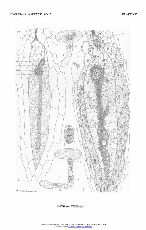
All figures were drawn with the aid of an Abbe camera lucida and reduced one-half in reproduction. Abbreviations are: ni,, male nucleus which fuses with the egg nucleus; in2, male nucleus which does not fuse with the egg nucleus; s,
20 BERRIDGE, ETHEL M., and SANDAY, ELIZABETH, Oogenesis and Ernbryogeny in Ephedra distachya. New Phytologist 6: I28-I34, i67-I74. pIS. 2, 3. I907.
This content downloaded from 194.29.185.134 on Thu, 15 May 2014 10:40:25 AMAll use subject to JSTOR Terms and Conditions
BOTANICAL CAZETTE, XLIV PLATEXX
1A07) onFPIEI)J5A
This content downloaded from 194.29.185.134 on Thu, 15 May 2014 10:40:25 AMAll use subject to JSTOR Terms and Conditions
BOTANICAL. GAZE 7'TE,-XLIV PLATE XXI
/ r 5j f
LAND on}FPIIIYJ.RA
This content downloaded from 194.29.185.134 on Thu, 15 May 2014 10:40:25 AMAll use subject to JSTOR Terms and Conditions
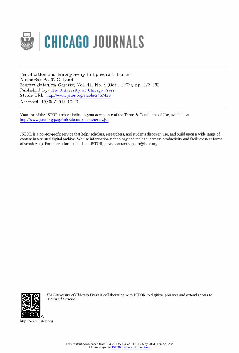
BOTANICAL GiAZETT7E, XLIV, PLATE XXII
14 ~ :.
15~~~~~~~~~~~~~~~~~~~~~~~~~~~~1
21
'1'~~1
0~~~~~4
;f.~ ~ ~ ~ 2
22
cmawa-ut- 2 5 2 %3 24~~~~~~~~~
LAND an EPUEDr
This content downloaded from 194.29.185.134 on Thu, 15 May 2014 10:40:25 AMAll use subject to JSTOR Terms and Conditions
I907] LAND-E1PIHEDRA TRIJFURCA 29I
stalk-cell nucleus; t, tube nucleus; pt, pollen tube; o, egg nucleus; v, ventral nucleus; p, proembryonal nucleus; st, suspensor tube; sn, suspensor nucleus; en, embryo-initial nucleus; s, suspensor; e, embryo-initial cell.
PLATE XX
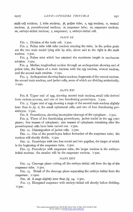
FIG. i. Division of the body cell. X500. FIG. 2. Pollen tube with tube nucleus entering the tube; in the pollen grain
are the two male nuclei lying side by side, above and to the right is the stalk nucleus. X 500.
FIG. 3. Pollen tube which has attained the maximum length in saccharose solution. X 500.
FIG. 4. Median longitudinal section through an archegonium showing end of pollen tube, the fusion of a male nucleus with the egg nucleus, ventral nucleus, and the second male nucleus. X25o.
FIG. 5. Archegonium showing fusion nucleus, fragments of the ventral nucleus. the second male nucleus, and jacket cells, some of which are dividing amitotically. X250.
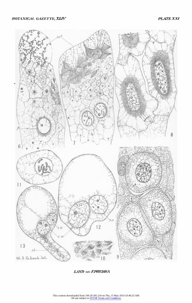
PLATE XXI
FIG. 6. Upper end of egg, showing second male nucleus, small cells derived from various sources, and one of two functioning proembryos. X 500.
FIG. 7. Upper end of egg showing a stage of the second male nucleus slightly later than in fig. 6, the small ephemeral cells, and two of four functioning pro- embryos. X 500.
FIG. 8. Proembryos, showing incomplete cleavage of the cytoplasm. X 5oo. FIG. 9. Three of five functioning proembryos; jacket nuclei in the egg cyto-
plasm; free masses of cytoplasm; also masses of cytoplasm remaining after the proembryonal cells have been carved out. X 500.
FIG. io. Disintegration of jacket cells. X 500. FIG. I I. One of the proembryos before formation of the suspensor tube; the
nucleus will shortly divide. X 500. FIG. 12. Proembryo with two free nuclei and two papillae, the larger of which
is the beginning of the suspensor tube. X 500. FIG. I3. Proembryo with suspensor tube; the larger nucleus is the embryo-
initial nucleus; the smaller will be the suspensor nucleus. X500.
PLATE XXII
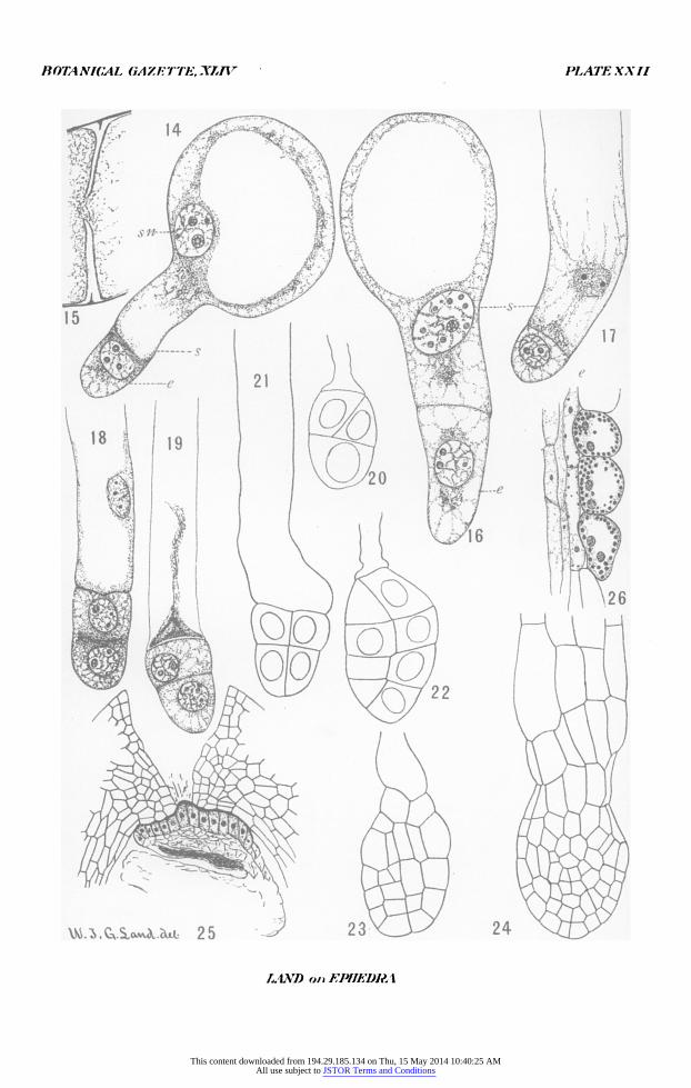
FIG. I4. Cleavage plane cutting off the embryo-initial cell from the tip of the suspensor tube. X 500.
FIG. I5. Detail of the cleavage plane separating the embryo initial from the suspensor. X I900.
FIG. i6. A stage slightly later than fig. I4. X500. FIG. I7. Elongated suspensor with embryo-initial cell shortly before dividing.
X500.
This content downloaded from 194.29.185.134 on Thu, 15 May 2014 10:40:25 AMAll use subject to JSTOR Terms and Conditions
292 BOTANICAL GAZETTE [OCTOBER
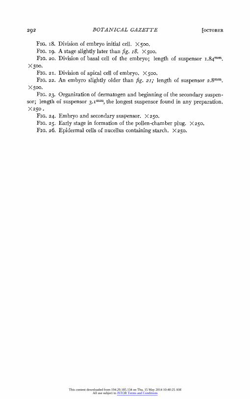
FIG. i8. Division of embryo initial cell. X 500. FIG. i9. A stage slightly later than fig. i8. X 500. FIG. 20. Division of basal cell of the embryo; length of suspensor i.84mm.
X500. FIG. 2i. Division of apical cell of embryo. X 500. FIG. 22. An embyro slightly older than fig. 21; length of suspensor 2.8mm.
X 500. FIG. 23. Organization of dermatogen and beginning of the secondary suspen-
sor; length of suspensor 3.Imm, the longest suspensor found in any preparation. X250.
FIG. 24. Embryo and secondary suspensor. X 250. FIG. 25. Early stage in formation of the pollen-chamber plug. X250. FIG. 26. Epidermal cells of nucellus containing starch. X250.
This content downloaded from 194.29.185.134 on Thu, 15 May 2014 10:40:25 AMAll use subject to JSTOR Terms and Conditions


![Evolving Synaptic Plasticity with an Evolutionary …...been suggested for such systems, including Artificial Ontogeny [8], Computational Embryogeny [9], Cellular Encoding [10,11],](https://img.pdfslide.net/doc/110x75/5f3f8ae8693e0a7d4e5ec431/evolving-synaptic-plasticity-with-an-evolutionary-been-suggested-for-such-systems.jpg)