Embed Size (px)
Citation preview

2613RESEARCH ARTICLE
INTRODUCTIONIn the adult, homeostasis, i.e. the control of the body’s internalenvironment, is mediated through the hypothalamo-pituitaryneuraxis. A central feature of this axis is the projection ofhypothalamic axons through an evagination of the ventralhypothalamic floor termed the infundibulum (Pelletier, 1991).Despite its key role in body function, little is understood of thecellular and molecular events that orchestrate formation of theinfundibulum and that sculpt and maintain it over time.
Much of the ventral hypothalamus arises from anterior ventralmidline floor plate-like cells (Manning et al., 2006). As one of themajor organisers of the CNS, floor plate (fp) cells instruct neuralcells to acquire distinctive fates and establish the elaborate neuronalnetworks that underlie CNS function (Jessell and Dodd, 1990;Ericson et al., 1997; Colamarino and Tessier-Lavigne, 1995;Placzek and Briscoe, 2005). Numerous lines of evidence suggestthat the fp is a non-uniform population, with cells along theanterior-posterior axis displaying distinct characteristics (for areview, see Placzek and Briscoe, 2005). In the forebrain, fp cellsare particularly heterogeneous and show dynamic changes in theirmolecular profiles and behaviour (Kapsimali et al., 2004; Manninget al., 2006; Ohyama et al., 2008). This raises the possibility thatsubsets of anterior fp contribute to defined ventral forebrainstructures, including the infundibulum.
In all vertebrates examined, FGFs are expressed within both fp-derived cells of the ventral hypothalamic midline (Manning et al.,2006) and the forming infundibulum (Tannahill et al., 1992;
Ohuchi et al., 2000; Herzog et al., 2004; Theil et al., 2008; Tsai etal., 2011), where they may play multiple roles in theneuroendocrine hypothalamus. FGFs act as spatial and proliferativecues for progenitors within Rathke’s pouch, which is the precursorof the anterior pituitary (Ericson et al., 1998; Norlin et al., 2000;Zhu et al., 2007), and additionally promote diverse aspects ofspecification of neuroendocrine neurons (Tsai et al., 2011).Similarly, emerging evidence supports a function for FGFsignalling in development of the infundibulum itself. In zebrafish,FGF signalling is required for the expression of Lhx2 (Seth et al.,2006), a Lim homeodomain transcription factor that is expressedwidely in the ventral hypothalamus and that in mouse is requiredfor appropriate proliferation and formation of the infundibulum.Mouse mutants that lack LHX2 expression show unusually highlevels of proliferation in the ventral diencephalic floor, withconcomitant failure of infundibular evagination (Zhao et al., 2010).In Fgf10 knockout mice, furthermore, the infundibulum similarlyfails to evaginate fully and infundibular cells undergo apoptosis(Ohuchi et al., 2000). These studies suggest a link between localproliferation and sculpted formation of the infundibulum, but anintegrated cellular mechanism linking these events remains unclear:LHX2 and Fgf10 are broadly expressed within the infundibulumand ventral diencephalon and could exert their actions on a numberof cell types.
Elsewhere in the CNS, and particularly well-described in theposterior neural tube, FGFs play a key role in neural proliferation(Mathis et al., 2001; Diez del Corral et al., 2003; Akai et al., 2005;Cayuso and Marti, 2005), where they govern expression of SOXgenes, HMG-box transcription factors that affect neuralproliferation and the maintenance of neural stem/progenitor cellrenewal (Bylund et al., 2003; Graham et al., 2003; Ellis et al., 2004;Pevny and Placzek 2005; Scott et al., 2010). In the anterior neuraltube, the SoxB1 transcription factor, SOX3, is expressed in thehypothalamus, around the forming infundibulum, and has beenpreviously implicated in infundibular formation. In mouse Sox3conditional knockouts, the infundibulum and adjacent ventralhypothalamus are thinner and show reduced proliferation rates
Development 138, 2613-2624 (2011) doi:10.1242/dev.062794© 2011. Published by The Company of Biologists Ltd
1MRC Centre for Developmental and Biomedical Genetics and Department ofBiomedical Science, University of Sheffield, Sheffield S10 2TN, UK. 2The RoslinInstitute, University of Edinburgh, Roslin EH25 9PS, UK.
*Author for correspondence ([email protected])†UCLA Department of Neurobiology, Los Angeles, CA 90095-7243, USA‡Nagasaki Medical School, Nagasaki 852-8523, Japan§University of Sydney, NSW2050, Australia
Accepted 23 March 2011
SUMMARYThe infundibulum links the nervous and endocrine systems, serving as a crucial integrating centre for body homeostasis. Here wedescribe that the chick infundibulum derives from two subsets of anterior ventral midline cells. One set remains at the ventralmidline and forms the posterior-ventral infundibulum. A second set migrates laterally, forming a collar around the midline. Weshow that collar cells are composed of Fgf3+ SOX3+ proliferating progenitors, the induction of which is SHH dependent, but themaintenance of which requires FGF signalling. Collar cells proliferate late into embryogenesis, can generate neurospheres thatpassage extensively, and differentiate to distinct fates, including hypothalamic neuronal fates and Fgf10+ anterior-dorsalinfundibular cells. Together, our study shows that a subset of anterior floor plate-like cells gives rise to Fgf3+ SOX3+ progenitorcells, demonstrates a dual origin of infundibular cells and reveals a crucial role for FGF signalling in governing extendedinfundibular growth.
KEY WORDS: FGF, Floor plate, Hypothalamus, Chick
FGF-dependent midline-derived progenitor cells inhypothalamic infundibular developmentCaroline Alayne Pearson1,†, Kyoji Ohyama1,‡, Liz Manning1,§, Soheil Aghamohammadzadeh1, Helen Sang2
and Marysia Placzek1,*
DEV
ELOPM
ENT
2614
(Rizzoti et al., 2004). This raises the possibility that SOX3 mightmaintain normal proliferation rates in ventral hypothalamicprecursors and hence govern infundibular morphogenesis. Humanstudies support this idea, showing that aberrant dosage of SOX3leads to infundibular hypoplasia and abnormal posterior pituitarydevelopment (Woods et al., 2005; di Iorgi et al., 2009). As yet, nostudy has analysed whether FGF signalling and SOX3 expressionare integrated in governing proliferation around the infundibulum.
Here we analyse formation of the infundibulum in the embryonicchick. We describe a dual origin of infundibular cells from twopopulations of ventral midline floor plate-like cells that differmarkedly in behaviour. One population persists at the ventralmidline and forms the posterior-ventral (p/v) infundibulum. Asecond population gives rise to a collar of cells that express Fgf3and SOX3 and are capable of extensive proliferation, with somecollar cell descendants differentiating to Fgf10+ anterior-dorsal(a/d) infundibular cells over an extended period, and other collarcells being retained at the neck of the infundibulum. SOX3+ collarprogenitors require FGF signalling for their maintenance andproliferation. Together, our studies reveal a mechanism to explainthe growth and functional characteristics of the infundibulum.
MATERIALS AND METHODSFate mappingHamburger and Hamilton stage (st) 9 embryos were fate mapped withDiI/DiO (Molecular Probes) as described (Manning et al., 2006) andallowed to develop up to embryonic day (E) 7 (n�40 embryos; 5-7 per timepoint). In neurosphere assays, DiI was targeted to st 9 prosencephalic neckcells, embryos incubated until E4, and collar cells dissected for neurospheregeneration (n�10).
Transplantation experimentsHypothalami were isolated with Dispase (Roche; 1 mg/ml, 40 minutes)from Roslin Green chicken embryos and collar or prospective p/vinfundibular cells subdissected and transplanted into the collar of isolatedwild-type (wt) hypothalami (n�14 and n�5 for homochronic andheterochronic grafts, respectively). Tissue was cultured for 72 hours inMatrigel (BD Biosciences). In situ hybridisation analysis for Fgf10 wascarried out prior to detection of GFP with anti-GFP antibody (Novacastra;1:200).
Explant culturesExplants of fp, collar or prospective p/v infundibulum were isolated fromsurrounding tissues (including Rathke’s pouch; n≥6) after Dispasetreatment and cultured in collagen (Placzek et al., 1993). SU54502(Calbiochem; 10 or 20 �M in DMSO), FGF10 and FGF3 (R&D Systems;10 and 100 ng/ml, respectively), FGF10- or FGF3-blocking antibodies[Santa Cruz Biotechnology; 50 ng/ml (Harada et al., 2002)] were added tothe culture medium.
Cell proliferation assaysExplants were cultured for 16 hours and pulsed with 10 �M BrdU for 1hour prior to fixation.
Immunohistochemistry and in situ hybridisationEmbryos, explants and neurospheres were analysed byimmunohistochemistry and in situ hybridisation according to standardtechniques (Placzek et al., 1993; Manning et al., 2006). Following cryostatsectioning (15-20 �m), the following antibodies were used: anti-SHH(1:50; 5E1, DSHB); anti-NKX2.1 (1:2000) (Ohyama et al., 2005); anti-PAX6 (1:50; DSHB); anti-PH3 (1:1000; Upstate); anti-BrdU (1:200;Novacastra); anti-TBX2 (1:1000; C. Goding) (Manning et al., 2006); anti-Musashi (1:200; Abcam); anti-SOX3 (1:500; T. Edlund, Umea, Sweden;Abcam); anti-TUJ1 (1:1000; Calbiochem); anti-pMAPK (1:50; CellSignaling); anti-SOX2 (1:500; Chemicon); anti-GFP (1:200; Novacastra);anti-P27 (1:1000; BD Biosciences); and anti-cCaspase 3 (1:1000; CellSignaling). Secondary antibodies (1:200; Jackson ImmunoResearch) were
conjugated to Cy3 or FITC. Images were taken using a Zeiss ApoTome orOlympus BX60 and Spot RT software v3.2 (Diagnostic Instruments). For3D reconstruction, overlap, channel visibility and orientation wereperformed using Image-Pro Analyser and Makromedia Fireworks; 3Drendering was performed using Volocity (version 5.4.1, Perkin Elmer).Following development, embryos or neurospheres were analysed aswholemount preparations or cryostat sections.
Neurosphere culturesTissues were dissected as for explants, trypsinised and mechanicallydissociated. Cells were filtered, plated at 90,000-210,000 cells/well intoultralow-binding plates (Nunc) in DMEM/F12 medium supplemented withB27/N21 and with 1:100 chick embryo extract, 20 ng/ml bFGF (FGF2) and20 ng/ml EGF (Molofsky et al., 2003) and incubated for 7 days at 37°C,5% CO2. Neurospheres were passaged after mechanical dissociation. Fordifferentiation, spheres were plated onto poly-D-lysine and fibronectin inmedium without EGF and cultured for 7 days.
Scanning electron microscopy (SEM)Hypothalami were fixed in 3% glutaraldehyde for 4 hours at 4°C,dehydrated, dried, mounted and coated with 25 nm gold (Edwards S150Bsputter coater). Specimens were imaged with a Philips XL-20 scanningelectron microscope at an accelerating voltage of 20 kV.
RESULTSDual origin of infundibular cells from anteriorfloor plateTo address whether anterior fp cells contribute to the infundibulum,we performed fate-mapping studies in chick embryos, tracing cellsfrom st 9 until E7 (Fig. 1A-C), when the infundibulum isstructurally distinct (Fig. 1D). DiI-labelled cells were detectedthroughout the infundibulum, with highest levels in posterior-ventral (p/v) regions (Fig. 1C).
SEM revealed that the infundibulum first becomes apparent atE4.5 (Fig. 1Di,ii) and that at E7 two morphologically discreteinfundibular cells can be detected: elongated cells that extend alongand immediately adjacent to the midline in the p/v infundibulum,and rounded cells in the a/d infundibulum (Fig. 1Diii,iv). Toinvestigate the origin of these cells, we refined the fate mapping,targeting small populations of anterior fp at st 9 with DiI/DiO andanalysing embryos after increasing time periods (Fig. 1E-P). Theseanalyses revealed a differential behaviour of fp cells that areinitially apposed. Fp cells just posterior to the prosencephalic neck(green cells, Fig. 1E,F) remained at the ventral midline (Fig. 1Finset, Fig. 1G-M) and by E7 contributed exclusively to midlinecells of the p/v infundibulum (Fig. 1N-P). By contrast, descendantsof fp cells that were at the prosencephalic neck at st 9 (red cells,Fig. 1E,F) moved laterally/posteriorly to form a semi-ellipse ofcells. These were initially apposed to the ventral midline population(Fig. 1F inset, Fig.1G) but appeared to become progressivelydispersed from it with time (Fig 1H).’
To analyse this behaviour in detail, we examined serial sectionsand performed 3D reconstructions over the period E3.5-4.5. Thisrevealed that, from anterior to posterior, and over time, clusters ofDiI-labelled cells become increasingly displaced from the ventralmidline by their descendants (Fig. 1I-M and see Movie 1 in thesupplementary material). Tracing to E7 revealed that cells from theprosencephalic neck give rise either to clusters of strongly labelledcells at the top of, or immediately adjacent to, the a/d infundibulum(Fig. 1N,O, arrows) or to scattered descendants in the a/dinfundibulum and adjacent hypothalamus (Fig. 1N, right-handpanel). We term the region occupied by these strongly labelled cellsthe ‘collar zone’ (white arrows in schematic, Fig. 1P).
RESEARCH ARTICLE Development 138 (12)
DEV
ELOPM
ENT
Prosencephalic neck descendants were never observed to contributeto the p/v infundibulum. These studies reveal a dual fp origin forthe a/d and p/v infundibulum.
FGF and SOX3 expression in the infundibulum andcollarTo begin to analyse the molecular profiles of the fpsubpopulations and their descendants, we examined theexpression of FGF genes previously reported to be expressed inthe embryonic ventral hypothalamus. No FGF expression wasdetected at st 9 (not shown). However, distinct subpopulationsof FGF-expressing fp cells were specified at this point: whenisolated and cultured to the equivalent of E2.5 (Fig. 2A,B), themore anterior prosencephalic neck (red, Fig. 2A) populationexpressed Fgf3 but not Fgf10 (80% Fgf3+; 90% Fgf10–; n�20
each; Fig. 2C, top panels), whereas the more posterior population(green, Fig. 2A) was Fgf3– Fgf10+ (100% Fgf3–; 80% Fgf10+;n�10 each; Fig. 2C, bottom panels).
These characteristics were revealed in vivo at E2.5 andmaintained to E7, the latest stage examined. Throughout thisperiod, midline cells expressed Fgf10, whereas Fgf3 was detectedin a semi-ellipse of cells increasingly displaced from the midline(Fig. 2D,E,G-I,O,P). Comparison of expression profiles and fate-mapping analyses indicated that fp cells at the prosencephalic neckgive rise to Fgf3+ collar zone cells, whereas posterior fp cells giverise to Fgf10+ p/v infundibular cells (Fig. 2D-F, E2.5; Fig. 2H,I,Iinset, E4.5; Fig. 2O,P,S, E7).
Between E2.5 and E4.5, Fgf10 expression expanded (compareFig. 2D with 2H) and by E7 marked the entire infundibulum (Fig.2M-O). This suggests that, after E2.5, Fgf10+ cells are derived
2615RESEARCH ARTICLEInfundibular formation from anterior floor plate progenitor cells
Fig. 1. Dual origin of the infundibulumfrom the anterior floor plate. (A)�Shhexpression in st 9 chick ventral midline floorplate (fp). (B)�Anterior fp targeted with DiI atst 9. (C)�A transverse section of an E7embryo at the level of the infundibulum(indicated in D), after DiI-labelling at st 9.Section is double labelled to detect DiI (red)and Shh (green). Low (Ci) and high (Cii)magnification views are shown. DiI labels p/vand a/d regions of the infundibulum andtight cell clusters adjacent to the a/dinfundibulum (arrows). The boxed region inCii indicates the region shown in Fig 1N, Fig.2R and Fig. 4A,B. (D)�SEM ventrolateral (D)and ventral (Di-iv) views of infundibulum(outlined in D). The boxed regions in Di andDiii are shown at high magnification in Dii(emerging infundibulum outlined) and Div,respectively. Bracketed arrow in Div points top/v cells. (F-I,N) Chick embryos injected withDiI/DiO at st 9 and analysed immediately inwholemounts (F) or in transverse sectionsthrough anterior or posterior parts of theinfundibulum after successive time periods [F inset, st 11 (8 hours); G, E2.5 (24 hours); I,E3.5 (48 hours); H, E4.5 (72 hours); N, E7(132 hours)]. I shows a subset of serialsections through an E3.5 infundibulum,used to obtain 3D reconstructions. (K,L,O) Three-dimensional confocalreconstructions at successive time periodsviewed ventrally or laterally (K),ventrolaterally (L) or transversely (O).(E,J,M,P) SEM views with fate-mappingschematic superimposed. In P, arrowsindicate collar cell zone, adjacent to a/dinfundibulum and defined through foci ofstrongly-labelled DiI cells (arrow in N,O).Arrows in E indicate directed movement ofprosencephalic neck fp cells; arrowheads inN indicate a/d infundibulum; colouredarrows in K and O indicate viewing axes. FP,floor plate; PM, prechordal mesoderm; a/d,anterior-dorsal infundibulum; p/v, posterior-ventral infundibulum. Scale bars: 100��m inCi,Di,Diii; 40��m in Cii,K,N,O; 50��m in D,L;50�m in Div; 30��m in G; 40��m in H,I.
DEV
ELOPM
ENT
2616
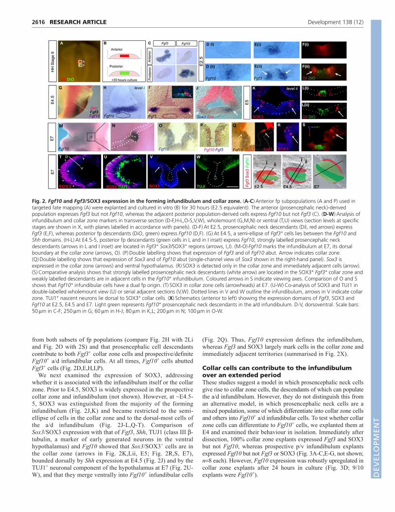
from both subsets of fp populations (compare Fig. 2H with 2Li and Fig. 2O with 2S) and that prosencephalic cell descendantscontribute to both Fgf3+ collar zone cells and prospective/definiteFgf10+ a/d infundibular cells. At all times, Fgf10+ cells abuttedFgf3+ cells (Fig. 2D,E,H,I,P).
We next examined the expression of SOX3, addressingwhether it is associated with the infundibulum itself or the collarzone. Prior to E4.5, SOX3 is widely expressed in the prospectivecollar zone and infundibulum (not shown). However, at ~E4.5-5, SOX3 was extinguished from the majority of the forminginfundibulum (Fig. 2J,K) and became restricted to the semi-ellipse of cells in the collar zone and to the dorsal-most cells ofthe a/d infundibulum (Fig. 2J-L,Q-T). Comparison ofSox3/SOX3 expression with that of Fgf3, Shh, TUJ1 (class III �-tubulin, a marker of early generated neurons in the ventralhypothalamus) and Fgf10 showed that Sox3/SOX3+ cells are inthe collar zone (arrows in Fig. 2K,Lii, E5; Fig. 2R,S, E7),bounded dorsally by Shh expression at E4.5 (Fig. 2J) and by theTUJ1+ neuronal component of the hypothalamus at E7 (Fig. 2U-W), and that they merge ventrally into Fgf10+ infundibular cells
(Fig. 2Q). Thus, Fgf10 expression defines the infundibulum,whereas Fgf3 and SOX3 largely mark cells in the collar zone andimmediately adjacent territories (summarised in Fig. 2X).
Collar cells can contribute to the infundibulumover an extended periodThese studies suggest a model in which prosencephalic neck cellsgive rise to collar zone cells, the descendants of which can populatethe a/d infundibulum. However, they do not distinguish this froman alternative model, in which prosencephalic neck cells are amixed population, some of which differentiate into collar zone cellsand others into Fgf10+ a/d infundibular cells. To test whether collarzone cells can differentiate to Fgf10+ cells, we explanted them atE4 and examined their behaviour in isolation. Immediately afterdissection, 100% collar zone explants expressed Fgf3 and SOX3but not Fgf10, whereas prospective p/v infundibulum explantsexpressed Fgf10 but not Fgf3 or SOX3 (Fig. 3A-C,E-G, not shown;n�8 each). However, Fgf10 expression was robustly upregulated incollar zone explants after 24 hours in culture (Fig. 3D; 9/10explants were Fgf10+).
RESEARCH ARTICLE Development 138 (12)
Fig. 2. Fgf10 and Fgf3/SOX3 expression in the forming infundibulum and collar zone. (A-C)�Anterior fp subpopulations (A and P) used intargeted fate mapping (A) were explanted and cultured in vitro (B) for 30 hours (E2.5 equivalent). The anterior (prosencephalic neck)-derivedpopulation expresses Fgf3 but not Fgf10, whereas the adjacent posterior population-derived cells express Fgf10 but not Fgf3 (C). (D-W)�Analysis ofinfundibulum and collar zone markers in transverse section (D-F,H-L,O-S,V,W), wholemount (G,M,N) or ventral (T,U) views (section levels at specificstages are shown in X, with planes labelled in accordance with panels). (D-F)�At E2.5, prosencephalic neck descendants (Dil, red arrows) expressFgf3 (E,F), whereas posterior fp descendants (DiO, green) express Fgf10 (D,F). (G)�At E4.5, a semi-ellipse of Fgf3+ cells lies between the Fgf10 andShh domains. (H-L)�At E4.5-5, posterior fp descendants (green cells in L and in I inset) express Fgf10, strongly labelled prosencephalic neckdescendants (arrows in L and I inset) are located in Fgf3+ Sox3/SOX3+ regions (arrows, I,J). (M-O)�Fgf10 marks the infundibulum at E7, its dorsalboundary at the collar zone (arrows, O). (P)�Double labelling shows that expression of Fgf3 and of Fgf10 abut. Arrow indicates collar zone.(Q)�Double labelling shows that expression of Sox3 and of Fgf10 abut (single-channel view of Sox3 shown in the right-hand panel). Sox3 isexpressed in the collar zone (arrows) and ventral hypothalamus. (R)�SOX3 is detected only in the collar zone and immediately adjacent cells (arrow).(S)�Comparative analysis shows that strongly labelled prosencephalic neck descendants (white arrow) are located in the SOX3+ Fgf3+ collar zone andweakly labelled descendants are in adjacent cells in the Fgf10+ infundibulum. Coloured arrows in S indicate viewing axes. Comparison of O and Sshows that Fgf10+ infundibular cells have a dual fp origin. (T)�SOX3 in collar zone cells (arrowheads) at E7. (U-W)�Co-analysis of SOX3 and TUJ1 indouble-labelled wholemount view (U) or serial adjacent sections (V,W). Dotted lines in V and W outline the infundibulum, arrows in V indicate collarzone. TUJ1+ nascent neurons lie dorsal to SOX3+ collar cells. (X)�Schematics (anterior to left) showing the expression domains of Fgf3, SOX3 andFgf10 at E2.5, E4.5 and E7. Light green represents Fgf10+ prosencephalic neck descendants in the a/d infundibulum. D-V, dorsoventral. Scale bars:50��m in C-F; 250��m in G; 60��m in H-J; 80��m in K,L; 200��m in N; 100��m in O-W.
DEV
ELOPM
ENT
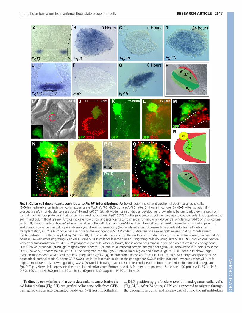
To directly test whether collar zone descendants can colonise thea/d infundibulum (Fig. 3H), we grafted collar zone cells from GFP-transgenic chicks into explanted wild-type (wt) host hypothalami
at E4.5, positioning grafts close to/within endogenous collar cells(Fig. 3I,J). After 24 hours, GFP+ cells appeared to migrate throughthe endogenous collar and medioventrally into the infundibulum
2617RESEARCH ARTICLEInfundibular formation from anterior floor plate progenitor cells
Fig. 3. Collar cell descendants contribute to Fgf10+ infundibulum. (A)�Boxed region indicates dissection of Fgf3+ collar zone cells. (B-D)�Immediately after isolation, collar explants are Fgf3+ Fgf10– (B,C) but are Fgf10+ after 24 hours in culture (D). (E-G)�After isolation (E),prospective p/v infundibular cells are Fgf3– (F) and Fgf10+ (G). (H) Model for infundibular development. p/v infundibulum (dark green) arises fromventral midline floor plate cells that remain in a midline position. Fgf3+ SOX3+ collar progenitors (red) can give rise to descendants that populate thea/d infundibulum (light green). Arrows indicate flow of collar descendants to form a/d infundibulum. (I-L)�Ventral wholemount (I-K) or thick coronalsection (L) views of infundibulum/collar region after collar cells from a Roslin-GFP embryo (head shown in inset, I) were transplanted adjacent toendogenous collar cells in wild-type (wt) embryos, shown schematically (I) or analysed after successive time points (J-L). Immediately aftertransplantation, GFP+ SOX3+ collar cells lie close to the endogenous SOX3+ collar (J). Analysis of a similar graft reveals that GFP+ cells streammedioventrally from the transplant by 24 hours (K, dotted white line indicates the endogenous collar region). The same transplant, analysed at 72hours (L), reveals more migrating GFP+ cells. Some SOX3+ collar cells remain in situ; migrating cells downregulate SOX3. (M)�Thick coronal sectionview after transplantation of E4.5 GFP+ prospective p/v cells. After 72 hours, transplanted cells remain in situ and do not cross the endogenousSOX3+ collar (outlined). (N-P)�High-magnification view of L (N) and serial adjacent section analysed for Fgf10 (O). Arrowhead in N points to someSOX3+ collar cells that remain in situ. GFP+ cells migrate into the Fgf10+ infundibular region and express Fgf10 (Pi,Pii). Inset in Pii shows high-magnification view of a GFP+ cell that has upregulated Fgf10. (Q)�Heterochronic transplant from E10 GFP+ to E4.5 wt embryo analysed after 72hours (thick coronal section). Some GFP+ SOX3+ collar cells remain in situ in the endogenous SOX3+ collar (outlined), whereas other GFP+ cellsmigrate medioventrally, downregulating SOX3. (R)�Model showing that collar cell descendants contribute to a/d infundibulum and upregulateFgf10. Top, yellow circle represents the transplanted collar zone. Bottom, see H. A-P, anterior to posterior. Scale bars: 100��m in A,E; 25��m in B-D,F,G; 100��m in H; 300��m in I; 50��m in J-L; 60��m in N,O; 30��m in P; 50��m in M,Q.
DEV
ELOPM
ENT
2618
(Fig. 3K). Explants were fixed after 72 hours to examine theexpression of collar and infundibular markers and confirm thisbehaviour. These analyses revealed that some GFP+ SOX3+ collarzone cells had remained in situ, with cells closest to theendogenous collar continuing to express SOX3 (Fig. 3L,N,arrowhead). Additionally, GFP+ cells had migrated through theendogenous collar zone into the Fgf10+ infundibulum (Fig.3L,N,O). The stream of GFP+ cells had increased in numberrelative to 24 hours previously (compare Fig. 3K with 3L).Migrating cells emanating from the graft did not express SOX3 butupregulated Fgf10 (Fig. 3O,P) and did not appear to re-enter theendogenous SOX3+ collar zone, suggesting that cells differentiatingfrom collar zone cells remain spatially separate from them. Inaddition to the robust flow of cells into the infundibulum, anoccasional GFP+ cell with neuronal morphology extended dorsallyfrom the graft (not shown). These experiments show that collarzone cells can differentiate, giving rise to cells that colonise theinfundibulum and that upregulate Fgf10.
The maintained expression of SOX3 and Fgf3 in the collar zonesuggests that collar zone cells are retained late into embryogenesis;we therefore asked whether they can contribute progeny to theinfundibulum over an extended period. Heterochronic (E10 GFP+
to E4.5 wt) grafting experiments showed that E10 collar cellsdisplay identical behaviour to those at E4.5: some remained in situand maintained expression of SOX3, whereas some migrated intothe infundibulum (Fig. 3Q). Thus, collar cells retain the ability topopulate the infundibulum into late embryogenesis.
Finally, we addressed whether the directed migratory behaviouris restricted to collar zone cells or whether Fgf10+ p/v infundibularcells behave similarly. GFP+ Fgf10+ p/v infundibular cells weresubdissected (Fig. 3E), similarly grafted (Fig. 3I) and examinedafter 3 days. Such grafts did not invade the SOX3+ collar zone nordid they migrate beyond it into the infundibulum (Fig. 3M).Together, these experiments suggest that the extended growth ofthe infundibulum relies on a protracted inflow of collar zone-derived descendants (schematised in Fig. 3R).
Differential cell proliferation in infundibularformationThe idea that cells from the collar zone contribute to the a/dinfundibulum over a period of days, together with our observationsthat relatively weak Di labelling is detected in a/d as compared
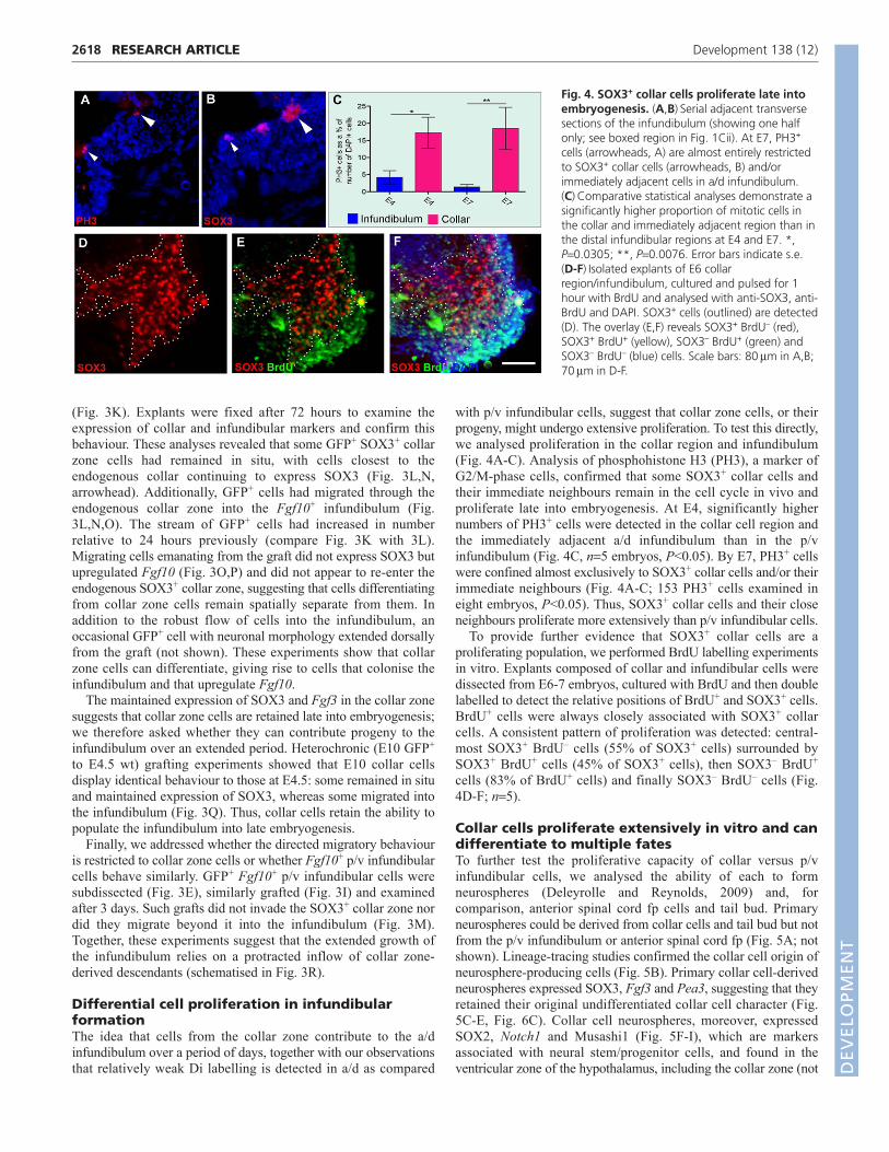
with p/v infundibular cells, suggest that collar zone cells, or theirprogeny, might undergo extensive proliferation. To test this directly,we analysed proliferation in the collar region and infundibulum(Fig. 4A-C). Analysis of phosphohistone H3 (PH3), a marker ofG2/M-phase cells, confirmed that some SOX3+ collar cells andtheir immediate neighbours remain in the cell cycle in vivo andproliferate late into embryogenesis. At E4, significantly highernumbers of PH3+ cells were detected in the collar cell region andthe immediately adjacent a/d infundibulum than in the p/vinfundibulum (Fig. 4C, n�5 embryos, P<0.05). By E7, PH3+ cellswere confined almost exclusively to SOX3+ collar cells and/or theirimmediate neighbours (Fig. 4A-C; 153 PH3+ cells examined ineight embryos, P<0.05). Thus, SOX3+ collar cells and their closeneighbours proliferate more extensively than p/v infundibular cells.
To provide further evidence that SOX3+ collar cells are aproliferating population, we performed BrdU labelling experimentsin vitro. Explants composed of collar and infundibular cells weredissected from E6-7 embryos, cultured with BrdU and then doublelabelled to detect the relative positions of BrdU+ and SOX3+ cells.BrdU+ cells were always closely associated with SOX3+ collarcells. A consistent pattern of proliferation was detected: central-most SOX3+ BrdU– cells (55% of SOX3+ cells) surrounded bySOX3+ BrdU+ cells (45% of SOX3+ cells), then SOX3– BrdU+
cells (83% of BrdU+ cells) and finally SOX3– BrdU– cells (Fig.4D-F; n�5).
Collar cells proliferate extensively in vitro and candifferentiate to multiple fatesTo further test the proliferative capacity of collar versus p/vinfundibular cells, we analysed the ability of each to formneurospheres (Deleyrolle and Reynolds, 2009) and, forcomparison, anterior spinal cord fp cells and tail bud. Primaryneurospheres could be derived from collar cells and tail bud but notfrom the p/v infundibulum or anterior spinal cord fp (Fig. 5A; notshown). Lineage-tracing studies confirmed the collar cell origin ofneurosphere-producing cells (Fig. 5B). Primary collar cell-derivedneurospheres expressed SOX3, Fgf3 and Pea3, suggesting that theyretained their original undifferentiated collar cell character (Fig.5C-E, Fig. 6C). Collar cell neurospheres, moreover, expressedSOX2, Notch1 and Musashi1 (Fig. 5F-I), which are markersassociated with neural stem/progenitor cells, and found in theventricular zone of the hypothalamus, including the collar zone (not
RESEARCH ARTICLE Development 138 (12)
Fig. 4. SOX3+ collar cells proliferate late intoembryogenesis. (A,B)�Serial adjacent transversesections of the infundibulum (showing one halfonly; see boxed region in Fig. 1Cii). At E7, PH3+
cells (arrowheads, A) are almost entirely restrictedto SOX3+ collar cells (arrowheads, B) and/orimmediately adjacent cells in a/d infundibulum.(C)�Comparative statistical analyses demonstrate asignificantly higher proportion of mitotic cells inthe collar and immediately adjacent region than inthe distal infundibular regions at E4 and E7. *,P�0.0305; **, P�0.0076. Error bars indicate s.e.(D-F)�Isolated explants of E6 collarregion/infundibulum, cultured and pulsed for 1hour with BrdU and analysed with anti-SOX3, anti-BrdU and DAPI. SOX3+ cells (outlined) are detected(D). The overlay (E,F) reveals SOX3+ BrdU– (red),SOX3+ BrdU+ (yellow), SOX3– BrdU+ (green) andSOX3– BrdU– (blue) cells. Scale bars: 80��m in A,B;70��m in D-F.
DEV
ELOPM
ENT
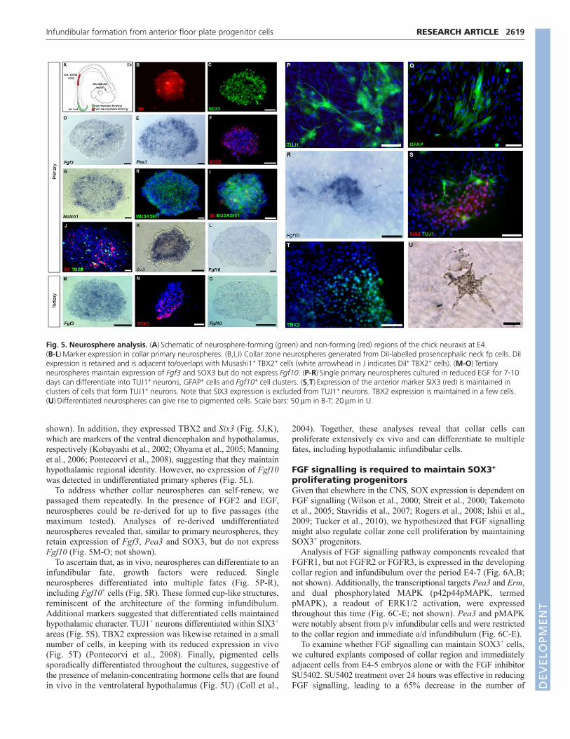
shown). In addition, they expressed TBX2 and Six3 (Fig. 5J,K),which are markers of the ventral diencephalon and hypothalamus,respectively (Kobayashi et al., 2002; Ohyama et al., 2005; Manninget al., 2006; Pontecorvi et al., 2008), suggesting that they maintainhypothalamic regional identity. However, no expression of Fgf10was detected in undifferentiated primary spheres (Fig. 5L).
To address whether collar neurospheres can self-renew, wepassaged them repeatedly. In the presence of FGF2 and EGF,neurospheres could be re-derived for up to five passages (themaximum tested). Analyses of re-derived undifferentiatedneurospheres revealed that, similar to primary neurospheres, theyretain expression of Fgf3, Pea3 and SOX3, but do not expressFgf10 (Fig. 5M-O; not shown).
To ascertain that, as in vivo, neurospheres can differentiate to aninfundibular fate, growth factors were reduced. Singleneurospheres differentiated into multiple fates (Fig. 5P-R),including Fgf10+ cells (Fig. 5R). These formed cup-like structures,reminiscent of the architecture of the forming infundibulum.Additional markers suggested that differentiated cells maintainedhypothalamic character. TUJ1+ neurons differentiated within SIX3+
areas (Fig. 5S). TBX2 expression was likewise retained in a smallnumber of cells, in keeping with its reduced expression in vivo(Fig. 5T) (Pontecorvi et al., 2008). Finally, pigmented cellssporadically differentiated throughout the cultures, suggestive ofthe presence of melanin-concentrating hormone cells that are foundin vivo in the ventrolateral hypothalamus (Fig. 5U) (Coll et al.,
2004). Together, these analyses reveal that collar cells canproliferate extensively ex vivo and can differentiate to multiplefates, including hypothalamic infundibular cells.
FGF signalling is required to maintain SOX3+
proliferating progenitorsGiven that elsewhere in the CNS, SOX expression is dependent onFGF signalling (Wilson et al., 2000; Streit et al., 2000; Takemotoet al., 2005; Stavridis et al., 2007; Rogers et al., 2008; Ishii et al.,2009; Tucker et al., 2010), we hypothesized that FGF signallingmight also regulate collar zone cell proliferation by maintainingSOX3+ progenitors.
Analysis of FGF signalling pathway components revealed thatFGFR1, but not FGFR2 or FGFR3, is expressed in the developingcollar region and infundibulum over the period E4-7 (Fig. 6A,B;not shown). Additionally, the transcriptional targets Pea3 and Erm,and dual phosphorylated MAPK (p42p44pMAPK, termedpMAPK), a readout of ERK1/2 activation, were expressedthroughout this time (Fig. 6C-E; not shown). Pea3 and pMAPKwere notably absent from p/v infundibular cells and were restrictedto the collar region and immediate a/d infundibulum (Fig. 6C-E).
To examine whether FGF signalling can maintain SOX3+ cells,we cultured explants composed of collar region and immediatelyadjacent cells from E4-5 embryos alone or with the FGF inhibitorSU5402. SU5402 treatment over 24 hours was effective in reducingFGF signalling, leading to a 65% decrease in the number of
2619RESEARCH ARTICLEInfundibular formation from anterior floor plate progenitor cells
Fig. 5. Neurosphere analysis. (A)�Schematic of neurosphere-forming (green) and non-forming (red) regions of the chick neuraxis at E4. (B-L)�Marker expression in collar primary neurospheres. (B,I,J) Collar zone neurospheres generated from DiI-labelled prosencephalic neck fp cells. DiIexpression is retained and is adjacent to/overlaps with Musashi1+ TBX2+ cells (white arrowhead in J indicates DiI+ TBX2+ cells). (M-O)�Tertiaryneurospheres maintain expression of Fgf3 and SOX3 but do not express Fgf10. (P-R)�Single primary neurospheres cultured in reduced EGF for 7-10days can differentiate into TUJ1+ neurons, GFAP+ cells and Fgf10+ cell clusters. (S,T)�Expression of the anterior marker SIX3 (red) is maintained inclusters of cells that form TUJ1+ neurons. Note that SIX3 expression is excluded from TUJ1+ neurons. TBX2 expression is maintained in a few cells.(U)�Differentiated neurospheres can give rise to pigmented cells. Scale bars: 50��m in B-T; 20��m in U.
DEV
ELOPM
ENT

2620
pMAPK-expressing cells and similarly reduced the extent of Pea3expression (n�40; not shown). Treatment over 24 hours resulted ina complete loss of SOX3+ cells, without a similar immediatedecrease in NKX2.1+ infundibular progenitors (Fig. 6G-L). Thisindicates that FGF signalling may selectively maintain SOX3+ cells,rather than exerting a generalised effect on all progenitor cells.
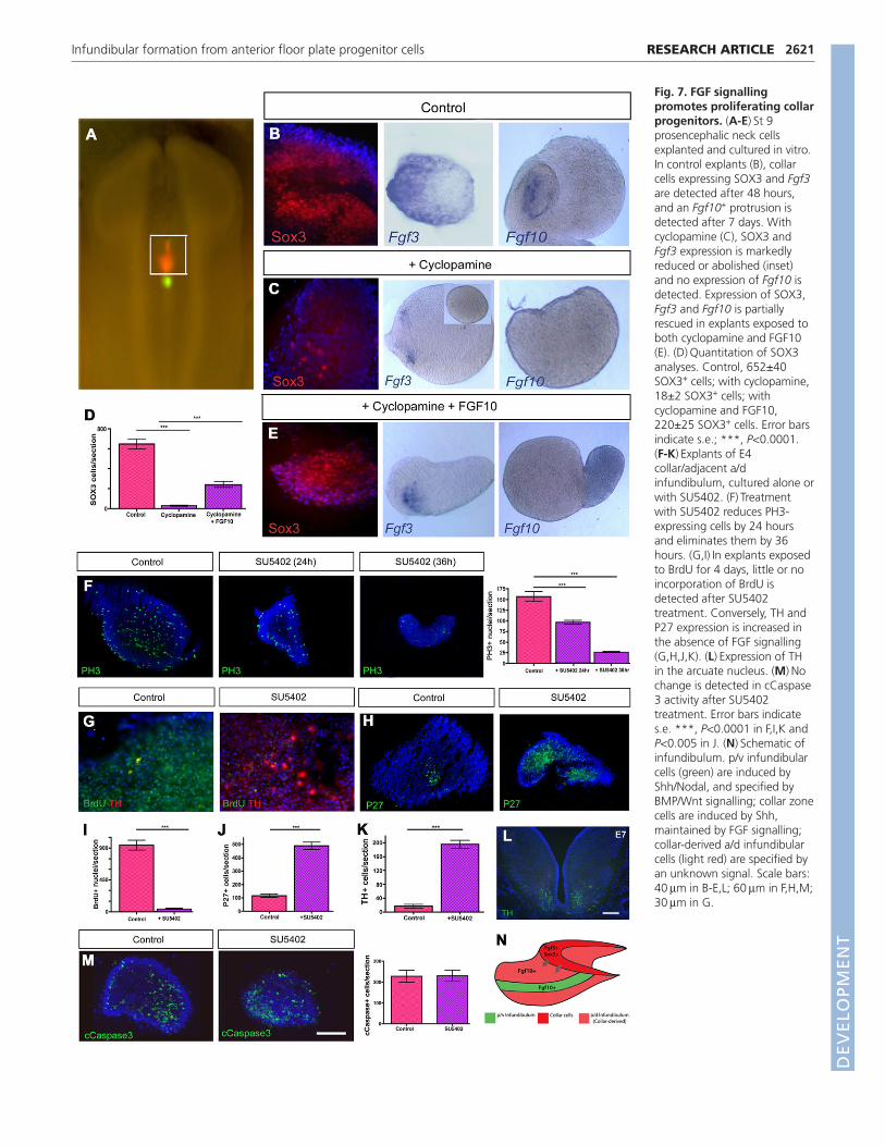
Although SU5402 is widely used as an inhibitor of FGFsignalling, it does not distinguish whether it is FGF10, FGF3 orboth ligands that contribute to SOX3+ cell maintenance. Wetherefore analysed the effects of FGF10- or FGF3-blockingantibodies by culturing explants alone or in the presence of eitheror both antibodies. In the absence of FGF10 signalling there was asignificant decrease in Pea3 expression and in the number ofpMAPK+ and SOX3+ cells (P<0.0001 and P<0.0006, respectively).By contrast, blockade of FGF3 signalling resulted in a less severedecrease in Pea3 expression and in pMAPK+ and SOX3+ cellnumber (P<0.0035 and P<0.046, respectively). However, exposureof explants to both blocking antibodies together resulted in asubstantial reduction or the complete loss of Pea3 expression andin a highly significant decrease in pMAPK+ and SOX3+ cellnumber (P<0.0001 for both; n�8-10 explants each; Fig. 6M-Y).This suggests that SOX3+ cells might require both FGF3 from thecollar region and FGF10 from the forming infundibulum.
Studies in mouse have shown that expression of Fgf3 in theventral forebrain is governed, at least in part, by SHH (Powles etal., 2004), raising the possibility that collar cells are SHHdependent. To examine this in chick, we dissected st 9 prospectivecollar cells (Fig. 7A) and cultured them alone or with cyclopamine,an inhibitor of SHH signalling. Cyclopamine treatment reduced theexpression of SOX3 and Fgf3 after a short culture period and ofFgf10 after protracted culture (Fig. 7B,C). Thus, as with manyother hypothalamic progenitors (Ohyama et al., 2008), collar cellinduction appears to depend on early SHH signalling. We next usedthis assay to establish whether FGF ligands can expand collar cells,rescuing their numbers after a reduction in SHH signalling.Exposure of prospective collar cells to a combination ofcyclopamine and FGF10 resulted in a partial rescue of SOX3-expressing cells and Fgf3 expression. Moreover, after an extendedperiod, Fgf10 expression was detected in cells protruding from themain body of the explant (Fig. 7D,E). These experiments supportthe idea that FGF10 expands collar cells and suggests that FGFsmight govern collar cell proliferation.
To test this latter contention directly, we asked whether the lossof SOX3+ cells following a reduction in FGF signalling isaccompanied by a decrease in progenitor cell proliferation.Treatment of E4.5 collar region explants with SU5402 not only
RESEARCH ARTICLE Development 138 (12)
Fig. 6. FGF signalling maintains SOX3+ progenitors. (A-E)�Transverse sections showing expression of FGF signalling components. The formingp/v infundibulum and Rathke’s pouch are outlined (C). The boxed region in D is shown at higher magnification in E. (F)�Expression of NKX2.1 in theinfundibulum. (G-T)�Explants of collar/infundibulum, dissected at E4, cultured alone or with FGF inhibitors. (G-L)�SU5402 significantly affects thenumber of SOX3+ cells but not the number of NKX2.1+ cells. (M-P)�Many pMAPK+ cells are seen in control explants (514±39.7), fewer with FGF3-blocking antibody (321±11.45), and significant reductions are detected in the presence of FGF10-blocking antibody (134±5.8) or both blockingantibodies (15±14.6). (Q-T)�Pea3 expression declines after exposure to FGF3 antibody, still further after exposure to FGF10 antibody, and is notdetected after exposure to both antibodies. (U-Y)�SOX3 cell number declines after exposure to FGF3 antibody, still further after exposure to FGF10antibody, and few SOX3+ cells are detected after exposure to both antibodies. Error bars indicate s.e. *, P�0.0006; **, P�0.0001. Scale bars: 50��min A,B,E-K,M-X; 60��m in C; 100��m in D; 40��m in Q-T.
DEV
ELOPM
ENT
2621RESEARCH ARTICLEInfundibular formation from anterior floor plate progenitor cells
Fig. 7. FGF signallingpromotes proliferating collarprogenitors. (A-E)�St 9prosencephalic neck cellsexplanted and cultured in vitro.In control explants (B), collarcells expressing SOX3 and Fgf3are detected after 48 hours,and an Fgf10+ protrusion isdetected after 7 days. Withcyclopamine (C), SOX3 andFgf3 expression is markedlyreduced or abolished (inset)and no expression of Fgf10 isdetected. Expression of SOX3,Fgf3 and Fgf10 is partiallyrescued in explants exposed toboth cyclopamine and FGF10(E). (D)�Quantitation of SOX3analyses. Control, 652±40SOX3+ cells; with cyclopamine,18±2 SOX3+ cells; withcyclopamine and FGF10,220±25 SOX3+ cells. Error barsindicate s.e.; ***, P<0.0001.(F-K)�Explants of E4collar/adjacent a/dinfundibulum, cultured alone orwith SU5402. (F)�Treatmentwith SU5402 reduces PH3-expressing cells by 24 hoursand eliminates them by 36hours. (G,I)�In explants exposedto BrdU for 4 days, little or noincorporation of BrdU isdetected after SU5402treatment. Conversely, TH andP27 expression is increased inthe absence of FGF signalling(G,H,J,K). (L)�Expression of THin the arcuate nucleus. (M)�Nochange is detected in cCaspase3 activity after SU5402treatment. Error bars indicates.e. ***, P<0.0001 in F,I,K andP<0.005 in J. (N)�Schematic ofinfundibulum. p/v infundibularcells (green) are induced byShh/Nodal, and specified byBMP/Wnt signalling; collar zonecells are induced by Shh,maintained by FGF signalling;collar-derived a/d infundibularcells (light red) are specified byan unknown signal. Scale bars:40��m in B-E,L; 60��m in F,H,M;30��m in G.
DEV
ELOPM
ENT
2622
eliminated SOX3 expression (Fig. 6G,H) but led to a significantdecrease in cycling cells (Fig. 7F). A 50% reduction in PH3+ cellswas observed after 24 hours and a 90% reduction after 36 hours(n�5, P<0.0001). Similarly, when BrdU was administered tocontrol or SU5402-treated explants, significantly fewer BrdU+ cellswere found following a reduction in FGF signalling (n�6,P<0.0001; Fig. 7G,I). Reduced proliferation was accompanied byenhanced differentiation: we detected a highly significant increasein p27, a marker of post-mitotic cells (n�7, P<0.001; Fig. 7H,J),and in tyrosine hydroxylase (TH)+ dopaminergic neurons thatdifferentiate in the ventral hypothalamic arcuate nucleus (Fig.7G,K,L), after reduction of FGF signalling. Exposure to SU5402did not appear to promote an increase in cell death: no differencewas detected in the rate of apoptosis, as measured through activatedcleaved (c) Caspase 3 activity (n�5, P�0.96; Fig. 7M), in controlversus SU5402-exposed explants. Together, these results suggestthat decreased FGF signalling leads to a reduction in collar cellproliferation.
DISCUSSIONThe infundibulum plays a pivotal role in vertebrates, linking thenervous and endocrine systems, and its proper development iscrucial to homeostasis. Previous studies have suggested that theinfundibulum derives from the ventral midline of thehypothalamus and that its development is triggered through earlysignalling events between the hypothalamic midline andRathke’s pouch (Pelletier, 1991; Dasen et al., 2001; Hermesz etal., 2003; Rizzoti et al., 2004). Here, we demonstrate that twoanterior fp subsets that are initially induced by Nodal and SHHsignalling (for a review, see Placzek and Briscoe, 2005) fashionthe infundibulum, governing its protracted sculpted evagination.The p/v infundibular cells derive from a set of anterior fp cellsthat are specified through BMP/Wnt signals to express Fgf10(Fig. 7N) (Kapsimali et al., 2004; Manning et al., 2006) and thatremain in a ventral midline location. By contrast, a/dinfundibular cells derive from a second subset of anterior fp cellsthat migrate laterally and posteriorly to form a collar of cellsaround the forming p/v infundibulum. SOX3+ collar cells are aproliferative neural progenitor population that, although initiallyinduced by SHH, is dependent on FGF signalling. In addition toproliferation, collar cells can differentiate to multiple fates,including Fgf10+ cells that populate the a/d infundibulum (Fig.7N). Proliferating collar cells are retained at the junction of theinfundibulum and hypothalamus at least until late intoembryogenesis. These findings have implications for thedevelopment, function and maintenance of the infundibulum.
Dual origin of the infundibulumMany lines of evidence in our study support a dual fp origin forFgf10+ infundibular cells. Our fate-mapping studies reveal that oneset of anterior fp cells remains at the midline, giving rise to cellsthat populate the p/v infundibulum, whereas a second adjacent set(‘prosencephalic neck’ cells) gives rise to a collar of cells, fromwhich the a/d infundibulum forms. The two fp populations displaymarkedly different behaviours. Forming p/v infundibular cells donot proliferate extensively, as evidenced by the retention of stronglineage label expression and a lack of active mitosis, and theirdescendants remain at the ventral midline: little or no cell mixingis observed in double fate-mapping analyses and graftingexperiments show that prospective p/v infundibular cells do notcontribute to the a/d infundibulum. The final fate of p/vinfundibular cells in unclear, but a likely possibility is that they
give rise to the posterior pituitary/neurohypophysis, which is theregion in the later embryo to which magnocellular hypothalamicaxons project.
By contrast, cells of the a/d infundibulum form from apopulation of cells that undergo extensive migration andproliferation. The behaviour of isolated explants (Fig. 7A,B)suggests that prosencephalic neck cells intrinsically migrateposteriorly/laterally to form collar zone cells. Some cells in thecollar zone appear to proliferate little or slowly, as judged by labelretention; however, in general, extensive proliferation occurs in thecollar zone. Our observations raise the possibility that the collarzone has aspects of a stem cell-like niche, in which slowly dividingcells can give rise to rapidly proliferating progenitors, some ofwhich differentiate to a/d infundibular fates. Neurosphere analyses,explant culture and grafting studies support this interpretation,showing that collar zone descendants can differentiate into multiplefates, including Fgf10+ cells that colonise the a/d infundibulum.
Although we cannot exclude the possibility that there is anadditional source of cells that contributes to the a/d infundibulum,our data strongly suggest a model in which cells of the a/dinfundibulum derive from collar cells, which in turn originate fromprosencephalic neck fp. Our grafting studies show, moreover, that thecollar zone is able to contribute cells to the Fgf10+ infundibulumover an extensive period of time, at least until E10. This observationsuggests that, in the late embryonic period, collar zone cells that nowlie at the interface of the infundibulum and hypothalamus cancontinue to shape and/or maintain the infundibulum.
FGF signalling governs proliferating SOX3+ collarprogenitorsEmerging studies from a number of vertebrates suggest that FGFsignalling plays a pivotal role in development of theneuroendocrine hypothalamus and the pituitary gland, and raise thenotion that FGFs might govern the development of theinfundibulum itself, potentially via effects on proliferation (for areview, see Tsai et al., 2011). Our studies suggest a mechanism forFGF function in infundibular growth, showing that FGF signallingis necessary for the proliferation of collar cells.
Our studies reveal, though, that the collar zone is not ahomogeneous population, and we cannot unequivocally establishwhich cells respond directly to FGF signalling, nor which collarzone cell type gives rise to a/d infundibular cells. However, in themouse, SOX3 has been shown to play a crucial role in infundibulardevelopment (Rizzoti et al., 2004) and our observations support theview that SOX3+ cells play an intimate role in a/d infundibularformation in chick: in cyclopamine-treated explants, the reductionof SOX3 is accompanied by a reduction in Fgf10+ cells;conversely, the rescue of SOX3 is accompanied by the rescue ofFgf10+ cells.
Our studies demonstrate, furthermore, that SOX3+ collar cellsare proliferative progenitors. In vivo, SOX3+ cells are maintainedlate into embryogenesis and are mitotically active, as evidenced bydetection of PH3. In vitro, SOX3+ cells can proliferate, as judgedby uptake of BrdU, but are not depleted, their numbers remainingrelatively constant between E5 and E7. Notably, only a subset ofSOX3+ cells appears to undergo rapid division. This, together withthe observation from the fate-mapping studies that SOX3+ cellsspan label-retaining collar zone regions and immediately adjacentlabel-diluted regions in the dorsal-most a/d infundibulum, suggestdifferential proliferation in subsets of SOX3+ cells. The existenceof distinct subsets of SOX3+ cells could account for the lack of anyapparent change in SOX3 expression in the Lhx2-null mouse, in
RESEARCH ARTICLE Development 138 (12)
DEV
ELOPM
ENT
which inappropriately high levels of proliferation are detected inthe ventral diencephalic floor, with concomitant failure ofinfundibular evagination (Zhao et al., 2010).
How SOX3 exerts its actions and the nature of SOX3+ cellsremain unclear. Several members of the SOX family are expressedin neural stem and progenitor cells, where they are thought to playcrucial roles in cell proliferation and in the maintenance of theneural stem and progenitor state (Pevny and Placzek, 2005; Scottet al., 2010). SOX family members operate in a context-dependentmanner, interacting with partner proteins, including other SOXproteins, to effect their actions. It seems likely that additional SOXproteins might play a role in the collar zone, interacting withSOX3+ cell subsets.
Proliferating SOX3+ collar cells are dependent on FGF signalling.A reduction in FGF signalling results in the loss of SOX3+ cells andin the gradual depletion of proliferative progenitors. The decrease inproliferation does not appear to be accompanied by changes inapoptosis, but instead by an increase in differentiated cells.Conversely, SOX3+ cells can be rescued by FGF10 aftercyclopamine treatment. Although we cannot prove that FGF operatesdirectly on SOX3+ cells, FGF signalling is not simply a permissiveproliferative signal for all progenitor cells: NKX2.1+ infundibularprogenitors are not acutely affected by reduced levels of FGF. Ourstudies are therefore consistent with a model in which SOX3+ cellscan either proliferate or are capable of giving rise to differentiatedprogeny, including descendants that contribute to the a/dinfundibulum over an extended period. As yet, we do not understandhow collar cells normally differentiate to infundibular cells, but thedownregulation of FGF signalling components, including Pea3 andpMAPK, in differentiating collar cells that enter the a/d infundibulumsuggests a loss of competence to FGF signalling.
One likely early source of FGFs for the maintenance of collarcells is the p/v infundibular cell population. This suggests a novelrole for ventral midline FGF10+ cells: signalling to adjacent floorplate-like cells to sustain them in a proliferative state. The findingthat FGF signalling from ventral midline cells may sustainproliferative properties in adjacent lateral fp cells might be relevantin other regions of the neuraxis. SoxB1 gene expression ismaintained in lateral fp cells that show sustained proliferation(Pevny et al., 1998), whereas expression of FGFs, namely Fgf3 andthe isoform altFgf2, has been noted in ventral midline fp cells inthe posterior neuraxis (Shi et al., 2009; Borja et al., 1996).
Our neurosphere analyses and grafting experiments show thatcollar cells can give rise not just to infundibular cells, but also toneuronal cells. This suggests that anterior fp cells that lie at theprosencephalic neck at st 9 have neuronal potential, a propertypreviously ascribed only to midbrain ventral midline fp cells(Hynes et al., 1995; Andersson et al., 2006; Ono et al., 2007), andthat, in addition to their role in infundibular formation, collar cellsmight contribute to, and shape, neuronal components of thehypothalamus.
In summary, our data suggest the presence of a spatiallyrestricted progenitor zone that forms around the anterior end ofventral midline cells of the neural tube. We propose that this zoneshapes both the infundibulum and, potentially, the overlyinghypothalamus, and that it can contribute cells to the infundibulumover an extended period. Intriguingly, other studies have shownthat there is a second proliferative zone at the most caudal regionof the forming neural tube, in which FGF signalling maintains cellsin a proliferative, undifferentiated state (Diez del Corral et al.,
2003; Delfino-Machin et al., 2005; McGrew et al., 2008). The twoends of the neural tube therefore share the ability to promote FGFsignalling and establish proliferative zones.
AcknowledgementsWe thank P. Ellis, H. Thornton and C. Hill for help with immunohistochemistryand SEM; and I. Mason. T. Jessell, K. Storey, J. Lopez Rios, K. Katsube, T.Edlund, C. Goding and H. Okano for probes and antibodies. This work wassupported through the MRC, the BBSRC (provision of Roslin Green eggs) andthe Wellcome Trust. Deposited in PMC for release after 6 months.
Competing interests statementThe authors declare no competing financial interests.
Supplementary materialSupplementary material for this article is available athttp://dev.biologists.org/lookup/suppl/doi:10.1242/dev.062794/-/DC1
ReferencesAkai, J., Halley, P. A. and Storey, K. G. (2005). FGF-dependent Notch signaling
maintains the spinal cord stem zone. Genes Dev. 19, 2877-2887.Andersson, E., Tryggvason, U., Deng, Q., Friling, S., Alekseenko, Z., Robert,
B., Perlmann, T. and Ericson, J. (2006). Identification of intrinsic determinantsof midbrain dopamine neurons. Cell 124, 393-405.
Borja, A. Z. M., Murphy, C. and Zeller, R. (1996). AltFGF-2, a novel ER-associated FGF-2 protein isoform: its embryonic distribution and functionalanalysis during neural tube development. Dev. Biol. 180, 680-692.
Bylund, M., Andersson, E., Novitch, B. G. and Muhr, J. (2003). Vertebrateneurogenesis is counteracted by Sox1-3 activity. Nat. Neurosci. 6, 1162-1168.
Cayuso, J. and Martí, E. (2005). Morphogens in motion: growth control of theneural tube. J. Neurobiol. 64, 376-387.
Colamarino, S. A. and Tessier-Lavigne, M. (1995). The role of the floor plate inaxon guidance. Annu. Rev. Neurosci. 18, 497-529.
Coll, A. P., Farooqi, I. S., Challis, B. G., Yeo, G. S. and O’Rahilly, S. (2004).Proopiomelanocortin and energy balance: insights from human and murinegenetics. J. Clin. Endocrinol. Metab. 89, 2557-2562.
Dasen, J. S., Barbera, J. P., Herman, T. S., Connell, S. O., Olson, L., Ju, B.,Tollkuhn, J., Baek, S. H., Rose, D. W. and Rosenfeld, M. G. (2001). Temporalregulation of a paired-like homeodomain repressor/TLE corepressor complex anda related activator is required for pituitary organogenesis. Genes Dev. 15, 3193-3207.
Deleyrolle, L. P. and Reynolds, B. A. (2009). Isolation, expansion, anddifferentiation of adult Mammalian neural stem and progenitor cells using theneurosphere assay. Methods Mol. Biol. 549, 91-101.
Delfino-Machín, M., Lunn, J. S., Breitkreuz, D. N., Akai, J. and Storey, K. G.(2005). Specification and maintenance of the spinal cord stem zone.Development 132, 4273-4283.
di Iorgi, N., Secco, A., Napoli, F., Calandra, E., Rossi, A. and Maghnie, M.(2009). Developmental abnormalities of the posterior pituitary gland. Endocr.Dev. 14, 83-94.
Diez del Corral, R., Olivera-Martinez, I., Goriely, A., Gale, E., Maden, M. andStorey, K. (2003). Opposing FGF and retinoid pathways control ventral neuralpattern, neuronal differentiation, and segmentation during body axis extension.Neuron 40, 65-79.
Ellis, P., Fagan, B. M., Magness, S. T., Hutton, S., Taranova, O., Hayashi, S.,McMahon, A., Rao, M. and Pevny, L. (2004). SOX2, a persistent marker formultipotential neural stem cells derived from embryonic stem cells, the embryoor the adult. Dev. Neurosci. 26, 148-165.
Ericson, J., Briscoe, J., Rashbass, P., van Heyningen, V. and Jessell, T. M.(1997). Graded sonic hedgehog signaling and the specification of cell fate in theventral neural tube. Cold Spring Harb. Symp. Quant. Biol. 62, 451-466.
Ericson, J., Norlin, S., Jessell, T. M. and Edlund, T. (1998). Integrated FGF andBMP signaling controls the progression of progenitor cell differentiation and theemergence of pattern in the embryonic anterior pituitary. Development 125,1005-1015.
Graham, V., Khudyakov, J., Ellis, P. and Pevny, L. (2003). SOX2 functions tomaintain neural progenitor identity. Neuron 39, 749-765.
Harada, H., Toyono, T., Toyoshima, K., Yamasaki, M., Itoh, N., Kato, S.,Sekine, K. and Ohuchi, H. (2002). FGF10 maintains stem cell compartment indeveloping mouse incisors. Development 129, 1533-1541.
Hermesz, E., Williams-Simons, L. and Mahon, K. A. (2003). A novel inducibleelement, activated by contact with Rathke’s pouch, is present in the regulatoryregion of the Rpx/Hesx1 homeobox gene. Dev. Biol. 260, 68-78.
Herzog, W., Sonntag, C., von der Hardt, S., Roehl, H. H., Varga, Z. M. andHammerschmidt, M. (2004). Fgf3 signaling from the ventral diencephalon isrequired for early specification and subsequent survival of the zebrafishadenohypophysis. Development 131, 3681-3692.
2623RESEARCH ARTICLEInfundibular formation from anterior floor plate progenitor cells
DEV
ELOPM
ENT
2624
Hynes, M., Porter, J. A., Chiang, C., Chang, D., Tessier-Lavigne, M., Beachy, P.A. and Rosenthal, A. (1995). Induction of midbrain dopaminergic neurons bySonic hedgehog. Neuron 15, 35-44.
Ishii, Y., Weinberg, K., Oda-Ishii, I., Coughlin, L. and Mikawa, T. (2009).Morphogenesis and cytodifferentiation of the avian retinal pigmentedepithelium require downregulation of Group B1 Sox genes. Development 136,2579-2589.
Jessell, T. M. and Dodd, J. (1990). Floor plate-derived signals and the control ofneural cell pattern in vertebrates. Harvey Lect. 86, 87-128.
Kapsimali, M., Caneparo, L., Houart, C. and Wilson, S. W. (2004). Inhibition ofWnt/Axin/{beta}-catenin pathway activity promotes ventral CNS midline tissue toadopt hypothalamic rather than floor plate identity. Development 131, 5923-5933.
Kobayashi, D., Kobayashi, M., Matsumoto, K., Ogura, T., Nakafuku, M. andShimamura, K. (2002). Early subdivisions in the neural plate define distinctcompetence for inductive signals. Development 129, 83-93.
Manning, L., Ohyama, K., Saeger, B., Hatano, O., Wilson, S. A., Logan, M.and Placzek, M. (2006). Regional morphogenesis in the hypothalamus: a BMP-Tbx2 pathway coordinates fate and proliferation through Shh downregulation.Dev. Cell 11, 873-885.
Mathis, L., Kulesa, P. M. and Fraser, S. E. (2001). FGF receptor signalling isrequired to maintain neural progenitors during Hensen’s node progression. Nat.Cell Biol. 3, 559-566.
McGrew, M. J., Sherman, A., Liuico, S. G., Ellard, F. M., Radcliffe, P. A.,Gilhooley, H. J., Mitrophanous, K. A., Cambray, N., Wilson, V. and Sang,H. (2008). Localised axial progenitor cell populations in the avian tail bud are notcommitted to a posterior Hox identity. Development 135, 2289-2299.
Molofsky, A. V., Pardal, R., Iwashita, T., Park, I. K., Clarke, M. F. andMorrison, S. J. (2003). Bmi-1 dependence distinguishes neural stem cell self-renewal from progenitor proliferation. Nature, 425, 962-967.
Norlin, S., Nordström, U. and Edlund, T. (2000). Fibroblast growth factorsignaling is required for the proliferation and patterning of progenitor cells inthe developing anterior pituitary. Mech. Dev. 96, 175-182.
Ohuchi, H., Hori, Y., Yamasaki, M., Harada, H., Sekine, K., Kato, S. and Itoh,N. (2000). FGF10 acts as a major ligand for FGF receptor 2 IIIb in mouse multi-organ development. Biochem. Biophys. Res. Commun. 277, 643-649.
Ohyama, K., Ellis, P., Kimura, S. and Placzek, M. (2005). Directed differentiationof neural cells to hypothalamic dopaminergic neurons. Development 132, 5185-5197.
Ohyama, K., Das, R. and Placzek, M. (2008). Temporal progression ofhypothalamic patterning by a dual action of BMP. Development 135, 3325-3331.
Ono, Y., Nakatani, T., Sakamoto, Y., Mizuhara, E., Minaki, Y., Kumai, M.,Hamaguchi, A., Nishimura, M., Inoue, Y., Hayashi, H. et al. (2007).Differences in neurogenic potential in floor plate cells along an anteroposteriorlocation: midbrain dopaminergic neurons originate from mesencephalic floorplate cells. Development 134, 3213-3225.
Pelletier, G. (1991). Anatomy of the hypothalamic-pituitary axis. Methods Achiev.Exp. Pathol. 14, 1-22.
Pevny, L. and Placzek, M. (2005). SOX genes and neural progenitor identity. Curr.Opin. Neurobiol. 15, 7-13.
Pevny, L. H., Sockanathan, S., Placzek, M. and Lovell-Badge, R. (1998). A rolefor SOX1 in neural determination. Development 125, 1967-1978.
Placzek, M. and Briscoe, J. (2005). The floor plate; multiple cells, multiple signals.Nat. Rev. Neurosci. 6, 230-240.
Placzek, M., Jessell, T. M. and Dodd, J. (1993). Induction of floor platedifferentiation by contact-dependent, homeogenetic signals. Development 117,205-218.
Pontecorvi, M., Goding, C. R., Richardson, W. D. and Kessaris, N. (2008).Expression of Tbx2 and Tbx3 in the developing hypothalamic-pituitary axis. GeneExpr. Patterns 8, 411-417.
Powles, N., Marshall, H., Economou, A., Chiang, C., Murakami, A., Dickson,C., Krumlauf, R. and Maconochie, M. (2004) Regulatory analysis of themouse Fgf3 gene: control of embryonic expression patterns and dependenceupon sonic hedgehog (Shh) signalling. Dev. Dyn. 230, 44-56.
Rizzoti, K., Brunelli, S., Carmignac, D., Thomas, P. Q., Robinson, I. C. andLovell-Badge, R. (2004). SOX3 is required during the formation of thehypothalamo-pituitary axis. Nat. Genet. 36, 247-255.
Rogers, C. D., Archer, T. C., Cunningham, D. D., Grammer, T. C. and Casey, E.M. (2008). Sox3 expression is maintained by FGF signaling and restricted to theneural plate by Vent proteins in the Xenopus embryo. Dev. Biol. 313, 307-319.
Scott, C. E., Wynn, S. L., Sesay, A., Cruz, C., Cheung, M., Gomez Gaviro, M.V., Booth, S., Gao, B., Cheah K. S..E, Lovell-Badge, R. et al. (2010). SOX9induces and maintains neural stem cells. Nat. Neurosci. 13, 1181-1190.
Seth, A., Culverwell, J., Walkowicz, M., Toro, S., Rick, J. M., Neuhauss, S. C.,Varga, Z. M. and Karlstrom, R.O. (2006). Belladonna/(Ihx2) is required forneural patterning and midline axon guidance in the zebrafish forebrain.Development 133, 725-735.
Shi, W., Peyrot, S. M., Munro, E. and Levine, M. (2009). FGF3 in the floor platedirects notochord convergent extension in the Ciona tadpole. Development 136,23-28.
Stavridis, M. P., Lunn, J. S., Collins, B. J. and Storey, K. G. (2007). A discreteperiod of FGF-induced Erk1/2 signalling is required for vertebrate neuralspecification. Development 134, 2889-2894.
Streit, A., Berliner, A. J., Papanayotou, C., Sirulnik, A. and Stern, C. D. (2000)Initiation of neural induction by FGF signalling before gastrulation. Nature 406,74-78.
Takemoto, T., Uchikawa, M., Kamachi, Y. and Kondoh, H. (2005).Convergence of Wnt and FGF signals in the genesis of posterior neural platethrough activation of the Sox2 enhancer N-1. Development 133, 297-306.
Tannahill, D., Isaacs, H. V., Close, M. J., Peters, G. and Slack, J. M. (1992).Developmental expression of the Xenopus int-2 (FGF-3) gene: activation bymesodermal and neural induction. Development 115, 695-702.
Theil, T., Dominguez-Frutos, E. and Schimmang, T. (2008). Differentialrequirements for Fgf3 and Fgf8 during mouse forebrain development. Dev. Dyn.237, 3417-3423.
Tsai, P. S, Brooks, L. R., Kavanaugh, S. I. and Chung, W. C. (2011). Fibroblastgrowth factor signalling in the developing neuroendocrine hypothalamus. Front.Neuroendocrinol. 32, 95-107.
Tucker, E. S., Lehtinen, M. K., Maynard, T., Zirlinger, M., Dulac, C., Rawson,N., Pevny, L. and Lamantia, A. S. (2010). Proliferative and transcriptionalidentity of distinct classes of neural precursors in the mammalian olfactoryepithelium. Development 137, 2471-2481.
Wilson, S. I., Graziano, E., Harland, R., Jessell, T. M. and Edlund, T. (2000) Anearly requirement for FGF signalling in the acquisition of neural cell fate in thechick embryo. Curr. Biol. 10, 421-429.
Woods, K. S., Cundall, M., Turton, J., Rizotti, K., Mehta, A., Palmer, R.,Wong, J., Chong, W. K., Al-Zyoud, M., El-Ali, M. et al. (2005). Over- andunderdosage of SOX3 is associated with infundibular hypoplasia andhypopituitarism. Am. J. Hum. Genet. 76, 833-849.
Zhao, Y., Mailloux, C. M., Hermesz, E., Palkovits, M. and Westphal, H.(2010). A role of the LIM-homeobox gene Lhx2 in the regulation of pituitarydevelopment. Dev. Biol. 337, 313-323.
Zhu, X., Gleiberman, A. S. and Rosenfeld, M. G. (2007). Molecular physiologyof pituitary development: signaling and transcriptional networks. Physiol. Rev.87, 933-963.
RESEARCH ARTICLE Development 138 (12)
DEV
ELOPM
ENT