Embed Size (px)
Citation preview
INFECTION AND IMMUNITY,0019-9567/01/$04.0010 DOI: 10.1128/IAI.69.9.5813–5822.2001
Sept. 2001, p. 5813–5822 Vol. 69, No. 9
Filarial Antigens Impair the Function of Human Dendritic Cellsduring Differentiation
ROSHANAK TOLOUEI SEMNANI,1 HELEN SABZEVARI,2 RAMESH IYER,1
AND THOMAS B. NUTMAN1*
Laboratory of Parasitic Diseases, National Institute of Allergy and Infectious Diseases,1 and Laboratory of TumorImmunology, National Cancer Institute,2 National Institutes of Health, Bethesda, Maryland 20892
Received 7 March 2001/Returned for modification 2 May 2001/Accepted 14 June 2001
The antigen-specific T-cell unresponsiveness seen in lymphatic filariasis is mediated, in part, by diminishedantigen-presenting cell function and is most specific for microfilariae (MF), the parasite stage found in largenumbers in the peripheral circulation. We investigated the effect of MF antigen (MFAg) on dendritic cells (DC)in both their differentiation process from monocyte precursors and also after they have developed into DC.When MFAg was added to cultures of monocytes during their differentiation process to immature DC, theproduction of interleukin 12 (IL-12) p40, p70 protein, and IL-10 was significantly (P < 0.03) inhibited inresponse to Staphylococcus aureus Cowan (SAC) and SAC-gamma interferon (IFN-g) (60% to 80% inhibition).IL-10 was also inhibited (P 5 0.04) in response to CD40 ligand–IFN-g. Moreover, MFAg inhibited the mRNAexpression of IL-12 p40 and IL-10 as assessed by RNA protection assays. This effect was antigen specific, asanother parasite antigen (soluble Toxoplasma gondii antigen) did not inhibit the production of these cytokines.This effect was also not a result of diminished cell viability nor of an alteration in surface expression of mostcostimulatory surface molecules, including major histocompatibility complex class I and class II. In contrastto exposure throughout the differentiation process, MFAg added to immature DC had no effect on DC cytokineexpression. Although MF-differentiated DC were capable of inducing an allogeneic mixed lymphocyte reaction,they did so to a significantly lesser degree than DC without antigen exposure. These data collectively suggestthat once DC are differentiated from their precursor cells, they become resistant to changes by MFAg.
Among the various outcomes associated with infection withthe lymphatic-dwelling filariae (lymphedema, adenolym-phangitis, elephantiasis, and tropical pulmonary eosinophilia),the most immunologically intriguing is a subclinical conditionassociated with both high levels of circulating microfilariae(MF) (or parasite antigen) and the inability to proliferate orproduce gamma interferon (IFN-g) in response to parasiteantigen (24). This lack of cellular responsiveness has beenshown to be primarily directed at the parasite stage found inthe blood circulation (MF) (21), a stage that represents (at thequantitative level) the major repository of parasite antigen.Whether the antigen-specific cellular hyporesponsiveness is acause or a result of the heavy intravascular parasite burdenremains to be determined.
The regulatory controls on the T-cell responses in thesepatently infected individuals affect type 1 responses preferen-tially, with interleukin 10 (IL-10) and transforming growthfactor beta being the cytokines most often implicated in me-diating this type 1 downregulation (15, 38). Indeed, IL-10 pro-duction is not only elevated in asymptomatic (or subclinical)MF individuals but is also preferentially induced by the MFstage of the parasite (22, 23). While many factors may beinvoked for modulating the immune response to parasite an-tigen in these asymptomatic individuals, including cross-regu-latory cytokines (15, 38), in utero sensitization to parasite an-tigen (48, 55), the antigenic composition of the parasitesthemselves (migration inhibition factor [33] or transforming
growth factor beta homologs [8]), and underlying genetic fac-tors, the deviation of the immune system away from a type 1response suggests that early priming events may play a criticalrole in determining the nature of the secondary (and long-lasting) response (48). While the type of antigen-presentingcells (APC) (5) and the antigen dose used at priming (2) haveeach been implicated as major determining factors in the dif-ferentiation process from naı̈ve to “mature” memory cells, it isthe cytokine milieu, most notably the IL-12–IL-4 balance at thetime of priming, that may be the most important factor (3, 30,32, 39, 40, 42, 56, 58).
IL-12 stands at the interface between the innate and adap-tive immune responses and has been shown to play a majorrole in initiating type 1 responses (43). With its ability tostimulate IFN-g and to modulate IL-4 production, it is a crit-ical cytokine in most responses to microbial pathogens, theexception being helminth parasites (31). Indeed, helminthsmost typically induce an immune response characterized byhigh levels of serum immunoglobulin E (IgE) and peripheralblood eosinophilia with concomitant increase in frequencies ofIL-4- and IL-5-producing T cells (20).
The cells that clearly initiate the early immune response toparasitic worms (just as with other pathogens) are the dendriticcells (DC), a major source of IL-12 and IL-10. Immature DChave a propensity to take up antigen in the periphery, mature,and migrate into secondary lymphoid organs, where they primeantigen-specific T cells (4). In the case of lymphatic-dwellingfilariae, MF must travel from the afferent lymphatics to theperipheral circulation. In so doing, these MF must encounterDC at different stages of their maturation and may influence
* Corresponding author. Mailing address: LPD/NIAID, 4 CenterDr., Room 4/126, NIH, Bethesda, MD 20892, Phone: (301) 496-5398.Fax: (301) 480-3757. E-mail: [email protected].
5813
on May 17, 2018 by guest
http://iai.asm.org/
Dow
nloaded from
the function of these cells by altering their function and/ortheir maturation process.
Thus, the objective of our study is to investigate the role ofthese MF antigens (MFAg) on the differentiation process ofDC from CD141 precursor monocytes as well as their effect onimmature DC. We demonstrate that the exposure of DC toMFAg at the beginning and during their differentiation frommonocytes inhibits the production of IL-12 and IL-10 followingactivation with Staphylococcus aureus Cowan (SAC) and IFN-g(SAC–IFN-g) and CD40 ligand (CD40L) and IFN-g (CD40L–IFN-g) at both the protein and mRNA levels. Furthermore,exposure to this antigen reduces the capacity of DC to stimu-late an allogeneic mixed lymphocyte reaction (MLR).
MATERIALS AND METHODS
MFAg preparation. Soluble MFAg was made from ;108 live Brugia malayi MF(provided by John McCall, University of Georgia, Athens, Ga.) as describedpreviously (48). Briefly, MF were collected by peritoneal lavage of infected jirdsand were separated from peritoneal cells by Ficoll diatrizoate density centrifu-gation. The MF were then washed repeatedly in RPMI medium with antibioticsand cultured overnight at 37°C in 5% CO2. Worms were harvested the followingday, washed with phosphate-buffered saline (PBS), and frozen at 220°C. Thefrozen MF were pulverized, sonicated, and extracted in PBS at 37°C for 4 h andthan at 4°C overnight. Following centrifugation at 20,000 3 g for 30 min, thesupernatant was passed through a 0.45-mm-pore-size filter and stored in aliquotsat 270°C. The antigen was tested for endotoxin (QCL-1000 kit; BioWhittaker)and found to be endotoxin free.
In vitro generation of DC. CD141 peripheral blood-derived monocytes wereisolated from leukopacks from healthy donors by counterflow centrifugal elu-triation (7). Monocytes were cryopreserved at 5 3 107/vial and were thawed forculture in six-well tissue culture plates at 2 3 106 to 3 3 106/ml (no. 3596; Costar)in complete RPMI 1640 (BioWhittaker) supplemented with 20 mM glutamine(BioWhittaker), 10% heat-inactivated fetal calf serum (Harlan Bioproducts forScience), 100 IU of penicillin/ml, and 100 mg of streptomycin (Biofluids, Inc.)/ml.Recombinant human IL-4 and recombinant human granulocyte-macrophagecolony-stimulating factor (PeproTech) were added to the culture at 50 ng/ml ondays 1, 4, and 7 of culture. When the effect of MFAg was studied on thedifferentiation process of DC, the antigen was added on the same days ascytokines (days 1, 4, and 7) at a final concentration of 50 mg/ml. Cells wereharvested at day 10 of culture with versene-EDTA (Biofluids, Inc.), washed twicewith PBS (without Ca21-Mg21), and used for flow cytometry analysis or otherfunctional studies. DC harvested at day 10 were repeatedly shown to be CD1a1,HLA-DR1, CD861, CD401, CD32, CD142/lo, CD192, and CD562 by flowcytometry (FACSCalibur; Becton Dickinson).
When the effect of MFAg was studied during the later stages of DC differen-tiation, the antigen was added at the day of harvest (day 10).
Isolation of T cells. Blood was obtained from normal volunteer blood donorsat the National Institutes of Health by apheresis, and lymphocytes were isolatedusing elutriation. They were washed twice with PBS and frozen in aliquots. Whenneeded, the cells were thawed and washed. Resting CD41 T cells were subse-quently obtained by negative selection as described (12), using a cocktail ofmonoclonal antibody (MAb) and rigorous immunomagnetic negative selectionwith BioMag beads (Polysciences, Inc.) bound to goat anti-mouse IgG (heavyplus light chains). The purity of the isolated cells was shown by flow cytometry tobe greater than 97%. The selected CD41 T cells were free of monocytes basedboth on flow cytometry and on the criterion that there is no proliferative re-sponse to optimal concentrations (1/200 dilution) of phytohemagglutinin (Mform; Gibco-BRL).
In vitro activation of DC. On day 10 of culture, DC were harvested andcultured at 0.5 3 106/ml in a 48-well tissue culture plate in media alone oractivated with SAC (10 mg/ml), SAC–IFN-g (1 ng/ml), soluble CD40L (2 mg/ml),or CD40L–IFN-g. Supernatants were collected at 16 or 48 h. For RNase pro-tection assays, RNA was prepared 16 h after activation.
Flow cytometry. Staining of cells with antibody was carried out according tostandard protocols. Propidium iodide (Sigma Chemical Co.) was used to excludenonviable cells from the analysis. DC (0.2 3 106 to 0.5 3 106) were harvested andwashed with fluorescence-activated cell sorter media (Hanks balanced salt solu-tion) without phenol red and without Ca21-Mg21 (BioWhittaker) containing0.2% human serum albumin (Sigma) and 0.2% sodium azide (Sigma). Cells were
incubated with human gamma globulin (Sigma) at 10 mg/ml for 10 min at 4°C toinhibit subsequent binding of MAb to FcR. Then cells were incubated withspecific MAb conjugated with fluorescein isothiocyanate (FITC) or phyco-erythrin (PE) at saturating concentrations for 30 min at 4°C, washed twice withfluorescence-activated cell sorter media, and analyzed using a FACSCalibur(Becton Dickinson) and CellQuest software. All antibodies used were mouseanti-human MAb and consisted of the following: CD1a-FITC (clone BB-5;Biosource); CD11a-FITC (clone MEM25; Caltag); CD11b-FITC (clone CR3;Caltag); CD11c-PE (clone 3.9; Caltag); CD14 (clone B-A8-FITC; Biosource);CD18 (clone CLB-LFA-1/1; Caltag); CD40-FITC (clone 5C3; PharMingen);CD54 (intercellular adhesion molecule 1 [ICAM-1]) (clone MEM111; Caltag);CD58-FITC (clone IC3; PharMingen); CD80 (B7-1)-PE (clone L307.4; BectonDickinson); CD86 (B7-2)-FITC (clone 2331; PharMingen); CD83-PE (cloneHB15e, PharMingen); HLA-A,B, C-FITC (clone G46-2.6; PharMingen); HLA-DR-FITC (clone L243; PharMingen). For isotype controls, FITC-mouse IgG1(clone MOPC-21), PE-mouse IgG1 (clone MOPC-21), and FITC-mouse IgG2b(clone 27-35) (all from PharMingen) were used.
RNase protection assay. RNA was prepared using RNAqueous (Ambion).RNA populations were analyzed using a multiprobe RNase protection assay.Defined riboprobes for human cytokines were purchased from PharMingen.Assays were performed as described previously (11). Radioactivity contained inbands on dried polyacrylamide gels was quantified using a Storm PhosphorIm-ager (Molecular Dynamics). The net counts per minute (cpm) for a given bandwas calculated by the following formula:
cpm of cytokine gene 2 cpm of background
and was expressed as a percentage of the housekeeping gene transcript glycer-aldehyde-3-phosphate dehydrogenase (GAPDH).
Immunofluorescent staining of DC. DC or MF-differentiated DC were har-vested on day 10 of culture. Cells were washed with PBS and were cytospun. Cellswere fixed in acetone-methanol (1:1) at 220°C. They were stained with poly-clonal rabbit anti-MF antibody or control rabbit sera for 1 h. After washing, thesecondary antibody FITC-conjugated AffiniPure F(ab9)2 fragment goat anti-rabbit IgG heavy plus light chains was used to detect rabbit anti-MF antibody.Immunofluorescence microscopy was used to detect the uptake of MFAg.
MLR. Purified CD41 T cells (50,000) were cultured in 96-well U-bottommicroplates with 5,000 or 10,000 DC. Thymidine incorporation was measured onday 7 after a 24-h pulse with [3H]thymidine solution (5 mCi/ml, 2 mCi/mmolspecific activity; New England Nuclear). Incorporation of radioactive label wasmeasured using liquid scintillation spectroscopy. Results are expressed as thearithmetic mean counts per minute of triplicate cultures.
Cytokine assays. All cytokines were detected in culture supernatants using acytokine-specific enzyme-linked immunosorbent assay. For IL-12 p70, pairedantibodies (R&D Systems) were used; for IL-12 p40 and IL-10, PharMingenpaired antibodies were used. Assays were performed according to the manufac-turer’s guidelines. The lower limits of detection for the assays were as follows: forIL-12 p70, 33 pg/ml; for IL-12 p40, 78 pg/ml; and for IL-10, 39 pg/ml. A PGE2immunoassay was performed using an R&D Systems kit (catalog no. DE0100).
Statistical analysis. The nonparametric Wilcoxon signed rank test was used toexamine the significant effects of culture conditions on cytokine secretion. Allstatistics were performed with StatView 5 (SAS Institute).
RESULTS
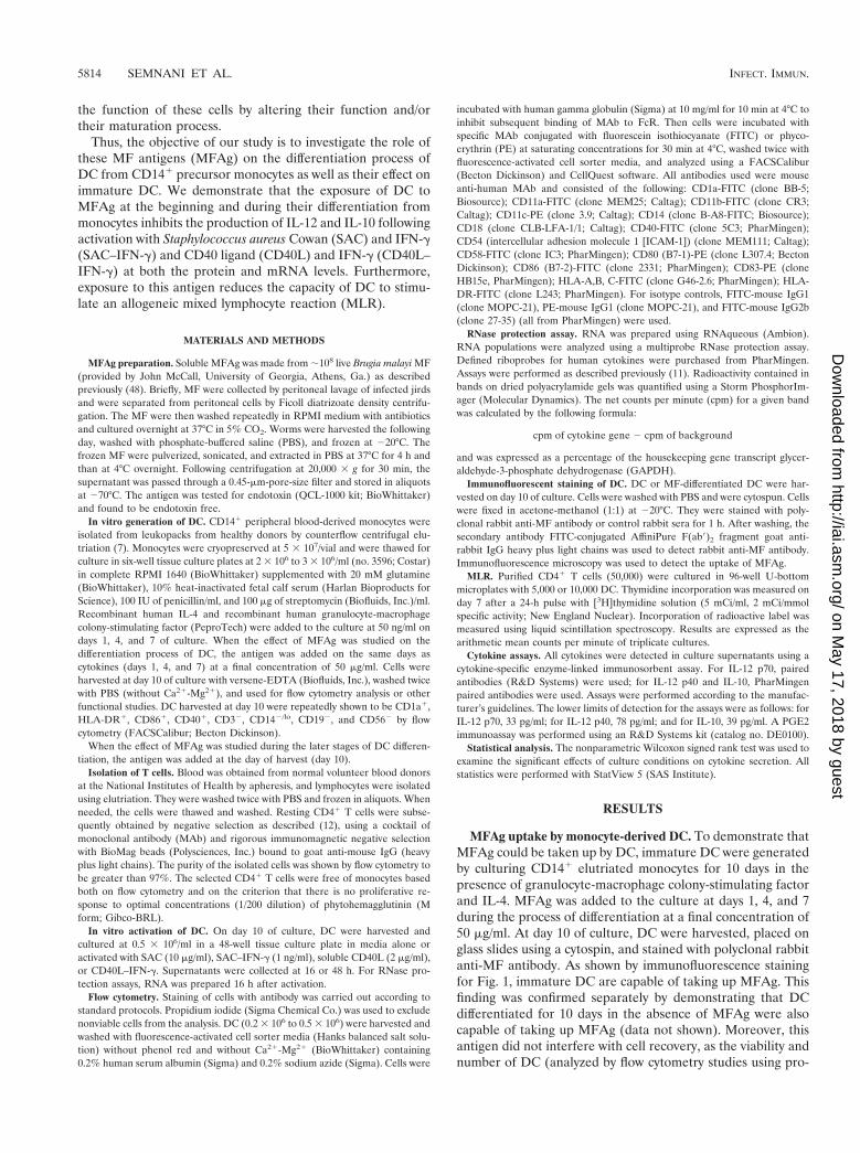
MFAg uptake by monocyte-derived DC. To demonstrate thatMFAg could be taken up by DC, immature DC were generatedby culturing CD141 elutriated monocytes for 10 days in thepresence of granulocyte-macrophage colony-stimulating factorand IL-4. MFAg was added to the culture at days 1, 4, and 7during the process of differentiation at a final concentration of50 mg/ml. At day 10 of culture, DC were harvested, placed onglass slides using a cytospin, and stained with polyclonal rabbitanti-MF antibody. As shown by immunofluorescence stainingfor Fig. 1, immature DC are capable of taking up MFAg. Thisfinding was confirmed separately by demonstrating that DCdifferentiated for 10 days in the absence of MFAg were alsocapable of taking up MFAg (data not shown). Moreover, thisantigen did not interfere with cell recovery, as the viability andnumber of DC (analyzed by flow cytometry studies using pro-
5814 SEMNANI ET AL. INFECT. IMMUN.
on May 17, 2018 by guest
http://iai.asm.org/
Dow
nloaded from
pidium iodide staining; data not shown) were similar for un-stimulated and antigen-differentiated DC.
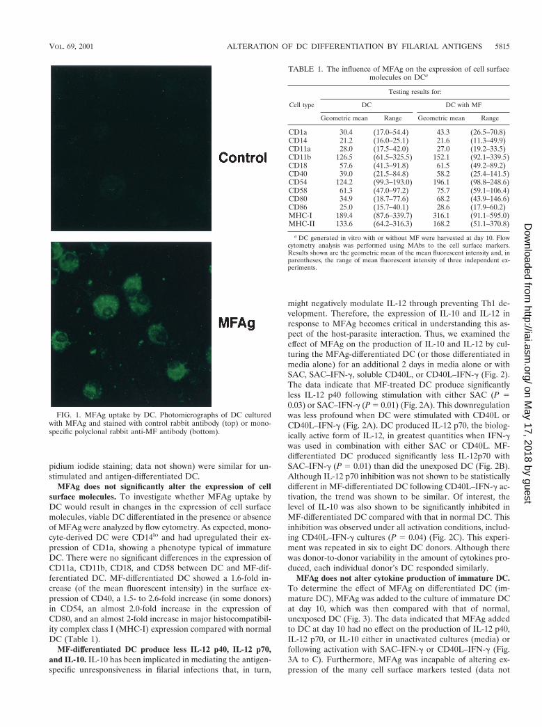
MFAg does not significantly alter the expression of cellsurface molecules. To investigate whether MFAg uptake byDC would result in changes in the expression of cell surfacemolecules, viable DC differentiated in the presence or absenceof MFAg were analyzed by flow cytometry. As expected, mono-cyte-derived DC were CD14lo and had upregulated their ex-pression of CD1a, showing a phenotype typical of immatureDC. There were no significant differences in the expression ofCD11a, CD11b, CD18, and CD58 between DC and MF-dif-ferentiated DC. MF-differentiated DC showed a 1.6-fold in-crease (of the mean fluorescent intensity) in the surface ex-pression of CD40, a 1.5- to 2.6-fold increase (in some donors)in CD54, an almost 2.0-fold increase in the expression ofCD80, and an almost 2-fold increase in major histocompatibil-ity complex class I (MHC-I) expression compared with normalDC (Table 1).
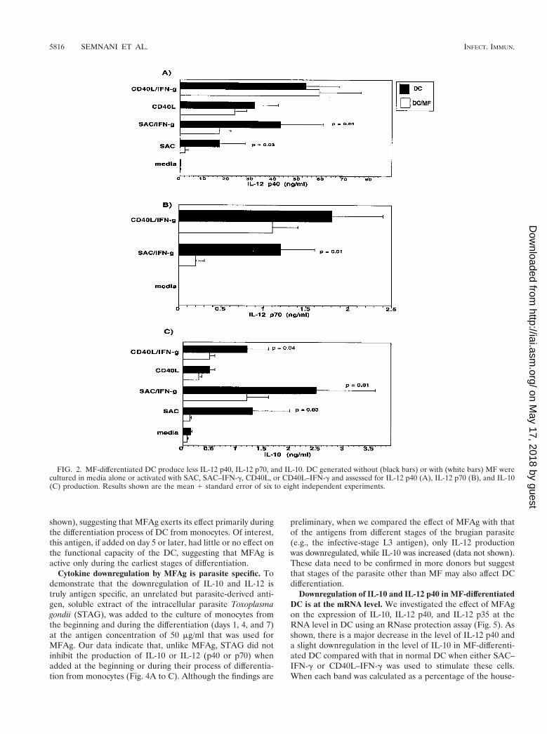
MF-differentiated DC produce less IL-12 p40, IL-12 p70,and IL-10. IL-10 has been implicated in mediating the antigen-specific unresponsiveness in filarial infections that, in turn,
might negatively modulate IL-12 through preventing Th1 de-velopment. Therefore, the expression of IL-10 and IL-12 inresponse to MFAg becomes critical in understanding this as-pect of the host-parasite interaction. Thus, we examined theeffect of MFAg on the production of IL-10 and IL-12 by cul-turing the MFAg-differentiated DC (or those differentiated inmedia alone) for an additional 2 days in media alone or withSAC, SAC–IFN-g, soluble CD40L, or CD40L–IFN-g (Fig. 2).The data indicate that MF-treated DC produce significantlyless IL-12 p40 following stimulation with either SAC (P 50.03) or SAC–IFN-g (P 5 0.01) (Fig. 2A). This downregulationwas less profound when DC were stimulated with CD40L orCD40L–IFN-g (Fig. 2A). DC produced IL-12 p70, the biolog-ically active form of IL-12, in greatest quantities when IFN-gwas used in combination with either SAC or CD40L. MF-differentiated DC produced significantly less IL-12p70 withSAC–IFN-g (P 5 0.01) than did the unexposed DC (Fig. 2B).Although IL-12 p70 inhibition was not shown to be statisticallydifferent in MF-differentiated DC following CD40L–IFN-g ac-tivation, the trend was shown to be similar. Of interest, thelevel of IL-10 was also shown to be significantly inhibited inMF-differentiated DC compared with that in normal DC. Thisinhibition was observed under all activation conditions, includ-ing CD40L–IFN-g cultures (P 5 0.04) (Fig. 2C). This experi-ment was repeated in six to eight DC donors. Although therewas donor-to-donor variability in the amount of cytokines pro-duced, each individual donor’s DC responded similarly.
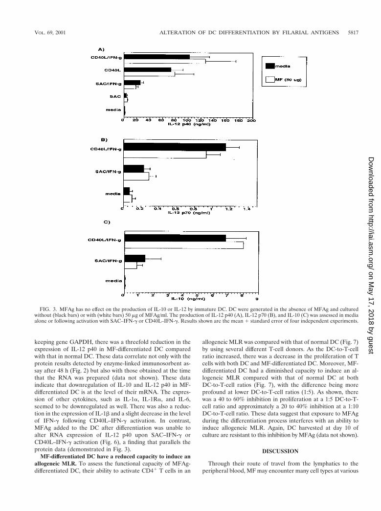
MFAg does not alter cytokine production of immature DC.To determine the effect of MFAg on differentiated DC (im-mature DC), MFAg was added to the culture of immature DCat day 10, which was then compared with that of normal,unexposed DC (Fig. 3). The data indicated that MFAg addedto DC at day 10 had no effect on the production of IL-12 p40,IL-12 p70, or IL-10 either in unactivated cultures (media) orfollowing activation with SAC–IFN-g or CD40L–IFN-g (Fig.3A to C). Furthermore, MFAg was incapable of altering ex-pression of the many cell surface markers tested (data not
TABLE 1. The influence of MFAg on the expression of cell surfacemolecules on DCa
Cell type
Testing results for:
DC DC with MF
Geometric mean Range Geometric mean Range
CD1a 30.4 (17.0–54.4) 43.3 (26.5–70.8)CD14 21.2 (16.0–25.1) 21.6 (11.3–49.9)CD11a 28.0 (17.5–42.0) 27.0 (19.2–33.5)CD11b 126.5 (61.5–325.5) 152.1 (92.1–339.5)CD18 57.6 (41.3–91.8) 61.5 (49.2–89.2)CD40 39.0 (21.5–84.8) 58.2 (25.4–141.5)CD54 124.2 (99.3–193.0) 196.1 (98.8–248.6)CD58 61.3 (47.0–97.2) 75.7 (59.1–106.4)CD80 34.9 (18.7–77.6) 68.2 (43.9–146.6)CD86 25.0 (15.7–40.1) 28.6 (17.9–60.2)MHC-I 189.4 (87.6–339.7) 316.1 (91.1–595.0)MHC-II 133.6 (64.2–316.3) 168.2 (51.1–370.8)
a DC generated in vitro with or without MF were harvested at day 10. Flowcytometry analysis was performed using MAbs to the cell surface markers.Results shown are the geometric mean of the mean fluorescent intensity and, inparentheses, the range of mean fluorescent intensity of three independent ex-periments.
FIG. 1. MFAg uptake by DC. Photomicrographs of DC culturedwith MFAg and stained with control rabbit antibody (top) or mono-specific polyclonal rabbit anti-MF antibody (bottom).
VOL. 69, 2001 ALTERATION OF DC DIFFERENTIATION BY FILARIAL ANTIGENS 5815
on May 17, 2018 by guest
http://iai.asm.org/
Dow
nloaded from
shown), suggesting that MFAg exerts its effect primarily duringthe differentiation process of DC from monocytes. Of interest,this antigen, if added on day 5 or later, had little or no effect onthe functional capacity of the DC, suggesting that MFAg isactive only during the earliest stages of differentiation.
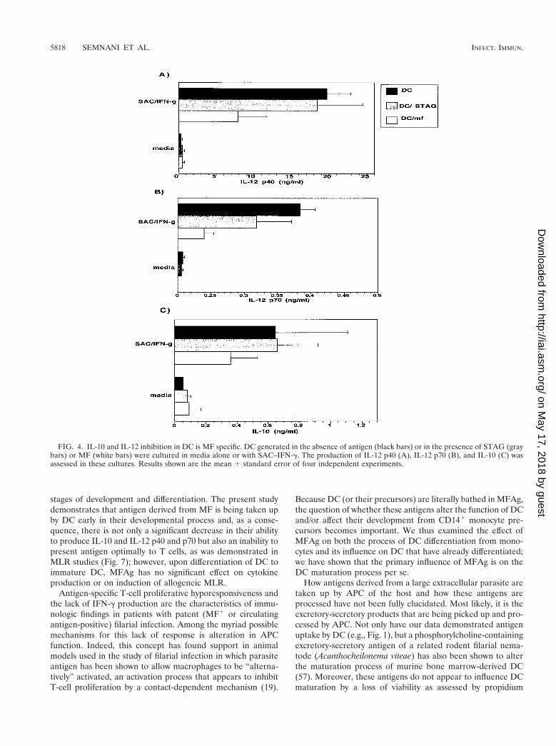
Cytokine downregulation by MFAg is parasite specific. Todemonstrate that the downregulation of IL-10 and IL-12 istruly antigen specific, an unrelated but parasite-derived anti-gen, soluble extract of the intracellular parasite Toxoplasmagondii (STAG), was added to the culture of monocytes fromthe beginning and during the differentiation (days 1, 4, and 7)at the antigen concentration of 50 mg/ml that was used forMFAg. Our data indicate that, unlike MFAg, STAG did notinhibit the production of IL-10 or IL-12 (p40 or p70) whenadded at the beginning or during their process of differentia-tion from monocytes (Fig. 4A to C). Although the findings are
preliminary, when we compared the effect of MFAg with thatof the antigens from different stages of the brugian parasite(e.g., the infective-stage L3 antigen), only IL-12 productionwas downregulated, while IL-10 was increased (data not shown).These data need to be confirmed in more donors but suggestthat stages of the parasite other than MF may also affect DCdifferentiation.
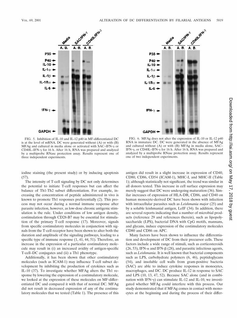
Downregulation of IL-10 and IL-12 p40 in MF-differentiatedDC is at the mRNA level. We investigated the effect of MFAgon the expression of IL-10, IL-12 p40, and IL-12 p35 at theRNA level in DC using an RNase protection assay (Fig. 5). Asshown, there is a major decrease in the level of IL-12 p40 anda slight downregulation in the level of IL-10 in MF-differenti-ated DC compared with that in normal DC when either SAC–IFN-g or CD40L–IFN-g was used to stimulate these cells.When each band was calculated as a percentage of the house-
FIG. 2. MF-differentiated DC produce less IL-12 p40, IL-12 p70, and IL-10. DC generated without (black bars) or with (white bars) MF werecultured in media alone or activated with SAC, SAC–IFN-g, CD40L, or CD40L–IFN-g and assessed for IL-12 p40 (A), IL-12 p70 (B), and IL-10(C) production. Results shown are the mean 1 standard error of six to eight independent experiments.
5816 SEMNANI ET AL. INFECT. IMMUN.
on May 17, 2018 by guest
http://iai.asm.org/
Dow
nloaded from
keeping gene GAPDH, there was a threefold reduction in theexpression of IL-12 p40 in MF-differentiated DC comparedwith that in normal DC. These data correlate not only with theprotein results detected by enzyme-linked immunosorbent as-say after 48 h (Fig. 2) but also with those obtained at the timethat the RNA was prepared (data not shown). These dataindicate that downregulation of IL-10 and IL-12 p40 in MF-differentiated DC is at the level of their mRNA. The expres-sion of other cytokines, such as IL-1a, IL-1Ra, and IL-6,seemed to be downregulated as well. There was also a reduc-tion in the expression of IL-1b and a slight decrease in the levelof IFN-g following CD40L–IFN-g activation. In contrast,MFAg added to the DC after differentiation was unable toalter RNA expression of IL-12 p40 upon SAC–IFN-g orCD40L–IFN-g activation (Fig. 6), a finding that parallels theprotein data (demonstrated in Fig. 3).
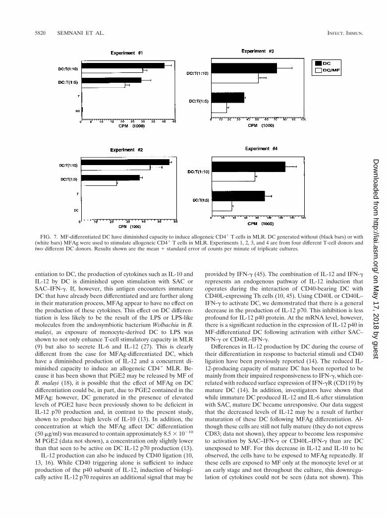
MF-differentiated DC have a reduced capacity to induce anallogeneic MLR. To assess the functional capacity of MFAg-differentiated DC, their ability to activate CD41 T cells in an
allogeneic MLR was compared with that of normal DC (Fig. 7)by using several different T-cell donors. As the DC-to-T-cellratio increased, there was a decrease in the proliferation of Tcells with both DC and MF-differentiated DC. Moreover, MF-differentiated DC had a diminished capacity to induce an al-logeneic MLR compared with that of normal DC at bothDC-to-T-cell ratios (Fig. 7), with the difference being moreprofound at lower DC-to-T-cell ratios (1:5). As shown, therewas a 40 to 60% inhibition in proliferation at a 1:5 DC-to-T-cell ratio and approximately a 20 to 40% inhibition at a 1:10DC-to-T-cell ratio. These data suggest that exposure to MFAgduring the differentiation process interferes with an ability toinduce allogeneic MLR. Again, DC harvested at day 10 ofculture are resistant to this inhibition by MFAg (data not shown).
DISCUSSION
Through their route of travel from the lymphatics to theperipheral blood, MF may encounter many cell types at various
FIG. 3. MFAg has no effect on the production of IL-10 or IL-12 by immature DC. DC were generated in the absence of MFAg and culturedwithout (black bars) or with (white bars) 50 mg of MFAg/ml. The production of IL-12 p40 (A), IL-12 p70 (B), and IL-10 (C) was assessed in mediaalone or following activation with SAC–IFN-g or CD40L–IFN-g. Results shown are the mean 1 standard error of four independent experiments.
VOL. 69, 2001 ALTERATION OF DC DIFFERENTIATION BY FILARIAL ANTIGENS 5817
on May 17, 2018 by guest
http://iai.asm.org/
Dow
nloaded from
stages of development and differentiation. The present studydemonstrates that antigen derived from MF is being taken upby DC early in their developmental process and, as a conse-quence, there is not only a significant decrease in their abilityto produce IL-10 and IL-12 p40 and p70 but also an inability topresent antigen optimally to T cells, as was demonstrated inMLR studies (Fig. 7); however, upon differentiation of DC toimmature DC, MFAg has no significant effect on cytokineproduction or on induction of allogeneic MLR.
Antigen-specific T-cell proliferative hyporesponsiveness andthe lack of IFN-g production are the characteristics of immu-nologic findings in patients with patent (MF1 or circulatingantigen-positive) filarial infection. Among the myriad possiblemechanisms for this lack of response is alteration in APCfunction. Indeed, this concept has found support in animalmodels used in the study of filarial infection in which parasiteantigen has been shown to allow macrophages to be “alterna-tively” activated, an activation process that appears to inhibitT-cell proliferation by a contact-dependent mechanism (19).
Because DC (or their precursors) are literally bathed in MFAg,the question of whether these antigens alter the function of DCand/or affect their development from CD141 monocyte pre-cursors becomes important. We thus examined the effect ofMFAg on both the process of DC differentiation from mono-cytes and its influence on DC that have already differentiated;we have shown that the primary influence of MFAg is on theDC maturation process per se.
How antigens derived from a large extracellular parasite aretaken up by APC of the host and how these antigens areprocessed have not been fully elucidated. Most likely, it is theexcretory-secretory products that are being picked up and pro-cessed by APC. Not only have our data demonstrated antigenuptake by DC (e.g., Fig. 1), but a phosphorylcholine-containingexcretory-secretory antigen of a related rodent filarial nema-tode (Acanthocheilonema viteae) has also been shown to alterthe maturation process of murine bone marrow-derived DC(57). Moreover, these antigens do not appear to influence DCmaturation by a loss of viability as assessed by propidium
FIG. 4. IL-10 and IL-12 inhibition in DC is MF specific. DC generated in the absence of antigen (black bars) or in the presence of STAG (graybars) or MF (white bars) were cultured in media alone or with SAC–IFN-g. The production of IL-12 p40 (A), IL-12 p70 (B), and IL-10 (C) wasassessed in these cultures. Results shown are the mean 1 standard error of four independent experiments.
5818 SEMNANI ET AL. INFECT. IMMUN.
on May 17, 2018 by guest
http://iai.asm.org/
Dow
nloaded from
iodine staining (the present study) or by inducing apoptosis(57).
The intensity of T-cell signaling by DC not only determinesthe potential to initiate T-cell responses but can affect thebalance of Th1-Th2 subset differentiation. For example, in-creasing the concentration of peptide administered in vivo isknown to promote Th1 responses preferentially (2). This pro-cess may not occur during a normal immune response afterparasite infection, however, as low-dose chronic antigenic stim-ulation is the rule. Under conditions of low antigen density,costimulation through CD28-B7 may be essential for stimula-tion of the primary T-cell response (17). Moreover, signalsfrom specific costimulatory molecules in conjunction with sig-nals from the T-cell receptor have been shown to alter both theduration and amplitude of the signaling pathways, leading to aspecific type of immune response (1, 41, 44, 51). Therefore, anincrease in the expression of a particular costimulatory mole-cule may result in (i) an increased avidity of antigen-specificT-cell–DC conjugates and (ii) a Th1 phenotype.
Additionally, it has been shown that other costimulatorymolecules (such as ICAM-1) may influence T-cell subset de-velopment by inhibiting the production of cytokines such asIL-10 (37). To investigate whether MFAg alters the Th1 re-sponse by lowering the expression of a costimulatory molecule,we looked at the expression of these molecules on MF-differ-entiated DC and compared it with that of normal DC. MFAgdid not result in decreased expression of any of the costimu-latory molecules that we tested (Table 1). The presence of this
antigen did result in a slight increase in expression of CD40,CD80, CD86, CD54 (ICAM-1), MHC-I, and MHC-II (Table1); although statistically not significant, the trend was similar inall donors tested. This increase in cell surface expression maymerely suggest that DC were undergoing maturation (36). Sim-ilar increases of expression of HLA-DR, CD86, and CD40 onhuman monocyte-derived DC have been shown with infectionwith intracellular parasites such as Leishmania major (25) andby a soluble leishmanial antigen, LeIF (34). In addition, thereare several reports indicating that a number of microbial prod-ucts (reference 29 and references therein), such as lipopoly-saccharide (LPS), bacterial DNA with CpG motifs, mannans,and glycans, induce expression of the costimulatory moleculesCD80 and CD86 on APC.
Many factors have been shown to influence the differentia-tion and development of DC from their precursor cells. Thesefactors include a wide range of stimuli, such as corticosteroids(26, 53), IFN-a and IFN-b (28), and parasitic infectious agents,such as Leishmania. It is well known that bacterial componentssuch as LPS, carbohydrate polymers (6, 46), peptidoglycans(54), and insoluble cell walls from gram-positive bacteria(SAC) are able to induce cytokine responses in monocytes,macrophages, and DC. DC produce IL-12 in response to SACand LPS (10, 13, 47, 52). Because SAC alone (and in combi-nation with IFN-g) can stimulate IL-12 and IL-10, we investi-gated whether MFAg could interfere with this process. Ourstudy demonstrated that if MFAg comes in contact with mono-cytes at the beginning and during the process of their differ-
FIG. 5. Inhibition of IL-10 and IL-12 p40 in MF-differentiated DCis at the level of mRNA. DC were generated without (A) or with (B)MFAg and cultured in media alone or activated with SAC–IFN-g orCD40L–IFN-g for 16 h. After 16 h, RNA was prepared and analyzedby a multiprobe RNase protection assay. Results represent one ofthree independent experiments.
FIG. 6. MFAg does not alter the expression of IL-10 or IL-12 p40RNA in immature DC. DC were generated in the absence of MFAgand cultured without (A) or with (B) MFAg in media alone, SAC–IFN-g, or CD40L–IFN-g for 16 h. After 16 h, RNA was prepared andanalyzed by a multiprobe RNase protection assay. Results representone of two independent experiments.
VOL. 69, 2001 ALTERATION OF DC DIFFERENTIATION BY FILARIAL ANTIGENS 5819
on May 17, 2018 by guest
http://iai.asm.org/
Dow
nloaded from
entiation to DC, the production of cytokines such as IL-10 andIL-12 by DC is diminished upon stimulation with SAC orSAC–IFN-g. If, however, this antigen encounters immatureDC that have already been differentiated and are further alongin their maturation process, MFAg appear to have no effect onthe production of these cytokines. This effect on DC differen-tiation is less likely to be the result of the LPS or LPS-likemolecules from the andosymbiotic bacterium Wolbachia in B.malayi, as exposure of monocyte-derived DC to LPS wasshown to not only enhance T-cell stimulatory capacity in MLR(9) but also to secrete IL-6 and IL-12 (27). This is clearlydifferent from the case for MFAg-differentiated DC, whichhave a diminished production of IL-12 and a concurrent di-minished capacity to induce an allogeneic CD41 MLR. Be-cause it has been shown that PGE2 may be released by MF ofB. malayi (18), it is possible that the effect of MFAg on DCdifferentiation could be, in part, due to PGE2 contained in theMFAg: however, DC generated in the presence of elevatedlevels of PGE2 have been previously shown to be deficient inIL-12 p70 production and, in contrast to the present study,shown to produce high levels of IL-10 (13). In addition, theconcentration at which the MFAg affect DC differentiation(50 mg/ml) was measured to contain approximately 8.5 3 10210
M PGE2 (data not shown), a concentration only slightly lowerthan that seen to be active on DC IL-12 p70 production (13).
IL-12 production can also be induced by CD40 ligation (10,13, 16). While CD40 triggering alone is sufficient to induceproduction of the p40 subunit of IL-12, induction of biologi-cally active IL-12 p70 requires an additional signal that may be
provided by IFN-g (45). The combination of IL-12 and IFN-grepresents an endogenous pathway of IL-12 induction thatoperates during the interaction of CD40-bearing DC withCD40L-expressing Th cells (10, 45). Using CD40L or CD40L–IFN-g to activate DC, we demonstrated that there is a generaldecrease in the production of IL-12 p70. This inhibition is lessprofound for IL-12 p40 protein. At the mRNA level, however,there is a significant reduction in the expression of IL-12 p40 inMF-differentiated DC following activation with either SAC–IFN-g or CD40L–IFN-g.
Differences in IL-12 production by DC during the course oftheir differentiation in response to bacterial stimuli and CD40ligation have been previously reported (14). The reduced IL-12-producing capacity of mature DC has been reported to bemainly from their impaired responsiveness to IFN-g, which cor-related with reduced surface expression of IFN-gR (CD119) bymature DC (14). In addition, investigators have shown thatwhile immature DC produced IL-12 and IL-6 after stimulationwith SAC, mature DC became unresponsive. Our data suggestthat the decreased levels of IL-12 may be a result of furthermaturation of these DC following MFAg differentiation. Al-though these cells are still not fully mature (they do not expressCD83; data not shown), they appear to become less responsiveto activation by SAC–IFN-g or CD40L–IFN-g than are DCunexposed to MF. For this decrease in IL-12 and IL-10 to beobserved, the cells have to be exposed to MFAg repeatedly. Ifthese cells are exposed to MF only at the monocyte level or atan early stage and not throughout the culture, this downregu-lation of cytokines could not be seen (data not shown). This
FIG. 7. MF-differentiated DC have diminished capacity to induce allogeneic CD41 T cells in MLR. DC generated without (black bars) or with(white bars) MFAg were used to stimulate allogeneic CD41 T cells in MLR. Experiments 1, 2, 3, and 4 are from four different T-cell donors andtwo different DC donors. Results shown are the mean 1 standard error of counts per minute of triplicate cultures.
5820 SEMNANI ET AL. INFECT. IMMUN.
on May 17, 2018 by guest
http://iai.asm.org/
Dow
nloaded from
has in vivo relevance, in that maturing DC are consistently ex-posed to MFAg in the blood of MF patients.
It has recently been reported that a particular subset of DCis capable of inducing Th2 responses. For example, DC derivedfrom the lymphoid lineage (35) or DC generated in the pres-ence of PGE2 are capable of inducing a type 2 T-cell response(13). In fact, a phosphorylcholine-containing glycoprotein, ES-62, secreted by the filarial nematode A. viteae has also beenshown to induce maturation of the so-called DC2 (57). ThisES-62-exposed DC produced significantly less IL-12 but notIL-10 than did LPS-generated DC following CD41 T-cell in-teraction. This is somewhat different from the results shown inthe present study, which indicated a general inhibition in cy-tokine production associated with a reduced capacity to induceallogeneic CD41 T cells in MLR. These differences may beattributed to both the source of antigen and the nature of thehost cells. In the studies done by Whelan et al. (57), bonemarrow-derived DC from mice were exposed to a specificprotein (ES-62), whereas the present study utilizes monocyte-derived DC and has as its antigen a crude parasite extract thatcontains many filaria-derived antigens. Therefore, it is likelythat human DC present very different epitopes based on thedifferences in the antigens that they encounter. Of note, bothMFAg-exposed and -unexposed DC were capable of inducingactivation and differentiation of naı̈ve Th cells in an anti-CD3-dependent manner (data not shown); preliminary results indi-cate that there were no significant differences in the cytokinesproduced by autologous, activated T cells in response to MF-differentiated DC. One possible explanation for the lack of dif-ferences in proliferation using anti-CD3 could be the strengthof the first signal. Anti-CD3 is clearly a strong first signal,which may override the inhibitory signals that might be sent byMF-differentiated DC. Furthermore, preliminary results in-dicated that allogeneic CD41 T cells in the presence of MF-differentiated DC or normal DC produced high but similarlevels of IFN-g, pointing toward the potency of the anti-CD3signal (data not shown). Moreover, there were no significantMF-induced differences in IL-5 or IL-10 production by these Tcells; however, because the precursor frequency of naı̈ve Tcells to MFAg is extremely low (range, 1/87,000 to 1/200,000)(48), it is almost impossible to compare the potency of MF-differentiated DC with the normal potency in an autologous,antigen-driven system.
Although the underlying mechanisms may differ, it is clearthat parasites as diverse as unicellular protozoa (Leishmania,plasmodia [49], and trypanosomes [50]) and multicellular hel-minths may influence the development and differentiation ofDC. Whether they do so in a manner that alters the nature ofthe effector T-cell responses remains to be elucidated.
ACKNOWLEDGMENTS
We thank Elaine K. Thomas (Immunex Corporation, Seattle,Wash.) for providing soluble CD40L and Alan Sher and Sara Hieny forproviding STAG. We also thank Brenda Rae Marshall for editorialhelp.
REFERENCES
1. Bluestone, J. A. 1995. New perspectives of CD28–B7-mediated T cell co-stimulation. Immunity 2:555–559.
2. Constant, S., A. Pfeiffer, T. Woodard, T. Pasqualini, and K. Bottomly. 1995.Extent of T cell receptor ligation can determine the functional differentiation
of naive CD41 T cells. J. Exp. Med. 182:1591–1596.3. Demeure, C. E., C. Y. Wu, U. Shu, P. V. Schneider, C. Heusser, H. Yssel, and
G. Delespesse. 1994. In vitro maturation of human neonatal CD41 T lym-phocytes. II. Cytokines present at priming modulate the development oflymphokine production. J. Immunol. 152:4775–4782.
4. Dieu, M. C., B. Vanbervliet, A. Vicari, J. M. Bridon, E. Oldham, A. Yahia, F.Briere, A. Zlotnik, S. Lebecque, and C. Caux. 1998. Selective recruitment ofimmature and mature dendritic cells by distinct chemokines expressed indifferent anatomic sites. J. Exp. Med. 188:373–386.
5. Duncan, D. D., and S. L. Swain. 1994. Role of antigen-presenting cells in thepolarized development of helper T cell subsets: evidence for differentialcytokine production by Th0 cells in response to antigen presentation by Bcells and macrophages. Eur. J. Immunol. 24:2506–2514.
6. Espevik, T., M. Ottelei, G. Skjek-Braek, L. Ryan, S. D. Wright, and A.Sundan. 1993. The involvement of CD14 in stimulation of cytokine produc-tion by uronic acid polymers. Eur. J. Immunol. 23:255–261.
7. Gerrard, T. L., C. H. Jurgenson, and A. S. Fauci. 1983. Differential effect ofmonoclonal anti-DR antibody on monocytes in antigen- and mitogen-stim-ulated responses: mechanism of inhibition and relationship to interleukin 1secretion. Cell. Immunol. 82:394–402.
8. Gomez-Escobar, N., W. F. Gregory, and R. M. Maizels. 2000. Identificationof tgh-2, a filarial nematode homolog of Caenorhabditis elegans daf-7 andhuman transforming growth factor b, expressed in microfilarial and adultstages of Brugia malayi. Infect. Immun. 68:6402–6410.
9. Hertz, C. J., S. M. Kiertscher, P. J. Godowski, D. A. Bouis, M. V. Norgard,M. D. Roth, and R. L. Modlin. 2001. Microbial lipopeptides stimulate den-dritic cell maturation via toll-like receptor 2. J. Immunol. 166:2444–2450.
10. Hilkens, C. M. U., P. Kalinski, M. de Boer, and M. L. Kapsenberg. 1997.Human dendritic cells require exogenous interleukin-12-inducing factors todirect the development of naive T-helper cells toward the Th1 phenotype.Blood 90:1920–1926.
11. Hodge, J. W., H. Sabzevari, A. G. Yafal, L. Gritz, M. G. Lorenz, and J.Schlom. 1999. A triad of costimulatory molecules synergize to amplify T-cellactivation. Cancer Res. 59:5800–5807.
12. Horgan, K. J., and S. Shaw. 1991. Immunomagnetic purification of T cellpopulations, p. 7.4.1-7.4.5. In J. E. Coligan, A. M. Kruisbeek, D. H. Margu-lies, E. M. Shevach, and W. Strober (ed.), Current protocols in immunology.Wiley Interscience, New York, N.Y.
13. Kalinski, P., C. M. U. Hilkens, A. Snijders, F. G. M. Snijdewint, and M. L.Kapsenberg. 1997. IL-12-deficient dendritic cells, generated in the presenceof prostoglandin E2, promote type 2 cytokine production in maturing humannaive T helper cells. J. Immunol. 159:28–35.
14. Kalinski, P., J. H. Schuitemaker, C. M. Hilkens, E. A. Wierenga, and M. L.Kapsenberg. 1999. Final maturation of dendritic cells is associated withimpaired responsiveness to IFN-g and to bacterial IL-12 inducers: decreasedability of mature dendritic cells to produce IL-12 during the interaction withTh cells. J. Immunol. 162:3231–3236.
15. King, C. L., S. Mahanty, V. Kumaraswami, J. S. Abrams, J. Regunathan, K.Jayaraman, E. A. Otteson, and T. B. Nutman. 1993. Cytokine control ofparasite-specific anergy in human lymphatic filariasis. Preferential inductionof a regulatory T helper Type 2 lymphocyte subset. J. Clin. Investig. 92:1667–1673.
16. Koch, F., U. Stanzl, P. Jennewein, K. Janke, C. Heufler, E. Kampgen, N.Romani, and G. Schuler. 1996. High level IL-12 production by murinedendritic cells: upregulation via MHC class II and CD40 molecules anddownregulation by IL-4 and IL-10. J. Exp. Med. 184:741–746.
17. Lenschow, D. J., T. L. Walunas, and J. A. Bluestone. 1996. CD28/B7 systemof T cell costimulation. Annu. Rev. Immunol. 14:233–258.
18. Liu, L. X., J. E. Buhlmann, and P. F. Weller. 1992. Release of prostaglandinE2 by microfilariae of Wuchereria bancrofti and Brugia malayi. Am. J. Trop.Med. Hyg. 46:520–523.
19. Loke, P., A. S. MacDonald, A. Robb, R. M. Maizels, and J. E. Allen. 2000.Alternatively activated macrophages induced by nematode infection inhibitproliferation via cell-to-cell contact. Eur. J. Immunol. 30:2669–2678.
20. Mahanty, S., C. L. King, V. Kumaraswami, J. Regunathan, A. Maya, K.Jayaraman, J. S. Abrams, E. A. Ottesen, and T. B. Nutman. 1993. IL-4- andIL-5-secreting lymphocyte populations are preferentially stimulated by par-asite-derived antigens in human tissue invasive nematode infections. J. Im-munol. 151:3704–3711.
21. Mahanty, S., H. E. Luke, V. Kumaraswami, P. R. Narayanan, V. Vijaysheka-ran, and T. B. Nutman. 1996. Stage-specific induction of cytokines regulatesthe immune response in lymphatic filariasis. Exp. Parasitol. 84:282–290.
22. Mahanty, S., S. N. Mollis, M. Ravichandran, J. S. Abrams, V. Kuma-raswami, K. Jayaraman, E. A. Ottesen, and T. B. Nutman. 1996. High levelsof spontaneous and parasite antigen-driven interleukin-10 production areassociated with antigen-specific hyporesponsiveness in human lymphatic fi-lariasis. J. Infect. Dis. 173:769–773.
23. Mahanty, S., and T. B. Nutman. 1995. Immunoregulation in human lym-phatic filariasis: the role of interleukin 10. Parasite Immunol. 17:385–392.
24. Maizels, R. M., J. E. Allen, and M. Yazdanbakhsh. 2000. Immunology oflymphatic filariasis: current controversies, p. 217–243. In T. B. Nutman (ed.),
VOL. 69, 2001 ALTERATION OF DC DIFFERENTIATION BY FILARIAL ANTIGENS 5821
on May 17, 2018 by guest
http://iai.asm.org/
Dow
nloaded from
dsfaLymphatic filariasis. Imperial College Press, London, United Kingdom.25. Marovich, M. A., M. A. McDowell, E. K. Thomas, and T. B. Nutman. 2000.
IL-12p70 production by Leishmania major-harboring human dendritic cells isa CD40/CD40 ligand-dependent process. J. Immunol. 164:5858–5865.
26. Matyszak, M. K., S. Citterio, M. Rescigno, and P. Ricciardi-Castagnoli.2000. Differential effects of corticosteroids during different stages of den-dritic cell maturation. Eur. J. Immunol. 30:1233–1242.
27. McRae, B. L., B. A. Beilfuss, and G. A. van Seventer. 2000. Interferon-bdifferentially regulates CD40-induced cytokine secretion by human dendriticcells. J. Immunol. 164:23–28.
28. McRae, B. L., T. Nagai, R. T. Semnani, J. M. van Seventer, and G. A. vanSeventer. 2000. Interferon-a and -b inhibit the in vitro differentiation ofimmunocompetent human dendritic cells from CD141 precursors. Blood96:210–217.
29. Medzhitov, R., and C. A. Janeway. 1997. Innate immunity: impact on theadaptive immune response. Curr. Opin. Immunol. 9:4–9.
30. Nakamura, T., K. Kamogawa, K. Bottomly, and R. A. Flavell. 1997. Polar-ization of IL-4 and IFN-g producing CD41 T cells following activation ofnaive CD41 T cells. J. Immunol. 158:1085–1094.
31. Nutman, T. B. 1996. Immune responses to helminth parasites, p. 561–570. InR. R. Rich, T. A. Fleisher, B. D. Schwartz, W. T. Shearer, and W. Strober(ed.), Clinical immunology: principles and practice. Mosby Year-Book, St.Louis, Mo.
32. Openshaw, P., E. E. Murphy, N. A. Hosken, V. Maino, K. Davis, K. M.Murphy, and A. O’Garra. 1995. Heterogeneity of intracellular cytokine syn-thesis at the single-cell level in polarized T helper 1 and T helper 2 popu-lation. J. Exp. Med. 182:1357–1367.
33. Pastrana, D. V., N. Raghavan, P. Fitzgerald, S. W. Eisinger, C. Metz, R.Bucala, R. P. Schleimer, C. Bickel, and A. L. Scott. 1998. Filarial nematodeparasites secrete a homologue of the human cytokine macrophage migrationinhibitory factor. Infect. Immun. 66:5955–5963.
34. Probst, P., Y. A. W. Skeiky, M. Steeves, A. Gervassi, K. H. Grabstein, andS. G. Reed. 1997. A Leishmania protein that modulates interleukin (IL)-12,IL-10 and tumor necrosis factor-a production and expression of B7–1 inhuman monocyte-derived antigen-presenting cells. Eur. J. Immunol.27:2634–2642.
35. Rissoan, M.-C., V. Soumelis, N. Kadowaki, G. Grouard, F. Briere, R. Male-fyt, and Y.-J. Liu. 1999. Reciprocal control of T helper cell and dendritic celldifferentiation. Science 283:1183–1186.
36. Sallusto, F., M. Cella, C. Danieli, and A. Lanzavecchia. 1995. Dendritic cellsuse macropinocytosis and the mannose receptor to concentrate macromol-ecules in the major histocompatibility complex class II compartment: down-regulation by cytokines and bacterial products. J. Exp. Med. 182:389–400.
37. Salomon, B., and J. A. Bluestone. 1998. LFA-1 interaction with ICAM-1 andICAM-2 regulates Th2 cytokine production. J. Immunol. 161:5138–5142.
38. Sartono, E., Y. C. M. Kruize, F. Partono, A. Kurniawan-Atmadja, R. M.Maizels, and M. Yazdanbakhsh. 1995. Specific T cell unresponsiveness inhuman filariasis: diversity in underlying mechanisms. Parasite Immunol. 17:587–594.
39. Schmitt, E., P. Hoehn, T. Germann, and E. Rude. 1994. Differential effectsof interleukin 12 on the development of naive mouse CD41 T cells. Eur.J. Immunol. 24:343–347.
40. Schmitt, E., P. Hoehn, S. Huels, S. Goedert, N. Palm, E. Rude, and T.Germann. 1994. T helper type 1 development of naive CD41 T cells requiresthe coordinate action of interleukin 12 and interferon-g and is inhibited bytransforming growth factor-b. Eur. J. Immunol. 24:793–798.
41. Schweitzer, A. N., F. Borriello, R. C. Wong, A. K. Abbas, and A. H. Sharpe.1997. Role of costimulators in T cell differentiation: studies using antigen-presenting cells lacking expression of CD80 or CD86. J. Immunol. 158:2713–2722.
42. Seder, R. A. 1996. High-dose IL-2 and IL-15 enhance the in vitro priming of
naive CD41 T cells for IFN-g but have differential effects on priming forIL-4. J. Immunol. 156:2413–2422.
43. Seder, R. A., R. T. Gazzinelli, A. Sher, and W. E. Paul. 1993. Interleukin 12acts directly on CD41 T cells to enhance priming for interferon g productionand diminishes interleukin 4 inhibition of such priming. Proc. Natl. Acad.Sci. USA 90:10188–10192.
44. Semnani, R. T., T. B. Nutman, P. Hochman, S. Shaw, and G. A. van Seventer.1994. Costimulation by purified intercellular adhesion molecule 1 and lym-phocyte function-associated antigen 3 induces distinct proliferation, cytokineand cell surface antigen profiles in human “naive” and “memory” CD41 Tcells. J. Exp. Med. 180:2125–2135.
45. Snijders, A., P. Kalinski, C. M. Hilkens, and M. L. Kapsenberg. 1998. Highlevel IL-12 production by human dendritic cells requires two signals. Int.Immunol. 10:1593–1598.
46. Soell, M., E. Lett, F. Holveck, M. Scholler, D. Wachsmann, and J. P. Klein.1995. Activation of human monocytes by streptococcal rhamnose glucosepolymers is mediated by CD14 antigen, and mannan binding protein inhibitsTNF-a release. J. Immunol. 154:851–860.
47. Sousa, C. R. E., S. Hieny, T. Scharton-Kersten, D. Jankovic, H. Charest,R. N. Germani, and A. Sher. 1997. In vivo microbial stimulation inducesrapid CD40 ligand-independent production of interleukin 12 by dendriticcells and their redistribution to T cell areas. J. Exp. Med. 186:1819–1829.
48. Steel, C., and T. B. Nutman. 1998. Helminth antigens selectively differentiateunsensitized CD45RA1 CD41 human T cells in vitro. J. Immunol. 160:351–360.
49. Urban, B. C., D. J. Ferguson, A. Pain, N. Willcox, M. Plebanski, J. M.Austyn, and D. J. Roberts. 1999. Plasmodium falciparum-infected erythro-cytes modulate the maturation of dendritic cells. Nature 400:73–77.
50. Van Overtvelt, L., N. Vanderheyde, V. Verhasselt, J. Ismaili, L. De Vos, M.Goldman, F. Willems, and B. Vray. 1999. Trypanosoma cruzi infects humandendritic cells and prevents their maturation: inhibition of cytokines, HLA-DR, and costimulatory molecules. Infect. Immun. 67:4033–4040.
51. van Seventer, G. A., R. T. Semnani, E. M. Palmer, B. L. McRae, and J. M.van Seventer. 1998. Integrins and T helper cell activation. Transplant. Proc.30:4270–4274.
52. Verhasselt, V., C. Buelens, F. Willems, D. De Groote, N. Haeffiner-Cavaillon,and M. Goldman. 1997. Bacterial lipopolysaccharide stimulates the produc-tion of cytokines and the expression of costimulatory molecules by humanperipheral blood dendritic cells: evidence for a soluble CD14-dependentpathway. J. Immunol. 158:2919–2925.
53. Vieira, P. L., P. Kalinski, E. A. Wierenga, M. L. Kapsenberg, and E. C. deJong. 1998. Glucocorticoids inhibit bioactive IL-12 p70 production by invitro-generated human dendritic cells without affecting their T cell stimula-tory potential. J. Immunol. 161:5245–5251.
54. Weidemann, B., H. Brade, E. T. Rietschel, R. Dziarski, V. Bazil, S. Ku-sumoto, H. D. Flad, and A. Ulmer. 1994. Soluble peptidoglycan-inducedmonokine production can be blocked by anti-CD14 monoclonal antibodiesand by lipid A partial structures. J. Clin. Investig. 62:4709–4715.
55. Weil, G. J., R. Hussain, V. Kumaraswami, S. P. Tripathy, K. S. Phillips, andE. A. Ottesen. 1983. Prenatal allergic sensitization to helminth antigens inoffspring of parasite-infected mothers. J. Clin. Investig. 71:1124–1129.
56. Wenner, C. A., M. L. Guler, S. E. Macatonia, A. O’Garra, and K. M.Murphy. 1996. Roles of IFN-g in IL-12-induced T helper cell-1 develop-ment. J. Immunol. 156:1442–1447.
57. Whelan, M., M. M. Harnett, K. M. Houston, V. Patel, W. Harnett, and K. P.Rigley. 2000. A filarial nematode-secreted product signals dendritic cells toacquire a phenotype that drives development of Th2 cells. J. Immunol.164:6453–6460.
58. Yoshimoto, K., S. L. Swain, and L. M. Bradley. 1996. Enhanced developmentof Th2-like primary CD4 effectors in response to sustained exposure tolimited rIL-4 in vivo. J. Immunol. 156:3267–3274.
Editor: J. M. Mansfield
5822 SEMNANI ET AL. INFECT. IMMUN.
on May 17, 2018 by guest
http://iai.asm.org/
Dow
nloaded from