Embed Size (px)
Citation preview
F I N E S T R U C T U R E OF T H E C I L I A
OF R O T I F E R S
A L B E R T I . L A N S I N G , Ph .D. , and F R A N C O I S L A M Y , P h . D .
From the Department of Anatomy, School of Medicine, University of Pittsburgh, and the Marine Biological Laboratory, Woods Hole
A B S T R A C T
The fine s t ructure of the coronal cilia of the rotifer Philodina citrina has been studied in detail. Specimens were fixed with OsO,~ and embedded in butyl methyl methacrylate , Epon 812, or Vestopal and sectioned with a Porter-Blum microtome. The details of struc- ture of the rootlets, basal bodies, basal plates, and free cilia are described. The general s tructure of the rotifer ciliary appara tus conforms well to tha t established for o ther species. One of the main observations is the difference in structure of the per ipheral filaments in the opposing halves of a cross-section of the free cilium. Also, in longi tudinal sections evidence is offered for the existence of a helical s tructure in the per ipheral filaments.
This is the first of a series of studies on the electron microscopical structures of rotifers. The rotifer, which has not been systemically character ized from a cytological viewpoint, is unusual ly well suited for study of senescence in tha t its life span is measured in days, it manifests de te rmina te de- ve lopment so tha t all body cells are of the same age, it is easily reared in homozygous stocks by virtue of its par thenogenet ic reproduct ion, and it lends itself well to s tandardized breeding in the laboratory ( l l ) . Previous studies (12, 13) which established the existence of a transmissible, cumu- lative, and reversible accelerator of senescence in rotifers have st imulated interest in the fine cytol- ogy of the rotifer.
In an a t t empt to extend these early studies to
character izat ion of the fine s tructural and cyto-
chemical changes tha t may be associated with
senescence in the rotifer, a beginning is being
made with analysis of the coronal cilia. Strobo-
scopic measurement of the rate of ciliary beat has confirmed an old impression (12) tha t the
cilia beat more slowly in the senile rotifer. Dur ing
most of the life span the coronal cilia beat at 1200
complete strokes per minute , bu t in the senile
rotifer pilot studies indicate tha t they slow down to 900 to 1000 beats per minute.
This report summarizes the current status of our study of the normal fine structure of the cilia of the rotifer. I t is our intent ion to establish a base line for analysis of possible age changes which may be correlated with the decrease in beat rate. Some of our observations have encouraged us to develop a model of ciliary structure which may fur ther our unders tand ing of the na ture of ciliary beat.
M A T E R I A L A N D M E T I I O D S
The bulk of these observations have been made on Philodina citrina with occasional reference to a giant rotifer of the genus Rotifer which as yet has not been fully identified. The animals were raised in mass and isolation culture essentially as described previously (1 l). Instead of artificial pond water, deep well water from the Pittsburgh environs buffered at pH 8.0 was used. The animals were fed a uniform diet of Chlorella vulgaris raised under artificial light on non-nutrient agar slants as before.
For electron microscopy the excess pond water in the pyrex depression slides used to raise rotifers was carefully drawn off with fine pipettes and 1 or 2 per
799
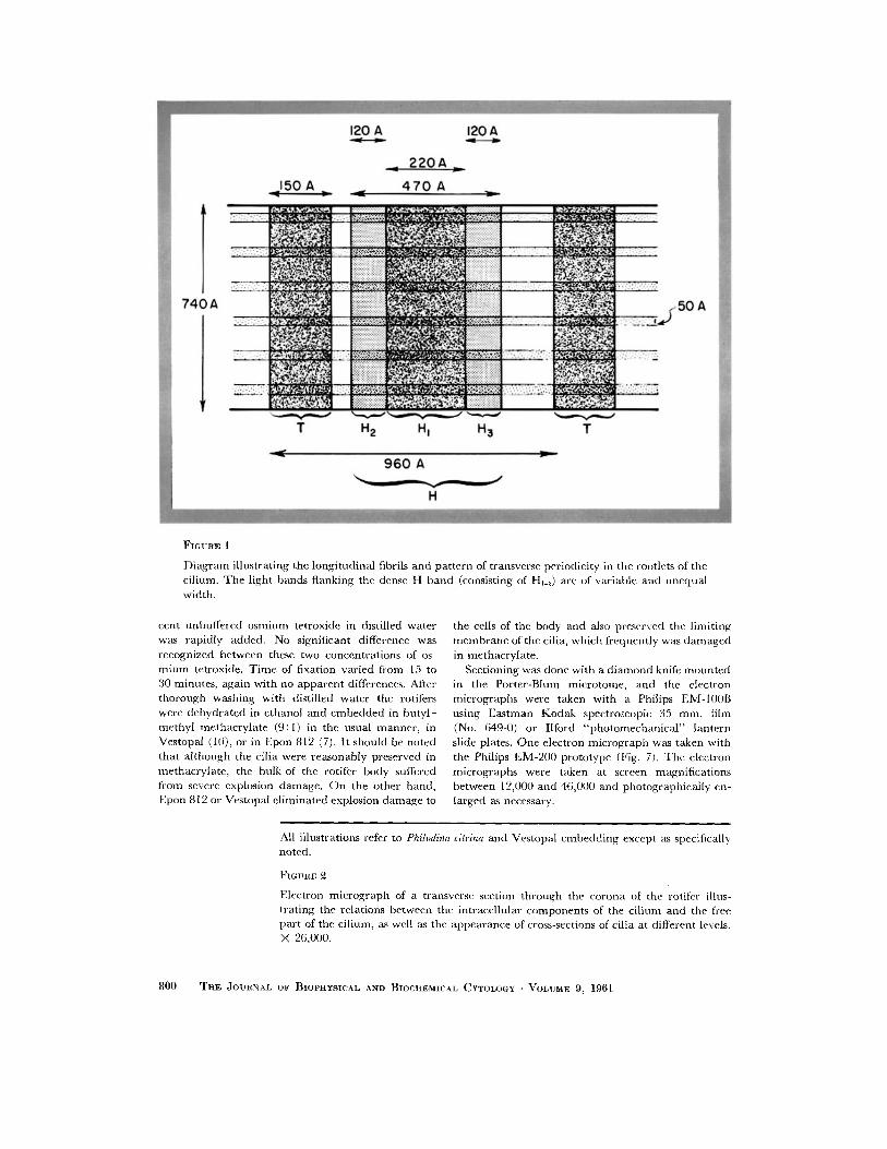
~'I GURE 1
Di ag ram illustrating the longi tudinal fibrils and pa t te rn of transverse periodicity in the rootlets of the cilium. T h e light bands f lanking the dense H band (consisting of Hj_a) are of variable and unequa l width.
cent unbuffered o smi um tetroxide in distilled water was rapidly added. No signiticant difference was recognized between these two concentrat ions of os- m i u m tetroxide. T i m e of fixation varied from 15 to 30 minutes , again with no apparen t differences. After thorough wash ing with distilled water the rotifers were dehydra ted in e thanol and embedded in bu ty l - methyl methacry la te (9:1) in the usual manne r , in Vestopal (10), or in Epon 812 (7). It should be noted tha t a l though the cilia were reasonably preserved in methacryla te , the bulk of the rotifer body suffered from severe explosion damage . O n the other hand , Epon 812 or Vestopal e l iminated explosion d a m a g e to
the cells of the body and also preserved the l imit ing m e m b r a n e of the cilia, which frequent ly was d a m a g e d in methacryla te .
Sectioning was done with a d i amond knife m o u n t e d in the Por ter-Blum microtome, and the electron micrographs were taken with a Philips EM-100B using Eas tman Kodak spectroscopic 35 m m . film (No. 649-0) or Ilford " p h o t o m e c h a n i c a l " lantern slide plates. One electron mic rograph was taken with the Philips EM-200 prototype (Fig. 7). The electron micrographs were taken at screen magnif icat ions between 12,000 and 46,000 and photographical ly en- larged as necessary,
All illustrations refer to Philodina citrina and Vestopal embedd ing except as specifically noted.
FIGIJRE
Electron mic rograph of a transverse section th rough the corona of the rotifer illus- 1rating the relations between the intracel lular componen ts of the ci l ium and the free par t of the cilium, as well as the appearance of cross-sections of cilia at different levels. X 26,000.
800 THE JOURNAL OF BIOPHYSICAL AND BIOCHEMICAL CYTOLOGY • VOLUME 9, 1961

A. I. LANSING AND F. LAMY Fine Structure of Cilia of Rotifers 801
O B S E R V A T I O N S
Rootlets
The coronal cilia of rotifcrs possess very well developed single rootlets whose transverse periods fade indistinctly in the region of junct ion with the basal body. These rootlets appear to have a mean m a x i m u m diameter of 740 A with a range of 722 to 758 A. The repeat ing transverse period, T to T (see Fig. 1), has a mean spacing of 960 A with a range of 922 to 1022 A. Between these narrow T bands of a mean width of 150 A (range 103 to 173 A) there is the p rominen t H band whose mean width is 470 A (range 427 to 530 A). In suitably thin sections it is possible to resolve three distinct bands in the H band : a thick central band with a mean width of 220 A, designated HI, is flanked by two bands designated Hu and H.~ each of which has a mean thickness of 120 A. The H band is not equidis tant between the T bands bu t ra ther is displaced toward one of these bands so tha t one band of low electron density between H and T is twice the width of the other.
In several preparat ions, one of which is illus- t ra ted in Fig. 7, we have been able to resolve line longi tudinal fibrils, apparent ly six in n u m b e r across the diameter of the rootlet, which arc equally spaced. At the point of intersection of these longitudinal fibers, which measure 50 A, and H2 or H.~ there are slight thickenings which may be due to superposition of longitudinal ly and transversely disposed mater ia l or may con- stitute actual nodules. Thus far we have not been able to clarify this point,
Basal Body
Every longi tudinal section including bo th rootlet and basal body tha t we have examined is charac- terized by a narrow region, at the point of junc- tion of these two structures, in which the trans- verse periods of the rootlet becomes very indistinct and blend rapidly into a dense fibrous ma t char- acteristic of the wall of the basal body (Figs. 10
and 14). In bo th methacry la te -embedded and Epon 812-embedded mater ia l the medul la of the basal body is character ized by a very low electron density. The basal body characteristically is located in a pro tuberance of cytoplasm like a hillock.
In suitably thin sections it is appa ren t tha t the basal body is a complex structure. The dense fibrous wall is confined to the proximal two-thirds of the basal body, while the distal th i rd is of relatively low density and is composed of longi- tudinal ly oriented filaments, nine in number . These filaments are of the same approximate diameter as the filaments of the fi'ee par t of the cil ium and are interconnected by a dense mater ia l (Figs. 3 and 6). The nine filaments within the substance of the basal body pass through the basal plate of the cil ium and are cont inuous with the per ipheral filaments of the fi'ee cilium.
Fig. 4 illustrates the ana tomica l cont inui ty between the cross-striated rootlet, the basal body, the basal plate, and the per iphera l f i laments of the fi'ce cilium.
So far as the medul lary par t of the basal body is concerned, our sections have not enabled us adequate ly to characterize this region. Specimens fixed in 5 per cent K M n O 4 showed a very low electron opacity for the medul la of the basal body. With 1 or 2 per cent OsO4 the medul la showed indistinct, sparsely distr ibuted granules, while similarly fixed mater ia l stained with lead acetate as r ecommended by Dal ton (5) revealed the presence of some strands and granules not unlike mater ia l found in the adjacent cytoplasmic sub- stance.
Basal Plate
The basal plate of the cil ium is strikingly con- spicuous because of its intense density. It is best described as a shallow cup or pan whose bot tom is only one-third as thick as the lateral walls. The mater ia l which makes up the walls and base of the basal plate appears to be homogeneous.
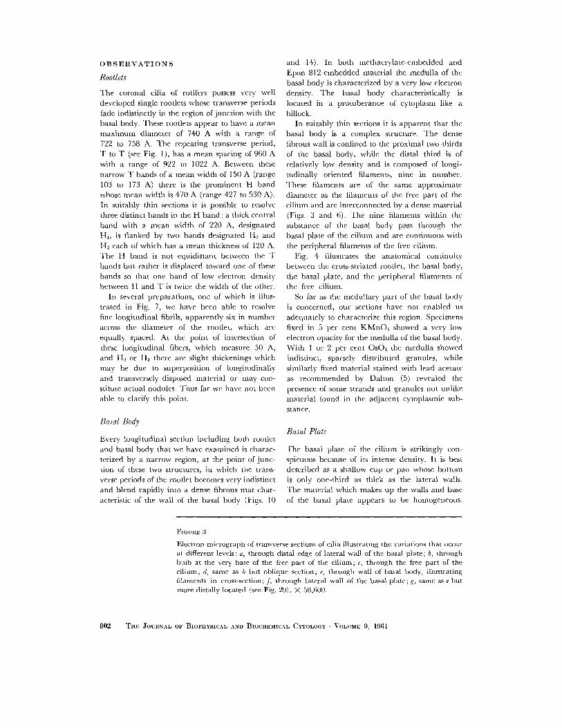
FIGURE 3
Electron micrograph of transverse sections of cilia illustrating the variations that occur at different levels: a, through distal edge of lateral wall of the basal plate; b, through bulb at the very base of the free part of the cilium; c, through the free part of the cilium; d, same as b but oblique section; e, through wall of basal body, illustrating filaments in cross-section; f, through lateral wall of the basal plate; g, same as e but more distally located (see Fig. 20). X 58,600.
802 THE JOURNAL OF BIOPHYSICAL AND BIOCHEMICAL CYTOLOGY - VOLUME 9, 1961
A. I. ],.~NS~N~ ~ND F. LAMY Fine ,qtructure of (!ilia of Rotifers 803

As already noted, the peripheral filaments of the free part of the cilium pass through the lateral wall of the basal plate and are continuous with filaments in the basal body. The paired central filaments of the free cilium extend down to, and appear to fuse with, the floor of the basal plate. At the region of junction of the central filaments with the floor of the basal plate there is a slight swelling and increased density. In no case have we observed central filaments extending below the basal plate into the basal body.
Free Cilium
In the main, the structure of the free part of the cilium of the rotifer conforms to that described for other species (Fig. 2), the literature for which has recently been summarized (8). In addition to the established pattern of nine pairs of peripheral filaments and two separated central filaments, we have, in suitable cross-sections, observed the existence of the "a rms" and "spokes" described by Afzelius (1) and substantiated by Gibbons and Grimstone (8). The radial distribution of the "spokes" is illustrated in Fig. 8.
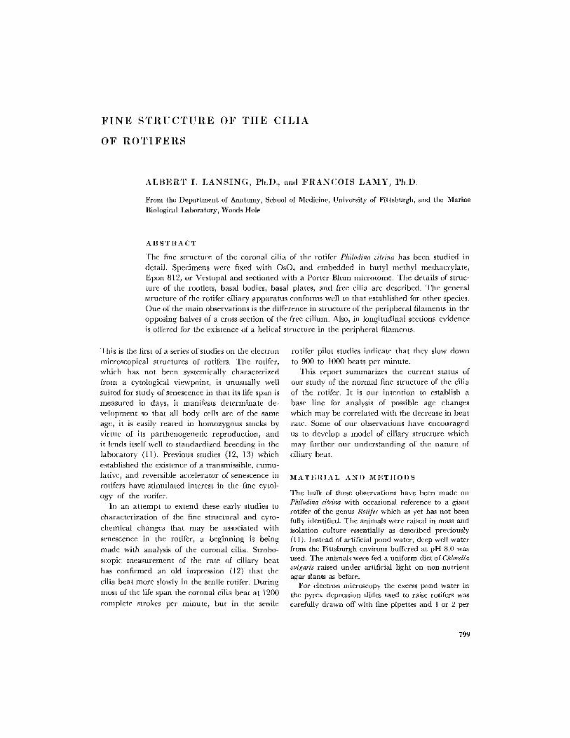
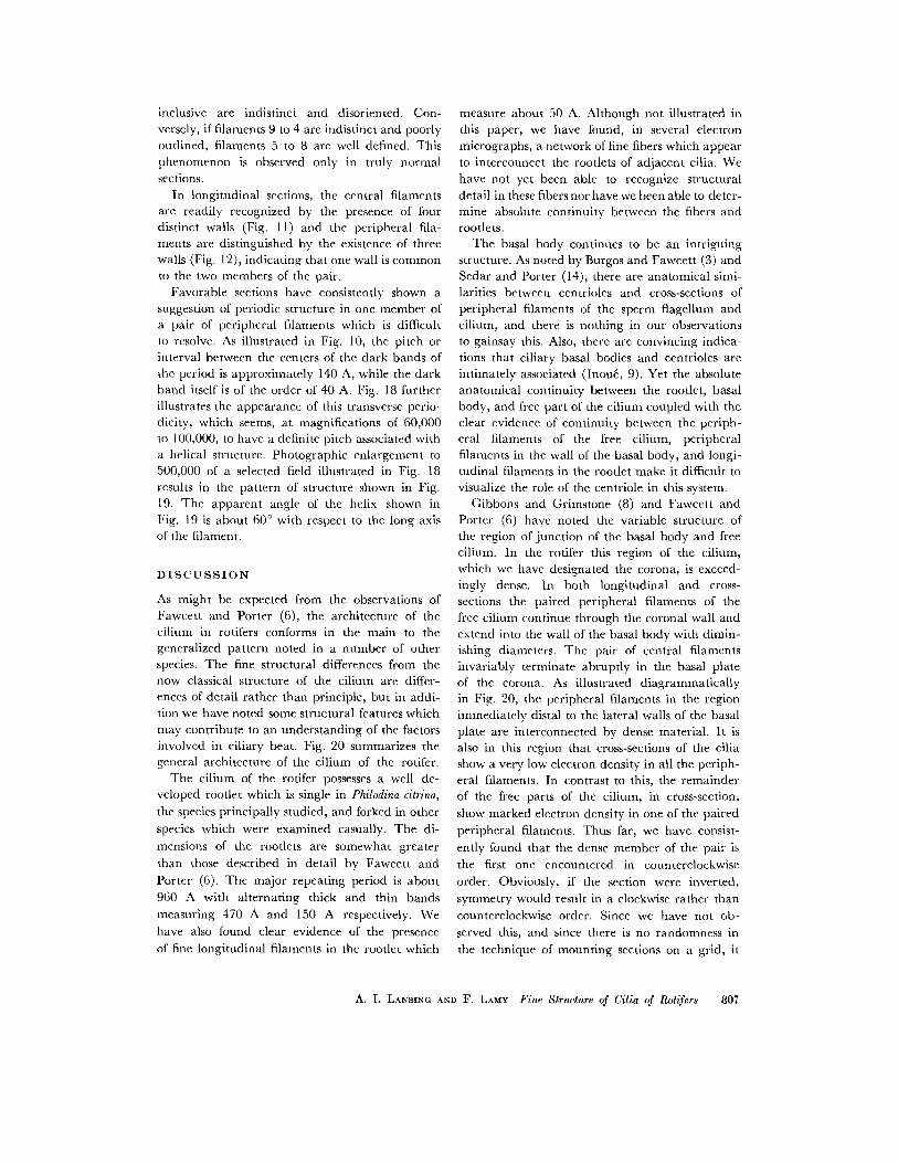
Fig. 15 summarizes observations made on methacrylate-, Vestopal-, and Epon-embedded material regarding the relatively fixed radial distribution of the peripheral filaments. Based upon analysis of 18 sections of cilia cut in normal section (as judged by sharp, circular outlining of the pair of central filaments), it seems that the distribution of peripheral filaments conforms well to the pattern suggested by Cleland and Roths- child (4). Projection through the long axis of the central filaments will pass through one pair of peripheral filaments (Fig. 15). Similarly, pro- jection of an axis perpendicular to the long axis through the central filaments will also pass through
a pair of peripheral filaments. In keeping with the numbering system of Bradfield (2) and others, the peripheral pair in the perpendicular axis is numbered 1 and the other filament pairs are numbered consecutively 2 to 9 in the direction of the f l amen t pair in the longitudinal axis. Inspec- tion of Fig. 15 further indicates that the positions of filament pairs 2 through 7 are quite fixed, while the positions of pairs 4, 5, and 6 show consid- erable variability. Also, while the interval between filament pairs 1 and 2, 2 and 3, 5 and 6, 6 and 7, and 7 and 8 is slightly over 40 °, the spacing be- tween 3 and 4, 4 and 5, 8 and 9, and 9 and 1 is roughly 10 ° less. In selecting the 18 sections for measurement, care was exercised not only to use normal sections, but also to avoid compression- distorted specimens which would not exhibit a cir- cular distribution of the peripheral filaments. This observation is consistent with that of Cleland and Rothschild (4), who noted that the peripheral filaments of the bandicoot spermatzoon are not distributed in a "equiangular radial way."
Of further interest in cross-sections of cilia was the observation that the members of the peripheral pairs of filaments are not identical (Fig. 13). One member is slightly larger than the other and
truly circular, whereas the smaller member ap- pears to be an arc of a circle with one wall in
common with its mate. In addition, as illustrated in Fig. 17, one member of a pair of peripheral
filaments exhibits a greater electron opacity than
its mate. Counting counterclockwise it appears
that the first member of a pair is the one that exhibits the greater electron opacity.
Figs. 8 and 9 illustrate a phenomenon of consis- tent occurrence in all sections cut perpendicular to the long axis of the cilium except those at the
very base of the cilium (Fig. 16). In sections of the
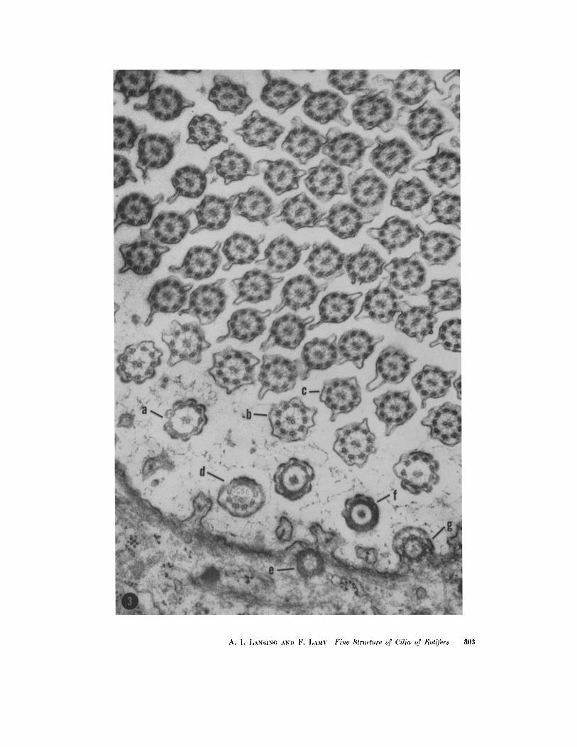
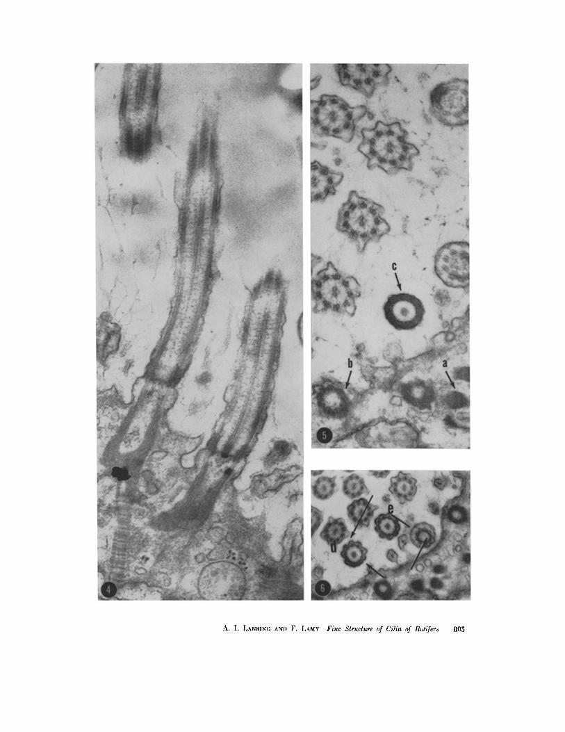
FIGURE ,,t
Longitudinal section through two adjacent cilia showing the anatomical continuity between rootlet, basal body, and fl'ee cilium. X 54,900.
FIG (~RE 5
Transversc section illustrating the rootlet (a), wall of the basal body (b), and lateral wall of the basal body (c). X 54,900.
FIGURE 6
Transverse section showing the connections between peripheral filaments in the lateral wall of the basal body (d) and a section just proximal to the basal plate (e). X 40,400.
804 TItE JOURNAL OF BIOPnYSIC3_L AND BIOCIIEMICAL CYTOLOGY • VOLUME O, 1961
A. I. LANSING AND F. LAMY Fine Slructure of Cilia of Rot~yers 805

FIGURE 7
Longitudinal section through a rootlet illustrating the longitudinal fibrils and transverse periodic structure. Epon 812-embedded, Philips EM-200, Eastman Kodak medium contrast plate. X 93,(i00.
FIGURES 8 ~lll(l ,(}
Transverse sections through free parts of cilia illustrating the almost semicircular differences in ap- pearance of peripheral filaments (arrows). Peripheral filaments in one semicircle are sharply outlined while thc filaments in the opposite semicircle are indistinct. X 56,000.
latter, the central pair of filaments and all nine pairs of peripheral filaments are sharply outlined as would be expected in a normal section. In all other sections it seems clear that peripheral
filaments 9, 1, 2, 3, and 4 differ in appearance from f laments 5, 6, 7, and 8. When filaments 9 to 4 inclusive appear sharply outlined along with the pair of central filaments, filaments 5 to 8
8116 THE JOURNAL OF BIOPHYSICAL AND BIOCHE~VIICAL CYTOLOGY • VOLUME 9, 1961
inclusive are indistinct and disoriented. Con- versely, if filaments 9 to 4 are indistinct and poorly outlined, filaments 5 to 8 are well defined. This phenomenon is observed only in truly normal sections.
In longitudinal sections, the central filaments are readily recognized by the presence of four distinct walls (Fig. 11) and the peripheral fila- ments are distinguished by the existence of three walls (Fig. 12), indicating that one wall is common to the two members of the pair.
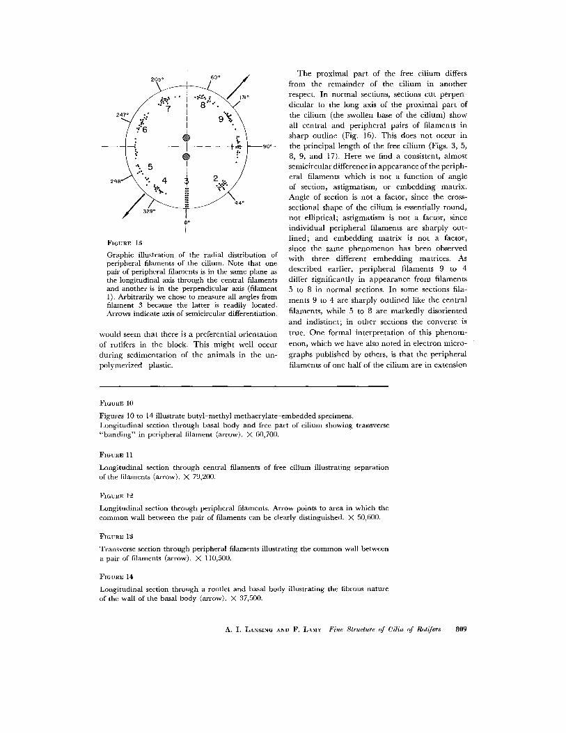
Favorable sections have consistently shown a suggestion of periodic structure in one member of a pair of peripheral filaments which is difficult to resolve. As illustrated in Fig. 10, the pitch or interval between the centers of the dark bands of the period is approximately 140 A, while the dark band itself is of the order of 40 A. Fig. 18 further illustrates the appearance of this transverse perio- dicity, which seems, at magnifications of 60,000 to 100,000, to have a definite pitch associated with a helical structure. Photographic enlargement to 500,000 of a selected field illustrated in Fig. 18 results in the pattern of structure shown in Fig. 19. The apparent angle of the helix shown in Fig. 19 is about 60 ° with respect to the long axis of the filament.
D I S C U S S I O N
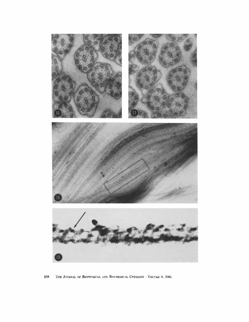
As might be expected from the observations of Fawcett and Porter (6), the architecture of the cilium in rotifcrs conforms in the main to the generalized pattern noted in a number of other species. The fine structural differences from the now classical structure of the cilium are differ- ences of detail rather than principle, but in addi- tion we have noted some structural features which may contribute to an understanding of the factors involved in ciliary beat. Fig. 20 summarizes the general architecture of the cilium of the rotifer.
The cilium of the rotifer possesses a well de- veloped rootlet which is single in Philodina citrina, the species principally studied, and forked in other
species which were examined casually. The di-
mensions of the rootlets are somewhat greater
than those described in detail by Fawcett and
Porter (6). The major repeating period is about 960 A with alternating thick and thin bands measuring 470 A and 150 A respectively. We have also found clear evidence of the presence
of fine longitudinal filaments in the rootlet which
measure about 50 A. Although not illustrated in this paper, we have found, in several electron micrographs, a network of fine fibers which appear to interconnect the rootlets of adjacent cilia. We have not yet been able to recognize structural detail in these fibers nor have we been able to deter- mine absolute continuity between the fibers and rootlets.
The basal body continues to be an intriguing structure. As noted by Burgos and Fawcett (3) and Sedar and Porter (14), there are anatomical simi- larities between centrioles and cross-sections of peripheral filaments of the sperm flagellum and cilium, and there is nothing in our observations to gainsay this. Also, there are convincing indica- tions that ciliary basal bodies and centrioles are intimately associated (Inoufi, 9). Yet the absolute anatomical continuity between the rootlet, basal body, and free part of the cilium coupled with the clear evidence of continuity between the periph- eral filaments of the free cilium, peripheral filaments in the wall of the basal body, and longi- tudinal filaments in the rootlet make it difficult to visualize the role of the centriole in this system.
Gibbons and Grimstone (8) and Fawcett and Porter (6) have noted the variable structure of the region of junction of the basal body and free cilium. In the rotifer this region of the cilium, which we have designated the corona, is exceed- ingly dense. In both longitudinal and cross- sections the paired peripheral filaments of the free cilium continue through the coronal wall and extend into the wall of the basal body with dimin- ishing diameters. The pair of central filaments invariably terminate abruptly in the basal plate of the corona. As illustrated diagrammatically in Fig. 20, the peripheral filaments in the region immediately distal to the lateral walls of the basal plate are interconnected by dense material. It is also in this region that cross-sections of the cilia show a very low electron density in all the periph- eral filaments. In contrast to this, the remainder of the free parts of the cilium, in cross-section,
show marked electron density in one of the paired peripheral filaments. Thus far, we have consist-
ently found that the dense member of the pair is the first one encountered in counterclockwise order. Obviously, if the section were inverted, symmetry would result in a clockwise rather than counterclockwise order. Since we have not ob- served this, and since there is no randomness in
the technique of mounting sections on a grid, it
A. I. LANSING AND F. LAMV Fine Structure of Cilia of Rotlfers 807

808 TIIE JOURNAL OF BIOPHYSICAL AND BIOCIfEMICAL CYTOLOGY • VOLUME 9,
2 0 5 ° 160 ° j "
/ 8,.....-,,< 2!~ / ..:': 9"~
7.4
\ ' : 5 i " / .:.. I
~. :%. ~ " r , / /)--z__i__-/.." \,o
,o 1
FIGURE 15
Graphic illustration of the radial distribution of peripheral filaments of the cilium. Note that one pair of peripheral filaments is in the same plane as the longitudinal axis through the central filaments and another is in the perpendicular axis (filament 1). Arbitrarily we chose to measure all angles from filament 3 because the latter is readily located. Arrows indicate axis of semicircular differentiation.
would seem tha t there is a preferential or ientat ion of rotifers in the block. This migh t well occur dur ing sedimenta t ion of the animals in the un- polymerized plastic.
The proximal par t of the free ci l ium differs from the r emainder of the cil ium in ano ther respect. In normal sections, sections cut perpen- dicular to the long axis of the proximal par t of the cil ium (the swollen base of the cilium) show all central and per ipheral pairs of fi laments in sharp outl ine (Fig. 16). This does not occur in the principal length of the free cil ium (Figs. 3, 5, 8, 9, and 17). Here we find a consistent, a lmost semicircular difference in appearance of the per iph- eral fi laments which is no t a function of angle of section, astigmatism, or embedd ing matrix. Angle of section is no t a factor, since the cross- sectional shape of the ci l ium is essentially round, not elliptical; ast igmatism is no t a factor, since individual per ipheral fi laments are sharply out- l ined; and embedd ing mat r ix is not a factor, since the same phenomenon has been observed with three different embedd ing matrices. As described earlier, per ipheral filaments 9 to 4 differ significantly in appearance from filaments 5 to 8 in normal sections. In some sections fila-
ments 9 to 4 are sharply out l ined like the cent ra l
filaments, while 5 to 8 are markedly disoriented
and indist inct; in other sections the converse is
true. One formal in terpre ta t ion of this phenom-
enon, which we have also noted in electron micro-
graphs publ ished by others, is tha t the per ipheral
fi laments of one half of the ci l ium are in extension
FIGURE l0
Figures l0 to 14 illustrate butyl-methyl mcthacrylate-cmbedded specimens. Longitudinal section through basal body and free part of cilium showing transverse "band ing" in peripheral filament (arrow). X 60,700.
FIGURE 11
Longitudinal section through ccntral filaments of free cilium illustrating separation of the filaments (arrow). X 79,200.
FIGURE 1~
Longitudinal section through peripheral filaments. Arrow points to area in which the common wall between the pair of filaments can be clearly distinguished. X 50,600.
FIGURE 13
Transverse section through peripheral filaments illustrating the common wall between a pair of filaments (arrow). X 110,500.
FIGURE 14
Longitudinal section through a rootlet and basal body illustrating the fibrous nature of the wall of the basal body (arrow). X 37,500.
A. I. LANSING AND F. LAMY Fine Structure of Cilia of tb)t~fers 809
8 |0 ThE JOURNAL OF BIOPtIYSICAL AND BIOClIEMICAL CYTOLOGY • V O L U M E 9, 1961
FIGURE ~00
S emi d i ag rammat i c reconst ruct ion of the rotifer ci l ium in longi tudinal and transverse axes. Note tha t the central fi laments, unl ike the per iphera l fi laments, do not extend into the wall of the basal body.
FIGURE 16
Transverse section t h rough the bu lbar base of the free par t of the cilium. In this region all per iphera l f i laments are sharply out l ined in the no rma l section and are of equally low electron opacity. X 104,000.
FmURE 17
Transverse section th rough the ma i n body of the free ci l ium il lustrat ing the differencc in electron opaci ty between m e m b e r s of a pair of per ipheral filaments. X 104,000.
FmURE 18
Longi tud ina l section th rough free cilium. Area in enclosure is enlargcd in Fig. 19. X 107,000.
FIGURE 19
Enla rgemen t of area shown in Fig. 18 i l lustrating the existence of a helical s t ruc ture (arrow) wi thin the per ipheral fi lament. X approx imate ly 500,000.
A. I. LANSING AND F. LAMY Fine Slructure of Cilia of Rotifers 811

while those of the other half are in contraction. This would result in loss of parallel relationship between filaments as well as possible differences in crystallinity between filaments which are ex- tended or contracted. In the light of this observa- tion it is clear that the plane of beat of the cilium is not perpendicular to the plane of the two central filaments, but rather makes an angle of approxi- mately 40 ° with the accepted plane of beat as described by Fawcett and Porter (6) and Brad- field (2).
I t is not difficult to visualize the possibility that a pendular beat by the cilium could be derived from a contraction of peripheral filaments in one semicircle of the cilium with a corresponding
R E F E R E N C E S
1. AFZELIUS, B., J. Biophysic. and Biochem. Cytol., 1959, 5, 269.
2. BRADFIELD, J. R. G., Syrup. Soc. Exp. Biol., 1955, no. 9, 306.
3. BUROOS, M. H., and FAWCETT, D. W. , J. Bio- physic, and Biochem. Cytol., 1955, 1, 287.
4. CLELAND, K. W., and ROTHSCHILD, LORD, Proc. Roy. Soc. London, Series B, 1959, 150, 24.
5. DALTON, A. J., and ZEIGEL, R. F., J. Biophysic. and Biochem. Cytol., 1960, 7,409.
6. FAWGETT, D, W., and PORTER, K. R., J. Morphol., 1954, 94, 221.
7. FINCK, H., J. Biophysic. and Biochem. Cylo[., 1960, 7, 27.
extension of peripheral filaments in the opposing semicircle. The observation of a helical structure in longitudinal sections of the peripheral fila- ments is consistent with the possibility that a
contractile and expansile system does exist in the
peripheral filaments.
Current studies are being conducted to explore further the nature of the helical structure in the
peripheral filaments and to determine the possible
influence of this system on ciliary beat.
This work has been aided by grants H-2560 (C3) and H-2975 (C3) from the United States Public Health Service. Received for publication, December 7, 1960.
8. GIBBONS, I. R., and GRIMSTONE, A. V., J. Bio- physic, and Biochem. Cytol., 1960, 7, 697.
9. INOU~, S., in Biophysical Seienee--A Study Pro- gram, (J. L. Oneley, editor), New York, .]. Wiley and Sons, 1959.
10. KELLENBERGER, ]~., SCHWAB, W., and RYTER, A., Experientia, 1956, 12,421.
l l. LANSING, A. I., J. Exp. Zool., 1942, 91, 195. 12. LANSING, A. I., J. Gerontol., 1947, 2, 228. 13. LANSING, A. I., Proc. Nat. Acad. Sc., 1948, 34, 304. 14. SEDAR, A. W., and PORTER, K. R., J. Biophysic.
and Biochem. Cytol., 1955, 1, 583.
812 THE JOURNAL OF BIOPHYSICAL.AND BIOCHEMICAL CYTOLOGY • VOLUME 9, 1961