Embed Size (px)
Citation preview
Memorial Sloan Kettering Cancer Center
IRB Number: 18-267 A(3)
Approval date: 20-Jan-2020
Page 1 of 33
First-in-human imaging of multiple myeloma using 89Zr-DFO-daratumumab, a CD38-targeting monoclonal antibody
PROTOCOL FACE PAGE FOR
MSK THERAPEUTIC/DIAGNOSTIC PROTOCOL
Principal Investigator/Department: Gary Ulaner, MD, PhD Radiology
Co-Principal Investigator(s)/Department:
Ola Landgren, MD Jason Lewis, PhD Joseph O’Donoghue, PhD
Medicine Radiology Medical Physics
Investigator(s)/Department: Mithat Gonen, PhD Pat Zanzonico, PhD Adam Kesner, PhD Assen Kirov, PhD Neha Korde, MD Oscar Lahoud, MD Alexander Lesokhin, MD Nikoletta Lendvai, MD, PhD Hani Hassoun, MD Sham Mailankody, MBBS Jonathan Peled, MD, PhD Michael Scordo, MD Eric Smith, MD, PhD Malin Hultcrantz, MD, PhD David Chung, MD, PhD Heather Landau, MD Sergio Giralt, MD Gunjan Shah, MD Virginia Klimek, MD Audrey Hamilton, MD Mila Gorsky, MD Paul Hamlin, MD Philip Caron, MD, PhD Colette Owens, MD
Biostatistics Medical Physics Medical Physics Medical Physics Medicine/Myeloma Medicine/Myeloma Medicine/Myeloma Medicine/Myeloma Medicine/Myeloma Medicine/Myeloma Medicine/Myeloma Medicine/Myeloma Medicine/Myeloma Medicine/ Myeloma Medicine/ABMT Medicine/ABMT Medicine/ABMT Medicine/ABMT Medicine Medicine Medicine Medicine Medicine Medicine
Consenting Professional(s)/Department:
Gary Ulaner, MD, PhD Ola Landgren, MD Neha Korde, MD Oscar Lahoud, MD Alexander Lesokhin, MD Nikoletta Lendvai, MD, PhD Hani Hassoun, MD Sham Mailankody, MBBS Jonathan Peled, MD, PhD
Radiology Medicine/Myeloma Medicine/Myeloma Medicine/Myeloma Medicine/MyelomaMedicine/Myeloma Medicine/Myeloma Medicine/Myeloma Medicine/Myeloma Medicine/Myeloma

Memorial Sloan Kettering Cancer Center
IRB Number: 18-267 A(3)
Approval date: 20-Jan-2020
Page 2 of 33
Michael Scordo, MD Eric Smith, MD, PhD Malin Hultcrantz, MD, PhD David Chung, MD, PhD Heather Landau, MD Sergio Giralt, MD Gunjan Shah, MD Virgina Klimek, MD Audrey Hamilton, MD Mila Gorsky, MD Paul Hamlin, MD Philip Caron, MD, PhD
Colette Owens, MD
Medicine/Myeloma Medicine/ Myeloma Medicine/ABMT Medicine/ABMT Medicine/ABMT Medicine/ABMT Medicine Medicine Medicine Medicine Medicine Medicine Medicine
Please Note: A Consenting Professional must have completed the mandatory Human
Subjects Education and Certification Program.
OneMSK Sites
Manhattan
Memorial Sloan Kettering Cancer Center 1275 York Avenue
New York, New York 10065
Memorial Sloan Kettering Cancer Center
IRB Number: 18-267 A(3)
Approval date: 20-Jan-2020
Page 3 of 33
Table of Contents
1.0 PROTOCOL SUMMARY AND/OR SCHEMA ....................................................................... 4
2.0 OBJECTIVES AND SCIENTIFIC AIMS ................................................................................ 5
3.0 BACKGROUND AND RATIONALE ..................................................................................... 6
4.0 OVERVIEW OF STUDY DESIGN/INTERVENTION ................................................................. 11
4.1 Design................................................................................................................................. 11
4.2 Intervention .............................................................................................................................. 12
5.0 THERAPEUTIC/DIAGNOSTIC AGENTS ........................................................................... 13
6.0 CRITERIA FOR SUBJECT ELIGIBILITY ............................................................................... 15
6.1 Subject Inclusion Criteria ..................................................................................................... 15
6.2 Subject Exclusion Criteria ................................................................................................... 15
7.0 RECRUITMENT PLAN .................................................................................................................... 15
8.0 PRETREATMENT EVALUATION ...................................................................................... 16
9.0 TREATMENT/INTERVENTION PLAN ................................................................................ 17
10.0 EVALUATION DURING TREATMENT/INTERVENTION .................................................... 17
11.0 TOXICITIES/SIDE EFFECTS .................................................................................................... 18
12.0 CRITERIA FOR THERAPEUTIC RESPONSE/OUTCOME ASSESSMENT ......................... 19
13.0 CRITERIA FOR REMOVAL FROM STUDY ....................................................................... 22
14.0 BIOSTATISTICS ............................................................................................................... 22
15.0 RESEARCH PARTICIPANT REGISTRATION AND RANDOMIZATION PROCEDURES
24
15.1 Research Participant Registration ....................................................................................... 24
15.2 Randomization .................................................................................................................... 24
16.0 DAT A M ANAGEMENT ISSUES ........................................................................................ 24
16.1 Quality Assurance............................................................................................................... 24
16.2 Data and Safety Monitoring .................................................................................................... 24
17.0 PROTECTION OF HUMAN SUBJECTS ............................................................................ 26
17.1 Privacy ................................................................................................................................ 26
17.2 Serious Adverse Event (SAE) Reporting ................................................................................ 26
17.2.1 ........................................................................................................................................ 28
18.0 INFORMED CONSENT PROCEDURES................................................................................... 28
19.0 REFERENCES ....................................................................................................................... 29
20.0 APPENDICES ................................................................................................................... 30

Memorial Sloan Kettering Cancer Center
IRB Number: 18-267 A(3)
Approval date: 20-Jan-2020
Page 4 of 33
1.0 PROTOCOL SUMMARY AND/OR SCHEMA
This is a phase I/II study with the goal to assess the feasibility of using the anti-CD38
monoclonal antibody daratumumab, labeled with Zirconium-89 (89Zr ) through
deferoxamine (DFO), known as 89Zr-DFO-daratumumab, for PET imaging of multiple
myeloma. CD38 is an established therapeutic target in multiple myeloma that is expressed
at high density by almost all myeloma cells. Daratumumab binds directly to CD38.
Imaging with 89Zr-DFO-daratumumab may therefore provide a sensitive and specific way
to detect and stage multiple myeloma. In addition, the degree of uptake of 89Zr-DFO-
daratumumab may predict the effectiveness of daratumumab therapy. Finally,
daratumumab labeled with a beta or alpha emitter may eventually be used to treat multiple
myeloma.
The study has two phases: a short phase 1 to confirm the safety of the imaging agent,
identify the suitable antibody mass for imaging, and identify the most appropriate time for
imaging; and a subsequent phase 2 to correlate tumor uptake on 89Zr-DFO-daratumumab
PET serum M protein concentration, percentage plasma cells on bone marrow biopsy, and
patient response to daratumumab/lenalidomide therapy.The total patient population is up
to 30 (6-12 evaluable patients in phase 1 and 18-24 evaluable patients in phase 2). The
study outline is as follows:
Phase I (total of 6-12 evaluable patients). At least 6 and up to 12 evaluable patients will be
needed to determine pharmacokinetics and radiation dosimetry for 89Zr-DFO-
daratumumab. Only the number of patients needed to determine pharmacokinetics and
radiation dosimetry will be enrolled in phase I.
Three patients will be administered 2 mCi of 89Zr-daratumumab in a total
of 50 mg of daratumumab antibody.
Accrue patients with multiple myeloma and at least one malignant lesion on
CT, MR, or FDG PET/CT within 60 days of protocol enrollment.
Memorial Sloan Kettering Cancer Center
IRB Number: 18-267 A(3)
Approval date: 20-Jan-2020
Page 5 of 33
Phase II (total of 18-24 evaluable patients). After pharmacokinetics and radiation
dosimetry are determined in phase I, additional patients will be enrolled in phase II.
2.1 OBJECTIVES AND SCIENTIFIC AIMS
Phase I objectives
• Determine the safety, pharmacokinetics, and radiation dosimetry of 89Zr-DFO-
daratumumab PET/CT imaging of CD38 expression in multiple myeloma.
• Determine the optimal parameters for imaging with 89Zr-DFO-daratumumab, including
antibody mass and time post-administration imaging.
Administer 89Zr-daratumumab at a dose and antibody amount, as
optimized in phase I. Obtain PET/CT imaging at the time point optimized in
phase I.
Correlate 89Zr-daratumumab PET uptake with serum M protein
concentration, percentage plasma cells on bone marrow biopsy, and
patient response to daratumumab/lenalidomide therapy.
Accrue patients with multiple myeloma and at least one malignant lesion on
CT, MR, or FDG PET/CT within 60 days of protocol enrollment.
Administered activity (1 to 5 mCi) of radioactivity and total amount of
administered antibody (3 to 50 mg) will be adjusted in subsequent patients
in order to maximize image quality.
Patients in phase I will have up to 4 PET/CT scans, multiple blood draws,
whole-body counts, and safety monitoring to determine pharmacokinetics,
radiation dosimetry, and safety of 89Zr-DFO-daratumumab for PET/CT
imaging.

Memorial Sloan Kettering Cancer Center
IRB Number: 18-267 A(3)
Approval date: 20-Jan-2020
Page 6 of 33
Phase II objective
• Obtain data on the correlation between tumor uptake of 89Zr-DFO-daratumumab with
patient serum M protein concentration, percentage of plasma cells on bone marrow
biopsy, and patient response to daratumumab/lenalidomidetherapy
Patients who are inevaluable will be replaced until 30 evaluable patients have been
accrued to both Phase I and Phase II of this study.
3.0 BACKGROUND AND RATIONALE
Multiple myeloma
Multiple myelomais a plasma cell neoplasm and the secondmost commonhematologic malignancy
in adults (1). Large independent studies have shown that the genetic and molecular landscape
underlying multiple myeloma pathogenesis is massively heterogeneous, including several recurrent
mutations in KRAS, NRAS, TP53, and other genes (2-4). However, no single mutation is seen in
all or most patients with multiple myeloma. Prospective studies have demonstrated that multiple
myeloma is consistently preceded by a precursor state—monoclonal gammopathy of undetermined
significance and smoldering myeloma (5, 6). Smoldering myelomais an earlier, asymptomatic stage
of myeloma and is defined based on clinical and laboratory characteristics. It carries an increased
risk of progression to multiple myeloma (7). Only limited information is available regarding the
genetic profiles of smoldering myeloma and preliminary studies have suggested comparable
mutational load and copy number profiles of myeloma cells in smoldering myeloma and multiple
myeloma patients (8, 9).
Previously we reported results from two prospective studies including patients with high-risk
smoldering myeloma and newly diagnosed multiple myeloma (10). In both cohorts, all patients were
treated uniformly with 8 cycles of combination therapy including carfilzomib, lenalidomide, and
dexamethasone (KRd) followed by two years of lenalidomide maintenance (KRd-R). We recently
expanded our early results (10) by enrolling additional patients with high-risk smoldering myeloma
and capturing a longer median follow-up of almost four years. Our promising early results of high
overall response rate and minimal residual disease (MRD) negativity among high-risk smoldering
myeloma patients treated with modern KRd-R combination therapy (10) prompted us to
prospectively investigate the baseline genetic landscape and patterns of mutations in our cohort of
patients. As a reference group, we included 40 patients with newly diagnosed multiple myeloma
who received the same KRd-R therapy (10). In brief; the overall response rate was 100%. With
median potential follow-up of 43.3 months, 10 (63%) remain in MRD negativity and the estimated
4-year progression-free and overall survival is 71% and 100%, respectively (in press). Importantly,
we report differences in mutational patterns in patients with high-risk smoldering myeloma and
newly diagnosed multiple myeloma, reflected in a lower frequency of mutations in significant
myeloma genes (6.6% vs. 45%) and NF-KB pathway genes (6.6% vs. 25%) (in press). Based on
these data, important differences are evident in the genetic landscape of high-risk smoldering

Memorial Sloan Kettering Cancer Center
IRB Number: 18-267 A(3)
Approval date: 20-Jan-2020
Page 7 of 33
myeloma and newly diagnosed multiple myeloma patients, suggestive of a more treatment-
responsive biology in early disease.
As mentioned above, we have pioneered the development of modern therapy in early disease,
which has resulted in unprecedented high rates of MRD negativity (10). However, we still see MRD-
positive disease in many patients (10). Also, many patients convert back from MRD-negative to
MRD-positive status. A major clinical limitation with current MRD assays is the fact that they are
based on blind bone marrow biopsies/aspirates (11). It is clinically well known that myeloma can
grow in a patchy manner in the bone marrow, particularly at stages with lower-level disease burden.
Therefore, current bone marrow biopsy/aspirate-MRD assays are inherently limited by potential
sampling error (i.e., false MRD negativity) (11). Furthermore, current standard PET/CT assessment
using FDG as a tracer is limited for the assessment of myeloma. It is based on glucose uptake and
cell proliferation. Because myeloma cells typically have low cell proliferation, FDG is only positive
in about 70% of patients with established myeloma bone disease. In addition, there is a high rate
of false positivity due to inflammatory and degenerative causes (12). Clearly, myeloma-specific PET
tracers are currently lacking.
Current monitoring of treated multiple myeloma patients is based on blood tests focusing on
secreted abnormal immunoglobulins (monoclonal proteins and free light chains). Based on current
guidelines, therapy is not initiated until the patient develops symptoms or the biomarkers are
elevated (13). Similar to multiple myeloma patients, in patients with smoldering myeloma the current
monitoring is based on blood tests focusing on secreted abnormal immunoglobulins. This remains
the current standard although is it well established that all myeloma cells do not secrete proteins
(14-16).
Design of a targeted PET tracer for multiple myeloma
To overcome the inherent dilemmas with blind bone marrow biopsies/aspirates and the limitations
of blood-based protein markers, we have developed a targeted immuno-PET imaging agent to rule
out MRD in multiple myeloma and its precursors. Specifically, we have developed a PET
tracer based upon daratumumab for tracking CD38 expression. CD38 is expressed on the surface
of virtually every multiple myeloma cell. Daratumumab (Darzalex®, Janssen Biotech) is a clinically
approved antibody targeting CD38 for the treatment of multiple myeloma. In our laboratory,
Daratumumab has been prepared for radiolabeling with 89Zr (t1/2 = 78.4 h) through conjugation
with desferrioxamine (DFO). Western blot, flow cytometry, saturation binding assays, and
internalization assays have been utilized to characterize CD38 expression and binding of
daratumumab in an OPM2 myeloma cell line. A murine xenograft model of the OPM2 cell line has
been generated for further in vivo studies. Longitudinal PET imaging is being performed following
injection of 5-10 MBq 89Zr-DFO-daratumumab out to 120 h post-injection. Based on our
successful preclinical results, this project is designed as a first-in-human trial of our CD38-
targeted immuno-PET imaging agent. Primary objectives are to determine the safety,
pharmacokinetics, and radiation dosimetry of 89Zr-DFO-daratumumab PET/CT imaging of CD38
Memorial Sloan Kettering Cancer Center
IRB Number: 18-267 A(3)
Approval date: 20-Jan-2020
Page 8 of 33
expression in multiple myeloma. In addition, we will determine the optimal timing and mass of
antibody for imaging and acquire preliminary data on the sensitivity and specificity of 89Zr-DFO-
daratumumab to visualize multiple myeloma lesions in patients. These are the first steps to
develop a more sensitive and specific imaging test to assess MRD in multiple myeloma.
89Zirconium (89Zr) was chosen as the radionuclide for immuno-PET due to favorable decay
characteristics. It has a 78.4 h half life, allowing for multiple-day circulation times for antibodies,
which are needed to increase target-to-background uptake. Standardized methods for the routine
production and isolation of high-purity and high-specific-activity 89Zr using a small cyclotron are
currently used at MSK and other institutions. Optimized cyclotron conditions reveal high average
yields of 1.52 ± 0.11 mCi/muA*h at a proton beam energy of 15 MeV and current of 15 muA using
a solid, commercially available 89 Y-foil target (0.1 mm, 100% natural abundance). The effective
specific activity of 89Zr was found to be in the range of 5.28−13.43 mCi/microg (470−1195
Ci/mmol) of zirconium. Radiolabeling studies using the trihydroxamate ligand desferrioxamine B
(DFO) gave 100% radiochemical yields in <15 min at room temperature, and in vitro stability
measurements confirmed that (89Zr) Zr-DFO is stable with respect to ligand dissociation in human
serum for more than 7 days.
Preclinical data on 89Zr-DFO-daratumumab PET/CT
Before animal studies were conducted, the expression of CD38 on OPM2 cells was determined
by flow cytometry. After conjugation of daratumumab to the DFO chelate, preservation of the
immunoreactivity (>90%) of the antibody was determined by Lindmo assay in OPM2 cells.
For the preliminary study, NSG mice were injected with a human multiple myeloma (MM) tumor cell
line (OPM2) intravenously approximately four weeks before administration of the radiolabeled
antibody. The intravenous injection of 1×10⁶ OPM2s in NSG miceis a well-validated model to study
MM bone disease (4). Myeloma cells accumulate in the bone marrow of the femurs and sternum
primarily. We used OPM2 cells that were stably transduced with firefly luciferase to allow for in vivo
imaging.
Due to the NSG background of the mice used in this study, one group of mice received a coinjection
with an excess of human IgG. It has been shown that the biodistribution of radiolabeled antibodies
is affected by the lack of endogenous IgG in mouse strains bearing a SCID mutation (Sharma, SK,
et al. Cancer Res; 78(7) April 2018). This manifests itself in reduced tumor uptake in these mice,
along with higher nonspecific spleen and bone uptake. This uncharacteristic distribution can be
blocked by coninjection of an isotype IgG.
Mice were separated into three groups (Figure 1A) (n=5). Groups 1 and 2 were injected with OPM2
cells; Group 3 consisted of healthy mice. Each mouse was injected with approximately 200μCi (70
μg) of 89Zr-DFO-daratumumab via the tail vein. Group 1 was co-injected with cold IgG (400 μg) to
block nontarget uptake in the spleen. PET/CT imaging was conducted at 24, 96, and 144 hours
post-injection. Images are coronal maximum intensity projections. Biodistribution was performed at

Memorial Sloan Kettering Cancer Center
IRB Number: 18-267 A(3)
Approval date: 20-Jan-2020
Page 9 of 33
120 hours post-injection for Group 2 and at 144 hours post-injection for Groups 1 and 3 (Figure
1B).
Approximately 24 hours prior to PET/CT imaging, mice were imaged by BLI so as to correlate the
antibody distribution to the actual distribution of OPM2 cells. Free 89Zr which is not chelated to
DFO in vivo is osteophilic and will become incorporated into bone due to its high affinity for
phosphate. Because the OPM2 cells reside in the bone marrow in this model, it was necessary to
extract the bone marrow from the bone for a more accurate view of the biodistribution. Both
hindlimbs from each mouse were collected and the bone marrow was extracted by centrifugation.
With this technique, about 30 milligrams of bone marrow per mouse can be reproducibly obtained.

Memorial Sloan Kettering Cancer Center
IRB Number: 18-267 A(3)
Approval date: 20-Jan-2020
Page 10 of 33
Memorial Sloan Kettering Cancer Center
IRB Number: 18-267 A(3)
Approval date: 20-Jan-2020
Page 11 of 33
Figure 1: (A) Group 1 was co-injected with cold IgG (400 μg) to block nontarget uptake in the spleen. PET/CT imaging
was conducted at 24, 96, and 144 hours post-injection. Images are coronal maximum intensity projections. (B)
Biodistribution was performed at 120 hours post-injection for Group 2 and at 144 hours post-injection for Groups 1 and
3.
At 24 hours post-injection, Group 1 mice showed a significant amount of 89Zr-dartumamab in the
blood circulation. Group 2 miceshow earlier uptake in the bones of the hind limbs and pelvic region,
but also increased accumulation in the liver and spleen as compared to Group 1. At later time
points, Group 1 mice show increasing accumulation in the hind limbs, pelvic region, and sternum.
Group 2 mice also show higher accumulation in the hind limb region, but the sternum cannot be
resolved due to significant uptake in the liver and spleen. The uptake in these bones is consistent
targeted imaging of multiple myeloma in this cancer model and confirmed by the bioluminescence
data. Importantly, the healthy control mice show significantly lower uptake in the bone as seen by
imaging. (Data from the laboratory of Dr. Jason Lewis, unpublished data.)
4.1 OVERVIEW OF STUDY DESIGN/INTERVENTION
4.2 Design
This protocol will perform first-in-human imaging of patients with 89Zr-DFO-daratumumab.
Patients with known CD38-positive malignancy will be recruited.
For phase I patients:
• Biodistribution of 89Zr-DFO-daratumumab will be obtained from the serial PET/CT
images.
• Blood pharmacokinetics of 89Zr-DFO-daratumumab will be obtained from serial
blood draws.
• Radiation dosimetry will be determined from whole-body counts, blood counts, and
PET/CT images.
• Safety of the imaging dose of 89Zr-DFO-daratumumab will be documented through
assessment of symptoms and physical examinations.
• The optimal antibody mass of non-radiolabeled (cold) daratumumab to be added to
radiolabeled 89Zr-DFO-daratumumab will be determined by administration of
varying amounts of “cold” (i.e., non-radioactive) daratumumab.
• The optimal timing of 89Zr-DFO-daratumumab PET/CT imaging will be determined
by sequential scanning of patients over 1-8 days post-administration.
For phase II patients we will correlate tumor uptake of 89Zr-DFO-daratumumab with:
• Serum M protein concentration;
• Percentage of plasma cells on bone marrow biopsy; and
• Patient response to daratumumab/lenalidomidetherapy.
Memorial Sloan Kettering Cancer Center
IRB Number: 18-267 A(3)
Approval date: 20-Jan-2020
Page 12 of 33
Serum M protein concentration and bone marrow biopsy will be required for all phase
II patients. As all myeloma patients will have clinically standard serum M protein
concentration and bone marrow biopsy results, this should not impede patient
recruitment.
4.3 Intervention
89Zr-DFO-daratumumab PET/CT and PET/CT interpretation: 89Zr-DFO-daratumumab
PET/CT studies will be performed as hybrid PET/CT examinations for attenuation
correction, lesion localization, and availability of additional findings on CT images. The
patient will be positioned on a GE PET/CT scanner. The CT component will be obtained
utilizing a low mA (80 mA) to minimize radiation exposure. 3D imaging will be obtained
from skull apex to feet (approximately 10 bed positions). 89Zr-DFO-daratumumab PET/CT
images will be reconstructed using iterative reconstruction and displayed in a multiplanar
format. All routinely applied corrections will be implemented and the reconstructed images
parameterized in terms of standard uptake values (SUVs).
89Zr-DFO-daratumumab PET/CT scans will be interpreted by a nuclear medicine physician
experienced in the use of routine and novel research PET radiotracers. Physiologic 89Zr-
DFO-daratumumab uptake will be determined from the images. Physiologic 89Zr-DFO-
daratumumab may be expected in the blood pool, liver, spleen, and kidneys, similar to
other radiolabeled antibodies. Radiotracer uptake in areas that are not physiologic will be
graded both qualitatively and semiquantitatively. For qualitative scoring, we will use a
scale of one to five where 1 = definitely normal, 2 = probably normal, 3 = equivocal, 4 =
probably abnormal, and 5 = definitely abnormal. Semiquantitative analysis of tracer uptake
will be performed for all lesions receiving a score of 4 or 5, as well as for apparently
normal background blood pool. Three-dimensional regions of interest (ROIs) will be placed
over grade 4 and 5 lesions and the mediastinal blood pool and tracer uptake will be
quantified using maximum SUV = decay-corrected maximum ROI activity (μCi/ml) /
(injected dose (μCi) / body weight (g)). 89Zr-DFO-daratumumab PET/CT images are for
research purposes only, and will not be used to influence patient care. This approach was
recently utilized for first-in-human work with 89Zr-DFO-pertuzumab, MSK IRB 17-059.
For patients in phase I, the following will be performed to determine 89Zr-DFO-
daratumumab tissue distribution, pharmacokinetics, and radiation dosimetry:
1. 89Zr-DFO-daratumumab kinetic measurements: Blood and serum samples will be
weighed and counted in a scintillation well counter calibrated for 89Zr. Immediately before
or after each PET/CT imaging session, whole-body activities will be measured using a
scintillation detector probe, with whole-body net count rates converted to the percent of
the administered activity by normalization to the first whole-body net count rate (i.e., the
count rate measured shortly after administration of the 89Zr-DFO-daratumumab).

Memorial Sloan Kettering Cancer Center
IRB Number: 18-267 A(3)
Approval date: 20-Jan-2020
Page 13 of 33
2. Timing of 89Zr-DFO-daratumumab PET/CT: Radiolabeled antibodies normally require
several days to clear blood pool, accumulate in targets, and yield reliable tumor images
(17). 89Zr-DFO-daratumumab will be administered on day 0. PET/CT images will be
obtained on post-administration days 1, 2-4, 5-6, and/or 7-8 following administration of 89Zr-DFO-daratumumab to determine the optimal time point for imaging. (At least three
PET/CT images at different time points are required to calculate dosimetry—i.e., the
radiation dose received throughout the body—for the novel radiotracer. We will obtain
three or four sets of images at the time points described above.)
For patients in phase II, a single PET/CT scan will be performed using the dose of 89Zr-
DFO-daratumumab, antibody mass of daratumumab, and timing determined from phase I
patients. No blood samples for dosimetry or whole-body counts will be performed. The
research PET/CT will be performed to correlate 89Zr-DFO-daratumumab PET uptake with
serum M protein concentration (measured prior to 89Zr-DFO-daratumumab PET/CT),
percentage plasma cells on bone marrow biopsy (measured prior to 89Zr-DFO-
daratumumab PET/CT), and patient response to daratumumab/lenalidomide therapy.
The radiologist evaluated the 89Zr-DFO-daratumumab PET/CT will be blinded to patient
serum M protein concentration and percentage of plasma cells on bone marrow biopsy.
As patient response to daratumumab/lenalidomide therapy will be subsequently
determined, that information will not be avialable at the time of 89Zr-DFO-daratumumab
PET/CT. Combined daratumumab/lenalidomide therapy is a standard-of-care
combination for treatment of myeloma, with response rates of up to 90%. Single agent
daratumumab therapy has lower response rates, and thus the combination therapy is the
current appropriate standard of care. Please see section 14.0 for statistical details.
5.1 THERAPEUTIC/DIAGNOSTIC AGENTS
89Zr-DFO-daratumumab: 89Zr-DFO-daratumumab is an investigational new drug (not FDA
approved) produced on demand by the Cyclotron-Radiochemistry Core at MSK (under the
direction of Dr. Jason Lewis), under GMP conditions adequate for clinical trials. 89Zr-DFO-
daratumumab is composed of the native CD38 targeting drug daratumumab labeled with the
positron-emitting radionuclide zirconium 89 (89Zr) through the linker DFO. Daratumumab is an
FDA-approved monoclonal antibody. 89Zr is a metallo-radionuclide with a half-life of 78 hours,
long enough to allow favorable biodistribution of radiolabeled antibodies. The final product will be
radiolabeled and tested for sterility, endotoxin, identity, purity, and potency as described in the
chemistry, manufacturing, and controls section of the IND application. 89Zr-DFO-daratumumab will
be administered intravenously over a 30-60 minute period.
No patient will receive 89Zr-DFO-daratumumab until the FDA approves the IND.
Memorial Sloan Kettering Cancer Center
IRB Number: 18-267 A(3)
Approval date: 20-Jan-2020
Page 14 of 33
Administered activity of 89Zr-DFO-daratumumab: For phase 1 patients, the administered activity of
89Zr-DFO-daratumumab will be between 1 and 5 mCi in 3 mg radiolabeled (“hot”) antibody.
Successful imaging of other 89Zr-labeled antibodies will be performed with 1-5 mCi (17, 18).
Changes in administered activity alter counts of radioactivity detected by the PET/CT camera and
may affect image quality. The administered activity of 89Zr-DFO-daratumumab will start at 2 mCi,
as this is an administered activity which has resulted in successful imaging in past radio-antibody
imaging trials, including the principle investigator’s trial of 89Zr-DFO-pertuzumab (IRB 17-059). If
2 mCi does not provide sufficient radioactive counts for successful imaging, then we will increase
the administered dose to 5 mCi. If 2 mCi provides more radioactive counts than is needed for
successful imaging, then we will attempt imaging with 1 mCi, to see if successful imaging can be
obtained with a lower exposure to radiation dose. This approach was recently utilized for first-in-
human work with 89Zr-DFO-pertuzumab, MSK IRB 17-059.
For phase 2 patients, the optimal dose of 89Zr-DFO-daratumumab determined in phase I will be
utilized.
Antibody mass of 89Zr-DFO-daratumumab: Radiolabeled PET antibodies have been shown to
demonstrate superior imaging when administered together with cold antibody to block non-
specific binding (17). For phase I patients, to determine the optimal antibody mass for imaging, 0,
17, or 47 mg of unlabelled (“cold”) antibody will be added to the radiolabeled antibody to produce
total administered antibody masses of 3, 20, and 50 mg. Antibody masses between 20 and 50 mg
per dose have provided the best image contrast in several prior studies with radiolabeled
antibodies at MSK (18-20). Changes in total amount of administered antibody alter distribution of
specific and non-specific binding of antibody and may affect image quality. The first three patients
will be imaged with a total antibody mass of 50 mg based on MSK’s prior experience with Zr-
labeled antibodies, as higher antibody mass usually results in better imaging due to cold antibody
occupying non-specific in vivo binding sites. Antibody mass will be reduced to 20 mg in the next
three patients to evaluate the effect on image quality. The choice of total antibody mass for each
group of three patients will depend on the quality of images already obtained from prior patients.
Thus, in this trial the first three patients will receive an antibody mass of 50 mg, and the fourth
through sixth patients will receive an antibody mass of 20 mg. If an improvement in image quality
is observed at 20 mg, or if image quality at 50 mg and 20 mg is comparable, then any subsequent
patients will receive an antibody mass of 3 mg. If image quality worsens at 20 mg, then the
antibody mass will be increased back to 50 mg for the remaining patients. The means of
determining whether one antibody mass is superior to another is described in section 14.0,
Biostatistics.
In summary, the antibody mass received by patients will be:
• Patients 1, 2 and 3: 50 mg
• Patients 4, 5, 6: 20 mg
• Any subsequent patients: 50 mg, 20 mg, or 3 mg, depending on imaging results of the
prior 6 patients.
Memorial Sloan Kettering Cancer Center
IRB Number: 18-267 A(3)
Approval date: 20-Jan-2020
Page 15 of 33
For any further patients, antibody mass will be determined based on imaging results from prior
patients. This approach was recently utilized for first-in-human work with 89Zr-DFO-pertuzumab,
MSK IRB 17-059.
For phase 2 patients, the optimal daratumumab antibody mass determined in phase I will be
utilized.
6.1 CRITERIA FOR SUBJECT ELIGIBILITY
6.2 Subject Inclusion Criteria
• Age 21 years or greater
• Histologically/Immunohistochemistry confirmed CD38-positive multiple myeloma
• At least one tumor lesion on CT, MRI, or FDG PET/CT within 60 days of protocol
enrollment
• ECOG performance status 0 to 2
• For Phase II patients only: plan for initiation of standard-of-care
daratumumab/lenalidomide therapy.
6.3 Subject Exclusion Criteria
• Life expectancy < 3 months
• Pregnancy or lactation
• Patients who cannot undergo PET/CT scanning because of weight limits. PET/CT
scanners may not be able to function with patients over 450 pounds.
• History of anaphylactic reaction to humanized or human antibodies or a Grade 3 or 4
administration reaction during a daratumumab administration.
7.0 RECRUITMENT PLAN
Patients who meet the above inclusion and exclusion criteria will be invited to participate in the
study by their primary oncologist. We will invite men and women of all races/ethnicities on an
equal basis.
Consenting professionals will meet with the patients and obtain informed consent in either the
oncologist’s clinic or the Molecular Imaging and Therapy Service clinic. As patients in Phase I will
require multiple visits, a small honorarium ($400) will be provided to patients that complete the
trial, in order to compensate them for their time and expenses. No payments will be offered to
patients in phase II.

Memorial Sloan Kettering Cancer Center
IRB Number: 18-267 A(3)
Approval date: 20-Jan-2020
Page 16 of 33
If the investigator is a member of the treatment team, s/he will screen their patients’ medical
records for suitable research study participants and discuss the study and their potential for
enrolling in the research study. Potential subjects contacted by their treating physician will be
referred to the investigator/ research staff of the study.
The principal investigator may also screen the medical records of patients with whom they do not
have a treatment relationship for the limited purpose of identifying patients who would be eligible
to enroll in the study and to record appropriate contact information in order to approach
these patients regarding the possibility of enrolling in the study.
During the initial conversation between the investigator/ research staff and the patient, the patient
may be asked to provide certain health information that is necessary to the recruitment and
enrollment process. The investigator/ research staff may also review portions of their medical
records at MSKCC in order to further assess eligibility. They will use the information provided by
the patient and/or medical record to confirm that the patient is eligible and to contact the patient
regarding study enrollment. If the patient turns out to be ineligible for the research study, the
research staff will destroy all information collected on the patient during the initial conversation
and medical records review, except for any information that must be maintained for screening log
purposes.
In most cases, the initial contact with the prospective subject will be conducted by either the
treatment team, investigator or the research staff working in consultation with the treatment team.
The recruitment process outlined presents no more than minimal risk to the privacy of the patients
who are screened and minimal protected health information (PHI) will be maintained as part of a
screening log. For these reasons, we seek a (partial) limited waiver of authorization for the
purposes of (1) reviewing medical records to identify potential research subjects and obtain
information relevant to the enrollment process; (2) conversing with patients regarding possible
enrollment; (3) handling of PHI contained within those records and provided by the potential
subjects; and (4) maintaining information in a screening log of patients approached (if applicable).
We anticipate an accrual rate of 1-2 patients per month. We may accrue multiple patients at the
same time however, in phase I, tracer administration for the next patient will wait until the prior
patient has completed all anticipated scans.
8.1 PRETREATMENT EVALUATION Prior to enrollment in the protocol, the following will be available:
• History and physical exam
• Histology demonstrating CD38-positive multiple myeloma.
• Clinically standard imaging scans (CT, MR, FDG PET/CT) demonstrating at least one
focus of suspected malignancy within 60 days of protocol enrollment
• Negative blood pregnancy test for women of childbearing potential

Memorial Sloan Kettering Cancer Center
IRB Number: 18-267 A(3)
Approval date: 20-Jan-2020
Page 17 of 33
9.1 TREATMENT/INTERVENTION PLAN For women of childbearing age, pregnancy will be excluded before 89Zr-DFO-daratumumab
administration. This will be accomplished by the use of a serum pregnancy test within two weeks
of 89Zr-DFO-daratumumab administration or a urine pregnancy test on the day of 89Zr-DFO-
daratumumab administration.
89Zr-DFO-daratumumab administration: 89Zr-DFO-daratumumab will be administered
intravenously on day 0.
Prior to 89Zr-DFO-daratumumab administration, the patient will be premedicated with
Acetaminophen 650 mg orally, Diphenhydramine 25mg intravenously, and Dexamethasone 20
mg intravenously.
Physical exam and vital signs will be obtained prior to the 89Zr-DFO-daratumumab administration,
as well as 30-60 minutes post-administration to document the absence of any acute toxicity from
the low imaging dose of 89Zr-DFO-daratumumab.
10.1 EVALUATION DURING TREATMENT/INTERVENTION For Phase I patients: Patients in phase I will have up to 4 PET/CT scans, multiple blood draws,
whole-body counts, and safety monitoring to determine pharmacokinetics, radiation dosimetry,
and safety of 89Zr-DFO-daratumumab for PET/CT imaging.
89Zr-DFO-daratumumab PET/CT: Patients will undergo up to 4 serial 89Zr-DFO-daratumumab
PET/CT scans. PET/CT images will be obtained on days 1, 2-4, 5-6, and/or 7-8 following
administration of 89Zr-DFO-daratumumab. All CT scans will be acquired as low-dose CT scans to
minimize radiation exposure. PET/CT images will be obtained from skull apex to feet
(approximately 10 bed positions) and reconstructed using our standard iterative reconstruction
algorithm.
Radiation dosimetry of 89Zr-DFO-daratumumab: The measured time-dependent blood, whole-
body, and PET-derived organ activities will be fit to exponential functions and the corresponding 89Zr residence times calculated by integration of the respective exponential functions, accounting
for the physical decay of the 89Zr. The resulting residence times will then be entered into the
OLINDA radiation dosimetry program and mean organ-absorbed doses (in rad and rad/mCi) and
the effective dose (in rem and rem/mCi) calculated.

Memorial Sloan Kettering Cancer Center
IRB Number: 18-267 A(3)
Approval date: 20-Jan-2020
Page 18 of 33
W hole-body count measurements: Patients will undergo whole-body count rate measurements
using a sodium iodide (NaI) probe placed approximately 3 meters from the patient. Pre- and post-
first-void measurements will be performed within 6 hours of administration of 89Zr-DFO-
daratumumab, as well as each time the patient returns for PET/CT scanning.
Blood samples for determination of pharmacokinetics of 89Zr-DFO-daratumumab: Approximately
3-5 ml of blood will be drawn at each of the following time points. Drawn blood will be collected in
lavender-top tubes (to prevent coagulation).
• Day 0: Pre-dose
• Day 0: Within 30 minutes of completion of dose administration
• Day 0: 30-60 minutes of completion of dose administration
• Day 0: 60-120 minutes of completion of dose administration
• On each return visit for PET/CT imaging
After all samples have been collected, weighed blood and serum aliquots will be counted in a
scintillation well counter calibrated for 89Zr and counts will be corrected for time of decay.
For Phase II patients: 89Zr-DFO-daratumumab will be administered at a dose and antibody
amount as optimized in phase 1. PET/CT imaging will be obtained at the time point optimized in
phase I. 89Zr-DFO-daratumumab PET ue will be correlated with serum M protein, percent plasma
cells on bone marrow biopsy, and patient response to daratumumab therapy as described in
section 4.2.
11.0 TOXICITIES/SIDE EFFECTS
89Zr-DFO-daratumumab: 89Zr-DFO-daratumumab is a diagnostic (not therapeutic) agent, and the
doses of 89Zr-DFO-daratumumab used for PET/CT are very low compared to approved
therapeutic daratumumab doses, and are expected to have a very low incidence of adverse
events. Prior results from patients who received 89Zr-DFO-trastuzumab as part of protocol #14-
156 demonstrated no grade 3 or 4 toxicities, with one patient reporting chills the night of antibody
administration, which is a recognized side effect of antibody administration. Nevertheless,
patients will be monitored closely for evidence of adverse events, including vital signs before and
after tracer administration. Phase I patients will be scheduled for a brief visit to the nuclear
medicine clinic the day after tracer administration to confirm that there have been no side effects
requiring treatment. If a severe adverse effect (Common Terminology Criteria for Adverse Events
grade 3 or 4) attributable to 89Zr-DFO-daratumumab occurs in any patient, then further use of 89Zr-
DFO-daratumumab will be suspended and the protocol will be reviewed with the MSK Data Safety
Monitoring Committee.
Less Likely: Infusion or allergic reactions, which may include fevers, chills, tiredness, rashes,
hives, tachycardia, or shortness of breath.
Memorial Sloan Kettering Cancer Center
IRB Number: 18-267 A(3)
Approval date: 20-Jan-2020
Page 19 of 33
Radiation risk: The effective dose from 2 mCi of 89Zr-DFO-daratumumab is estimated to be
between 1.74 and 1.90 centiGray (see Appendix 1). A low milliampere CT scan, performed as
part of the hybrid 89Zr-DFO-daratumumab PET/CT, contributes an additional effective dose of 0.9
rem. The effective dose from an experimental 89Zr-DFO-daratumumab PET and up to 4 CT
examinations is in the range of 7.1 to 7.4 centiGray, which is comparable to the dose from other 89Zr-labeled radiolabeled antibodies received by oncology patients in MSK clinical trials. A table of
projected radiation doses to normal tissues based on extrapolation from animal biodistribution,
including contribution from CT scans, is shown in Appendix 1. No specific patient instructions are
required for the low level of radioactivity encountered from the doses of 89Zr administered in this
protocol (21).
Pregnancy risk: Even low diagnostic levels of radiation, such as those that will be received in this
protocol from the investigational 89Zr-DFO-daratumumab PET/CT studies, have been associated
with a risk of inducing childhood cancer. A negative pregnancy test will be required before a
patient is accrued to this protocol. Patients on this protocol will be advised not to become
pregnant for at least one month following administration of 89Zr-DFO-daratumumab.
12.0 CRITERIA FOR THERAPEUTIC RESPONSE/OUTCOME ASSESSMENT There is no therapy arm or measurement of response to the imaging agent. This is a diagnostic
imaging study; therefore, no criteria for therapeutic response are included. The doses of 89Zr-
DFO-daratumumab used for imaging are very low compared to therapeutic daratumumab doses,
and we do not expect a significant therapeutic effect from the diagnostic (not therapeutic) dose of
this agent. This protocol will be a first-in-human CD38-targeted imaging study with 89Zr-DFO-
daratumumab PET/CT.

Memorial Sloan Kettering Cancer Center
IRB Number: 18-267 A(3)
Approval date: 20-Jan-2020
Page 20 of 33
Flow chart for phase 1 patients
Screening On study
Within 60 days
Day 0
Day 1
Day 2-4
Day 5-6
Day
7-8
Informed consent X
Medical history X
Physical exam X Prior radiographic studies (CT,
MR, FDG PET/CT) reviewed X
Histology/Immunohistochemistry
proof of CD38-positive myeloma X
Vital signs X 89Zr-DFO-daratumumab
administration
X
Adverse events monitored X X X X X
Whole-body counts X X X X X
Blood samples for
pharmacokinetics
X X X X X
PET/CT scan X X X X
Memorial Sloan Kettering Cancer Center
IRB Number: 18-267 A(3)
Approval date: 20-Jan-2020
Page 21 of 33
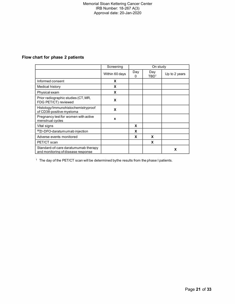
Flow chart for phase 2 patients
Screening On study
Within 60 days Day
0
Day
TBD1
Up to 2 years
Informed consent X Medical history X
Physical exam X
Prior radiographic studies (CT, MR,
FDG PET/CT) reviewed X
Histology/Immunohistochemistryproof of CD38-positive myeloma
X
Pregnancy test for women with active menstrual cycles
x
Vital signs X 89Zr-DFO-daratumumab injection X
Adverse events monitored X X
PET/CT scan X Standard-of-care daratumumab therapy and monitoring of disease response
X
1 The day of the PET/CT scan will be determined bythe results from the phase I patients.

Memorial Sloan Kettering Cancer Center
IRB Number: 18-267 A(3)
Approval date: 20-Jan-2020
Page 22 of 33
13.0 CRITERIAFOR REMOVAL FROM STUDY
• Patients may withdraw from the protocol voluntarily at any time.
• Development of unacceptable toxicity.
• The patient is found to be ineligible for the protocol as designated in the section on Criteria
for Patient/Subject Eligibility (i.e., a change in diagnosis).
14.0 BIOSTATISTICS This is a pilot first-in-human trial of 89Zr-DFO-daratumumab in patients with multiple myeloma.
In phase I, we will determine the tissue distribution, pharmacokinetics, radiation dosimetry, and
safety of 89Zr-DFO-daratumumab for PET/CT imaging.
The organ/tissue uptake and dosimetry following IV injection of 89Zr-DFO-daratumumab will be
determined. Standardized uptake value (SUV) in various organs will be estimated from VOI
analysis of clinical images and converted to activity-time curves. The areas under the activity-time
curves will be derived by integration, converted to residence times, and used as input to the
OLINDA/EXM dosimetry program to obtain absorbed dose estimates for normal tissues.
Additionally, SUVmean, max and peak for lesions will be summarized with descriptive statistics.
Pharmacokinetic analysis will be performed using a compartmental analysis (SAAM or GraphPad
V5); standard parameters such as AUC, clearance, volume of distribution of central compartment,
and Co will be reported. Descriptive statistics will be tabulated.
There are three scan parameteers that will be optimized in phase I: administered radioactivity,
antibody mass, and time of scan. The primarily method of determining these variables will be
quantitative, based of the proportion of lesions identified of the research 89Zr-DFO-daratumumab
scans. The number of lesions in the patient will be determined from the lesions seen on CT, MR,
and FDG PET/CT studies perfromed within the last 60 days. Then the number of 89Zr-DFO-
daratumumab avid lesions will be counted on the 89Zr-DFO-daratumumab PET/CT. The greatest
proportion of lesion lesions detected on 89Zr-DFO-daratumumab PET will be the primary method
of determining optimal scan parameters. Since it is possible that the same proportion of lesions
may be identified using different scan parameters and that contrast may vary between the
different imaging days, a consensus visual assessment of a group of experienced Molecular
Imaging and Therapy investigator who will grade the studies separately (GU, CR, and JO), will be
used to make the final decision. This qualitative assesement will be a secondary method, after
using the primary quantitative comparison of number of lesions visualized. It is possible that
these three scan parameters will interact to effect optimal scan parameters. By sequential
modification of administered activity and antibody mass and obtaining imaging at multiple time
points for each combination of activity and antibody mass, we will obtain adequate comparisons
with which to make quantitative and qualitative comparisons. This approach has been
successfully used in prior radio-antibody imaging trials, including the principle investigator’s trial of
89Zr-DFO-pertuzumab (IRB 17-059).

Memorial Sloan Kettering Cancer Center
IRB Number: 18-267 A(3)
Approval date: 20-Jan-2020
Page 23 of 33
89Zr-DFO-daratumumab is a diagnostic (not therapeutic) agent, and the doses of 89Zr-DFO-
daratumumab used for PET/CT are very low compared to approved therapeutic daratumumab
doses. We expected to have a very low incidence of adverse events. Prior results from patients
who received 89Zr-DFO-trastuzumab as part of protocol #14-156 demonstrated no grade 3 or 4
toxicities, with one patient reporting chills the night of antibody administration, which is a
recognized side effect of antibody administration. Nevertheless, phase I patients will be monitored
closely for evidence of adverse events, including vital signs before and after tracer administration
and a brief visit to the Nuclear Medicine Clinic the day after tracer administration to confirm that
there are no side effects requiring treatment. This diagnostic agent is not expected to result in
significant adverse effects, but the number and propotion of patients with any adverse effect from
tracer administration will be deermined.
In the phase II portion, we will correlate tumor uptake of 89Zr-DFO-daratumumab with serum M
protein concentration, percentage plasma cells on bone marrow biopsy and patient response to
daratumumab/lenalidomidetherapy. The total planned sample size for both phases is 30
evaluable patients; hence Phase II component will enroll 18-24 evaluable patients, depending on
how many patients were needed for the Phase I. The total sample size was chosen given
budgetary concerns.
Phase II patients will be monitored for adverse effects, including vital signs before and after
tracer administration. This disagnostic agent is not expected to result in significant adverse
effects, but the number and propotion of patients with any adverse effect from tracer
administration will be deermined.
Serum M protein concentration: Serum M protein concentration (including heavy-chains and
serum free light-chains) is a standard of care laboratory measurement and reported as a
continuous metric. High serum M protein concentration represents high tumor burden in the
blood. We will explore the correlation between 89Zr-DFO-daratumumab PET SUVmax and serum
M protein concentration using scatterplots and Spearman’s rank correlation.
Percentage plasma cells on bone marrow biopsy: Percentage plasma cells on bone marrow
biopsy is a standard of care pathology measurement and reported as a continuous metric. High
percentage plasma cells on bone marrow biopsy represents a high tumor burden. We will explore
the correlation between 89Zr-DFO-daratumumab PET SUVmax and percentage plasma cells on
bone marrow biopsy by scatterplots and Spearman’s rank correlation.
Patient response to daratumumab/lenalidomidetherapy: We will evaluate response by one
dichotomous and one continuous method. Dichotomous evaluation will categorize patients as
complete/partial response vs. stable disease/progressive disease, as defined by International
Myeloma Working Group consensus criteria for response (IMWG). Continuous evaluation will
assess best anti-tumor response as a percentage of change from baseline by IMWG, and will be
graphed as a waterfall plot. We will explore the correlation between 89Zr-DFO-daratumumab PET
SUVmax and anti-tumor efficacy of daratumumab using a Wilcoxon test and an ROC curve in the
case of IMWG response and scatterplots with Spearman rank correlation in the case of percent
change from baseline.
Memorial Sloan Kettering Cancer Center
IRB Number: 18-267 A(3)
Approval date: 20-Jan-2020
Page 24 of 33
15.1 RESEARCH PARTICIPANT REGISTRATION AND RANDOMIZATION PROCEDURES
15.2 Research Participant Registration
Confirm eligibility as defined in the section entitled Inclusion/Exclusion Criteria. Obtain
informed consent, by following procedures defined in section entitled Informed
Consent Procedures. During the registration process registering individuals will be
required to complete a protocol specific Eligibility Checklist. The individual signing the
Eligibility Checklist is confirming whether or not the participant is eligible to enroll in the
study. Study staff are responsible for ensuring that all institutional requirements
necessary to enroll a participant to the study have been completed. See related
Clinical Research Policy and Procedure #401 (Protocol Participant Registration).
15.3 Randomization
N/A
16.1 DATA MANAGEMENT ISSUES
16.2 Quality Assurance
Accrual rates and extent and accuracy of evaluations and follow-up will be monitored
periodically throughout the study period and potential problems will be brought to the
attention of the study team for discussion and action.
16.3 Data and Safety Monitoring
The Data and Safety Monitoring (DSM) Plans at Memorial Sloan-Kettering Cancer Center were
approved by the National Cancer Institute in September 2001. The plans address the new policies
set forth by the NCI in the document entitled “Policy of the National Cancer Institute for Data and
Safety Monitoring of Clinical Trials” which can be found at:
http://cancertrials.nci.nih.gov/researchers/dsm/index.html. The DSM Plans at MSK were
established and are monitored by the Clinical Research Administration. The MSK Data and Safety
Monitoring Plans can be found on the MSK Intranet
at:https://one.mskcc.org/sites/pub/clinresearch/Pages/protocol-review-committees/data-and- safety-
monitoring-committee.aspx
.
There are several different mechanisms by which clinical trials are monitored for data,
safety and quality. There are institutional processes in place for quality assurance (e.g.,
protocol monitoring, compliance and data verification audits, therapeutic response, and
staff education on clinical research QA) and departmental procedures for quality control,
plus there are two institutional committees that are responsible for monitoring the activities
of our clinical trials programs. The committees: Data and Safety Monitoring Committee

Memorial Sloan Kettering Cancer Center
IRB Number: 18-267 A(3)
Approval date: 20-Jan-2020
Page 25 of 33
(DSMC) for Phase I and II clinical trials, and the Data and Safety Monitoring Board
(DSMB) for Phase III clinical trials, report to the Center’s Research Council and
Institutional Review Board.
During the protocol development and review process, each protocol will be assessed for
its level of risk and degree of monitoring required. Every type of protocol (e.g., NIH
sponsored, in-house sponsored, industrial sponsored, NCI cooperative group, etc.) Will be
addressed and the monitoring procedures will be established at the time of protocol
activation.

Memorial Sloan Kettering Cancer Center
IRB Number: 18-267 A(3)
Approval date: 20-Jan-2020
Page 26 of 33
17.1 PROTECTION OF HUMAN SUBJECTS
Participation in this trial is voluntary. All patients will be required to sign a statement of informed
consent, which must conform to IRB guidelines.
Confidentiality: All patient records will be kept as confidential as is possible under the law. No
individual identifiers will be used in any reports or publication resulting from this study, but the data
will be used in the interest of the ongoing research.
Benefits: There is no guarantee of any benefits.
Incentives: $400 will be provided to patients/subjects for participation in Phase I of the study. No
incentive will be provided to patients in phase II.
Costs: The research 89Zr-DFO-daratumumab radiotracer and 89Zr-DFO-daratumumab PET/CT
scan(s) will be performed without charge. Patients in phase II requiring a bone marrow biopsy will be
performed without charge. The patient will be responsible for the costs of standard medical care.
Alternatives: The patient can choose not to be on this study and follow the treatment outlined by his
or her treating physician.
Treatment and Compensation: If the patient is injured as a result of participating in this study,
emergency care, hospitalization, and outpatient care will be made available by the hospital and
billed to the patient and his insurance company as part of his medical expenses. If the patient
desires additional information about the consent process, research patient’s rights, or research-
related injury, he/she may call the Patient Representative’s office at (212) 639-8254
17.2 Privacy
MSK's Privacy Office may allow the use and disclosure of protected health information pursuant to a
completed and signed Research Authorization form. The use and disclosure of protected health
information will be limited to the individuals described in the Research Authorization form. A
Research Authorization form must be completed by the Principal Investigator and approved by the
IRB and Privacy Board (IRB/PB)
17.3 Serious Adverse Event (SAE) Reporting
An adverse event is considered serious if it results in ANY of the following outcomes:
• Death
• A life-threatening adverse event
• An adverse event that results in inpatient hospitalization or prolongation of existing
hospitalization
Memorial Sloan Kettering Cancer Center
IRB Number: 18-267 A(3)
Approval date: 20-Jan-2020
Page 27 of 33
• A persistent or significant incapacity or substantial disruption of the ability to conduct
normal life functions
• A congenital anomaly/birth defect
• Important Medical Events (IME) that may not result in death, be life threatening, or
require hospitalization may be considered serious when, based upon medical judgment,
they may jeopardize the patient or subject and may require medical or surgical
intervention to prevent one of the outcomes listed in this definition
Note: Hospital admission for a planned procedure/disease treatment is not considered an
SAE.
SAE reporting is required as soon as the participant signs consent. SAE reporting is required
for 30-days after the participant’s last investigational treatment or intervention. Any events
that occur after the 30-day period and that are at least possibly related to protocol treatment
must be reported.
If an SAE requires submission to the IRB office per IRB SOP RR-408 ‘Reporting of Serious
Adverse Events’, the SAE report must be sent to the IRB within 5 calendar days of the event.
The IRB requires a Clinical Research Database (CRDB) SAE report be submitted
electronically to the SAE Office as follows:
For IND/IDE trials: Reports that include a Grade 5 SAE should be sent to
[email protected]. All other reports should be sent to [email protected].
For all other trials: Reports that include a Grade 5 SAE should be sent to
[email protected]. All other reports should be sent to [email protected].
The report should contain the following information:
Fields populated from CRDB:
• Subject’s initials
• Medical record number
• Disease/histology (if applicable)
• Protocol number and title
Data needing to be entered:
• The date the adverse event occurred
• The adverse event
• The grade of the event
• Relationship of the adverse event to the treatment (drug, device, or intervention)
• If the AE was expected
• The severity of the AE
• The intervention
• Detailed text that includes the following

Memorial Sloan Kettering Cancer Center
IRB Number: 18-267 A(3)
Approval date: 20-Jan-2020
Page 28 of 33
o A explanation of how the AE was handled
o A description of the subject’s condition
o Indication if the subject remains on the study
• If an amendment will need to be made to the protocol and/or consent form
• If the SAE is an Unanticipated Problem
The PI’s signature and the date it was signed are required on the completed report.
For IND/IDE protocols:
The CRDB SAE report should be completed as per above instructions. If appropriate, the
report will be forwarded to the FDA by the SAE staff through the IND Office
17.2.1
Any additional SAE reporting information required by the sponsor or drug supplier should be
included in this section.
18.1 INFORMED CONSENT PROCEDURES
Before protocol-specified procedures are carried out, consenting professionals will explain
full details of the protocol and study procedures as well as the risks involved to participants
prior to their inclusion in the study. Participants will also be informed that they are free to
withdraw from the study at any time. All participants must sign an IRB/PB-approved consent
form indicating their consent to participate. This consent form meets the requirements of the
Code of Federal Regulations and the Institutional Review Board/Privacy Board of this Center.
The consent form will include the following:
1. The nature and objectives, potential risks and benefits of the intended study.
2. The length of study and the likely follow-up required.
3. Alternatives to the proposed study. (This will include available standard and
investigational therapies. In addition, patients will be offered an option of supportive
care for therapeutic studies.)
4. The name of the investigator(s) responsible for the protocol.
5. The right of the participant to accept or refuse study interventions/interactions and to
withdraw from participation at any time.
Before any protocol-specific procedures can be carried out, the consenting professional will
fully explain the aspects of patient privacy concerning research specific information. In
addition to signing the IRB Informed Consent, all patients must agree to the Research
Authorization component of the informed consent form.
Each participant and consenting professional will sign the consent form. The participant must
receive a copy of the signed informed consent form.
Memorial Sloan Kettering Cancer Center
IRB Number: 18-267 A(3)
Approval date: 20-Jan-2020
Page 29 of 33
19.0 REFERENCES
1. Siegel RL, Miller KD, Jemal A. Cancer statistics, 2016. CA Cancer J Clin. 2016;66(1):7-30.
2. Lohr JG, Stojanov P, Carter SL, et al. Widespread genetic heterogeneity in multiple myeloma: implications for targeted therapy. Cancer Cell. 2014;25(1):91-101. 3. Bolli N, Avet-Loiseau H, Wedge DC, et al. Heterogeneity of genomic evolution and mutational profiles in multiple myeloma. Nat Commun. 2014;5:2997. 4. Lawson MA, Paton-Hough JM, Evans HR, et al. NOD/SCID-GAMMA mice are an ideal strain to assess the efficacy of therapeutic agents used in the treatment of myeloma bone disease. PloS one. 2015;10(3):e0119546. 5. Landgren O, Kyle RA, Pfeiffer RM, et al. Monoclonal gammopathy of undetermined significance (MGUS) consistently precedes multiple myeloma: a prospective study. Blood. 2009;113(22):5412-7. 6. Weiss BM, Abadie J, Verma P, Howard RS, Kuehl WM. A monoclonal gammopathy precedes multiple myeloma in most patients. Blood. 2009;113(22):5418-22. 7. Ghobrial IM, Landgren O. How I treat smoldering multiple myeloma. Blood. 2014;124(23):3380-8. 8. Walker BA, Boyle EM, Wardell CP, et al. Mutational Spectrum, Copy Number Changes, and Outcome: Results of a Sequencing Study of Patients With Newly Diagnosed Myeloma. J Clin Oncol. 2015;33(33):3911-20. 9. Zhao S, Choi M, Heuck C, et al. Serial exome analysis of disease progression in premalignant gammopathies. Leukemia. 2014;28(7):1548-52. 10. Korde N, Roschewski M, Zingone A, et al. Treatment With Carfilzomib-Lenalidomide- Dexamethasone With Lenalidomide Extension in Patients With Smoldering or Newly Diagnosed Multiple Myeloma. JAMA Oncol. 2015;1(6):746-54. 11. Mailankody S, Korde N, Lesokhin AM, et al. Minimal residual disease in multiple myeloma: bringing the bench to the bedside. Nat Rev Clin Oncol. 2015;12(5):286-95. 12. Hillengass J, Landgren O. Challenges and opportunities of novel imaging techniques in monoclonal plasma cell disorders: imaging "early myeloma". Leuk Lymphoma. 2013;54(7):1355-63. 13. Rajkumar SV, Dimopoulos MA, Palumbo A, et al. International Myeloma Working Group updated criteria for the diagnosis of multiple myeloma. Lancet Oncol. 2014;15(12):e538-48. 14. Chawla SS, Kumar SK, Dispenzieri A, et al. Clinical Course and Prognosis of Non-Secretory Multiple Myeloma. Eur J Haematol. 2015. 15. Dimopoulos MA, Kastritis E, Terpos E. Non-secretory myeloma: one, two, or more entities? Oncology (Williston Park). 2013;27(9):930-2. 16. Lopes da Silva R, Monteiro A, Veiga J. Non-secretory multiple myeloma relapsing as extramedullary liver plasmacytomas. J Gastrointestin Liver Dis. 2011;20(1):81-3. 17. Dijkers EC, Oude Munnink TH, Kosterink JG, et al. Biodistribution of 89Zr-trastuzumab and PET imaging of HER2-positive lesions in patients with metastatic breast cancer. Clinical pharmacology and therapeutics. 2010;87(5):586-92. 18. Ulaner GA, Hyman DM, Ross DS, et al. Detection of HER2-Positive Metastases in Patients with HER2-Negative Primary Breast Cancer Using 89Zr-Trastuzumab PET/CT. J Nucl Med. 2016;57(10):1523-8. 19. Divgi CR, Uzzo RG, Gatsonis C, et al. Positron emission tomography/computed tomography identification of clear cell renal cell carcinoma: results from the REDECT trial. J Clin Oncol. 2013;31(2):187-94. 20. O'Donoghue JA, Smith-Jones PM, Humm JL, et al. 124I-huA33 antibody uptake is driven by A33 antigen concentration in tissues from colorectal cancer patients imaged by immuno-PET. J Nucl Med. 2011;52(12):1878-85. 21. Williamson MJ, Dauer LT. Activity thresholds for patient instruction and release for positron emission tomography radionuclides. Health Phys. 2014;106(3):341-52.

Memorial Sloan Kettering Cancer Center
IRB Number: 18-267 A(3)
Approval date: 20-Jan-2020
Page 30 of 33
20.0 APPENDICES
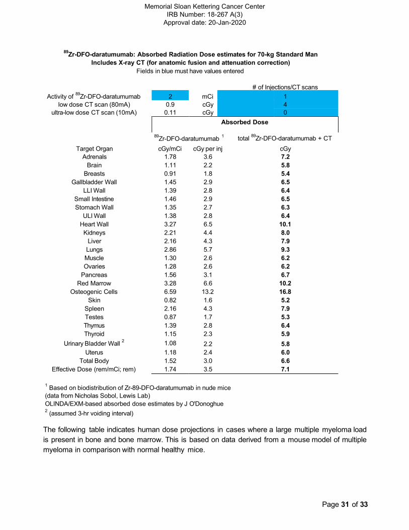
Appendix 1: 89Zr-DFO-daratumumab Radiation Dosimetry
A primary objective of this study is to quantify the biodistribution and pharmacokinetics of 89Zr-DFO-
daratumumab in human patients and thereby generate absorbed radiation dose estimates.
Absorbed dose projections for humans, based on animal biodistribution data from the lab of Dr
Jason Lewis, are provided below.
Page 31 of 33
Memorial Sloan Kettering Cancer Center
IRB Number: 18-267 A(3)
Approval date: 20-Jan-2020
89Zr-DFO-daratumumab: Absorbed Radiation Dose estimates for 70-kg Standard Man
Includes X-ray CT (for anatomic fusion and attenuation correction)
Fields in blue must have values entered
# of Injections/CT scans
Activity of 89
Zr-DFO-daratumumab 2 mCi 1
low dose CT scan (80mA) 0.9 cGy 4
ultra-low dose CT scan (10mA) 0.11 cGy 0
Absorbed Dose
89Zr-DFO-daratumumab
1 total
89Zr-DFO-daratumumab + CT
Target Organ
Adrenals
cGy/mCi
1.78
cGy per inj
3.6
cGy
7.2
Brain 1.11 2.2 5.8
Breasts 0.91 1.8 5.4
Gallbladder Wall 1.45 2.9 6.5
LLI Wall 1.39 2.8 6.4
Small Intestine 1.46 2.9 6.5
Stomach Wall 1.35 2.7 6.3
ULI Wall 1.38 2.8 6.4
Heart Wall 3.27 6.5 10.1
Kidneys 2.21 4.4 8.0
Liver 2.16 4.3 7.9
Lungs 2.86 5.7 9.3
Muscle 1.30 2.6 6.2
Ovaries 1.28 2.6 6.2
Pancreas 1.56 3.1 6.7
Red Marrow 3.28 6.6 10.2
Osteogenic Cells 6.59 13.2 16.8
Skin 0.82 1.6 5.2
Spleen 2.16 4.3 7.9
Testes 0.87 1.7 5.3
Thymus 1.39 2.8 6.4
Thyroid 1.15 2.3 5.9
Urinary Bladder Wall 2 1.08 2.2 5.8
Uterus 1.18 2.4 6.0
Total Body 1.52 3.0 6.6
Effective Dose (rem/mCi; rem) 1.74 3.5 7.1
1 Based on biodistribution of Zr-89-DFO-daratumumab in nude mice
(data from Nicholas Sobol, Lewis Lab)
OLINDA/EXM-based absorbed dose estimates by J O'Donoghue 2
(assumed 3-hr voiding interval)
The following table indicates human dose projections in cases where a large multiple myeloma load
is present in bone and bone marrow. This is based on data derived from a mouse model of multiple
myeloma in comparison with normal healthy mice.

Page 32 of 33
Memorial Sloan Kettering Cancer Center
IRB Number: 18-267 A(3)
Approval date: 20-Jan-2020
89Zr-DFO-daratumumab: Absorbed Radiation Dose estimates for 70-kg Standard Man
Includes X-ray CT (for anatomic fusion and attenuation correction)
Fields in blue must have values entered
# of Injections/CT scans
Activity of 89
Zr-DFO-daratumumab 2 mCi 1
low dose CT scan (80mA) 0.9 cGy 4
ultra-low dose CT scan (10mA) 0.11 cGy 0
Absorbed Dose
89Zr-DFO-daratumumab
1 total
89Zr-DFO-daratumumab + CT
Target Organ
Adrenals
cGy/mCi
1.90
cGy per inj
3.8
cGy
7.4
Brain 1.14 2.3 5.9
Breasts 0.79 1.6 5.2
Gallbladder Wall 1.30 2.6 6.2
LLI Wall 1.34 2.7 6.3
Small Intestine 1.31 2.6 6.2
Stomach Wall 1.20 2.4 6.0
ULI Wall 1.24 2.5 6.1
Heart Wall 3.31 6.6 10.2
Kidneys 2.29 4.6 8.2
Liver 2.20 4.4 8.0
Lungs 2.94 5.9 9.5
Muscle 1.34 2.7 6.3
Ovaries 1.19 2.4 6.0
Pancreas 1.47 2.9 6.5
Red Marrow 4.85 9.7 13.3
Osteogenic Cells 9.40 18.8 22.4
Skin 0.80 1.6 5.2
Spleen 2.16 4.3 7.9
Testes 0.69 1.4 5.0
Thymus 1.28 2.6 6.2
Thyroid 1.09 2.2 5.8
Urinary Bladder Wall 2 0.81 1.6 5.2
Uterus 0.98 2.0 5.6
Total Body 1.65 3.3 6.9
Effective Dose (rem/mCi; rem) 1.90 3.8 7.4
1 Based on biodistribution of Zr-89-DFO-daratumumab in normal and MM mice
(data from Nicholas Sobol, Lewis Lab)
OLINDA/EXM-based absorbed dose estimates by J O'Donoghue 2
(assumed 3-hr voiding interval)

Page 33 of 33
Memorial Sloan Kettering Cancer Center
IRB Number: 18-267 A(3)
Approval date: 20-Jan-2020
Absorbed Dose Projections for Phase 2
In phase 2, a single PET/CT scan will be acquired following administration of 1-5 mCi of 89Zr-DFO-
daratumumab at the mass dose and time deemed optimal from phase 1 Absorbed dose projections
for low and high disease burdens for the minimum and maximum activities envisaged are provided
below:
89Zr-DFO-daratumumab Phase 2:
Absorbed Radiation Dose estimates for 70-kg Standard Man including X-ray CT
min max
Activity of 89
Zr-DFO-daratumumab (mCi) 1 5
low dose CT scan (80mA) 0.9 cGy
ultra-low dose CT scan (10mA) 0.11 cGy
Target Organ
Adrenals
Brain
Breasts
Gallbladder Wall
LLI Wall
Small Intestine
Stomach Wall
ULI Wall
Heart Wall
Kidneys
Liver
Lungs
Muscle
Ovaries
Pancreas
Red Marrow
Osteogenic Cells
Skin
Spleen
Testes
Thymus
Thyroid
Urinary Bladder Wall 2
Uterus
Total Body
Effective Dose (rem/mCi; rem)
# of Injections/CT scans
1
1
0
1 Based on biodistribution of Zr-89-DFO-daratumumab in nude mice (Nicholas Sobol, Lewis Lab)
OLINDA/EXM-based absorbed dose estimates by J O'Donoghue 2
(assumed 3-hr voiding interval)
Absorbed dose for 89
Zr-DFO-daratumumab + CT (cGy)
Low disease burden High disease burden
min max min max
2.7 9.8 2.8 10.4
2.0 6.5 2.0 6.6
1.8 5.5 1.7 4.8
2.3 8.1 2.2 7.4
2.3 7.8 2.2 7.6
2.4 8.2 2.2 7.4
2.3 7.7 2.1 6.9
2.3 7.8 2.1 7.1
4.2 17.3 4.2 17.5
3.1 11.9 3.2 12.3
3.1 11.7 3.1 11.9
3.8 15.2 3.8 15.6
2.2 7.4 2.2 7.6
2.2 7.3 2.1 6.9
2.5 8.7 2.4 8.2
4.2 17.3 5.7 25.1
7.5 33.8 10.3 47.9
1.7 5.0 1.7 4.9
3.1 11.7 3.1 11.7
1.8 5.2 1.6 4.4
2.3 7.8 2.2 7.3
2.1 6.7 2.0 6.4
2.0 6.3 1.7 4.9
2.1 6.8 1.9 5.8
2.4 8.5 2.5 9.1
2.6 9.6 2.8 10.4


![Preclinical Development of CD38-Targeted [89Zr]Zr-DFO ...jnm.snmjournals.org/content/59/2/216.full.pdf · an unmet need for innovative and personalized therapeutic ap-proaches that](https://img.pdfslide.net/doc/110x75/5f099d517e708231d427adb7/preclinical-development-of-cd38-targeted-89zrzr-dfo-jnm-an-unmet-need-for.jpg)


![[89Zr]Zr(oxinate)4 Allows Direct Radiolabelling and PET](https://img.pdfslide.net/doc/110x75/62730480589fbe3641342cb3/89zrzroxinate4-allows-direct-radiolabelling-and-pet-.jpg)