Embed Size (px)
Citation preview
FISIOLOGIA DIGESTIVA (BCM II)FISIOLOGIA DIGESTIVA (BCM II)
Clase 3: Fisiopatología EsofágicaClase 3: Fisiopatología Esofágica
Dr. Michel Baró AlisteDr. Michel Baró Aliste
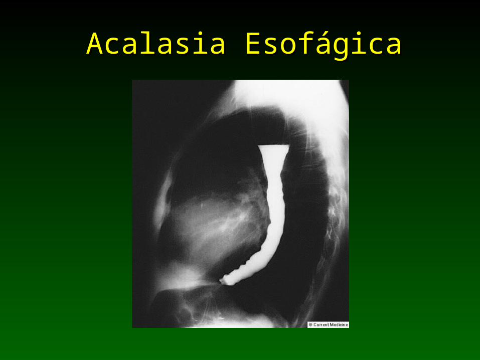
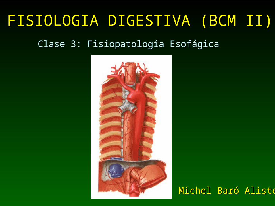
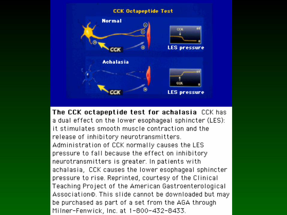
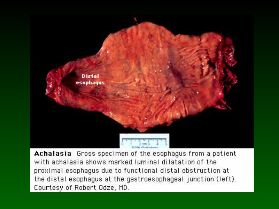
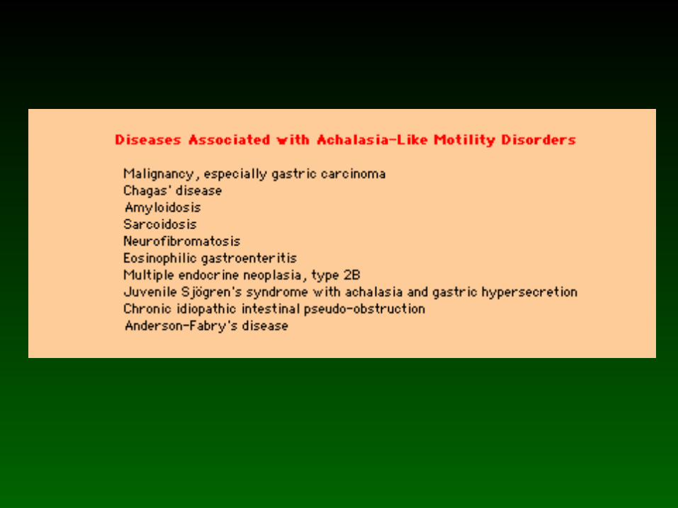
Acalasia EsofágicaAcalasia Esofágica
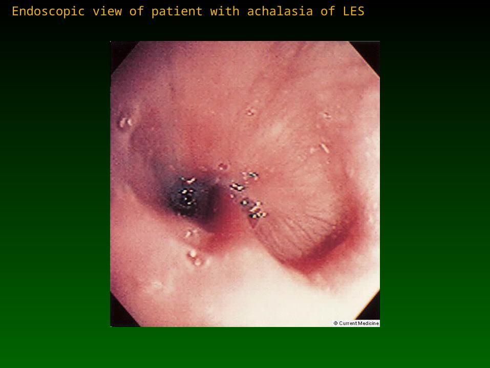
Endoscopic view of patient with achalasia of LES
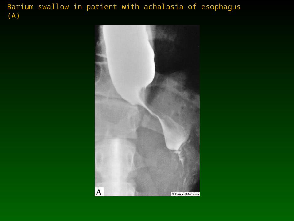
Barium swallow in patient with achalasia of esophagus (A)
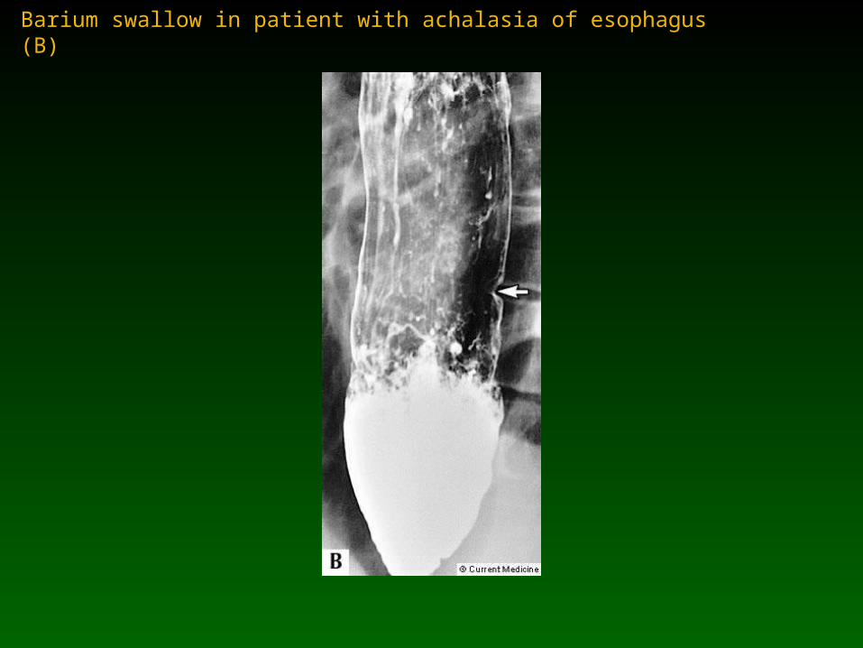
Barium swallow in patient with achalasia of esophagus (B)

Lateral radiograph of barium swallow in patient with achalasia of esophagus
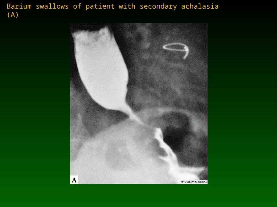
Barium swallows of patient with secondary achalasia (A)
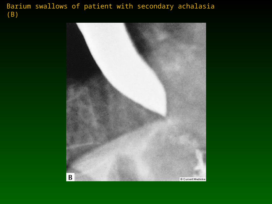
Barium swallows of patient with secondary achalasia (B)
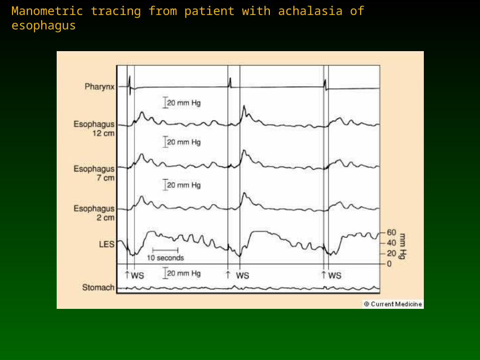
Manometric tracing from patient with achalasia of esophagus

Pressure recording and fluoroscopy during barium swallow in achalasia
Onda de presión hidrostática
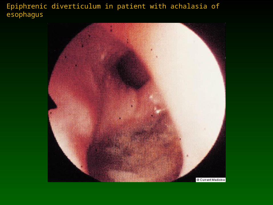
Epiphrenic diverticulum in patient with achalasia of esophagus
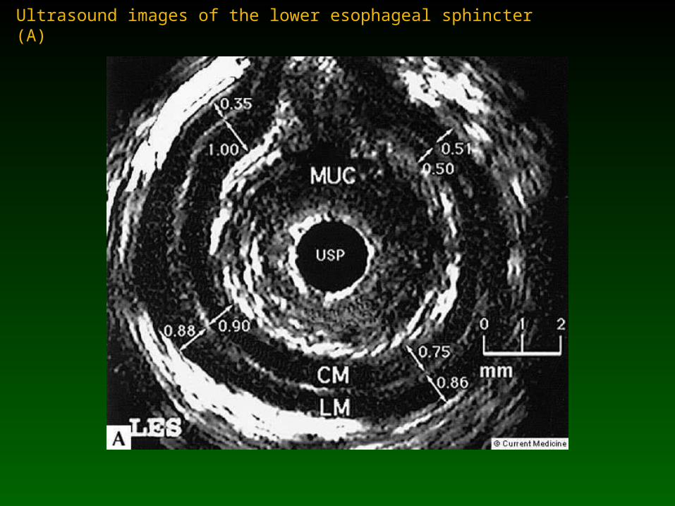
Ultrasound images of the lower esophageal sphincter (A)
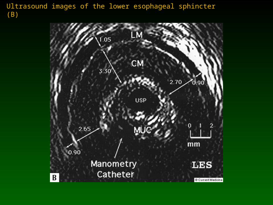
Ultrasound images of the lower esophageal sphincter (B)
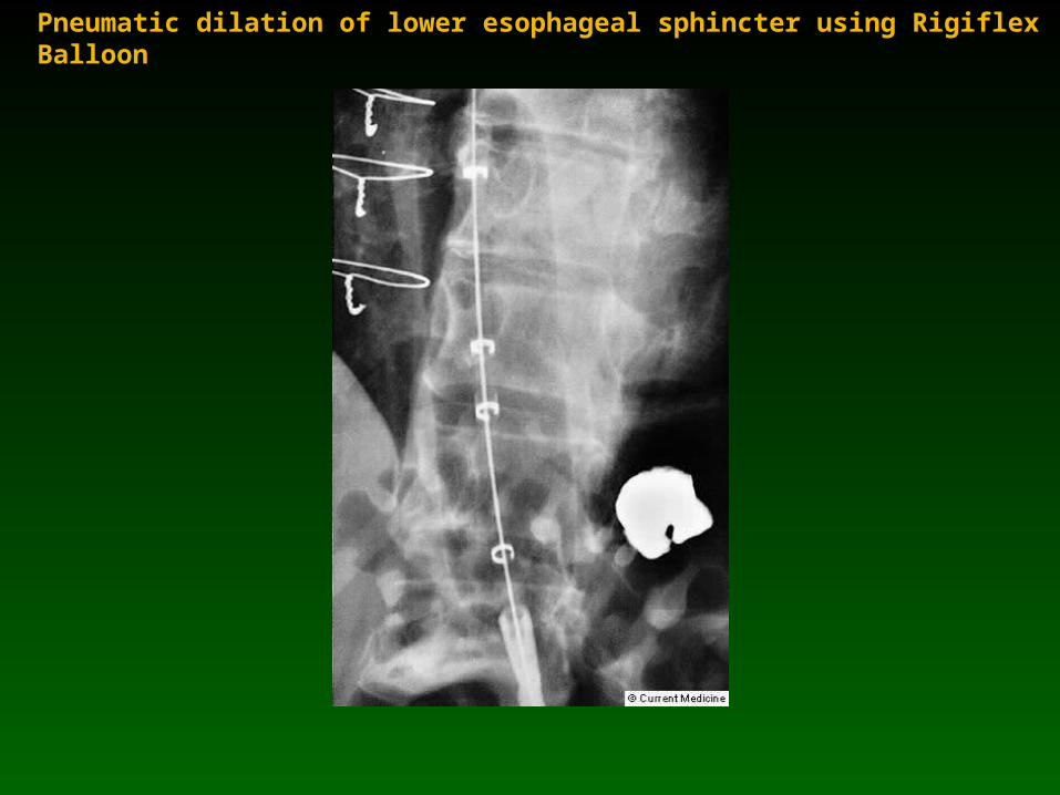
Pneumatic dilation of lower esophageal sphincter using Rigiflex Balloon
Otros Trastornos Motores del Otros Trastornos Motores del EsófagoEsófago
• Espasmo difuso del esófago
• Esófago en cascanueces
• Esclerodermia
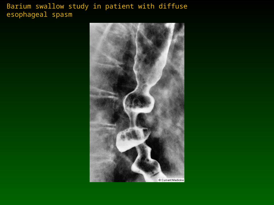
Barium swallow study in patient with diffuse esophageal spasm
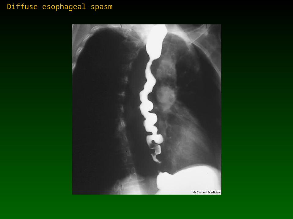
Diffuse esophageal spasm
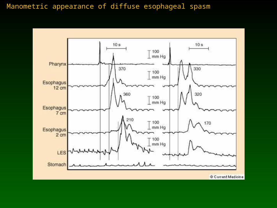
Manometric appearance of diffuse esophageal spasm
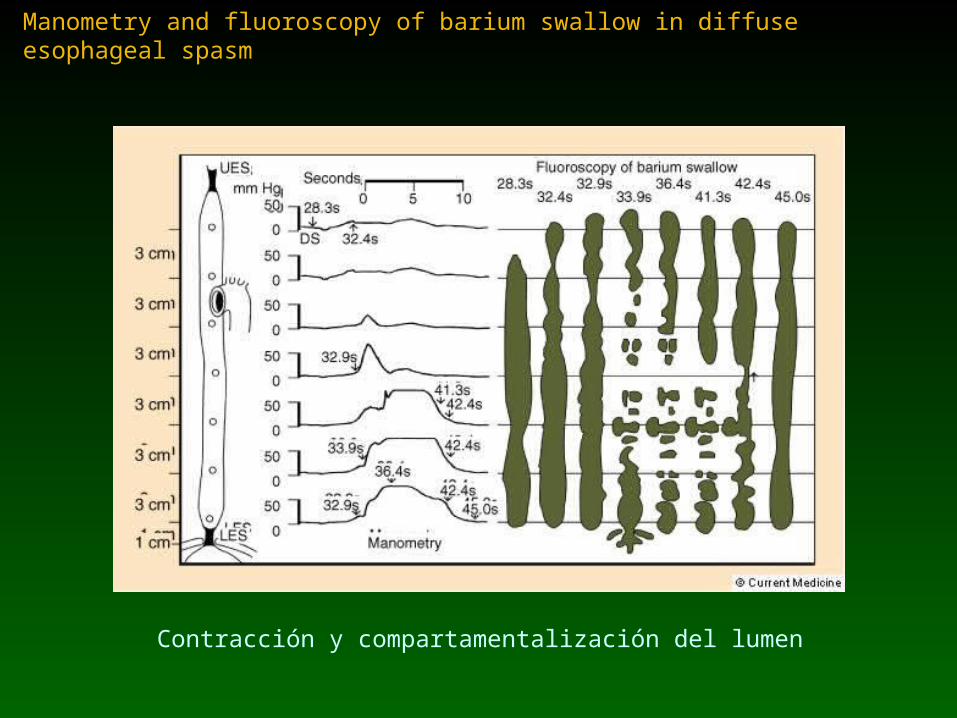
Manometry and fluoroscopy of barium swallow in diffuse esophageal spasm
Contracción y compartamentalización del lumen
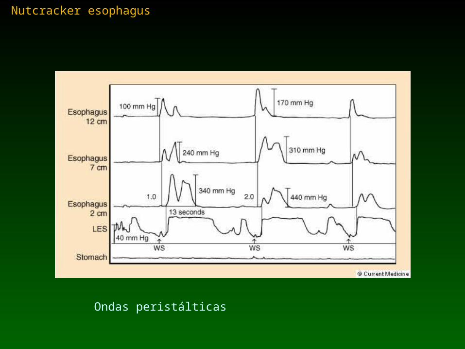
Nutcracker esophagus
Ondas peristálticas
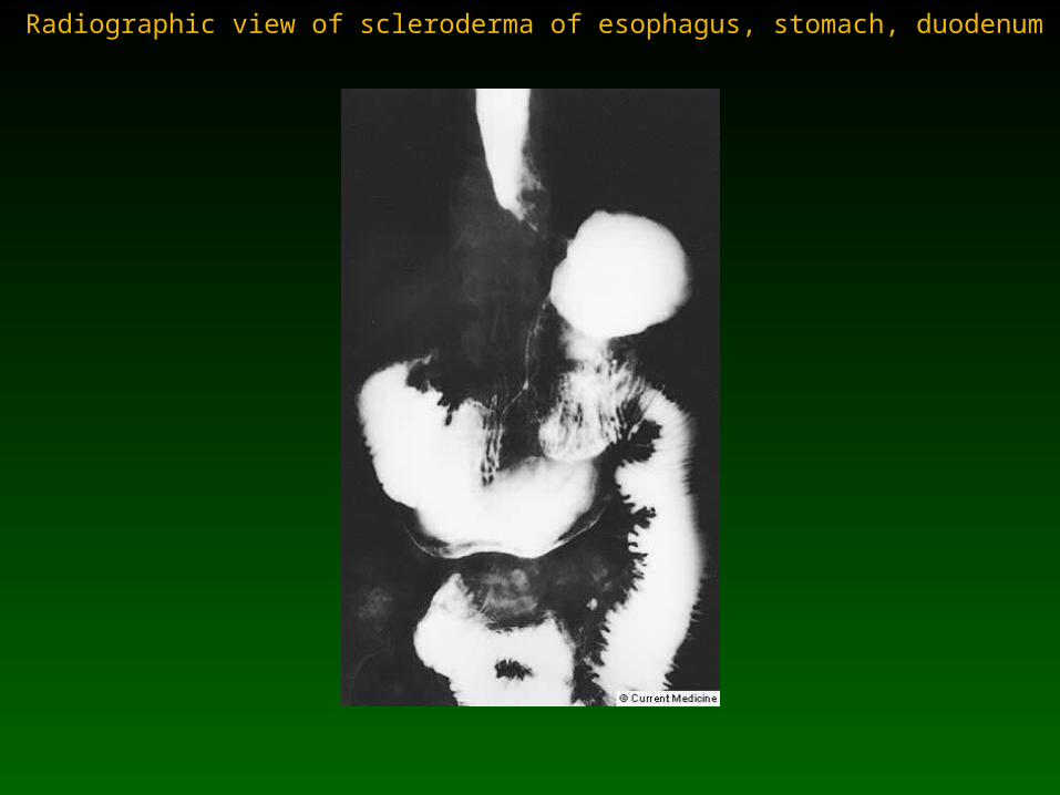
Radiographic view of scleroderma of esophagus, stomach, duodenum
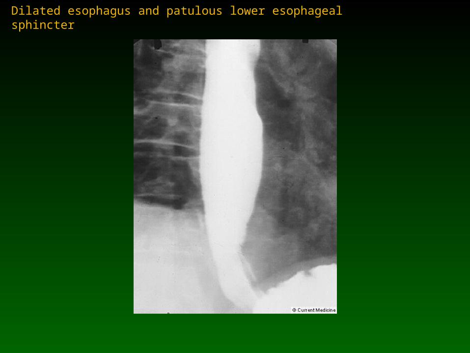
Dilated esophagus and patulous lower esophageal sphincter

Manometric tracing from patient with severe involvement of scleroderma

Esophageal function in woman with scleroderma and esophageal motor impairment (A)
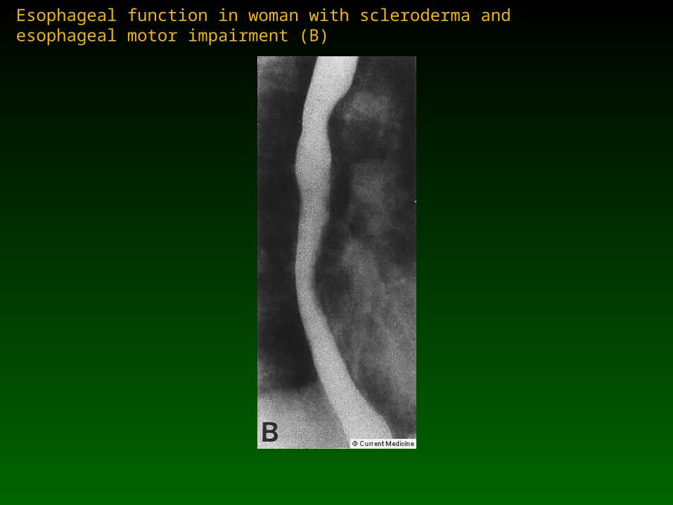
Esophageal function in woman with scleroderma and esophageal motor impairment (B)
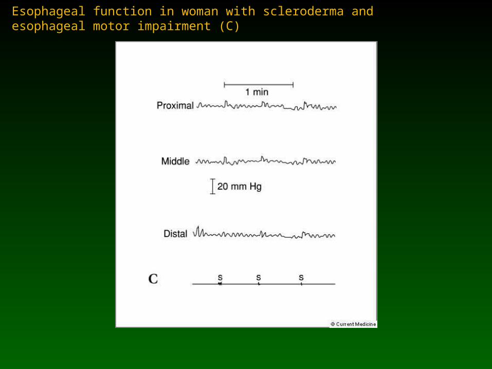
Esophageal function in woman with scleroderma and esophageal motor impairment (C)
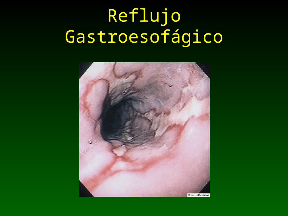
Reflujo GastroesofágicoReflujo Gastroesofágico
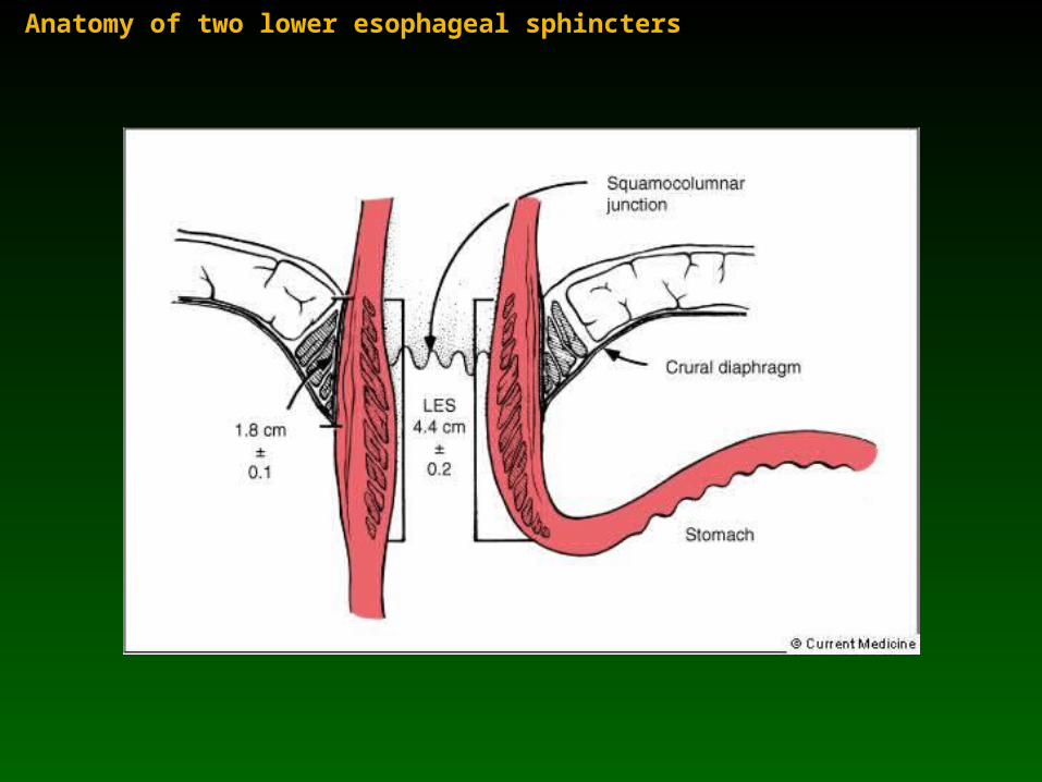
Anatomy of two lower esophageal sphincters

Contribution of LES/crural diaphragm to esophagogastric junction pressure
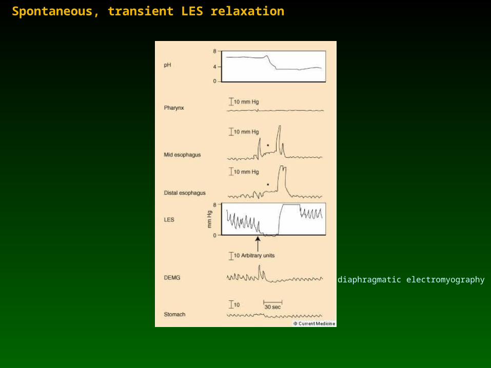
Spontaneous, transient LES relaxation
diaphragmatic electromyography
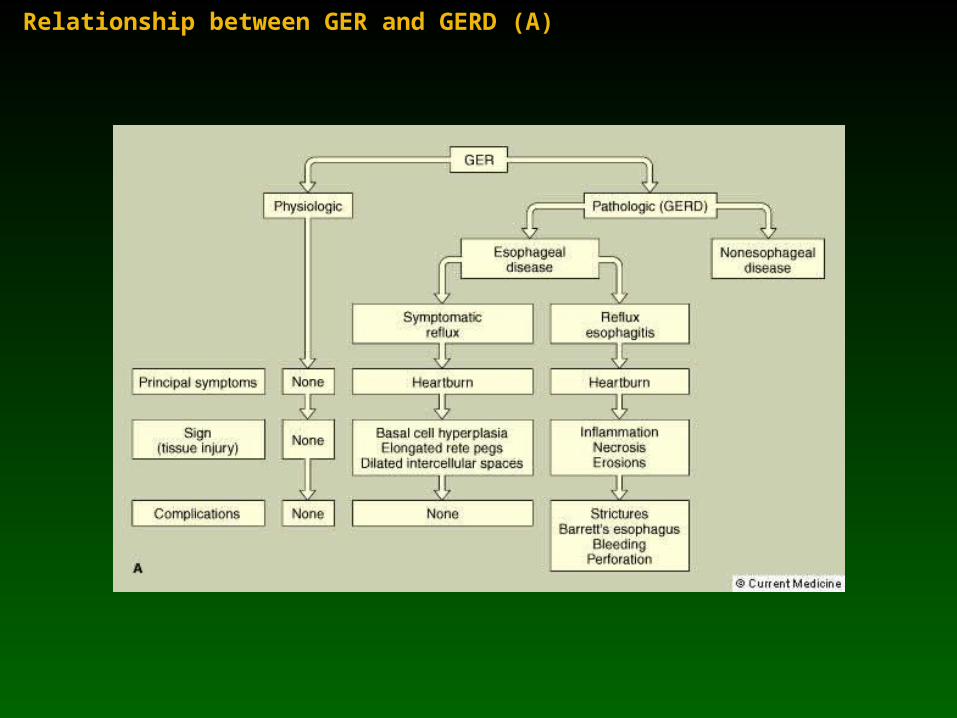
Relationship between GER and GERD (A)
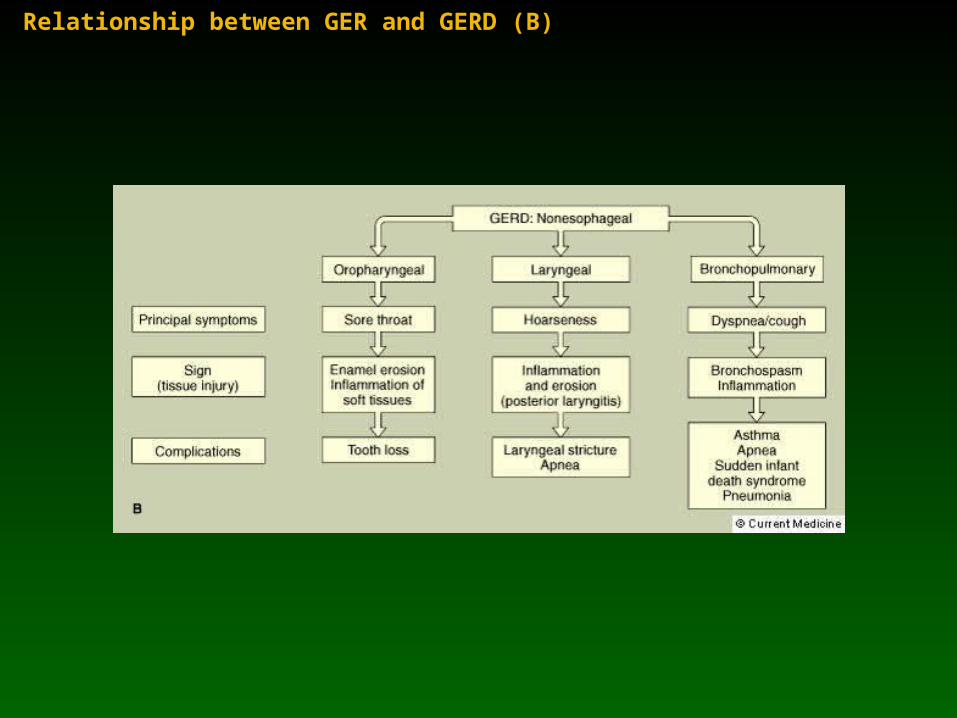
Relationship between GER and GERD (B)
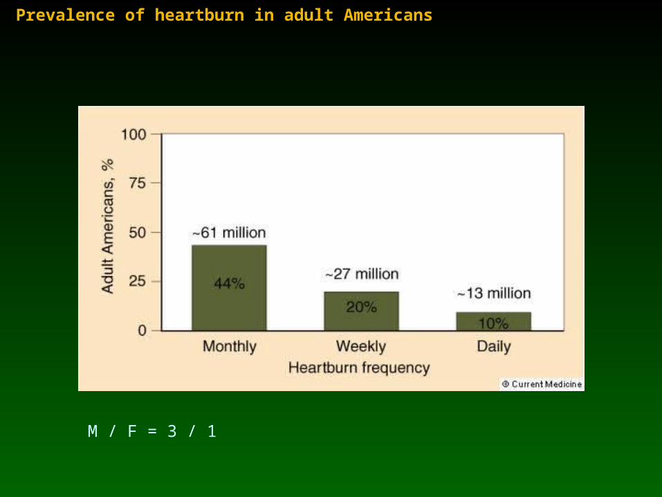
Prevalence of heartburn in adult Americans
M / F = 3 / 1
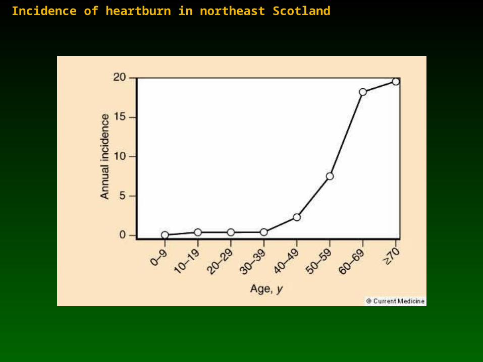
Incidence of heartburn in northeast Scotland
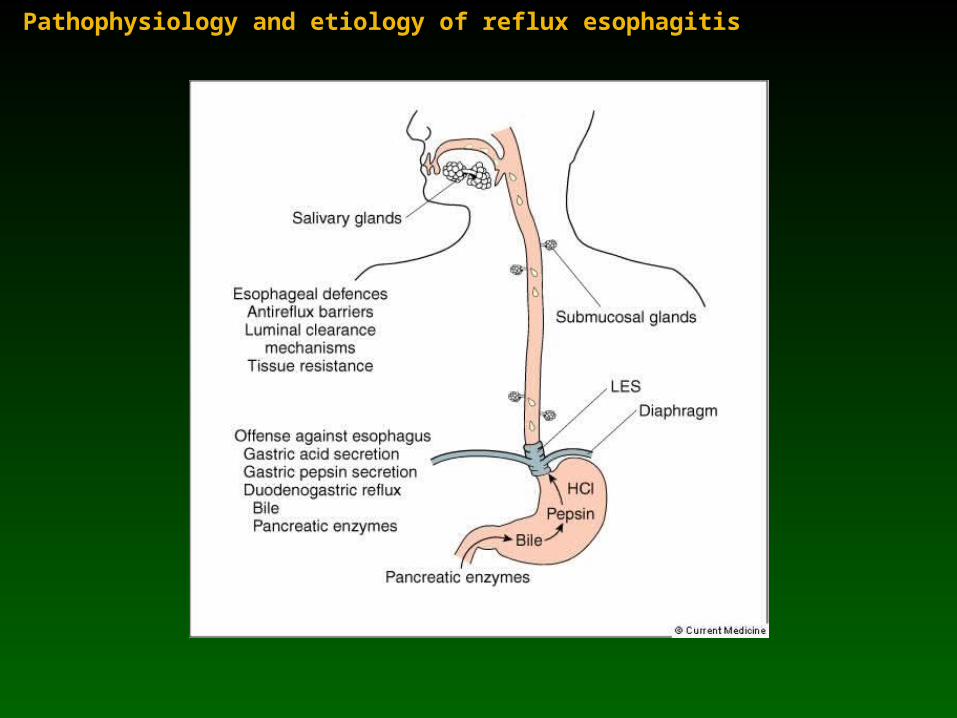
Pathophysiology and etiology of reflux esophagitis
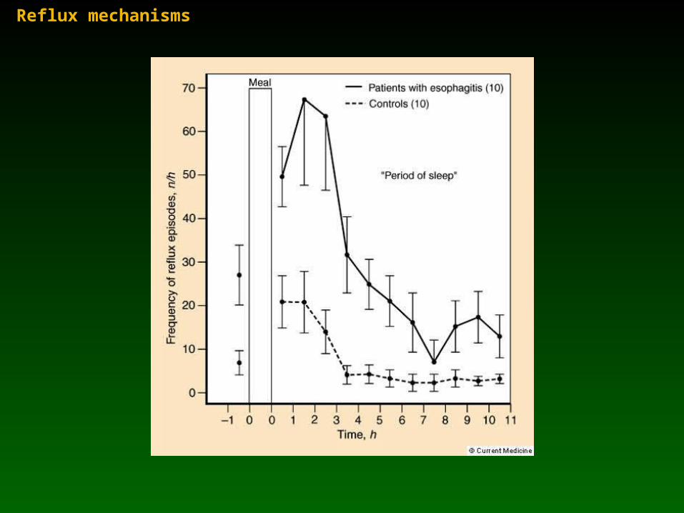
Reflux mechanisms
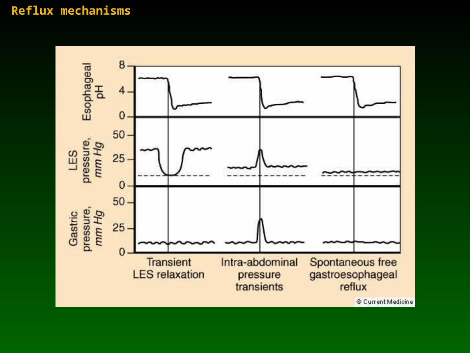
Reflux mechanisms
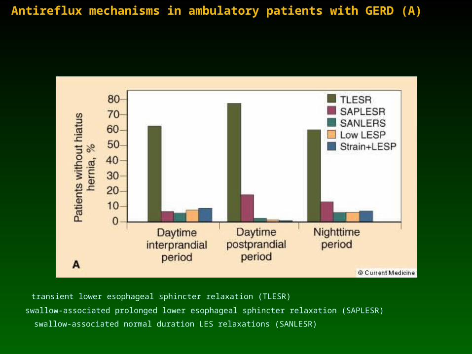
Antireflux mechanisms in ambulatory patients with GERD (A)
transient lower esophageal sphincter relaxation (TLESR)
swallow-associated prolonged lower esophageal sphincter relaxation (SAPLESR)
swallow-associated normal duration LES relaxations (SANLESR)
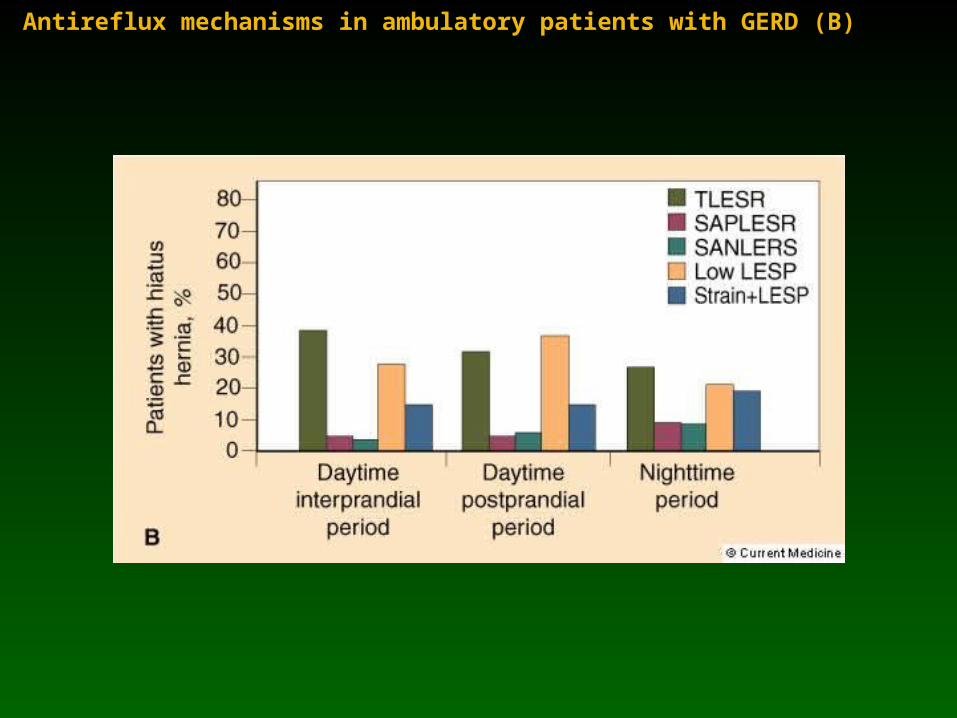
Antireflux mechanisms in ambulatory patients with GERD (B)
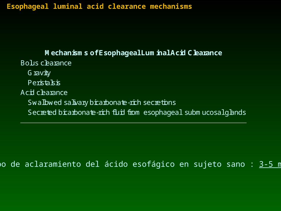
Esophageal luminal acid clearance mechanisms
Mechanisms of Esophageal Luminal Acid Clearance Bolus clearance Gravity Peristalsis Acid clearance Swallowed salivary bicarbonate-rich secretions Secreted bicarbonate-rich fluid from esophageal submucosal glands
Tiempo de aclaramiento del ácido esofágico en sujeto sano : 3-5 minutos

Esophageal luminal acid clearance mechanisms
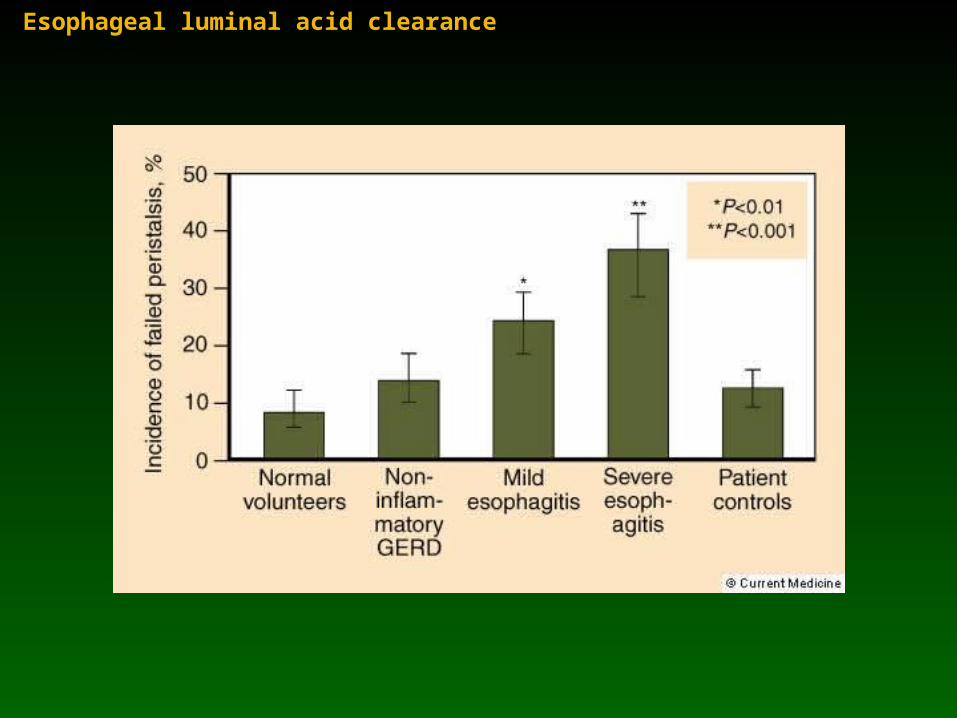
Esophageal luminal acid clearance
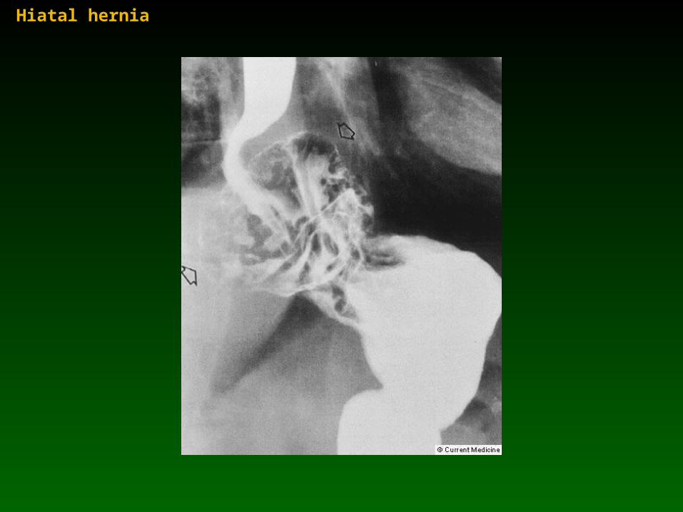
Hiatal hernia
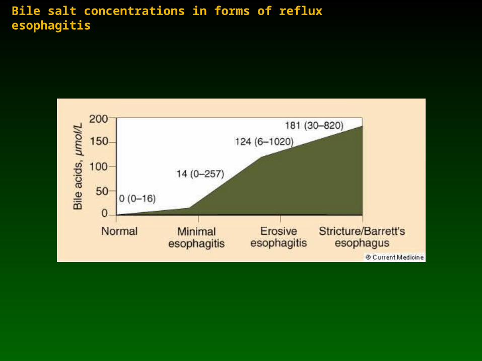
Bile salt concentrations in forms of reflux esophagitis
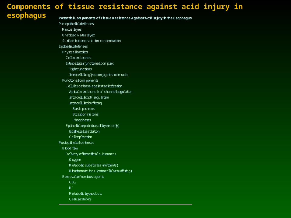
Components of tissue resistance against acid injury in esophagusPotential Components of Tissue Resistance Against Acid Injury in the Esophagus
Pre-epithelial defenses
Mucus layer
Unstirred water layer
Surface bicarbonate ion concentration
Epithelial defenses
Physical barriers
Cell membranes
Intercellular junctional complex
Tight junctions
Intercellular glycoconjugates or mucin
Functional components
Cellular defense against acidification
Apical membrane Na+ channel regulation
Intracellular pH regulation
Intracellular buffering
Basic proteins
Bicarbonate ions
Phosphates
Epithelial repair (basal layers only)
Epithelial restitution
Cell replication
Postepithelial defenses
Blood flow
Delivery of beneficial substances
Oxygen
Metabolic substrates (nutrients)
Bicarbonate ions (extracellular buffering)
Removal of noxious agents
CO2
H+
Metabolic byproducts
Cellular debris
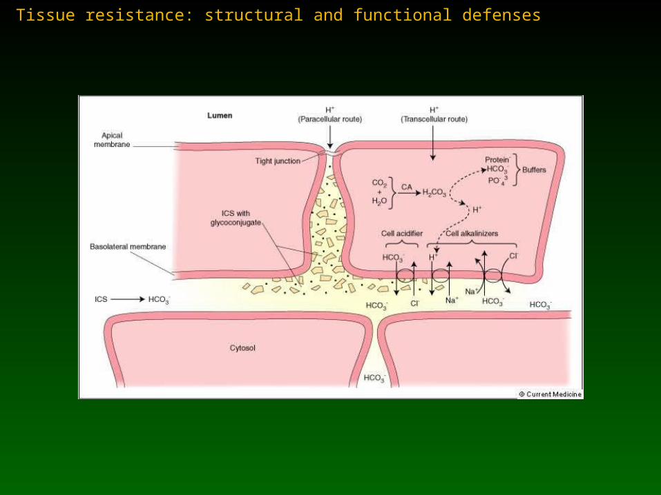
Tissue resistance: structural and functional defenses
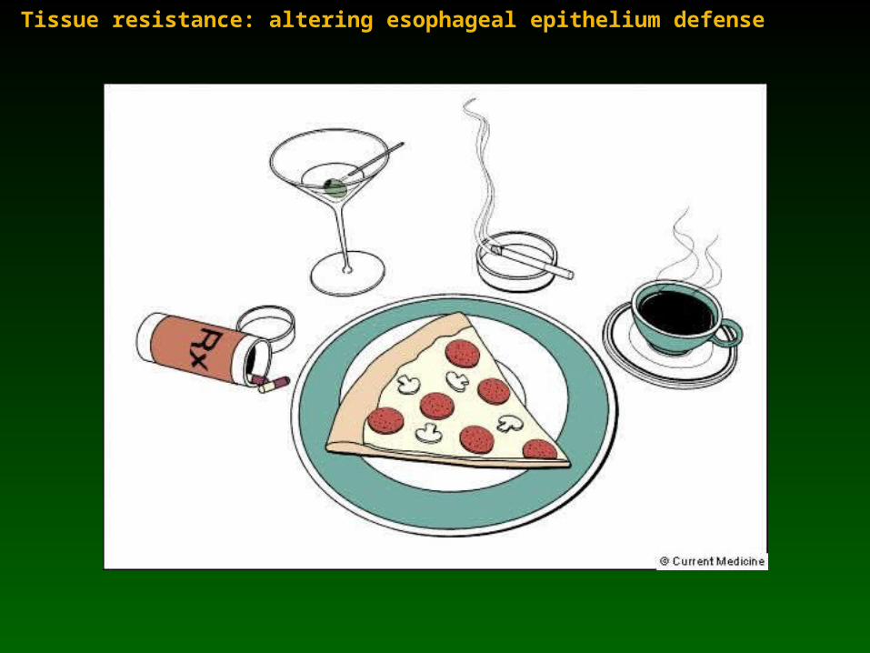
Tissue resistance: altering esophageal epithelium defense
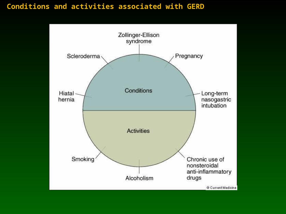
Conditions and activities associated with GERD
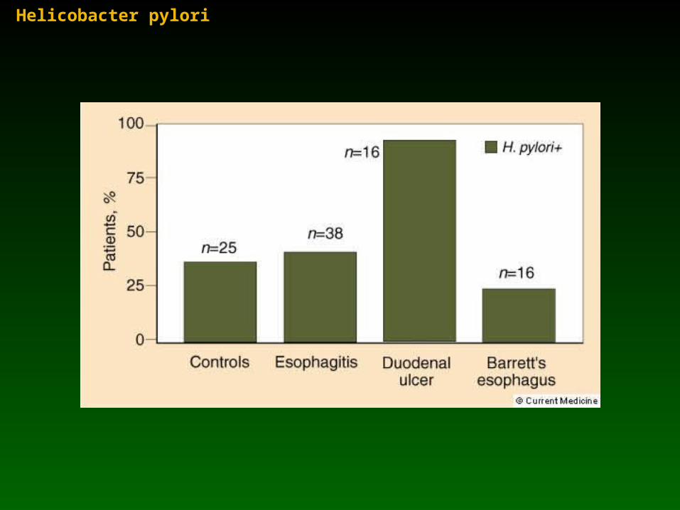
Helicobacter pylori
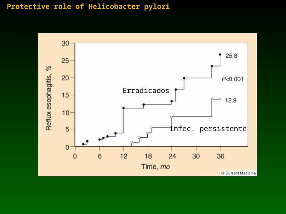
Protective role of Helicobacter pylori
Erradicados
Infec. persistente
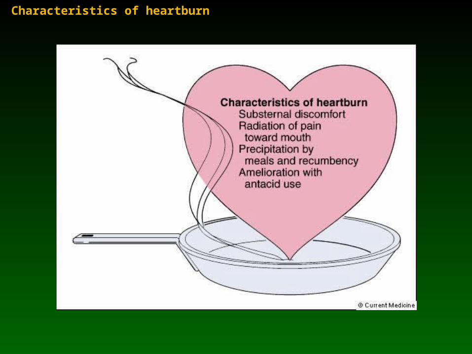
Characteristics of heartburn
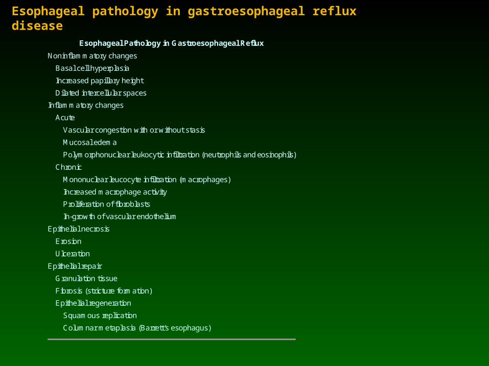
Esophageal pathology in gastroesophageal reflux disease
Esophageal Pathology in Gastroesophageal Reflux
Noninflammatory changes
Basal cell hyperplasia
Increased papillary height
Dilated intercellular spaces
Inflammatory changes
Acute
Vascular congestion with or without stasis
Mucosal edema
Polymorphonuclear leukocytic infiltration (neutrophils and eosinophils)
Chronic
Mononuclear leucocyte infiltration (macrophages)
Increased macrophage activity
Proliferation of fibroblasts
In-growth of vascular endothelium
Epithelial necrosis
Erosion
Ulceration
Epithelial repair
Granulation tissue
Fibrosis (stricture formation)
Epithelial regeneration
Squamous replication
Columnar metaplasia (Barrett's esophagus)

Histopathology of reflux damage to esophagus
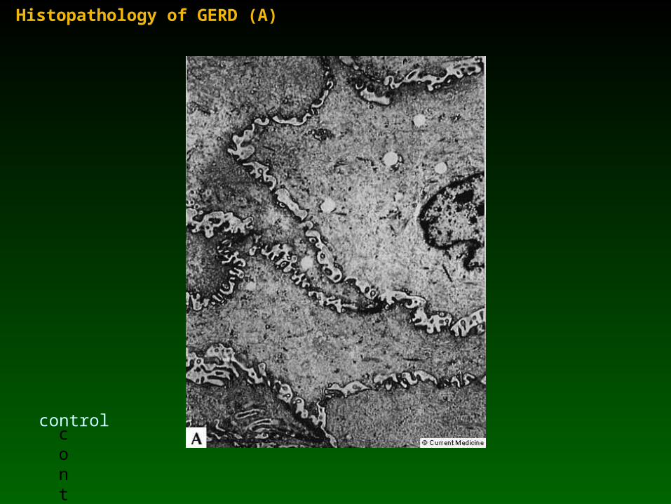
Histopathology of GERD (A)
control
control
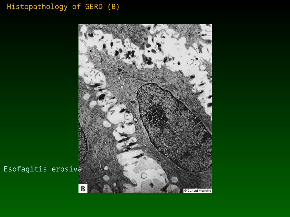
Histopathology of GERD (B)
Esofagitis erosiva
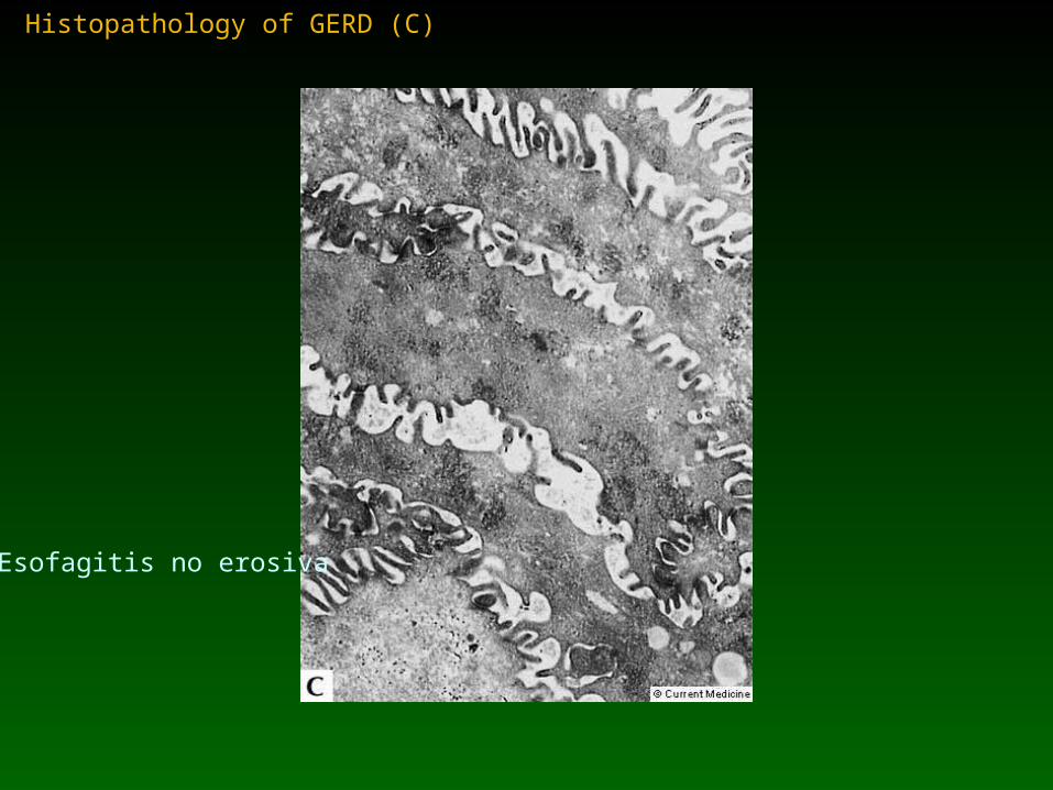
Histopathology of GERD (C)
Esofagitis no erosiva
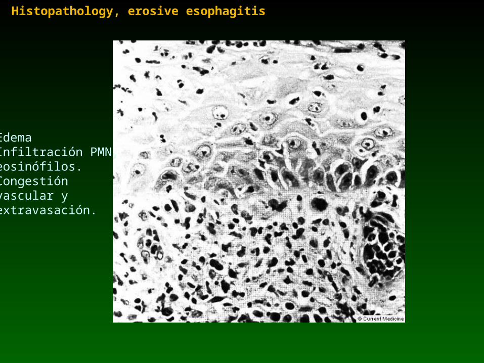
Histopathology, erosive esophagitis
EdemaInfiltración PMN, eosinófilos.Congestión vascular yextravasación.
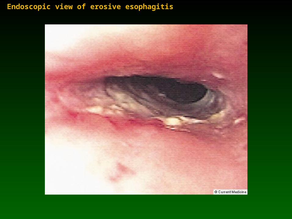
Endoscopic view of erosive esophagitis
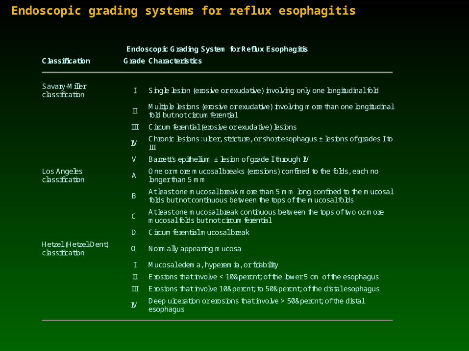
Endoscopic grading systems for reflux esophagitis
Endoscopic Grading System for Reflux Esophagitis
Classification Grade Characteristics
Savary-Miller classification I Single lesion (erosive or exudative) involving only one longitudinal fold
II Multiple lesions (erosive or exudative) involving more than one longitudinal fold but not circumferential
III Circumferential (erosive or exudative) lesions
IV Chronic lesions: ulcer, stricture, or short esophagus ± lesions of grades I to III
V Barrett's epithelium ± lesion of grade I through IV
Los Angeles classification A
One or more mucosal breaks (erosions) confined to the folds, each no longer than 5 mm
B At least one mucosal break more than 5 mm long confined to the mucosal folds but not continuous between the tops of the mucosal folds
C At least one mucosal break continuous between the tops of two or more mucosal folds but not circumferential
D Circumferential mucosal break
Hetzel (Hetzel-Dent) classification O Normally appearing mucosa
I Mucosal edema, hyperemia, or friability
II Erosions that involve < 10% of the lower 5 cm of the esophagus
III Erosions that involve 10% to 50% of the distal esophagus
IV Deep ulceration or erosions that involve > 50% of the distal esophagus
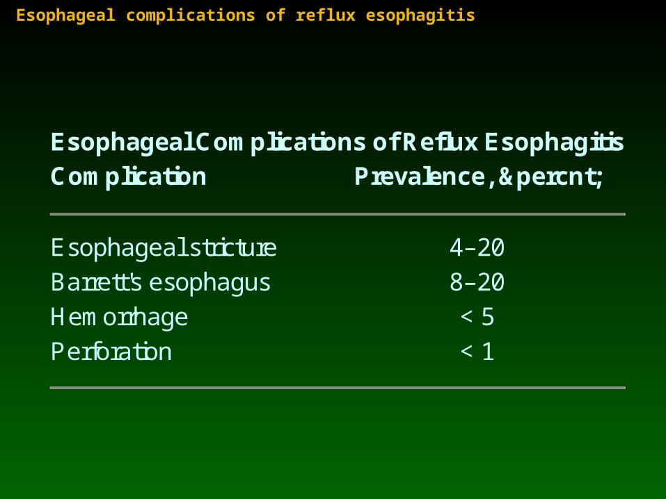
Esophageal complications of reflux esophagitis
Esophageal Complications of Reflux Esophagitis Complication Prevalence, %
Esophageal stricture 4–20
Barrett's esophagus 8–20
Hemorrhage < 5
Perforation < 1
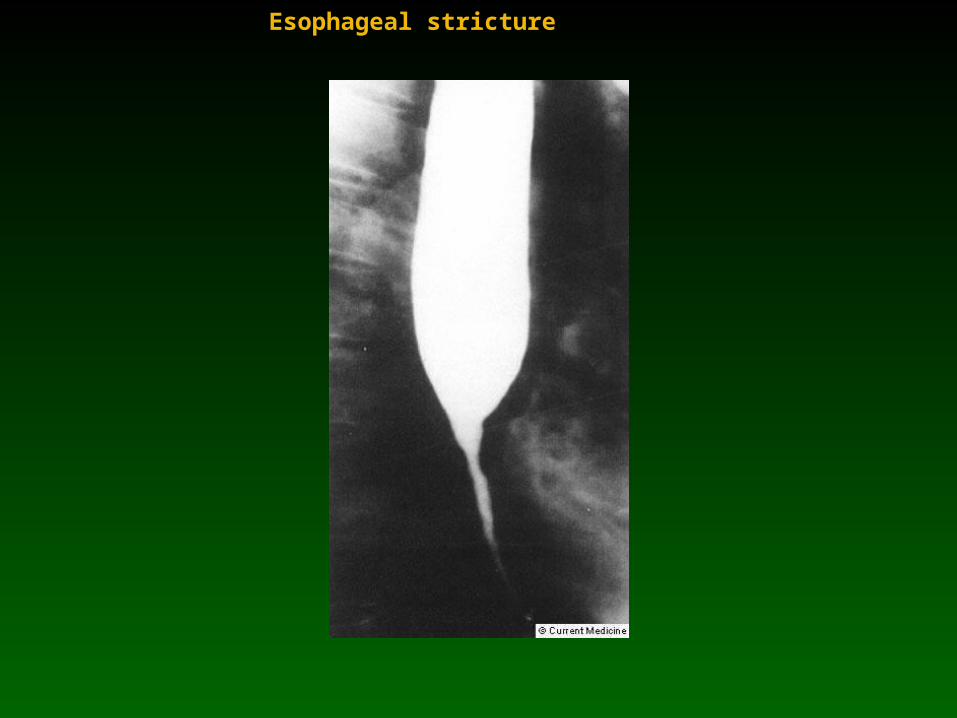
Esophageal stricture
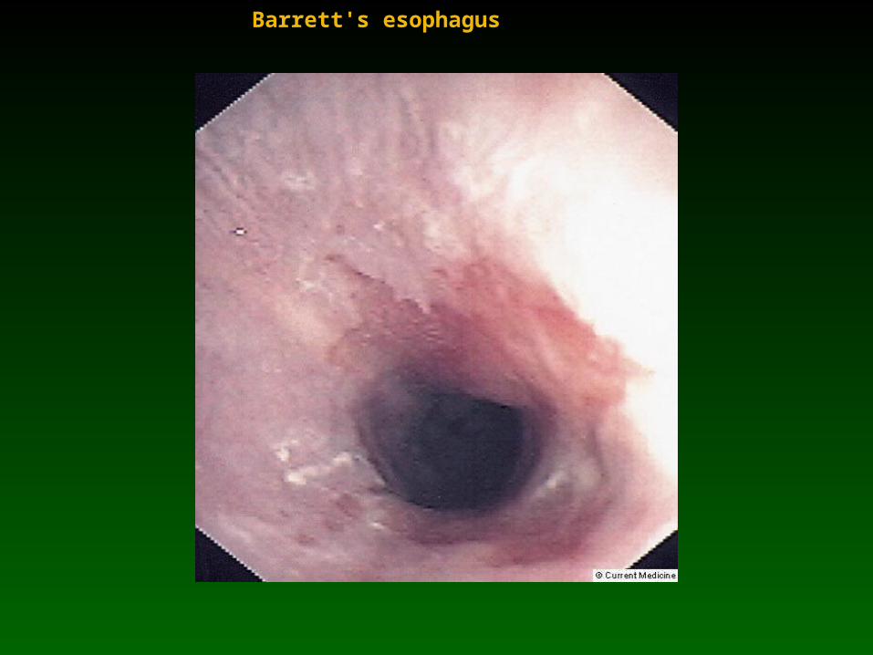
Barrett's esophagus
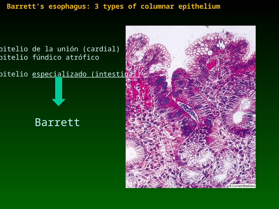
Barrett's esophagus: 3 types of columnar epithelium
1-Epitelio de la unión (cardial)2-Epitelio fúndico atrófico
3-Epitelio especializado (intestinal)
Barrett
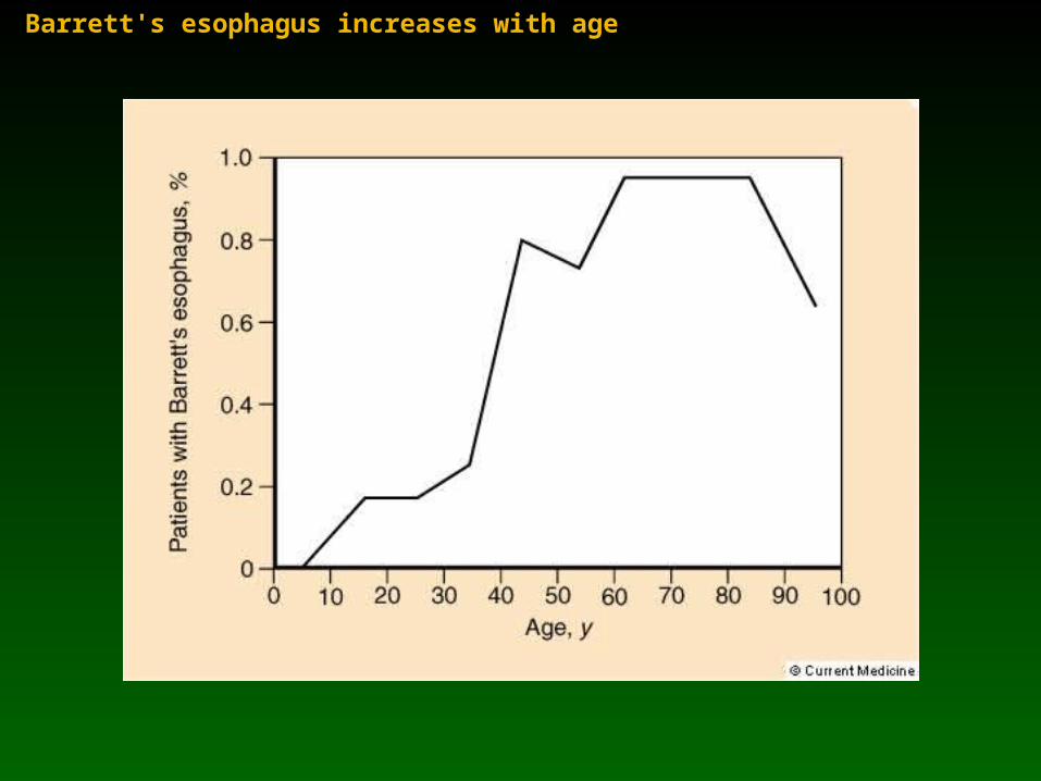
Barrett's esophagus increases with age
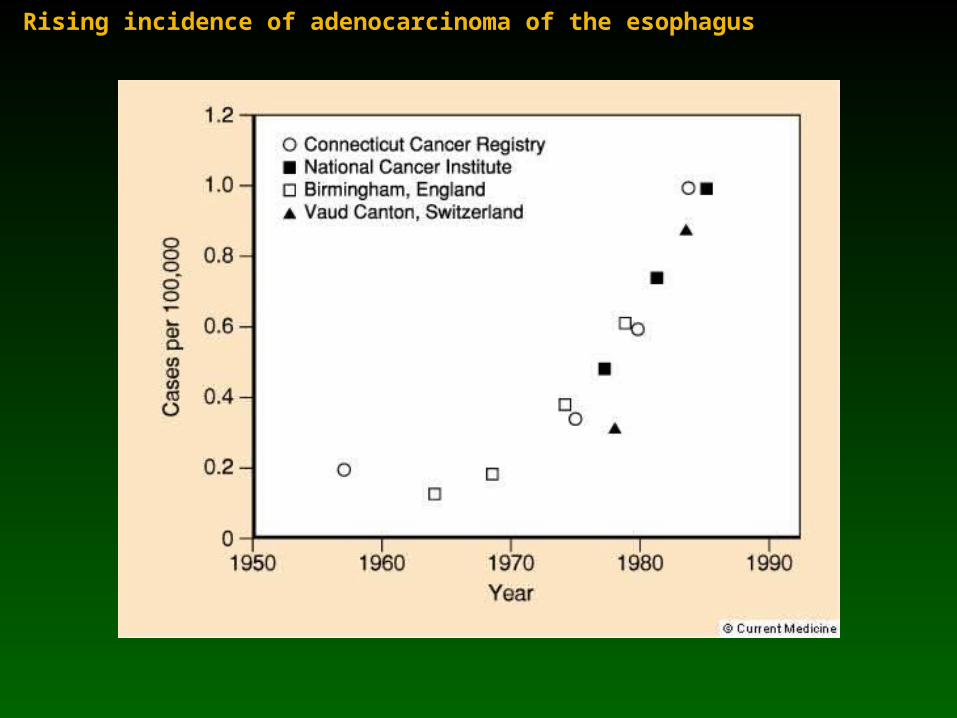
Rising incidence of adenocarcinoma of the esophagus
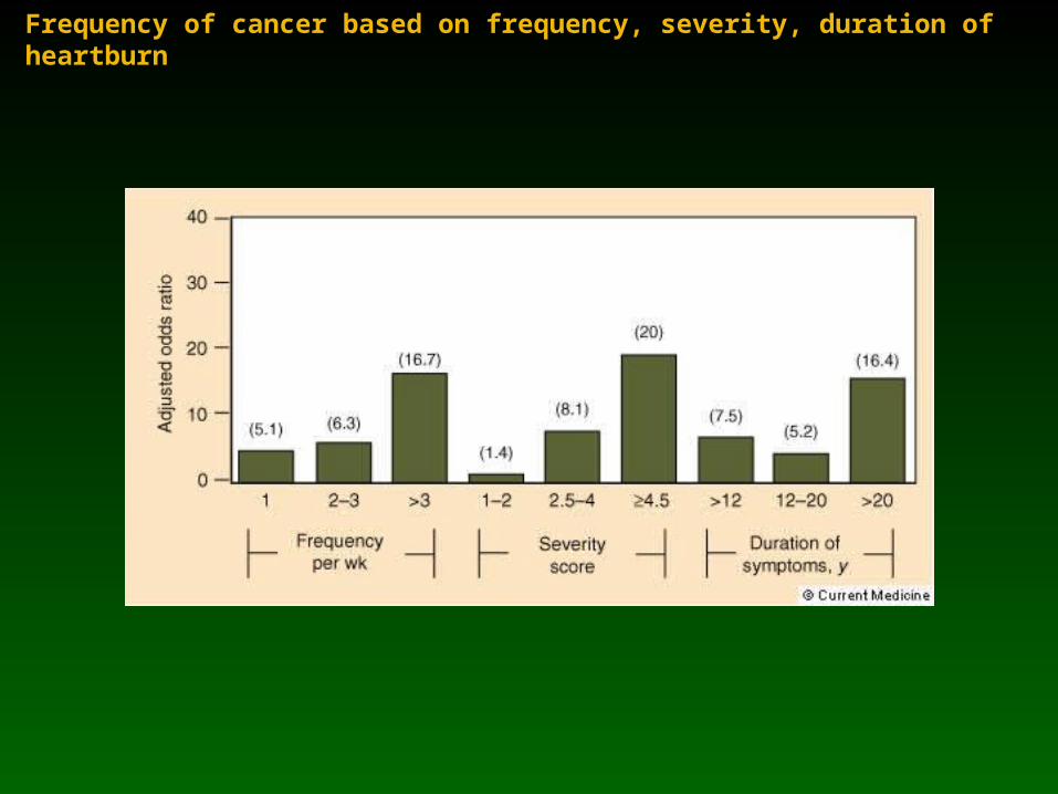
Frequency of cancer based on frequency, severity, duration of heartburn
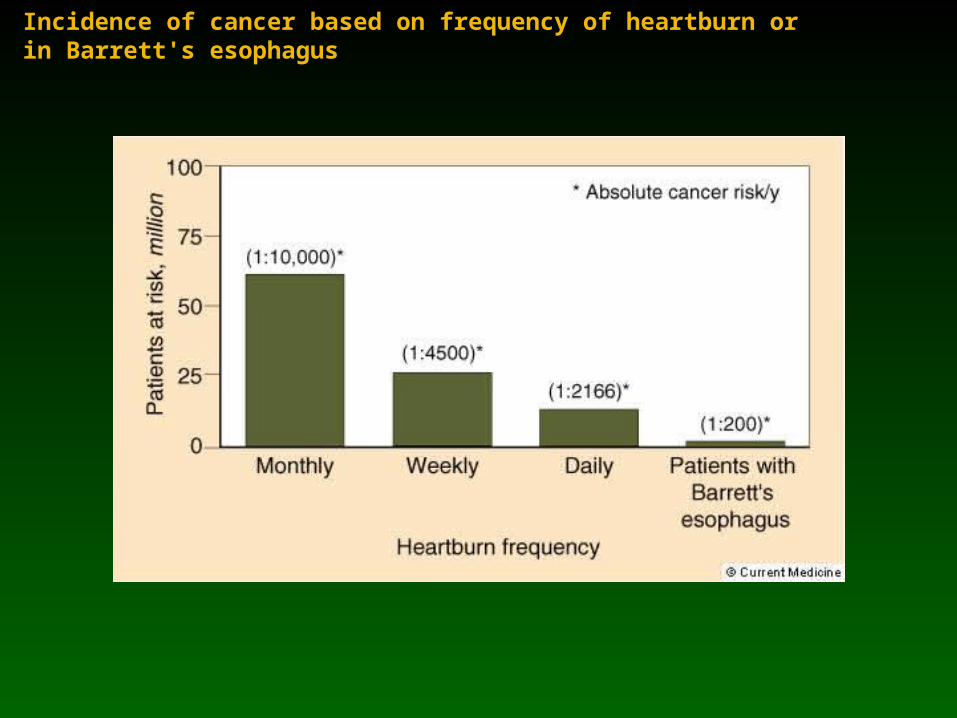
Incidence of cancer based on frequency of heartburn or in Barrett's esophagus
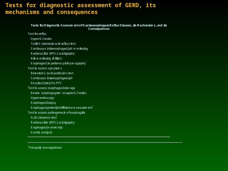
Tests for diagnostic assessment of GERD, its mechanisms and consequences
Tests for Diagnostic Assessment of Gastroesophageal Reflux Disease, its Mechanisms, and its Consequences
Test for reflux
Upper GI series
Tuttle's (standard acid reflux) test
Continuous intraesophageal pH monitoring
Radionuclide (99Tc) scintigraphy
Bile monitoring (Bilitec)
Esophageal impedence plethysmography*
Test to assess symptoms
Bernstein's (acid-perfusion) test
Continuous intraesophageal pH
Empirical trial of a PPI
Test to assess esophageal damage
Barium esophagogram or upper GI series
Upper endoscopy
Esophageal biopsy
Esophageal potential difference measurement*
Test to assess pathogenesis of esophagitis
Acid clearance test*
Radionuclide (99Tc) scintigraphy
Esophageal manometry
Gastric analysis
*Principally investigational.
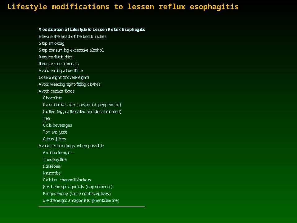
Lifestyle modifications to lessen reflux esophagitis
Modification of Lifestyle to Lessen Reflux Esophagitis
Elevate the head of the bed 6 inches
Stop smoking
Stop consuming excessive alcohol
Reduce fat in diet
Reduce size of meals
Avoid eating at bedtime
Lose weight (if overweight)
Avoid wearing tight-fitting clothes
Avoid certain foods
Chocolate
Carminatives (eg, spearmint, peppermint)
Coffee (eg, caffeinated and decaffeinated)
Tea
Cola beverages
Tomato juice
Citrus juices
Avoid certain drugs, when possible
Anticholinergics
Theophylline
Diazepam
Narcotics
Calcium channel blockers
β-Adrenergic agonists (isoproterenol)
Progesterone (some contraceptives)
α-Adrenergic antagonists (phentolamine)
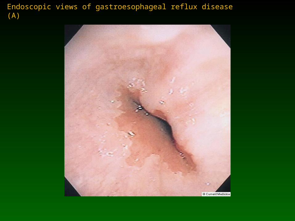
Endoscopic views of gastroesophageal reflux disease (A)

Endoscopic views of gastroesophageal reflux disease (B)
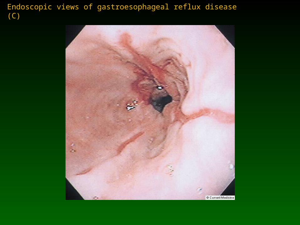
Endoscopic views of gastroesophageal reflux disease (C)
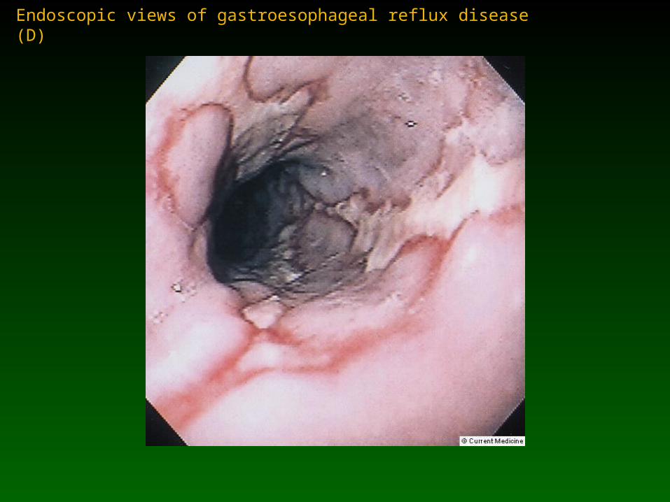
Endoscopic views of gastroesophageal reflux disease (D)
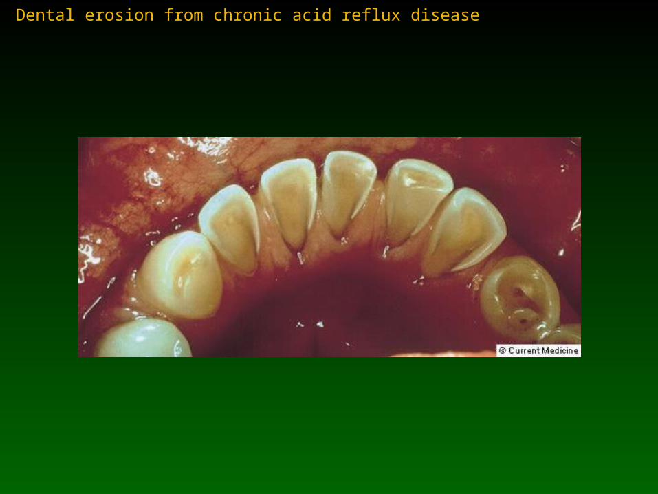
Dental erosion from chronic acid reflux disease
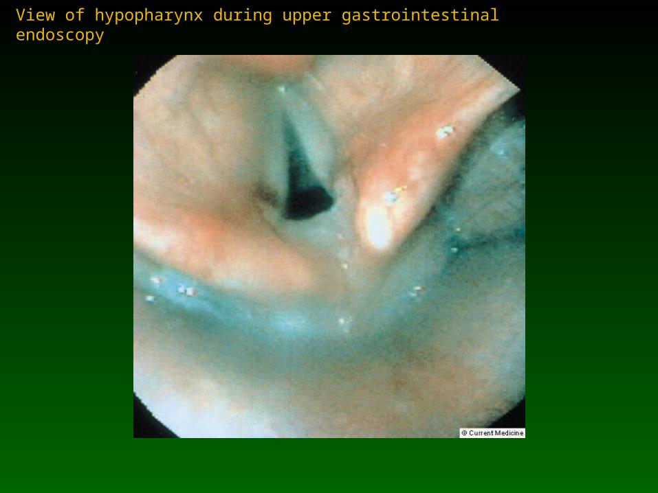
View of hypopharynx during upper gastrointestinal endoscopy

Fin