Embed Size (px)
Citation preview
Chemistry & Biology
Article
Fluorescent Probes of Tissue TransglutaminaseReveal Its Association with Arterial StiffeningNicolas Chabot,1 Simon Moreau,2 Amina Mulani,1 Pierre Moreau,2 and Jeffrey W. Keillor1,*1Departement de chimie, Universite de Montreal, C.P. 6128, Succursale centre-ville, Montreal, QC H3C 3J7, Canada2Faculte de pharmacie, Universite de Montreal, C.P. 6128, Succursale centre-ville, Montreal, QC H3C 3J7, Canada
*Correspondence: [email protected] 10.1016/j.chembiol.2010.06.019
SUMMARY
Tissue transglutaminase (TG2) catalyzes the cross-linking of proteins. TG2 has been implicated infibrosis and vascular calcification, both of whichlead to a common feature of aging known as arterialstiffness. In order to probe the role of TG2 in arterialrigidification, we have prepared a fluorescent irre-versible inhibitor as a probe for TG2 activity (RhodB-PGG-K(Acr)-LPF-OH). This probe was synthesizedon solid support, characterized kinetically (kinact =0.68 min-1, KI = 79 mM), and then used to stain theaorta from rats used as a model of isolated systolichypertension (ISH). Interestingly, TG2 activity wasthus shown to increase over 4 weeks of the hyperten-sion model, corresponding with the previouslyobserved increase in arterial stiffness. These resultsclearly suggest an association between TG2 andthe phenomenon of arterial rigidification.
INTRODUCTION
Transglutaminases (TGases) (EC 2.3.2.13) are a family of
Ca2+-dependent enzymes that catalyze an acyl transfer reaction
from the g-carboxamide group of a peptide-bound glutamine
residue to the 3-amino group of a peptide-bound lysine residue,
resulting in the formation of an isopeptidic amide bond that may
crosslink peptides or proteins (Figure 1). Acyl transfer from
glutamine to other primary amines and even water can also be
mediated by TGases (Achyuthan et al., 1993; Folk and Cole,
1966; Greenberg et al., 1991). Mammalian TGases from tissue,
epidermis, and plasma have been extensively characterized.
Of these, tissue TGase (tissue transglutaminase [TG2]) has
been shown to participate in biological processes such as endo-
cytosis (Davies et al., 1980; Levitzki et al., 1980), apoptosis
(Fesus et al., 1987), and cell growth regulation (Birckbichler
et al., 1983). TG2 is mostly a cytosolic protein, but it has also
been detected in the nucleus (Lesort et al., 1998) and can be
secreted from the cell. Outside the cell, it plays one of its most
important biological roles, crosslinking the extracellular matrix,
thus making it less susceptible to proteolytic degradation (Aes-
chlimann and Thomazy, 2000). When TGase-mediated cross-
linking activity is not carefully regulated, it may also be involved
in a number of physiological disorders, such as acne (De Young
Chemistry & Biology 17, 1143–1
et al., 1984), the formation of cataracts (Azari et al., 1981),
immune system diseases (Fesus, 1982), psoriasis (Schroeder
et al., 1992), Alzheimer’s disease (Norlund et al., 1999; Selkoe
et al., 1982), Huntington’s disease (Dedeoglu et al., 2002; Mas-
troberardino et al., 2002), Celiac disease (Piper et al., 2002),
and cancer metastasis (Choi et al., 2005; Mehta, 2009).
TG2 has also been implicated in fibrosis (Griffin et al., 1979;
Johnson et al., 2007; Small et al., 1999) and vascular calcification
(Faverman et al., 2008; Johnson et al., 2008; Kaartinen et al.,
2007). In large conduit arteries these two phenomena lead to
a common feature of aging known as arterial stiffness. Vascular
calcification is an active phenomenon involving the modulation
of matrix Gla protein (Schurgers et al., 2008) and the phenotypic
modulation of vascular smooth muscle cells (Shanahan et al.,
2000). On the other hand, fibrosis may be explained by an
increased collagen/elastin ratio in the extracellular matrix (John-
son et al., 2001). As we described previously this is due to elastin
degradation (Bouvet et al., 2008) as well as the accumulation of
collagen (Essalihi et al., 2007). TGase has been shown to partic-
ipate in this phenomenon by crosslinking collagen in the extra-
cellular matrix (Ientile et al., 2007; Johnson et al., 1997, 2007).
The collagen network being rather rigid, its accumulation in
arteries has been associated with increased vascular stiffness
(Arribas et al., 2006; Bruel et al., 1998; Safar et al., 2003; Zieman
et al., 2005).
The hemodynamic consequences of arterial stiffening are an
increased systolic blood pressure, unchanged or slightly re-
duced diastolic blood pressure, increased pulse pressure, and
the development of isolated systolic hypertension (ISH). We
have developed a rat model of ISH, known as the warfarin/
vitamin K (WVK) model, which mimics the physiopathological
process of the disease observed in man (Bouvet et al., 2008;
Essalihi et al., 2003, 2004, 2005). It consists in blocking the
vitamin K-dependant maturation of a physiological inhibitor of
calcification, matrix Gla protein, with warfarin, while preventing
the rats from bleeding with the concomitant injection of Vitamin
K1. With this model we obtain an elevation of vascular calcifica-
tion, collagen content, and vascular stiffening within 4 weeks of
treatments.
TG2 is involved in vascular calcification and collagen accumu-
lation. These phenomena are known to cause arterial stiffening
and occur in ISH. Therefore, our working hypothesis is that
TG2 is involved in the stiffening of large arteries associated
with ISH. Interrogation of the putative role of TG2 in arterial rigid-
ification requires a sensitive method for detecting active TG2 in
samples of arterial tissue. To that end we have designed
substrate analog probes that are capable of reacting with TG2,
150, October 29, 2010 ª2010 Elsevier Ltd All rights reserved 1143
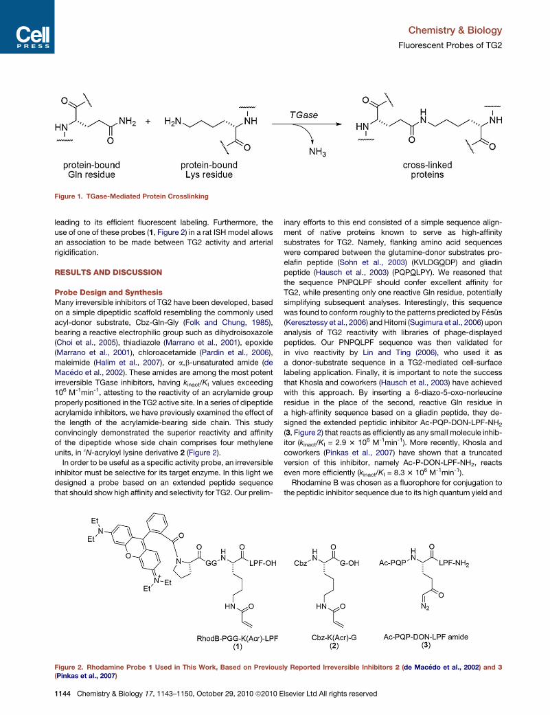
Figure 1. TGase-Mediated Protein Crosslinking
Chemistry & Biology
Fluorescent Probes of TG2
leading to its efficient fluorescent labeling. Furthermore, the
use of one of these probes (1, Figure 2) in a rat ISH model allows
an association to be made between TG2 activity and arterial
rigidification.
RESULTS AND DISCUSSION
Probe Design and SynthesisMany irreversible inhibitors of TG2 have been developed, based
on a simple dipeptidic scaffold resembling the commonly used
acyl-donor substrate, Cbz-Gln-Gly (Folk and Chung, 1985),
bearing a reactive electrophilic group such as dihydroisoxazole
(Choi et al., 2005), thiadiazole (Marrano et al., 2001), epoxide
(Marrano et al., 2001), chloroacetamide (Pardin et al., 2006),
maleimide (Halim et al., 2007), or a,b-unsaturated amide (de
Macedo et al., 2002). These amides are among the most potent
irreversible TGase inhibitors, having kinact/KI values exceeding
106 M-1min-1, attesting to the reactivity of an acrylamide group
properly positioned in the TG2 active site. In a series of dipeptide
acrylamide inhibitors, we have previously examined the effect of
the length of the acrylamide-bearing side chain. This study
convincingly demonstrated the superior reactivity and affinity
of the dipeptide whose side chain comprises four methylene
units, in 3N-acryloyl lysine derivative 2 (Figure 2).
In order to be useful as a specific activity probe, an irreversible
inhibitor must be selective for its target enzyme. In this light we
designed a probe based on an extended peptide sequence
that should show high affinity and selectivity for TG2. Our prelim-
Figure 2. Rhodamine Probe 1 Used in This Work, Based on Previous(Pinkas et al., 2007)
1144 Chemistry & Biology 17, 1143–1150, October 29, 2010 ª2010 E
inary efforts to this end consisted of a simple sequence align-
ment of native proteins known to serve as high-affinity
substrates for TG2. Namely, flanking amino acid sequences
were compared between the glutamine-donor substrates pro-
elafin peptide (Sohn et al., 2003) (KVLDGQDP) and gliadin
peptide (Hausch et al., 2003) (PQPQLPY). We reasoned that
the sequence PNPQLPF should confer excellent affinity for
TG2, while presenting only one reactive Gln residue, potentially
simplifying subsequent analyses. Interestingly, this sequence
was found to conform roughly to the patterns predicted by Fesus
(Keresztessy et al., 2006) andHitomi (Sugimura et al., 2006) upon
analysis of TG2 reactivity with libraries of phage-displayed
peptides. Our PNPQLPF sequence was then validated for
in vivo reactivity by Lin and Ting (2006), who used it as
a donor-substrate sequence in a TG2-mediated cell-surface
labeling application. Finally, it is important to note the success
that Khosla and coworkers (Hausch et al., 2003) have achieved
with this approach. By inserting a 6-diazo-5-oxo-norleucine
residue in the place of the second, reactive Gln residue in
a high-affinity sequence based on a gliadin peptide, they de-
signed the extended peptidic inhibitor Ac-PQP-DON-LPF-NH2
(3, Figure 2) that reacts as efficiently as any small molecule inhib-
itor (kinact/KI = 2.9 3 106 M-1min-1). More recently, Khosla and
coworkers (Pinkas et al., 2007) have shown that a truncated
version of this inhibitor, namely Ac-P-DON-LPF-NH2, reacts
even more efficiently (kinact/KI = 8.3 3 106 M-1min-1).
Rhodamine B was chosen as a fluorophore for conjugation to
the peptidic inhibitor sequence due to its high quantum yield and
ly Reported Irreversible Inhibitors 2 (de Macedo et al., 2002) and 3
lsevier Ltd All rights reserved
Figure 3. Solid-Phase Synthesis of Rhodamine Labeling Agent 1 and Control Compound 4
Table 1. Kinetic Parameters Measured for TG2 Affinity Labels
Studied Herein
Compound R = X =
kinact(min-1)
KI
(mM)
kinact / KI
(mM-1 min-1)
1 rhodamine B Pro 0.68 ± 0.20 79 ± 44 0.009 ± 0.008
4 Fmoc Pro 0.46 ± 0.08 42 ± 17 0.011 ± 0.006
5 rhodamine B Gly 0.19 ± 0.02 6 ± 2 0.032 ± 0.014
6 Fmoc Gly 0.36 ± 0.06 22 ± 8 0.016 ± 0.008
Chemistry & Biology
Fluorescent Probes of TG2
red emission, easily distinguishable from intrinsic cellular fluo-
rescence. Furthermore, free rhodamine B is affordable, and its
incorporation into a peptide sequence can be achieved through
straightforward coupling procedures. In order to ensure that the
fluorophore does not decrease the affinity of the peptide
sequence for TG2 and that the adjacent peptide does not
quench the fluorescence of the fluorophore, it was deemed
necessary to attach the rhodamine to the peptide sequence
through a spacer moiety. It has been reported that secondary
amides of rhodamine are less fluorescent than their parent
acid, presumably due to lactamisation. For this reason, the
rhodamine moiety of inhibitor 1 was attached to the peptide
sequence through a tertiary amide linkage with a proline residue.
The nonfluorescent control compound 4 (Figure 3) was designed
according to the same criteria, having the same reactive electro-
phile, substrate peptide sequence, and spacer sequence as
inhibitor 1 but lacking the rhodamine B fluorophore.
To facilitate the preparation of these labeling agents, a strategy
was developed to allow their synthesis on solid support
(Figure 3). In general the devised synthetic route follows typical
Fmoc-based peptide synthesis using Wang resin. Notably, the
approach shown in Figure 3 allows the acryloyl group to be
added to the 3-amino group of the Lys residue, without requiring
cleavage from the resin. aN-Fmoc-3N-Boc-protected Lys was
first prepared from a known protocol (Albericio et al., 1990)
and then added to the resin-bound LPF tripeptide. Removal of
the Boc group from the side chain of the doubly protected
residue, without cleavage from the resin, was critical to this
approach. This was achieved using TMSOTf, according to the
procedure published by Lejeune et al. (2003). The acryloyl group
was then added using acrylic acid and the coupling agent EEDQ,
in the absence of base. These conditions were found to be supe-
rior to addition of acrylic anhydride, for example, giving an excel-
lent yield without degradation of the electrophilic acryloyl group.
Chemistry & Biology 17, 1143–1
Our procedure for Fmoc-based peptide coupling did not affect
the integrity of the acryloyl group either, allowing a peptide
spacer sequence to be added subsequently. Finally, the rhoda-
mine fluorophore was added to the N-terminal of the peptide
chain in excellent yield, using the same mild coupling conditions
as for the addition of the acryloyl group. After cleavage from the
resin, peptides were purified by preparative HPLC prior to kinetic
evaluation or application.
The effect of the spacer sequence on the fluorescence of the
rhodamine probe was also studied. Two additional probes
were prepared, where the N-terminal proline residue of peptides
1 and 4 was replaced with glycine, resulting in peptides having
spacer sequences GGG and numbered 5 and 6, respectively
(Table 1). The fluorescence of probe 1, having a tertiary amide
rhodamine linkage, was found to be comparable to free rhoda-
mine B. However, peptide 5, wherein the rhodamine moiety is
attached to the peptide through a secondary amide, was found
to be 13-fold less fluorescent. This is consistent with the putative
formation of a lactam isomer having a significantly lower
quantum yield (Adamczyk and Grote, 2000).
Finally, a negative control compound was prepared, bearing
a rhodamine B moiety, but with a glycine residue in place of
the electrophilic acryloyl-lysine residue. This control compound
(RhodB-PGG-G-LPF-OH, 7) was synthesized on solid support
150, October 29, 2010 ª2010 Elsevier Ltd All rights reserved 1145
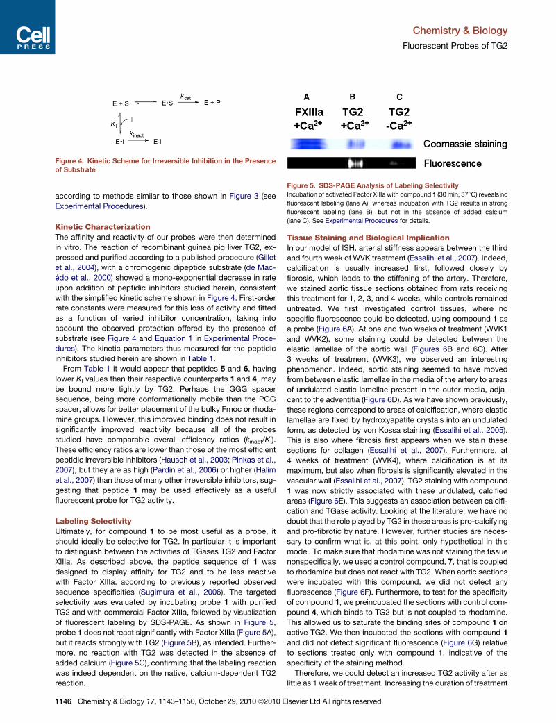
Figure 4. Kinetic Scheme for Irreversible Inhibition in the Presence
of Substrate
Figure 5. SDS-PAGE Analysis of Labeling Selectivity
Incubation of activated Factor XIIIa with compound 1 (30 min, 37�C) reveals nofluorescent labeling (lane A), whereas incubation with TG2 results in strong
fluorescent labeling (lane B), but not in the absence of added calcium
(lane C). See Experimental Procedures for details.
Chemistry & Biology
Fluorescent Probes of TG2
according to methods similar to those shown in Figure 3 (see
Experimental Procedures).
Kinetic CharacterizationThe affinity and reactivity of our probes were then determined
in vitro. The reaction of recombinant guinea pig liver TG2, ex-
pressed and purified according to a published procedure (Gillet
et al., 2004), with a chromogenic dipeptide substrate (de Mac-
edo et al., 2000) showed a mono-exponential decrease in rate
upon addition of peptidic inhibitors studied herein, consistent
with the simplified kinetic scheme shown in Figure 4. First-order
rate constants were measured for this loss of activity and fitted
as a function of varied inhibitor concentration, taking into
account the observed protection offered by the presence of
substrate (see Figure 4 and Equation 1 in Experimental Proce-
dures). The kinetic parameters thus measured for the peptidic
inhibitors studied herein are shown in Table 1.
From Table 1 it would appear that peptides 5 and 6, having
lower KI values than their respective counterparts 1 and 4, may
be bound more tightly by TG2. Perhaps the GGG spacer
sequence, being more conformationally mobile than the PGG
spacer, allows for better placement of the bulky Fmoc or rhoda-
mine groups. However, this improved binding does not result in
significantly improved reactivity because all of the probes
studied have comparable overall efficiency ratios (kinact/KI).
These efficiency ratios are lower than those of the most efficient
peptidic irreversible inhibitors (Hausch et al., 2003; Pinkas et al.,
2007), but they are as high (Pardin et al., 2006) or higher (Halim
et al., 2007) than those of many other irreversible inhibitors, sug-
gesting that peptide 1 may be used effectively as a useful
fluorescent probe for TG2 activity.
Labeling SelectivityUltimately, for compound 1 to be most useful as a probe, it
should ideally be selective for TG2. In particular it is important
to distinguish between the activities of TGases TG2 and Factor
XIIIa. As described above, the peptide sequence of 1 was
designed to display affinity for TG2 and to be less reactive
with Factor XIIIa, according to previously reported observed
sequence specificities (Sugimura et al., 2006). The targeted
selectivity was evaluated by incubating probe 1 with purified
TG2 and with commercial Factor XIIIa, followed by visualization
of fluorescent labeling by SDS-PAGE. As shown in Figure 5,
probe 1 does not react significantly with Factor XIIIa (Figure 5A),
but it reacts strongly with TG2 (Figure 5B), as intended. Further-
more, no reaction with TG2 was detected in the absence of
added calcium (Figure 5C), confirming that the labeling reaction
was indeed dependent on the native, calcium-dependent TG2
reaction.
1146 Chemistry & Biology 17, 1143–1150, October 29, 2010 ª2010 E
Tissue Staining and Biological ImplicationIn our model of ISH, arterial stiffness appears between the third
and fourth week of WVK treatment (Essalihi et al., 2007). Indeed,
calcification is usually increased first, followed closely by
fibrosis, which leads to the stiffening of the artery. Therefore,
we stained aortic tissue sections obtained from rats receiving
this treatment for 1, 2, 3, and 4 weeks, while controls remained
untreated. We first investigated control tissues, where no
specific fluorescence could be detected, using compound 1 as
a probe (Figure 6A). At one and two weeks of treatment (WVK1
and WVK2), some staining could be detected between the
elastic lamellae of the aortic wall (Figures 6B and 6C). After
3 weeks of treatment (WVK3), we observed an interesting
phenomenon. Indeed, aortic staining seemed to have moved
from between elastic lamellae in the media of the artery to areas
of undulated elastic lamellae present in the outer media, adja-
cent to the adventitia (Figure 6D). As we have shown previously,
these regions correspond to areas of calcification, where elastic
lamellae are fixed by hydroxyapatite crystals into an undulated
form, as detected by von Kossa staining (Essalihi et al., 2005).
This is also where fibrosis first appears when we stain these
sections for collagen (Essalihi et al., 2007). Furthermore, at
4 weeks of treatment (WVK4), where calcification is at its
maximum, but also when fibrosis is significantly elevated in the
vascular wall (Essalihi et al., 2007), TG2 staining with compound
1 was now strictly associated with these undulated, calcified
areas (Figure 6E). This suggests an association between calcifi-
cation and TGase activity. Looking at the literature, we have no
doubt that the role played by TG2 in these areas is pro-calcifying
and pro-fibrotic by nature. However, further studies are neces-
sary to confirm what is, at this point, only hypothetical in this
model. To make sure that rhodamine was not staining the tissue
nonspecifically, we used a control compound, 7, that is coupled
to rhodamine but does not react with TG2. When aortic sections
were incubated with this compound, we did not detect any
fluorescence (Figure 6F). Furthermore, to test for the specificity
of compound 1, we preincubated the sections with control com-
pound 4, which binds to TG2 but is not coupled to rhodamine.
This allowed us to saturate the binding sites of compound 1 on
active TG2. We then incubated the sections with compound 1
and did not detect significant fluorescence (Figure 6G) relative
to sections treated only with compound 1, indicative of the
specificity of the staining method.
Therefore, we could detect an increased TG2 activity after as
little as 1 week of treatment. Increasing the duration of treatment
lsevier Ltd All rights reserved

Figure 6. Fluorescent Staining of Aortic TG2
Staining of aortic tissues using irreversible TG2 inhibitors as fluorescent probes on control rats (A),WVK1 rats (B),WVK2 rats (C),WVK3 rats (D), andWVK4 rats (E).
(F) Negative control using unreactive rhodamine derivative 7 onWVK4 rats. (G) Preincubation of compound 4 followed by incubation of compound 1 to determine
the specificity of compound 1 staining on WVK4 rats. Arrows point to areas of intense TG2 labeling. A, adventitia; L, lumen; M, media of the aortic wall.
Chemistry & Biology
Fluorescent Probes of TG2
led to a staining pattern that was associated with areas of calci-
fication and fibrosis. This suggests that TG2 activity is increased
during the process of calcification and fibrosis associated with
arterial stiffness and ISH. In future work it would be interesting
to use these same irreversible inhibitors in the context of ISH,
but as blocking agents rather than probes, to measure their
impact on calcification, fibrosis, and arterial stiffness.
SIGNIFICANCE
New fluorescent irreversible inhibitors were designed as
labeling agents for TG2. These inhibitors were based on a
peptidic sequence designed to confer affinity for TG2 and
selectivity over Factor XIIIa, as demonstrated by SDS-
PAGE. As such, these inhibitors may be useful for ‘‘chemical
knockout’’ experiments intended to distinguish the biolog-
ical activities of the two closely related TGases. The fluores-
cence of the inhibitors presented herein was also optimized
with respect to the spacer fragment that links the rhodamine
B fluorophore to the peptide sequence. The most fluores-
cent inhibitor was then used to probe TG2 activity in a
WVK rat model for ISH. Fluorescent staining of aortic tissue
revealed that over a 4 week period, TG2 activity increases in
parallel with arterial stiffness, suggesting an association
between TG2 activity and arterial rigidification.
EXPERIMENTAL PROCEDURES
General Synthesis
All Fmoc-protected amino acids, resins, and coupling reagents were
purchased from GL Biochem; Wang resin was purchased from NovaBiochem.
All other reagents were obtained from Sigma-Aldrich. Reactions requiring
anhydrous conditions were carried out under a dry nitrogen atmosphere
Chemistry & Biology 17, 1143–1
employing conventional benchtop techniques. 1H- and 13C-NMR spectra
were recorded on AMXR400 and AMX300 spectrometers and were referenced
to the residual proton or 13C signal of the solvent. Mass spectra were deter-
mined by FAB+ ionization on an AutoSpec Q spectrometer at the Regional
Mass Spectrometry Centre at the Universite de Montreal.
Reactor tubes for solid-phase peptide synthesis were obtained from
Supelco, and shaking was performed on a shaker. All resins were swelled
in DMF, and washing steps were performed using CH2Cl2 and DMF (EMD
Chemicals). Purification of all peptides was performed using a preparative
HPLC method. Mass spectral data (MS, LCMS) were obtained using two
different columns—column A: Gemini C18, 1503 4.6 mm, 5 m (Phenomenex,
Torrance, CA); and column B: Synergi Polar-RP, 1503 4.6 mm, 4 m (Phenom-
enex, Torrance, CA). Crude peptides were purified using a preparative Synergi
Polar-RP, 100 3 21.20 mm column (Phenomenex, Torrance, CA) on a Varian
(Prep Star) HPLC system at the Regional Mass Spectrometry Centre at the
Universite de Montreal.
General Procedure for Fmoc Peptide Synthesis of Irreversible
Inhibitors
All peptides were synthesized using standard solid-phase Fmoc chemistry.
Briefly, the first Fmoc protected amino acid (5.5 mmol) was coupled to
Wang resin (1.1 mmol) using DIC (5.5 mmol) and DMAP (0.11 mmol) in DMF
(5 resin volumes). The level of loading of the amino acid on the resin after
the first coupling step was determined by spectroscopic measurement of
the UV absorbance of the piperidine dibenzofulvalene adduct formed during
Fmoc deprotection. This loading level was used as the resin loading capacity
for all subsequent steps. The remaining free hydroxyl functionalities were
capped by treating the resin with a mixture of acetic anhydride/pyridine
(2:3), followed by shaking for 2 hr. After washing with DMF (three times with
10 resin volumes), DCM (three times with 10 resin volumes), and ether (three
times with 10 resin volumes), the Fmoc group was removed by incubating
three times with piperidine in DMF (20% v/v; 10 resin volumes) for 5 min,
followed by washing with DMF (three times with 10 resin volumes), DCM (three
times with 10 resin volumes), and ether (three times with 10 resin volumes) in
preparation for the next amide coupling. Deprotection was verified by a posi-
tive Kaiser test on a sample of a few beads. Then each Fmoc protected amino
acid (1.7 mmol) was coupled to Wang resin preloaded with the necessary
carboxyl-terminal amino acid (0.68 mmol) in DMF (5 resin volumes) using
150, October 29, 2010 ª2010 Elsevier Ltd All rights reserved 1147
Chemistry & Biology
Fluorescent Probes of TG2
HOBT (1.7 mmol) and DIC (1.7 mmol). This operation was performed twice for
30 min. Coupling was verified by a negative Kaiser test on a sample of a few
beads. The peptide was then cleaved from the resin (1 g) by incubating with
TFA:DCM (1:1) for 2 hr. The peptide was precipitated from the cleavage solu-
tion using diethyl ether and hexane, and its purity was determined by HPLC
using two different columns—column A: Gemini C18, 1503 4.6 mm, 5m (Phe-
nomenex, Torrance, CA); and column B: Synergi Polar-RP, 150 3 4.6 mm, 4m
(Phenomenex, Torrance, CA). The crude peptide was purified using a prepara-
tive Synergi Polar-RP, 100 3 21.20 mm column (Phenomenex, Torrance, CA)
on a Varian (Prep Star) HPLC system.
Boc Deprotection Protocol
To a reactor containing 1 g of the Wang resin-supported Fmoc-peptide
(0.68 mmol, according to the measured loading level) were added 30 ml of
deprotection mixture, freshly prepared from 470 ml TEA (2 eq.), 1.09 ml of
TMSOTf (0.2 M), and 28.44 ml of anhydrous DCE. The resin was shaken
for 10 min, then filtered and washed with 5 3 5 ml of DCM, 2 3 5 ml of DIEA
10% in DCM, and 3 3 5 ml of DCE. Deprotection was carried out for another
10 min with a fresh deprotection mixture. The resin was filtered then washed
with 5 3 5 ml of DCM, 2 3 5 ml of DIEA 10% in DCM, 2 3 5 ml of DCM, 2 3
5 ml of DMF, and 2 3 5 ml of Et2O. Deprotection was verified by a positive
Kaiser test on a sample of a few beads.
Acrylation and Rhodamine B-Coupling Protocol
To the Wang resin-supported peptide (1 g, 0.68 mmol), either Fmoc protected
in the case of acrylation or deprotected in the case of rhodamine B coupling,
swollen in anhydrous DCM (5 resin volumes), was added acrylic acid
(1.7 mmol) and EEDQ (1.7 mmol). The reactionmixture was shaken for 1 hr, fol-
lowed by washing with DMF (three times with 10 resin volumes), DCM (three
times with 10 resin volumes), and ether (three times with 10 resin volumes).
This operation was performed twice.
HPLC Analysis and Purification
For HPLC analysis, the peptide was cleaved from the resin as described in
General Procedure for Fmoc Peptide Synthesis of Irreversible Inhibitors. The
crude material was purified by preparative Synergi Polar-RP (100 3
21.20 mm) column, on a Varian (Prep Star) HPLC system using 80%–95% of
MeOH in water as eluant, a flow rate of 8 ml/min, and the detector set at
254 nm. The collected fractions were freeze-dried to give the peptide in the
form of a powder. The areas under the peaks were determined using LC/
MSD Chem Station (Agilent Technologies).
Synthesis of Irreversible Inhibitors
RhodamineB-prolinyl-glycinyl-glycinyl-lysinyl(acryloyl)-leucinyl-prolinyl-phe-
nylalaninate (1).This peptide was cleaved from the resin and purified by
preparative HPLC as described previously. The collected fractions were
freeze-dried to give the desired irreversible peptidic inhibitor 1 as a pink
powder (overall yield: 10%; 98% purity by column A, 96% purity by column
B). HRMS m/z (M+H+): calcd 1193.63938; found 1193.63908.
Fmoc-Prolinyl-glycinyl-glycinyl-lysinyl(acryloyl)-leucinyl-prolinyl-phenylala-
ninate (4).This peptide was cleaved from the resin and purified by preparative
HPLC as described previously. The collected fractions were freeze-dried to
give the desired irreversible peptidic inhibitor 4 as a white powder (8% overall
yield; 91% purity by column A, 90% purity by column B). HRMS m/z (M+H+):
calcd 991.49238; found 991.49072.
Fmoc-Glycinyl-glycinyl-glycinyl-lysinyl(acryloyl)-leucinyl-prolinyl-phenylala-
ninate (5).This peptide was cleaved from the resin and purified by preparative
HPLC as described previously. The collected fractions were freeze-dried to
give the desired irreversible peptidic inhibitor 5 (6% of yield ; 98% purity by
column A, 99.9% purity by column B) as a white powder. HRMS m/z
(M+H+): calcd 951.46108; found 951.46215.
RhodamineB-glycinyl-glycinyl-glycinyl-lysinyl(acryloyl)-leucinyl-prolinyl-phe-
nylalaninate (6).This peptide was cleaved from the resin and purified by prepar-
ative HPLC as described previously. The collected fractions were freeze-dried
to give the desired irreversible peptidic inhibitor 6 (yield: 10%; 81% purity by
column A, 80% purity by column B) as a pink powder. HRMS m/z (M+H+):
calcd 1153.60808; found 1153.60589.RhodamineB-prolinyl-glycinyl-glycinyl-
glycinyl-leucinyl-prolinyl-phenylalaninylate (7).This peptide was cleaved from
the resin and purified by preparative HPLC as described previously. The
1148 Chemistry & Biology 17, 1143–1150, October 29, 2010 ª2010 E
collected fractions were freeze-dried to give the desired control probe 7 as a
pink powder (yield: 30%; 99% purity by column A, 98% purity by column B).
HRMS m/z (M+H+): calcd 1068.55532; found 1068.55472.
Kinetic Methods for Irreversible Inhibition
All assays were performed in triplicate. Kinetic runs were recorded on a
UV-visible spectrophotometer at 405 nm and 25�C, in a buffer composed of
50 mM CaCl2, 50 mM EDTA, and 0.1 M MOPS (pH 7.0). All aqueous solutions
were prepared using deionized water. All kinetic assays were carried out using
900 ml of buffer, 50 ml of 0.15 mg/ml TGase, 25 ml of the chromogenic substrate
Cbz-Glu(g-p-nitrophenyl ester)Gly (Leblanc et al., 2001) dissolved in DMF
(54 mM, corresponding to 6-fold KM), and 25 ml of inhibitor. Final concentrations
of inhibitors ranged from 10 mM to the solubility limit of each compound. Stock
solutions of the inhibitors were also prepared in DMF such that the final
concentration of this co-solvent was constant at 5% v/v. Kinetic runs were
initiated by enzyme addition, and evaluation was carried out by the method
of Stone and Hofsteenge (1985), as described previously (de Macedo et al.,
2002; Marrano et al., 2001). Mono-exponential time-dependent inactivation
was observed for all of the inhibitors studied herein. Observed first-order
rate constants of inactivation were determined from nonlinear regression,
using a mono-exponential model. These rate constants were in turn fit to
Equation 1 by nonlinear regression, providing the kinetic parameters kinactand KI as defined in Figure 4 and shown in Table 1.
kobs =kinact½I�
½I�+KI
�1+ ½S�
KM
�: (1)
Enzyme Preparation
Recombinant guinea pig liver TG2 was overexpressed and purified from E. coli
following our previously published procedure (Gillet et al., 2004). The purified
TG2 solution used in all assays had a specific activity of 30 U/mg in 25 mM
Tris-acetate (pH 7) according to the hydroxamate activity assay (Nemes
et al., 2005), in which Cbz-L-Gln-Gly and hydroxylamine are used as acyl-
donor and acyl-acceptor substrates, respectively. Recombinant human TG2,
used in the selectivity tests, was expressed and purified as reported previously
(Piper et al., 2002). Its activity was verified using a previously reported chromo-
genic activity assay (Leblanc et al., 2001). Factor XIII was purchased from
Zedira (Darmstadt, Germany). It was activated using thrombin and its activity
verified by a fluorogenic assay, according to the supplier’s protocols.
Selectivity Tests
Parallel incubation experiments were performed in three 1.5 ml Eppendorf
tubes. To the first was added Factor XIII (3 mg/ml (activated with 0.3 NIH units
of thrombin), in 56.8 mM Tris (pH 7.5), 11.36 mM CaCl2, 113.6 mM NaCl,
0.113% w/v PEG 8000, 5.68 mM GlyOMedHCl, 5.68 mg/l hexadimethrine
bromide, and compound 1 (90 mM dissolved in DMF, 0.9% final vol/vol).
To the second was added human TG2 (0.868 mg/ml in 25 mM Tris [pH 7.2],
150 mM NaCl, 1 mM EDTA, and 1 mM TCEP) in the presence of calcium
(0.6 M) and compound 1 (90 mM dissolved in DMF, 0.9% final vol/vol). The
third tube was identical to the second, but with 5 mM EDTA and no added
calcium. All three tubes were incubated for 30 min at 37�C, then analyzed
by 10% SDS-PAGE. The resulting gel was exposed under a trans-UV lamp
to determine fluorescent labeling and then stained with Coomassie blue to
reveal protein.
Animal Treatments
Male Wistar rats weighing 175–200 g (n = 9 per group) were obtained from
Charles River Breeding Laboratories (Saint-Constant, QC, Canada). They
received warfarin (20 mg/kg/day in drinking water) and vitamin K (phylloqui-
none, 15 mg/kg/day sc injection) (WVK) during 1, 2, 3, and 4 weeks. Dosages
were adjusted every second day. Control rats consisted of age-matched
untreated rats. All animal experiments were approved by the Animal Care
and Use Committee of Universite de Montreal.
Tissue Preparation
Animals were sacrificed with a lethal dose of pentobarbital (65 mg/kg), and the
aorta was harvested. A small section (0.5–1 cm) of aorta was frozen at �80�Cin Tissue-Tek OCT compound (Sakura Finetek Inc., Torrance, CA). These
aorta sections were later cut into 10 mm thick cryosections with a cryostat
(Leica Microsystems, model CM3050S, Richmond Hill, ON, Canada),
collected on Colorfrost/Plus Microscope slides (Fisher scientific, Ottawa,
ON, Canada), and kept at �20�C.
lsevier Ltd All rights reserved
Chemistry & Biology
Fluorescent Probes of TG2
Tissue Staining
Tissue slides were allowed to thaw at room temperature for 20 min. OCT was
removed, and tissues were permeabilized in PBS containing 0.1% triton for
20 min. Tissue sections were then circled using an ImmEdge PEN (Vector
Laboratories, Burlingame, CA). A 1 mM solution of compound 1was prepared
in a buffer containing 0.1MTris HCl (pH 7.5), 0.15MNaCl, and 5mMCaCl2 and
applied to the tissue sections for 15 min at 37�C. Slides were then rinsed four
times (5 min each) under agitation in PBS. After rinsing in distilled water for
5 min, slides were dried briefly before applying a solution of Mowiol/p-phenyl-
enediamine (9:1 vol/vol) (Calbiochem, San Diego, CA, USA, and Sigma-Al-
drich, Canada, respectively) to prevent bleaching. As a negative control, we
applied 1 mM of an inactive form of the inhibitor, compound 7, that is also
coupled to rhodamine. Furthermore, to make sure that compound 1 does
not bind nonspecifically in the tissue, we applied 5 mM of control compound
4, an irreversible inhibitor that is not coupled to rhodamine, for 45 min at
37�C, before applying 1 mM of the normal rhodamine-coupled inhibitor (1),
to the same section. Because compound 1 binds to the active form of TG2,
fluorescence was evaluated as an index of TG2 activity. To do so, slides
were photographed with a fluorescent microscope (Axioskop 40, Carl Zeiss
Canada Ltd., Toronto, Ontario, Canada, with SPOT RT Slider, Diagnostic
Instruments Inc., Sterling Heights, MI, USA).
ACKNOWLEDGMENTS
This study was supported by the Canadian Institutes for Health Research
(CIHR), the Natural Sciences and Engineering Research Council of Canada
(NSERC), and the Groupe de recherche universitaire sur les medicaments
(GRUM). S.M. received a studentship from the Fonds de Recherche en Sante
du Quebec (FRSQ). N.C. and A.M. are grateful for bursaries from GRUM.
Received: February 3, 2010
Revised: June 28, 2010
Accepted: June 30, 2010
Published: October 28, 2010
REFERENCES
Achyuthan, K.E., Slaughter, T.F., Santiago, M.A., Enghild, J.J., andGreenberg,
C.S. (1993). Factor XIIIa-derived peptides inhibit transglutaminase activity.
Localization of substrate recognition sites. J. Biol. Chem. 268, 21284–21292.
Adamczyk, M., and Grote, J. (2000). Efficient synthesis of rhodamine conju-
gates through the 20-position. Bioorg. Med. Chem. Lett. 10, 1539–1541.
Aeschlimann, D., and Thomazy, V. (2000). Protein crosslinking in assembly and
remodeling of extracellular matrices: the role of transglutaminase. Connect.
Tissue Res. 41, 1–27.
Albericio, F., Nicolas, E., Rizo, J., Ruiz-Gayo, M., Pedroso, E., and Giralt, E.
(1990). Convenient syntheses of fluorenylmethyl-based side chain derivatives
of glutamic and aspartic acids, lysine, and cysteine. Synthesis 1990 2,
119–122.
Arribas, S.M., Hinek, A., and Gonzalez, M.C. (2006). Elastic fibres and vascular
structure in hypertension. Pharmacol. Ther. 111, 771–791.
Azari, P., Rahim, I., and Clarkson, D.P. (1981). Transglutaminase activity in
normal and hereditary cataractous rat lens and its partial purification. Curr.
Eye Res. 1, 463–469.
Birckbichler, P.J., Orr, G.R., Patterson, M.K., Conway, E., Carter, H.A., and
Maxwell, M.D. (1983). Enhanced transglutaminase activity in transformed
human lung fibroblast cells after exposure to sodium butyrate. Biochim. Bio-
phys. Acta 763, 27–34.
Bouvet, C., Moreau, S., Blanchette, J., de Blois, D., and Moreau, P. (2008).
Sequential activation of matrix metalloproteinase 9 and transforming growth
factor beta in arterial elastocalcinosis. Arterioscler. Thromb. Vasc. Biol. 28,
856–862.
Bruel, A., Ortoft, G., and Oxlund, H. (1998). Inhibition of cross-links in collagen
is associated with reduced stiffness of the aorta in young rats. Atherosclerosis
140, 135–145.
Chemistry & Biology 17, 1143–1
Choi, K., Siegel, M., Piper, J.L., Yuan, L., Cho, E., Strnad, P., Omary, B., Rich,
K.M., and Khosla, C. (2005). Chemistry and biology of dihydroisoxazole
derivatives: selective inhibitors of human transglutaminase 2. Chem. Biol.
12, 469–475.
Davies, P.J.A., Davies, D.R., Levitzki, A., Maxfield, F.R., Milhaud, P.,
Willingham, M.C., and Pastan, I.H. (1980). Transglutaminase is essential in
receptor-mediated endocytosis of alpha 2-macroglobulin and polypeptide
hormones. Nature 283, 162–167.
de Macedo, P., Marrano, C., and Keillor, J.W. (2000). A direct and continuous
spectrophotometric assay for transglutaminase mediated transpeptidation
activity. Anal. Biochem. 285, 16–20.
de Macedo, P., Marrano, C., and Keillor, J.W. (2002). Synthesis of dipeptide-
bound epoxides and a,b-unsaturated amides as potential irreversible transglu-
taminase inhibitors. Bioorg. Med. Chem. 10, 355–360.
De Young, L., Ballaron, S., and Epstein, W. (1984). Transglutaminase activity
in human and rabbit ear comedogenesis: a histochemical study. J. Invest.
Dermatol. 82, 275–279.
Dedeoglu, A., Kubilus, J.K., Jeitner, T.M.,Matson, S.A., Bogdanov, M., Kowall,
N.W., Matson, W.R., Cooper, A.J., Ratan, R.R., Beal, M.F., et al. (2002).
Therapeutic effects of cystamine in a murine model of Huntington’s disease.
J. Neurosci. 22, 8942–8950.
Essalihi, R., Dao, H.H., Gilbert, L.A., Bouvet, C., Semerjian, Y., McKee, M.D.,
and Moreau, P. (2005). Regression of medial elastocalcinosis in rat aorta:
a new vascular function for carbonic anhydrase. Circulation 112, 1628–1635.
Essalihi, R., Dao, H.H., Yamaguchi, N., and Moreau, P. (2003). A new model of
isolated systolic hypertension induced by chronic warfarin and vitamin K1
treatment. Am. J. Hypertens. 16, 103–110.
Essalihi, R., Ouellette, V., Dao, H.H., McKee, M.D., and Moreau, P. (2004).
Phenotypic modulation of vascular smooth muscle cells during medial arterial
calcification: a role for endothelin? J. Cardiovasc. Pharmacol. 44 (Suppl 1 ),
S147–S150.
Essalihi, R., Zandvliet, M.L., Moreau, S., Gilbert, L.A., Bouvet, C., Lenoel, C.,
Nekka, F., McKee, M.D., and Moreau, P. (2007). Distinct effects of amlodipine
treatment on vascular elastocalcinosis and stiffness in a rat model of isolated
systolic hypertension. J. Hypertens. 25, 1879–1886.
Faverman, L., Mikhaylova, L., Malmquist, J., and Nurminskaya, M. (2008).
Extracellular transglutaminase 2 activates beta-catenin signaling in calcifying
vascular smooth muscle cells. FEBS Lett. 582, 1552–1557.
Fesus, L. (1982). Transglutaminase activation: significance with respect to
immunologic phenomena. Surv. Immunol. Res. 1, 297–304.
Fesus, L., Thomazy, V., and Falus, A. (1987). Induction and activation of tissue
transglutaminase during programmed cell death. FEBS Lett. 224, 104–108.
Folk, J.E., and Chung, S.I. (1985). Transglutaminases. Methods Enzymol. 113,
358–375.
Folk, J.E., and Cole, P.W. (1966). Mechanism of action of guinea pig liver trans-
glutaminase. I. Purification and properties of the enzyme: identification of
a functional cysteine essential for activity. J. Biol. Chem. 241, 5518–5525.
Gillet, S.M.F.G., Chica, R.A., Keillor, J.W., and Pelletier, J.N. (2004).
Expression and rapid purification of highly active hexahistidine-tagged guinea
pig liver transglutaminase. Protein Expr. Purif. 33, 256–264.
Greenberg, C.S., Birchbichler, P.J., and Rice, R.H. (1991). Transglutaminases:
multifunctional cross-linking enzymes that stabilize tissues. FASEB J. 5, 3071–
3077.
Griffin, M., Smith, L.L., and Wynne, J. (1979). Changes in transglutaminase
activity in an experimental model of pulmonary fibrosis induced by paraquat.
Br. J. Exp. Pathol. 60, 653–661.
Halim, D., Caron, K., and Keillor, J.W. (2007). Synthesis and evaluation of
peptidic maleimides as transglutaminase inhibitors. Bioorg. Med. Chem.
Lett. 17, 305–308.
Hausch, F., Halttunen, T., Maki, M., and Khosla, C. (2003). Design, synthesis,
and evaluation of gluten peptide analogs as selective inhibitors of human
tissue transglutaminase. Chem. Biol. 10, 225–231.
Ientile, R., Caccamo, D., and Griffin, M. (2007). Tissue transglutaminase and
the stress response. Amino Acids 33, 385–394.
150, October 29, 2010 ª2010 Elsevier Ltd All rights reserved 1149
Chemistry & Biology
Fluorescent Probes of TG2
Johnson, T.S., Griffin, M., Thomas, G.L., Skill, J., Cox, A., Yang, B., Nicholas,
B., Birckbichler, P.J., Muchaneta-Kubara, C., and Meguid El Nahas, A. (1997).
The role of transglutaminase in the rat subtotal nephrectomy model of renal
fibrosis. J. Clin. Invest. 99, 2950–2960.
Johnson, C.P., Baugh, R., Wilson, C.A., and Burns, J. (2001). Age related
changes in the tunica media of the vertebral artery: implications for the
assessment of vessels injured by trauma. J. Clin. Pathol. 54, 139–145.
Johnson, T.S., Fisher, M., Haylor, J.L., Hau, Z., Skill, N.J., Jones, R., Saint, R.,
Coutts, I., Vickers, M.E., El Nahas, A.M., and Griffin, M. (2007). Transglutami-
nase inhibition reduces fibrosis and preserves function in experimental chronic
kidney disease. J. Am. Soc. Nephrol. 18, 3078–3088.
Johnson, K.A., Polewski, M., and Terkeltaub, R.A. (2008). Transglutaminase 2
is central to induction of the arterial calcification program by smooth muscle
cells. Circ. Res. 102, 529–537.
Kaartinen, M.T., Murshed, M., Karsenty, G., and McKee, M.D. (2007).
Osteopontin upregulation and polymerization by transglutaminase 2 in calci-
fied arteries of Matrix Gla protein-deficient mice. J. Histochem. Cytochem.
55, 375–386.
Keresztessy, Z., Csosz, E., Harsfalvi, J., Csomos, K., Gray, J., Lightowlers,
R.N., Lakey, J.H., Balajthy, Z., and Fesus, L. (2006). Phage display selection
of efficient glutamine-donor substrate peptides for transglutaminase 2. Protein
Sci. 15, 2466–2480.
Leblanc, A., Gravel, C., Labelle, J., and Keillor, J.W. (2001). Kinetic studies of
guinea pig liver transglutaminase reveal a general-base-catalyzed deacylation
mechanism. Biochemistry 40, 8335–8342.
Lejeune, V., Martinez, J., and Cavelier, F. (2003). Towards a selective Boc
deprotection on acid cleavable Wang resin. Tetrahedron Lett. 44, 4757–4759.
Lesort, M., Attanavanich, K., Zhang, J., and Johnson, G.V. (1998). Distinct
nuclear localization and activity of tissue transglutaminase. J. Biol. Chem.
273, 11991–11994.
Levitzki, A., Willingham, M., and Pastan, I.H. (1980). Evidence for participation
of transglutaminase in receptor-mediated endocytosis. Proc. Natl. Acad. Sci.
USA 77, 2706–2710.
Marrano, C., de Macedo, P., and Keillor, J. (2001). Evaluation of novel
dipeptide-bound a,b-unsaturated amides and epoxides as irreversible inhibi-
tors of guinea pig liver transglutaminase. Bioorg. Med. Chem. 9, 1923–1928.
Mastroberardino, P.G., Iannicola, C., Nardacci, R., Bernassola, F.,
De Laurenzi, V., Melino, G., Moreno, S., Pavone, F., Oliverio, S., Fesus, L.,
and Piacentini, M. (2002). ‘‘Tissue’’ transglutaminase ablation reduces
neuronal death and prolongs survival in a mouse model of Huntington’s
disease. Cell Death Differ. 9, 873–880.
Mehta, K. (2009). Biological and therapeutic significance of tissue transgluta-
minase in pancreatic cancer. Amino Acids 36, 709–716.
Nemes, Z., Petrovski, G., and Fesus, L. (2005). Tools for the detection and
quantitation of protein transglutamination. Anal. Biochem. 342, 1–10.
1150 Chemistry & Biology 17, 1143–1150, October 29, 2010 ª2010 E
Norlund, M.A., Lee, J.M., Zainelli, G.M., and Muma, N.A. (1999). Elevated
transglutaminase-induced bonds in PHF tau in Alzheimer’s disease. Brain
Res. 851, 154–163.
Pardin, C., Gillet, S.M.F.G., and Keillor, J.W. (2006). Synthesis and evaluation
of peptidic irreversible inhibitors of tissue transglutaminase. Bioorg. Med.
Chem. 14, 8379–8385.
Pinkas, D.M., Strop, P., Brunger, A.T., and Khosla, C. (2007). Transglutami-
nase 2 undergoes a large conformational change upon activation. PLoS
Biol. 5 (12), e327.
Piper, J.L., Gray, G.M., and Khosla, C. (2002). High selectivity of human tissue
transglutaminase for immunoactive gliadin peptides: implications for celiac
sprue. Biochemistry 41, 386–393.
Safar, M.E., Levy, B.I., and Struijker-Boudier, H. (2003). Current perspectives
on arterial stiffness and pulse pressure in hypertension and cardiovascular
diseases. Circulation 107, 2864–2869.
Schroeder, W.T., Thacher, S.M., Stewart-Galetka, S., Annarella, M., Chema,
D., Siciliano, M.J., Davies, P.J.A., Tang, H.Y., Sowa, B.A., and Duvic, M.
(1992). Type I keratinocyte transglutaminase: expression in human skin and
psoriasis. J. Invest. Dermatol. 99, 27–34.
Schurgers, L.J., Cranenburg, E.C., and Vermeer, C. (2008). Matrix Gla-protein:
the calcification inhibitor in need of vitamin K. Thromb. Haemost. 100,
593–603.
Selkoe, D.J., Abraham, C., and Ihara, Y. (1982). Brain transglutaminase: in vitro
crosslinking of human neurofilament proteins into insoluble polymers. Proc.
Natl. Acad. Sci. USA 79, 6070–6074.
Shanahan, C.M., Proudfoot, D., Tyson, K.L., Cary, N.R., Edmonds, M., and
Weissberg, P.L. (2000). Expression of mineralisation-regulating proteins in
association with human vascular calcification. Z. Kardiol. 89 (Suppl 2 ), 63–68.
Small, K., Feng, J.F., Lorenz, J., Donnelly, E.T., Yu, A., Im, M.J., Dorn, G.W.,
2nd, and Liggett, S.B. (1999). Cardiac specific overexpression of transglutami-
nase II (G(h)) results in a unique hypertrophy phenotype independent of
phospholipase C activation. J. Biol. Chem. 274, 21291–21296.
Sohn, J., Kim, T., Young-Hee, Y., Kim, J., and Kim, S. (2003). Novel transglu-
taminase inhibitors reverse the inflammation of allergic conjunctivitis. J. Clin.
Invest. 111, 121–128.
Stone, S.R., and Hofsteenge, J. (1985). Specificity of activated human protein
C. Biochem. J. 230, 497–500.
Sugimura, Y., Hosono, M., Wada, F., Yoshimura, T., Maki, M., and Hitomi, K.
(2006). Screening for the preferred substrate sequence of transglutaminase
using a phage-displayed peptide library: identification of peptide substrates
for TGASE 2 and Factor XIIIA. J. Biol. Chem. 281, 17699–17706.
Lin, C.W., and Ting, A.Y. (2006). Transglutaminase-catalyzed site-specific
conjugation of small-molecule probes to proteins in vitro and on the surface
of living cells. J. Am. Chem. Soc. 128, 4542–4543.
Zieman, S.J., Melenovsky, V., and Kass, D.A. (2005). Mechanisms, pathophys-
iology, and therapy of arterial stiffness. Arterioscler. Thromb. Vasc. Biol. 25,
932–943.
lsevier Ltd All rights reserved