Embed Size (px)
Citation preview
Version 4 Last Updated 16 April 2015
Instructions for Use
For the rapid, sensitive and accurate measurement of myeloperoxidase activity in various samples.
This product is for research use only and is not intended for diagnostic use.
ab111749Myeloperoxidase (MPO) Activity Assay Kit (Fluorometric)
Discover more at www.abcam.com 1
Table of Contents
INTRODUCTION1. BACKGROUND 22. ASSAY SUMMARY 3
GENERAL INFORMATION3. PRECAUTIONS 44. STORAGE AND STABILITY 45. MATERIALS SUPPLIED 56. MATERIALS REQUIRED, NOT SUPPLIED 57. LIMITATIONS 68. TECHNICAL HINTS 7
ASSAY PREPARATION9. REAGENT PREPARATION 810. STANDARD PREPARATION 911. SAMPLE PREPARATION 10
ASSAY PROCEDURE and DETECTION12. ASSAY PROCEDURE and DETECTION 12
DATA ANALYSIS13. CALCULATIONS 1414. TYPICAL DATA 16
RESOURCES15. QUICK ASSAY PROCEDURE 1816. TROUBLESHOOTING 1917. FAQ 2118. INTERFERENCES 2319. NOTES 24
Discover more at www.abcam.com 2
INTRODUCTION
1. BACKGROUND
Myeloperoxidase (MPO) Assay Kit (fluorometric) (ab111749) provides a rapid, simple, sensitive, and reliable method to study MPO activity. MPO catalyzes the production of sodium hypochlorite (NaClO) from hydrogen peroxide (H2O2) and sodium chloride (NaCl). Subsequently, NaClO will react stoichiometrically with the free radical sensor aminophenyl fluorescein (APF) to generate fluorescein, which can be detected at Ex/Em = 485/525 nm. This kit can be used to detect MPO activity as low as 0.5 μU per well.
Myeloperoxidase (MPO, EC 1.11.1.7) is a peroxidase enzyme most abundantly present in neutrophil granulocytes. It is a green hemoprotein found in neutrophils and monocytes that catalyzes the reaction of hydrogen peroxide and halide ions to form cytotoxic acids and other intermediates that play a role in the oxygen-dependent killing of tumor cells and microorganisms. Its heme pigment causes the green color in secretions rich in neutrophils, such as pus and some forms of mucus. Furthermore, it can oxidize tyrosine to a tyrosyl radical using hydrogen peroxide as an oxidizing agent.
Discover more at www.abcam.com 3
INTRODUCTION
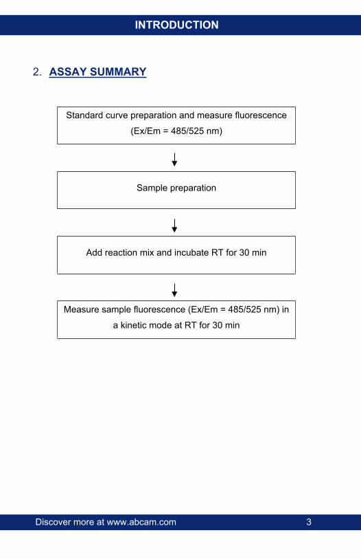
2. ASSAY SUMMARY
Standard curve preparation and measure fluorescence
(Ex/Em = 485/525 nm)
Sample preparation
Add reaction mix and incubate RT for 30 min
Measure sample fluorescence (Ex/Em = 485/525 nm) in
a kinetic mode at RT for 30 min
Discover more at www.abcam.com 4
GENERAL INFORMATION
3. PRECAUTIONSPlease read these instructions carefully prior to beginning the assay.All kit components have been formulated and quality control tested to function successfully as a kit. Modifications to the kit components or procedures may result in loss of performance.
4. STORAGE AND STABILITYStore kit at -20ºC in the dark immediately upon receipt. Kit has a storage time of 1 year from receipt, providing components have not been reconstituted.Refer to list of materials supplied for storage conditions of individual components. Observe the storage conditions for individual prepared components in section 5.Aliquot components in working volumes before storing at the recommended temperature. Reconstituted components are stable for 2 months.
Discover more at www.abcam.com 5
GENERAL INFORMATION
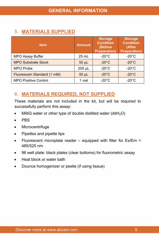
5. MATERIALS SUPPLIED
Item AmountStorage
Condition(Before
Preparation)
StorageCondition
(After Preparation)
MPO Assay Buffer 25 mL -20°C -20°CMPO Substrate Stock 50 µL -20°C -20°CMPO Probe 200 µL -20°C -20°CFluorescein Standard (1 mM) 50 µL -20°C -20°CMPO Positive Control 1 vial -20°C -20°C
6. MATERIALS REQUIRED, NOT SUPPLIEDThese materials are not included in the kit, but will be required to successfully perform this assay:
MilliQ water or other type of double distilled water (ddH2O)
PBS
Microcentrifuge
Pipettes and pipette tips
Fluorescent microplate reader – equipped with filter for Ex/Em = 485/525 nm
96 well plate: black plates (clear bottoms) for fluorometric assay
Heat block or water bath
Dounce homogenizer or pestle (if using tissue)
Discover more at www.abcam.com 6
GENERAL INFORMATION
7. LIMITATIONS Assay kit intended for research use only. Not for use in diagnostic
procedures.
Do not use kit or components if it has exceeded the expiration date on the kit labels.
Do not mix or substitute reagents or materials from other kit lots or vendors. Kits are QC tested as a set of components and performance cannot be guaranteed if utilized separately or substituted.
Discover more at www.abcam.com 7
GENERAL INFORMATION
8. TECHNICAL HINTS This kit is sold based on number of tests. A ‘test’ simply
refers to a single assay well. The number of wells that contain sample, control or standard will vary by product. Review the protocol completely to confirm this kit meets your requirements. Please contact our Technical Support staff with any questions.
Keep enzymes, heat labile components and samples on ice during the assay.
Make sure all buffers and solutions are at room temperature before starting the experiment.
Samples generating values higher than the highest standard should be further diluted in the appropriate sample dilution buffers.
Avoid foaming or bubbles when mixing or reconstituting components.
Avoid cross contamination of samples or reagents by changing tips between sample, standard and reagent additions.
Ensure plates are properly sealed or covered during incubation steps.
Make sure you have the right type of plate for your detection method of choice.
Make sure the heat block/water bath and microplate reader are switched on.
Discover more at www.abcam.com 8
ASSAY PREPARATION
9. REAGENT PREPARATION Briefly centrifuge small vials at low speed prior to opening.
9.1 MPO Assay Buffer:Ready to use as supplied. Equilibrate to room temperature before use. Store at -20°C.
9.2 MPO Substrate Stock:Ready to use as supplied. Aliquot substrate so that you have enough volume to perform the desired number of assays. Store at -20°C.Dilute 4 µL substrate stock with 700 µL MPO Assay Buffer to prepare the MPO substrate solution. Keep on ice while in use.
9.3 MPO Probe:Ready to use as supplied. Aliquot probe so that you have enough volume to perform the desired number of assays. Store at -20°C protected from light. Once the probe is thawed, use within two months.
9.4 Fluorescein Standard (1 mM):Ready to use as supplied. Aliquot standard so that you have enough volume to perform the desired number of assays. Store at -20°C. Keep on ice while in use.
9.5 MPO Positive Control:Reconstitute in 50 µL Assay Buffer. Aliquot probe so that you have enough volume to perform the desired number of assays. Use within 1 month. Avoid freeze thaw cycles. Store at -20°C.
Discover more at www.abcam.com 9
ASSAY PREPARATION
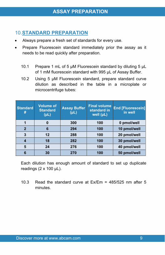
10.STANDARD PREPARATION Always prepare a fresh set of standards for every use.
Prepare Fluorescein standard immediately prior the assay as it needs to be read quickly after preparation.
10.1 Prepare 1 mL of 5 µM Fluorescein standard by diluting 5 µL of 1 mM fluorescein standard with 995 µL of Assay Buffer.
10.2 Using 5 µM Fluorescein standard, prepare standard curve dilution as described in the table in a microplate or microcentrifuge tubes:
Standard#
Volume of Standard
(µL)Assay Buffer
(µL)Final volume standard in
well (µL)End [Fluorescein]
in well
1 0 300 100 0 pmol/well2 6 294 100 10 pmol/well3 12 288 100 20 pmol/well4 18 282 100 30 pmol/well5 24 276 100 40 pmol/well6 30 270 100 50 pmol/well
Each dilution has enough amount of standard to set up duplicate readings (2 x 100 µL).
10.3 Read the standard curve at Ex/Em = 485/525 nm after 5 minutes.
ASSAY PRE
Discover more at www.abcam.com 10
ASSAY PREPARATION
11.SAMPLE PREPARATIONGeneral Sample information: We recommend performing several dilutions of your sample to
ensure the readings are within the standard value range.
We recommend that you use fresh samples. If you cannot perform the assay at the same time, we suggest that you complete the Sample Preparation step before storing the samples. Alternatively, if that is not possible, we suggest that you snap freeze cells or tissue in liquid nitrogen upon extraction and store the samples immediately at -80°C. When you are ready to test your samples, thaw them on ice. Be aware however that this might affect the stability of your samples and the readings can be lower than expected.
11.1 Cell (adherent or suspension) samples:11.1.1 Harvest the amount of cells necessary for each assay (initial
recommendation = 2 x 10⁶ cells).11.1.2 Wash cells with cold PBS.11.1.3 Resuspend cells in 4 volumes of ice cold Assay Buffer.11.1.4 Homogenize cells quickly by pipetting up and down a few
times.11.1.5 Centrifuge sample for 10 minutes at 4°C at 13,000 x g using
a cold microcentrifuge to remove any insoluble material.11.1.6 Collect supernatant and transfer to a clean tube.11.1.7 Keep on ice.
11.2 Tissue samples:11.2.1 Harvest the amount of tissue necessary for each assay
(initial recommendation = 10 mg).11.2.2 Wash tissue in cold PBS.11.2.3 Resuspend tissue in 4 volumes (~400 µL) of ice cold Assay
Buffer.
ASSAY PRE
Discover more at www.abcam.com 11
ASSAY PREPARATION
11.2.4 Homogenize tissue with a Dounce homogenizer sitting on ice, with 10 – 15 passes.
11.2.5 Centrifuge samples for 2 – 5 minutes at 4°C at top speed using a cold microcentrifuge to remove any insoluble material.
11.2.6 Collect supernatant and transfer to a clean tube.11.2.7 Keep on ice.
11.3 Plasma, Serum and Urine and other biological fluids: Serum and urine samples can be tested directly by adding sample to the microplate wells.However, to find the optimal values and ensure your readings will fall within the standard values, we recommend performing several dilutions of the sample (1/2 – 1/5 – 1/10).
NOTE: We suggest using different volumes of sample to ensure readings are within the Standard Curve range.
Discover more at www.abcam.com 12
ASSAY PROCEDURE and DETECTION
12.ASSAY PROCEDURE and DETECTION● Equilibrate all materials and prepared reagents to room
temperature prior to use.● It is recommended to assay all standards, controls and
samples in duplicate.12.1 Set up Reaction wells:- Standard wells = 100 µL standard dilutions.- Sample wells = 1 – 50 µL samples (adjust volume to
50 µL/well with Assay Buffer).- OPTIONAL – MPO Positive control = 10 µL Positive control
(adjust volume to 50 µL/well with Assay Buffer.12.2 MPO Reaction Mix:
Prepare 50 µL of MPO Reaction Mix for each reaction
Component Reaction Mix (µL)
MPO Assay Buffer 46MPO Substrate Solution 2MPO Probe 2
Mix enough reagents for the number of assays (samples and positive control) to be performed. Prepare a master mix of the Reaction Mix to ensure consistency. We recommend the following calculation:X µL component x (Number samples + positive control +1).
12.3 Add 50 µL of MPO Reaction Mix into each sample and positive control wells. Mix thoroughly. DO NOT ADD REACTION MIX TO STANDARD WELLS.
12.4 Measure fluorescence on a microplate reader at Ex/Em = 485/525 nm in a kinetic mode, every 2 – 3 minutes, for 30 minutes at RT protected from light.STANDARD: measure fluorescence of standard curve 5 minutes after preparation (Section 10.3).
ASSAY PRE
Discover more at www.abcam.com 13
ASSAY PROCEDURE and DETECTION
NOTE: Sample incubation time can vary depending on MPO activity. We recommend measuring the fluorescence in kinetic mode and choosing two time points (T1 and T2) in the linear range to calculate the MPO activity of the samples.

Discover more at www.abcam.com 14
DATA ANALYSIS
13.CALCULATIONS Samples producing signals greater than that of the highest
standard should be further diluted in appropriate buffer and reanalyzed, then multiplying the concentration found by the appropriate dilution factor.
For statistical reasons, we recommend each sample should be assayed with a minimum of two replicates (duplicates).
13.1 Average the duplicate reading for each standard and sample.
13.2 Subtract the mean absorbance value of the blank (Standard #1) from all standard and sample readings. This is the corrected absorbance.
13.3 Plot the corrected absorbance values for each standard as a function of the final concentration of MPO.
13.4 Draw the best smooth curve through these points to construct the standard curve. Most plate reader software or Excel can plot these values and curve fit. Calculate the trendline equation based on your standard curve data (use the equation that provides the most accurate fit).
13.5 Activity of MPO is calculated as:ΔRFU = (RFU2 – RFUBG2) – (RFU1 – RFUBG1)Where:RFU1 and RFU2 is the sample reading at time T1 and T2 respectively
13.6 Use the ΔRFU to obtain B pmol of fluorescein generated by MPO.
13.1 MPO activity (in pmol/min/mL or µU/mL) in the test samples is calculated as:
𝑀𝑃𝑂 𝐴𝑐𝑡𝑖𝑣𝑖𝑡𝑦 = ( 𝐵Δ𝑇 𝑥 𝑉) ∗ 𝐷
= pmol/min/mL = µU/mL
Where:
Discover more at www.abcam.com 15
DATA ANALYSIS
B = Amount of fluorescein from Standard Curve (pmol).ΔT = Reaction time (hour).V = pretreated sample volume added into the reaction well (µL).D = sample dilution factor.
Unit Definition:1 Unit MPO activity = amount of MPO that oxidizes the APF substrate to generate 1.0 µmol of fluorescein per minute at 25°C.
Discover more at www.abcam.com 16
DATA ANALYSIS
14.TYPICAL DATATYPICAL STANDARD CURVE – Data provided for demonstration purposes only. A new standard curve must be generated for each assay performed.
Figure 1. Typical fluorescein Standard calibration curve using fluorometric reading.
Discover more at www.abcam.com 17
DATA ANALYSIS
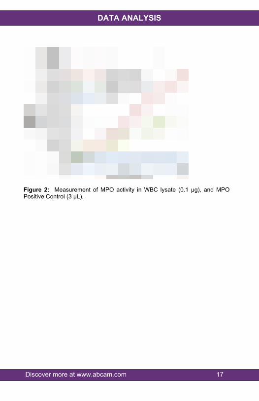
Figure 2: Measurement of MPO activity in WBC lysate (0.1 µg), and MPO Positive Control (3 µL).
Discover more at www.abcam.com 18
RESOURCES
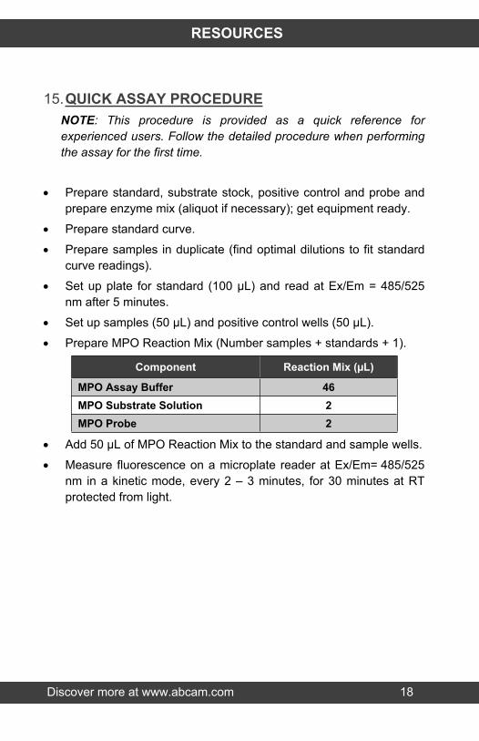
15.QUICK ASSAY PROCEDURENOTE: This procedure is provided as a quick reference for experienced users. Follow the detailed procedure when performing the assay for the first time.
Prepare standard, substrate stock, positive control and probe and prepare enzyme mix (aliquot if necessary); get equipment ready.
Prepare standard curve.
Prepare samples in duplicate (find optimal dilutions to fit standard curve readings).
Set up plate for standard (100 µL) and read at Ex/Em = 485/525 nm after 5 minutes.
Set up samples (50 µL) and positive control wells (50 µL).
Prepare MPO Reaction Mix (Number samples + standards + 1).
Component Reaction Mix (µL)
MPO Assay Buffer 46MPO Substrate Solution 2MPO Probe 2
Add 50 µL of MPO Reaction Mix to the standard and sample wells.
Measure fluorescence on a microplate reader at Ex/Em= 485/525 nm in a kinetic mode, every 2 – 3 minutes, for 30 minutes at RT protected from light.

Discover more at www.abcam.com 19
RESOURCES
16.TROUBLESHOOTING
Problem Cause Solution
Use of ice-cold buffer Buffers must be at room temperature
Plate read at incorrect wavelength
Check the wavelength and filter settings of instrument
Assay not
workingUse of a different 96-
well plate
Colorimetric: Clear platesFluorometric: black wells/clear
bottom plateSamples not
deproteinized (if indicated on protocol)
Use PCA precipitation protocol for deproteinization
Cells/tissue samples not homogenized
completely
Use Dounce homogenizer, increase number of strokes
Samples used after multiple free/ thaw
cycles
Aliquot and freeze samples if needed to use multiple times
Use of old or inappropriately stored
samples
Use fresh samples or store at - 80°C (after snap freeze in liquid
nitrogen) till use
Sample with erratic readings
Presence of interfering substance
in the sample
Check protocol for interfering substances; deproteinize samples
Improperly thawed components
Thaw all components completely and mix gently before use
Allowing reagents to sit for extended times
on ice
Always thaw and prepare fresh reaction mix before use
Lower/ Higher readings in samples and Standards Incorrect incubation
times or temperaturesVerify correct incubation times and temperatures in protocol
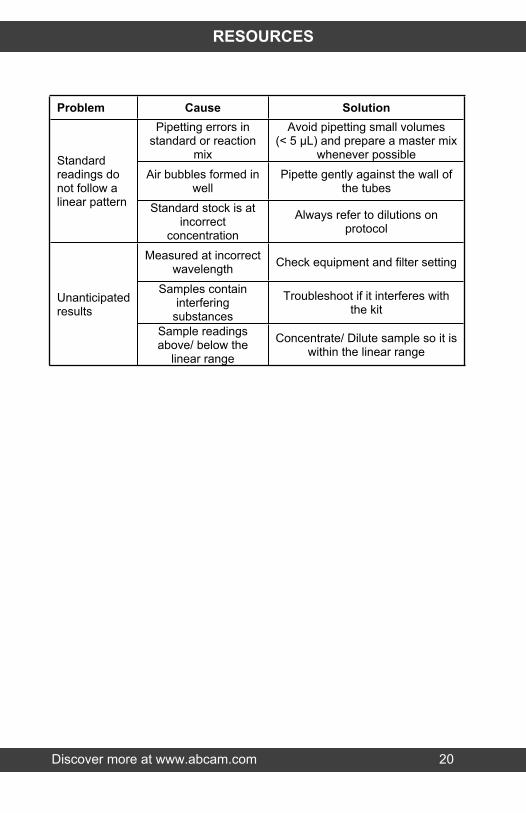
Discover more at www.abcam.com 20
RESOURCES
Problem Cause SolutionPipetting errors in
standard or reaction mix
Avoid pipetting small volumes (< 5 µL) and prepare a master mix
whenever possibleAir bubbles formed in
wellPipette gently against the wall of
the tubes
Standard readings do not follow a linear pattern Standard stock is at
incorrect concentration
Always refer to dilutions on protocol
Measured at incorrect wavelength Check equipment and filter setting
Samples contain interfering
substances
Troubleshoot if it interferes with the kit
Unanticipated results
Sample readings above/ below the
linear range
Concentrate/ Dilute sample so it is within the linear range
Discover more at www.abcam.com 21
RESOURCES
17.FAQCan this kit be used with frozen cells/tissue?We always prefer fresh samples for the most accurate results. It is possible to use frozen cells/tissue samples as long as they have been stored with minimum delay after collection and have been stored at -80C without freeze-thaw. However, the MPO enzyme is quite vulnerable to losing activity during storage. The functional enzyme contains disulphide bonds and if reduced, the enzyme can lose activity. It also contains heme which can get oxidized by the peroxide generated by MPO activity and the result is that the enzyme loses activity by auto-oxidation.
I have used this product (ab111749) and its colorimetric equivalent (ab105136) but got very different raw data for the increasing dilutions of the samples. Why?It is very important to be able to distinguish the two kits by principle. For MPO Activity Colorimetric (ab105136), the lower the OD, the higher the MPO activity. If you add too much sample, the OD will be too low and it will probably be below the detection limit of absorbance instruments. For MPO Activity Fluorometric (ab111749), the higher the RFU, the higher the MPO activity. So, for this kit, adding too much sample can saturate the detector and the substrate can be limiting. This will result in discrepant differences between dilutions.
How much cells/tissue is needed for this assay?There is no set amount of cells/tissue needed for this assay. The amount depends on how much active MPO is present in the samples. Typically 1 – 2 x 10⁶ cells and 10 – 100 mg tissue can be used per assay.
What is the activity of the positive control? How can the value be higher to compare with samples?
Discover more at www.abcam.com 22
RESOURCES
The positive control is only a benchmark sample. As long as the values are within the range of the standard curve this is fine. The positive control is not be used to compare values with the samples. The positive control is provided to validate that the assay components are all working. If the values are low, the customer can add more volume to get higher values but this is not necessary as long as the values are within the std. curve range. MPO is a very vulnerable enzyme to freeze-thaw and can lose activity with storage over time.
What is the dilution factor used for?If a certain volume of neat sample is added to the well and volume is made up with the assay buffer up to 50 µL, then dilution factor does not apply. If the sample is prediluted before adding to the well, then the dilution factor is used. For example, if 10 µL of a 5X diluted sample is used, then V=0.01 mL and Dilution factor =5.
How does the sensitivity compare between MPO Activity Assay Kit (Colorimetric) (ab105136) and this product (ab111749)?This kit (ab111749) is a fluorometric assay and is at least 10 times more sensitive than ab105136. The standard curve range for ab105136 is 1 – 50 nmoles of TNB formed, whereas the standard curve range for ab111749 is between 1 – 50 pmoles of Fluorescein.
Can RIPA buffer be used to prepare samples for this kit?For any enzyme assay, we do not recommend RIPA buffer since it contains SDS and this can denature proteins and affect enzyme activity. We have tested and recommend using the assay buffer provided in the kit for best results.
Discover more at www.abcam.com 23
RESOURCES
18. INTERFERENCESThese chemicals or biological materials will cause interferences in this assay causing compromised results or complete failure:
RIPA: contains SDS which can destroy/decrease the activity of the enzyme.
EDTA: MPO is an enzyme and needs some ions/cofactors to function. As EDTA is a chelator, it might interfere with the enzymatic activity and make MPO non-functional.

Discover more at www.abcam.com 24
RESOURCES
19.NOTES

Discover more at www.abcam.com 25
RESOURCES

Discover more at www.abcam.com 26
RESOURCES

RESOURCES 27
UK, EU and ROWEmail: [email protected] | Tel: +44-(0)1223-696000
AustriaEmail: [email protected] | Tel: 019-288-259
FranceEmail: [email protected] | Tel: 01-46-94-62-96 GermanyEmail: [email protected] | Tel: 030-896-779-154 SpainEmail: [email protected] | Tel: 911-146-554 SwitzerlandEmail: [email protected] Tel (Deutsch): 0435-016-424 | Tel (Français): 0615-000-530
US and Latin AmericaEmail: [email protected] | Tel: 888-77-ABCAM (22226)
CanadaEmail: [email protected] | Tel: 877-749-8807
China and Asia Pacific Email: [email protected] | Tel: 108008523689 (中國聯通) JapanEmail: [email protected] | Tel: +81-(0)3-6231-0940
www.abcam.com | www.abcam.cn | www.abcam.co.jp
Copyright © 2015 Abcam, All Rights Reserved. The Abcam logo is a registered trademark.
All information / detail is correct at time of going to print.


![Welcome [] · both MPO and CRP had a 4.3-fold risk vs. patients with only one elevated marker 1Modified from Heslop CL et al. Myeloperoxidase and C-reactive protein have combined](https://img.pdfslide.net/doc/110x75/5fc2e8320603913c6031ea49/welcome-both-mpo-and-crp-had-a-43-fold-risk-vs-patients-with-only-one-elevated.jpg)