Embed Size (px)
Citation preview

Toxicology Section
Analytical Manual - Standard Operating
Procedures (Version 2.3) Forensic Analysis Division

Toxicology Section Analytical Manual - SOPs
Forensic Analysis Division
Toxicology Standard Operating Procedures Version: FAD-Tox-SOP-2.3 Issued By: Section Manager Issue Date: 2016-05-04 Uncontrolled When Printed Page 2 of 174
Table of Contents
1. Introduction .............................................................................................................................................. 4
2. Safety ........................................................................................................................................................ 4
3. Evidence Handling ..................................................................................................................................... 5
4. Technical and Administrative Review ..................................................................................................... 10
5. Preparation of Drug-Free Matrix ............................................................................................................ 13
6. Verification of Relative Concentrations of Working Standard Solutions ................................................ 15
7. Preparation and Validation of Drug Standards, Spiking Solutions, Controls and Reagents ................... 17
8. In-Process Calibration and Quality Control for Drug Screening/Confirmation Testing .......................... 23
9. General Guidelines for Instruments and Equipment .............................................................................. 26
10. Cleaning and Deactivating GC-MS Injection Liners (Silanizing Glassware) ........................................... 33
11. Operation and Maintenance of Zymark Turbo Vap LV ......................................................................... 36
12. Operation and Maintenance of CEREX Pressure Processors ................................................................ 39
13. Operation and Maintenance of the TECAN Freedom Evo 75 ............................................................... 42
14. Operation and Maintenance of the TECAN HydroFlex Plate Washer ................................................... 46
15. Operation and Maintenance of the TECAN Sunrise Plate Reader ........................................................ 49
16. Enzyme-Linked Immunosorbent Assay (ELISA) ..................................................................................... 51
17. Reagents for Drug Screening/Confirmation Analyses ........................................................................... 66
18. Evaluation of Results from Gas Chromatography-Mass Spectroscopy................................................. 69
19. Alprazolam Confirmation by Gas Chromatography-Mass Spectrometry ............................................. 73
20. Basic, Acidic and Neutral Drugs by Gas Chromatography-Mass Spectrometry* .................................. 78
21. Benzodiazepine Confirmation by Gas Chromatography-Mass Spectrometry ...................................... 85
22. Cannabinoid Confirmation by Gas Chromatography-Mass Spectrometry* ......................................... 92
23. Carisoprodol/Meprobamate Confirmation by Gas Chromatography-Mass Spectrometry .................. 99
24. Fentanyl Confirmation by Gas Chromatography-Mass Spectrometry ................................................ 103
25. Ketamine Confirmation by Gas Chromatography-Mass Spectrometry .............................................. 107
26. Keto Opioids Confirmation by Gas Chromatography-Mass Spectrometry ......................................... 111
27. Methadone/EDDP Confirmation by Gas Chromatography-Mass Spectrometry ................................ 116
29. Phencyclidine Confirmation by Gas Chromatography-Mass Spectrometry ....................................... 128
30. Stimulants Confirmation by Gas Chromatography-Mass Spectrometry ............................................ 136

Toxicology Section Analytical Manual - SOPs
Forensic Analysis Division
Toxicology Standard Operating Procedures Version: FAD-Tox-SOP-2.3 Issued By: Section Manager Issue Date: 2016-05-04 Uncontrolled When Printed Page 3 of 174
31. Zolpidem Confirmation by Gas Chromatography-Mass Spectrometry .............................................. 142
32. Analysis of Alcohol and Other Volatiles by Headspace GC/FID .......................................................... 146
33. Quantitative Method Validation ......................................................................................................... 155
34. Validation of Reportable Qualitative Analytical Methods .................................................................. 165
35. Validation of Manufactured Kits Used for Screening Analysis............................................................ 170
36. Appendix 1. Abbreviations .................................................................................................................. 172

Toxicology Section Analytical Manual - SOPs
Forensic Analysis Division
Toxicology Standard Operating Procedures Version: FAD-Tox-SOP-2.3 Issued By: Section Manager Issue Date: 2016-05-04 Uncontrolled When Printed Page 4 of 174
1. Introduction
1.1. This Toxicology Standard Operating Procedures manual (“manual” or “SOP manual”) is intended
to consolidate the policies and procedures specific to the toxicology section of the Houston
Forensic Science Center (“HFSC”). Any conflict between this document and any governing
policies established by HFSC, such as the overall quality assurance system, shall be resolved in
favor of the HFSC policy. This manual is part of an overall quality assurance system in the
Houston Forensic Science Center. Any policies established in the governing quality system or
established by the Houston Forensic Science Center will supersede any requirements stated in
this manual. However, this document may add additional guidance that supplements what has
already been established. This document is an amalgamation of multiple existing policies and
procedures specific to the toxicology section. This document supersedes any existing policies
and procedures that are not incorporated into the document.
2. Safety
2.1. These procedures must be conducted in accordance with the existing HFSC and Toxicology
health and safety manuals and the current Quality Manual. All biological samples shall be
treated with universal precautions. Appropriate personal protective equipment should be worn
at all times. Flammable liquids and vapors may cause eye, skin, and respiratory tract irritation.
Derivatization reagents are toxic and must be handled in a chemical safety hood or well-
ventilated area. Safety Data Sheets (SDS) are available in the laboratory and/or electronically.

Toxicology Section Analytical Manual - SOPs
Forensic Analysis Division
Toxicology Standard Operating Procedures Version: FAD-Tox-SOP-2.3 Issued By: Section Manager Issue Date: 2016-05-04 Uncontrolled When Printed Page 5 of 174
3. Evidence Handling
3.1. Purpose
3.1.1. This document outlines the handling procedures of submitted evidence into the
Toxicology Section.
3.2. Scope
3.2.1. This procedure is used for the handling and storage of evidence within the Toxicology
Section.
3.3. Submission Of Evidence
3.3.1. Evidence to be analyzed by the Toxicology Section is received from law enforcement
agencies or the judicial system.
3.4. Storage Of Evidence
3.4.1. Toxicology evidence is routinely stored in refrigerators within the toxicology section.
Freezer storage is an acceptable alternative. All refrigerators and freezers in the
toxicology section are monitored using TempAlert, which is further detailed in the Quality
Manual, or equivalent system.
Acceptable refrigerator temperature range: > 0 - 10°C
Acceptable freezer temperature range: ≤ 0°C
3.4.2. If a refrigerator/freezer stops functioning and exceeds the acceptable temperature
range, evidence will be moved to another functioning refrigerator/freezer and transfer
documented.
3.4.3. Evidence must be kept separately from drug standards, reagents, and other analytical
substances by storage in a separate refrigerator or freezer.
3.5. Receiving Evidence
3.5.1. It is the responsibility of Toxicology personnel to maintain the integrity of evidence at
all times while in their custody. All evidence must be protected from loss, cross-transfer,
contamination, or deleterious change.
3.6. Upon receipt of evidence into the Toxicology Section:
3.6.1. Evidence handling personnel will examine evidence container(s) to ensure that proper
seals, which are further detailed in the Quality Manual, are in place.
3.6.2. All evidence transfers must be documented electronically as part of the chain of
custody and include comments pertaining to any evidence processing. If needed, a paper
chain of custody will suffice using the HFSC Chain of Custody Form.

Toxicology Section Analytical Manual - SOPs
Forensic Analysis Division
Toxicology Standard Operating Procedures Version: FAD-Tox-SOP-2.3 Issued By: Section Manager Issue Date: 2016-05-04 Uncontrolled When Printed Page 6 of 174
3.6.3. The outer-most container, e.g. a bag, envelope, or box containing specimens for a
case, must be marked with a unique case identifier. This may be accomplished by adding
a Laboratory Information Management System (LIMS) barcode label associated with the
evidence to the container.
3.6.4. A submission form, or an electronic equivalent, must be filled out for all submitted
evidence and contain the following information: agency case number, subject name(s),
and description(s) of evidence. If date(s) of birth is used as an identifier on the evidence,
the date of birth must be included in the submission information. The information
displayed in LIMS will serve as the reference for verifying the accuracy of submitted
information. If any of the above information is missing, the evidence will be returned to
the submitting agency and a rejection report will be issued.
3.7. Accessioning
3.7.1. All specimens related to a case will be compared with the submission information and
evidence packaging documentation for proper accessioning (inventory). The accessioner
will mark all inner evidence packaging with unique case identifier and initials, as well as
sub-itemize and mark specimens with the sub-item number, unique case identifier, date,
and initials. This may also be accomplished by printing LIMS mini barcode labels which
include the sub-item number, unique case identifier, date, and unique employee
number.
3.8. Discrepancies
3.8.1. Major discrepancies will result in a report to the customer indicating "the evidence has
been rejected for analysis due to [standard phrase highlighted in bold for each
discrepancy]" or an equivalent statement, and that the evidence will be returned to the
submitting agency. All such discrepancies will be documented in the case record; minor
discrepancies will be captured within evidence photographs.
3.8.2. Major discrepancies include:
3.8.2.1. Inconsistent subject name: this includes instances where the name is missing on
any pieces of evidence, when the name is not exactly the same throughout all pieces of
evidence, or subject name between the evidence and submission information do not
match.
3.8.2.2. Date of birth of subject given on evidence contradicts date of birth given in
submission information
3.8.2.3. Inconsistent agency case numbers: this includes instances where the case number
varies between any pieces of evidence or the case number between the evidence and
submission information do not match.

Toxicology Section Analytical Manual - SOPs
Forensic Analysis Division
Toxicology Standard Operating Procedures Version: FAD-Tox-SOP-2.3 Issued By: Section Manager Issue Date: 2016-05-04 Uncontrolled When Printed Page 7 of 174
3.8.2.4. Specimens missing affixed labels or specimens with affixed labels missing
pertinent information: this includes at least two of the following: subject name, agency
case number, or date of birth.
3.8.2.5. Specimen ID number inconsistent with Specimen ID Form: this includes instances
where the case samples labeled with the barcode (specimen ID number) linked with a
specific submission information form (specimen ID form), do not match. The barcode
label can be substituted for the necessary identifiers described in 3.8.2.4.
3.8.2.6. Outer-most evidence container not being properly sealed
3.8.2.7. All specimens are compromised, e.g. leaking, cracked, or tampered container(s)
3.8.2.8. Inconsistent evidence descriptions between evidence received, submission
information, and evidence documentation
3.8.3. Minor discrepancies
Minor discrepancies include any missing or contradicting information not pertaining to
major discrepancies.
3.9. Specimen Condition
3.9.1. If aliquoting of a biological specimen can be conducted without the need for
homogenization, the specimen is deemed acceptable (normal) for toxicological analysis.
The condition of the specimen before analysis will be captured in evidence photographs
(refer to 3.10). If a specimen is subject to homogenization prior to analysis, the action will
be documented in the case record.
3.10. Photographs
3.10.1. Evidence must have representative images uploaded into LIMS. The naming scheme
of Item# – Picture#, e.g. Item1 – 1, may be used. Pictures may include:
Outer-most evidence container
Inner evidence container
Blood vial(s) in plastic tubing
Blood vial(s) at all angles
Urine specimen container(s) at all angles
Any other exhibit(s)
3.11. Sub-itemizing
3.11.1. A description of specimens within a case, along with the corresponding sub-item
number, must be documented on the Evidence Description and Review Form (LAB-32) or
an electronic equivalent. Evidence must be sub-itemized in LIMS and correspond with the
documentation. For cases with multiple evidence items, separate evidence forms will be
used for each item.

Toxicology Section Analytical Manual - SOPs
Forensic Analysis Division
Toxicology Standard Operating Procedures Version: FAD-Tox-SOP-2.3 Issued By: Section Manager Issue Date: 2016-05-04 Uncontrolled When Printed Page 8 of 174
3.11.1.1. Common parent item descriptions in LIMS include: ONE TOXICOLOGY KIT, TWO
BLOOD VIALS, ONE BLOOD VIAL, or ONE URINE SPECIMEN
3.11.1.2. Common sub-item descriptions include:
one color top tube
one color top tube with replacement top
one replacement top tube
one plastic container
3.11.2. For cases that have multiple color top tubes, the best suited one will be analyzed as
follows: grey>lavender>pink>tan>royal blue (if it contains anticoagulant). The following
color top tubes require discussion with manager/supervisor and client to decide the
appropriateness of testing in the event they are the only type provided: gold or
red/grey, orange, light green or green/grey, white, red, royal blue(if it contains a clot
activator), green, light blue, and replacement top. Yellow top BD brand tubes will not be
analyzed.
3.12. Assignments/Tasks
3.12.1. Based on type of offense and type of evidence submitted, the following
assignments/tasks will be added to LIMS, unless otherwise requested or specified in the
case record. For cases with multiple subjects associated with separate evidence items, an
assignment/task will be added for each subject. Three types of reports may be issued:
alcohol, negative screening, and confirmation.
3.12.2. DWI or DUID
Blood specimens – alcohol analysis, if <0.10 g/100mL Drug Screen, if positive
Confirmation
Urine specimens (if submitted and if blood specimen is not available) – Drug Screen, if
positive Confirmation
3.12.3. Vehicular Homicide or Death Due to Accident
Blood specimens – alcohol analysis Drug Screen, if positive Confirmation
Urine specimens (if submitted and if blood specimen is not available) – Drug Screen, if
positive Confirmation
3.12.4. Sexual Assault (Toxicology Kit usually contains both blood and urine specimens)
Blood Specimens only – alcohol analysis Drug Screen, if positive Confirmation
Blood Specimens – alcohol analysis
Urine Specimen – Drug Screen, if positive Confirmation
3.12.5. Other – Assignment will be made based on client request or consult.
3.13. Outsourced Cases
3.13.1. Evidence to be outsourced to an external laboratory must be processed in the
following manner:

Toxicology Section Analytical Manual - SOPs
Forensic Analysis Division
Toxicology Standard Operating Procedures Version: FAD-Tox-SOP-2.3 Issued By: Section Manager Issue Date: 2016-05-04 Uncontrolled When Printed Page 9 of 174
3.13.1.1. Verify correct evidence case number.
3.13.1.2. Add a barcode label or manually transfer evidence custody in LIMS.
3.13.1.3. Document outsourcing of evidence through chain of custody in LIMS.
3.13.1.4. Verify a photograph of the evidence has been taken.
3.13.1.5. Seal the evidence for shipment.
3.13.2. Evidence that has been outsourced for testing and has been returned must be
verified and documented prior to being sealed and returned to the submitting agency.
This is accomplished in the following manner:
3.13.2.1. Verify correct evidence case number.
3.13.2.2. Add a barcode label or manually transfer evidence custody in LIMS.
3.13.2.3. Document receiving of evidence through chain of custody in LIMS.
3.13.2.4. Photograph evidence.
3.13.2.5. Seal evidence.
3.14. Returning of Evidence
3.14.1. All submitted items will be returned to submitting agency.
3.14.2. Before evidence is sealed, the contents will be checked for proper labeling; one or
more pictures would have been taken of the tested specimen after analysis to record the
initials of analysts having opened the container to conduct analysis.
3.14.3. Outer evidence containers will be properly sealed and labeled with initials of the
individual placing the seal on the item and date the seal was placed before returning
evidence to the submitting agency. A part of the initials or date must extend over the
edge of the seal onto the container.

Toxicology Section Analytical Manual - SOPs
Forensic Analysis Division
Toxicology Standard Operating Procedures Version: FAD-Tox-SOP-2.3 Issued By: Section Manager Issue Date: 2016-05-04 Uncontrolled When Printed Page 10 of 174
4. Technical and Administrative Review
4.1. Purpose
4.1.1. This document outlines the technical and administrative review process for batch
analysis of human toxicology specimens.
4.2. Scope
4.2.1. This procedure is used to manage the technical and administrative review of toxicology
documentation.
4.3. Review Process
4.3.1. Following batch casework analysis, a batch file will be created by the casework analyst
conducting the analysis. The casework analyst will then enter case-related information
into the Laboratory Information Management System (LIMS). For alcohol analysis and
some drug screening and confirmation analyses, this will result in case reports. The batch
file and case reports will be initially reviewed by the casework analyst and afterwards
technically reviewed by another qualified individual using the appropriate Batch Review
Checklist. Upon successful completion of the batch and case report technical review
(simultaneously for alcohol analysis and separately for drug analysis), each case will then
be administratively reviewed by another individual. Instances where more than one drug
confirmation analysis is needed, the batch file and case file technical review will be
completed at separate times, not necessarily by the same analyst. Below is a schematic
of the overall review process:
4.3.2. Any errors caught during technical or administrative review must be addressed before
proceeding to the next task in the review process. If an error is caught during
administrative review of a case file, the error must be corrected and the case file
technically and administratively reviewed once more.
4.3.3. If the batch file is printed, the casework analyst will scan and store the documentation
in the proper digital location following successful batch file technical review. If the batch
file exists digitally, it will be moved, if not already, to the proper digital location. Printed
or digital case-specific documentation will then be moved to each respective case record.

Toxicology Section Analytical Manual - SOPs
Forensic Analysis Division
Toxicology Standard Operating Procedures Version: FAD-Tox-SOP-2.3 Issued By: Section Manager Issue Date: 2016-05-04 Uncontrolled When Printed Page 11 of 174
Batch File
4.3.4. A batch file will be created, printed or digitally, by the casework analyst.
4.3.5. Batch files must include:
Batch Review Checklist
Work list
Sequence
Calibration/Controls
Data
Worksheet summarizing the data and detailing other information concerning testing
4.3.6. Batch files may include:
Method
Blood and urine batch report
Any corrective action documentation
4.4. Case File
4.4.1. A case file is generated, printed or digitally, upon submission of evidence to the
Houston Forensic Science Center. Each case file must include the following:
4.4.2. Examination Documentation
Data relevant to the case
Worksheet summarizing the results and detailing other information concerning testing
4.4.2.1. Note: For each report generated (e.g., alcohol, negative screening, and/or positive
screening/confirmation), the case record will include total number of examination pages
associated with the report.
4.4.3. Administrative Documentation
Evidence Description and Review Form, or electronic equivalent
Photographs of submitted evidence
4.4.4. Administrative documentation may include:
Case File Review Checklist
Submission Form
Report
Discovery Order
Correspondence (phone, email, and/or other types of communication)
Any recovery or corrective action documentation
Other documentation, e.g. outside agency forms

Toxicology Section Analytical Manual - SOPs
Forensic Analysis Division
Toxicology Standard Operating Procedures Version: FAD-Tox-SOP-2.3 Issued By: Section Manager Issue Date: 2016-05-04 Uncontrolled When Printed Page 12 of 174
4.5. Batch Review Checklist
4.5.1. Batch review checklists, which are part of the case record, detail the aspects of the
batch file requiring inspection for technical accuracy. These have been developed for
alcohol (LAB-70), immunoassay screening (LAB-75), and GC-MS screening and/or
confirmation (LAB-73). Equivalent electronic checklists may also be used.
4.6. Case File Review Checklist
4.6.1. Case file review checklists, which are part of the case record, detail the aspects of the
case file requiring inspection for technical accuracy for GC-MS screening and/or
confirmation. This has been developed for GC-MS screening and confirmation (LAB-74).
Equivalent electronic checklists may also be used.
4.7. Administrative Review Checklist
4.7.1. The following items denote what constitutes as a case file administrative review:
4.7.1.1. All comments and/or strikethroughs, if any, initialed
4.7.1.2. All pages have correct unique case identifier
4.7.1.3. Added documentation initialed by the individual adding to the case record
4.7.1.4. Evidence Description and Review Form:
4.7.1.4.1. Initials/signature of individual that accessioned and dated
4.7.1.4.2. Outer evidence container and sealed sections completed
4.7.1.4.3. All items received listed, sub-items numbered, and volumes written
4.7.1.5. LIMS:
4.7.1.5.1. Toxicology request added, if needed
4.7.1.5.2. Representative images of evidence uploaded
4.7.1.5.3. Review chain of custody for consistency with documentation
4.7.1.6. Report:
4.7.1.6.1. Review unique case identifier
4.7.1.6.2. Date and name of technical reviewer are listed
4.7.1.6.3. Review name of Related Individual(s)
4.7.1.6.4. Review date of birth of Related Individual(s)
4.7.1.6.5. Review all items listed and descriptions are consistent with documentation
4.7.1.6.6. Statement included for each item untested
4.7.1.6.7. Statement included for each item requiring additional testing
4.7.1.6.8. Any comments/discrepancies are clear and consistent with documentation
4.7.1.6.9. Reviewed for clerical errors

Toxicology Section Analytical Manual - SOPs
Forensic Analysis Division
Toxicology Standard Operating Procedures Version: FAD-Tox-SOP-2.3 Issued By: Section Manager Issue Date: 2016-05-04 Uncontrolled When Printed Page 13 of 174
5. Preparation of Drug-Free Matrix
5.1. Purpose
5.1.1. Preparation and preservation of drug-free matrices using sodium fluoride and
potassium oxalate.
5.2. Scope
5.2.1. Drug-free matrices are used for the preparation of calibrators and controls for
screening and confirmatory toxicology tests.
5.2.2. Drug-free blood containing sodium fluoride preservative and potassium oxalate as
anticoagulant is purchased from a commercial vendor. Other biological matrices may be
prepared as needed.
5.3. Safety/Quality Assurance
5.3.1. This procedure must be conducted in accordance with the HFSC and Toxicology Health
and Safety Manuals and the Quality Manual.
5.4. Reagents
Sodium fluoride (ACS grade or better)
Potassium oxalate (ACS grade or better)
5.5. Equipment
Stirrer
Glass media bottles
Stir bars
Top loading balance
Homogenizer (for preparation of tissues only)
5.6. Procedure
5.6.1. Drug-Free Blood:
5.6.1.1. Drug-free blood containing sodium fluoride preservative (1%) and potassium
oxalate (0.2%) as anticoagulant is purchased from a commercial vendor. If blood does
not contain the preservative and anticoagulant, it can be prepared in-house at the
specified concentrations. Store drug-free blood in a refrigerator (expires after 12
months from the preparation date).
5.6.2. Drug-Free Urine for Immunoassay and GC-MS qualitative analyses:
5.6.2.1. Human urine from drug-free individuals is collected into a disposable plastic
specimen cup or other collection containers and refrigerated.

Toxicology Section Analytical Manual - SOPs
Forensic Analysis Division
Toxicology Standard Operating Procedures Version: FAD-Tox-SOP-2.3 Issued By: Section Manager Issue Date: 2016-05-04 Uncontrolled When Printed Page 14 of 174
5.6.2.2. Once approximately 500 mL of urine has been collected it should be pooled and
tested by immunoassay and/or GC-MS screen to ensure it is drug-free.
5.6.2.3. Verified drug-free urine shall be transferred into individual plastic containers each
containing approximately 10 mL and frozen.
5.6.2.4. On day of use, thaw sufficient urine to complete assay and combine before use.
5.6.2.5. Alternatively, commercial drug-free human urine products can be used once they
are demonstrated not to interfere with the analytical assays in service.
5.6.2.6. Store drug-free urine in a refrigerator or a freezer (12 month expiration).
5.6.3. Quality Assurance/Quality Control
5.6.3.1. All drug-free matrices are appropriately tested using screening or confirmatory drug
testing prior to use. Documentation is maintained in a retrievable format.
5.6.3.2. Drug-free blood or urine is evaluated in triplicate for each relevant assay as negative
control samples (with internal standard if applicable). The samples should not trigger
positive results or affect performance of the assays. When relevant, signal abundance
after corrected by internal standard should be less than 10% of that of LOQ.

Toxicology Section Analytical Manual - SOPs
Forensic Analysis Division
Toxicology Standard Operating Procedures Version: FAD-Tox-SOP-2.3 Issued By: Section Manager Issue Date: 2016-05-04 Uncontrolled When Printed Page 15 of 174
6. Verification of Relative Concentrations of Working Standard Solutions
6.1. Purpose
6.1.1. This procedure may be performed to compare a new working standard solution with
an old working standard solution or to compare the concentration of a calibration
working solution to the concentration of a control working solution.
6.2. Scope
6.2.1. This SOP is applicable to all laboratory staff and this procedure is not routinely required
to be performed unless specified in a protocol, method, or SOP.
6.3. Procedure
6.3.1. Working standard solutions (also called spiking solutions) are the solutions used to
spike a calibration curve and controls or the solution used to prepare bulk quality control
(QC) samples. If multiple working standard solutions are prepared using serial dilution
(dilution of one working solution to prepare a second working solution) then this
procedure may only need to be performed on the most dilute solution. If the
concentration of the most dilute solution is verified to be accurate then it can be
assumed that the more concentrated solutions from which it was prepared are also
accurate.
6.3.2. Each of the two working standards are to be precisely diluted in triplicate in an
appropriate solvent so that they may be analyzed directly by the instrumental technique
that will be used for analysis of subject specimens. Each dilution is to be analyzed in
duplicate, giving a total of 12 analyses.
6.3.2.1. For example, if the compound of interest is to be analyzed by GC-MS, dilutions will
be prepared from each stock solution by diluting each stock solution with reconstitution
solvent and then analyzing by GC-MS. Each dilution will be prepared in triplicate and
injected in duplicate, giving a total of 12 injections. These dilutions must be prepared so
that they are within the linear response range of the GC-MS instrument and the samples
must be derivatized if the method requires. A typical procedure would read:
6.3.2.1.1. Prepare triplicate dilutions of each of the working standard solutions by
adding 20 µL of the stock standards into conical tubes.
6.3.2.1.2. Add 200 µL reconstitution solvent to each tube and vortex briefly to mix.
6.3.2.1.3. Transfer to autosampler vials and analyze using the normal analytical
conditions. Make duplicate injections of each dilution.
6.3.2.1.4. Note: Aliquot volumes may change. However, each method must be
evaluated to ensure the final sample has an appropriate concentration. It is
important that both working standard solutions are prepared using the same
volumes.

Toxicology Section Analytical Manual - SOPs
Forensic Analysis Division
Toxicology Standard Operating Procedures Version: FAD-Tox-SOP-2.3 Issued By: Section Manager Issue Date: 2016-05-04 Uncontrolled When Printed Page 16 of 174
6.4. Data Analysis
6.4.1. Using the peak area or peak height response, determine the response factor of the
stock solutions by dividing the average response of the duplicate injections for each stock
solution by the solution concentration. Calculate the response factor ratio by dividing the
response factors of one of the stock solutions by the response factor for the other stock
solution. A ratio of 1.0 ± 0.05 gives confidence in the preparation of the stock solutions.
Document the verification results on the Excel template located in the Q Drive.
6.4.2. NOTE: A ratio outside of ± 0.05 may be acceptable for a given assay. If the ratio falls
outside this range, consult with the section supervisor.

Toxicology Section Analytical Manual - SOPs
Forensic Analysis Division
Toxicology Standard Operating Procedures Version: FAD-Tox-SOP-2.3 Issued By: Section Manager Issue Date: 2016-05-04 Uncontrolled When Printed Page 17 of 174
7. Preparation and Validation of Drug Standards, Spiking Solutions, Controls and Reagents
7.1. Purpose
7.1.1. Organic solvents should be HPLC grade or higher and inorganic reagents (e.g. salts)
should be ACS grade. Deionized water should be obtained using a Millipore Direct Q UV3
water system or from an equivalent source. Any internally prepared calibrators or
controls may be purchased from an appropriate vendor in lieu of preparation in-house.
7.1.2. All drug standards, spiking solutions, and quality control preparations must be
documented on LAB-68 Reagent Preparation Worksheet or equivalent and include key
information regarding drug standard and chemical names, manufacturers, lot numbers,
preparation and verification dates, and by whom the solution was prepared and verified.
Reference drug standards, controls, and reagents used in the laboratory must be of
sufficient quality for their intended use.
7.2. Scope
7.2.1. This procedure can be used for the qualitative or quantitative analysis of all toxicology
specimens.
7.3. Definitions
7.3.1. Certified Reference Material (CRM): Drug standard purchased from an approved
vendor which includes a certificate of analysis verifying the concentration.
7.3.2. Drug Standard: Any chemical other than the sample used in the preparation of
standard solutions for calibrators, controls or internal reference. CRM should be used as
drug standards whenever possible.
7.3.3. Calibration Sample: Analytical standard used to fix, set or check the graduations or
scale of an analytical procedure.
7.3.4. Internal Standard (IS): An analyte (generally of similar chemical structure to an analyte
being measured) that is added, in a known concentration, to all samples (calibrators, QCs
and unknowns) in an analytical method, and that functions as a reference marker for that
sample, against which the analyte of interest can be measured.
7.3.5. Fortified QC Sample: A sample of similar matrix to the unknown case sample, which
has been spiked with a predetermined amount of the analyte(s) of interest. QC samples
can be prepared in-house or purchased from an approved vendor.
7.3.6. Spiking Solution: Solution prepared by diluting a drug standard to a pre-determined
concentration and used to prepare calibration or QC samples.
7.3.7. Reagent: A chemical, chemical mixture or dilution of a chemical substance used in
toxicological analysis.

Toxicology Section Analytical Manual - SOPs
Forensic Analysis Division
Toxicology Standard Operating Procedures Version: FAD-Tox-SOP-2.3 Issued By: Section Manager Issue Date: 2016-05-04 Uncontrolled When Printed Page 18 of 174
7.4. Drug Standards – Purchasing, Storage, and Expiration
7.4.1. Vendors should supply a certificate of analysis that contains specific free and formula
molecular weights, purity, storage conditions, solubility, and a lot number. Information
may include an expiration or re-test date.
7.4.2. Drug standards purchased as liquids in sealed ampoules expire on the date indicated
by the manufacturer. Solid drug standards expire on the date indicated by the
manufacturer.
7.4.3. Once a drug standard, or internal standard solution is prepared or diluted, it expires
within one year or on the earliest CRM expiration date, unless otherwise specified in the
analysis-specific procedures.
7.4.4. If a drug/internal standard is transferred, the expiration date follows the CRM
expiration date, unless otherwise specified in the analysis-specific procedures.
7.4.5. Expired drug standards and reagents should be discarded or clearly labeled not for
casework.
7.5. Calibration Samples
7.5.1. Assay calibration must be performed as validated and described in the analytical
method.
7.5.2. QC Samples: Types of controls, in order of preference:
7.5.2.1. Commercial controls
7.5.2.2. In-house controls prepared in bulk
7.5.2.3. Controls prepared at the time of analysis using a “spiking solution”
7.5.3. Preparation of spiking solutions/calibration samples, internal standard solutions,
controls, and reagents
7.5.3.1. Standards must be made and stored in accordance with the SOP. An equivalent
procedure may be used if it is documented on the appropriate preparation log.
7.5.3.2. In-house prepared controls and/or spiking solutions should be prepared from a
different manufacturer than the CRM used to prepare calibration samples.
If a drug standard is not available from a different manufacturer then a different lot
from the same manufacturer can be used. If a different lot from the same manufacturer
is not available, different ampoules should be used to make separate stock solutions for
controls/spiking solutions and calibration samples.
7.5.3.3. QC stock solutions must be prepared by a different analyst than the stock solutions
used for calibration samples.
7.5.3.4. Information regarding preparation must be documented using the appropriate form
or an equivalent form or method, for example:
7.5.3.4.1. LAB-27: Working Stock/Standard Preparation Log;
7.5.3.4.2. LAB-68: Reagent Preparation Worksheet

Toxicology Section Analytical Manual - SOPs
Forensic Analysis Division
Toxicology Standard Operating Procedures Version: FAD-Tox-SOP-2.3 Issued By: Section Manager Issue Date: 2016-05-04 Uncontrolled When Printed Page 19 of 174
7.5.4. All solutions/samples must be labeled accordingly. The label must contain at a
minimum the solution name and concentration, solution preparation date, initials of the
preparer, and expiration date.
7.5.5. Spiking solutions, QC samples, and IS solutions must be verified to ensure they have
been correctly prepared before being used in casework. Upon verification, the verifying
analyst shall note the date of verification and initial that date.
7.5.6. The concentration of purchased control material must be verified prior to being used
with casework. Upon verification, the verifying analyst shall note the date of verification
and initial that date. Verification runs must be documented and data kept in a retrievable
format in the laboratory.
7.6. Validation of New Lots of Spiking Solutions/Calibration Samples
7.6.1. Validation batch must include:
7.6.1.1. Current calibrator set
7.6.1.2. New calibrator set
7.6.1.3. QC samples
7.6.2. Evaluation of new spiking solutions/calibration samples
7.6.2.1. Calculate a run as normal, using current calibrator set as “calibrators”
7.6.2.2. Treat the new calibrators and quality controls as unknowns and determine their
calculated values
7.6.2.3. If possible, repeat steps using new calibrator set as “calibrators”
7.6.2.4. Open the Calibrator Validation Excel Spreadsheet Template
7.6.2.5. Open template and save with name of procedure and new calibrator lot number
7.6.2.6. Right click on the worksheet tab, select “move or copy”, check “create a copy” and
select “move to end”. Repeat for every analyte included in the calibration samples.
7.6.2.7. Rename each sheet to match analyte being evaluated
7.6.3. Acceptance Criteria
7.6.3.1. When the current calibrator lot results and new calibrator lot results are entered
into the spreadsheet, a chart will be generated comparing the two sets of data.
7.6.3.2. The slope of best-fit line is acceptable if:
7.6.3.2.1. It is between 0.85 and 1.15, and either
7.6.3.2.2. It is between 0.95 and 1.05, or
7.6.3.2.3. The uncertainty range (95% confidence interval) contains 1.
7.6.3.3. The y-intercept of the best fit line is acceptable if:
7.6.3.3.1. The uncertainty range (95% confidence interval) contains 0.
7.6.3.4. The QC values when calculated vs. the new calibrator must be within the percent
acceptance used when validating the particular QC (typically 20%)
Toxicology Section Analytical Manual - SOPs
Forensic Analysis Division
Toxicology Standard Operating Procedures Version: FAD-Tox-SOP-2.3 Issued By: Section Manager Issue Date: 2016-05-04 Uncontrolled When Printed Page 20 of 174
7.6.3.5. If criteria are met, the pass/review fields will read “Pass”.
7.6.3.6. If the criteria are not met, the pass/review fields will read “Review”.
7.6.3.7. Further supplemental information is available to assist in evaluating how the
calibrators compare:
7.6.3.7.1. The percent difference between the two calibrator results will be calculated
and will be shaded if the new calibrator is more than ± 15% different than the
current calibrator.
7.6.4. Review/Approval
7.6.4.1. After all appropriate data has been entered to the file, it is to be saved and the
appropriate Supervisor, Team Leader, or designee is to be notified.
7.6.4.2. If all criteria are met, the supervisor, team leader, or designee can enter “yes” in the
approval field and enter their name and date in the review area, thereby approving the
new calibration lot.
7.6.4.3. If any criteria are not met, review of the new lot of calibrator can only be performed
by the supervisor.
7.6.4.4. If upon further review the supervisor decides that the new calibration lot is
acceptable, appropriate comments are to be placed in the Comments field specifying
why it was accepted. The supervisor must then enter “yes” in the approval field, along
with their name and date in the review area, thereby approving the calibration lot.
7.6.4.5. If upon further review the supervisor decides that the new calibration lot is not
acceptable, appropriate comments are to be placed in the Comments field specifying
the appropriate steps to be taken. The supervisor must then enter “no” in the approval
field, along with their name and date in the review area, thereby verifying that the
review is complete.
7.7. Validating and Re-validating Internal Standards
7.7.1. Validating a New Lot of Internal Standard
7.7.1.1. Add the amount of internal standard noted in the bench procedure to blank matrix.
Run in duplicate.
7.7.1.2. Compare the area(s) or height(s) of the new internal standard to the area or height
of the current internal standard in a blank matrix sample.
7.7.1.3. The two results should match within ± 30%. For results outside this range, consult a
supervisor or manager.
7.7.2. Re-validating an Existing Lot of Internal Standard
7.7.2.1. Prior to the expiration date, the current lot of internal standard should be evaluated
to determine if the expiration date can be extended. Compare the internal standard in a
current run to the chromatograms from the original validation.

Toxicology Section Analytical Manual - SOPs
Forensic Analysis Division
Toxicology Standard Operating Procedures Version: FAD-Tox-SOP-2.3 Issued By: Section Manager Issue Date: 2016-05-04 Uncontrolled When Printed Page 21 of 174
7.7.2.2. Fortify a blank matrix sample with the normal amount of internal standard.
7.7.2.3. Evaluate the blank matrix to ensure the internal standard has not degraded or
become contaminated
7.7.2.4. Any peak for the analyte of interest should be < 10% of the area or height observed
in the lowest calibration sample.
7.7.2.5. Using at least two blank matrixes from the current run, compare the area or height
of the internal standard to the original validation data. The internal standard should be
within ± 30% of the validation data.
7.7.2.6. If the internal standard meets acceptance criteria, place a new label on the internal
standard bottle and attach re-validation paperwork to original paperwork.
7.7.2.7. If the internal standard is deemed unacceptable after investigation, prepare a new
lot of IS and validate as described above.
7.8. Establishing Target Concentration and Acceptance Range for QC Samples in Quantitative
Assays
7.8.1. Commercial controls
7.8.1.1. Follow the manufacturer’s instructions for material preparation, and then perform
four separate analytical runs with three replicates per run or a minimum of 12 replicates
over more than one run to establish the mean. Perform these determinations in parallel
with the existing controls to verify performance. Acceptable criterion is that the mean
value is within ± 15% of nominal. The controls should have verified QC result data from
the manufacturer whenever possible to designate the nominal value. This validation is
conducted if the lot number for commercial controls changes.
7.8.2. In-house controls
7.8.2.1. The target is defined to be the average calculated concentration of a total of at least
12 analyses performed in a minimum of four separate analytical runs, with a minimum
of three replicates per run. The verified target must be within ± 15% of the nominal
value.
7.8.2.2. Daily spiked QCs: The target is defined as the nominal value.
7.9. Establishing Performance for QC Samples for Qualitative Assays
7.9.1. Commercial controls
7.9.1.1. Follow the manufacturer’s instructions and perform a minimum of four separate
analytical runs with a minimum of three replicates per run. Perform these
determinations in parallel with the existing controls to verify performance. All samples
must provide accepted results.
7.9.2. In-house controls

Toxicology Section Analytical Manual - SOPs
Forensic Analysis Division
Toxicology Standard Operating Procedures Version: FAD-Tox-SOP-2.3 Issued By: Section Manager Issue Date: 2016-05-04 Uncontrolled When Printed Page 22 of 174
7.9.2.1. The target is defined as the average calculated concentration of a total of at least 12
analyses performed in a minimum of four separate analytical runs, with a minimum of
three replicates per run. All samples must provide expected results.
7.10. Validation of Newly Prepared Reagents
7.10.1. Run a set of controls using the newly prepared reagent in parallel with controls made
using the existing reagent.
7.10.2. The reagent will be considered validated if controls meet the acceptance criteria
defined by the method.

Toxicology Section Analytical Manual - SOPs
Forensic Analysis Division
Toxicology Standard Operating Procedures Version: FAD-Tox-SOP-2.3 Issued By: Section Manager Issue Date: 2016-05-04 Uncontrolled When Printed Page 23 of 174
8. In-Process Calibration and Quality Control for Drug Screening/Confirmation Testing
8.1. Purpose
8.1.1. This procedure describes the preparation and implementation of a calibration curve
and in-process QC samples. This procedure is designed to provide a means of detecting
potential problems with assay performance and to ensure accurate and reliable test
results.
8.2. Scope
8.2.1. These are default procedures for QC of all validated qualitative and quantitative bio-
analytical assays applicable to all analytical SOPs not having specific QC protocols.
8.3. Definitions
8.3.1. Calibration Protocol: A written procedure, which describes the preparation of
calibration samples, the processing of these samples and the method-specific calculation
model that is to be used.
8.3.2. Calibration Sample: An analytical standard used to fix, set or check the graduations or
scale of an analytical procedure.
8.3.3. Carryover: An analyte that is retained from one sample into another sample, usually
the sample immediately following that contains an elevated concentration of the analyte
of interest.
8.3.4. Certified Reference Material (CRM): Drug standard purchased from an approved
vendor that includes a certificate of analysis verifying the concentration.
8.3.5. Fortified Quality Control Sample: A sample of similar matrix to the unknown case
sample, which has been spiked with a predetermined amount of the analyte(s) of
interest.
8.3.6. Internal Standard: An analyte (generally of similar chemical structure to an analyte
being measured) that is added, in a known concentration, to all samples (calibrators,
QC’s, unknowns) in an analytical method, and that functions as a reference marker for
that sample, against which the analyte of interest can be measured.
8.3.7. Linear Range: Typically the Limit of Quantification (LOQ) to the Upper Limit of
Quantification (ULOQ) are administratively defined as the concentration of the lowest
and highest calibrator used in preparation of the calibration curve.
8.3.8. Matrix: The material into which is spiked known amounts of an analyte(s) of interest in
order to calibrate the method or to track method performance.
8.3.9. Neat: A systematic representative of an analyte of interest that is free from a mixture
or dilution.
8.3.10. Negative Control: Matrix fortified with internal standard. The negative control may
also contain the analyte of interest at a concentration below the LOQ or cutoff of the

Toxicology Section Analytical Manual - SOPs
Forensic Analysis Division
Toxicology Standard Operating Procedures Version: FAD-Tox-SOP-2.3 Issued By: Section Manager Issue Date: 2016-05-04 Uncontrolled When Printed Page 24 of 174
assay.
8.4. Calibration of Quantitative Assays
8.4.1. Calibration protocol must be performed as validated and described in the analytical
method.
8.4.2. A matrix blank must be included in each analytical run but this blank will not be used in
the generation of the calibration curve unless specifically indicated in the analytical
method.
8.4.3. Unless otherwise specified in the analytical procedure, no fewer than three calibration
levels, spanning the linear range of the assay (not including the blank), may be used for a
linear calibration curve. The three concentrations must span the linear range of the
assay.
8.4.4. No fewer than four non-zero points spanning the analytical range of the assay may be
used for a quadratic curve at any time.
8.4.5. For the calibration curve to be accepted, the back-calculated results for each calibrator
must calculate to within ± 20% of its target value. A variance of ± 25% is allowed at the
LOQ.
8.4.6. In some situations one point may be eliminated from the calibration curve to improve
the quality of the curve fit. This does not include the blank. Elimination of two points
should only be made in exceptional circumstances when other evidence supports the use
of analytical data from that particular analysis batch. The approval of a supervisor or
designee is required when two points are discarded.
8.4.7. When a calibrator is excluded the fact that it was excluded and the reason must be
clearly documented with the data for that batch.
8.4.8. The lowest acceptable calibrator for a given batch is the reporting limit, unless
otherwise specified in the analytical method.
8.4.9. A negative control should be included after the highest calibrator in each analytical run
to monitor for carryover.
8.4.10. For SIM and MRM analyses, ion ratios for all calibrators must be with in ± 20% or two
standard deviations relative to the average ratio from all calibrators used in the
calibration curve. Any calibrators that do not meet these requirements must be excluded
from the curve.
8.5. Calibration of Qualitative Assays
8.5.1. A cutoff calibrator must be included in every analytical run.
8.5.2. Results of unknown samples are determined to be positive or negative when evaluated
against the response of the cut-off calibrator.
8.6. Control of Quantitative Assays

Toxicology Section Analytical Manual - SOPs
Forensic Analysis Division
Toxicology Standard Operating Procedures Version: FAD-Tox-SOP-2.3 Issued By: Section Manager Issue Date: 2016-05-04 Uncontrolled When Printed Page 25 of 174
8.6.1. Each analytical run must contain at least one matrix blank, two low QC sample and two
high QC samples, which are extracted at the same time as the calibration curve and case
specimens.
8.6.1.1. Low QC concentration should be no more than three times the target LOQ of the
assay.
8.6.1.2. High QC concentration should be no less than 75% of the target ULOQ.
8.6.2. The number of QC samples must be at least 5% of the total number of unknown
samples in the run.
8.6.3. A low QC sample should be injected after the last case specimen for each run.
8.6.4. QC samples must be included for every analyte being quantified by the method.
8.6.5. Acceptance criteria for QC samples are included in Section 32.11 for alcohol analysis
and Section 18.3 for GC-MS analysis.
8.7. Control of Qualitative Assays
8.7.1. Each analytical batch of a qualitative assay must include a matrix blank and at least one
positive control, i.e. a sample of the control matrix fortified to a concentration within
three times that of the reporting limit for the assay with all drug classes or individual
compounds for which the assay is designed to detect. For immunoassay methods, the
positive control may be spiked at a concentration 200% above the positivity threshold.
8.7.2. Each analytical batch must include a low QC sample fortified at no less than 50% of the
cutoff calibrator concentration.
8.7.3. A run may be accepted if system suitability (if applicable) is acceptable, and if all QC
samples perform as expected (i.e., negative controls and reagent blank give negative
responses and positive control(s) give(s) a positive response).
Toxicology Section Analytical Manual - SOPs
Forensic Analysis Division
Toxicology Standard Operating Procedures Version: FAD-Tox-SOP-2.3 Issued By: Section Manager Issue Date: 2016-05-04 Uncontrolled When Printed Page 26 of 174
9. General Guidelines for Instruments and Equipment
9.1. Purpose
9.1.1. Instrumentation and equipment must be regularly maintained to ensure precision and
accuracy in the various assays used by the toxicology section.
9.2. Scope
9.2.1. These guidelines are intended to describe proper operation, maintenance, and
performance verification procedures for key instrumentation.
9.3. Mass Spectrometers
9.3.1. Tune Verification and Autotunes
9.3.1.1. An autotune is used to evaluate the instrument’s performance and to check for
leaks. An autotune must be performed prior to each analytical run on the instrument,
after any maintenance, and may be done at other intervals as deemed necessary by the
analyst. During an autotune, the MSD is calibrated by tuning the instrument to ensure
the mass-to-charge ratios (m/z) are assigned correctly and the scan ratio is set
properly. This procedure also serves as a check for air leaks.
9.3.1.1.1. Each day that an autotune is performed, it should be documented on the GC-
MS Maintenance Log (LAB-24) or an equivalent form. A copy of the most recent
tune file should be recorded in a retrievable format.
9.3.1.1.2. Following an EI autotune on the mass spectrometer, the tune report should
be examined. If the tune does not meet the criteria for the application, then action
should be taken to determine why it does not meet said criteria. For example, the
system may need a refreshed autotune, a source cleaning, or there may be an air
leak.
9.3.1.1.3. If an instrument does not pass the tune verification, no casework will be
performed using that instrument until the problem is resolved and the tune
verification falls within acceptable specifications.
9.3.1.1.3.1. Tune Specifications
9.3.1.1.3.1.1. The three tuning masses must be within ± 0.2 amu of 69.00,
219.00, and 502.00 amu.
9.3.1.1.3.1.2. The peak widths of the three tuning masses must be within ±0.10
amu of 0.60 amu.
9.3.1.1.3.1.3. The ratio of mass 70 to 69 must be within 0.5 – 1.6%.
9.3.1.1.3.1.4. The ratio of mass 220 to 219 must be within 3.2 – 5.4 %.
9.3.1.1.3.1.5. The ratio of mass 503 to 502 must be within 7.9 – 12.3%.
9.3.1.1.3.1.6. The ratio of mass 219 to 69 must be > 30%.
9.3.1.1.3.1.7. The ratio of mass 502 to 69 must be > 3%.
9.3.1.1.3.1.8. The abundance of any peaks less than 69 amu must not be greater
than 10% of the base peak abundance. Peaks at 18, 28, and 32 amu are

Toxicology Section Analytical Manual - SOPs
Forensic Analysis Division
Toxicology Standard Operating Procedures Version: FAD-Tox-SOP-2.3 Issued By: Section Manager Issue Date: 2016-05-04 Uncontrolled When Printed Page 27 of 174
indicative of water, nitrogen, and oxygen, respectively, and may indicate an
air leak.
9.3.2. Maintenance
9.3.2.1. Maintenance should be performed following the manufacturer’s guidelines or more
frequently as needed. Refer to Agilent 5975 Series MSD Operation Manual or
equivalent. All maintenance and repairs should be documented on the GC-MS
Maintenance Log (LAB-24) or an equivalent form.
9.3.2.1.1. Before each use:
Perform an Autotune
Verify tank pressure
Check the wash solvents (Note: The solvent vials may be rinsed and filled or refilled
as needed)
Wash Syringe
Check the rough pump oil level
9.3.2.1.2. Every six months to one year
Replace the rough pump oil
Check calibration vial and refill PFTBA as necessary
Check diffusion pump oil and replace if necessary
9.3.2.1.3. As needed (depending upon instrument and sample throughput)
Change the septum
Check and replace the inlet liner
Check and replace the gold seal
Clip the column
Replace/Switch filament(s)
Replace gas cylinders
Clean the ion source
9.3.3. Methods
9.3.3.1. Electronic backups of the methods and data files are recommended for each
instrument. An electronic copy of the method is located in the Instrument Method
Folder or equivalent. Methods are updated regularly following routine instrument
maintenance. It is recommended that the analyst initial and date any updated methods.
9.3.4. Sample Preparation and Sequence Set-up
9.3.4.1. Samples should be prepared for analysis according to the section’s standards or
specific SOP.
9.3.4.2. The data file path must clearly identify the location and storage of the data. The
convention for the data file storage should include the date and name of the examiner.
9.3.4.3. Retain GC-MS analysis data in the case record.
9.4. Pipettes

Toxicology Section Analytical Manual - SOPs
Forensic Analysis Division
Toxicology Standard Operating Procedures Version: FAD-Tox-SOP-2.3 Issued By: Section Manager Issue Date: 2016-05-04 Uncontrolled When Printed Page 28 of 174
9.4.1. Method of Use
9.4.1.1. Refer to appropriate manuals for proper handling and use.
9.4.2. Performance Verification and Maintenance
9.4.2.1. Each pipette should be externally calibrated and certified by an approved calibration
vendor at least once per calendar year.
9.4.2.2. Pipettes that dispense volumes greater than 25 µL are evaluated at a minimum of
two times per calendar year, at least one by an approved calibration vendor. Internal
performance verification procedures may also be used in combination with external
calibration.
9.4.2.3. Due to the inability to gravimetrically verify the small volumes (2.0 µL) associated
with low volume pipettes that dispense volumes ≤25 µL, these are not subject to in-
house verification. These pipettes are calibrated and certified annually by an outside
vendor.
9.4.2.4. Pipette verifications are also performed following any routine maintenance or
cleaning.
9.4.2.5. Verification procedure:
9.4.2.5.1. Room temperature deionized water should be pipetted into a weighing vessel
on an analytical balance.
9.4.2.5.2. The pipette should be checked using three points over the full range with five
replicates at each volume unless verified concentrations correspond to critical
measurements of the particular pipette.
9.4.2.5.3. The mass of the water delivered should be recorded on the Pipette
Performance Verification (LAB-41) or by an equivalent method.
9.4.2.5.4. The average value of each pipette should fall within the verification tolerance
ranges set by the external vendor’s calibration document (± 3%) before it may be
used for casework.
9.4.2.6. Follow manufacturer’s instructions for troubleshooting maintenance if needed.
9.4.2.7. If a pipette fails a performance verification check or if an analyst has reason to
believe that a pipette is not working properly they must:
9.4.2.7.1. Perform a pipette verification and if the pipette is not in proper working
order:
9.4.2.7.1.1. Clearly mark the pipette “OUT OF SERVICE”.
9.4.2.7.1.2. Inform the laboratory manager. No laboratory case work will be
performed using the pipette until the problem is corrected.
9.4.2.7.1.3. Repair or send out the pipette for repairs.
9.4.2.7.1.4. Verify that the pipette is working correctly and falls with-in the proper
tolerances following routine repair, maintenance, or calibration.
9.4.2.7.1.5. Documentation must be maintained in a retrievable format in the
laboratory.
9.4.2.7.2. Occasionally, a pipette may be out of service even if no problem has been
identified, such as newly purchased pipettes pending verification or calibration. In

Toxicology Section Analytical Manual - SOPs
Forensic Analysis Division
Toxicology Standard Operating Procedures Version: FAD-Tox-SOP-2.3 Issued By: Section Manager Issue Date: 2016-05-04 Uncontrolled When Printed Page 29 of 174
the event a pipette is out of service (inactive, in repair, etc.) the pipette shall be
marked “OUT OF SERVICE” and the appropriate dates for the period documented
using Pipette Performance Verification (LAB-41) or an equivalent form.
9.4.2.7.3. A volumetric or positive displacement pipette is intended for the quantitative
transfer of a liquid. On occasion, however, pipettes are used only for qualitative
purposes (i.e., transfer steps during derivatizations). All pipettes that dispense
volumes above 25 µL marked as “OUT OF SERVICE” are not subject to external or
internal performance verifications.
9.4.2.7.4. A pipette that has failed at one of the three points may be used qualitatively
and must be marked “QUALITATIVE ONLY”.
9.5. pH Meter
9.5.1. Refer to the User’s Manual for detailed instructions on proper handling and use.
9.5.2. A three-point performance check is a more accurate and preferred method for
measuring pH of a solution when pH accuracy better than ± 0.1 is required. It is
recommended that this check is performed monthly or as needed depending on use.
9.5.3. At time of use, if a three-point check has not been done, the pH meter should be
verified using a pH buffer as close to the sample pH as possible.
9.6. Refrigerators
9.6.1. Maintenance
9.6.1.1. Refrigerators and freezers should remain clean and organized at all times. If a spill
occurs, appropriate cleaning procedures should be performed.
9.6.2. If a refrigerator or freezer is open for an extended period (e.g., cleaning, inventory),
this should be documented on the Universal Maintenance Log (LAB-47) or by an
equivalent method.
9.6.3. If a refrigerator or freezer stops functioning and exceeds the acceptable temperature
range, evidence will be moved to another functioning refrigerator or freezer and the
transfer documented.
9.7. NIST Traceable Thermometers
9.7.1. In the event the TempAlert monitoring system is not working, temperature
measurements for refrigerators, freezers, and chart recorders must be performed using
NIST traceable thermometers.
9.7.2. If any thermometer is suspected of not working properly, laboratory management
should be notified and a record made. The manual may be used for troubleshooting, but
if the problem is not resolved in a timely manner, it should immediately be removed
from service. Once repaired and verified to be within acceptable specifications, the
equipment may be returned to service.

Toxicology Section Analytical Manual - SOPs
Forensic Analysis Division
Toxicology Standard Operating Procedures Version: FAD-Tox-SOP-2.3 Issued By: Section Manager Issue Date: 2016-05-04 Uncontrolled When Printed Page 30 of 174
9.8. Balances
9.8.1. Method of Use
9.8.1.1. Refer to the appropriate operating instructions for proper handling and use.
9.8.2. Performance Verification and Maintenance
9.8.2.1. Performance verification and maintenance should be performed following the
manufacturer’s guidelines and the schedule described below.
9.8.2.2. Balances
9.8.2.2.1. Balances must be calibrated and certified with a traceable certificate by an
external vendor once per year.
9.8.2.2.2. An internal performance check must be conducted weekly using NIST
traceable reference weights. . This is documented using form LAB-14, or an
equivalent method.
9.8.2.2.3. Additional performance verifications may be performed as necessary.
9.8.2.2.4. Balances should be checked for accuracy each time the balance is moved and
after maintenance is performed.
9.8.2.3. Weights
9.8.2.3.1. Weights used to performance check the balance shall be sent to a vendor for
recertification every year.
9.8.2.3.2. Laboratory weights should be inspected after the annual recertification of the
balance by performing an internal performance verification. This is documented
using form LAB-14, or an equivalent method.
9.8.2.3.3. Laboratory weights should be stored, transported, and handled using pre-
cautions to protect them from contamination and deterioration.
9.8.2.4. General
9.8.2.4.1. Maintain a log book, or electronic equivalent, with the results of performance
verifications of balances and weight sets, maintenance, and certification.
9.8.2.4.2. If the result from a performance check is outside of the acceptable range, the
balance will be immediately taken out of service until maintenance and/or
certification are performed by an approved vendor. Laboratory management should
be notified and the problem documented on the Universal Maintenance Log (LAB-
47), or an equivalent method.
9.8.2.4.3. Since the tolerances of electronic balances vary, instrument specifications
must be checked to determine the appropriate criteria for satisfactory
performance. The following general specifications may be used.
Balance Class Weights Acceptable Range
Analytical 0.002 g 0.0015-0.0025 g

Toxicology Section Analytical Manual - SOPs
Forensic Analysis Division
Toxicology Standard Operating Procedures Version: FAD-Tox-SOP-2.3 Issued By: Section Manager Issue Date: 2016-05-04 Uncontrolled When Printed Page 31 of 174
1 g 0.9500-1.0500 g
5 g 4.9500-5.0500 g
Top Loading
1 g 0.95-1.05 g
5 g 4.95-5.05 g
100 g 99.50-100.50 g
9.9. Heating Block
9.9.1. Method of Use
9.9.1.1. Refer to the appropriate operating instruction manual for proper handling and use.
9.9.2. Performance Verification and Maintenance
The thermometer in the heat block will be verified off of a NIST traceable thermometer
once a month. The temperature on the thermometer in the heat block must read within
±5°C of the NIST traceable thermometer.
9.10. Literature References
9.10.1. Agilent Technologies. Agilent G1701EA GC-MSD ChemStation: Getting Started, Part
Number G1701-90062, 2007.
9.10.2. Agilent Technologies. Agilent 5975 Mass Selective Detector: Hydrogen Safety, Part
Number G3170-90010, 2005.
9.10.3. Agilent Technologies. Agilent 5975 Series MSD: Operation Manual, Part Number
G3170-90030, 2010.
9.10.4. Agilent Technologies. Agilent 7890A Gas Chromatography: Quick Reference, Part
Number G3430-90009, 2007.
9.10.5. Agilent Technologies. Agilent MSD ChemStation Software: User Information CD, Part
Number G1701-60125.
9.10.6. Agilent Technologies. Evaluate Tune System Verification Report.
9.10.7. Dawling S. Gas Chromatography in Clarke’s Analysis of Drugs and Poisons, 3rd Edition.
Moffat AC; Ossleton MD; Widdop B; (Ed). The Pharmaceutical Press, London, 2004, pp.
425-499.
9.10.8. McLafferty FW. Interpretation of Mass Spectra, 3rd Edition. University Science Books,
Mill Valley, CA, 1980.
9.10.9. Skoog DA (Ed.). Principles of Instrumental Analysis, 3rd Edition. Saunders College
Publishing, New York, 1985, pp. 523-535, 554.
9.10.10. Smith RM. Understanding Mass Spectra: A Basic Approach, 2nd Edition. John Wiley
and Sons, Inc., Hoboken, NJ, 2004.

Toxicology Section Analytical Manual - SOPs
Forensic Analysis Division
Toxicology Standard Operating Procedures Version: FAD-Tox-SOP-2.3 Issued By: Section Manager Issue Date: 2016-05-04 Uncontrolled When Printed Page 32 of 174
9.10.11. Watson D. Mass Spectrometry in Clarke’s Analysis of Drugs and Poisons, 3rd Edition.
Moffat AC; Ossleton MD; Widdop B; (Ed). The Pharmaceutical Press, London, 2004, pp.
379-391.
9.10.12. Watson JT. Introduction to Mass Spectroscopy: Biomedical, Environmental, and
Forensic Applications. Raven Press Books, New York, NY, 1976.
9.10.13. Gilson. Microman Pipette Manual. Middleton, WI, 2006.
9.10.14. International Organization for Standardization. ISO 8655 Piston-Operated
Volumetric Apparatus. 2002.
9.10.15. VWR. VWR Signature Ergonomic High-Performance Pipettor Manual. West Chester,
PA.
9.10.16. Beckman Instruments, Inc., Fullerton, CA 92835. Users’ manual A59443AA May
2008.
9.10.17. Dickson. SC3 Temperature Recorder Quick Start Guide.
9.10.18. VWR. VWR Chromatography Refrigerator Manual, 2007.
9.10.19. Mettler-Toledo. Operating Instructions AB-S/FACT, 2007.
9.10.20. Mettler-Toledo. Operating Instructions PB-S/FACT, 2007.

Toxicology Section Analytical Manual - SOPs
Forensic Analysis Division
Toxicology Standard Operating Procedures Version: FAD-Tox-SOP-2.3 Issued By: Section Manager Issue Date: 2016-05-04 Uncontrolled When Printed Page 33 of 174
10. Cleaning and Deactivating GC-MS Injection Liners (Silanizing Glassware)
10.1. Purpose
10.1.1. The use of silanized glassware may be necessary for some toxicological procedures. A
silanizing reagent is used to deactivate the surface of the glass and reduce adsorptive
losses that may occur with polar drugs (and/or metabolites), particularly those that may
be present at very low concentrations. Refer to the respective procedures to determine
if silanized glassware is necessary.
10.2. Scope
10.2.1. The continued use of a GC-MS liner will eventually result in the accumulation of
contaminants in the liner, causing a loss in performance, and making it unsuitable for
use. Following routine use, GC-MS liners can be reused if appropriate cleaning and
chemical deactivation steps are followed. This SOP describes the cleaning, deactivation,
and silanization process.
10.3. Reagents
10.3.1. Chromic Acid
10.3.1.1. May be prepared using concentrated sulfuric acid and potassium dichromate.
10.3.1.2. A recommended preparation includes 20 mL potassium dichromate, 60 mL
concentrated sulfuric acid and 200 mL deionized water. Quantity may be increased as
needed using appropriate ratios (1:3:10).
10.3.1.3. The solution should be a dark orange-red color; should it turn green, it should be
disposed of using proper disposal procedures.
10.3.2. Silanizing reagent: 1,1,1,3,3,3-Hexamethyldisilazane
10.3.2.1. CAUTION: Flammable liquid, extremely harmful when inhaled and ingested,
extremely corrosive to skin, mucous membranes, and respiratory tract. May cause eye
burns.
10.4. Equipment and Materials
Hot Plate
Beakers
Forceps
Pasteur pipettes
Sonicator
Vacuum oven
Hamilton 500 µL syringe or equivalent

Toxicology Section Analytical Manual - SOPs
Forensic Analysis Division
Toxicology Standard Operating Procedures Version: FAD-Tox-SOP-2.3 Issued By: Section Manager Issue Date: 2016-05-04 Uncontrolled When Printed Page 34 of 174
10.5. Procedure for Cleaning and Deactivation
10.5.1. Carefully place the chromic acid solution on a hot plate in a hood and heat to
approximately 80°C.
10.5.2. Using forceps, gently place the liners in the chromic acid solution with the top of liner
pointing up.
10.5.3. Using a Pasteur pipette, continuously fill the liners with the chromic acid solution.
Allow the solution to move through the liners cleaning and removing interior soiling.
10.5.4. When all soiling appears to be removed from the interior, carefully transfer the liners
to a clean beaker to finish draining. Heavily soiled liners may need more rinses in the
chromic acid solution to become clean.
10.5.5. Carefully rinse the beaker with the drained liners with deionized water in order to
remove all traces of the chromic acid solution. Individual liners may be rinsed with
deionized water to facilitate this process.
10.5.6. Sonicate the liners in deionized water for 10 minutes. Repeat with fresh deionized
water.
10.5.7. Sonicate the liners in methanol for 10 minutes. Briefly allow the liners to dry.
10.5.8. Cleaned and deactivated liners should be handled only with gloves and should be
stored in a location to protect them from possible sources of contamination.
10.5.9. Liners will be silanized prior to use unless disposable liners are used. To demonstrate
that this disposable item is not contributing to contamination, a wash sample is analyzed
prior to each batch and a negative control sample is analyzed prior to case samples in
each batch.
10.6. Procedure for Silanization
10.6.1. Place glassware items to be silanized into the vacuum oven. Ensure that glassware
does not obstruct the flow of gases or vapors.
10.6.2. Heat the oven to 160°C.
10.6.3. Ensure that both vent valves are closed.
10.6.4. Turn on the vacuum and slowly open the vacuum valve to allow the vacuum to rise.
The needle on the gauge will move.
10.6.5. When the vacuum reaches approximately 15 psi, close the vacuum valve.
10.6.6. Turn off the vacuum.
10.6.7. Prepare the injection by drawing up 100 µL of 1,1,1,3,3,3-Hexamethyldisilazane into a
Hamilton gas tight syringe. Carefully open the injector valve just until the vacuum begins
to steadily fall. Inject the 1,1,1,3,3,3-Hexamethyldisilazane. The oven must keep a
vacuum of at least -10 psi during silanization.
10.6.8. Close the injector valve.
10.6.9. Silanize glassware for at least 1 hour.
10.6.10. Open the vacuum valve so that the vacuum starts to fall gradually to zero.

Toxicology Section Analytical Manual - SOPs
Forensic Analysis Division
Toxicology Standard Operating Procedures Version: FAD-Tox-SOP-2.3 Issued By: Section Manager Issue Date: 2016-05-04 Uncontrolled When Printed Page 35 of 174
10.6.11. Vent the oven by opening the injection valve.
10.6.12. Turn off the oven and allow the contents to cool to room temperature.
10.6.13. Remove the glassware. Store silanized glassware in a clearly marked receptacle.
10.7. Literature and Supporting Documentation
10.7.1. Operator Manual, NAPCO Vacuum Oven, Thermo Scientific; Manual P/N 3177871.

Toxicology Section Analytical Manual - SOPs
Forensic Analysis Division
Toxicology Standard Operating Procedures Version: FAD-Tox-SOP-2.3 Issued By: Section Manager Issue Date: 2016-05-04 Uncontrolled When Printed Page 36 of 174
11. Operation and Maintenance of Zymark Turbo Vap LV
11.1. Purpose
11.1.1. During toxicological extraction, samples require evaporation in order to concentrate
the isolated material. The Zymark Turbo Vap® LV is a microprocessor-controlled
evaporator that provides simultaneous and automated concentration of multiple
samples, unattended operation, convenience, and speed. The Turbo Vap will allow high
capacity evaporation at a maximum of 50 samples processed simultaneously.
11.2. Scope
11.2.1. The Turbo Vap will be used routinely during extractions in order to concentrate
extracts or as needed. Analysts should be knowledgeable regarding the use, precautions,
and maintenance of this evaporator.
11.3. Equipment
Zymark Tubo Vap LV Workstation and accessories
Compatible test tube rack
Inert gas supply (nitrogen)
Distilled or deionized water
Timer
Siphon tube
Spectrum Clear Bath
11.4. Precautions
11.4.1. The workstation must be placed in an appropriate location with available gas and
electrical sources, as well as adequate ventilation. The workstation may either be placed
inside a fume hood or the exhaust duct supplied with the unit must be utilized. This must
go to a suitable ventilation system vented outside the laboratory.
11.4.1.1. Note: Exhaust gases may be hazardous. Consult the Safety Data Sheets for all
solvents used.
11.4.2. The workstation must be used on a flat, level, stable surface.
11.4.3. The workstation must never be used with hydrogen or other flammable gases which
may explode or catch on fire.
11.4.4. DO NOT move the unit when the bath is full of water. It is a burn hazard.
11.4.5. DO NOT operate the Turbo Vap LV without water in the bath to avoid the risk of fire
or burn injuries.
11.4.6. To avoid injury, DO NOT EXCEED 100 psi maximum inlet pressure.
Toxicology Section Analytical Manual - SOPs
Forensic Analysis Division
Toxicology Standard Operating Procedures Version: FAD-Tox-SOP-2.3 Issued By: Section Manager Issue Date: 2016-05-04 Uncontrolled When Printed Page 37 of 174
11.5. Procedure
11.5.1. Turn on the unit and gas supply.
11.5.2. Check the water bath and fill to the appropriate level with distilled or deionized
water. The water bath level should be as high as the initial level of solvent in sample
tubes unless micro-centrifuge racks and tubes are utilized.
11.5.3. Check the gas supply and pressure.
11.5.4. Set the water bath temperature as specified in the respective procedure. The bath
will be at the correct temperature when the TEMP light stops blinking, in approximately
20 minutes.
11.5.5. Set the gas pressure and time setting in accordance with the SOP.
11.5.6. Turn on the rows containing sample tubes by pressing the corresponding tube
stations pushbuttons. There are 5 rows of 10 stations available for use. Partial rows may
have unused gas nozzles. These should be plugged with supplied stoppers for optimum
gas flow and sample protection against moisture.
11.5.6.1. Note: Leaving empty rows unplugged while in use can cause condensation on the
unit lid, which may in turn contaminate the sample.
11.5.7. Load sample tubes into the evaporator by opening the cover and placing the sample
racks into the water bath.
11.5.8. Close the cover and press START. To stop an evaporation run, simply press STOP. To
pause an evaporation run, press the START/PAUSE pushbutton. To shut off the
evaporation in any row of test tubes, press the corresponding TUBE STATIONS
pushbutton.
11.5.9. When the cycle is complete, the gas automatically shuts off and the evaporator
buzzer will sound every 30 seconds. Lift lid and leave open as soon as possible. Remove
tubes and blot with absorbent material to remove moisture if desired.
11.5.9.1. Note: Highly volatile samples can be lost if they are allowed to remain in the unit.
11.5.10. When use of the TurboVap is complete for the day, turn the unit power and gas
supply off. Lift the cover and keep it open.
11.6. Maintenance
11.6.1. Routine maintenance of the Turbo Vap LV eliminates the need for frequent cleaning
due to cloudy or bacteria infested water.
11.6.1.1. Note: Cleaning the water bath may cause exposure to bacterial or viral hazards.
Use good laboratory operating procedures when dealing with liquids.
11.6.2. Turn the evaporator’s AC power OFF and unplug the power cord.
11.6.3. Open the cover and remove the rack.
11.6.4. Siphon the water out of the bath.
11.6.5. Use an appropriate cleaner if desired, to wipe any residue from the bath walls. Rinse
the bath and re-siphon the liquid. Also clean the rack itself.

Toxicology Section Analytical Manual - SOPs
Forensic Analysis Division
Toxicology Standard Operating Procedures Version: FAD-Tox-SOP-2.3 Issued By: Section Manager Issue Date: 2016-05-04 Uncontrolled When Printed Page 38 of 174
11.6.6. Pour approximately 1 L distilled or deionized water into bath. Add 15 drops ofClear
Bath.
11.6.7. Add more water until liquid level is at standard operating height.
11.6.8. Refer to user manual for other maintenance issues including but not limited to fuse
replacement, leak checks, and troubleshooting.
11.7. Literature and Supporting Documentation
11.7.1. Operator’s Manual. TurboVap LV Evaporator Workstation. Zymark Corporation. P/N
71467. Rev. 3.
Toxicology Section Analytical Manual - SOPs
Forensic Analysis Division
Toxicology Standard Operating Procedures Version: FAD-Tox-SOP-2.3 Issued By: Section Manager Issue Date: 2016-05-04 Uncontrolled When Printed Page 39 of 174
12. Operation and Maintenance of CEREX Pressure Processors
12.1. Purpose
12.1.1. During solid phase extraction (SPE), samples require processing and drying which may
be carried out using positive pressure. The CEREX® Pressure Processor is equipped with
48 individual, restricted gas ports to allow for positive pressure processing of solid phase
extraction columns. It is also equipped with on/off switches, in order to preserve gas
and save time by not having to plug ports not in use.
12.2. Scope
12.2.1. Each of two positive pressure manifolds, CEREX System 48 and CEREX System 48-II,
will be used routinely during solid phase extraction in the toxicology section of the
laboratory. Analysts should be knowledgeable regarding use and maintenance of these
processors.
12.3. Equipment
CEREX System 48 (“System 48”) or CEREX System 48-II (“CEREX 48”)
Collection tube rack, 16x100mm
SPE Rack, 6CC, for 48 Place Processor
Waste bin(s) and waste bin rack
Nitrogen and Air compressed gas cylinder/supply
12.4. Precautions
12.4.1. The SPE Processor does not include a filter on the source gas input. A clean, oil-free
gas source is essential to prevent sample contamination.
12.4.2. Compressed nitrogen is the recommended pressure source. If necessary, the unit can
operate using high purity filtered air.
12.4.2.1. Note: The positive pressure processor must be used in a fume hood in order to
prevent inhalation of aspirated biological fluids and organic solvents.
12.5. Procedure
12.5.1. Once samples are ready for extraction, place SPE columns into SPE rack.
12.5.2. Place a waste bin in the waste bin rack and set the SPE rack on top. The black marker
on the rack leg should be in the back right corner for correct alignment of racks, column
openings, and gas flow.
12.5.3. Turn on gas flow to the unit.
12.5.4. To compress the manifold, place the assembled collection and column racks on the
slide platform. Slide rack to the back of the unit using the small handle on the front of
the slide platform. The platform will stop when it reaches the stop located at the back of
the unit, under the manifold.
12.5.5. To begin applying pressure to the rack assembly:

Toxicology Section Analytical Manual - SOPs
Forensic Analysis Division
Toxicology Standard Operating Procedures Version: FAD-Tox-SOP-2.3 Issued By: Section Manager Issue Date: 2016-05-04 Uncontrolled When Printed Page 40 of 174
12.5.5.1. System 48: Simultaneously lift the black switches on each side of the unit. This will
move the rack assembly up and against the manifold.
12.5.5.2. CEREX 48: Simultaneously push the blue buttons on each side of the front of the
unit until the manifold moves down onto the rack assembly.
12.5.5.2.1. Note: Releasing the blue buttons too soon will cause the depression
mechanism to automatically open and return to the manifold to its original position.
12.5.6. Using the 4 toggle switches on the top of the manifold, turn on the rows that will be
needed. (If rows are not needed, they may be turned off, if desired, to save nitrogen.)
12.5.7. To adjust the flow, use the “SPE flow rate” knob located on the front of the unit.
Three different settings are available to use during extraction.
12.5.7.1. ‘Off’ – No gas flows to the manifold. This setting should be used when
compression and decompression of the manifold are taking place.
12.5.7.2. ‘Adjust Flow’ – The gas is delivered through the ADJ FLOW regulator and then
through the rotameter which is controlled by the needle valve located at its base. This
flow is optimal when precise, slow flow is required for the columns. This is the most
gentle, limiting flow setting.
12.5.7.3. ‘Max Flow’ – The gas delivery system provides rapid gas flow to the manifold which
can be controlled by adjusting the regulator located below the ‘Max Flow’ pressure
gauge on the front of the unit. This pressure range can be used to maximize flow
through the columns and during column drying.
12.5.7.3.1. Note: The ‘max flow’ regulator is a ‘locking’ knob. It must be adjusted by
pulling out the knob. Once the desired pressure is set, it can be ‘locked in’ by
pushing in on the knob.
12.5.8. Decompression of the manifold is done by the following procedure
12.5.8.1. System 48: Simultaneously push down the black switches on each side of the unit.
This will move the rack assembly down and away from the column assembly.
12.5.8.2. CEREX 48: Simultaneously push the blue buttons on each side of the front of the
unit until the manifold moves up, away from the rack assembly.
12.6. Maintenance
12.6.1. Both System 48 and CEREX 48 are constructed of anodized aluminum, stainless steel,
and solvent resistant plastics. However, the following items should be observed during
the operation:
12.6.2. Solvent spillage or overflow should be cleaned immediately in order to prevent
instrument damage.
12.6.3. The column seal is silicone rubber and should be cleaned with methanol after each
use.
12.7. Literature

Toxicology Section Analytical Manual - SOPs
Forensic Analysis Division
Toxicology Standard Operating Procedures Version: FAD-Tox-SOP-2.3 Issued By: Section Manager Issue Date: 2016-05-04 Uncontrolled When Printed Page 41 of 174
12.7.1. Operating Instruction. System 48-II CEREX Pressure Processor. CEREX 48-II manual
rev.

Toxicology Section Analytical Manual - SOPs
Forensic Analysis Division
Toxicology Standard Operating Procedures Version: FAD-Tox-SOP-2.3 Issued By: Section Manager Issue Date: 2016-05-04 Uncontrolled When Printed Page 42 of 174
13. Operation and Maintenance of the TECAN Freedom Evo 75
13.1. Purpose
13.1.1. The TECAN Freedom Evo 75 is an instrument programmed to aliquot fixed quantities
of sample and reagent into 96-well microliter plates for use in the Enzyme-Linked
Immunosorbent Assay (ELISA).
13.2. Scope
13.2.1. This document describes the operation and maintenance of the Tecan Freedom Evo
75.
13.3. Equipment
TECAN Freedom Evo, Model: FREEDOM Evo 75
13.4. Reagents and Materials
13.4.1. Commercial immunoassay kits (“strips”) for drugs-of-abuse testing.
13.4.2. Deionized water.
13.4.3. 1M HCl (hydrochloric acid): Add 84.6 mL concentrated hydrochloric acid (HCl) to a 1
liter volumetric flask containing deionized water. Bring to volume with deionized water.
Store at room temperature (12 month expiration).
13.4.4. 1M NaOH (sodium hydroxide): Dissolve 40.0 g sodium hydroxide pellets in 1L
deionized water. Store at room temperature (12 month expiration).
13.5. Instrument Operation
13.5.1. Place the subsample tubes into racks in accordance with the plate map. Place a high
QC after every 10 samples, and after the last sample in the run. Make sure to place the
conjugate tubes with the conjugate solution corresponding to its microplate. All
reagents, samples and strips must be at room temperature before the start of the run.
13.5.2. Flushing Tips
13.5.2.1. Double click on “Evoware Standard” icon.
13.5.2.2. Enter username and password and click enter.
13.5.2.3. Check “Edit and existing script”.
13.5.2.4. Click the “Start your Selection” arrow.
13.5.2.5. Click on favorites; double click on “PP_2TipN02_96”.
13.5.2.6. Click on the “Commands” tab. Double click on “Flush”.
13.5.2.7. Make sure path is clear for moving the tips.
13.5.2.8. Click “Start Flushing Tips”. The instrument flushes both tips with deionized water
before starting the run.

Toxicology Section Analytical Manual - SOPs
Forensic Analysis Division
Toxicology Standard Operating Procedures Version: FAD-Tox-SOP-2.3 Issued By: Section Manager Issue Date: 2016-05-04 Uncontrolled When Printed Page 43 of 174
13.5.2.9. Click “Ok”.
13.5.2.10. Repeat flushing for at least 4 cycles (repeat steps 7 & 8) to prime the instrument.
Check to see if any bubbles are trapped in the instrument’s tubing.
13.5.2.11. When flushing is complete, go to “File”, click “Exit, unload drivers”.
13.5.2.12. The display screen stating “Should Evoware move all arms to their home
positions?” Click Yes.
13.5.3. To Start Running a Sample Set Using NaviTrak-Operating System (OS):
13.5.3.1. Double click “NaviTrak2” icon and click “start”
13.5.3.2. Under the “Test Panel” tab, click “Recall Panel”
13.5.3.3. For analyzing blood samples and all assays click the appropriate panel (i.e. Blank
panel.pnl, Blood 12 assay.pnl, Blood 11 assay.pnl, or Blood Oxy Only.pnl). This
automatically brings up the panel that has the aliquots of blood that need to be added
into the wells.
13.5.3.4. Similarly, for urine samples and all assays click the appropriate panel (i.e. Blank
panel.pnl, Urine 11 assay.pnl, Urine 10 assay.pnl, or Urine Oxy Only.pnl).
13.5.3.5. Verify the “TMB on the deck” is checked.
13.5.3.6. Under the “Sample List” tab, create the sequence by filling out the “ID” column of
the second box (controls, samples, and a high QC after every 10 samples and the end of
the sequence).
13.5.3.7. Enter sample IDs reflecting the order of the samples in the sample tray, no two
boxes can have the identical name (i.e. High, High2).
13.5.3.8. Click “Save Sample List” print sequence by clicking “Print Samples List”, and save
run by clicking “Save Job.”
13.5.3.9. Take a printout of the sample list and place in batch folder.
13.5.3.10. Review the sequence and sample IDs under the “Review” tab.
13.5.3.11. Click the “Run Evo” button to start run. The dialogue box displays if the machine
detects any error. The instrument pipettes the indicated amount of controls and
samples into the wells in each plate and 100 µL of the conjugate solution. A timer
automatically pops up. Allow the timer to proceed through the incubation countdown.
13.5.3.12. Incubate for 1 hour, away from light.
13.5.3.13. After incubation, a command box appears instructing to wash the plates in the
plate washer and replace them on the deck. (See procedures for operation of plate
reader.)
13.5.3.14. After washing the plates, place them back into their respective places and pour
the TMB substrate into the TMB substrate container. Place the TMB substrate container
into its respective position on the instrument. Click “OK”.
13.5.3.15. The instrument pipettes 100 µL of TMB substrate into the washed wells.
Incubate for 30 minutes, away from light.
Toxicology Section Analytical Manual - SOPs
Forensic Analysis Division
Toxicology Standard Operating Procedures Version: FAD-Tox-SOP-2.3 Issued By: Section Manager Issue Date: 2016-05-04 Uncontrolled When Printed Page 44 of 174
13.5.3.16. Place the plates back into their respective places on the instrument prior to the
completion of the 30 minute incubation period.
13.5.3.17. After 30 minutes of incubation, the instrument automatically pipettes 100 µL of
STOP solution into the wells. The plates are ready to be read on the plate reader.
13.5.4. For analyzing individual assays
13.5.4.1. To enter the aliquots manually for blood and urine runs, select “Unlock.”
13.5.4.2. Right click the panel to add or delete a microplate
13.5.4.3. Microplate names can be changed by clicking in the associated white box. Verify
that the amount to be aliquoted is correct.
13.5.4.4. Click on the white drop down box and select the ELISA kit test numbers based on
the drug that needs to be tested on the microplates.
13.5.4.5. Check the “TMB on the deck” box.
13.5.4.6. Click the “Lock” button when complete and saving the panel is optional, by clicking
“Save Panel.”
13.6. Maintenance of the TECAN EVO 75
13.6.1. Follow the preventative maintenance procedure below as needed or before running
case work. Documentation of the maintenance performed is on the TECAN/Plate Washer
Maintenance Log (LAB-61) or an equivalent method.
13.6.1.1. Thoroughly flush the system with deionized water from the system fluid container.
13.6.1.2. Remove racks from the instrument surface. Carefully clean the work surface using
a chloride disinfectant wipe and a KimWipe.
13.6.1.3. Empty all waste containers.
13.6.1.4. Clean the Teflon sample tip by gently wiping it with a lint-free tissue and deionized
water.
13.6.1.5. Check the level of the system fluid container. It is good practice to replace system
liquid at least once a week and to keep container filled.
13.6.1.6. Check the syringes for leaks, bubbles or internal contamination. If required, clean
the syringes, taking care in removing syringes. If the syringes are leaking, replace the
caps on the syringe plungers.
13.6.1.7. Check around the valve for signs of moisture.
13.6.1.8. Check the green Teflon coating of the stainless steel pipette tip for any damage.
13.6.1.9. Check for air bubbles or contamination in the pipetting tubing. Tighten the tubing
connections or replace the tubing as required.
13.6.1.10. Clean the toothed rack with a lint-free tissue.
13.6.1.11. Clean the system liquid container with a mild soap. Make sure to thoroughly rise
out container before filling with deionized water.
Toxicology Section Analytical Manual - SOPs
Forensic Analysis Division
Toxicology Standard Operating Procedures Version: FAD-Tox-SOP-2.3 Issued By: Section Manager Issue Date: 2016-05-04 Uncontrolled When Printed Page 45 of 174
13.6.2. Acid-Base Wash
13.6.2.1. Performing an Acid-Base wash using 1N HCl and 1N NaOH is recommended for the
maintenance of the Tecan Evo 75. This procedure can be done as needed. An acid-base
wash must be completed after preventative maintenance has been performed.
13.6.2.2. Double click on the “NaviTrak2 Shortcut” icon.
13.6.2.3. Click “Start.”
13.6.2.4. Click on the “Utilities” tab.
13.6.2.5. Click the “Acid/Base Wash” button.
13.6.2.6. Click the “Run” button and follow the pop-up window instructions.
13.6.2.7. Document in the maintenance log.
13.6.2.8. A verification run must be completed after every preventative maintenance. All
controls must be run in duplicate and meet the acceptance criteria (Please refer to
section 16.11 for details).
13.7. References
13.7.1. TECAN Operating Manual Freedom EVO 75 BG/N: 30023958.02.
Toxicology Section Analytical Manual - SOPs
Forensic Analysis Division
Toxicology Standard Operating Procedures Version: FAD-Tox-SOP-2.3 Issued By: Section Manager Issue Date: 2016-05-04 Uncontrolled When Printed Page 46 of 174
14. Operation and Maintenance of the TECAN HydroFlex Plate Washer
14.1. Purpose
14.1.1. The TECAN Hydroflex Plate Washer is used to rinse microtiter plates that are used for
drugs-of-abuse screening by Enzyme- Linked Immunosorbent Assay (“ELISA”).
14.2. Scope
14.2.1. This document describes the operation and maintenance of the TECAN HydroFlex.
Further operational instruction is found in Section 16.
14.3. Equipment
TECAN HydroFlex Plate Washer
14.4. Reagents and Materials
Deionized Water
14.5. Procedure
14.5.1. Switch on the plate washer.
14.5.2. Use the “>” key to select between options, “OK” to confirm, and “□” to go back.
14.5.3. Priming: Select “Procedures”, then “Prime”. Select “Time10 sec” and press “OK” to
start priming. The system must be primed with the liquid that will be used to wash the
plates. Repeat the priming steps several times.
14.5.4. After the system is primed, place the microplate to be tested into the corresponding
slot in the instrument.
14.5.5. From “Program”, select “Start” and then select “ELISA”. Press OK on “Run 1: ELISA”.
14.5.6. Enter the number of strips on the plate. The plate washer washes 2 strips at a time.
“S” denotes the presence of a strip and “N” denotes no strip. Example: If 4 strips need to
be washed, the screen display should be ‘SSSSNNNNNNNN’.
14.5.7. Click “OK”.
14.5.8. The plate washer washes the wells 6 times.
14.5.9. After washing is complete, place the plates back into its respective slot on the TECAN
EVO 75 instrument.
14.6. Maintenance (as needed)
14.6.1. Documentation of the maintenance performed is on the TECAN/Plate Washer
Maintenance Log (LAB-61) or an equivalent method.
14.6.2. Rinsing – Rinsing is performed to flush the liquid system and to prevent needle
blockages. During the rinse procedures, the needles are soaked in the prime tray.
14.6.3. Rinse Menu – The rinse menu has the following options:

Toxicology Section Analytical Manual - SOPs
Forensic Analysis Division
Toxicology Standard Operating Procedures Version: FAD-Tox-SOP-2.3 Issued By: Section Manager Issue Date: 2016-05-04 Uncontrolled When Printed Page 47 of 174
14.6.3.1. Rinse Day – This is done if the instrument is to be left to stand for a short time of
up to two hours. Rinse day can be performed with wash buffer or deionized water.
14.6.3.1.1. Procedure
14.6.3.1.1.1. Switch on the Plate Washer.
14.6.3.1.1.2. Press “>” key to find Procedures.
14.6.3.1.1.3. Press OK on Procedures.
14.6.3.1.1.4. Select “Rinse”.
14.6.3.1.1.5. Press OK to enter the Rinse menu.
14.6.3.1.1.6. Select “Rinse Day” procedure.
14.6.3.1.1.7. Confirm that the rinsing solution and the tube connecting the solution
to the plate washer are connected.
14.6.3.1.1.8. Confirm waste bottle is empty.
14.6.3.1.1.9. Select “channel 1”. The tube connecting the plate washer to the rinse
bottle has the number on it.
14.6.3.1.1.10. Select the duration time or the volume.
14.6.3.1.1.11. Once the rinse procedure is complete, press “stop” to abort the rinse
procedure.
14.6.3.1.1.12. END appears on the display when the procedure is completed. The
manifold remains immersed in the prime tray until END is pressed. Press END to
finish the rinse procedure and the firmware returns to the Program menu.
14.6.3.2. Rinse Night –the instrument is left to stand 12 hours or longer with the manifold
soaking in deionized water. Rinse night must be performed with deionized water only.
14.6.3.2.1. Procedure
14.6.3.2.1.1. Switch on the Plate Washer.
14.6.3.2.1.2. Press “>” key to find Procedures.
14.6.3.2.1.3. Press “OK” on Procedures.
14.6.3.2.1.4. Select “Rinse”.
14.6.3.2.1.5. Press OK to enter the Rinse menu.
14.6.3.2.1.6. Select “Rinse Night” procedure.
14.6.3.2.1.7. Confirm that the rinsing solution and the tube connecting the solution
to the plate washer are connected.
14.6.3.2.1.8. Confirm waste bottle is empty.
14.6.3.2.1.9. Select “channel 1”. The tube connecting the plate washer to the rinse
bottle has the number on it.
14.6.3.2.1.10. Select “OK” and the wash system is rinsed.
14.6.3.2.1.11. The prime tray is filled with liquid and the manifold is lowered into it.
14.6.3.2.1.12. Once the rinse procedure is complete, press “stop” to abort the rinse
procedure.
14.6.3.2.1.13. Do not switch off the instrument.

Toxicology Section Analytical Manual - SOPs
Forensic Analysis Division
Toxicology Standard Operating Procedures Version: FAD-Tox-SOP-2.3 Issued By: Section Manager Issue Date: 2016-05-04 Uncontrolled When Printed Page 48 of 174
14.6.3.2.1.14. END appears on the display when the procedure is completed. The
manifold remains immersed in the prime tray until END is pressed. Press END to
finish the rinse procedure and the firmware returns to the Program menu.
14.6.3.2.1.15. In case of instrument malfunction, inform Laboratory Management
immediately and document the problem on lab form LAB-62 (ELISA
Troubleshooting Log) or use an equivalent method. Take necessary corrective
action.
14.7. References
14.7.1. TECAN HydroFlex Plate Washer Operating Manual, Document Part No: 30026397,
2008-02

Toxicology Section Analytical Manual - SOPs
Forensic Analysis Division
Toxicology Standard Operating Procedures Version: FAD-Tox-SOP-2.3 Issued By: Section Manager Issue Date: 2016-05-04 Uncontrolled When Printed Page 49 of 174
15. Operation and Maintenance of the TECAN Sunrise Plate Reader
15.1. Purpose
15.1.1. The TECAN Sunrise Plate Reader is a spectrophotometer, used to measure
absorbance of 96-well microtiter plates in the Enzyme- Linked Immunosorbent Assay
(“ELISA”).
15.2. Scope
15.2.1. This document describes the operation and maintenance of the TECAN Sunrise plate
reader. Further operational instruction is found in Section 16.
15.3. Equipment
TECAN Sunrise Plate Reader
15.4. Procedure
15.4.1. Double click on Magellan 7.2 icon on the desk top.
15.4.2. Click the Start Measurement Wizard.
15.4.3. Click on the green button.
15.4.4. Click the Use Predefined Method
15.4.5. Click on blood or urine folder depending on the samples.
15.4.6. Click on the respective plate that needs to be read. (e.g., thc_b.mth for THC blood
samples. Similarly, THC_U.mth for urine samples.)
15.4.7. Verify that the date is correct in the workspace.
15.4.8. Verify the correct method is selected.
15.4.9. Under the Sample ID List, click on “Insert”.
15.4.10. Click on “import”.
15.4.11. Select “Tecan-Files” from the files drop down box.
15.4.12. Select the microplate that needs to be read. Click on “open”.
15.4.13. The plate map with the list of samples that has been loaded in the racks is displayed.
15.4.14. Click “OK”.
15.4.15. Click on “Start”. The UV spectrophotometer measures the absorbance at two
wavelengths, 450nm and 620nm.
15.4.16. Click on “Finish”.
15.4.17. Click “Save” to save the data.
15.4.18. In case of instrument malfunction, inform Laboratory Management immediately and
document the problem on the lab form LAB-62 (ELISA Troubleshooting Log) or using an
equivalent method. Take any necessary correction action.

Toxicology Section Analytical Manual - SOPs
Forensic Analysis Division
Toxicology Standard Operating Procedures Version: FAD-Tox-SOP-2.3 Issued By: Section Manager Issue Date: 2016-05-04 Uncontrolled When Printed Page 50 of 174
15.4.19. Note: For further instructions on the Sunrise Absorbance Reader, refer to the
operating manual “TECAN instructions for the Use of Sunrise Absorbance Reader +
Option Touchscreen Color”.
15.5. References
15.5.1. TECAN Sunrise Plate Reader Operating Manual, Document Part No.: 30041769, 2008-
11; Document Revision No.: 1.5.

Toxicology Section Analytical Manual - SOPs
Forensic Analysis Division
Toxicology Standard Operating Procedures Version: FAD-Tox-SOP-2.3 Issued By: Section Manager Issue Date: 2016-05-04 Uncontrolled When Printed Page 51 of 174
16. Enzyme-Linked Immunosorbent Assay (ELISA)
16.1. Purpose
16.1.1. Preliminary screening of blood or urine samples for certain drugs or classes of drugs
may be performed using Enzyme-Linked Immunosorbent Assay (ELISA). ELISA relies on
drug-specific antibodies which are attached to polystyrene wells on a 96 well microliter
plate. The unknown sample is fortified to the plate with a drug-enzyme conjugate
(horseradish peroxidase). Any free drug in the sample competes with the conjugate for
antibody binding sites on the surface of the well. After the well is washed, a
chromogenic substrate is added, and a color is produced by catalysis by horseradish
peroxidase. The enzymatic reaction is stopped by using dilute hydrochloric acid and then
the absorbance is measured at 450 and 620 nm. The intensity of the color is inversely
proportional to the concentration of drug in the sample.
16.2. Scope
16.2.1. This protocol describes usage of ELISA as a preliminary, qualitative screen for certain
drugs or classes of drugs.
16.3. Reagents and Kits (Supplied by the Vendor)
16.3.1. Antibody coated polystyrene microliter plates. These are purchased as commercial
kits (Immunalysis Corporation).
Cannabinoids (THCA) Direct ELISA Kit
Cocaine Metabolite (BE Specific) Direct ELISA Kit
Opiates Direct ELISA Kit
PCP Direct ELISA Kit
Methamphetamine Direct ELISA Kit
Benzodiazepines Direct ELISA Kit
Amphetamine Direct ELISA Kit
Barbiturate Direct ELISA Kit
Carisoprodol Direct ELISA Kit
Methadone Direct ELISA Kit
Oxycodone Direct ELISA Kit
Oxycodone/Oxymorphone Direct ELISA Kit
Zolpidem Direct ELISA Kit
16.3.2. Enzyme conjugate: Horseradish peroxidase labeled drug and diluted in a protein
matrix with protein stabilizers.
16.3.3. Substrate Reagent: Each bottle contains 3, 3’, 5, 5’- tetramethylbenzidine and urea
peroxidase in buffer.
16.3.4. Stop Reagent: Each bottle contains 1N hydrochloric acid (HCl).

Toxicology Section Analytical Manual - SOPs
Forensic Analysis Division
Toxicology Standard Operating Procedures Version: FAD-Tox-SOP-2.3 Issued By: Section Manager Issue Date: 2016-05-04 Uncontrolled When Printed Page 52 of 174
16.3.5. Forensic Diluent (pH 7.0): Phosphate buffered saline (PBS) consisting of 150 mM
saline in 100 mM phosphate buffer.
16.3.5.1. PBS may be prepared in-house:
16.3.5.1.1. Add 14.0 g of dibasic sodium phosphate to a 1000 mL volumetric flask and
bring to volume with deionized water while stirring. (If needed, the buffer can be
made in larger or smaller volumes by maintaining weight to volume ratio).
16.3.5.1.2. In a second 500 mL volumetric flask, add 6.0 g of monobasic sodium
phosphate and bring to volume with deionized water while stirring.
16.3.5.1.3. Mix the two solutions together until a pH of 7.0 is obtained.
16.3.5.1.4. Add sufficient sodium chloride to bring the concentration to 150mM.
16.3.5.1.5. For 1 L phosphate buffer, add 8.8 g sodium chloride.
16.3.5.1.6. Store refrigerated (expiration 6 months).
16.3.6. ELISA Stock standards for blood and urine: Concentrations of certified reference
materials different from the charts below may be used to obtain the same final
concentration.
16.3.7. Note: Allow all reagents to come to room temperature before use.
16.4. Stock Standard Solutions for Blood
16.4.1. Calibrators and controls, unless otherwise specified, are stored in the freezer for 1
year as stated in 7.4.3.
16.4.2. When preparing the calibrator and control working stock solutions, the solutions are
mixed for approximately 10 minutes. A rotator or magnetic stirrer can be used.
16.4.3. Calibrators
16.4.3.1. Calibrator Sub-stock Standard
16.4.3.1.1. Prepare by spiking the following amounts of the Stock Standards into a 25
mL class A volumetric flask and dilute to volume with 100mM PBS Buffer.
Certified Reference Material* Concentration (µl) Spiked into 25 mL of 100mM PBS
Buffer
Final Concentration (ng/mL)
Morphine Sulfate 100 µg/mL 50 200
d-Amphetamine Sulfate 100 µg/mL 50 200
(+) Methamphetamine Hydrochloride
100 µg/mL 50 200
Secobarbital 100 µg/mL 100 400
Oxazepam 100 µg/mL 50 200
11-nor-9-carboxy-THC 100 µg/mL 25 100
Benzoylecgonine 100 µg/mL 50 200
Carisoprodol 1 mg/mL 125 5000
(±) Methadone Hydrochloride 100 µg/mL 62.5 250
Phencyclidine Hydrochloride 100 µg/mL 25 100
Zolpidem 100 µg/mL 25 100

Toxicology Section Analytical Manual - SOPs
Forensic Analysis Division
Toxicology Standard Operating Procedures Version: FAD-Tox-SOP-2.3 Issued By: Section Manager Issue Date: 2016-05-04 Uncontrolled When Printed Page 53 of 174
*Cerilliant or equivalent
Storage: Transfer to silanized 10 mL amber glass bottles, cap tightly, store in refrigerator. Discard 1 year
from preparation.
16.4.3.2. Calibrator
16.4.3.2.1. Preparation
16.4.3.2.1.1. Transfer 5 mL of the Calibrator Sub-stock Standard to an appropriately
labeled 50 mL class A volumetric flask and bring to volume (QS) with Unfortified
Blood (synthetic) and mix. Aliquot out 150 µl into appropriately labeled 12 x 75
mm test tubes.
Calibrator Final Concentration (ng/mL)
Morphine 20
d-Amphetamine 20
d-Methamphetamine 20
Secobarbital 40
Oxazepam 20
11-nor-9-carboxy-THC 10
Benzoylecgonine 20
Carisoprodol 500
Methadone 25
Phencyclidine 10
Zolpidem 10
16.4.3.3. Oxycodone Sub-stock Standard 10 ng/µL 16.4.3.3.1. Preparation
16.4.3.3.1.1. Prepare by spiking 100 µL of the 1mg/mL Oxycodone in methanol into 10mL class A volumetric flask and dilute to volume (QS) with methanol.
16.4.3.4. Oxycodone Positive Calibrator - 10 ng/mL
16.4.3.4.1. Preparation 16.4.3.4.1.1. Transfer 50 µL of the Oxycodone Sub-stock Standard 10 ng/µL to an
appropriately labeled 50 mL Class A volumetric flask and bring to volume (QS) with Unfortified Blood (synthetic) and mix.
16.4.3.4.1.2. Aliquot out 150 µL into appropriately labeled 12 x 75 mm test tubes.
16.4.4. Controls 16.4.4.1. Stock standards used for QC solutions are prepared from either a separate lot or a
separate weighing of the standard reference material. 16.4.4.2. Blank: Unfortified Blood (Synthetic) - For all analytes

Toxicology Section Analytical Manual - SOPs
Forensic Analysis Division
Toxicology Standard Operating Procedures Version: FAD-Tox-SOP-2.3 Issued By: Section Manager Issue Date: 2016-05-04 Uncontrolled When Printed Page 54 of 174
16.4.4.2.1. Source: Immunalysis Corp.
16.4.4.2.1.1. For Blank: Unfortified Blood (Synthetic) Store in freezer until thawed.
Thawed blood is stored refrigerated for 90 days.
16.4.4.2.2. Equivalent Source: Utak Laboratories 16.4.4.2.3. Before preparation of a new lot of calibrators/controls, run a set of three
blank controls for all analytes to ensure there is no contamination. Should be
comparable to current lot.
16.4.4.2.4. Transfer 100 mL of the Unfortified Blood (synthetic) into 100 µl aliquots in
appropriately labeled 12 x 75 mm test tubes to be used as the Blank.
16.4.4.3. Negative Control Sub-stock standard 16.4.4.3.1. Preparation
16.4.4.3.1.1. Prepare by spiking the following amounts of the Stock Standards into a 50 mL class A volumetric flask and dilute to volume with 100mM PBS Buffer.
Certified Reference Material* Concentration (µl) Spiked into 50mL of 100mM
PBS Buffer
Final Concentration (ng/mL)
Morphine Sulfate 100 µg/mL 10 200
d-Amphetamine Sulfate 100 µg/mL 10 200
(+) Methamphetamine Hydrochloride
100 µg/mL 10 200
Secobarbital 100 µg/mL 20 400
Oxazepam 100 µg/mL 10 200
11-nor-9-carboxy-THC 100 µg/mL 50 100
Benzoylecgonine 100 µg/mL 10 200
Carisoprodol 1 mg/mL 250 5000
(±) Methadone Hydrochloride 100 µg/mL 12.5 250
Phencyclidine Hydrochloride 100 µg/mL 50 100
Zolpidem 100 µg/mL 25 50
*Cerilliant or equivalent Storage: Transfer to silanized 10 mL amber glass bottles, cap tightly, and store in refrigerator.
16.4.4.4. Negative Control 16.4.4.4.1. Preparation
16.4.4.4.1.1. Transfer 2.5 mL of the Control Sub-stock Standard to an appropriately labeled 50 mL class A volumetric flask and bring to volume (QS) with Unfortified Blood (Synthetic) and mix.
16.4.4.4.1.2. Aliquot out 150 µL into appropriately labeled 12 x 75 mm test tubes
Negative Control Final Concentration (ng/mL)
Morphine 10
d-Amphetamine 10
d-Methamphetamine 10
Secobarbital 20

Toxicology Section Analytical Manual - SOPs
Forensic Analysis Division
Toxicology Standard Operating Procedures Version: FAD-Tox-SOP-2.3 Issued By: Section Manager Issue Date: 2016-05-04 Uncontrolled When Printed Page 55 of 174
Oxazepam 10
11-nor-9-carboxy-THC 5
Benzoylecgonine 10
Carisoprodol 250
Methadone 12.5
Phencyclidine 5
Zolpidem 2.5
16.4.4.5. High Control Sub-stock standard 16.4.4.5.1. Preparation
16.4.4.5.1.1. Prepare by spiking the following amounts of the Stock Standards into a 25 mL class A volumetric flask and dilute to volume with 100mM PBS Buffer.
Certified Reference Material*
Concentration (µl) Spiked into 25 mL of 100mM PBS
Buffer
Final Concentration (ng/mL)
Morphine Sulfate 100 µg/mL 50 200
d-Amphetamine Sulfate
100 µg/mL 50 200
(+) Methamphetamine
Hydrochloride 100 µg/mL 50 200
Secobarbital 100 µg/mL 100 400
Oxazepam 100 µg/mL 50 200
11-nor-9-carboxy-THC 100 µg/mL 25 100
Benzoylecgonine 100 µg/mL 50 200
Carisoprodol 1 mg/mL 125 5000
(±) Methadone Hydrochloride
100 µg/mL 62.5 250
Phencyclidine Hydrochloride
100 µg/mL 25 100
Zolpidem 100 µg/mL 25 100
*Cerilliant or equivalent
Storage: Transfer to silanized 10 mL amber glass bottles, cap tightly, and store in a refrigerator. Discard
1 year from preparation.
16.4.4.6. High Control
16.4.4.6.1. Preparation

Toxicology Section Analytical Manual - SOPs
Forensic Analysis Division
Toxicology Standard Operating Procedures Version: FAD-Tox-SOP-2.3 Issued By: Section Manager Issue Date: 2016-05-04 Uncontrolled When Printed Page 56 of 174
16.4.4.6.1.1. Transfer 10 mL of the High Control Sub-stock Standard to an
appropriately labeled 50 mL class A volumetric flask and bring to volume (QS)
with Unfortified Blood (Synthetic) and mix.
16.4.4.6.1.2. Aliquot out 150 µL into appropriately labeled 12 x 75 mm test tubes.
High Control Final Concentration (ng/mL)
Morphine 40
d-Amphetamine 40
d-Methamphetamine 40
Secobarbital 800
Oxazepam 40
11-nor-9-carboxy-THC 20
Benzoylecgonine 40
Carisoprodol 1000
Methadone 50
Phencyclidine 20
Zolpidem 20
16.4.4.7. Oxycodone Control Sub-stock standard 10 ng/µL
16.4.4.7.1. Preparation
16.4.4.7.1.1. Prepare by spiking 100 µL of the 1 mg/mL Oxycodone in methanol into
10mL class A volumetric flask and dilute to volume (QS) with methanol.
16.4.4.8. Oxycodone Negative Control - 5 ng/mL
16.4.4.8.1. Preparation
16.4.4.8.1.1. Transfer 25 µL of the Oxycodone Control Sub-stock Standard 10 ng/µL
to an appropriately labeled 50 mL Class A volumetric flask and bring to volume
(QS) with Unfortified Blood (Synthetic) and mix.
16.4.4.8.1.2. Aliquot out 150 µL into appropriately labeled 12 x 75 mm test tubes.
16.4.4.9. Oxycodone High Control - 20 ng/mL
16.4.4.9.1. Preparation
16.4.4.9.1.1. Transfer 100 µL of the Oxycodone Control Sub-stock Standard 10
ng/µL to an appropriately labeled 50 mL Class A volumetric flask and bring to
volume (QS) with Unfortified Blood (Synthetic) and mix.
16.4.4.9.1.2. Aliquot out 150 µL into appropriately labeled 12 x 75 mm test tubes.
16.5. Stock Standard Solutions for Urine
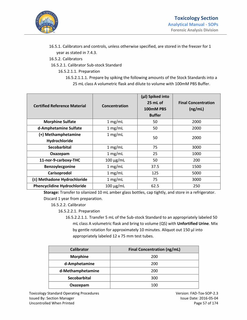
Toxicology Section Analytical Manual - SOPs
Forensic Analysis Division
Toxicology Standard Operating Procedures Version: FAD-Tox-SOP-2.3 Issued By: Section Manager Issue Date: 2016-05-04 Uncontrolled When Printed Page 57 of 174
16.5.1. Calibrators and controls, unless otherwise specified, are stored in the freezer for 1
year as stated in 7.4.3.
16.5.2. Calibrators
16.5.2.1. Calibrator Sub-stock Standard
16.5.2.1.1. Preparation
16.5.2.1.1.1. Prepare by spiking the following amounts of the Stock Standards into a
25 mL class A volumetric flask and dilute to volume with 100mM PBS Buffer.
Certified Reference Material Concentration
(µl) Spiked into
25 mL of
100mM PBS
Buffer
Final Concentration
(ng/mL)
Morphine Sulfate 1 mg/mL 50 2000
d-Amphetamine Sulfate 1 mg/mL 50 2000
(+) Methamphetamine
Hydrochloride
1 mg/mL 50 2000
Secobarbital 1 mg/mL 75 3000
Oxazepam 1 mg/mL 25 1000
11-nor-9-carboxy-THC 100 µg/mL 50 200
Benzoylecgonine 1 mg/mL 37.5 1500
Carisoprodol 1 mg/mL 125 5000
(±) Methadone Hydrochloride 1 mg/mL 75 3000
Phencyclidine Hydrochloride 100 µg/mL 62.5 250
Storage: Transfer to silanized 10 mL amber glass bottles, cap tightly, and store in a refrigerator.
Discard 1 year from preparation.
16.5.2.2. Calibrator
16.5.2.2.1. Preparation
16.5.2.2.1.1. Transfer 5 mL of the Sub-stock Standard to an appropriately labeled 50
mL class A volumetric flask and bring to volume (QS) with Unfortified Urine. Mix
by gentle rotation for approximately 10 minutes. Aliquot out 150 µl into
appropriately labeled 12 x 75 mm test tubes.
Calibrator Final Concentration (ng/mL)
Morphine 200
d-Amphetamine 200
d-Methamphetamine 200
Secobarbital 300
Oxazepam 100

Toxicology Section Analytical Manual - SOPs
Forensic Analysis Division
Toxicology Standard Operating Procedures Version: FAD-Tox-SOP-2.3 Issued By: Section Manager Issue Date: 2016-05-04 Uncontrolled When Printed Page 58 of 174
11-nor-9-carboxy-THC 20
Benzoylecgonine 150
Carisoprodol 500
Methadone 300
Phencyclidine 25
Storage: Store in freezer. Discard 1 year from preparation.
16.5.2.3. Oxycodone Sub-stock Standard 10 ng/µL
16.5.2.3.1. Preparation
16.5.2.3.1.1. Prepare by spiking 100 µL of the 1mg/mL Oxycodone in methanol into
10mL class A volumetric flask and dilute to volume (QS) with methanol.
16.5.2.3.2. Storage
16.5.2.3.2.1. Transfer to silanized 10 mL amber vials, cap tightly. Store in a freezer.
Discard 1 year from preparation.
16.5.2.4. Oxycodone Positive Calibrator - 100 ng/mL
16.5.2.4.1. Preparation
16.5.2.4.1.1. Transfer 500 µL of the Oxycodone Sub-stock Standard 10 ng/µL to an
appropriately labeled 50 mL Class A volumetric flask and bring to volume (QS)
with Unfortified Urine and mix.
16.5.2.4.1.2. Aliquot out 150 µL into appropriately labeled 12 x 75 mm test tubes.
16.5.3. Controls
16.5.3.1. Stock standards used for QC solutions are prepared from either a separate lot or a
separate weighing of the standard reference material.
16.5.3.2. Blank: Unfortified Urine - For all analytes
16.5.3.2.1. Before preparation of a new lot of working standards/controls, run a set of
three blank controls for all analytes to ensure there is no contamination. Should be
comparable to current lot.
16.5.3.2.2. Transfer 100 mL of the Unfortified Urine into 100 µL aliquots in
appropriately labeled 12 x 75 mm test tubes to be used as the Blank.
16.5.3.3. Control Sub-stock standard
16.5.3.3.1. Preparation
16.5.3.3.1.1. Prepare by spiking the following amounts of the Stock Standards into a
50 mL class A volumetric flask and dilute to volume with 100mM PBS Buffer.
Certified Reference Material* Concentration (µl) Spiked into Final Concentration

Toxicology Section Analytical Manual - SOPs
Forensic Analysis Division
Toxicology Standard Operating Procedures Version: FAD-Tox-SOP-2.3 Issued By: Section Manager Issue Date: 2016-05-04 Uncontrolled When Printed Page 59 of 174
50mL of 100mM
PBS Buffer
(ng/mL)
Morphine Sulfate 1 mg/mL 100 2000
d-Amphetamine Sulfate 1 mg/mL 100 2000
(+) Methamphetamine
Hydrochloride
1 mg/mL 100 2000
Secobarbital 1 mg/mL 150 3000
Oxazepam 1 mg/mL 50 1000
11-nor-9-carboxy-THC 100 µg/mL 100 200
Benzoylecgonine 1 mg/mL 75 1500
Carisoprodol 1 mg/mL 250 5000
(±) Methadone Hydrochloride 1 mg/mL 150 3000
Phencyclidine Hydrochloride 100 µg/mL 125 250
*Cerilliant or equivalent
Storage: Transfer to silanized 10 mL amber glass bottles, cap tightly, and store in a refrigerator. Discard
1 year from preparation.
16.5.3.4. Negative Control
16.5.3.4.1. Preparation
16.5.3.4.1.1. Transfer 2.5 mL of the Control Sub-stock Standard to an appropriately
labeled 50 mL class A volumetric flask and bring to volume (QS) with Unfortified
Urine and mix.
16.5.3.4.1.2. Aliquot out 150 µL into appropriately labeled 12 x 75 mm test tubes.
Negative Control Final Concentration (ng/mL)
Morphine 100
d-Amphetamine 100
d-Methamphetamine 100
Secobarbital 150
Oxazepam 50
11-nor-9-carboxy-THC 10
Benzoylecgonine 75
Carisoprodol 250
Methadone 150
Phencyclidine 12.5
16.5.3.5. High Control
16.5.3.5.1. Preparation

Toxicology Section Analytical Manual - SOPs
Forensic Analysis Division
Toxicology Standard Operating Procedures Version: FAD-Tox-SOP-2.3 Issued By: Section Manager Issue Date: 2016-05-04 Uncontrolled When Printed Page 60 of 174
16.5.3.5.1.1. Transfer 10 mL of the Control Sub-stock Standard to an appropriately
labeled 50 mL class A volumetric flask and bring to volume (QS) with Unfortified
Urine and mix.
16.5.3.5.1.2. Aliquot out 150 µL into appropriately labeled 12 x 75 mm test tubes.
High Control Final Concentration (ng/mL)
Morphine 400
d-Amphetamine 400
d-Methamphetamine 400
Secobarbital 600
Oxazepam 200
11-nor-9-carboxy-THC 40
Benzoylecgonine 300
Carisoprodol 1000
Methadone 600
Phencyclidine 50
16.5.3.6. Oxycodone Control Sub-stock standard 20 ng/µL
16.5.3.6.1. Preparation
16.5.3.6.1.1. Prepare by spiking 200 µL of the 1mg/mL Oxycodone in methanol into
10mL class A volumetric flask and dilute to volume (QS) with methanol.
16.5.3.6.1.2. Storage
16.5.3.6.1.2.1. Transfer to silanized 10mL amber vials, cap tightly. Store in
freezer. Discard 1 year from preparation.
16.5.3.7. Oxycodone Low Control - 50 ng/mL
16.5.3.7.1. Preparation
16.5.3.7.1.1. Transfer 125 µL of the Oxycodone Sub-stock Standard 20 ng/µL to an
appropriately labeled 50 mL Class A volumetric flask and bring to volume (QS)
with Unfortified Urine and mix.
16.5.3.7.1.2. Aliquot out 150 µL into appropriately labeled 12 x 75 mm test tubes.
16.5.3.8. Oxycodone High Control - 200 ng/mL
16.5.3.8.1. Preparation
16.5.3.8.1.1. Transfer 500 µL of the Oxycodone Sub-stock Standard 20 ng/µL to an
appropriately labeled 50 mL Class A volumetric flask and bring to volume (QS)
with Unfortified Urine and mix.
16.5.3.8.1.2. Aliquot out 150 µL into appropriately labeled 12 x 75 mm test tubes.
16.6. Equipment

Toxicology Section Analytical Manual - SOPs
Forensic Analysis Division
Toxicology Standard Operating Procedures Version: FAD-Tox-SOP-2.3 Issued By: Section Manager Issue Date: 2016-05-04 Uncontrolled When Printed Page 61 of 174
TECAN Freedom Evo 75
TECAN HydroFlex Plate Washer
TECAN Sunrise Plate Reader
Vortex mixer
Air Displacement pipettes (100-1000 µL; 20-200 µL; 2-20 µL)
Centrifuge
Sonicator
Homogenizer
16.7. Procedure for Blood and Urine
16.7.1. In each run, the following controls are included:
16.7.1.1. Blank control (in duplicate) – drug free matrix.
16.7.1.2. Calibrator (in duplicate) – fortified with target drugs at the specified threshold.
16.7.1.3. Negative QC (in duplicate) – fortified with target drugs at half the concentration
found in the positive control with the exception of Zolpidem.
16.7.1.4. High QC (in duplicate) – fortified with target drugs at two times the concentration
found in the Calibrator.
16.7.2. Calibrator Concentrations
16.7.2.1. Samples with absorbance readings BELOW the positive control are considered
PRESUMPTIVE POSITIVE. Confirmatory analysis is routinely indicated for all
presumptive positive cases.
16.7.2.2. Note: Confirmatory analysis may be requested on any sample, regardless of
preliminary screening results.
Calibrator Blood (ng/mL) Urine (ng/mL)
Morphine 20 200
d-Amphetamine 20 200
d-Methamphetamine 20 200
Secobarbital 40 300
Oxazepam 20 100
11-nor-9-carboxy-THC 10 20
Benzoylecgonine 20 150
Carisoprodol 500 500
Methadone 25 300
Phencyclidine 10 25
Zolpidem 10 ---
Oxycodone 10 100

Toxicology Section Analytical Manual - SOPs
Forensic Analysis Division
Toxicology Standard Operating Procedures Version: FAD-Tox-SOP-2.3 Issued By: Section Manager Issue Date: 2016-05-04 Uncontrolled When Printed Page 62 of 174
16.7.3. Sample Preparation
16.7.3.1. Prior to aliquoting, transfer a portion of each case specimen into a separate
transfer container. Transferring and aliquoting occurs one case specimen at a time and
the transfer container is discarded after each aliquot.
16.7.3.2. Add 900 µL of Forensic diluent (PBS buffer) to each subsample tube.
16.7.3.3. Blood 1:10 dilution with forensic diluent (phosphate buffered saline)
16.7.3.3.1. Pipette 100µL of the calibrators, controls, and unknowns into subsample
tubes and vortex.
16.7.3.3.1.1. Note: If blood samples are clotted, remove clots and decant sample
into new subsample tube. Sonication, centrifugation, or other sample
preparation steps may be necessary for degraded specimens.
16.7.3.4. Urine 1:10 dilution with forensic diluent (phosphate buffered saline)
16.7.3.4.1. Add 900 µL of Forensic diluent (PBS buffer) to each subsample tube.
16.7.3.4.2. Pipette 100µL of the calibrators, controls, and unknowns into subsample
tubes and vortex.
16.7.3.4.2.1. Note: If samples contain particulate material, centrifugation may be
performed before transfer into the subsample tube.
16.7.4. Placement of samples
16.7.4.1. Place unknowns and QCs in appropriate positions in subsample racks (see chart
below). Note: A High QC must be run in between every ten case samples and at the end
of every run).
1 2 3 4 5 6
A BLANK SAMPLE SAMPLE SAMPLE SAMPLE HIGH QC
B BLANK SAMPLE SAMPLE SAMPLE SAMPLE SAMPLE
C CAL QC SAMPLE HIGH QC SAMPLE SAMPLE SAMPLE
D CAL QC SAMPLE SAMPLE SAMPLE SAMPLE SAMPLE
E NEG QC SAMPLE SAMPLE SAMPLE SAMPLE SAMPLE
F NEG QC SAMPLE SAMPLE HIGH QC SAMPLE SAMPLE
G HIGH QC SAMPLE SAMPLE SAMPLE SAMPLE SAMPLE
H HIGH QC SAMPLE SAMPLE SAMPLE SAMPLE HIGH QC
16.7.5. To edit or create a method:
Go to the main menu
Click on “create/edit a method.”
Click “OK.”
Click “open.”

Toxicology Section Analytical Manual - SOPs
Forensic Analysis Division
Toxicology Standard Operating Procedures Version: FAD-Tox-SOP-2.3 Issued By: Section Manager Issue Date: 2016-05-04 Uncontrolled When Printed Page 63 of 174
Click on the ELISA kit test number
Click on “Endpoint measurement.”
Click on “define evaluation.” The plate map is displayed.
Click on the boxes that need to be changed.
Select the identifiers Acronym.
Click “Fill Selection.”
Click “Finish.”
Click “Save.”
16.7.6. The instrument dispenses the following volumes for blood assays
Assay Volume Blood (µL)* Volume Conjugate (µL) Volume TMB Substrate (µL) Volume Stop Solution (µL)
Morphine 10 100 100 100
d-Amphetamine 25 100 100 100
d-Methamphetamine 25 100 100 100
Secobarbital 10 100 100 100
Oxazepam 30 100 100 100
11-nor-D9 THC-COOH 50 100 100 100
Benzoylecgonine 40 100 100 100
Carisoprodol 10 100 100 100
Methadone 15 100 100 100
Phencyclidine 10 100 100 100
Zolpidem 100 100 100 100
Oxycodone 10 100 100 100
*Diluted matrix (1:10 in PBS or Forensic Diluent)
16.7.7. The instrument dispenses the following volumes for urine assays
Assay Volume Urine
(µL)*
Volume
Conjugate (µL)
Volume TMB
Substrate (µL)
Volume Stop
Solution (µL)
Morphine 10 100 100 100
d-Amphetamine 10 100 100 100
d-Methamphetamine 10 100 100 100
Secobarbital 10 100 100 100
Oxazepam 15 100 100 100
11-nor-D9 THC-COOH 50 100 100 100
Benzoylecgonine 10 100 100 100
Carisoprodol 10 100 100 100
Methadone 10 100 100 100
Phencyclidine 10 100 100 100

Toxicology Section Analytical Manual - SOPs
Forensic Analysis Division
Toxicology Standard Operating Procedures Version: FAD-Tox-SOP-2.3 Issued By: Section Manager Issue Date: 2016-05-04 Uncontrolled When Printed Page 64 of 174
Oxycodone 40 100 100 100
*Diluted matrix (1:10 in PBS or Forensic Diluent)
16.7.8. Samples incubate with the drug-enzyme conjugate for 1 hour and with the enzyme
substrate for 30 minutes.
16.8. Other Matrices
16.8.1. Appropriate sample preparation steps must be used for tissues or other alternative
matrices that may be encountered. Tissues are routinely homogenized using deionized
water (1:1) prior to analysis. Additional water may be added to achieve full
homogenization. Document the conditions for homogenization in the case folder.
Following homogenization, tissues and solids are analyzed in accordance with blood
samples.
16.8.2. Sample preparation for alternative matrices may include homogenization, filtration,
sonication, centrifugation, or dilution. Liquid-based alternative matrices are analyzed in
accordance with urine samples.
16.8.3. Document all sample preparation steps in the case folder.
16.9. Interpretation of Results
16.9.1. All samples with B/B0 (% binding) readings BELOW the calibrator (mean of duplicate
analysis) are presumptive positive.
16.9.2. Confirmatory analyses are requested on all presumptive positive samples unless
specifically directed otherwise by the submitting agency or client.
16.9.3. Results are qualitative in nature. Quantitative results are not possible due to the non-
linear binding characteristics of the assay and antibody specificity.
16.10. Quality Assurance
16.10.1. ELISA QC Logs
16.10.1.1. The ELISA operator is responsible for completing ELISA QC Logs (for blood and
urine), ELISA batch reports, and submitting data for review.
16.10.1.2. % Binding is calculated numerically as follows:
Calibrator and Controls: %Binding = (Avg. Abs. calibrator/Avg. Abs. blank control) x 100
Case Samples: %Binding = (Abs. case sample/Avg. Abs. blank control) x 100
16.10.1.3. Example:

Toxicology Section Analytical Manual - SOPs
Forensic Analysis Division
Toxicology Standard Operating Procedures Version: FAD-Tox-SOP-2.3 Issued By: Section Manager Issue Date: 2016-05-04 Uncontrolled When Printed Page 65 of 174
A calibrator produces an average absorbance reading of 0.66 (Avg. Abs. calibrator =0.66);
the average absorbance for the blank control is 2.0 (Avg. Abs. blank control =2.0). The %
Binding is 0.66/2.0) x 100 = 33%
16.10.1.4. The control charts are reviewed with the ELISA batch file. Only a copy of the
ELISA results is placed in the case folder. ELISA results are reviewed and stored in batch
files in the laboratory.
16.11. Acceptance Criteria
16.11.1. During technical review of the ELISA batch file, the following acceptance criteria
apply to all assays:
16.11.1.1. % Binding for the CALIBRATOR must be < 70%, and
16.11.1.2. % CV for the BLANK CONTROL (n=2) must be < 20%
16.11.1.3. The average absorbance and % binding values should be as follows: BLANK
CONTROL> NEGATIVE CONTROL> CALIBRATOR> HIGH CONTROL
16.11.1.4. The average absorbance of the BLANK CONTROL should be greater than 1.5 for
all assays.
16.11.2. Assays not meeting both of these criteria should be repeated.
16.12. Literature and Supporting Documentation
16.12.1. J.L. Farrell, S. Kerrigan, B.K. Logan in Recommendations for Toxicological
Investigation of drug Impaired Driving. J Forensic Sci. (2013).
16.12.2. Smith, M. Immunoassay in Principles of Forensic Toxicology, 3rd Edition. Levine B
(Ed). AACC Press, Washington DC, 2009, pp. 117-138.
16.12.3. Kerrigan, S and Phillips Jr., W.H. Comparison of ELISAs for Opiates,
Methamphetamine, Cocaine Metabolite, Benzodiazepines, Phencyclidine and
Cannabinoids in Whole Blood and Urine, Clin Chem: 47, 540-547 (2001).
16.12.4. TECAN Freedom Evo 75, Operators Manual.
16.12.5. TECAN HydroFlex Plate Washer, Operators Manual.
16.12.6. TECAN Sunrise Plate Reader, Operation Manual.

Toxicology Section Analytical Manual - SOPs
Forensic Analysis Division
Toxicology Standard Operating Procedures Version: FAD-Tox-SOP-2.3 Issued By: Section Manager Issue Date: 2016-05-04 Uncontrolled When Printed Page 66 of 174
17. Reagents for Drug Screening/Confirmation Analyses
17.1. Purpose
17.1.1. This document outlines the preparation of reagents used in drug
screening/confirmation analysis.
17.2. Safety
17.2.1. This procedure must be conducted in accordance with the HFSC Health and Safety
Manual and Quality Manual. All case specimens should be treated with universal blood
borne pathogen precautions. Appropriate personal protective equipment must be worn
during sample and reagent preparation and when handling caustic chemicals. Flammable
liquids and vapors may cause eye, skin and respiratory tract irritation. Derivatization
reagents are toxic and must be handled in a chemical safety hood or well-ventilated area.
Safety Data Sheets (SDS) are available in the laboratory.
17.3. Reagents
17.3.1. Organic solvents are HPLC grade or higher and inorganic reagents (e.g. salts, acids)
are ACS grade. Deionized water is prepared using a Millipore Direct Q UV3 water system
(or equivalent). Preparations of reagents are documented in a retrievable format.
Glacial acetic acid
Hydrochloric acid
Ammonium hydroxide
Dibasic sodium phosphate
Methylene chloride
Isopropanol
Methanol
Ethyl acetate
Deionized (DI) water [7732-18-5]
17.3.2. 1 M Acetic acid
17.3.2.1. Add 28.6 mL of concentrated glacial acetic acid to a 500 mL volumetric flask half-
filled with deionized water. Bring to volume with deionized water and mix.
17.3.2.2. Storage: Room temperature
17.3.2.3. Discard: 1 year
17.3.3. 100 mM, pH 6.0 Phosphate buffer
17.3.3.1. Add 7.0 g of dibasic sodium phosphate to a 500 mL volumetric flask and
bring to volume with deionized water while stirring. In a second 1000 mL
volumetric flask, add 12.0 g of monobasic sodium phosphate and bring to

Toxicology Section Analytical Manual - SOPs
Forensic Analysis Division
Toxicology Standard Operating Procedures Version: FAD-Tox-SOP-2.3 Issued By: Section Manager Issue Date: 2016-05-04 Uncontrolled When Printed Page 67 of 174
volume with deionized water while stirring. Adjust pH of the monobasic sodium
phosphate by adding the dibasic sodium phosphate drop-wise until the pH
reaches 6.0.
17.3.3.2. Storage: Room temperature
17.3.3.3. Discard: 6 months
17.3.4. 100 mM sodium phosphate buffer, pH 6.8 – Deconjugation Buffer
17.3.4.1. Add 7.0 g of dibasic sodium phosphate to a 500 mL volumetric flask and
bring to volume with deionized water while stirring. In a second 500 mL
volumetric flask, add 6.0 g of monobasic sodium phosphate and bring to volume
with deionized water while stirring. Mix the two solutions together until a pH of
6.8 is obtained
17.3.4.2. Storage: Refrigerated
17.3.4.3. Discard: 6 months
17.3.5. 80:20 Methylene Chloride/isopropanol
17.3.5.1. In a 500 mL volumetric flask, add 100 mL of isopropanol to 400 mL of
methylene chloride.
17.3.5.2. Storage: Room temperature
17.3.5.3. Discard: 6 months
17.3.6. 2% Ammonium hydroxide (concentrated) in 80:20 methylene
chloride/isopropanol – Elution Solvent
17.3.6.1. In a 25 mL volumetric flask or a graduated cylinder, add 0.5 mL
concentrated ammonium hydroxide. Bring to volume with 80:20 methylene
chloride/isopropanol. Mix vigorously.
17.3.6.2. Storage of Reagents: Room temperature
17.3.6.3. Discard: after each daily use
17.3.7. 2% Ammonium hydroxide (concentrated) in ethyl acetate with 1 mL of
methanol
17.3.7.1. In a 50 mL volumetric flask or a graduated cylinder, partially filled with
ethyl acetate, add 0.5 mL concentrated ammonium hydroxide. Bring volume to
25 mL with ethyl acetate. Add 1 mL of Methanol. Mix vigorously.
17.3.7.2. Storage of reagents: Room temperature
17.3.7.3. Discard: after each daily use
17.3.8. 1% Hydrochloric acid in methanol – Acidic Methanol

Toxicology Section Analytical Manual - SOPs
Forensic Analysis Division
Toxicology Standard Operating Procedures Version: FAD-Tox-SOP-2.3 Issued By: Section Manager Issue Date: 2016-05-04 Uncontrolled When Printed Page 68 of 174
17.3.8.1. Add 1 mL of concentrated hydrochloric acid into a 100 mL volumetric flask
using a volumetric pipette. Bring to volume with methanol.
17.3.8.2. Storage: Room temperature
17.3.8.3. Discard: 3 months
17.3.9. 0.1 M Hydrochloric acid
17.3.9.1. Add 8.4 mL concentrated hydrochloric acid to a 1 liter volumetric flask
containing deionized water. Bring to volume with deionized water.
17.3.9.2. Storage: Room temperature
17.3.9.3. Discard: 12 months
17.3.10. 1 M Hydrochloric acid
17.3.10.1. Add 84.6 mL concentrated hydrochloric acid to a 1 liter volumetric flask
containing deionized water. Bring to volume with deionized water.
17.3.10.2. Storage: Room temperature
17.3.10.3. Discard: 12 months

Toxicology Section Analytical Manual - SOPs
Forensic Analysis Division
Toxicology Standard Operating Procedures Version: FAD-Tox-SOP-2.3 Issued By: Section Manager Issue Date: 2016-05-04 Uncontrolled When Printed Page 69 of 174
18. Evaluation of Results from Gas Chromatography-Mass Spectroscopy
18.1. Purpose
18.1.1. The GC-MS is composed of two major building blocks: the gas chromatograph (GC)
and the mass spectrometer (MS). The GC utilizes a capillary column. The dimensions
(length, diameter, film thickness) and the stationary phase properties of the column
facilitate the separation of complex mixtures. Different compounds in a sample and their
varied relative affinity for the stationary phase of the column will promote separation of
these compounds as the sample travels through the length of the column. The
compounds are retained by the column and then elute from the column at different
times (retention time). The mass spectrometer is the detector portion of the instrument.
As the compounds elute off the column and enter the MS, they are ionized, fragmented,
separated and detected based upon their mass-to-charge ratio (m/z). The results
obtained from GC-MS analysis must be evaluated to determine the acceptability of the
results. This applies to both quantitative and qualitative results.
18.2. Scope
18.2.1. This policy applies to all GC-MS analyses.
18.3. Quality Controls
18.3.1. Positive and negative QCs must be included in each run. Each run must end with a
negative and positive control. For quantitative analysis 5% of the run must consist of
positive control samples. For qualitative analysis each batch must contain a negative
(drug-free) extract and an extracted drug standard.
18.3.2. For quantitative analysis, an in-house or external QC must be included. Record the
calculated concentrations in the appropriate QC Log.
18.3.3. Acceptance criteria of quality controls are that they must be ± 20 of the targeted
value and the retention time must be ± 2% of the average of the calibrators.
18.3.4. For SIM analyses, acceptance criteria for ion ratios are ± 20% or two standard
deviations of the average of the calibrators used in the calibration response curve.
However, it is recognized that some ions ratios are concentration-dependent and that
comparison to a calibrator of similar concentration may be necessary rather than the
average ratio for the curve. This exception shall be documented in case record or batch
summary raw data file.
18.4. Blood And Urine
18.4.1. Blood is the preferred specimen for impairment cases as it reveals what compounds
may be affecting the individual and the concentration of the compounds may be
associated with impairment. Urine is an acceptable specimen but is typically not
quantitated due to variations in volume.

Toxicology Section Analytical Manual - SOPs
Forensic Analysis Division
Toxicology Standard Operating Procedures Version: FAD-Tox-SOP-2.3 Issued By: Section Manager Issue Date: 2016-05-04 Uncontrolled When Printed Page 70 of 174
18.5. Tissues And Other Matrices
18.5.1. Appropriate sample preparation steps must be used for tissues or alternative
matrices that may be encountered. Tissues are routinely homogenized using deionized
water (1:1) prior to analysis. Additional water may be added to achieve full
homogenization. Document the conditions for homogenization in the case folder.
Following homogenization, tissues and other solids are analyzed in accordance with
blood samples.
18.5.2. Sample preparation for alternative matrices may include homogenization, filtration,
sonication, centrifugation, and/or dilution. Liquid-based alternative matrices are
analyzed in accordance with urine samples.
18.5.3. All sample preparation steps, including appropriate matrix matched controls must be
documented in the case record.
18.6. Qualitative Analysis
18.6.1. All mass spectral libraries and/or reference spectra must be approved by the
Technical Leader or Toxicology Manager.
18.6.2. The following acceptance criteria may be used as guidelines, but not as absolutes, to
identify a substance through its mass spectrum:
18.6.2.1. Usually the base peak will be the same and isotope clusters will contain the same
major ions and relative abundances.
18.6.2.2. When compared to a reference spectrum, the spectra must contain strong
similarities.
18.6.2.3. Any differences between the reference spectrum and the unknown spectrum must
be carefully evaluated for acceptability.
18.6.3. The following criteria must be met for qualitative identification of a compound:
18.6.3.1. Retention times (RT) and/or relative retention times (RRT) must be within ± 2% of
the target.
18.6.3.2. Case mass spectrum must be of comparable quality to a reference spectrum in
mass assignment and intensity.
18.6.3.3. Any extraneous ions of significant intensity that are not found in the reference
spectrum must be demonstrated as being from background or known interference by
using an extracted ion chromatogram.
18.6.4. Qualitative identification is determined using characteristic retention time and mass
spectral characteristics. Deuterated internal standards in each sample are used to
identify the correct retention time of the drug. For SIM, all three characteristic ions must
be present. Ion intensity and ratios should be taken into consideration. Ion ratios should
be within ± 20% or two standard deviations of the average ratio from all calibrators used
in the calibration response curve. However, it is recognized that some ion ratios are
concentration dependent and that comparison to a calibrator of a similar concentration

Toxicology Section Analytical Manual - SOPs
Forensic Analysis Division
Toxicology Standard Operating Procedures Version: FAD-Tox-SOP-2.3 Issued By: Section Manager Issue Date: 2016-05-04 Uncontrolled When Printed Page 71 of 174
may be necessary rather than the average ratio of the curve. This exception shall be
documented in case notes or batch summary raw data file. These are evaluated on a case
by case basis and are subjected to technical and administrative review.
18.7. Quantitative Analysis
18.7.1. Qualitative identification must be met from section 18.6.4.
18.7.2. Concentration of a drug is determined using linear regression analysis. At least 4
calibrators and a blank blood sample must be used for each quantitation. R2 values of at
least 0.99 should be attained following linear regression analysis. If more than three
calibrators are included in the run, two may be excluded if the response factor is not
within ± 20% of the remaining calibrators or the ion ratios are unacceptable. The
response factor is defined as follows: (Peak Area of the Internal Standard/Peak Area of
the Calibrator) x Calibrator Concentration. If the response factor is within range, it may
be permissible to exclude the point from the calibration provided that the independent
control is still within acceptable limits and the reviewer is in agreement. The calculated
concentration of a calibrator must be within ± 20% of the expected value, with the
exception of the LOQ, which must be within ±25% of the expected value. When
determining the acceptability of a calibrator, the concentration range is taken into
account. Acceptable ion ratios must be met as described in “In-Process Calibration and
Quality Control for Drug Screening/Confirmation Testing,” section 8.4.10 in order for a
calibrator to be included in the calibration response curve. If a calibrator needs to be
excluded from the curve, the action should be described and justified in the batch file.
18.7.3. Quantitative values above or below the highest and lowest calibrators should be
reported as greater than or less than the appropriate calibrator concentration. Dilution of
a specimen may be necessary for samples that contain an elevated concentration of
drug. Any changes in sample volume that deviate from the technical procedure must be
documented in the case folder.
18.7.4. For quantitative reporting, the calculated value of the in-house or external QC should
be within ± 20% of the target concentration. Alternatively, quantitative results may be
reported if the QC is within three standard deviations of the established mean.
18.8. Reinjection Guidelines
18.8.1. Reinjections can be made up to 48 hours after the tune for that particular analytical
run, per Agilent’s acceptance criteria, unless otherwise stated in specific SOP.
18.8.2. Reinjections must be run with negative control and bracketed by both a low and high
quality control. The quality control must meet the acceptance criteria in order for the
reinjections to be reported.
18.9. Toxicology Reporting Guidelines

Toxicology Section Analytical Manual - SOPs
Forensic Analysis Division
Toxicology Standard Operating Procedures Version: FAD-Tox-SOP-2.3 Issued By: Section Manager Issue Date: 2016-05-04 Uncontrolled When Printed Page 72 of 174
18.9.1. Qualitative vs. Quantitative Results
18.9.1.1. Drugs in urine are reported qualitatively unless specifically noted otherwise in the
standard operating procedure. Quantitative drug determinations in urine cannot be
interpreted pharmacologically due to differences in total urine volume and elimination
rate.
18.9.1.2. Unless specifically requested by the client, it is the policy of the laboratory not to
report certain substances that are routinely encountered in toxicological specimens
including caffeine, nicotine, and their metabolites and byproducts.
18.9.1.3. Blood is the preferred specimen for quantitative drug determination. Blood drug
results may be reported either qualitatively or quantitatively. The laboratory reserves
the right to determine whether qualitative or quantitative results are reported as
determined by:
Class of drug
Case type/offense type
Other toxicological findings
Data and/or specimen quality
Quantity of sample
Technical abilities of the laboratory
18.9.2. Screening and Confirmatory Tests
18.9.2.1. It is the policy of the laboratory not to report positive immunoassay screening
results in the absence of confirmation. All presumptive positive drug test results by
immunoassay are subject to confirmatory analysis using GC-MS or equivalent, provided
there is sufficient specimen.
18.9.2.2. Preliminary screening tests such as immunoassay have limited scope and utility.
They are predominantly used to direct the scope of other analytical testing.
Immunoassay results are not reported unless they are negative and no additional tests
are performed. In those instances, the report clearly states the scope of testing was
limited to immunoassay and lists the drug classes and appropriate cutoffs.

Toxicology Section Analytical Manual - SOPs
Forensic Analysis Division
Toxicology Standard Operating Procedures Version: FAD-Tox-SOP-2.3 Issued By: Section Manager Issue Date: 2016-05-04 Uncontrolled When Printed Page 73 of 174
19. Alprazolam Confirmation by Gas Chromatography-Mass Spectrometry
19.1. Purpose
19.1.1. A targeted analysis is performed for confirmatory analysis of alprazolam by solid
phase extraction (SPE) and gas chromatography-mass spectrometry (GC-MS).
Quantitative and/or qualitative analysis are performed using deuterated internal
standard and selective ion monitoring (SIM) in electron ionization (EI) mode.
19.2. Scope
19.2.1. This procedure should be used for confirmatory analysis of alprazolam from
toxicology specimens, typically blood. This procedure is recommended if quantitative
analysis is desired. Qualitative results may also be reported. For routine full scan
qualitative analysis, refer to Basic, Acidic and Neutral Drugs by GC-MS.
19.3. Reagents And Standards
19.3.1. Alprazolam Working Standard for Calibrators and In-house Controls
19.3.1.1. 10 ng/µL Alprazolam Working Standard: Add 100 µL of alprazolam (1.0 mg/mL) to
a 10 mL volumetric flask and bring to volume with methanol. Store refrigerated (3
month expiration).
19.3.1.2. 1 ng/µL Alprazolam Working Standard: Add 1 mL of 10 ng/µL working standard to
a 10 mL volumetric flask and bring to volume with methanol. Store refrigerated (3
month expiration).
19.3.2. Alprazolam-D5 Internal Standard Solution
19.3.2.1. 10 ng/µL Alprazolam – D5 STOCK Internal Standard: Add 1 mL of Alprazolam-D5
(100 µg/mL) to a 10 mL volumetric flask and bring to volume with methanol. Store
refrigerated (12 month expiration).
19.3.2.2. 1 ng/µL Alprazolam – D5 WORKING Internal Standard: Add 1 mL of Alprazolam-D5
(10 ng/µL) to a 10 mL volumetric flask and bring to volume with methanol. Store
refrigerated (6 month expiration).
19.3.3. 1M Acetic Acid
19.3.4. 100 mM, pH 6.0 Phosphate Buffer
19.3.5. Methylene Chloride/Isopropanol (80:20)
19.3.6. Elution Solvent: 2% ammonium hydroxide (concentrated) in 80:20 methylene
chloride/isopropanol
19.4. Equipment And Materials
Air displacement pipettes (100-1000 µL; 20-200 µL; 2-20 µL)
Cerex PolyChrom Clin II SPE columns

Toxicology Section Analytical Manual - SOPs
Forensic Analysis Division
Toxicology Standard Operating Procedures Version: FAD-Tox-SOP-2.3 Issued By: Section Manager Issue Date: 2016-05-04 Uncontrolled When Printed Page 74 of 174
Vacuum manifold with disposable Teflon inserts or positive pressure SPE manifold
Pan balance
pH meter
Evaporator
Vortex mixer
Centrifuge
Tissue homogenizer
Sonicator
Heating block
Vacuum pump or house vacuum
Agilent 7890 GC 5975 MSD
Stir plate and stir bars
19.5. Instrumental Parameters
19.5.1. Capillary Column: 30 m HP-5MS Agilent J&W GC Column (or equivalent), 0.250 mm id
X 0.25 µm film thickness. The flow rate is 1.2 mL/min with an injection volume of 2 µL in
split mode (10:1).
19.5.2. GC-MS: Agilent 7890A
Initial Temperature: 140 °C
Hold for 0.5 minutes
30 °C/min to 290 °C
Hold for 2.5 minutes
50 °C/min to 310 °C
Hold for 2.6 minutes
Total Run Time: 11 minutes
Injector Temperature: 250 °C
Interface Temperature: 280 °C
MS Quads: 150 °C
MS Source: 230 °C
19.5.3. Wash solvents for autosampler: Methanol and ethyl acetate are used as the wash
solvents. A minimum of 6 pre and 6 post rinses are performed. Each rinse cycle consists
of 3 methanol rinses followed by 3 ethyl acetate rinses.
19.6. Extraction Procedure
1.1.1. Blood, Urine and Other Matrices:
19.6.1. In a round bottom glass culture tube, add 1 mL of specimen and 100 µL Working
Internal Standard Solution (1 ng/µL) while vortex mixing (total concentration = 100
Toxicology Section Analytical Manual - SOPs
Forensic Analysis Division
Toxicology Standard Operating Procedures Version: FAD-Tox-SOP-2.3 Issued By: Section Manager Issue Date: 2016-05-04 Uncontrolled When Printed Page 75 of 174
ng/mL). NOTE IF THE SAMPLE IS KNOWN TO CONTAIN A HIGH CONCENTRATION OF
DRUG, ALIQUOT A SMALLER VOLUME, ADD BUFFER AS THE DILUENT AND DOCUMENT IN
THE CASE RECORD.
19.6.2. Fortify all controls and calibrators while vortex mixing using the appropriate amount
of working standard. Refer to the fortification guide for guidance on preparation of
calibrators. CONTROLS AND CALIBRATORS MUST COME FROM INDEPENDENT STOCK
SOLUTIONS. A minimum of one negative and one positive control must be included in
each run for qualitative analysis. For quantitative analysis, a minimum of one
quantitative QC must be included in each run. An in-house whole blood QC of 100 ng/mL
is recommended. Enter results into the appropriate QC Log after each run. Use
appropriate external controls (e.g., UTAK Whole Blood Controls) when available.
19.6.3. Add 2 mL of 100 mM, pH 6.0 sodium phosphate buffer to each tube. Vortex.
Sonication and/or centrifugation may be used if necessary. Document any additional
sample pretreatment in the case folder.
19.6.4. Place collection tubes or reservoir (for waste) into vacuum manifold rack or positive
pressure apparatus.
19.6.5. Centrifuge samples for at least 10 minutes at 4000 rpm.
19.6.6. Pour sample onto Polychrom Clin II columns. Use sufficient vacuum/pressure to draw
samples through the columns.
19.6.7. Wash column with 1 mL deionized water.
19.6.8. Remove waste tubes/reservoir and replace with fresh waste tubes/reservoir.
19.6.9. Wash column with 1 mL 1 M acetic acid.
19.6.10. Dry column on full vacuum/pressure for 5 minutes.
19.6.11. Wash column with 1 mL hexane.
19.6.12. Place conical collection tubes into the SPE manifold. If the Teflon inserts (vacuum
manifold only) appear dirty, replace them.
19.6.13. Elute drug with 1 mL ethyl acetate.
19.6.14. Turn on vacuum or apply positive pressure for a few seconds to ensure all elution
solvent has drained from the column.
19.6.15. Remove conical collection tubes and replace with waste tubes/reservoir.
19.6.16. Wash column with 1 mL methanol.
19.6.17. Dry column on full vacuum/pressure for 5 minutes.
19.6.18. Replace waste tubes/reservoir with conical collection tubes containing the
acidic/neutral fraction. If the Teflon inserts (vacuum manifold only) appear dirty, replace
them.
19.6.19. Elute drug with 1 mL elution solvent (2% ammonium hydroxide in methylene
chloride/isopropyl alcohol 80:20). ELUTION SOLVENT MUST BE MADE FRESH.
19.6.20. Turn on vacuum or apply positive pressure for a few seconds to ensure all elution
solvent has drained from the column.

Toxicology Section Analytical Manual - SOPs
Forensic Analysis Division
Toxicology Standard Operating Procedures Version: FAD-Tox-SOP-2.3 Issued By: Section Manager Issue Date: 2016-05-04 Uncontrolled When Printed Page 76 of 174
19.6.21. Evaporate the combined basic and acidic/neutral fractions to dryness under nitrogen
for approximately 10 minutes at 50-60°C. Reconstitute in 30 µL ethyl acetate, vortex, and
transfer to autosampler vials.
19.6.22. Inject 2 µL onto the GC-MS using ALPRAZOLAM.M.
19.7. Instrumental Analysis
19.7.1. Ensure that the autotune and testmix have been completed.
Volume of
Blood (mL)
Target Concentration
(ng/mL)
Working Standard
Concentration (ng/µL = µg/mL)
Volume
Added (µL)
1 20 1 20
1 50 1 50
1 100 1 100
1 250 10 25
1 500 10 50
19.8. Interpretation Of Results
19.8.1. Assay performance including but not limited to precision, accuracy, limits of
detection, quantitation, and linearity are summarized in the validation documentation.
19.8.2. The limit of detection in whole blood is 10 ng/mL and the limit of quantitation is 20
ng/mL.
19.9. Fortification Guide
Volume of
Blood (mL)
Target Concentration
(ng/mL)
Working Standard
Concentration (ng/µL = µg/mL)
Volume
Added (µL)
1 20 1 20
1 50 1 50
1 100 1 100
1 250 10 25
1 500 10 50
19.10. Literature And Supporting Documentation
19.10.1. Cairns, Eric R., et al. Quantitative Analysis of Alprazolam and Triazolam in
Hemolysed Whole Blood and Liver Digest by GC-MS/NICI with Deuterated Internal
Standards. J Anal Toxicol 1994 Jan-Feb; Vol. 18 (1), pp. 1-6.
19.10.2. Tiscione, Nicholas B., et al. Quantitation of Benzodiazepines in Whole Blood by
Electron Impact-Gas Chromatography-Mass Spectrometry. J Anal Toxicol 2008 Oct.; Vol.
32 (8), pp. 644-652.

Toxicology Section Analytical Manual - SOPs
Forensic Analysis Division
Toxicology Standard Operating Procedures Version: FAD-Tox-SOP-2.3 Issued By: Section Manager Issue Date: 2016-05-04 Uncontrolled When Printed Page 77 of 174
19.10.3. Levine, Barry. Central Nervous System Depressants in Principles of Forensic
Toxicology, 2nd Ed., Levine, Barry, Ed. AACC Press, Washington DC, pp. 173-186 (2003).
19.10.4. Baselt, Randall C. Disposition of Toxic Drugs and Chemicals in Man, 7th Ed.,
Biomedical Publications, Foster City, CA, p 35-36 (2004).
19.10.5. Method file: ALPRAZOLAM.M.

Toxicology Section Analytical Manual - SOPs
Forensic Analysis Division
Toxicology Standard Operating Procedures Version: FAD-Tox-SOP-2.3 Issued By: Section Manager Issue Date: 2016-05-04 Uncontrolled When Printed Page 78 of 174
20. Basic, Acidic and Neutral Drugs by Gas Chromatography-Mass Spectrometry
20.1. Purpose
20.1.1. Basic, acidic, and neutral drugs are extracted from the matrix using Cerex Polychrom
Clin II solid phase extraction (SPE) columns and gas chromatography-mass spectrometry
(GC-MS) analysis in electron ionization (EI) mode. This procedure is used to identify a
wide range of basic, acidic, and neutral drugs. Mepivacaine and pentobarbital-d5 are
used as internal standards.
20.2. Scope
20.2.1. This procedure functions as an initial screen as well as a qualitative confirmation for
multiple drugs of interest in blood and urine specimens.
20.2.2. This procedure may be suitable for the qualitative confirmation of benzodiazepines,
barbiturates, anticonvulsants, antihistamines, tricyclic antidepressants, non-tricyclic
antidepressants (e.g., selective serotonin reuptake inhibitors (SSRIs) and related
compounds), muscle relaxants, narcotic analgesics, dissociative anesthetics, and other
abused drugs. Other targeted procedures are available for quantitative analysis of
several basic, acidic, and neutral drugs.
20.3. Standards and Solutions
20.3.1. Basic Drug Cut-off Calibrator: a methanolic solution of commonly encountered basic
drugs. Refer to the Reagent QC Log for the composition and concentration of the current
basic drug mixture.
20.3.2. Basic Drug Quality Control: a methanolic solution of commonly encountered basic
drugs. Refer to the Reagent QC Log for composition and concentration of the current
basic drug mixture.
20.3.3. Basic Internal Standard: a methanolic solution of mepivacaine at 10 µg/mL.
20.3.4. Acidic-Neutral (AN) Drug Cut-off Calibrator: a methanolic solution of commonly
encountered acidic and neutral drugs. Refer to the Reagent QC Log for the composition
and concentration of the current AN drug mixture.
20.3.5. Acidic-Neutral (AN) Drug Quality Control: a methanolic solution of commonly
encountered acidic and neutral drugs. Refer to the Reagent QC Log for composition and
concentration of the current AN drug mixture.
20.3.6. Acid-Neutral (AN) Internal Standard: a methanolic solution of pentobarbital-D5 at 10
µg/mL.
20.3.7. 1 mM Acetic acid
20.3.8. 100 mM, pH 6.0 Phosphate buffer
20.3.9. Elution Solvent: 2% ammonium hydroxide (concentrated) in 80:20 methylene
chloride/isopropanol
20.3.10. Acidic Methanol (1 % HCl in methanol)

Toxicology Section Analytical Manual - SOPs
Forensic Analysis Division
Toxicology Standard Operating Procedures Version: FAD-Tox-SOP-2.3 Issued By: Section Manager Issue Date: 2016-05-04 Uncontrolled When Printed Page 79 of 174
20.4. Equipment and Materials
Air displacement pipettes (100-1000 µL; 20-200 µL)
Eppendorf repeater pipette
Cerex Poly Chrom II SPE columns
Vacuum manifold with disposable Teflon inserts or positive pressure SPE manifold
Analytical balance
pH meter
Evaporator
Vortex mixer
Centrifuge
Tissue homogenizer
Sonicator
Agilent 7890 GC 5975 MSD
20.5. Instrumental Parameters
20.5.1. Capillary Column: 30m DB-5MS Agilent J&W Column (or equivalent), 0.25mm id X
0.25µm film thickness. The flow rate is 1.38 mL/min with an injection volume of 2 µL in
pulsed split mode (4:1).
20.5.2. GC-MS: Agilent 7890A
Initial Temperature:
110°C hold for 0.5 minutes
20°C/min to 180°C for 2.5 minutes
5°C/min to 230°C for 0.1 minutes
25°C/min to 320°C for 4.5 minutes
Total Run Time: 24.7 minutes
Injector Temperature: 250°C
Interface Temperature: 280°C
MS Quads: 150°C
MS Source: 230°C
The above instrument conditions may be modified to adjust or improve separation and sensitivity
after appropriate validation.
20.5.3. Wash Solvents for the autosampler: Methanol and ethyl acetate are used as the wash
solvents. A minimum of 6 pre and 6 post injection rinses are performed. Each rinse cycle
consists of 3 methanol rinses followed by 3 ethyl acetate rinses.
20.6. Basic Extraction Procedure
20.6.1. Allow all biological specimens to come to room temperature before starting the
procedure.
20.6.2. Label all round bottom screw cap tubes accordingly.

Toxicology Section Analytical Manual - SOPs
Forensic Analysis Division
Toxicology Standard Operating Procedures Version: FAD-Tox-SOP-2.3 Issued By: Section Manager Issue Date: 2016-05-04 Uncontrolled When Printed Page 80 of 174
20.6.3. Add corresponding drug standard to cut-off and positive controls then dry down at
approximately 50°C under nitrogen at 20 psi for 1 minute.
20.6.4. Pipet 2 mL of drug-free blood/urine for matrix blank/negative control/positive
controls and case samples into the appropriate labeled tubes.
20.6.5. Using the repeater pipet, add 100 µL of internal standard to each sample. Vortex.
20.6.6. Add 4 mL of 100 mM sodium phosphate buffer, pH 6. Vortex.
20.6.7. Centrifuge tubes at approximately 4000 rpm for 10 minutes.
20.6.8. Place labeled SPE cartridges in the extraction manifold. Throughout the SPE, it is
important not to permit SPE sorbent to dry, unless specified.
20.6.8.1. Pour samples into appropriate SPE columns. Aspirate slowly so that the sample
takes at least 2 minutes to pass through the column.
20.6.8.2. Add 1 mL of deionized water to each column and aspirate.
20.6.8.3. Add 1 mL of 1M acetic acid to each column and aspirate. Dry columns under full
pressure for 5 minutes.
20.6.8.4. Add 1 mL of methanol to each column and aspirate. Dry columns under full
pressure for 5 minutes.
20.6.8.5. Elute basic drugs by adding 1 mL of methylene chloride: isopropyl alcohol (80:20)
with 2% ammonium hydroxide. Prepare fresh daily.
20.6.9. Add 30 µL of acidic methanol to all elution tubes.
20.6.10. Evaporate eluates to dryness at approximately 50°C under nitrogen at 20 psi for 7-9
minutes.
20.6.11. Reconstitute in 30 µL of ethyl acetate using the repeater pipet, vortex, and transfer
to autosampler vials.
20.6.12. Inject 2 µL onto the GC-MS using BANSCREEN.M. Data Analysis method
BASICDRUG.M
20.7. Acidic-Neutral Extraction Procedure
20.7.1. Allow all biological specimens to come to room temperature before starting the
procedure.
20.7.2. Label all round bottom screw cap tubes accordingly.
20.7.3. Add corresponding drug standard to cut-off and positive controls then dry down at
approximately 50°C under nitrogen at 20 psi for 1 minute.
20.7.4. Pipet 500 µL of drug-free blood/urine for matrix blank/negative control/positive
controls and case samples into the appropriate labeled tubes.
20.7.5. Using the repeater pipet add 50 µL of internal standard. Vortex
20.7.6. Add 4 mL of 100 mM sodium phosphate buffer, pH 6. Vortex
20.7.7. Centrifuge tubes at approximately 4000 rpm for 10 minutes
20.7.8. Place labeled SPE cartridges in the extraction manifold. Throughout the SPE, it is
important not to permit SPE sorbent to dry, unless specified.
20.7.8.1. Pour samples into appropriate SPE columns. Aspirate slowly so that the sample
takes at least 2 minutes to pass through the column.
20.7.8.2. Add 1mL of deionized water to each column and aspirate

Toxicology Section Analytical Manual - SOPs
Forensic Analysis Division
Toxicology Standard Operating Procedures Version: FAD-Tox-SOP-2.3 Issued By: Section Manager Issue Date: 2016-05-04 Uncontrolled When Printed Page 81 of 174
20.7.8.3. Add 1mL of 1M acetic acid to each column and aspirate. Dry columns under full
pressure for 5 minutes.
20.7.8.4. Add 1mL of hexane to each column and aspirate. Dry columns at 20 psi pressure
for 2 minutes.
20.7.8.5. Elute acidic and neutral drugs by adding 1 mL of ethyl acetate.
20.7.9. Add 30 µL of acidic methanol to all elution tubes.
20.7.10. Evaporate eluates to dryness at approximately 50°C under nitrogen at 20 psi for 5-7
minutes.
20.7.11. Reconstitute in 30 µL of ethyl acetate using the repeater pipet, vortex, and transfer
to autosampler vials.
20.7.12. Inject 2 µL onto the GC-MS using BANSCREEN.M. Data analysis method
ACIDICDRUGS.M
20.8. Instrumental Analysis
20.8.1. Ensure that the tune verification and daily QC have been completed.
20.8.2. Full-Scan acquisition: BANSCREEN.M
20.9. Sequence Table
20.9.1. Every screen batch must contain a matrix blank, negative control, and 10% positive
controls of applicable matrix.
20.9.2. Every confirmation batch must contain a matrix blank, negative control, cut-off
calibrator, and 10% positive controls of applicable matrix.
Ex; Blood and Urine Confirmation
Blood Matrix Blank
Negative Blood
Negative Urine
Cut-off Blood
Positive Blood
10 Blood Case Samples
Positive Blood
10 Blood Case Samples
Positive Blood
Positive Urine
10 Urine Case Samples
Positive Urine
20.10. Interpretation of Results Using Deconvolution Reporting Software (DRS)
20.10.1. The following criteria will be used to consider a positive finding without doing
additional library searches.
20.10.1.1. The match factor should be within the range of 60 to 100. Typically, any
component having a match factor greater than 80 may be considered positive,

Toxicology Section Analytical Manual - SOPs
Forensic Analysis Division
Toxicology Standard Operating Procedures Version: FAD-Tox-SOP-2.3 Issued By: Section Manager Issue Date: 2016-05-04 Uncontrolled When Printed Page 82 of 174
depending on the abundance. Components with a match factor less than 80 but greater
than 60 may also be considered positive but require careful scrutiny of the data to
ensure that retention times and peak shape are acceptable, and unique ions are
present.
20.10.1.2. Retention time has already been factored into the deconvolution match factor.
Components with good spectral matches but a retention time not matching the
databases are automatically penalized according to difference in expected and actual
retention times. There may be occasions where retention time shifts occur due to the
presence of overloading constituents. These instances may require alternate means of
peak identification (i.e., screener or library searching).
20.10.1.3. Additional confirmatory data can be generated within the AMDIS software to
display ion overlays and the ability to search unidentified deconvolution spectra using
the NIST library.
20.10.1.4. When determining the presence or absence of a drug, the following table should
be used as a guideline.
Determining Factors Positive None Detected
Response
Unambiguous/Solid.
Approximately more than 3
times signal/noise ratio.
Less than approximately 3
times the signal/noise ratio.
Peak Shape Defined Peak Shape
No symmetrical peak shape
or almost down to the
baseline
Spectra
Most of the characteristic
ions are present, the most
abundant ones, at the correct
ratio.
Many of the characteristic
ions are missing, including
the ones with a significant
relative abundance and/or
with the correct ion ratio.
DRS Match From 60-100 Less than 80
Chemstation Quantification
Must be greater than or equal
to the concentration of the
cut-off and acceptable
positive control must
bracketed the sample.
The concentration is less
than the cut-off and/or the
sample is not bracketed by
acceptable positive controls.
20.11. Run Analysis Procedure
20.11.1. Screen Data Analysis
20.11.1.1. QC Review
20.11.1.2. General review of the Batch (to be done at the end of the run)
Toxicology Section Analytical Manual - SOPs
Forensic Analysis Division
Toxicology Standard Operating Procedures Version: FAD-Tox-SOP-2.3 Issued By: Section Manager Issue Date: 2016-05-04 Uncontrolled When Printed Page 83 of 174
20.11.1.2.1. Examine the MS chromatogram of the matrix blank for significant peaks
that are not considered normal. If unusual peaks are found, identify on the MS. If
unusual findings are observed, review data with supervisor for decision on
acceptability.
20.11.1.2.2. Review chromatography of positive and negative controls throughout the
batch by the MS for unusual peak shape, tailing, fronting, splitting, or interferences.
Questionable performance must be reviewed with the supervisor for acceptability.
20.11.1.2.3. Examine the entire run to assure that samples are in proper order and the
analysis was performed successfully (e.g., no baseline injections, no baseline
problems, etc.).
20.11.1.3. Batch Review of Controls
20.11.1.3.1. For screen analysis, the compounds contained in these controls will be
identified using DRS.
20.11.1.3.2. All drugs in positive controls must be detected by DRS to be acceptable.
20.11.1.3.3. All blanks should be negative; if a blank is positive, it should be evaluated
for carry-over to ensure the results of case samples are not compromised.
20.11.1.4. Case Sample review
20.11.1.4.1. Case samples should be reviewed in order they were injected to allow for
monitoring of performance over the course of the run.
20.11.1.4.2. 2/3 of positive controls must pass to report negative case samples.
20.11.1.4.3. Internal standards in all case samples must be detected by DRS.
20.11.1.4.4. Case samples with a DRS match greater than 60 will be sent to
confirmation.
20.11.1.4.5. If DRS identifies a compound not presently in the current drug mixtures at a
significant concentration, the case sample will be evaluated to see if outsourcing is
necessary for confirmation.
20.11.2. Qualitative Confirmation Data Analysis
20.11.2.1. QC Review
20.11.2.2. General review of the Batch (to be done at the end of the run)
20.11.2.2.1. Examine the MS chromatogram of the matrix blank for significant peaks
that are not considered normal. If unusual peaks are found, identify on the MS. If
unusual findings are observed, review data with supervisor for decision on
acceptability.
20.11.2.2.2. Review chromatography of positive and negative controls throughout the
batch by the MS for unusual peak shape, tailing, fronting, splitting, or interferences.
Questionable performance must be reviewed with the supervisor for acceptability.
20.11.2.2.3. Examine the entire run to assure that samples are in proper order and the
analysis was performed successfully (e.g., no baseline injections, no baseline
problems, etc.)
20.11.2.2.4. Evaluate run recovery by comparing IS recovery of case samples to IS
recovery of the cut-off calibrator and controls.
20.11.3. Batch Review of Controls

Toxicology Section Analytical Manual - SOPs
Forensic Analysis Division
Toxicology Standard Operating Procedures Version: FAD-Tox-SOP-2.3 Issued By: Section Manager Issue Date: 2016-05-04 Uncontrolled When Printed Page 84 of 174
20.11.3.1. For qualitative-only analysis, the compounds contained in these controls will be
identified using DRS and Chemstation.
20.11.3.2. All drugs in positive controls must be present and above the cut-off
concentration.
20.11.3.3. All blanks should be negative; if a blank is positive it should be evaluated for
carry-over to ensure the results of case samples are not compromised.
20.11.4. Case Sample review
20.11.4.1. Case samples should be reviewed in order they were injected to allow for
monitoring of performance over the course of the run.
20.11.4.2. IS response must be above 50% of the average of the cut-off calibrator and all
controls and no higher than 150% to report qualitative, confirmed results.
20.11.4.3. Case samples must be bracketed by acceptable positive controls.
20.11.4.4. Batch outcomes not described in 11.1 and 11.2 will be evaluated on a case by
case basis and acceptability will be determined by a Supervisor.
20.12. Literature and Supporting Documentation
20.12.1. Chen, Xiao-Hua, et al. Isolation of Acidic, Neutral, and Basic Drugs from Whole Blood
Using A Single Mixed-Mode Solid-Phase Extraction Column. J Anal Toxicol 1992 Nov-Dec;
Vol. 16 (6), pp. 351-355.
20.12.2. Method File: BAN.M

Toxicology Section Analytical Manual - SOPs
Forensic Analysis Division
Toxicology Standard Operating Procedures Version: FAD-Tox-SOP-2.3 Issued By: Section Manager Issue Date: 2016-05-04 Uncontrolled When Printed Page 85 of 174
21. Benzodiazepine Confirmation by Gas Chromatography-Mass Spectrometry
21.1. Principle
21.1.1. A targeted analysis is performed for desalkylflurazepam, flurazepam, nordiazepam,
oxazepam, diazepam, lorazepam, temazepam, clonazepam, and alpha-
hydroxyalprazolam. Drugs are isolated from the matrix using liquid-liquid extraction
(LLE). Trimethylsilyl derivatives of each drug and deuterated internal standards are
prepared and analyzed using GC-MS, selective ion monitoring (SIM), and electron (EI)
ionization.
21.2. Scope
21.2.1. Confirmatory analysis of target analytes from toxicology specimens including but not
limited to blood and urine. Urine confirmations are reported only qualitatively.
21.3. Reagents and Standards
21.3.1. BENZO Working Standards for Calibrators and In-house Controls
21.3.1.1. Two working standards are routinely used. Each contains desalkylflurazepam,
flurazepam, nordiazepam, oxazepam, diazepam, lorazepam, temazepam, clonazepam,
and alpha-hydroxyalprazolam at either 10 ng/µL or 1 ng/µL. Additional working
standards may be prepared as needed.
21.3.1.2. Preparation of 10 ng/µL BENZO Working Standard: Add 1000 µL of each drug
standard (0.1 mg/mL) to a 10mL volumetric flask and bring to volume with methanol.
21.3.1.3. Preparation of 1 ng/µL Working Standard: Add 1000 µL of the 10 ng/µL BENZO
Working Standard to a 10 mL volumetric flask and bring to volume with methanol.
21.3.2. Internal Standard Solution
21.3.2.1. This solution consists of desalkylflurazepam-d4, nordiazepam-d5, oxazepam-d5,
diazepam-d5, lorazepam-d4, temazepam-d5, clonazepam-d4, and alph-
hydroxyalprazolam-d5 (2 ng/µL) in methanol.
21.3.2.2. Preparation of (2 ng/µL) Internal Standard Solution: Add 200 µL of each deuterated
drug standard (0.1 mg/mL) to a 10 mL volumetric flask and bring to volume with
methanol.
21.3.3. KCl Stock Solution, 200 mg/mL: Add 20 g KCl to a 100 mL volumetric flask. Bring to
volume with deionized water. Store at room temperature (12 month expiration).
21.3.4. NaOH Stock Solution, 100 mg/mL: Add 10 g NaOH to a 100 mL volumetric
flask. Bring to volume with deionized water. Store at room temperature (12 month
expiration).
21.3.5. KCl/NaOH Working Solution, pH 12: Add 4.8 mL NaOH Stock Solution and 18.6 mL
KCl Stock Solution to a 1 L volumetric flask. Bring to volume with deionized water. Check

Toxicology Section Analytical Manual - SOPs
Forensic Analysis Division
Toxicology Standard Operating Procedures Version: FAD-Tox-SOP-2.3 Issued By: Section Manager Issue Date: 2016-05-04 Uncontrolled When Printed Page 86 of 174
pH to ensure 12. Adjust pH as needed. Store at room temperature (12 month
expiration).
21.3.6. 1:10 KCl/NaOH Working Solution, pH 12: Add 100 mL KCl/NaOH Working Solution to
a 1 L volumetric flask. Bring to volume with deionized water. Store at room temperature
(12 month expiration).
21.3.7. 1-Chlorobutane
21.3.8. BSTFA (N,O-bis(trimethylsilyl)trifluoroacetamide) for derivatization. Must be
refrigerated.
21.3.9. Deconjugation Enzyme: E-Coli Type IX-A β-Glucuronidase (125,000 Units). To one
vial of 125,000 Units, add 25 mL of deconjugation buffer and mix gently. Store frozen
aliquots to prevent freeze-thaw cycles. Expiration of reconstituted enzyme reagent is 30
days. Do not re-freeze once reconstituted.
21.3.10. Deconjugation Buffer (100 mM sodium phosphate buffer, pH 6.8): Add 7.0 g of
dibasic sodium phosphate to a 500 mL volumetric flask and bring to volume with
deionized water while stirring. In a second 500 mL volumetric flask, add 6.0 g of
monobasic sodium phosphate and bring to volume with deionized water while
stirring. Mix the two solutions together until a pH of 6.8 is obtained. Refrigerated
storage is recommended (expiration 6 months).
21.3.11. Conjugated Oxazepam Working Standard (10 ng/µL): An oxazepam-glucuronide
working standard is used. Add 100 µL of 0.1 mg/mL oxazepam-glucuronide drug
standard to a 10 mL volumetric flask. Bring to volume with methanol. Other conjugated
benzodiazepines may be used (e.g. lorazepam-glucuronide) if oxazepam-glucuronide is
not available.
21.4. Equipment and Materials
Air displacement pipettes (100-1000 µL; 20-200 µL; 2-20 µL)
Tube rotator
Pan balance
pH meter
Evaporator
Vortex mixer
Centrifuge
Tissue homogenizer
Sonicator
Heating block
Vacuum pump or house vacuum
Agilent 7890 GC 5975 MSD
21.5. Instrumental Parameters

Toxicology Section Analytical Manual - SOPs
Forensic Analysis Division
Toxicology Standard Operating Procedures Version: FAD-Tox-SOP-2.3 Issued By: Section Manager Issue Date: 2016-05-04 Uncontrolled When Printed Page 87 of 174
21.5.1. Capillary Column: 30 m HP-5MS Agilent J&W GC Column (or equivalent), 0.250 mm id
X 0.25 µm film thickness. The flow rate is 1.5 mL/min with an injection volume of 2 µL in
splitless mode.
21.5.2. GC-MS: Agilent 7890A
Initial Temperature: 180 °C
Hold for 0.25 minutes
15 °C/min to 240 °C
Hold for 3 minutes
30 °C/min to 290 °C
Hold for 5 minutes
Total Run Time: 13.917 minutes
Injector Temperature: 250 °C
Interface Temperature: 280 °C
MS Quads: 150 °C
MS Source: 230 °C
21.5.3. Wash solvents for autosampler: Methanol and ethyl acetate are used as the wash
solvents. A minimum of 6 pre and 6 post rinses are performed. Each rinse cycle consists
of 3 methanol rinses followed by 3 ethyl acetate rinses.
21.6. Extraction Procedure
21.6.1. Blood
21.6.1.1. In a round bottom glass centrifuge tube, add 1 mL of blood. NOTE: IF THE SAMPLE
IS KNOWN TO CONTAIN A HIGH CONCENTRATION OF DRUG, ALIQUOT A SMALLER
VOLUME AND DOCUMENT IN THE CASE FOLDER. For qualitative analysis include a
minimum of one negative and one positive control in each run. For preparation of a 100
ng/mL positive control, add 100 µL of BENZO Working Standard Solution (1 ng/µL) to 1
mL blood while vortex mixing.
21.6.1.2. Add 50 µL of Internal Standard (2 ng/µL) while vortex mixing. Final concentration
of internal standard in each sample is 100 ng/mL.
21.6.1.3. Fortify all controls and calibrators while vortex mixing using appropriate amount of
working standard. Refer to the fortification guide for preparation of calibrators.
CONTROLS AND CALIBRATORS MUST COME FROM INDEPENDENT STOCK SOLUTIONS. A
minimum of one in-house control should be included in each run. Use appropriate
external controls (eg: Utak Whole Blood Controls) when available.
21.6.1.4. Add 1 mL of 100 mM pH 6.8 phosphate buffer (deconjugation buffer) and vortex.
21.6.1.5. Add 2 mL KCl/NaOH Working Solution to each tube. Vortex.

Toxicology Section Analytical Manual - SOPs
Forensic Analysis Division
Toxicology Standard Operating Procedures Version: FAD-Tox-SOP-2.3 Issued By: Section Manager Issue Date: 2016-05-04 Uncontrolled When Printed Page 88 of 174
21.6.1.6. Add 6 mL n-butyl chloride to each tube. Cap tightly and mix on tube rotator for 5
minutes.
21.6.1.7. Centrifuge for 5 minutes at 4000 rpm.
21.6.1.8. Transfer the organic (upper) layer into a clean round bottom centrifuge tube.
21.6.1.9. Add 1 mL 1:10 KCl/NaOH, pH 12. NOTE: DO NOT VORTEX.
21.6.1.10. Rotate tubes for 5 minutes.
21.6.1.11. Centrifuge for 5 minutes at 4000 rpm.
21.6.1.12. Transfer the organic (upper) layer into a clean conical bottom glass tube.
21.6.1.13. Evaporate to dryness under nitrogen for 10 minutes at 50 °C.
21.6.1.14. IN THE FUME HOOD add 10 µL ethyl acetate and 25 µL BSTFA, vortex, and cap
tightly. Heat at 70 °C for 15 minutes.
21.6.1.15. Remove from heat block and cool to room temperature.
21.6.1.16. Transfer to autosampler vials. Reconstitution volumes may be adjusted with
ethyl acetate prior to injection when necessary.
21.6.1.17. Inject 2 µL onto the GC-MS using BENZOSIM.M.
21.6.2. Blood (Deconjugation)-Optional (This procedure is recommended when the ELISA
screen results for Benzodiazepines is high but confirmation by GCMS is negative)
21.6.2.1. If deconjugation of glucuronidated benzodiazepines in blood is required, proceed
as follows:
21.6.2.2. In a round bottom glass centrifuge tube, add 1 mL of sample. NOTE: IF THE
SAMPLE IS KNOWN TO CONTAIN A HIGH CONCENTRATION OF DRUG, ALIQUOT A
SMALLER VOLUME AND DOCUMENT IN THE CASE FOLDER. For qualitative analysis
include a minimum of one negative and one positive control in each run. For
preparation of a 100 ng/mL positive control, add 100 µL of BENZO Working Standard
Solution (1 ng/µL) to 1 mL blood while vortex mixing. For hydrolyzation of
glucuronidated drugs, a glucuronidated control must be included. For preparation of a
200 ng/mL oxazepam-glucuronide control, add 20 µL of oxazepam-glucuronide (10
ng/µL) to 1 mL blood while vortex mixing. Other glucuronidated drugs may be used
when available.
21.6.2.3. Add 1 mL of deconjugation buffer and vortex.
21.6.2.4. Add 100 µL deconjugation enzyme and vortex.
21.6.2.5. Cap and heat samples at 37 °C for 1 hour. Let cool to room temperature, then
proceed as outlined blow.
21.6.2.6. Add 50 µL Internal Standard Solution (2 ng/µL) while vortex mixing. The final
concentration of internal standard is 100 ng/mL.
21.6.2.7. Proceed from step 4 of blood procedure above.
21.6.3. Urine (All Specimens Are Deconjugated)

Toxicology Section Analytical Manual - SOPs
Forensic Analysis Division
Toxicology Standard Operating Procedures Version: FAD-Tox-SOP-2.3 Issued By: Section Manager Issue Date: 2016-05-04 Uncontrolled When Printed Page 89 of 174
21.6.3.1. In a round bottom glass centrifuge tube, add 1 mL of sample. NOTE: IF THE
SAMPLE IS KNOWN TO CONTAIN A HIGH CONCENTRATION OF DRUG, ALIQUOT A
SMALLER VOLUME AND DOCUMENT IN THE CASE FOLDER. For qualitative analysis
include a minimum of one negative and one positive control in each run. For
preparation of a 100 ng/mL positive control, add 100 µL of BENZO Working Standard
Solution (1 ng/µL) to 1 mL urine while vortex mixing. For hydrolyzation of
glucuronidated drugs, a glucuronidated control must be included. For preparation of a
200 ng/mL oxazepam-glucuronide control, add 20 µL of oxazepam-glucuronide (10
ng/µL) to 1 mL urine while vortex mixing. Other glucuronidated drugs may be used
when available.
21.6.3.2. Add 1 mL of deconjugation buffer to all tubes and vortex.
21.6.3.3. Add 100 µL deconjugation enzyme to all tubes and vortex.
21.6.3.4. Cap and heat samples at 37°C for 1 hour. Let cool to room temperature, then
proceed as outlined blow.
21.6.3.5. Add 100 µL Internal Standard Solution (2 ng/µL) while vortex mixing. The final
concentration of internal standard is 200 ng/mL.
21.6.3.6. Proceed from step 4 of blood procedure above.
21.7. Instrumental Analysis
21.7.1. Ensure that the autotune and testmix have been completed.
21.7.2. SIM acquisition: BENZOSIM.M.
Drug Quant Ion Qualifier Ions RT*
Desalkylflurazepam 360 325, 245 6.40
Flurazepam 86 387, 315 10.35
Nordiazepam 341 327, 227 6.55
Oxazepam 429 401, 313 7.61
Diazepam 283 256, 221 8.29
Lorazepam 429 449, 347 8.48
Temazepam 343 372, 283 9.19
Clonazepam 387 372, 352 9.55
α-Hydroxyalprazolam 396 383, 364 12.55
Internal Standards Quant Ion Qualifier Ions RT*
Desalkylflurazepam-D4** 364 329, 249 6.36
Nordiazepam-d5 347 332, 232 6.50
Oxazepam-d5 433 406, 318 7.57
Diazepam-d5 289 261, 226 8.28
Lorazepam-d4 433 453, 351 8.46
Temazepam-d5 348 377, 288 9.20

Toxicology Section Analytical Manual - SOPs
Forensic Analysis Division
Toxicology Standard Operating Procedures Version: FAD-Tox-SOP-2.3 Issued By: Section Manager Issue Date: 2016-05-04 Uncontrolled When Printed Page 90 of 174
Clonazepam-d4 391 376, 356 9.54
α-Hydroxyalprazolam-d5 401 388, 386 12.52
*Retention Time (RT) varies with column length. Deuterated (D4 and D5) internal standards are used
throughout. Refer to attached method for acquisition parameters. **Desalkylflurazepam-d4 is used for
both desalkylflurazepam and flurazepam.
21.8. Interpretation of Results
21.8.1. Assay performance including but not limited to precision, accuracy, limits of
detection, quantitation, and linearity are summarized in the validation documentation.
21.8.2. The limit of detection in blood and urine are as follows:
Drug LOD and LOQ (Blood) ng/mL LOD (Urine) ng/mL
Desalkylflurazepam 15 15
Flurazepam 10 10
Nordiazepam 15 20
Oxazepam 15 15
Diazepam 25 15
Lorazepam 25 20
Temazepam 10 10
Clonazepam 30 10
α-Hydroxyalprazolam 30 20
21.8.3. Quality Controls
21.8.3.1. For quantitative analysis, use external QCs (e.g. UTAK QCs) when available. UTAK
“Benzodiazepines Plus” 100 ng/mL QC contains 100 ng/mL of flurazepam, nordiazepam,
oxazepam, diazepam, lorazepam, temazepam, clonazepam*, and alpha-
hydroxyalprazolam. Record the calculated concentrations in the appropriate QC Log.
21.8.3.2. NOTE: Although present in the UTAK QC, clonazepam cannot be quantitated due
to an interference caused by prazepam (also in the UTAK QC) with the target ion.
21.8.3.3. Qualitative identification is determined using characteristic retention times and
mass spectral characteristics. Ion ratios should be within +/- 20% of the calibrator or QC.
However, ions of low intensity (<10%) may not be within this range and are acceptable if
they fall within 4% (absolute) of the established ion ratio. These are evaluated on a case
by case basis.
21.9. Fortification Guide
Volume of
Blood (mL)
Target Concentration
(ng/mL)
Working Standard Concentration
(ng/µL = µg/mL)
Volume
Added (µL)

Toxicology Section Analytical Manual - SOPs
Forensic Analysis Division
Toxicology Standard Operating Procedures Version: FAD-Tox-SOP-2.3 Issued By: Section Manager Issue Date: 2016-05-04 Uncontrolled When Printed Page 91 of 174
1 25 1 25
1 50 1 50
1 100 1 100
1 250 10 25
1 500 10 50
1 1000 10 100
21.10. Literature and Supporting Documentation
21.10.1. Gunnar, T; Ariniemi, K; Lillsunde, P. Determination of 14 benzodiazepines and
hydroxyl metabolites, zaleplon and zolpidem as tert-butyldimethylsilyl derivatives
compared with other common silylating reagents in whole blood by gas chromatography-
mass spectrometry. J Chrom B 2005 Nov-Dec; Vol. 818 (2), pp. 175-189.
21.10.2. Jufer-Phipps, RA; Levine, B. Benzodiazepines in Principles of Forensic Toxicology, 3rd
Edition. Levine B (Ed). AACC Press, Washington DC, 2009, pp 191-205.
21.10.3. Siek, TJ. Specimen Preparation: Liquid-Liquid Extraction in Principles of Forensic
Toxicology, 3rd Edition. Levine B (Ed). AACC Press, Washington DC, 2009, pp. 71-77.
21.10.4. Tiscione, NB, Shan, X; Alford, I; Yeatman, DT. Quantitation of Benzodiazepines and in
Whole Blood by Electron Impact-Gas Chromatography-Mass Spectrometry. J Anal Toxicol
2008 Nov-Dec; Vol. 32 (8), pp. 644-52.
21.10.5. Method file: BENZOSIM.M.

Toxicology Section Analytical Manual - SOPs
Forensic Analysis Division
Toxicology Standard Operating Procedures Version: FAD-Tox-SOP-2.3 Issued By: Section Manager Issue Date: 2016-05-04 Uncontrolled When Printed Page 92 of 174
22. Cannabinoid Confirmation by Gas Chromatography-Mass Spectrometry
22.1. Purpose
22.1.1. This document outlines the confirmatory procedure for quantitative blood and
qualitative urine analysis of Δ9-tetrahydrocannabinol (THC), 11-nor-9-carboxy-THC (THC-
COOH) and 11-hydroxy-THC (11-OH-THC) by Solid Phase Extraction (SPE), and Gas
Chromatography-Mass Spectrometry (GC-MS). Trimethylsilylating reagent
(BSTFA+1%TMCS), deuterated internal standards, and selective ion monitoring (SIM) in
electron ionization (EI) mode are used.
22.2. Scope
22.2.1. Confirmatory analysis of target analytes from toxicology specimens including blood
and urine. Urine confirmations are qualitatively reported.
22.3. Standards and Solutions
22.3.1. Cannabinoids Standards Solutions include THC, THC-COOH, and 11-hydroxy-delta9-
THC. Working Standards for Calibrators and In-house Controls are prepared from two
different sources. Three working standards are used at 10, 1, and 0.1 µg/mL.
22.3.2. Preparation of 10 µg/mL Cannabinoids Working Standard: Add 1mL of THC, THC-
COOH, and 11-OH-THC of 100 µg/mL or 100 µL of 1mg/mL to a 10 mL volumetric flask
and bring to volume with methanol.
22.3.3. Preparation of 1 µg/mL Cannabinoids Working Standard: Add 1000 µL of the 10
µg/mL Cannabinoids Working Standard to a 10 mL volumetric flask and bring to volume
with methanol.
22.3.4. Preparation of 0.1 µg/mL Cannabinoids Working Standard: Add 1000 µL of the 1
µg/mL Cannabinoids Working Standard to a 10 mL volumetric flask and bring to volume
with methanol.
22.3.5. Preparation of 10 µg/mL THC-COOH-Glucuronide Stock Standard: Add 1 mL of THC-
COOH-Glucuronide (100 µg/mL) to a 10 mL volumetric flask and bring to volume with
methanol.
22.3.6. Preparation of 1 µg/mL THC-COOH-Glucuronide Working Standard: Add 1mL of
THCCOOH-Glucuronide (10 µg/mL) to a 10 mL volumetric flask and bring to volume with
methanol.
22.3.7. Internal Standard Solution: This solution consists of THC-d3, THC-COOH-d3, and 11-
OH-THC-d3 in methanol at 2 µg/mL.
22.3.8. Preparation of (10 µg/mL) Internal Standard Stock Solution: Add 1000 µL of each of
the deuterated internal standard (100 µg/mL) or 100 µL of the 1 mg/mL to a 10 mL
volumetric flask and bring to volume with methanol.

Toxicology Section Analytical Manual - SOPs
Forensic Analysis Division
Toxicology Standard Operating Procedures Version: FAD-Tox-SOP-2.3 Issued By: Section Manager Issue Date: 2016-05-04 Uncontrolled When Printed Page 93 of 174
Preparation of (2 µg/mL) Internal Standard Solution: Add 2 mL of the 10 µg/mL Internal
Standard Stock Solution to a 10 mL volumetric flask and bring to volume with methanol.
22.4. Reagents
22.4.1. Cold Acetonitrile: HPLC grade acetonitrile used in this procedure can be stored in the
refrigerator or freezer prior to use.
22.4.2. SPE Wash Solution: 1% ammonium hydroxide (concentrated) in 85:15 H2O:
Acetonitrile, made fresh for each extraction.
22.4.3. Elution Solvent: Add 3% glacial acetic acid (concentrated) to 90:10 hexane:ethyl
acetate, made fresh for each extraction.
22.4.4. BSTFA+1%TMCS (N,O-bis(trimethylsilyl)trifluoroacetamide and Trimethylchlorosilane)
for derivatization.
22.4.5. 10M Potassium Hydroxide: Add 56.10 g of Potassium Hydroxide pellets to a 100 mL
volumetric flask containing approximately 50 mL of deionized water. Stir. Let it
completely dissolve and cool before bringing it up to volume (exothermic reaction may
have an effect on volume if the solution has not been cooled to room temperature).
Store it at room temperature.
22.5. Equipment and Materials
22.5.1. Air displacement pipettes (100-1000 µL; 20-200 µL)
22.5.2. Repeater pipet
22.5.3. Cerex THC PolyChrom Clin II SPE columns
22.5.4. Positive pressure SPE manifold
22.5.5. Analytical Balance
22.5.6. Evaporator
22.5.7. Vortex mixer
22.5.8. Centrifuge
22.5.9. Agilent 7890 GC/5975 MSD
22.5.10. Heating block
22.5.11. Round bottom tubes, conical tubes, GC-MS vials and caps
22.6. Instrumental Parameters
22.6.1. Capillary Column: 30 m HP-1MS Agilent J&W GC Column (or equivalent), 0.250 mm id
X 0.25 µm film thickness. The flow rate is 3 mL/min with an injection volume of 2 µL in
pulsed splitless mode.
Initial Temperature: 160 °C for 1 min
25°C/min to 230°C for 6 minutes
30°C /min to 260°C for 4 minutes
10°C/min to 270°C for 0.5 minutes

Toxicology Section Analytical Manual - SOPs
Forensic Analysis Division
Toxicology Standard Operating Procedures Version: FAD-Tox-SOP-2.3 Issued By: Section Manager Issue Date: 2016-05-04 Uncontrolled When Printed Page 94 of 174
30°C /min to 300°C for 5 minutes
Total Run Time: 22.3 minutes
Injector Temperature: 250 °C
Interface Temperature: 280 °C
MS Quads: 150 °C
MS Source: 230 °C
Note: The above instrument conditions may be modified to adjust or improve separation and
sensitivity after appropriate validation.
22.6.2. Wash solvents for autosampler: Methanol and ethyl acetate are used as the wash
solvents. A minimum of 6 pre and 6 post rinses are performed. Each rinse cycle consists
of 3 methanol rinses followed by 3 ethyl acetate rinses.
22.7. Blood Extraction Procedure (Blood samples must be run on HP-1 MS column)
22.7.1. In a round bottom tube add 1 mL of the negative blood.
22.7.2. Add 25 µL Internal Standard Solution (2 µg/mL) using a repeater pipet. Final
concentration of internal standard in each sample is 50 ng/mL. Vortex samples.
22.7.3. Fortify all controls and calibrators using appropriate amount of working standard.
Vortex samples. Refer to the fortification guide for guidance on preparation of calibrators
and controls.
22.7.4. Add 2 mL of cold acetonitrile. Vortex immediately.
22.7.5. Centrifuge for 15 minutes at 4000 rpm.
22.7.6. Pour off the supernatant layer into a clean round bottom glass tube. Add 2mL of
deionized water.
22.7.7. Place Cerex THC PolyChrom Clin II SPE columns and waste reservoir onto the positive
pressure apparatus.
22.7.8. Pour sample onto the SPE columns. Allow the samples to flow under gravity. Do not
use vacuum (or pressure) unless it is necessary.
22.7.9. Wash column with 1mL SPE wash solution. (1% ammonium hydroxide (concentrated)
in 85:15 H2O/Acetonitrile). Wash solution must be made fresh for each extraction.
22.7.10. Dry column on full vacuum/pressure for 5 minutes at 50psi.
22.7.11. Place conical glass collection tubes into the rack.
22.7.12. Elute THC with 2 mL ethyl acetate.
22.7.13. Dry column on full vacuum/pressure for 5 minutes at 50psi.
22.7.14. Elute THC-COOH with 2mL SPE elution solvent (3% glacial acetic acid in hexane/ethyl
acetate 90:10). Elution solvent must be prepared fresh for each extraction. Allow to elute
by gravity.
22.7.15. Turn on full vacuum (or apply pressure) for a few seconds to ensure all elution
solvent has drained from the column.

Toxicology Section Analytical Manual - SOPs
Forensic Analysis Division
Toxicology Standard Operating Procedures Version: FAD-Tox-SOP-2.3 Issued By: Section Manager Issue Date: 2016-05-04 Uncontrolled When Printed Page 95 of 174
22.7.16. Remove tubes from the rack and evaporate to dryness (approximately 20 minutes)
under nitrogen at 50°C.
22.7.17. Add 20 µL of ethyl acetate and 30 µL of BSTFA+1%TMCS in the fume hood. Vortex
well. Cap tightly. Heat at 70°C for 15 minutes.
22.7.18. Allow samples to cool to room temperature. Transfer to auto-sampler vials. Inject
2µL onto the GC-MS using THC.M.
22.8. Urine Extraction Procedure (urine samples must be run on an HP-5 MS column)
22.8.1. In a round bottom centrifuge tube, add 1 mL urine.
22.8.2. Fortify calibrator and controls according to the Urine fortification guide. Vortex
samples. A minimum of 2 positive controls, 2 glucuronide positive controls and 2
negative controls must be included in every run.
22.8.3. Add 50 µL of 10 M Potassium Hydroxide to each sample.
22.8.4. Heat the samples at 60°C for 15 minutes. Let it cool to room temperature.
22.8.5. Add 50 µL of Internal Standard Solution (2 µg/mL). The total concentration of Internal
Standard is 100 µg/mL.
22.8.6. If the sample is cloudy or contains particulates, centrifuge for 10 minutes at 4000
rpm.
22.8.7. Proceed from step 22.7.5 through 22.7.16 of blood procedure.
22.9. Sequence Table Example
Blood: Urine:
Blank Blood (no Internal Standard) Blank Urine Matrix (no Internal Standard)
Calibrator 1 Cutoff calibrator at 4 ng/mL
Calibrator 2 Glucuronide Positive at 8 ng/mL
Calibrator 3 High QC at 80 ng/mL
Calibrator 4 Negative
Calibrator 5 Positive QC at 8 ng/mL
Calibrator 6 10 case samples
Negative Positive QC at 8 ng/mL
LOD at 4 ng/mL 10 case samples
HQC/Low QC* Positive QC at 8 ng/mL
10 case samples
HQC/Low QC*
10 case samples
Low QC
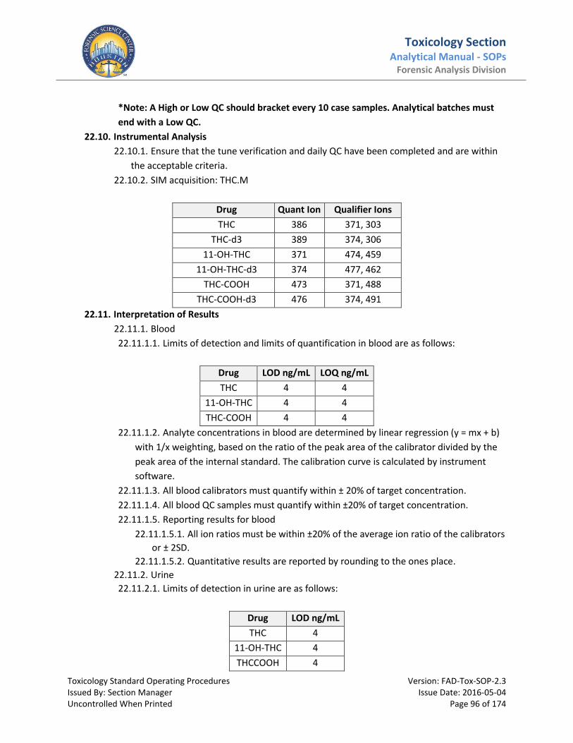
Toxicology Section Analytical Manual - SOPs
Forensic Analysis Division
Toxicology Standard Operating Procedures Version: FAD-Tox-SOP-2.3 Issued By: Section Manager Issue Date: 2016-05-04 Uncontrolled When Printed Page 96 of 174
*Note: A High or Low QC should bracket every 10 case samples. Analytical batches must
end with a Low QC.
22.10. Instrumental Analysis
22.10.1. Ensure that the tune verification and daily QC have been completed and are within
the acceptable criteria.
22.10.2. SIM acquisition: THC.M
Drug Quant Ion Qualifier Ions
THC 386 371, 303
THC-d3 389 374, 306
11-OH-THC 371 474, 459
11-OH-THC-d3 374 477, 462
THC-COOH 473 371, 488
THC-COOH-d3 476 374, 491
22.11. Interpretation of Results
22.11.1. Blood
22.11.1.1. Limits of detection and limits of quantification in blood are as follows:
Drug LOD ng/mL LOQ ng/mL
THC 4 4
11-OH-THC 4 4
THC-COOH 4 4
22.11.1.2. Analyte concentrations in blood are determined by linear regression (y = mx + b)
with 1/x weighting, based on the ratio of the peak area of the calibrator divided by the
peak area of the internal standard. The calibration curve is calculated by instrument
software.
22.11.1.3. All blood calibrators must quantify within ± 20% of target concentration.
22.11.1.4. All blood QC samples must quantify within ±20% of target concentration.
22.11.1.5. Reporting results for blood
22.11.1.5.1. All ion ratios must be within ±20% of the average ion ratio of the calibrators
or ± 2SD.
22.11.1.5.2. Quantitative results are reported by rounding to the ones place.
22.11.2. Urine
22.11.2.1. Limits of detection in urine are as follows:
Drug LOD ng/mL
THC 4
11-OH-THC 4
THCCOOH 4

Toxicology Section Analytical Manual - SOPs
Forensic Analysis Division
Toxicology Standard Operating Procedures Version: FAD-Tox-SOP-2.3 Issued By: Section Manager Issue Date: 2016-05-04 Uncontrolled When Printed Page 97 of 174
22.11.2.2. Urine is calibrated using a 1 point calibration curve.
22.11.2.3. All urine controls must give the expected result (negative must be negative,
positive must be positive).
22.11.2.4. All ion ratios must be within ±20% or ± 2SD of the acceptable average (for
example: cutoff, positive QC and High QC)
22.11.2.5. If the calculated concentration for THC, THC-COOH and 11-OH-THC is greater than
4 ng/mL cutoff calibrator the result is reported as “Positive”.
22.11.2.6. If the calculated concentration for THC, THC-COOH and 11-OH-THC is less than 4
ng/mL cutoff calibrator the result is reported as “None Detected.”
22.12. Fortification Guide (Blood)
22.12.1. Calibrators
Volume of Blood (mL)
Target Concentration (ng/mL)
Cannabinoids Working Standard Concentration (µg/mL)
Volume Added (µL)
1 4 0.1 40
1 10 0.1 100
1 25 1.0 25
1 50 1.0 50
1 75 1.0 75
1 100 1.0 100
22.12.2. Controls
Volume of Blood (mL)
Target Concentration (ng/mL)
Cannabinoids Working Standard Concentration (µg/mL)
Volume Added (µL)
1 8 (Low QC) 0.1 80
1 80 (HQC) 1 80
1 4 (LOD) 0.1 40
22.13. Fortification guide (urine)
Volume of Urine (1mL)
Target Concentration (ng/mL)
Cannabinoids Working Standard Solution(µg/mL)
Volume Added (µL)
1 cut off calibrator at 4 0.1 calibrator solution 40
1 Positive QC at 8 0.1 QC solution 80
1 Glucuronide THC-
COOH at 8 0.1 Glucuronide THC-COOH
solution 80
1 High QC at 80 1 QC solution 80
Toxicology Section Analytical Manual - SOPs
Forensic Analysis Division
Toxicology Standard Operating Procedures Version: FAD-Tox-SOP-2.3 Issued By: Section Manager Issue Date: 2016-05-04 Uncontrolled When Printed Page 98 of 174
22.14. Literature and Supporting Documentation
22.14.1. Huestis, MA. Cannabis in Principles of Forensic Toxicology, 3rd Edition. Levine B (Ed).
AACC Press, Washington DC, 2009, pp. 269-303.
22.14.2. Baselt, RC. Tetrahydrocannabinol in Disposition of Toxic Drugs and Chemicals in
Man, 8th Edition Foster City, CA. Biomedical Publications. 2009, pp. 1513-1518
22.14.3. Watanabe, Kanako: “11.2.2-Cannabinoids and their metabolites” in Drugs and
poisons in Humans. A Handbook of Practical Analysis. Springer-Verlag Berlin Heidelberg.
2005, pp. 187-194.
22.14.4. Abraham, TT; Lowe, TH; Pirnay, SO; Darwin, WD; Huestis, MA. Simultaneous GC-EI-
MS determination of D9-Tetrahydrocannabinol, 11-Hydroxy-D9-tetrahydrocannabinool,
and 11-nor-9-tetrahydrocannabinol in human urine following tandem enzyme-alkaline
hydrolysis. Journal of Analytical Toxicology, 31, October 2007, pp. 477-485
22.14.5. Method File: THC.M

Toxicology Section Analytical Manual - SOPs
Forensic Analysis Division
Toxicology Standard Operating Procedures Version: FAD-Tox-SOP-2.3 Issued By: Section Manager Issue Date: 2016-05-04 Uncontrolled When Printed Page 99 of 174
23. Carisoprodol/Meprobamate Confirmation by Gas Chromatography-Mass Spectrometry
23.1. Purpose
23.1.1. A targeted analysis is performed for confirmatory analysis of carisoprodol and/or
meprobamate by solid phase extraction (SPE) and gas chromatography-mass
spectrometry (GC-MS). Drugs are isolated from the matrix using an acidic/neutral
extraction. Deuterated internal standards and selective ion monitoring (SIM) are used in
electron ionization (EI) mode.
23.2. Scope
23.2.1. This procedure should be used for confirmatory analysis of carisoprodol and/or
meprobamate from toxicology specimens, typically blood. This procedure is
recommended if quantitative analysis is desired. Qualitative results may also be
reported. For routine full scan qualitative analysis of carisoprodol and/or meprobamate,
however, refer to Basic, Acidic and Neutral Drugs Confirmation by GC-MS).
23.3. Reagents and Standards
23.3.1. Carisoprodol/Meprobamate Working Standards for Calibrators and In-house Controls
23.3.1.1. Note: The concentration of carisoprodol in the working standard is half that of the
meprobamate.
23.3.1.2. Carisoprodol (0.02 mg/mL)/Meprobamate (0.04 mg/mL) Working Standard: Add
200 µL of carisoprodol (1.0 mg/mL) and 400 µL of meprobamate (1.0 mg/mL) to a 10 mL
volumetric flask, and bring to volume with methanol. Store refrigerated (6 month
expiration).
23.3.2. Internal Standard Solution: This solution consists of carisoprodol-D7 and
meprobamate-D7 in methanol. The final concentration is 10 ng/µL.
23.3.2.1. Working Internal Standard Solution (10 ng/µL): Add 1000 µL of carisoprodol-D7
(0.1 mg/mL) and 1000 µL meprobamate-D7 (0.1 mg/mL) to a 10 mL volumetric flask and
bring to volume with methanol. Store refrigerated (6 month expiration).
23.3.3. 100 mM sodium Phosphate Buffer pH 6.0
23.4. Equipment and Materials
Air displacement pipettes (100-1000 µL; 20-200 µL; 2-20 µL)
Cerex PolyChrom Clin II SPE columns
Vacuum manifold with disposable Teflon inserts or positive pressure SPE manifold
Pan balance
pH meter
Evaporator
Vortex mixer

Toxicology Section Analytical Manual - SOPs
Forensic Analysis Division
Toxicology Standard Operating Procedures Version: FAD-Tox-SOP-2.3 Issued By: Section Manager Issue Date: 2016-05-04 Uncontrolled When Printed Page 100 of 174
Centrifuge
Tissue homogenizer
Sonicator
Heating block
Vacuum pump or house vacuum
Agilent 7890 GC 5975 MSD
23.5. Instrumental Parameters
23.5.1. Capillary Column: 30 m HP-5MS Agilent J&W GC Column (or equivalent), 0.250 mm id
X 0.25 µm film thickness. The flow rate is 1.3 mL/min with an injection volume of 2 µL in
split mode (10:1).
23.5.2. GC-MS: Agilent 7890A
Initial Temperature: 140 °C
Hold for 0.5 minutes
30 °C/min to 290 °C
Hold for 2.5 minutes
50 °C/min to 310 °C
Hold for 2.6 minutes
Total Run Time: 11 minutes
Injector Temperature: 250 °C
Interface Temperature: 280 °C
MS Quads: 150 °C
MS Source: 230 °C
23.5.3. Wash solvents for autosampler: Methanol and ethyl acetate are used as the wash
solvents. A minimum of 6 pre and 6 post rinses are performed. Each rinse cycle consists
of 3 methanol rinses followed by 3 ethyl acetate rinses.
23.6. Extraction Procedure
23.6.1. Blood and All Other Matrices
23.6.2. In a round bottom glass culture tube, add 0.25 mL specimen and 2 mL of 100 mM
sodium phosphate buffer (pH 6.0). Vortex mix.
23.6.3. Add 100 µL of Working Internal Standard Solution (10 ng/µL) while vortex
mixing. The final concentration of internal standard in each sample is 4 µg/mL or 4
mg/L.
23.6.4. Fortify all controls and calibrators while vortex mixing using the appropriate amount
of working standard. The typical calibration range for quantitative analysis is 0-20 mg/L
carisoprodol and 0-40 mg/L meprobamate. Refer to the fortification guide for guidance

Toxicology Section Analytical Manual - SOPs
Forensic Analysis Division
Toxicology Standard Operating Procedures Version: FAD-Tox-SOP-2.3 Issued By: Section Manager Issue Date: 2016-05-04 Uncontrolled When Printed Page 101 of 174
on preparation of calibrators. CONTROLS AND CALIBRATORS MUST COME FROM
INDEPENDENT STOCK SOLUTIONS.
23.6.4.1. NOTE: IF THE SAMPLE IS KNOWN TO CONTAIN A HIGH CONCENTRATION OF DRUG,
ALIQUOT A SMALLER VOLUME, ADD BUFFER AS THE DILUENT AND DOCUMENT IN THE
CASE FOLDER.
23.6.5. A quantitative QC containing 4 mg/L carisoprodol and 8 mg/L meprobamate must be
used. Add 50 µL of 0.02/0.04 mg/mL in-house QC Working Standard Solution to 0.25 mL
of blank matrix. Use appropriate external controls (e.g., UTAK Whole Blood Controls) if
available.
23.6.6. Centrifuge samples for at least 10 minutes at 4000 rpm.
23.6.7. Place collection tubes or reservoir (for waste) into vacuum manifold rack.
23.6.8. Add the sample to Polychrom Clin II Column. Draw the sample through using
sufficient vacuum as necessary.
23.6.9. Wash column with 1 mL of deionized water.
23.6.10. Wash column with 1 mL of 1M acetic acid.
23.6.11. Dry column on full vacuum/pressure for 5 minutes. .
23.6.12. Wash column with 1 mL of hexane.
23.6.13. Elute drugs with 1 mL of ethyl acetate.
23.6.14. Turn on vacuum for a few seconds to ensure all elution solvent has drained from the
column.
23.6.15. Remove tubes from rack and evaporate to dryness (approximately 10 minutes)
under nitrogen at 50-60°C.
23.6.16. Reconstitute in 40 µL of ethyl acetate, vortex, and transfer to autosampler vials. An
appropriate volume of solvent should be used to prevent overloading.
23.6.17. Inject 2 µL onto the GC-MS using CARISO-MEPRO.M.
23.7. Instrumental Analysis
23.7.1. Ensure that the autotune and testmix have been completed.
23.7.2. SIM acquisition: CARIO-MEPRO.M. Use this method to perform targeted analysis for
carisoprodol and meprobamate.
Drug Quant Ion Qualifier Ions RT*
Meprobamate 144 83, 114 4.16
Carisoprodol 158 245, 184 4.43
Meprobamate-D7 151 89, 121 4.13
Carisoprodol-D7 165 252, 191 4.41
*Retention Time (RT) varies with column length. Deuterated (7)
internal standards are used throughout. Corresponding ions for
internal standards are M+7. Refer to attached method for acquisition
parameters.

Toxicology Section Analytical Manual - SOPs
Forensic Analysis Division
Toxicology Standard Operating Procedures Version: FAD-Tox-SOP-2.3 Issued By: Section Manager Issue Date: 2016-05-04 Uncontrolled When Printed Page 102 of 174
23.8. Interpretation of Results
23.8.1. Assay performance including but not limited to prevision, accuracy, limit of detection,
limit of quantitation, and linearity are summarized in the validation documentation.
23.8.2. Limits of detection in blood and urine are as follows:
Drug LOD and LOQ (Blood) mg/L
Meprobamate 1.0
Carisoprodol 0.5
23.8.3. In the event of a co-extractive interference that influences the ion ratios of the
internal standard (meprobamate-D7) it is permissible to use the second internal standard
(carisoprodol-D7) to report meprobamate subject to review and approval by the
Laboratory Manager. In doing so, all qualitative and quantitative reporting criteria must
be met.
23.9. Fortification Guide
Volume of
Blood (mL)
Carisoprodol Target
Concentration (mg/L)
Meprobamate Target
Concentration (mg/L)
Carisoprodol/Meprobamate Working
Standard Concentration (mg/mL)
Volume
Added (µL)
0.25 2 4 0.02/0.04 25
0.25 5 10 0.02/0.04 62.5
0.25 10 20 0.02/0.04 125
0.25 20 40 0.02/0.04 250
23.10. Literature and Supporting Documentation
23.10.1. Downey, Delisa and Kerrigan, Sarah. Quantitative Analysis of Carisoprodol and
Meprobamate in Whole Blood Using Benzylcarbamate and Deuterated Meprobamate as
Internal Standards. J Anal Toxicol 2009 June; Vol. 33, pp. 278-282.
23.10.2. Baselt, Randall C. Disposition of Toxic Drugs and Chemicals in Man, 7th Ed.,
Biomedical Publications, Foster City, CA, p 182-183 (2004).
23.10.3. Method file: CARISO-MEPRO.M.

Toxicology Section Analytical Manual - SOPs
Forensic Analysis Division
Toxicology Standard Operating Procedures Version: FAD-Tox-SOP-2.3 Issued By: Section Manager Issue Date: 2016-05-04 Uncontrolled When Printed Page 103 of 174
24. Fentanyl Confirmation by Gas Chromatography-Mass Spectrometry
24.1. Purpose
24.1.1. A targeted analysis is performed for confirmatory analysis of fentanyl by solid phase
extraction (SPE) and gas chromatography-mass spectrometry (GC-MS). Fentanyl is
isolated from the matrix using a basic extraction. Deuterated internal standard and
selective ion monitoring (SIM) are used in electron impact (EI) mode.
24.2. Scope
24.2.1. Confirmatory analysis of fentanyl from toxicology specimens including but not limited
to blood. This procedure is recommended if quantitative analysis is desired. Qualitative
results may also be reported. For routine full scan qualitative analysis, however, refer to
Basic, Acidic, and Neutral Drugs by GC-MS.
24.3. Reagents and Standards
24.3.1. Fentanyl Working Standards for Calibrators and In-house Controls
24.3.1.1. Preparation of 1 ng/µL Fentanyl Working Standard: Add 100 µL of Fentanyl (0.1
mg/mL) to a 10 mL volumetric flask and bring to volume with methanol. Store
refrigerated (3 month expiration).
24.3.1.2. Preparation of 0.1 ng/µL Fentanyl Working Standard: Add 1 mL of Fentanyl (1
ng/µL) to a 10 mL volumetric flask and bring to volume with methanol. Store
refrigerated (3 month expiration).
24.3.1.3. Preparation of (0.5 ng/µL) Internal Working Solution: Add 50 µL of fentanyl-D5.
deuterated drug standard (0.1 mg/mL) to a 10 mL volumetric flask and bring to volume
with methanol. Store refrigerated (3 month expiration).
24.3.2. 1M Acetic Acid
24.3.3. 100 mM, pH 6.0 Phosphate Buffer
24.3.4. Elution Solvent: 2% ammonium hydroxide (concentrated) in 80:20 methylene
chloride/isopropanol
24.3.5. Methylene Chloride/Isopropanol (80:20)
24.4. Equipment and Materials
Air displacement pipettes (100-1000 µL; 20-200 µL; 2-20 µL)
Cerex PolyChrom Clin II SPE columns
Vacuum manifold with disposable Teflon inserts or positive pressure SPE manifold
Pan balance
pH meter
Evaporator

Toxicology Section Analytical Manual - SOPs
Forensic Analysis Division
Toxicology Standard Operating Procedures Version: FAD-Tox-SOP-2.3 Issued By: Section Manager Issue Date: 2016-05-04 Uncontrolled When Printed Page 104 of 174
Vortex mixer
Centrifuge
Tissue homogenizer
Sonicator
Heating block
Vacuum pump or house vacuum
Agilent 7890 GC 5975 MSD
24.5. Instrumental Parameters
24.5.1. Capillary Column: 30 m HP-5MS Agilent J&W GC Column (or equivalent), 0.250 mm id
X 0.25 µm film thickness. The flow rate is 1.3 mL/min with an injection volume of 2 µL in
split mode (10:1).
24.5.2. GC-MS: Agilent 7890A
Initial Temperature: 140 °C
Hold for 0.5 minutes
30 °C/min to 290 °C
Hold for 2.5 minutes
50 °C/min to 310 °C
Hold for 2.6 minutes
Total Run Time: 11 minutes
Injector Temperature: 250 °C
Interface Temperature: 280 °C
MS Quads: 150 °C
MS Source: 230 °C
24.5.3. Wash solvents for autosampler: Methanol and ethyl acetate are used as the wash
solvents. A minimum of 6 pre and 6 post rinses are performed. Each rinse cycle consists
of 3 methanol rinses followed by 3 ethyl acetate rinses.
24.6. Extraction Procedure: Blood, Urine, and all Other Matrices
24.6.1. In a round bottom glass culture tube, add 2 mL of sample. NOTE IF THE SAMPLE IS
KNOWN TO CONTAIN A HIGH CONCENTRATION OF DRUG, ALIQUOT A SMALLER
VOLUME, ADD BUFFER AS THE DILUENT AND DOCUMENT IN THE CASE FOLDER.
24.6.2. Add 40 µL of Internal Standard (0.5 ng/µL) while vortex mixing. Final concentration of
internal standard in each sample is 10 ng/mL.
24.6.3. Fortify all controls and calibrators while vortex mixing using the appropriate amount
of working standard. Refer to the fortification guide for guidance on preparation of
calibrators. CONTROLS AND CALIBRATORS MUST COME FROM INDEPENDENT STOCK

Toxicology Section Analytical Manual - SOPs
Forensic Analysis Division
Toxicology Standard Operating Procedures Version: FAD-Tox-SOP-2.3 Issued By: Section Manager Issue Date: 2016-05-04 Uncontrolled When Printed Page 105 of 174
SOLUTIONS. Use appropriate external controls (e.g., UTAK Whole Blood Controls) when
available. It is recommended the controls be at 10 ng/mL.
24.6.4. Add 4 mL of 100 mM, pH 6.0 sodium phosphate buffer to each tube. Vortex.
24.6.5. Centrifuge the samples for at least 10 minutes at 4000 rpm.
24.6.6. Place collection tubes or reservoir (for waste) into vacuum manifold rack or positive
pressure apparatus.
24.6.7. Pour sample onto Polychrom Clin II columns. Use sufficient vacuum/pressure to draw
samples through the columns.
24.6.8. Wash column with 1 mL deionized water.
24.6.9. Wash column with 1 mL 1 M acetic acid.
24.6.10. Dry column on full vacuum/pressure for 5 minutes. .
24.6.11. Wash column with 1 mL hexane.
24.6.12. Wash column with 1 mL ethyl acetate.
24.6.13. Wash column with 1 mL methanol.
24.6.14. Dry column on full vacuum/pressure for 5 minutes. .
24.6.15. Replace waste tubes/reservoir with conical collection tubes. If the Teflon inserts
(vacuum manifold only) appear dirty, replace them.
24.6.16. Elute basic drugs with 1 mL elution solvent (2% ammonium hydroxide in methylene
chloride/isopropyl alcohol 80:20). ELUTION SOLVENT MUST BE MADE FRESH.
24.6.17. Turn on vacuum or apply positive pressure for a few seconds to ensure all elution
solvent has drained from the column.
24.6.18. Evaporate to dryness under nitrogen for approximately 10 minutes at 50-60 °C.
24.6.19. Reconstitute in 30 µL ethyl acetate (or appropriate volume), vortex, and transfer to
autosampler vials.
24.6.20. Inject 2 µL onto the GC-MS using FENTANYL.M.
24.7. Instrumental Analysis
24.7.1. Ensure that the autotune and testmix have been completed.
24.7.2. SIM acquisition: FENTANYL.M.
Drug Quant Ion Qualifier Ions RT*
Fentanyl 245 146, 189 7.73
Fentanyl – d5 250 151, 194 7.71
*Retention Time (RT) varies with column length. Deuterated (D5)
internal standards are used throughout. Corresponding ions for
internal standards are M+5. Refer to attached method for acquisition
parameters.
24.8. Interpretation of Results

Toxicology Section Analytical Manual - SOPs
Forensic Analysis Division
Toxicology Standard Operating Procedures Version: FAD-Tox-SOP-2.3 Issued By: Section Manager Issue Date: 2016-05-04 Uncontrolled When Printed Page 106 of 174
24.8.1. Assay performance including but not limited to precision, accuracy, limit of detection,
limit of quantitation, and linearity are summarized in the validation documentation.
24.8.2. The limit of detection and quantitation in whole blood and urine is 2.5 ng/mL.
24.9. Fortification Guide
Volume of
Blood (mL)
Target Concentration
(ng/mL)
Working Standard
Concentration (ng/µL = µg/mL)
Volume
Added (µL)
2 2.5 0.1 50
2 5 0.1 100
2 10 0.1 200
2 25 1 50
2 50 1 100
2 100 1 200
24.10. Literature and Supporting Documentation
24.10.1. Baselt, R.C. Fentanyl. Disposition of Toxic Drugs and Chemicals in Man. 8th edition.
Biomedical Publications, Foster City, CA, pp. 616-619.
24.10.2. Bronwyn, F. et al. Determination of subnanogram concentrations of Fentanyl in
plasma by gas chromatography-mass spectrometry: comparison with standard
radioimmunoassay. J Chrom B. 1997. 688, pp. 79-85.
24.10.3. Carson, H.J. et al. A fatality involving an unusual route of Fentanyl delivery: Chewing
and aspirating the transdermal patch. Legal Med. 2010. Vol. 12, pp. 157-159.
24.10.4. Kuhlman, J.J. Jr., et al. Fentanyl use, misuse, and abuse: a summary of 23
postmortem cases. J Anal Toxicol 2003; Vol. 27, pp. 499-504.
24.10.5. Watts, V. et al. Determination of Fentanyl in Whole Blood at Subnanogram
Concentrations by Dual Capillary Column Gas Chromatography with Nitrogen Sensitive
Detectors and Gas Chromatography/Mass Spectrometry. J Anal Toxicol 1988 October;
Vol. 12, pp. 246-254.
24.10.6. Method file: FENTANYL.M

Toxicology Section Analytical Manual - SOPs
Forensic Analysis Division
Toxicology Standard Operating Procedures Version: FAD-Tox-SOP-2.3 Issued By: Section Manager Issue Date: 2016-05-04 Uncontrolled When Printed Page 107 of 174
25. Ketamine Confirmation by Gas Chromatography-Mass Spectrometry
25.1. Purpose
25.1.1. A targeted analysis is performed for confirmatory analysis of ketamine by solid phase
extraction (SPE) and gas chromatography-mass spectrometry (GC-MS). Drugs are
isolated from the matrix using a basic extraction. A deuterated internal standard and
selective ion monitoring (SIM) are used in electron ionization (EI) mode.
25.2. Scope
25.2.1. This procedure should be used for confirmatory analysis of ketamine from toxicology
specimens, typically blood. This procedure is recommended if quantitative analysis is
desired. Qualitative results may also be reported. For routine full scan qualitative
analysis, however, refer to Basic, Acidic and Neutral Drugs Confirmation by GC-MS.
25.3. Reagents and Standards
25.3.1. Ketamine Working Standard for Calibrators and In-house Controls
25.3.1.1. 10 ng/µL Ketamine Working Standard: Add 100 µL of ketamine (1.0 mg/mL) to a
10 mL volumetric flask and bring to volume with methanol. Store refrigerated (3 month
expiration).
25.3.1.2. 1 ng/µL Ketamine Working Standard: Add 1 mL of 10 ng/µL working standard to a
10 mL volumetric flask and bring to volume with methanol. Store refrigerated (3 month
expiration).
25.3.2. Ketamine-D4 Internal Standard Solution
25.3.2.1. 10 ng/µL Ketamine-D4 Stock Internal Standard: Add 1 mL of Ketamine-D4 (100
µg/mL) to a 10 mL volumetric flask and bring to volume with methanol. Store
refrigerated (12 month expiration).
25.3.2.2. Preparation of 1 ng/µL Ketamine Working Internal Standard: Add 1 mL of 10 ng/µL
internal standard to a 10 mL volumetric flask and bring to volume with methanol. Store
refrigerated (6 month expiration).
25.3.3. 1M Acetic Acid
25.3.4. 100 mM pH 6.0 Phosphate Buffer
25.3.5. Methylene Chloride/Isopropanol (80:20)
25.3.6. Elution Solvent: 2% ammonium hydroxide (concentrated) in 80:20 methylene
chloride/isopropanol
25.4. Equipment and Materials
Air displacement pipettes (100-1000 µL; 20-200 µL; 2-20 µL)

Toxicology Section Analytical Manual - SOPs
Forensic Analysis Division
Toxicology Standard Operating Procedures Version: FAD-Tox-SOP-2.3 Issued By: Section Manager Issue Date: 2016-05-04 Uncontrolled When Printed Page 108 of 174
Cerex PolyChrom Clin II SPE columns
Vacuum manifold with disposable Teflon inserts or positive pressure SPE manifold
Pan balance
pH meter
Evaporator
Vortex mixer
Centrifuge
Tissue homogenizer
Sonicator
Heating block
Vacuum pump or house vacuum
Agilent 7890 GC 5975 MSD
25.5. Instrumental Parameters
25.5.1. Capillary Column: 30 m HP-5MS Agilent J&W GC Column (or equivalent), 0.250 mm id
X 0.25 µm film thickness. The flow rate is 1.2 mL/min with an injection volume of 2 µL in
split mode (10:1).
25.5.2. GC-MS: Agilent 7890A
Initial Temperature: 140 °C
Hold for 0.5 minutes
30 °C/min to 290 °C
Hold for 2.5 minutes
50 °C/min to 310 °C
Hold for 2.6 minutes
Total Run Time: 11 minutes
Injector Temperature: 250 °C
Interface Temperature: 280 °C
MS Quads: 150 °C
MS Source: 230 °C
25.5.3. Wash solvents for auto sampler: Methanol and ethyl acetate are used as the wash
solvents. A minimum of 6 pre and 6 post rinses are performed. Each rinse cycle consists
of 3 methanol rinses followed by 3 ethyl acetate rinses.
25.6. Extraction Procedure
25.6.1. Blood, Urine And Other Matrices
25.6.1.1. In a round bottom glass culture tube, add 1 mL of specimen and 100 µL WORKING
Internal Standard Solution (1 ng/µL) while vortex mixing (total concentration = 100

Toxicology Section Analytical Manual - SOPs
Forensic Analysis Division
Toxicology Standard Operating Procedures Version: FAD-Tox-SOP-2.3 Issued By: Section Manager Issue Date: 2016-05-04 Uncontrolled When Printed Page 109 of 174
ng/mL). NOTE: IF THE SAMPLE IS KNOWN TO CONTAIN A HIGH CONCENTRATION OF
DRUG, ALIQUOT A SMALLER VOLUME, ADD BUFFER AS THE DILUENT AND DOCUMENT
IN THE CASE FOLDER.
25.6.1.2. Fortify all controls and calibrators while vortex mixing using the appropriate
amount of working standard. Refer to the fortification guide for guidance on
preparation of calibrators. CONTROLS AND CALIBRATORS MUST COME FROM
INDEPENDENT STOCK SOLUTIONS. A minimum of one in-house control should be
included in each run. Use appropriate external controls (e.g., UTAK Whole Blood
Controls) when available. An in-house quantitative QC of 100 ng/mL is recommended.
25.6.1.3. Add 2 mL of 100 mM, pH 6.0 sodium phosphate buffer to each tube. Vortex.
25.6.1.4. Centrifuge samples for at least 10 minutes at 4000 rpm.
25.6.1.5. Place collection tubes or reservoir (for waste) into vacuum manifold rack or
positive pressure apparatus.
25.6.1.6. Pour sample onto Polychrom Clin II columns. Use sufficient vacuum/pressure to
draw samples through the columns.
25.6.1.7. Wash column with 1 mL deionized water.
25.6.1.8. Remove waste tubes/reservoir and replace with fresh waste tubes/reservoir.
25.6.1.9. Wash column with 1 mL 1 M acetic acid.
25.6.1.10. Dry column on full vacuum/pressure for 5 minutes.
25.6.1.11. Wash column with 1 mL hexane.
25.6.1.12. Wash column with 1 mL ethyl acetate.
25.6.1.13. Wash column with 1 mL methanol.
25.6.1.14. Dry column on full vacuum/pressure for 5 minutes.
25.6.1.15. Replace waste tubes/reservoir with conical collection tubes. If the Teflon inserts
(vacuum manifold only) appear dirty, replace them.
25.6.1.16. Elute basic drugs with 1mL elution solvent (2% ammonium hydroxide in
methylene chloride/isopropyl alcohol 80:20). ELUTION SOLVENT MUST BE MADE
FRESH.
25.6.1.17. Turn on vacuum or apply positive pressure for a few seconds to ensure all elution
solvent has drained from the column.
1.1.1.1. Evaporate to dryness under nitrogen for approximately 10 minutes at 50-60 °C.
25.6.1.18. Reconstitute in 30µL ethyl acetate (or an appropriate volume), vortex, and
transfer to autosampler vials.
25.6.1.19. Inject 2 µL onto the GC-MS using KETAMINE.M. (FOR URINE SAMPLES ONLY
USE—KETAMINE_URINE.M)
25.6.2. Dilutions: If the sample is known to contain a high concentration of drug, it should be
diluted prior to analysis. Aliquot the desired volume of blood and make up to volume
using phosphate buffer. DOCUMENT THESE CHANGES IN THE CASE FOLDER. Then
proceed from step 4 of the blood procedure.

Toxicology Section Analytical Manual - SOPs
Forensic Analysis Division
Toxicology Standard Operating Procedures Version: FAD-Tox-SOP-2.3 Issued By: Section Manager Issue Date: 2016-05-04 Uncontrolled When Printed Page 110 of 174
25.7. Instrumental Analysis
25.7.1. Ensure that the autotune and testmix have been completed.
25.7.2. SIM acquisition: KETAMINE_URINE.M. Use this method to perform targeted analyses
for ketamine in urine.
25.7.3. SIM acquisition: KETAMINE.M. Use this method to perform targeted analyses for
ketamine in blood.
Drug Quant Ion Qualifier Ions RT*
Ketamine-D4 213 184, 156 4.46
Ketamine 209 180, 152 4.47
*Retention Time (RT) varies with column length. Deuterated (D4)
internal standards are used throughout. Corresponding ions for
internal standards are M+4. Refer to attached method for acquisition
parameters.
25.8. Interpretation of Results
25.8.1. Assay performance including but not limited to precision, accuracy, limit of detection,
limit of quantitation, and linearity are summarized in the validation documentation.
25.8.2. Limits of detection and quantitation of ketamine in blood are 10 ng/mL.
25.9. Fortification Guide
Volume of
Blood (mL)
Target Concentration
(ng/mL)
Working Standard Concentration
(ng/µL = µg/mL)
Volume
Added (µL)
1 20 1 20
1 50 1 50
1 100 1 100
1 250 10 25
1 500 10 50
1 1000 10 100
25.10. Literature And Supporting Documentation
25.10.1. Baselt, Randall C. Disposition of Toxic Drugs and Chemicals in Man, 7th Ed.,
Biomedical Publications, Foster City, CA, p 586-588 (2004).
25.10.2. Jenkins, Amanda J. Hallucinogens in Principles of Forensic Toxicology, 2nd Ed., Levine,
Barry, Ed. AACC Press, Washington DC, pp. 265-283 (2003).
25.10.3. Method file: KETAMINE.M
25.10.4. Method file: KETAMINE_URINE.M

Toxicology Section Analytical Manual - SOPs
Forensic Analysis Division
Toxicology Standard Operating Procedures Version: FAD-Tox-SOP-2.3 Issued By: Section Manager Issue Date: 2016-05-04 Uncontrolled When Printed Page 111 of 174
26. Keto Opioids Confirmation by Gas Chromatography-Mass Spectrometry
26.1. Purpose
26.1.1. A targeted analysis is performed for confirmatory analysis of hydrocodone,
hydromorphone, oxycodone, and oxymorphone using solid phase extraction (SPE) and
gas chromatography-mass spectrometry (GC-MS). Drugs are isolated from the matrix
using a basic extraction. Deuterated internal standards and selective ion monitoring
(SIM) are used in electron impact (EI) mode.
26.2. Scope
26.2.1. Confirmatory analysis of keto opioids from toxicology specimens, including but not
limited to blood. Urine confirmations are reported only qualitatively and may also be
reported using Basic, Acidic and Neutral Drugs Confirmation by GC-MS.
26.3. Reagents and Standards
26.3.1. Keto Opioid Working Standard for Calibrators and In-house Controls
26.3.1.1. Two solutions will be needed for this analysis. The solutions contain
concentrations of hydrocodone, hydromorphone, oxycodone, and oxymorphone at 10
ng/µL and 1 ng/µL, respectively.
26.3.1.2. Preparation of 10 ng/µL Keto Opioid Working Standard: Add 100 µL of each keto
opioid (1.0 mg/mL) to a 10 mL volumetric flask and bring to volume with methanol.
Store refrigerated (6 month expiration).
26.3.1.3. Preparation of 1 ng/µL Working Standard: Add 1 mL of 10 ng/µL working standard
to a 10 mL volumetric flask and bring to volume with methanol. Store refrigerated (3
month expiration).
26.3.2. Keto Opioid Internal Standard Solution: This solution contains concentrations of
hydrocodone-D3, hydromorphone-D3, oxycodone-D3, and oxymorphone-D3.
26.3.2.1. Preparation of 10 ng/µL Keto Opioid Stock Internal Standard: Add 100 µL of each
keto opioid (100 µg/mL) to a 10 mL volumetric flask and bring to volume with methanol.
Store refrigerated (3 month expiration).
26.3.2.2. Preparation of 1 ng/µL Working Internal Standard: Add 1 mL of 10 ng/µL internal
standard to a 10 mL volumetric flask and bring to volume with methanol. Store
refrigerated (3 month expiration).
26.3.3. 1M Acetic Acid
26.3.4. 100 mM, pH 6.0 Phosphate Buffer
26.3.5. Methylene Chloride/Isopropanol (80:20)
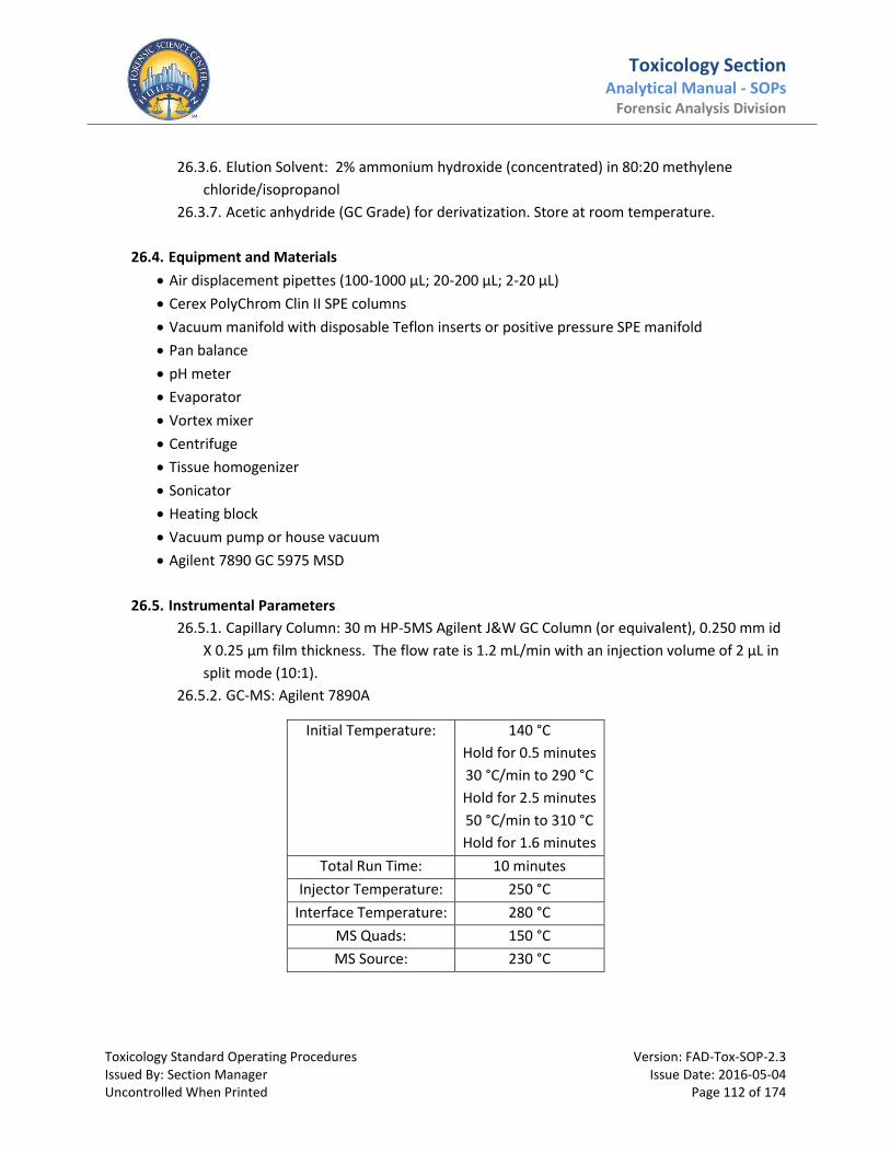
Toxicology Section Analytical Manual - SOPs
Forensic Analysis Division
Toxicology Standard Operating Procedures Version: FAD-Tox-SOP-2.3 Issued By: Section Manager Issue Date: 2016-05-04 Uncontrolled When Printed Page 112 of 174
26.3.6. Elution Solvent: 2% ammonium hydroxide (concentrated) in 80:20 methylene
chloride/isopropanol
26.3.7. Acetic anhydride (GC Grade) for derivatization. Store at room temperature.
26.4. Equipment and Materials
Air displacement pipettes (100-1000 µL; 20-200 µL; 2-20 µL)
Cerex PolyChrom Clin II SPE columns
Vacuum manifold with disposable Teflon inserts or positive pressure SPE manifold
Pan balance
pH meter
Evaporator
Vortex mixer
Centrifuge
Tissue homogenizer
Sonicator
Heating block
Vacuum pump or house vacuum
Agilent 7890 GC 5975 MSD
26.5. Instrumental Parameters
26.5.1. Capillary Column: 30 m HP-5MS Agilent J&W GC Column (or equivalent), 0.250 mm id
X 0.25 µm film thickness. The flow rate is 1.2 mL/min with an injection volume of 2 µL in
split mode (10:1).
26.5.2. GC-MS: Agilent 7890A
Initial Temperature: 140 °C
Hold for 0.5 minutes
30 °C/min to 290 °C
Hold for 2.5 minutes
50 °C/min to 310 °C
Hold for 1.6 minutes
Total Run Time: 10 minutes
Injector Temperature: 250 °C
Interface Temperature: 280 °C
MS Quads: 150 °C
MS Source: 230 °C

Toxicology Section Analytical Manual - SOPs
Forensic Analysis Division
Toxicology Standard Operating Procedures Version: FAD-Tox-SOP-2.3 Issued By: Section Manager Issue Date: 2016-05-04 Uncontrolled When Printed Page 113 of 174
26.5.3. Wash solvents for autosampler: Methanol and ethyl acetate are used as the wash
solvents. A minimum of 6 pre and 6 post rinses are performed. Each rinse cycle consists
of 3 methanol rinses followed by 3 ethyl acetate rinses.
26.6. Extraction Procedure (Use Silanized Glassware)
26.6.1. Blood, Urine, and all Other Matrices
26.6.2. In a round bottom glass culture tube, add 1 mL of specimen and 100 µL WORKING
Internal Standard Solution (1 ng/µL) while vortex mixing (total concentration = 100
ng/mL). NOTE: IF THE SAMPLE IS KNOWN TO CONTAIN A HIGH CONCENTRATION OF
DRUG, ALIQUOT A SMALLER VOLUME AND DOCUMENT IN THE CASE FOLDER.
26.6.3. Fortify all controls and calibrators while vortex mixing using the appropriate amount
of working standard. Refer to the fortification guide for guidance on preparation of
calibrators. CONTROLS AND CALIBRATORS MUST COME FROM INDEPENDENT STOCK
SOLUTIONS. Use appropriate external controls (e.g. UTAK Whole Blood Controls) when
available.
26.6.4. Add 2 mL of 100 mM, pH 6.0 sodium phosphate buffer to each tube. Vortex.
26.6.5. Place collection tubes or reservoir (for waste) into vacuum manifold rack or positive
pressure apparatus.
26.6.6. Pour sample onto Polychrom Clin II columns. Use sufficient vacuum/pressure to draw
samples through the columns.
26.6.7. Wash column with 1 mL deionized water.
26.6.8. Remove waste tubes/reservoir and replace with fresh waste tubes/reservoir.
26.6.9. Wash column with 1 mL 1 M acetic acid.
26.6.10. Dry column on full vacuum/pressure for 5 minutes.
26.6.11. Wash column with 1 mL hexane.
26.6.12. Wash column with 1 mL ethyl acetate.
26.6.13. Wash column with 1 mL methanol.
26.6.14. Dry column on full vacuum/pressure for 5 minutes.
26.6.15. Replace waste tubes/reservoir with conical collection tubes. If the Teflon inserts
(vacuum manifold only) appear dirty, replace them.
26.6.16. Elute basic drugs with 1 mL elution solvent (2% ammonium hydroxide in methylene
chloride/isopropyl alcohol 80:20). ELUTION SOLVENT MUST BE MADE FRESH DAILY.
26.6.17. Turn on vacuum or apply positive pressure for a few seconds to ensure all elution
solvent has drained from the column.
26.6.18. Evaporate to dryness under nitrogen for 15 minutes at 60 °C.
26.6.19. Reconstitute in 10µL ethyl acetate and 10 µL acetic anhydride (or other appropriate
volume of 1:1 ethyl acetate/acetic anhydride), vortex, and transfer to autosampler vials.
26.6.20. Inject 2 µL onto the GC-MS using KETO.M.

Toxicology Section Analytical Manual - SOPs
Forensic Analysis Division
Toxicology Standard Operating Procedures Version: FAD-Tox-SOP-2.3 Issued By: Section Manager Issue Date: 2016-05-04 Uncontrolled When Printed Page 114 of 174
26.7. Instrumental Analysis
26.7.1. Ensure that the autotune and testmix have been completed.
26.7.2. SIM acquisition: KETO.M.
Drug Quant Ion Qualifier Ions RT*
Hydrocodone-d3 302 273, 287 6.541
Hydrocodone 299 270, 284 6.550
Hydromorphone-d3 288 259, 330 7.178
Hydromorphone 285 256, 327 7.188
Oxycodone-d3 360 301, 317 6.876
Oxycodone 357 298, 314 6.889
Oxymorphone-d3 346 303, 388 7.472
Oxymorphone 343 300, 385 7.482
*Retention Time (RT) varies with column length. Deuterated (D3)
internal standards are used throughout. Corresponding ions for
internal standards are M+3.
26.8. Interpretation of Results
26.8.1. Assay performance including but not limited to precision, accuracy, limit of detection,
limit of quantitation, and linearity are summarized in the validation documentation.
Drug LOD and LOQ in Blood ng/mL LOD in Urine ng/mL
Hydrocodone 20 30
Hydromorphone 25 20
Oxycodone 20 20
Oxymorphone 25 20
26.9. Fortification Guide
Volume of
Blood (mL)
Target Concentration
(ng/mL)
Working Standard
Concentration (ng/µL = µg/mL)
Volume
Added (µL)
1 20 1 20
1 50 1 50
1 100 1 100
1 250 10 25
1 500 10 50
1 1000 10 100

Toxicology Section Analytical Manual - SOPs
Forensic Analysis Division
Toxicology Standard Operating Procedures Version: FAD-Tox-SOP-2.3 Issued By: Section Manager Issue Date: 2016-05-04 Uncontrolled When Printed Page 115 of 174
26.10. Literature And Supporting Documentation
26.10.1. Kerrigan S; Goldberger BA. Opioids in Principles of Forensic Toxicology, 3rd Edition.
Levine B (Ed). AACC Press, Washington DC, 2009, pp. 225-244.
26.10.2. Method file: KETO.M

Toxicology Section Analytical Manual - SOPs
Forensic Analysis Division
Toxicology Standard Operating Procedures Version: FAD-Tox-SOP-2.3 Issued By: Section Manager Issue Date: 2016-05-04 Uncontrolled When Printed Page 116 of 174
27. Methadone/EDDP Confirmation by Gas Chromatography-Mass Spectrometry
27.1. Purpose
27.1.1. A targeted analysis is performed for confirmatory analysis of methadone and its
metabolite 2-ethylidene-1,5-dimethyl-3,3-diphenylpyrrolidine (EDDP) by solid phase
extraction (SPE) and gas chromatography-mass spectrometry (GC-MS). Drugs are
isolated from the matrix using a basic extraction. Deuterated internal standards and
selective ion monitoring (SIM) are used in electron ionization (EI) mode.
27.2. Scope
27.2.1. Confirmatory analysis of Methadone/EDDP from toxicology specimens including but
not limited to blood. This procedure is recommended if quantitative analysis is desired.
Qualitative results may also be reported. For routine full scan qualitative analysis,
however, refer to Basic, Acidic and Neutral Drugs Confirmation by GC-MS.
27.3. Reagents and Standards
27.3.1. Methadone/EDDP Stock Standard for Calibrators and In-house Controls
27.3.1.1. Preparation of 0.1 mg/mL Methadone/EDDP Stock Standard: Add 1000 µL of
methadone and EDDP (1.0 mg/mL) to a 10mL volumetric flask and bring to volume with
methanol. Store refrigerated (6 month expiration).
27.3.2. Methadone/EDDP Working Standards
27.3.2.1. Preparation of 10 ng/µL Methadone/EDDP Working Standard: Add 1000 µL of
methadone (0.1 mg/mL) and 1000 µL of EDDP (0.1 mg/mL) to a 10 mL volumetric flask
and bring to volume with methanol. Store refrigerated (3 month expiration).
27.3.2.2. Preparation of a 1 ng/µL Methadone/EDDP Working Solution: Add 1000 µL of
Methadone/EDDP (10 ng/µL) to a 10 mL volumetric flask and bring to volume with
methanol. Store refrigerated (3 month expiration).
27.3.3. Internal Standard Solution: Methadone-D9/EDDP-D3 (5 ng/µL)
27.3.3.1. Preparation of 5 ng/µL Internal Standard Solution: Add 500 µL of Methadone-D9
and EDDP-D3 (0.1 mg/mL) to a 10 mL volumetric flask and bring to volume with
methanol. Store refrigerated (6 month expiration).
27.3.4. 1M Acetic Acid
27.3.5. 100 mM, pH 6.0 Phosphate Buffer
27.3.6. Methylene Chloride/Isopropanol (80:20)
27.3.7. Elution Solvent: 2% ammonium hydroxide (concentrated) in 80:20 methylene
chloride/isopropanol
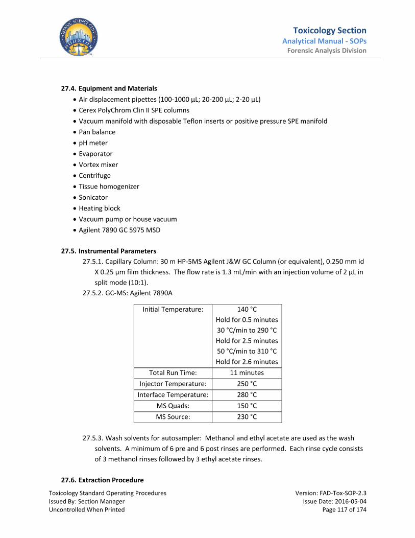
Toxicology Section Analytical Manual - SOPs
Forensic Analysis Division
Toxicology Standard Operating Procedures Version: FAD-Tox-SOP-2.3 Issued By: Section Manager Issue Date: 2016-05-04 Uncontrolled When Printed Page 117 of 174
27.4. Equipment and Materials
Air displacement pipettes (100-1000 µL; 20-200 µL; 2-20 µL)
Cerex PolyChrom Clin II SPE columns
Vacuum manifold with disposable Teflon inserts or positive pressure SPE manifold
Pan balance
pH meter
Evaporator
Vortex mixer
Centrifuge
Tissue homogenizer
Sonicator
Heating block
Vacuum pump or house vacuum
Agilent 7890 GC 5975 MSD
27.5. Instrumental Parameters
27.5.1. Capillary Column: 30 m HP-5MS Agilent J&W GC Column (or equivalent), 0.250 mm id
X 0.25 µm film thickness. The flow rate is 1.3 mL/min with an injection volume of 2 µL in
split mode (10:1).
27.5.2. GC-MS: Agilent 7890A
Initial Temperature: 140 °C
Hold for 0.5 minutes
30 °C/min to 290 °C
Hold for 2.5 minutes
50 °C/min to 310 °C
Hold for 2.6 minutes
Total Run Time: 11 minutes
Injector Temperature: 250 °C
Interface Temperature: 280 °C
MS Quads: 150 °C
MS Source: 230 °C
27.5.3. Wash solvents for autosampler: Methanol and ethyl acetate are used as the wash
solvents. A minimum of 6 pre and 6 post rinses are performed. Each rinse cycle consists
of 3 methanol rinses followed by 3 ethyl acetate rinses.
27.6. Extraction Procedure
Toxicology Section Analytical Manual - SOPs
Forensic Analysis Division
Toxicology Standard Operating Procedures Version: FAD-Tox-SOP-2.3 Issued By: Section Manager Issue Date: 2016-05-04 Uncontrolled When Printed Page 118 of 174
27.6.1. Blood, Urine, and all Other matrices
27.6.2. In a round bottom glass culture tube, add 1 mL of sample. NOTE: IF THE SAMPLE IS
KNOWN TO CONTAIN A HIGH CONCENTRATION OF DRUG, ALIQUOT A SMALLER VOLUME
AND DOCUMENT IN THE CASE FOLDER.
27.6.3. Add 50 µL of Internal Standard (5 ng/µL) while vortex mixing. Final concentration of
internal standard in each sample is 250 ng/mL.
27.6.4. Fortify all controls and calibrators while vortex mixing using the appropriate amount
of working standard. The typical calibration range for quantitative analysis is 0-1000
ng/mL for Methadone/EDDP. Refer to the fortification guide for guidance on preparation
of calibrators. CONTROLS AND CALIBRATORS MUST COME FROM INDEPENDENT STOCK
SOLUTIONS. An in-house control containing 100 ng/mL methadone/EDDP is
recommended. Use appropriate external controls (e.g. UTAK Whole Blood Controls)
when available.
27.6.5. Add 2 mL of 100 mM, pH 6.0 sodium phosphate buffer to each tube. Vortex.
27.6.6. Centrifuge samples for 10 minutes at 4000 rpm.
27.6.7. Place collection tubes or reservoir (for waste) into vacuum manifold rack or positive
pressure apparatus.
27.6.8. Pour sample onto Polychrom Clin II columns. Use sufficient vacuum/pressure to draw
samples through the columns.
27.6.9. Wash column with 1 mL deionized water.
27.6.10. Wash column with 1 mL 1 M acetic acid.
27.6.11. Dry column on full vacuum/pressure for 5 minutes.
27.6.12. Wash column with 1 mL hexane.
27.6.13. Wash column with 1 mL ethyl acetate.
27.6.14. Wash column with 1 mL methanol.
27.6.15. Dry column on full vacuum/pressure for 5 minutes.
27.6.16. Replace waste tubes/reservoir with conical collection tubes. If the Teflon inserts
(vacuum manifold only) appear dirty, replace them.
27.6.17. Elute basic drugs with 1 mL elution solvent (2% ammonium hydroxide in methylene
chloride/isopropyl alcohol 80:20). ELUTION SOLVENT MUST BE MADE FRESH.
27.6.18. Turn on vacuum or apply positive pressure for a few seconds to ensure all elution
solvent has drained from the column.
27.6.19. Evaporate to dryness under nitrogen for approximately 10 minutes at 50-60°C.
27.6.20. Reconstitute in 30µL ethyl acetate (or appropriate volume), vortex, and transfer to
autosampler vials.
27.6.21. Inject 2 µL onto the GC-MS using METHADONE-EDDP.M.
27.7. Instrumental Analysis
27.7.1. Ensure that the autotune and testmix have been completed.

Toxicology Section Analytical Manual - SOPs
Forensic Analysis Division
Toxicology Standard Operating Procedures Version: FAD-Tox-SOP-2.3 Issued By: Section Manager Issue Date: 2016-05-04 Uncontrolled When Printed Page 119 of 174
27.7.2. SIM acquisition: Methadone-EDDP.M.
Drug Quant Ion Qualifier Ions RT*
EDDP 277 200, 276 5.07
EDDP-D3 280 279, 203 5.06
Methadone 72 223, 294 5.4
75 226, 303 5.39
*Retention Time (RT) varies with column length. Deuterated (D3 and
D9) internal standards are used throughout.
27.8. Interpretation of Results
27.8.1. Assay performance including but not limited to precision, accuracy, limit of detection,
limit of quantitation, and linearity are summarized in the validation documentation.
27.8.2. The limits of detection and quantitation for methadone and EDDP are 20 ng/mL in
blood.
27.9. Fortification Guide
Volume of
Blood (mL)
Target Concentration
(ng/mL)
Working Standard
Concentration (ng/µL = µg/mL)
Volume
Added (µL)
1 20 1 20
1 50 1 50
1 100 1 100
1 250 10 25
1 500 10 50
1 1000 10 100
27.10. Literature and Supporting Documentation
27.10.1. Lucas, A.C.S. et al. Use of solid-phase microextraction (SPME) for the determination
of methadone and EDDP in human hair by GC-MS. Forensic Science International. 2000;
107, pp. 225-232.
27.10.2. Nikolaou, P.D. et al. Development and Validation of an EI-GC-MS Method for the
Determination of Methadone and its Major Metabolites (EDDP and EMDP) in Human
Breast Milk. J Anal Toxicol 2008 Sept; Vol. 32, pp. 478-484
27.10.3. Nikolaou, P.D. et al. Validated method for the simultaneous determination of
methadone and its main metabolites (EDDP and EMDP) in plasma of umbilical cord blood
by gas chromatography-mass spectrometry. J Chrom B. 2008, 867, pp. 219-225.
27.10.4. R.C. Baselt. Disposition of Toxic Drugs and Chemicals in Man. 8th edition. Biomedical
Publications, Foster City, CA, pp. 941-944.
Toxicology Section Analytical Manual - SOPs
Forensic Analysis Division
Toxicology Standard Operating Procedures Version: FAD-Tox-SOP-2.3 Issued By: Section Manager Issue Date: 2016-05-04 Uncontrolled When Printed Page 120 of 174
27.10.5. Kuhlman, J.J. Jr., et al. Fentanyl use, misuse, and abuse: a summary of 23
postmortem cases. J Anal Toxicol 2003; Vol. 27, pp. 499-504.
27.10.6. Method file: Methadone-EDDP.M
28. Opiates and Cocaine Confirmation by Gas Chromatography-Mass Spectrometry
28.1. Purpose
28.1.1. A targeted analysis is performed for morphine, codeine, 6-acetylmorphine (6-AM),
cocaine, benzoylecgonine (BE), ecgonine methyl ester (EME), and cocaethylene (CE).
Drugs are isolated from the matrix using solid phase extraction (SPE). Perfluoroacyl
derivatives are prepared and analyzed using gas chromatography-mass spectrometry
(GC-MS), deuterated internal standards and selective ion monitoring (SIM) in electron
impact (EI) mode.
28.2. Scope
28.2.1. Confirmatory analysis of target analytes from toxicology specimens including but not
limited to blood and urine. Urine confirmations are reported only qualitatively.
28.3. Standards and Solutions
28.3.1. Opiate and Cocaine (“OPICOC”) Working Standards for Calibrators and In-house
Controls
28.3.1.1. Two working standards are used. Each contains morphine, codeine, 6-AM,
cocaine, BE, EME and CE at either 10 ng/µL or 1 ng/µl, respectively.
28.3.1.2. Preparation of 10 ng/µl OPICOC Working Standard
28.3.1.2.1. Add 1000 µL of each drug standard (0.1 mg/mL) to a 10 mL volumetric flask
and bring to volume with acetonitrile. Store refrigerated (3 month expiration).
28.3.1.3. Preparation of a 1 ng/µL OPICOC Working Standard
28.3.1.3.1. Add 1000 µL of the 10 ng/µL OPICOC working standard to a 10 mL volumetric
flask and bring to volume with acetonitrile. Store refrigerated (3 month expiration).
28.3.2. Internal Standard Solution
28.3.2.1. This solution consists of morphine-d3, codeine-d3, cocaine-d3, BE-d3, EME-d3, CE-
d3 (1ng/µL) and 6-AM (0.5 ng/µL) in acetonitrile.
28.3.2.2. Preparation of (10 ng/µL) Internal Standard Stock Solution
28.3.2.2.1. Add 1000 µL of each deuterated drug standard (0.1 mg/ml) to a 10 mL
volumetric flask and bring to volume with acetonitrile. Store refrigerated (3 month
expiration).
28.3.2.3. Preparation of (1 ng/µL) Internal Standard Solution

Toxicology Section Analytical Manual - SOPs
Forensic Analysis Division
Toxicology Standard Operating Procedures Version: FAD-Tox-SOP-2.3 Issued By: Section Manager Issue Date: 2016-05-04 Uncontrolled When Printed Page 121 of 174
28.3.2.3.1. Add 1000 µL of 10 ng/µL internal standard stock solution to a 10 mL
volumetric flask and bring to a volume with acetonitrile. Store refrigerated (3
month expiration).
28.3.3. 0.1 M HCl
28.3.4. 1 M HCl
28.3.5. Cold Acetonitrile: HPLC grade acetonitrile used in this procedure should be stored in
the refrigerator at 0 °C to 10 °C prior to use.
28.3.6. Elution Solvent: 2% ammonium hydroxide (concentrated) in 80:20 methylene
chloride/isopropanol
28.3.7. Methylene Chloride/Isopropanol (80:20)
28.3.8. PFPA (pentafluoropropionic anhydride) and HFIP (1,1,1,3,3,3-hexafluoro-2-
propanol) for derivatization. Store both reagents at room temperature. Discard if there is
a change in color.
28.3.9. Deconjugation Enzyme: E-coli Type IX-A β-Glucuronidase (125,000 Units). To one
vial of 125,000 units, add 25 mL of deconjugation buffer and mix gently. Store frozen
(expiration of reconstituted enzyme reagent is 30 days).
28.3.10. Deconjugation Buffer (100 mM sodium phosphate buffer, pH 6.8)
28.3.11. Conjugated Morphine Working Standard (10 ng/µL): For total morphine
determination only, a morphine-3-glucuronide working standard is used. Add 1000 µL of
0.1 mg/ml morphine-3-glucuronide drug standard to a 10 mL volumetric flask. Bring to
volume with acetonitrile. Store refrigerated (6 month expiration).
28.3.12. Conjugated Morphine Internal Standard (10 ng/µL): For total morphine
determination only, a deuterated morphine-3-glucuronide internal standard is used. Add
1000 µL of a 0.1 mg/ml morphine-3-glucuronide-d3 drug standard to a 10 mL volumetric
flask. Bring to volume with acetonitrile. Store refrigerated (6 month expiration).
28.4. Equipment and Materials
Air displacement pipettes (100-1000 µL; 20-200 µL; 2-20 µL)
Cerex PolyChrom Clin II SPE columns
Vacuum manifold with disposable Teflon inserts or positive pressure SPE manifold
Pan balance
pH meter
Evaporator
Vortex mixer
Centrifuge
Tissue homogenizer
Sonicator
Heating block

Toxicology Section Analytical Manual - SOPs
Forensic Analysis Division
Toxicology Standard Operating Procedures Version: FAD-Tox-SOP-2.3 Issued By: Section Manager Issue Date: 2016-05-04 Uncontrolled When Printed Page 122 of 174
Vacuum pump or house vacuum
Agilent 7890 GC 5975 MSD
28.5. Instrumental Parameters
28.5.1. Capillary Column: 30 m HP-5MS Agilent J&W GC Column (or equivalent), 0.250 mm id
X 0.25 µm film thickness. The flow rate is 1.1 mL/min with an injection volume of 2 µL in
split mode (10:1)
28.5.2. GC-MS: Agilent 7890A
Initial Temperature: 100 °C
Hold for 0 minutes
30 °C/min to 230 °C
Hold for 5 minutes
30 °C/min to 290 °C
Hold for 5 minutes
Total Run Time: 16.333 minutes
Injector Temperature: 260 °C
Interface Temperature: 280 °C
MS Quads: 150 °C
MS Source: 230 °C
28.5.3. Wash solvents for autosampler: Methanol and ethyl acetate are used as the wash
solvents. A minimum of 6 pre and 6 post rinses are performed. Each rinse cycle consists
of 3 methanol rinses followed by 3 ethyl acetate rinses.
28.6. Extraction Procedure – Use silanized glassware for blood and urine extracts
28.6.1. Blood
28.6.1.1. In a round bottom glass centrifuge tube add 1 mL blood and 50 µL Internal
Standard Solution (1 ng/µL) while vortex mixing. Final concentration of internal
standard in each sample of 50 ng/mL for all target drugs except 6-AM (25 ng/ml). NOTE:
IF THE SAMPLE IS KNOWN TO CONTAIN A HIGH CONCENTRATION OF DRUG, ALIQUOT A
SMALL VOLUME AND DOCUMENT IN THE CASE RECORD.
28.6.1.2. Fortify all controls and calibrators while mixing using the appropriate amount of
working standard. The typical calibration range for quantitative analysis is 0-1000
ng/mL. Refer to the fortification guide for guidance on preparation of calibrators.
CONTROLS AND CALIBRATORS MUST COME FROM INDEPENDENT STOCK SOLUTIONS. A
minimum of one in-house control (100 ng/mL) should be included in each run. Use
appropriate external controls (e.g., UTAK Whole Blood Controls) when available.
28.6.1.3. Precipitate proteins by addition of 2 mL cold acetonitrile while vortex mixing.

Toxicology Section Analytical Manual - SOPs
Forensic Analysis Division
Toxicology Standard Operating Procedures Version: FAD-Tox-SOP-2.3 Issued By: Section Manager Issue Date: 2016-05-04 Uncontrolled When Printed Page 123 of 174
28.6.1.4. Centrifuge for 10 minutes at 4000 rpm.
28.6.1.5. Pour off the supernatant layer into a clean round bottom glass tube. Add 1 mL 0.1
M HCl.
28.6.1.6. Place collection tubes or reservoir (for waste) into vacuum manifold rack or on
positive pressure apparatus.
28.6.1.7. Pour acidified sample onto SPE columns. Samples should flow under gravity. Do
not use the vacuum (or pressure) unless it is necessary.
28.6.1.8. Wash column with 1 mL deionized water.
28.6.1.9. Wash column with 1 mL 0.1 M HCl.
28.6.1.10. Wash column with 1 mL methanol.
28.6.1.11. Wash column with 1 mL ethyl acetate.
28.6.1.12. Dry column on full vacuum/pressure for 5 minutes.
28.6.1.13. Place conical glass collection tubes into the rack.
28.6.1.14. Elute drugs with 1 mL elution solvent (2% ammonium hydroxide in methylene
chloride/isopropyl alcohol 80:20). Samples should flow under gravity. Do not use
vacuum (or pressure) unless it is necessary. ELUTION SOLVENT MUST BE MADE FRESH.
28.6.1.15. Turn on vacuum for a few seconds to ensure all elution solvent has drained from
the column.
28.6.1.16. Remove tubes from rack and evaporate to dryness (approximately 10 minutes)
under nitrogen at 50 °C.
28.6.1.17. IN THE FUME HOOD, add 75 µL PFPA and 75 µL HFIP. Cap tightly. Heat at 70 °C
for 15 minutes.
28.6.1.18. Evaporate to dryness under nitrogen for 10 minutes at 50 °C. Reconstitute in 30
µL ethyl acetate, vortex, and transfer to autosampler vials. It is acceptable to increase
the reconstitution volume prior to injection as needed to prevent sample overload. The
presumptive screening test results can be used as a guide.
28.6.1.19. Inject 2 µL onto the GC-MS using OPICOCSIM.M.
28.6.2. Urine
28.6.2.1. In a round bottom glass centrifuge tube, add 1 mL urine and 50 µL Internal
Standard Solution (1 ng/µL) while vortex mixing. Final concentration of internal
standard in each sample of 50 ng/mL.
28.6.2.2. For qualitative analysis include a minimum of one negative and one positive
control in each run. For preparation of 200 ng/mL positive control, add 20 µL of OPICOC
Working Standard Solution (10 ng/µL) to 1 mL urine while vortex mixing.
28.6.2.3. If sample is cloudy or contains particulates, centrifuge for 10 minutes, 4000 rpm.
28.6.2.4. Add 1 mL 1 M HCl and mix.
28.6.2.5. Proceed from step 6 through 17 of the blood procedure.

Toxicology Section Analytical Manual - SOPs
Forensic Analysis Division
Toxicology Standard Operating Procedures Version: FAD-Tox-SOP-2.3 Issued By: Section Manager Issue Date: 2016-05-04 Uncontrolled When Printed Page 124 of 174
28.6.2.6. Evaporate to dryness under nitrogen for 10 minutes at 50 °C. Reconstitute in 50 µL
ethyl acetate, vortex and transfer to autosampler vials. Samples containing extremely
elevated drug concentration (based on presumptive immunoassay results) may be
reconstituted using a larger volume of ethyl acetate if necessary.
28.6.2.7. Inject 2 µL onto the GC-MS using OPICOCSIM.M.
28.7. Total Morphine Determination
28.7.1. In a round bottom glass centrifuge tube, add 1 mL blood and 25 µL of 10 ng/µL
morphine-3-glucuronide-d3 Internal Standard Solution to all standards, QCs, and
samples.
28.7.1.1. Note: This is a different deuterated internal standard from the free opiate analysis.
Deuteration and glucuronidation of the internal standard ensures that the
deconjugation of opiates in the sample is monitored.
28.7.2. Fortify all controls and calibrators while vortex mixing using the appropriate amount
of conjugated morphine working standard. Refer to the fortification guide for guidance
on preparation of calibrators. A minimum of one in-house control should be included in
each run. CONTROLS AND CALIBRATORS MUST COME FROM INDEPENDENT STOCK
SOLUTIONS.
28.7.3. Add 0.2 mL deconjugation enzyme (5000 units). Incubate at 37 °C for 3 hours.
28.7.4. Proceed from step 3 of the blood procedure.
28.8. Instrumental Analysis
28.8.1. Ensure that the autotune and testmix have been completed.
28.8.2. SIM acquisition: OPICOCSIM.M. Use this method to perform targeted analysis for
morphine, codeine, 6-AM, cocaine, BE, EME and CE.
Drug Quant Ion Qualifier Ions RT*
Morphine 577 558, 414 9.43
Codeine 445 282, 266 9.97
6-AM 414 473, 361 10.60
Cocaine 303 182, 82 8.70
BE 439 318, 272 6.44
CE 317 272, 196 9.45
EME 345 317, 182 3.77
*Retention Time (RT) varies with column length. Deuterated (D3)
internal standards are used throughout. Corresponding ions for
internal standards are M+3. Refer to attached method for acquisition
parameters (OPICOCSIM.M)

Toxicology Section Analytical Manual - SOPs
Forensic Analysis Division
Toxicology Standard Operating Procedures Version: FAD-Tox-SOP-2.3 Issued By: Section Manager Issue Date: 2016-05-04 Uncontrolled When Printed Page 125 of 174
Internal Standard Internal Standard
Quant ions Qualifier Ions RT*
Morphine-d3 580 561,417 9.43
Codeine-d3 448 285,269 9.97
6-AM-d3 417 476, 364 10.60
Cocaine-d3 306 185, 85 8.70
BE-d3 442 321, 275 6.44
CE-d3 320 275, 199 9.45
EME-d3 348 317, 185 3.77
*Retention Time (RT) varies with column length. Deuterated (D3)
internal standards are used throughout. Corresponding ions for internal
standards are M+3. Refer to attached method for acquisition parameters
(OPICOCSIM.M)
28.9. Interpretation of Results
28.9.1. Assay performance including but not limited to precision, accuracy, limit of detection,
limit of quantitation, and linearity are summarized in the validation documentation.
28.9.2. Limits of detection in blood and urine are as follows:
Drug LOD and LOQ (Blood) ng/mL LOD (Urine) ng/mL
Morphine 20 10
Codeine 10 10
6-AM 5 10
Cocaine 5 10
Benzoylecgonine 5 10
Cocaethylene 5 10
Ecgonine Methyl Ester 5 20
28.9.3. Qualitative identification is determined using characteristic retention time (RT) and
mass spectral characteristics. For Selected Ion Monitoring (SIM), all three characteristic
ions must be present. Ion intensity and ratios should be taken into consideration. Ion
ratios should be within ± 20% of the calibrator or QC. However, ions of low intensity may
not be within this range for all case samples to approval of the technical leader or section
manager. If drugs other than the target analytes are suspected, samples may be re-
injected using alternative methods of acquisition. If keto-opioids (hydrocodone,
oxycodone, hydromorphone, or oxymorphone) are present, they should be re-extracted
and analyzed using the appropriate procedure.

Toxicology Section Analytical Manual - SOPs
Forensic Analysis Division
Toxicology Standard Operating Procedures Version: FAD-Tox-SOP-2.3 Issued By: Section Manager Issue Date: 2016-05-04 Uncontrolled When Printed Page 126 of 174
28.9.4. For quantitative analysis, calibrators are typically prepared at concentrations
between 20 and 1000 ng/mL. Although the limit of quantitation for some drugs is lower
than 20 ng/ml, concentrations in the range between the limit of quantitation and the
lowest calibrator may be reported as “less than 20 ng/mL”. See Quantitative Analysis
below. An independent control is included in each run.
28.10. Quality Controls
28.10.1. For quantitative analysis use external QCs (e.g., UTAK QCs) when available. Record
the calculated concentration in the appropriate QC Log.
28.10.1.1. UTAK Drugs of Abuse Level I QC contains 100 ng/mL of morphine, codeine,
cocaine, BE and CE.
28.10.1.2. UTAK Drugs of Abuse Level 2 QC contains 500 ng/mL of morphine, codeine,
cocaine, BE and CE.
28.10.2. If total morphine is to be determined, both the working standard and deuterated
internal standard are glucuronidated (morphine-3-glucuronide and morphine-3-
glucuronide-d3, respectively).
28.11. Fortification Guide
Volume of
Blood (mL)
Target Concentration
(ng/mL)
OPICOC Working Standard
Conc. (ng/µL = µg/mL)
Volume
Added (µL)
1 20 1 20
1 50 1 50
1 100 1 100
1 250 10 25
1 500 10 50
1 1000 10 100
28.12. Literature and Supporting Documentation
28.12.1. Kerrigan S; Goldberger DA. Opioids in Principles of Forensic Toxicology, 3rd Edition.
Levine B (Ed). AACC Press, Washington DC, 2009, pp. 225-244.
28.12.2. Isenschmid DS. Cocaine in Principles of Forensic Toxicology 3rd Edition. Levine B (Ed).
AACC Press, Washington DC, 2009, pp. 245-268.
28.12.3. Grinstead GF. A closer look at acetyl and pentafluoropropionyl derivatives for
quantitative analysis of morphine and codeine by gas chromatography/mass
spectrometry. J Anal Toxicology 1991 Nov-Dec; Vol. 15(6), pp. 293-8.
28.12.4. Adejan RE; Schmitt G; Wu M; Meyer C. Determination of cocaine and
benzoylecgonine by derivatization with iodomethane-D3 or PFPS/HFIP in human blood
Toxicology Section Analytical Manual - SOPs
Forensic Analysis Division
Toxicology Standard Operating Procedures Version: FAD-Tox-SOP-2.3 Issued By: Section Manager Issue Date: 2016-05-04 Uncontrolled When Printed Page 127 of 174
and urine using GC-MS (EI or PCI mode). J Anal Toxicology 1993 Jan-Feb; Vol 17(1), pp.
51-5.
28.12.5. Method File: OPICOC.M

Toxicology Section Analytical Manual - SOPs
Forensic Analysis Division
Toxicology Standard Operating Procedures Version: FAD-Tox-SOP-2.3 Issued By: Section Manager Issue Date: 2016-05-04 Uncontrolled When Printed Page 128 of 174
29. Phencyclidine Confirmation by Gas Chromatography-Mass Spectrometry
29.1. Purpose
29.1.1. This document outlines the confirmatory procedure for quantitative blood and
qualitative urine analysis of Phencyclidine (PCP) by Solid Phase Extraction (SPE) and Gas
Chromatography-Mass Spectrometry (GC-MS). Drugs are isolated from the matrix using
basic extraction. Deuterated Internal Standard and Selective Ion Monitoring (SIM) are
used in Electron Ionization mode.
29.2. Scope
29.2.1. This procedure is used for confirmatory analysis of PCP in blood and urine.
29.3. Safety
29.3.1. This procedure must be conducted in accordance with the HFSC Health and Safety
Manual and Quality Manual. All case specimens should be treated with Universal Blood
borne Pathogen Precautions. Appropriate personal protective equipment must be worn
during sample and reagent preparation and when handling caustic chemicals. Flammable
liquids and vapors may cause eye, skin and respiratory tract irritation. Derivatization
reagents are toxic and must be handled in a chemical safety hood or well-ventilated area.
Material Safety Data Sheets (MSDS) are available in the laboratory.
29.4. Reagents
29.4.1. Refer to SOP: Reagents for Drug Toxicology for preparation information
1M Glacial Acetic Acid
100 mM, pH 6.0 Phosphate buffer
80:20 Methylene Chloride/Isopropanol
Concentrated Ammonium Hydroxide
Deionized (DI) Water [7732-18-5]
Ethyl Acetate
Hexane
Methanol
Blank Blood (Potassium Oxalate and Sodium Fluoride preserved)
Blank Urine (No preservatives added)
29.5. Equipment
29.5.1. Instrumentation using a method that is approved and validated for use in the section
Air Displacement Pipettes (100-1000 µL; 20-200 µL; 2-20 µL)
Cerex PolyChrom Clin II SPE columns
Positive Pressure manifold or equivalent

Toxicology Section Analytical Manual - SOPs
Forensic Analysis Division
Toxicology Standard Operating Procedures Version: FAD-Tox-SOP-2.3 Issued By: Section Manager Issue Date: 2016-05-04 Uncontrolled When Printed Page 129 of 174
Analytical Balance
pH Meter
Evaporator
Vortex mixer
Centrifuge
Agilent 7890 GC 5975 MSD
Round bottom tubes, conical tubes, GCMS vials and caps.
29.6. Instrument Parameters
29.6.1. Capillary Column: 30 m HP-5MS Agilent J&W GC Column (or equivalent), 0.250 mm id
X 0.25 µm film thickness. The flow rate is 1.3 mL/min with an injection volume of 2 µL in
split mode (10:1)
29.6.2. GC-MS: Agilent 7890
Initial Temperature: 140°C
Hold for 0.5 min
30°C/min to 290°C
Hold for 2.5 min
50°C/min to 310°C
Hold for 2.6 min
Total Run Time: 11 min
Injector Temperature: 250°C
Interface Temperature: 280°C
MS Quads: 150°C
MS Source: 230°C
29.6.3. Wash solvents for autosampler: METHANOL and ETHYL ACETATE are used as the
wash solvents. A minimum of 6 pre and 6 post injection rinses are performed. Each rinse
cycle consists of 3 methanol rinses followed by 3 ethyl acetate rinses.
29.7. Maintenance
29.7.1. For detailed instructions on the operation of the GC-MS refer to TX-07-07
Maintenance and Use of the HP 5975 MSD.
29.8. Calibration
29.8.1. Calibrators and controls are prepared using drug-free urine or drug-free blood.
Preparation of stock solutions and working standards are documented in a retrievable
format.
29.8.2. Calibrator Solutions

Toxicology Section Analytical Manual - SOPs
Forensic Analysis Division
Toxicology Standard Operating Procedures Version: FAD-Tox-SOP-2.3 Issued By: Section Manager Issue Date: 2016-05-04 Uncontrolled When Printed Page 130 of 174
29.8.2.1. Stock Solution (0.1 mg/mL)
29.8.2.1.1. Add 1000 µL of Phencyclidine (1.0 mg/mL) to a 10 mL volumetric flask and
bring to volume with methanol.
29.8.2.1.2. Storage: Store refrigerated.
29.8.2.2. Working solution A (1 ng/µL)
29.8.2.2.1. Add 100 µL of 0.1 mg/mL stock solution to a 10 mL volumetric flask and bring
to volume with methanol.
29.8.2.2.2. Storage: Store refrigerated.
29.8.2.3. Working Solution B (0.1 ng/µL)
29.8.2.3.1. Add 1000 µL of 1 ng/µL working solution A to a 10 mL volumetric flask and
bring to volume with methanol.
29.8.2.3.2. Storage: Store refrigerated.
29.9. Internal Standard
29.9.1. This solution consists of Phencyclidine-D5.
29.9.2. I.S. Working Solution (1 ng/µL)
29.9.2.1. Add 100 µL of the deuterated drug standard (0.1 mg/mL) to a 10 mL volumetric
flask and bring to volume with methanol.
29.9.2.2. Storage: Store refrigerated. .
29.10. Quality Control Solutions
29.10.1. Note: Calibrators and controls must be prepared from separate lots or sources of
certified reference material. Controls may be purchased or prepared in-house. In-house
prepared controls shall be verified according to SOP: Preparation and Validation of Drug
Standards, Spiking Solutions, Controls and Reagents.
29.10.2. QC Stock Solution (10 ng/µL)
29.10.2.1. Transfer 250 µL of PCP reference material (1000 ng/µL) into a 25 mL volumetric
flask containing approximately 15 mL of methanol. Bring to volume with methanol.
Storage: Store frozen in amber vial.
29.10.3. PCP HIGH Blood QC Samples (80 ng/mL)
29.10.3.1. Transfer 750 µL of PCP Stock QC solution (10 ng/µL) into 100 mL volumetric flask
containing approximately 50 mL of room temperature blank blood. Bring to volume with
blank blood. Transfer 1.2 mL aliquots to appropriately labeled 2.0 mL snap-cap conical
tubes. Assign lot number and expiration date.
Storage: Store frozen.

Toxicology Section Analytical Manual - SOPs
Forensic Analysis Division
Toxicology Standard Operating Procedures Version: FAD-Tox-SOP-2.3 Issued By: Section Manager Issue Date: 2016-05-04 Uncontrolled When Printed Page 131 of 174
29.10.4. PCP LOW BLOOD QC Samples (15 ng/mL)
29.10.4.1. Transfer 150 µL of PCP Stock QC solution (10 ng/µL) into 100 mL volumetric flask
containing approximately 50 mL of room temperature blank blood. Bring it to volume
with blank blood. Transfer 1.2 aliquots to appropriately labeled 2.0 mL snap-cap conical
tubes. Assign lot number and expiration date.
Storage: Store frozen.
29.10.5. PCP Negative Urine QC Samples (5 ng/mL)
29.10.5.1. Transfer 50 µL of PCP Stock QC solution (10 ng/µL) into 100 mL volumetric flask
containing approximately 50 mL of room temperature blank urine. Bring it to volume
with blank urine. Transfer 1.2 aliquots to appropriately labeled 2.0 mL snap-cap conical
tubes. Assign lot number and expiration date.
Storage: Store frozen.
29.10.6. PCP Positive Urine QC Samples (20 ng/mL)
29.10.6.1. Transfer 200 µL of PCP Stock QC solution (10 ng/µL) into 100 mL volumetric flask
containing approximately 50 mL of room temperature blank urine. Bring it to volume
with blank urine. Transfer 1.2 aliquots to appropriately labeled 2.0 mL snap-cap conical
tubes. Assign lot number and expiration date.
Storage: Store frozen.
29.10.7. Dilution Control
29.10.7.1. A 2x dilution of High Blood QC is analyzed in the batch, if necessary, to verify
dilution results of case specimens.
29.11. Sample Preparation
29.11.1. Aliquot 1 mL of blank blood, controls, and case samples into labeled round bottom
glass culture tubes.
NOTE: Dilution of a specimen may be necessary for samples that contain elevated
concentration of drug. Any changes in sample volume that deviate from the technical
procedure must be documented in the case folder.
29.11.2. Fortify calibrators according to the table below:
Volume of
Blood (mL)
Phencyclidine
Target Concentration (ng/mL)
Phencyclidine Working
Standard Concentration(ng/µL)
Volume
Added (µL)
1 5 B (0.1) 50
1 10 B 100
Toxicology Section Analytical Manual - SOPs
Forensic Analysis Division
Toxicology Standard Operating Procedures Version: FAD-Tox-SOP-2.3 Issued By: Section Manager Issue Date: 2016-05-04 Uncontrolled When Printed Page 132 of 174
1 25 A (1) 25
1 50 A 50
1 100 A 100
Volume of
Urine (mL)
Phencyclidine
Target Concentration (ng/mL) Phencyclidine Working
Standard Concentration (ng/µL)
Volume
Added (µL)
1 10 B 100
29.11.3. Add 50 µL of Internal Standard Solution (1 ng/µL) while vortex mixing. Final
concentration of internal standard in each sample is 50 ng/mL.
29.11.4. Add 2 mL of 100 mM, pH 6.0 sodium phosphate buffer to each tube. Vortex.
Centrifuge at 4000 RPM for 10 minutes.
29.11.5. Place collection tubes or reservoir (for waste) into vacuum or positive pressure
manifold rack.
29.11.6. Add the sample to Polychrom Clin II Column. Draw the sample through using
sufficient vacuum/pressure when necessary.
29.11.7. Wash column with 1 mL of deionized water.
29.11.8. Wash column with 1 mL of 1M acetic acid.
29.11.9. Dry column on full vacuum/pressure for 5 minutes.
29.11.10. Wash column with 1 mL of hexane.
29.11.11. Wash column with 1 mL ethyl acetate.
29.11.12. Wash column with 1 mL methanol.
29.11.13. Dry column on full vacuum/pressure for 5 minutes.
29.11.14. Replace waste tubes/reservoir with labelled conical collection tubes.
29.11.15. Elute Basic Drugs with 1 mL methylene chloride/isopropyl alcohol (80:20) with 2%
ammonium hydroxide (PREPARE FRESH).
29.11.15.1. Preparation: 2% ammonium hydroxide (concentrated) in 80:20 methylene
chloride/isopropanol. For instance, in a 25 mL volumetric flask, add 0.5 mL concentrated
ammonium hydroxide. Bring to volume with 80:20 methylene chloride/isopropanol.
29.11.16. Turn on vacuum/pressure for a few seconds to ensure all elution solvent has
drained from the column.
29.11.17. Remove tubes from rack and evaporate to dryness (approximately 10 minutes)
under nitrogen at 50°C-60°C
29.11.18. Reconstitute in 30 µL of ethyl acetate (or an appropriate volume), vortex, and
transfer to autosampler vials.
29.11.19. Inject 2 µL onto the GC-MS using PCP.M method and SIM acquisition.
29.12. Sequence Table (Example Only)
Toxicology Section Analytical Manual - SOPs
Forensic Analysis Division
Toxicology Standard Operating Procedures Version: FAD-Tox-SOP-2.3 Issued By: Section Manager Issue Date: 2016-05-04 Uncontrolled When Printed Page 133 of 174
29.12.1. Note: A minimum of 2 low and 2 high controls must be included in every run that
contains blood; a minimum of 2 positive and 2 negative controls must be included in
every run that contains urine.
Blood Urine
Calibrator 1 Cutoff Calibrator
Calibrator 2 Negative
Calibrator 3 Positive QC
Calibrator 4 Negative QC
Calibrator 5 10 Case Samples
Negative Negative QC
High QC 10 Case Samples e.g.: 1,2,3,4,5
Low QC Positive QC
10 Case Samples 10 Case Samples
Low QC Positive QC
10 Case Samples e.g.: 1,2,3,4,5 Negative QC
High QC
10 Case Samples
High QC
Low QC
29.13. Method of Calculation
29.13.1. See SOP: In-Process Calibration and Quality Control for Toxicology Testing for
general information on calibration and control of assays.
Drug Quant Ion Qualifier Ions RT*
PCP 242 243, 200 4.70
PCP-d5 205 190, 247 4.69
*Retention Time (RT) varies with column length. Deuterated (D5) internal
standards are used throughout. Refer to the method for acquisition parameters of
PCP.M
29.13.2. Blood
29.13.3. The Limit of Detection (LOD) and Limit of Quantitation (LOQ) in blood are 5 ng/ml.
29.13.3.1. Analyte concentrations in blood are determined by linear regression (y = mx + b)
with 1/x weighting, based on the ratio of the peak area of the calibrator divided by the
peak area of the internal standard. The calibration curve is calculated by instrument
software.
29.13.3.2. All calibrators must back calculate to within ±20% of target
29.13.3.3. A maximum of 1 calibrator may be dropped from the curve. If a calibrator is not
used this must be documented on the Calibration Curve along with an explanation as to

Toxicology Section Analytical Manual - SOPs
Forensic Analysis Division
Toxicology Standard Operating Procedures Version: FAD-Tox-SOP-2.3 Issued By: Section Manager Issue Date: 2016-05-04 Uncontrolled When Printed Page 134 of 174
why it was not included.
29.13.4. Urine
29.13.4.1. The Limit of Detection (LOD) and Limit of Quantitation (LOQ) in blood are10
ng/ml.
29.13.4.2. Urine is calibrated using a 1 point calibration curve
29.14. Quality Control Acceptance Criteria
29.14.1. All Blood QC samples must quantify within ±20% of target concentration.
29.14.2. All urine controls must give the expected result (negative must be negative, positive
must be positive).
29.15. Reporting Results
29.15.1. Blood
29.15.1.1. All ion ratios must be within ±20% of the average ion ratio of the calibrators.
29.15.1.2. Quantitative results are reported by rounding to the ones place (ex. 5.6 ng/mL
reported as 6 ng/mL; 9.2 ng/mL reported as 9 ng/mL; 10.7 ng/mL reported as 11 ng/mL).
29.15.2. Urine
29.15.2.1. All ion ratios must be within ±20% of the ion ratio of the cutoff calibrators.
29.15.2.2. If the response ratio of the target ion for PCP is greater than the response ratio of
the 10 ng/mL cutoff calibrator the result is reported as “Positive”.
29.15.2.3. If the response ratio of the target ion for PCP is less than the response ratio of the
10 ng/mL cutoff calibrator the result is reported as “None Detected.”
29.16. Limitations
29.16.1. Validation was performed using human blood without preservatives for the
preparation of calibrators and controls. Evaluation of samples collected in gray top tubes
indicates that one of the additives in these tubes causes an elevated m/z 200 ion. As a
result, the qualifier ion ratio in samples containing <10 ng/mL does not fall within +/- 20%
of the average calibrator ion ratio. The quantitative results are not significantly affected.
29.17. Literature and Supporting Documentation
29.17.1. Kunsman, G.W., et al. Phencyclidine Blood Concentrations in DRE Cases. J Anal
Toxicol 1997 October; Vol. 21, pp. 498-502.
29.17.2. Grieshaber, A., et al. Stability of Phencyclidine in Stored Blood Samples. J Anal
Toxicol 1998 October; Vol. 22, pp. 515-519.
29.17.3. Pestaner, J.P., et al. Sudden Death During Arrest and Phencyclidine Intoxication.
American J Forensic Medicine and Pathology. 2003. June. Vol. 24. No. 2, pp. 119-122.

Toxicology Section Analytical Manual - SOPs
Forensic Analysis Division
Toxicology Standard Operating Procedures Version: FAD-Tox-SOP-2.3 Issued By: Section Manager Issue Date: 2016-05-04 Uncontrolled When Printed Page 135 of 174
29.17.4. R.C. Baselt. Phencyclidine. Disposition of Toxic Drugs and Chemicals in Man. 8th
edition. Biomedical Publications, Foster City, CA, pp. 1223-1226.
29.17.5. Method file: PCP.M

Toxicology Section Analytical Manual - SOPs
Forensic Analysis Division
Toxicology Standard Operating Procedures Version: FAD-Tox-SOP-2.3 Issued By: Section Manager Issue Date: 2016-05-04 Uncontrolled When Printed Page 136 of 174
30. Stimulants Confirmation by Gas Chromatography-Mass Spectrometry
30.1. Purpose
30.1.1. A targeted analysis is performed for methamphetamine, amphetamine, ephedrine,
pseudoephedrine, 3,4-methylenedioxymethamphetamine (MDMA), methylenedioxy-
amphetamine (MDA), and 3,4-methylenedioxy-N-ethylamphetamine(MDEA). Drugs are
isolated from the matrix using solid phase extraction (SPE). Perfluoroacyl derivatives are
prepared and analyzed using gas chromatography-mass spectrometry (GC-MS),
deuterated internal standards, and selective ion monitoring (SIM) in electron ionization
(EI) mode.
30.2. Scope
30.2.1. Confirmatory analysis of target analytes from toxicology specimens including but not
limited to blood and urine. Urine confirmations are reported only qualitatively.
30.3. Reagents and Standards
30.3.1. Stimulant Working Standards for Calibrators and In-house Controls
30.3.1.1. Two working standards are used. Each contains methamphetamine, amphetamine,
ephedrine, pseudoephedrine, MDEA, MDA, and MDMA at either 10 ng/µL or 1 ng/µL,
respectively.
30.3.1.2. Preparation of 10 ng/µL Stimulant Working Standard: Add 1000 µL of each drug
standard (0.1 mg/mL) to a 10 mL volumetric flask and bring to volume with methanol.
Store refrigerated (6 month expiration).
30.3.1.3. Preparation of 1 ng/µL Stimulant Working Standard: Add 1000 µL of the 10 ng/µL
Stimulant Working Standard to a 10 mL volumetric flask and bring to volume with
methanol. Store refrigerated (3 month expiration).
30.3.2. Internal Standard Solution
30.3.2.1. This solution consists of methamphetamine-d14, amphetamine-d11, ephedrine-
d3, pseudoephedrine-d3, MDEA-d5, MDA-d5, and MDMA-d5 in methanol at 10 ng/µL.
30.3.2.2. Preparation of (10 ng/µL) Internal Standard Solution: Add 1000 µL of each
deuterated drug standard (0.1 mg/mL) to a 10 mL volumetric flask and bring to volume
with methanol. Store refrigerated (6 month expiration).
30.3.3. 1M Acetic Acid
30.3.4. Acidic Methanol (1% HCl in methanol)
30.3.5. Phosphate Buffer pH 6.0
30.3.6. Elution Solvent: 2% ammonium hydroxide (concentrated) in ethyl acetate plus 1 mL
of methanol

Toxicology Section Analytical Manual - SOPs
Forensic Analysis Division
Toxicology Standard Operating Procedures Version: FAD-Tox-SOP-2.3 Issued By: Section Manager Issue Date: 2016-05-04 Uncontrolled When Printed Page 137 of 174
30.3.7. PFPA (pentafluoropropionic anhydride) for derivatization. Store at room temperature.
Discard if there is a change in color.
30.4. Equipment and Materials
Air displacement pipettes (100-1000 µL; 20-200 µL; 2-20 µL)
Cerex PolyChrom Clin II SPE columns
Vacuum manifold with disposable Teflon inserts or positive pressure SPE manifold
Pan balance
pH meter
Evaporator
Vortex mixer
Centrifuge
Tissue homogenizer
Sonicator
Heating block
Vacuum pump or house vacuum
Agilent 7890 GC 5975 MSD
Stir plate and stir bars
30.5. Instrumental Parameters
30.5.1. Capillary Column: 30 m HP-5MS Agilent J&W GC Column (or equivalent), 0.250 mm id
X 0.25 µm film thickness. The flow rate is 1.1 mL/min with an injection volume of 2 µL in
split mode (10:1).
30.5.2. GC-MS: Agilent 7890A
Initial Temperature: 80 °C
Hold for 0.5 minutes
30 °C/min to 290 °C
Hold for 5 minutes
Total Run Time: 12.5 minutes
Injector Temperature: 260 °C
Interface Temperature: 280 °C
MS Quads: 150 °C
MS Source: 230 °C
30.5.3. Wash solvents for autosampler: Methanol and ethyl acetate are used as the wash
solvents. A minimum of 6 pre and 6 post rinses are performed. Each rinse cycle consists
of 3 methanol rinses followed by 3 ethyl acetate rinses.
30.6. Extraction Procedure

Toxicology Section Analytical Manual - SOPs
Forensic Analysis Division
Toxicology Standard Operating Procedures Version: FAD-Tox-SOP-2.3 Issued By: Section Manager Issue Date: 2016-05-04 Uncontrolled When Printed Page 138 of 174
30.6.1. Sonication (15 minutes) and centrifugation (15 minutes at 4,000 rpm) is permitted for
biological samples such as blood, tissue homogenates, urine, or controls as needed.
30.6.2. Blood
30.6.2.1. In a round bottom glass centrifuge tube, add 1 mL blood and 25 µL Internal
Standard Solution (10 ng/µL) while vortex mixing. Final concentration of internal
standard in each sample is 250 ng/mL. NOTE IF THE SAMPLE IS KNOWN TO CONTAIN A
HIGH CONCENTRATION OF DRUG, ALIQUOT A SMALL VOLUME, ADD BUFFER AS THE
DILUENT AND DOCUMENT IN THE CASE FOLDER.
30.6.2.2. Fortify all controls and calibrators while vortex mixing using the appropriate
amount of working standard. The typical calibration range for quantitative analysis is 20
-1000 ng/mL. Refer to the fortification guide for guidance on preparation of calibrators.
CONTROLS AND CALIBRATORS MUST COME FROM INDEPENDENT STOCK SOLUTIONS.
An in-house control containing 100 ng/mL of each analyte is recommended. Use
appropriate external controls (e.g., UTAK Whole Blood Controls) when available.
30.6.2.3. Add 2 mL of phosphate buffer, pH 6.0. Vortex.
30.6.2.4. Centrifuge samples at 4,000 rpm for at least 10 minutes.
30.6.2.5. Place collection tubes or reservoir (for biological waste) into vacuum manifold
rack.
30.6.2.6. Pour sample onto SPE columns. If sample quality is poor, decant the supernatant
layer into a clean tube and avoid putting solid material onto the column.
30.6.2.7. Use sufficient vacuum to draw blood samples through the column (approximately
5 psi.).
30.6.2.8. Wash column with 1 mL deionized water.
30.6.2.9. Remove collection tubes or reservoir (for biological waste) and replace with
collection tubes or reservoir (for organic waste). Dispose of biological waste.
30.6.2.10. Wash column with 1 mL 1 M acetic acid
30.6.2.11. Dry column on full vacuum/pressure for 5 minutes.
30.6.2.12. Wash column with 1 mL hexane.
30.6.2.13. Wash column with 1 mL ethyl acetate.
30.6.2.14. Wash column with 1 mL methanol.
30.6.2.15. Place conical collection tubes into the rack. If using Teflon inserts and they appear
dirty, replace them.
30.6.2.16. Elute drugs with 1 mL elution solvent (2% ammonium hydroxide in ethyl acetate
plus 1 mL methanol).
30.6.2.17. Turn on vacuum/pressure for a few seconds to ensure all elution solvent has
drained from the column.
30.6.2.18. Remove tubes from rack and add 30 µL of acidic methanol.

Toxicology Section Analytical Manual - SOPs
Forensic Analysis Division
Toxicology Standard Operating Procedures Version: FAD-Tox-SOP-2.3 Issued By: Section Manager Issue Date: 2016-05-04 Uncontrolled When Printed Page 139 of 174
30.6.2.19. Evaporate to dryness under nitrogen at a temperature not to exceed 50° C, avoid
over drying samples.
30.6.2.20. IN THE FUME HOOD, add 50 µL ethyl acetate and 50 µL PFPA. Cap tightly. Vortex.
Heat at 70o C for 30 minutes. Allow samples to come to room temperature.
30.6.2.21. Evaporate to dryness under nitrogen at a temperature not to exceed 50° C.
30.6.2.22. Reconstitute in 20 µL ethyl acetate (or appropriate volume), vortex, and transfer
to autosampler vials.
30.6.2.23. Inject 2 µL onto the GC-MS using STIMSIM.M
30.6.3. Urine
30.6.3.1. In a round bottom centrifuge tube, add 1 mL urine and 25 µL Internal Standard
Solution (10 ng/µL) while vortex mixing. The total concentration of internal standard is
250 ng/mL.
30.6.3.2. For qualitative analysis include a minimum of one negative and one positive
control in each run. For preparation of a 200 ng/mL positive control, add 20 µL of
Stimulant Working Standard Solution (10 ng/µL) to 1 mL urine while vortex mixing.
30.6.3.3. Add 2 mL phosphate buffer (pH 6.0). Vortex.
30.6.3.4. If sample is cloudy or contains particulates, centrifuge for 15 minutes, 4000 rpm.
30.6.3.5. Proceed from step 4 through step 20 of blood procedure.
30.6.3.6. Evaporate to dryness under nitrogen at a temperature not to exceed 50° C.
Reconstitute in 40 µL ethyl acetate (or appropriate volume), vortex, and transfer to
autosampler vials. Samples suspected of containing high concentrations of drug may be
reconstituted in a larger volume of ethyl acetate if necessary. Presumptive
immunoassay test results can be used as a guide for reconstitution volume for urine
samples.
30.6.3.7. Inject 2 µL onto the GC-MS using STIMSIM.M.
30.7. Instrumental Analysis
30.7.1. Ensure that the autotune and testmix have been completed.
30.7.2. SIM acquisition: STIMSIM.M. Use this method to perform targeted analysis for
amphetamine, methamphetamine, ephedrine, pseudoephedrine, MDMA, MDA, and
MDEA.
Drug Quant Ion Qualifier Ions RT*
Amphetamine 190 118, 91 4.172
Methamphetamine 204 160, 118 4.676
Ephedrine 204 160,294 4.610
Pseudoephedrine 204 160, 294 4.854
MDMA 204 339, 162 6.022

Toxicology Section Analytical Manual - SOPs
Forensic Analysis Division
Toxicology Standard Operating Procedures Version: FAD-Tox-SOP-2.3 Issued By: Section Manager Issue Date: 2016-05-04 Uncontrolled When Printed Page 140 of 174
MDA 325 162, 135 5.565
MDEA 353 218, 190 6.164
Drug Internal Standard Quant Ion Qualifier Ions RT*
Amphetamine-D11 194 128,98 4.153
Methamphetamine-D14 211 163, 128 4.643
Ephedrine-D3 207 163,297 4.599
Pseudoephedrine-D3 207 163, 297 4.843
MDMA-D5 208 344, 163 6.009
MDA-D5 330 167, 136 5.554
MDEA-D5 358 223, 191 6.144
*Retention Time (RT) varies with column length. Deuterated internal standards are used throughout.
30.8. Interpretation of Results
Drug LOD (Blood) ng/mL LOQ (Blood) ng/mL LOD (Urine) ng/mL
Amphetamine 10 10 10
Methamphetamine 10 10 10
Ephedrine 10 10 10
Pseudoephedrine 10 10 10
MDMA 20 20 25
MDA 20 20 25
MDEA 20 20 25
30.8.1. Assay performance including but not limited to precision, accuracy, limit of detection,
limit of quantitation, and linearity are summarized in the validation documentation.
30.8.2. Limits of detection in blood and urine are as follows:
30.8.3. MDEA is reported qualitative only. For MDA and MDMA curve is loaded till 250 ng/ml
as higher concentration causes interferences in the Internal Standard. IF the sample
contains greater than 500ng/ml of MDA and MDMA then dilution is needed.
30.9. Quality Controls
30.9.1. Positive and negative QCs must be included in each run. For qualitative analysis, each
batch must contain a blank (drug-free) extract and an extracted drug standard.
30.9.2. For quantitative analysis, use additional external QCs (e.g., UTAK QCs) when
available. UTAK Drugs of Abuse Level I QC contains 100 ng/mL of amphetamine,
methamphetamine, MDA, MDEA, and MDMA. Record the calculated concentrations in
the appropriate QC Log.

Toxicology Section Analytical Manual - SOPs
Forensic Analysis Division
Toxicology Standard Operating Procedures Version: FAD-Tox-SOP-2.3 Issued By: Section Manager Issue Date: 2016-05-04 Uncontrolled When Printed Page 141 of 174
30.10. Fortification Guide
Volume of
Blood (mL)
Target Concentration
(ng/mL)
Stimulant Working Standard
Concentration (ng/µL = µg/mL)
Volume
Added (µL)
1 20 1 20
1 50 1 50
1 100 1 100
1 250 10 25
1 500 10 50
1 1000 10 100
30.11. Literature and Supporting Documentation
30.11.1. Villamor JL; Bermejo AM; Fernández P; Tabernero MJ. A new GC-MS method for the
determination of five amphetamines in human hair. J Anal Toxicol 2005 March; Vol.
29(2), pp.135-139.
30.11.2. Churley M; Robandt PV; Kuhnle JA; Lyons TP; Bruins MR. Extraction of amphetamine
and methamphetamine from urine specimens with Cerex Polycrom Clin II Solid Phase
Extraction columns and the Speedisk 48 Pressure Processor. J Anal Toxicol 2002
September; Vol 26(6), pp.347-354.
30.11.3. Valtier S; Cody JT. Evaluation of internal standards for the analysis of amphetamine
and methamphetamine. J Anal Toxicol 1995 October; Vol. 19 (6), pp. 375-380.
30.11.4. Awad T; DeRuiter J; Clark CR. GC-MS Analysis of Acylated Derivatives of the side
chain and ring regioisomers of methylenedioxymethamphetamine. J of Chromatographic
Science 2005 July; Vol. 43 (6), pp. 296-303.
30.11.5. Baselt RC. Disposition of Toxic Drugs and Chemicals in Man, 8th Edition. Biomedical
Publications, Foster City, CA, 2008.
30.11.6. Method file: STIMSIM.

Toxicology Section Analytical Manual - SOPs
Forensic Analysis Division
Toxicology Standard Operating Procedures Version: FAD-Tox-SOP-2.3 Issued By: Section Manager Issue Date: 2016-05-04 Uncontrolled When Printed Page 142 of 174
31. Zolpidem Confirmation by Gas Chromatography-Mass Spectrometry
31.1. Purpose
31.1.1. A targeted analysis is performed for confirmatory analysis of zolpidem by solid phase
extraction (SPE) and gas chromatography-mass spectrometry (GC-MS). Drugs are
isolated from the matrix using a basic extraction. Zolpidem D7 is used as the internal
standard and selective ion monitoring (SIM) is used in electron ionization (EI) mode.
31.2. Scope
31.2.1. Confirmatory analysis of zolpidem from toxicology specimens including but not
limited to blood. Urine confirmations are reported only qualitatively and are typically
reported using Basic, Acidic and Neutral Drugs Confirmation by GC-MS.
31.3. Reagents and Standards
31.3.1. Zolpidem Working Standard for Calibrators and In-house Controls
31.3.1.1. Preparation of 10 ng/µL Zolpidem Working Standard: Add 100 µL of zolpidem (1.0
mg/mL) to a 10 mL volumetric flask and bring to volume with methanol. Store
refrigerated (6 month expiration).
31.3.1.2. Preparation of 1 ng/µL Zolpidem Working Standard: Add 1 mL of 10 ng/µL working
standard to a 10 mL volumetric flask and bring to volume with methanol. Store
refrigerated (3 month expiration).
31.3.2. Internal Standard Solution
31.3.2.1. Preparation of 10 ng/µL Internal Standard Stock Solution: Add 1 mL of Zolpidem –
D7 (100 µg/mL) to a 10 mL volumetric flask and bring to volume with methanol. Store
refrigerated (6 month expiration).
31.3.2.2. Preparation of 1 ng/µL Working Internal Standard: Add 1 mL of 10 ng/µL Zolpidem
–D7 to a 10 mL volumetric flask and bring to volume with methanol. Store refrigerated
(6 month expiration).
31.3.3. 1M Acetic Acid
31.3.4. 100 mM, pH 6.0 Phosphate Buffer
31.3.5. Elution Solvent (2% ammonium hydroxide concentrated in 80:20 methylene
chloride/isopropanol)
31.3.6. Methylene Chloride/Isopropanol (80:20)
31.4. Equipment and Materials
Air displacement pipettes (100-1000 µL; 20-200 µL; 2-20 µL)
Cerex PolyChrom Clin II SPE columns

Toxicology Section Analytical Manual - SOPs
Forensic Analysis Division
Toxicology Standard Operating Procedures Version: FAD-Tox-SOP-2.3 Issued By: Section Manager Issue Date: 2016-05-04 Uncontrolled When Printed Page 143 of 174
Vacuum manifold with disposable Teflon inserts or positive pressure SPE manifold
Pan balance
pH meter
Evaporator
Vortex mixer
Centrifuge
Tissue homogenizer
Sonicator
Heating block
Vacuum pump or house vacuum
Agilent 7890 GC 5975 MSD
31.5. Instrumental Parameters
31.5.1. Capillary Column: 30 m HP-5MS Agilent J&W GC Column (or equivalent), 0.250 mm id
X 0.25 µm film thickness. The flow rate is 1.2 mL/min with an injection volume of 2 µL in
split mode (10:1).
31.5.2. GC-MS: Agilent 7890A
Initial Temperature: 140 °C
Hold for 0.5 minutes
30 °C/min to 290 °C
Hold for 2.5 minutes
50 °C/min to 310 °C
Hold for 2.6 minutes
Total Run Time: 11 minutes
Injector Temperature: 250 °C
Interface Temperature: 280 °C
MS Quads: 150 °C
MS Source: 230 °C
31.5.3. Wash solvents for autosampler: Methanol and ethyl acetate are used as the wash
solvents. A minimum of 6 pre and 6 post rinses are performed. Each rinse cycle consists
of 3 methanol rinses followed by 3 ethyl acetate rinses.
31.6. Extraction Procedure
31.6.1. Blood, Urine, and all Other matrices
31.6.1.1. In a round bottom glass culture tube, add 1 mL of specimen and 100 µL WORKING
Internal Standard Solution (1ng/uL) while vortex mixing (total concentration is 100
ng/mL).

Toxicology Section Analytical Manual - SOPs
Forensic Analysis Division
Toxicology Standard Operating Procedures Version: FAD-Tox-SOP-2.3 Issued By: Section Manager Issue Date: 2016-05-04 Uncontrolled When Printed Page 144 of 174
31.6.1.2. Dilutions: If the sample is known to contain a high concentration of drug, it should
be diluted prior to analysis. Aliquot the desired volume of blood and make up to
volume using phosphate buffer. DOCUMENT THESE CHANGES IN THE CASE RECORD.
Then proceed from step 4 of the blood procedure.
31.6.1.3. Fortify all controls and calibrators while vortex mixing using the appropriate
amount of working standard. Refer to the fortification guide for guidance on
preparation of calibrators. CONTROLS AND CALIBRATORS MUST COME FROM
INDEPENDENT STOCK SOLUTIONS. A minimum of one in-house control should be
included in each run. Use appropriate external controls (e.g., UTAK Whole Blood
Controls) when available. An in-house quantitative QC of 50 ng/mL is recommended.
31.6.1.4. Add 2 mL of 100 mM, pH 6.0 sodium phosphate buffer to each tube. Vortex.
31.6.1.5. Centrifuge all samples for at least 10 minutes at 4000 rpm.
31.6.1.6. Place collection tubes or reservoir (for waste) into vacuum manifold rack or
positive pressure apparatus.
31.6.1.7. Pour sample onto Polychrom Clin II columns. Use sufficient vacuum/pressure to
draw samples through the columns.
31.6.1.8. Wash column with 1 mL deionized water.
31.6.1.9. Remove waste tubes/reservoir and replace with fresh waste tubes/reservoir.
31.6.1.10. Wash column with 1 mL of 1 M acetic acid.
31.6.1.11. Dry column on full vacuum/pressure for 5 minutes.
31.6.1.12. Wash column with 1 mL hexane.
31.6.1.13. Wash column with 1 mL ethyl acetate.
31.6.1.14. Wash column with 1 mL methanol.
31.6.1.15. Dry column on full vacuum/pressure for 5 minutes.
31.6.1.16. Replace waste tubes/reservoir with conical collection tubes. If the Teflon inserts
(vacuum manifold only) appear dirty, replace them.
31.6.1.17. Elute basic drugs with 1mL of elution solvent (2% ammonium hydroxide in
methylene chloride/isopropyl alcohol 80:20). ELUTION SOLVENT MUST BE MADE FRESH.
31.6.1.18. Turn on vacuum or apply positive pressure for a few seconds to ensure all elution
solvent has drained from the column.
31.6.1.19. Evaporate to dryness under nitrogen for 15 minutes at 50-60°C. Reconstitute in
30 µL ethyl acetate (or an appropriate volume), vortex, and transfer to autosampler
vials.
31.6.1.20. Inject 2 µL onto the GC-MS using ZOLPIDEM.
31.7. Instrumental Analysis
31.7.1. Ensure that the autotune and testmix have been completed.
31.7.2. SIM acquisition: ZOLPIDEM.M. Use this method to perform targeted analysis for
zolpidem.

Toxicology Section Analytical Manual - SOPs
Forensic Analysis Division
Toxicology Standard Operating Procedures Version: FAD-Tox-SOP-2.3 Issued By: Section Manager Issue Date: 2016-05-04 Uncontrolled When Printed Page 145 of 174
Drug Quant Ion Qualifier Ions RT*
Zolpidem-D7 242 314, 223 8.05
Zolpidem 235 307, 219 8.27
*Retention Time (RT) varies with column length. Deuterated (D7) internal standards are
used throughout. Corresponding ions for internal standards are M+7.
31.8. Interpretation of Results
31.8.1. Assay performance including but not limited to precision, accuracy, limit of detection,
limit of quantitation, and linearity are summarized in the validation documentation.
31.8.2. Limit of detection and limit of quantitation of zolpidem in whole blood are 20 ng/mL
and 25 ng/mL, respectively.
31.9. Fortification Guide
Volume of Blood
(mL)
Target Concentration
(ng/mL)
Working Standard Concentration
(ng/µL = µg/mL)
Volume Added
(µL)
1 20 1 20
1 50 1 50
1 100 1 100
1 250 10 25
1 500 10 50
31.10. Literature and Supporting Documentation
31.10.1. Baselt, Randall C. Disposition of Toxic Drugs and Chemicals in Man, 7th Ed.,
Biomedical Publications, Foster City, CA, p 1212-1214 (2004).
31.10.2. Timothy P. Rohrig and Christine M. Moore. Zolpidem (Review). Forensic Science,
Medicine, and Pathology. Human Press (2005).
31.10.3. Method file: ZOLPIDEM.M

Toxicology Section Analytical Manual - SOPs
Forensic Analysis Division
Toxicology Standard Operating Procedures Version: FAD-Tox-SOP-2.3 Issued By: Section Manager Issue Date: 2016-05-04 Uncontrolled When Printed Page 146 of 174
32. Analysis of Alcohol and Other Volatiles by Headspace GC/FID
32.1. Purpose
32.1.1. This document outlines the procedures for the qualitative or quantitative analysis of
ethanol, methanol, acetone, and isopropanol in toxicology specimens using headspace
sampling and dual column gas chromatography with flame ionization detection (GC/FID).
32.2. Scope
32.2.1. This procedure is used for the qualitative or quantitative analysis of ethanol,
methanol, acetone, and isopropanol in toxicology specimens.
32.3. Reagents
Ethyl Alcohol (Ethanol, CH3CH2OH) [64-17-5] (200 proof)
n-Propanol (CH3CH2CH2OH) [71-23-8]
Isopropanol (CH3CH[OH]CH3) [67-63-0]
Acetone (CH3COCH3) [67-64-1]
Methanol (CH3OH) [67-56-1]
Deionized (DI) Water [7732-18-5]
Blank Blood (Potassium Oxalate and Sodium Fluoride preserved)
32.4. Equipment
Instrumentation using a method that is approved and validated for use in the section
Compressed gas cylinders or equivalent (Helium, Hydrogen, Air, and Nitrogen)
Vials, caps, stoppers, and crimpers
Hamilton pipettor-dilutor or equivalent
Volumetric flasks (50 mL, 100 mL, 500 mL, 1000 mL, etc.)
Analytical balance
Homogenizer
Centrifuge
Vortex mixer
32.5. Maintenance
32.5.1. Verification is performed after maintenance or changes in data acquisition. The
verification run must meet acceptance criteria outlined in this procedure. If acceptance
criteria are not met, appropriate measures must be taken to rectify the problem.
Verification runs must be documented in the maintenance log and data kept in a
retrievable format in the laboratory.

Toxicology Section Analytical Manual - SOPs
Forensic Analysis Division
Toxicology Standard Operating Procedures Version: FAD-Tox-SOP-2.3 Issued By: Section Manager Issue Date: 2016-05-04 Uncontrolled When Printed Page 147 of 174
32.5.2. A major verification, which includes three analyses of calibrators and at least three
replicates of MQC1, BQC2, EQC and LMQC, should be performed following any changes
in data acquisition.
32.5.3. A minor verification, which includes an analysis of calibrators and at least three
replicates of MQC1, BQC2, EQC, and LMQC, should be performed following any
preventative maintenance and/or major repairs.
32.6. Calibration
32.6.1. Ethanol Calibration (0.010 – 0.500 g/100 mL)
32.6.1.1. Ethanol calibrators must be run with the first batch of case specimens prepared by
an individual analyst each day. Subsequent batches prepared by the same analyst during
the same work shift must contain controls prepared along with the case specimens.
32.6.1.2. External (e.g., Cerilliant, Lipomed, or equivalent) NIST certified reference ethanol
standards or mixed volatile standards that include ethanol are used as calibrators for
ethanol calibration. See manufacturer’s Certificate of Analysis for storage and expiration
information. The following calibrator concentrations are used for ethanol calibration
unless otherwise specified in the case record:
Level 1: 0.010 g/100 mL
Level 2: 0.050 g/100 mL
Level 3: 0.100 g/100 mL
Level 4: 0.200 g/100 mL
Level 5: 0.400 g/100 mL
Level 6: 0.500 g/100 mL
32.6.1.3. External calibrator solutions typically come in vials containing approximately 1.2
mL of solution. Once opened, the contents of the ampoule may be transferred to a
labeled container, sealed, and stored in the refrigerator. Opened and transferred
solutions can be used for ethanol calibration for 1 month.
32.6.2. Methanol/Isopropanol/Acetone (M/I/A) Calibration (0.010 – 0.400 g/100 mL)
32.6.2.1. External (e.g., Cerilliant, Lipomed, or equivalent) NIST certified reference mixed
volatile standards are used as calibrators to establish the M/I/A calibration. The
following calibrator concentrations are used unless otherwise specified in the case
record:
Level 1: 0.010 g/100 mL
Level 2: 0.050 g/100 mL
Level 3: 0.100 g/100 mL
Level 4: 0.400 g/100 mL

Toxicology Section Analytical Manual - SOPs
Forensic Analysis Division
Toxicology Standard Operating Procedures Version: FAD-Tox-SOP-2.3 Issued By: Section Manager Issue Date: 2016-05-04 Uncontrolled When Printed Page 148 of 174
32.7. Internal Standard
32.7.1. n-Propanol Internal Standard Stock Solution (1.0 g/100 mL)
32.7.1.1. Weigh out 1.0 g of n-propanol in a 100 mL volumetric flask. Bring to volume with
DI water. This will give a 1.0% I.S. stock solution (1.0 g/100 mL).
Storage: Store refrigerated. Discard: 6 months.
32.7.2. n-Propanol Internal Standard Working Solution (0.010 g/100 mL)
32.7.2.1. Add 10 mL of the 1.0% I.S. stock solution to a 1000 mL volumetric flask. Bring to
volume with DI water. This will give a 0.010% I.S. working solution (0.010 g/100 mL).
Storage: Store at room temperature. Discard: 6 months.
32.8. Controls
32.8.1. Note: Calibrators and controls must be prepared from separate lots or sources of
certified reference material. Controls may be purchased or prepared in-house. In-house
prepared controls shall be verified to establish a target mean before use by replicates of
three over four runs, or a minimum of 12 replicates over several runs. Solution volumes
may proportionally change as long as the final concentration stays the same.
32.8.2. Whole Blood Controls
32.8.2.1. Purchased whole blood ethanol controls at low (BQC1) and high (BQC2)
concentrations will be used for quantitative ethanol analysis. A target mean shall be
established for each new lot of BQC1 and BQC2 by replicates of three over four runs, or
a minimum of 12 replicates over several runs.
Storage: Store refrigerated. Discard: Expiration date, 1 month opened.
32.8.2.2. If quantitative M/I/A analysis is required, a purchased mixed volatile whole blood
control (MQC) must be used in addition to the ethanol only blood controls (BQC1 and
BQC2). A target mean for each analyte shall be established for each new lot of MQC by
replicates of three over four runs, or a minimum of 12 replicates over several runs.
Storage: Store refrigerated. Discard: Expiration date, 1 month opened.
32.8.3. Low Aqueous Mixed Volatile Control (LMQC) (0.020 g/100 mL)
32.8.3.1. Chemical reagents are used to make the LMQC. Spike 25.4 µL each of ethanol,
methanol, isopropanol, and acetone into a 100 mL volumetric flask filled with
approximately 90 mL of DI water. Bring to volume with DI water. Cap and mix
thoroughly. Carefully transfer into containers that are pre-labeled. Tightly cap each
container.
Storage: Store in freezer. Discard: 1 year frozen, 1 month thawed.

Toxicology Section Analytical Manual - SOPs
Forensic Analysis Division
Toxicology Standard Operating Procedures Version: FAD-Tox-SOP-2.3 Issued By: Section Manager Issue Date: 2016-05-04 Uncontrolled When Printed Page 149 of 174
32.8.4. Aqueous Ethanol Control (EQC) – 0.080 g/100 mL
The EQC is a quality control from an external source. Once open, the contents of the vial
may be transferred to a labeled container and tightly capped.
Storage: Store refrigerated. Discard: Expiration date, 1 month opened.
32.8.5. High Aqueous Ethanol Control (HEQC) – 0.400 g/100 mL
32.8.5.1. The HEQC is a quality control from an external source. Once open, the contents of
the vial may be transferred to a labeled container and tightly capped.
Storage: Store refrigerated. Discard: Expiration date, 1 month opened.
32.8.5.2. HEQC can also be made in-house from chemical reagents. Spike 5.1 mL of ethanol
into a 100 mL volumetric flask filled with approximately 90 mL of DI water. Bring to
volume with DI water. Cap and mix thoroughly. Carefully transfer into pre-labeled
containers. Tightly cap each container.
Storage: Store in freezer. Discard: 1 year frozen, 1 month thawed.
32.8.6. Mixed Volatile System Suitability Control (SS)
32.8.6.1. The system suitability is a qualitative control used to confirm resolution prior to
each calibration. This can be an external aqueous control or remaining calibration
material.
Storage: Store refrigerated. Discard: CRM expiration date.
32.8.7. Dilution Control (DQC)
32.8.7.1. A dilution of the HEQC or other CRM with known concentration is analyzed in the
batch, if necessary, to verify dilution results of an alcoholic beverage or other liquid case
specimen(s).
32.9. Sample Preparation
32.9.1. Allow calibrators, controls, and case specimens to come to room temperature prior to
sampling.
32.9.2. Mix all calibrators, controls, and case specimens well prior to sampling by gentle
inversion or rocking. Avoid shaking.
32.9.3. The DI Water Control, consisting of 50 µL deionized (DI) water, is left open for the
duration of the sampling process. When all sampling is completed, add internal standard,
cap, and crimp tightly.
32.9.4. Dilutions will be performed with DI water prior to sampling.
32.9.4.1. When alcoholic beverages or other liquids are analyzed, fluid from the container is
diluted appropriately based on suspected sample prior to analysis. From the dilution, a
50 µL aliquot is analyzed. If the sample is unknown, start with a dilution of 1:50.
32.9.4.2. A DQC, using the same dilution as the case specimen, must be included in the
Toxicology Section Analytical Manual - SOPs
Forensic Analysis Division
Toxicology Standard Operating Procedures Version: FAD-Tox-SOP-2.3 Issued By: Section Manager Issue Date: 2016-05-04 Uncontrolled When Printed Page 150 of 174
batch when diluted alcoholic beverages or other liquids are analyzed.
32.9.5. Commonly used dilutions are as follows:
Suspected Sample Dilution Beverage or liquid (µL) Deionized Water (µL)
beer 1:20 50 950
wine or unknown 1:50 20 980
liquor 1:100 10 990
32.9.6. Before using the pipettor-dilutor, verify the appropriate settings. Prior to aliquoting,
transfer a portion of each case specimen into a separate and labeled transfer container.
Transferring and aliquoting occurs one case specimen at a time and the transfer
container is discarded after each aliquot. Using the pipettor-dilutor, aliquot 50 µL of the
calibrators, controls, or case specimens. The pipettor-dilutor will deliver 500 µL of IS
working solution along with the 50 µL sample aliquot into a headspace vial. Label the
headspace vial appropriately, cap, and crimp tightly.
32.9.7. Samples must be prepared and labeled appropriately in the same order they will be
analyzed (see sequence below).
32.9.8. Between each sample, rinse the pipettor-dilutor tubing at least two times by
aliquoting DI-water and dispensing the DI-water along with the internal standard
solution. End the rinsing by wiping the tubing.
32.9.9. All case specimens are analyzed in duplicate. A single aliquot will be taken from each
case specimen and recapped. Once all case specimens have been singly prepared for
analysis, they will be re-ordered prior to sampling a second time. This can be
accomplished by preparing case specimens in reverse order.
32.9.10. For cases that have multiple items submitted, one specimen from each item will be
analyzed. Preference will be given in the following order: grey>lavender>pink>tan>royal
blue (if it contains anticoagulant). See 3.11.2 of the Evidence Handling section for further
details.
32.9.11. The condition of a case specimen during analysis is deemed acceptable (normal)
unless otherwise recorded (e.g., clotted). If a case specimen is clotted, the specimen
must be homogenized using a tissue grinder or equivalent prior to analysis. See 3.9 of the
Evidence Handling section for further details.
32.9.12. Transfer vials from the sampling rack to the autosampler using batch sequence.
32.10. Sequence Tables
32.10.1. Example sequence:
Air Control
SS
Calibrators (Low to High)
Negative Control (Carryover check)
HEQC

Toxicology Section Analytical Manual - SOPs
Forensic Analysis Division
Toxicology Standard Operating Procedures Version: FAD-Tox-SOP-2.3 Issued By: Section Manager Issue Date: 2016-05-04 Uncontrolled When Printed Page 151 of 174
DQC2
MQC1
Up to 10 samples
BQC2
Up to 10 samples
BQC1
Up to 10 samples
Continue to alternate 10 samples with low and high controls
BQC1/2*
LMQC*
EQC*
DI Water Control* 1 Only necessary if quantifying M/I/A. 2 Only necessary if diluted case specimen(s) are included in batch.
*NOTE: Each sample batch shall end with BQC1/2, LMQC, EQC, and DI Water Control. The
DI Water Control will be injected twice at the end of the sequence, once with the
analytical method and once with the shutdown method. No data is produced during the
shutdown method.
32.10.2. The order of samples on the autosampler must be verified by a second reviewer
prior to analysis. This must be documented by initialing and dating the sequence.
32.10.3. Batch size will accommodate all accessioned case specimens or a maximum of 30
case specimens.
32.11. Calculation and Acceptance Criteria
32.11.1. Analyte concentrations are determined by linear regression (y = mx + b), based on
the ratio of the peak area of the analyte divided by the peak area of the internal
standard. The calibration curve is calculated by the instrument software.
32.11.2. Calibrators and Controls (Ethanol and M/I/A)
32.11.2.1. For volatile concentrations of reporting analyte(s) > 0.050 g/100 mL, obtained
values from FID1 A must be within ± 5% of target value.
32.11.2.2. For volatile concentrations of reporting analyte(s) ≤ 0.050 g/100 mL, obtained
values from FID1 A must be within ± 10% of target value.
32.11.2.3. The calibration curve must yield an R2 value of 0.99 or greater for reporting
analyte(s).
32.11.2.4. If a control or calibrator for reporting analyte(s) is not within the given range:
32.11.2.4.1. HEQC, EQC, and LMQC must be within the allowable range for reporting
analyte(s) to accept a batch.
32.11.2.4.2. LMQC and MQC must be within the allowable range for reporting analyte(s)

Toxicology Section Analytical Manual - SOPs
Forensic Analysis Division
Toxicology Standard Operating Procedures Version: FAD-Tox-SOP-2.3 Issued By: Section Manager Issue Date: 2016-05-04 Uncontrolled When Printed Page 152 of 174
to quantitatively report volatiles other than ethanol.
32.11.2.4.3. Case samples must be immediately bracketed by acceptable controls to
report the results.
32.11.2.4.4. Any non-negative case specimens bracketed by a control not within the
acceptable range must be re-analyzed. Qualitative results (i.e., negative cases) may
be reported upon documented review of the data by the Supervisor or Manager.
32.11.3. Internal Standard Recovery
32.11.3.1. For each batch, the consistency of internal standard must be evaluated.
32.11.3.2. The internal standard recovery, as indicated by the area counts of the integration,
for case samples must be within 0.5 to 2 times the internal standard recovery average of
the calibrators and controls within the batch.
32.11.3.3. If an unknown sample is not within this range, the sample may be re-injected
once. If it is still not within this range, the sample must be re-aliquotted.
32.11.4. Chromatography
32.11.4.1. All chromatography must be symmetric and well resolved from any interfering
peaks.
32.11.4.2. Peak symmetry will be evaluated for every peak associated with a reporting
analyte. The instrument software will generate and report the peak symmetry. A
number between 0.5 and 2 indicates acceptable symmetry.
32.11.4.3. Peak resolution will be evaluated for every peak associated with a reporting
analyte. The instrument software will generate and report the peak-to-peak valley ratio
for peaks that have an apparent shoulder or interfering peak. A number greater than 10
indicates acceptable resolution.
32.11.4.4. Any samples with unresolved or asymmetric peaks for reporting analyte(s) may
be re-injected once. If the same condition is found, the sample must be re-aliquoted.
32.11.5. Case specimens
32.11.5.1. Ethanol
32.11.5.1.1. Results from FID1 A and FID2 B per aliquot must be within ± 5% of each
other.
32.11.5.1.2. If the average, truncated to three decimals, of aliquot 1 and aliquot 2 from
FID1 A ≤ 0.050 g/100 mL, both FID1 A aliquot values must be ± 10% of the average.
𝐴𝑐𝑐𝑒𝑝𝑡𝑎𝑏𝑙𝑒 𝑅𝑎𝑛𝑔𝑒 = 𝐴𝑙𝑖𝑞𝑢𝑜𝑡 1 + 𝐴𝑙𝑖𝑞𝑢𝑜𝑡 2
2 𝑥 0.9 𝑡𝑜
𝐴𝑙𝑖𝑞𝑢𝑜𝑡 1 + 𝐴𝑙𝑖𝑞𝑢𝑜𝑡 2
2 𝑥 1.1

Toxicology Section Analytical Manual - SOPs
Forensic Analysis Division
Toxicology Standard Operating Procedures Version: FAD-Tox-SOP-2.3 Issued By: Section Manager Issue Date: 2016-05-04 Uncontrolled When Printed Page 153 of 174
32.11.5.1.3. If the average, truncated to three decimals, of aliquot 1 and aliquot 2 from
FID1 A > 0.050 g/100 mL, both FID1 A aliquot values must be ± 5% of the average.
𝐴𝑐𝑐𝑒𝑝𝑡𝑎𝑏𝑙𝑒 𝑅𝑎𝑛𝑔𝑒 = 𝐴𝑙𝑖𝑞𝑢𝑜𝑡 1 + 𝐴𝑙𝑖𝑞𝑢𝑜𝑡 2
2 𝑥 0.95 𝑡𝑜
𝐴𝑙𝑖𝑞𝑢𝑜𝑡 1 + 𝐴𝑙𝑖𝑞𝑢𝑜𝑡 2
2 𝑥 1.05
32.11.5.1.4. If a case specimen is not within the given range, that case specimen must
be re-analyzed. Documentation of the reason for reanalysis must be included in the
case record. Case specific data must be included in the case record. If after two
analyses the duplicate difference for each run exceeds the acceptable range, the
lowest obtained blood alcohol value from all four results (FID1 A) will be used. Any
further attempts should be investigated and discussed with the section Manager,
Supervisor, or Quality Manager.
32.11.5.2. M/I/A
32.11.5.2.1. These compounds can be reported qualitatively if the calculated
concentration is ≥ 0.010 g/100 mL and the retention time is within 2% of that
obtained for each reporting analyte in the LMQC.
32.11.5.2.2. To report quantitative results, the values from aliquot 1 and aliquot 2 on
FID1 A must be within ± 10% of the FID1 A average of aliquot 1 and aliquot 2,
truncated to three decimals.
𝐴𝑐𝑐𝑒𝑝𝑡𝑎𝑏𝑙𝑒 𝑅𝑎𝑛𝑔𝑒 = 𝐴𝑙𝑖𝑞𝑢𝑜𝑡 1 + 𝐴𝑙𝑖𝑞𝑢𝑜𝑡 2
2 𝑥 0.9 𝑡𝑜
𝐴𝑙𝑖𝑞𝑢𝑜𝑡 1 + 𝐴𝑙𝑖𝑞𝑢𝑜𝑡 2
2 𝑥 1.1
32.12. Uncertainty of Measurement (UM)
32.12.1. This is a statistical calculation of all known variables that contribute to the inherent
variance of the overall result at a desired confidence level. For this procedure, the
confidence level used is 99.73%, k=3. The uncertainty of measurement will be evaluated
yearly.
32.12.2. Uncertainty components that contribute to the calculated uncertainty of
measurement (UM%) include:
32.12.2.1. Measurement Reproducibility – accounts for the control with the largest Percent
Relative Standard Deviation or %RSD based on historical data.
32.12.2.2. CRM Uncertainty – accounts for the CRM used as a calibrator or control with the
largest reported uncertainty.
32.12.2.3. Pipettor-dilutor – accounts for the variability of the two syringes used by the
Hamilton pipettor-dilutor for creating samples for analysis. Values are obtained from

Toxicology Section Analytical Manual - SOPs
Forensic Analysis Division
Toxicology Standard Operating Procedures Version: FAD-Tox-SOP-2.3 Issued By: Section Manager Issue Date: 2016-05-04 Uncontrolled When Printed Page 154 of 174
calibration certificates.
32.12.2.4. Duplicates – accounts for the maximum allowed difference between analytically-
obtained values of duplicate case samples.
32.12.3. The Alcohol Uncertainty of Measurement Spreadsheet is available in the laboratory
in a retrievable format.
32.13. Reporting
32.13.1. Ethanol and M/I/A
32.13.1.1. Reported concentration will be the average of the results obtained from aliquot 1
and aliquot 2 on FID1 A, truncated to three decimal places. FID2 B is used as a semi-
quantitative confirmation only.
32.13.1.2. Reported uncertainty of measurement (UM) will be calculated using the following
formula and rounded up to three decimal places:
Reported UM = ± (Reported concentration x UM%)
32.13.2. Reporting Statements
32.13.2.1. Blood specimen results are reported in “grams per 100 milliliters of blood.”
32.13.2.2. The reported UM must be included on each report using the statement “The
measurement uncertainty associated with this value is ± x.xxx grams per 100 milliliters”
or an equivalent statement.
32.13.2.3. For volatiles in alcoholic beverages or other liquid specimens, results are reported
in “grams of ethanol/methanol/acetone/isopropanol per 100 milliliters of liquid.”
32.13.2.4. For serum/plasma specimens, the report should state “Item x.x was found to
contain x.xxx grams of ethanol per 100 milliliters of serum/plasma. The equivalent
whole blood concentration was calculated to be x.xxx - x.xxx grams of ethanol per 100
milliliters of blood using a conversion factor range of 1.15 to 1.2” or equivalent
statement.
32.13.2.5. For specimens that required homogenization, the report should state “This
sample was homogenized prior to analysis” or an equivalent statement.
32.13.2.6. If no volatiles are detected, or if results are below the lowest calibrator (0.010
g/100 mL), the report should state “no alcohol detected” or an equivalent statement.
32.13.2.7. If there is insufficient sample, the report should state “Insufficient sample for
analysis” or equivalent statement.
32.13.2.8. If the sample is unsuitable for analysis, the report should state “Sample
unsuitable for analysis” or equivalent statement.

Toxicology Section Analytical Manual - SOPs
Forensic Analysis Division
Toxicology Standard Operating Procedures Version: FAD-Tox-SOP-2.3 Issued By: Section Manager Issue Date: 2016-05-04 Uncontrolled When Printed Page 155 of 174
33. Quantitative Method Validation
33.1. Purpose
33.1.1. This procedure is intended to define the minimum parameters and sets of
experiments to validate a quantitative method.
33.2. Scope
33.2.1. This procedure applies to all quantitative bio-analytical methods.
33.2.2. Method validation is required to verify the performance parameters of a method are
fit for purpose. Validation is required after the following events occur:
33.2.2.1. Development of a new method.
33.2.2.2. Modification of a validated method to improve its performance or extend its use
beyond that for which it was originally validated.
33.2.2.3. Transfer of a validated method to a new instrument.
33.2.2.4. Modifications to existing methods or transfer of a validated method to new
instruments may not require re-validation of all parameters. Determination of validation
parameters which must be re-evaluated will be dependent upon the changes made to
the method.
33.3. Validation Parameters
33.3.1. The following validation parameters are recommended by the Scientific Working
Group for Forensic Toxicology (SWGTOX) Standard Practices for Method Validation in
Forensic Toxicology. Please refer to the document published May 20, 2013 for detailed
information on each parameter.
Calibration model/linearity
Limit of detection (LOD)
Limit of quantification (LOQ)
Bias and imprecision
Carryover
Interference studies
Matrix matching (if applicable, not included in SWGTOX guidelines)
Dilution integrity (if applicable)
Stability (if applicable)
Ionization suppression/enhancement (if applicable)*
*Note: At this time HFSC does not employ Liquid Chromatography-Tandem Mass
spectrometry for any analysis. Ionization suppression/enhancement is not
applicable to GC-MS testing.
Extraction efficiency and matrix effect
Blind sample analysis (if applicable; not included in the SWGTOX guidelines)

Toxicology Section Analytical Manual - SOPs
Forensic Analysis Division
Toxicology Standard Operating Procedures Version: FAD-Tox-SOP-2.3 Issued By: Section Manager Issue Date: 2016-05-04 Uncontrolled When Printed Page 156 of 174
33.4. Validation Experiments
33.4.1. Calibration Model and Linearity
33.4.1.1. The most appropriate calibration model should be determined during method
development. For methods using a detector that is theoretically linear over the covered
concentration range, it is recommended that initially a straight line concentration-
response model be adopted using weighted linear regression with inverse concentration
weighting to estimate slope and intercept. Other procedures may be probed, preferably
in the following sequence:
33.4.1.1.1. Straight line fit using weighted linear regression with inverse concentration-
squared weights
33.4.1.1.2. Straight line fit forced through the origin for single point calibrations only
33.4.1.1.3. Quadratic fit with inverse concentration using a weighted 2nd order
polynomial
33.4.1.1.4. There are three components to validation of the calibration model/linearity –
verification of weighting scheme, validation of linearity across the analytical
measurement range, and validation of calibration.
33.4.1.2. Procedure
33.4.1.2.1. Once the best calibration model has been determined, the following
experiment is to be performed to evaluate the effectiveness of the model:
33.4.1.2.2. Prepare a series of standards covering the concentration range of interest
with typically no more than a five-fold increase between sequential calibrators. At
least six (nonzero) concentration levels should be used.
33.4.1.2.3. A minimum of five replicates per concentration is required. The replicates
shall be in separate runs. The origin shall not be included as a calibration point.
33.4.1.3. Data Analysis
33.4.1.3.1. Use data from all five runs to produce combined calibration curves with no
weighting, 1/x weighting and 1/x2 weighting and process data calibration data using
all three calibration curves. This can be done by exporting the data and using a
validated worksheet in Microsoft Excel designed for this purpose.
33.4.1.3.1.1. These combined curves will be used to verify the weighting scheme.
33.4.1.3.2. Using the data from the appropriate weighting scheme, perform an
unweighted linear regression of Cnominal v. Ccalculated where Cnominal is nominal (labeled
or established) concentration of calibrator and Ccalculated is the concentration
obtained from calibration curve.
33.4.1.3.2.1. These data will be used to evaluate the linearity of the analytical
measurement range.
33.4.1.3.3. Process each individual run using the selected weighting scheme.
33.4.1.3.3.1. These data will be used to validate the calibration range.

Toxicology Section Analytical Manual - SOPs
Forensic Analysis Division
Toxicology Standard Operating Procedures Version: FAD-Tox-SOP-2.3 Issued By: Section Manager Issue Date: 2016-05-04 Uncontrolled When Printed Page 157 of 174
33.4.1.3.4. If fewer calibrators will be used to analyze case samples than were used in
validation, reprocess all data using only the calibrators selected to be used during
case analysis. Perform an unweighted linear regression of Cvalcal v. Ccasecal where
Cvalcal is the calculated concentrations of calibrators and controls using all calibrators
included in the validation experiments and Ccasecal is the calculated concentrations of
calibrators and controls using the calibrators that will be used during analysis of
case samples.
33.4.1.4. Acceptance Criteria
33.4.1.4.1. Verification of weighting scheme
33.4.1.4.1.1. Correct weighting must be verified by evaluating the ∑│%RE│ for at
least unweighted, 1/x and 1/x2. The least complex weighting scheme that
minimizes ∑│%RE│ should be used.
%RE =Ccalculated − Cnominal
Cnominal x 100
33.4.1.4.2. Validation of linearity
33.4.1.4.2.1. Visual examination of combined calibration curve with appropriate
weighting must verify linearity across analytical range
33.4.1.4.2.1.1. The linear regression of Cnominal vs Ccalculated must meet the
following requirements:
33.4.1.4.2.1.1.1. 95% CI of slope must include 1.
33.4.1.4.2.1.1.2. 95% CI of intercept must include 0.
33.4.1.4.3. Validation of calibration
33.4.1.4.3.1. Visual examination of calibration curve must verify linearity across
analytical range.
33.4.1.4.3.1.1. %RE of each calibrator, as determined by the weighted linear
regression of the individual run, must be ≤ ± 20%.
33.4.1.4.4. Validation of casework calibration
33.4.1.4.4.1. The linear regression of Cvalcal vs Ccasecal must meet the following
requirements:
33.4.1.4.4.1.1. 95% CI of slope must include 1.
33.4.1.4.4.1.2. 95% CI of intercept must include 0.

Toxicology Section Analytical Manual - SOPs
Forensic Analysis Division
Toxicology Standard Operating Procedures Version: FAD-Tox-SOP-2.3 Issued By: Section Manager Issue Date: 2016-05-04 Uncontrolled When Printed Page 158 of 174
33.4.1.4.5. Note: All remaining validation experiments should be evaluated using results
from the data analysis which includes only the calibrators which are to be used in
the analysis of case samples.
33.4.2. Limit of Detection (LOD)
33.4.2.1. Procedure
33.4.2.1.1. Analyze a minimum of three different sources of blank matrix fortified at
decreasing concentrations in duplicate for three days.
33.4.2.2. Data Analysis
33.4.2.2.1. Visually inspect the chromatograms to evaluate the retention time, peak
shape, mass spectral ion ratios, and any other criteria used to identify the analyte of
interest.
33.4.2.2.2. Use the instrument software to evaluate the signal-to-noise ratio in the
blanks and the LOD samples.
33.4.2.3. Acceptance Criteria
33.4.2.3.1. The LOD is the lowest concentration that:
33.4.2.3.1.1. Yields a reproducible instrument response greater than or equal to
three times the noise level of the background signal from the negative samples.
33.4.2.3.1.2. Achieves acceptable predefined detection and identification criteria
(e.g., retention time, peak shape, mass spectral ion ratios).
33.4.3. Limit of Quantitation (LOQ)
33.4.3.1. Procedure
33.4.3.1.1. Prepare a pooled fortified matrix sample at the same concentration as the
lowest calibrator.
33.4.3.1.2. Analyze a minimum of three replicates a run in five different runs.
33.4.3.2. Data analysis
33.4.3.2.1. Use the established calibration curve to quantify the analyte of interest.
33.4.3.2.2. Calculate the bias
𝐵𝑖𝑎𝑠 = 𝐴𝑣𝑒𝑟𝑎𝑔𝑒 𝑜𝑓 𝑐𝑎𝑙𝑐𝑢𝑙𝑎𝑡𝑒𝑑 𝑐𝑜𝑛𝑐𝑒𝑛𝑡𝑟𝑎𝑡𝑖𝑜𝑛𝑠 − 𝑁𝑜𝑚𝑖𝑛𝑎𝑙 𝑐𝑜𝑛𝑐𝑒𝑛𝑡𝑟𝑎𝑡𝑖𝑜𝑛
𝑁𝑜𝑚𝑖𝑛𝑎𝑙 𝐶𝑜𝑛𝑐𝑒𝑛𝑡𝑟𝑎𝑡𝑖𝑜𝑛 𝑥 100
33.4.3.2.3. Calculate the within-run and between-run imprecision

Toxicology Section Analytical Manual - SOPs
Forensic Analysis Division
Toxicology Standard Operating Procedures Version: FAD-Tox-SOP-2.3 Issued By: Section Manager Issue Date: 2016-05-04 Uncontrolled When Printed Page 159 of 174
𝑊𝑖𝑡ℎ𝑖𝑛 − 𝑅𝑢𝑛 𝐼𝑚𝑝𝑟𝑒𝑐𝑖𝑠𝑖𝑜𝑛 = 𝑆𝑡𝑎𝑛𝑑𝑎𝑟𝑑 𝑑𝑒𝑣𝑖𝑎𝑡𝑖𝑜𝑛 𝑜𝑓 𝑠𝑖𝑛𝑔𝑙𝑒 𝑟𝑢𝑛
𝐴𝑣𝑒𝑟𝑎𝑔𝑒 𝑜𝑓 𝑠𝑖𝑛𝑔𝑙𝑒 𝑟𝑢𝑛 𝑥 100
𝐵𝑒𝑡𝑤𝑒𝑒𝑛 − 𝑅𝑢𝑛 𝐼𝑚𝑝𝑟𝑒𝑐𝑖𝑠𝑖𝑜𝑛 = 𝑆𝑡𝑎𝑛𝑑𝑎𝑟𝑑 𝑑𝑒𝑣𝑖𝑎𝑡𝑖𝑜𝑛 𝑜𝑓 𝑎𝑙𝑙 𝑟𝑢𝑛𝑠
𝐴𝑣𝑒𝑟𝑎𝑔𝑒 𝑜𝑓 𝑎𝑙𝑙 𝑟𝑢𝑛𝑠 𝑥 100
33.4.3.3. Acceptance Criteria
33.4.3.3.1. Bias: %Bias ≤ 25%
33.4.3.3.2. Within-Run Imprecision: %CV ≤ 25%
33.4.3.3.3. Between-Run Imprecision: %CV ≤ 25%
33.4.4. Bias and Precision
33.4.4.1. Procedure
33.4.4.1.1. Prepare pooled fortified matrix samples using a minimum of three replicates
per run at three control concentrations (low, medium, and high).
33.4.4.1.2. Analyze a minimum of three replicates per run in five different runs.
33.4.4.2. Data Analysis
33.4.4.2.1. Use the established calibration curve to calculate the concentration of the
analyte of interest.
33.4.4.2.2. Calculate bias, within-run and between-run precision.
33.4.4.3. Acceptance Criteria
33.4.4.3.1. Bias: %Bias ≤ 20%
33.4.4.3.2. Within-Run Precision: %CV ≤ 20%
33.4.4.3.3. Between-Run Precision: %CV ≤ 20%
33.4.5. Carryover
33.4.5.1. Procedure
33.4.5.1.1. Analyze blank matrix samples with IS (i.e., negative control) following the
high calibrator in each validation run.
33.4.5.2. Data Analysis
33.4.5.2.1. Use the established calibration curve to calculate the concentration of the
analyte of interest.
33.4.5.3. Acceptance Criteria
33.4.5.3.1. Quantitative result must be ≤ 25% of LOQ.
33.4.6. Interference Studies

Toxicology Section Analytical Manual - SOPs
Forensic Analysis Division
Toxicology Standard Operating Procedures Version: FAD-Tox-SOP-2.3 Issued By: Section Manager Issue Date: 2016-05-04 Uncontrolled When Printed Page 160 of 174
33.4.6.1. Procedure
33.4.6.1.1. Matrix interference – Analyze a minimum of ten different sources of blank
matrix (without IS).
33.4.6.1.2. Interference from stable-isotope internal standards – Analyze a blank matrix
fortified with IS but no analyte of interest in each validation run.
33.4.6.1.3. Interference from commonly encountered exogenous analytes – Analyze
blank matrix fortified with analytes of interest at the low control concentration and
potential interferences at high therapeutic or lethal concentrations.
33.4.6.2. Data analysis
33.4.6.2.1. Matrix interference – Evaluate response of any peak at the retention time of
the analyte of interest.
33.4.6.2.2. Interference from stable-isotope internal standards – Evaluate response of
any peak at the retention time of the analyte of interest.
33.4.6.2.3. Interference from commonly encountered exogenous analytes – Use the
established calibration curve to calculate the concentration of the analyte of
interest.
33.4.6.3. Acceptance Criteria
33.4.6.3.1. Matrix interference – Response of blank matrix must be ≤ 10% of the
average response of LOQ.
33.4.6.3.2. Interference from stable-isotope internal standards – Response of blank
matrix must be ≤ 10% of the average response of LOQ.
33.4.6.3.3. Interference from commonly encountered exogenous analytes -
Concentration of analytes of interest must meet acceptance criteria of the method.
33.4.7. Matrix Matching
33.4.7.1. If matrices other than that used to create the calibration will be analyzed, the
ability of the method to accurately quantify analytes in that matrix must be evaluated.
Example: A method that uses blood calibrators but will be used to analyze serum
samples.
33.4.7.2. Procedure
33.4.7.2.1. Perform bias and precision studies in each matrix that will be analyzed using
the method.
33.4.7.2.2. Ideally multiple sources of each matrix should be used for these studies.
33.4.7.3. Data Analysis
33.4.7.3.1. Use the established calibration curve to calculate the concentration of the
analyte of interest.

Toxicology Section Analytical Manual - SOPs
Forensic Analysis Division
Toxicology Standard Operating Procedures Version: FAD-Tox-SOP-2.3 Issued By: Section Manager Issue Date: 2016-05-04 Uncontrolled When Printed Page 161 of 174
33.4.7.3.2. Calculate bias, within-run and between-run precision.
33.4.7.4. Acceptance Criteria
33.4.7.4.1. Bias: %Bias ≤ 20%.
33.4.7.4.2. Within-Run Precision: %CV ≤ 20%.
33.4.7.4.3. Between-Run Precision: %CV ≤ 20%.
33.4.8. Dilution integrity (if applicable)
33.4.8.1. Procedure
33.4.8.1.1. Dilute the high control with blank matrix (evaluate dilution ratios that may
be used on case specimens). Common dilutions are 2x (1 part sample + 1 part
diluent), 5x (1 part sample + 4 parts diluent), and 10x (1 part sample + 9 parts
diluent).
33.4.8.1.2. Analyze a minimum of two replicates of each dilution per run for at least
three runs.
33.4.8.2. Data Analysis
33.4.8.2.1. Calculate bias and precision as described above.
33.4.8.2.2. Note: The average control concentrations from the bias and precision
experiments can be used as the target for evaluating dilution integrity.
33.4.8.3. Acceptance Criteria
33.4.8.3.1. All diluted samples must quantify within control acceptance criteria (typically
20%) of the average value obtained in the Bias and Precision studies.
33.4.9. Stability
33.4.9.1. Procedure
33.4.9.1.1. Storage stability – A literature search should be performed to determine if
storage stability for the analyte of interest has been reported. If no information can
be found then follow the procedure below.
33.4.9.1.1.1. Determine what conditions will be evaluated. At minimum, stability
should be investigated in gray-top tubes stored at room temperature,
refrigerated temperature and frozen. If other types are expected to be received,
they should also be investigated.
33.4.9.1.1.2. Note: If analytes are determined to be unstable at all storage
conditions, an investigation of the effects of light protection should be
considered.
33.4.9.1.1.3. Prepare enough control material at low and high control
concentrations to complete entire stability experiment.
33.4.9.1.1.4. Store specimens at determined temperatures.
Toxicology Section Analytical Manual - SOPs
Forensic Analysis Division
Toxicology Standard Operating Procedures Version: FAD-Tox-SOP-2.3 Issued By: Section Manager Issue Date: 2016-05-04 Uncontrolled When Printed Page 162 of 174
33.4.9.1.1.5. Analyze samples in triplicate after storage for 0, 7, 14, and 30 days and
6 months. Note: It is permissible to use Day 1 validation QC results as “Day 0”
for stability purposes.
33.4.9.1.2. In-Process stability – If it is expected that a break may need to take place
during sample preparation, the stability of the analyte over that time period should
be evaluated.
33.4.9.1.2.1. Determine the point in the procedure at which a break may occur and
how long the break may be. In the case of procedures where solid phase or
liquid extractions are performed, the studied step should be the final extraction
or elution solvent.
33.4.9.1.2.2. Prepare a calibration curve and controls according to the normal
procedure.
33.4.9.1.2.3. Prepare a second set of controls, in duplicate, but delay completion of
preparation at the predetermined step and for the correct time.
33.4.9.1.3. Autosampler stability – Both the analyte(s) and the internal standard in
processed QC samples will be tested for stability. This will demonstrate that extracts
are stable while in the autosampler awaiting analysis. In addition, determination of
stability beyond 12 hours will allow for the re-injection of samples in the event of
instrument malfunction.
33.4.9.1.3.1. Determine the maximum residence time of the extracts in the
autosampler to be tested. Include the time a run may sit unanalyzed over a
weekend in the auto-sampler due to an instrument malfunction. A different
approach may be necessary for unstable molecules.
33.4.9.1.3.2. Set aside a validation run that includes calibrators, and at least two low
and two high controls to be re-injected. These samples will be added to another
validation run with a freshly prepared calibrator.
33.4.9.1.3.3. Prepare a calibration curve along with at least four low and four high
controls.
33.4.9.1.3.3.1. Inject the calibration curve only and one set (at least two low and
two high) controls.
33.4.9.1.3.3.2. Store the remaining vials in the autosampler for at least 12
hours. The interval chosen should represent the longest period that the
processed extracts will remain at autosampler conditions during the course
of an analytical run.
33.4.9.1.3.3.3. At the end of the predetermined time period inject all controls
(half will be unanalyzed, half will be re-injects).
33.4.9.2. Data Analysis
33.4.9.2.1. Use the established calibration curve to calculate the concentration of the
analyte of interest.

Toxicology Section Analytical Manual - SOPs
Forensic Analysis Division
Toxicology Standard Operating Procedures Version: FAD-Tox-SOP-2.3 Issued By: Section Manager Issue Date: 2016-05-04 Uncontrolled When Printed Page 163 of 174
33.4.9.3. Acceptance Criteria
33.4.9.3.1. All storage stability samples must quantify within control acceptance criteria
(typically 20%) of the method compared to the average concentration on Day 0.
33.4.9.3.2. All in-process and autosampler stability samples must quantify within control
acceptance criteria of the average value obtained in the Bias and Precision studies.
33.4.10. Extraction Efficiency and Matrix Effect
33.4.10.1. Procedure
33.4.10.1.1. Prepare three groups of samples fortified with analyte at three different
concentrations spanning the calibration curve (n=5 at each concentration).
33.4.10.1.2. Fortify blank matrix samples with QC solution before sample extraction
(Group 1).
33.4.10.1.3. Fortify blank matrix samples with QC solution after sample extraction
(Group 2).
33.4.10.1.4. Fortify elution solvent with QC solution (neat samples, Group 3).
33.4.10.2. Data Analysis
33.4.10.2.1. Determine a mean analyte area of samples in each group at each
concentration.
33.4.10.2.2. Calculate the extraction efficiency:
𝐸𝑥𝑡𝑟𝑎𝑐𝑡𝑖𝑜𝑛 𝐸𝑓𝑓𝑖𝑐𝑖𝑒𝑛𝑐𝑦 = 𝑀𝑒𝑎𝑛 𝑝𝑒𝑎𝑘 𝑎𝑟𝑒𝑎 𝑜𝑓 𝑠𝑎𝑚𝑝𝑙𝑒𝑠 𝑓𝑜𝑟𝑡𝑖𝑓𝑖𝑒𝑑 𝑏𝑒𝑓𝑜𝑟𝑒 𝑒𝑥𝑡𝑟𝑎𝑐𝑡𝑖𝑜𝑛
𝑀𝑒𝑎𝑛 𝑝𝑒𝑎𝑘 𝑎𝑟𝑒𝑎 𝑜𝑓 𝑠𝑎𝑚𝑝𝑙𝑒𝑠 𝑓𝑜𝑟𝑡𝑖𝑓𝑖𝑒𝑑 𝑎𝑓𝑡𝑒𝑟 𝑒𝑥𝑡𝑟𝑎𝑐𝑡𝑖𝑜𝑛 𝑥 100
33.4.10.2.3. Calculate the matrix effect:
𝑀𝑎𝑡𝑟𝑖𝑥 𝑒𝑓𝑓𝑒𝑐𝑡 = (𝑀𝑒𝑎𝑛 𝑝𝑒𝑎𝑘 𝑎𝑟𝑒𝑎 𝑜𝑓 𝑠𝑎𝑚𝑝𝑙𝑒𝑠 𝑓𝑜𝑟𝑡𝑖𝑓𝑖𝑒𝑑 𝑎𝑓𝑡𝑒𝑟 𝑒𝑥𝑡𝑟𝑎𝑐𝑡𝑖𝑜𝑛
𝑀𝑒𝑎𝑛 𝑝𝑒𝑎𝑘 𝑎𝑟𝑒𝑎 𝑜𝑓 𝑛𝑒𝑎𝑡 𝑠𝑎𝑚𝑝𝑙𝑒𝑠− 1) 𝑥 100
33.4.10.3. Acceptance Criteria
33.4.10.3.1. If a method achieves desired limit of quantification and/or detection for the
analyte, its extraction efficiency and matrix effect are considered acceptable.
33.4.11. Blind Sample Analysis (if applicable)
33.4.11.1. Procedure
33.4.11.1.1. Prepare a total of 20 blind samples; the expected concentrations of these
samples should be blinded (unknown) to the analyst performing the validation.
These samples can be discarded/de-identified authentic samples known to be
positive or negative for analyte(s) of interest or fortified samples. Ten of the

Toxicology Section Analytical Manual - SOPs
Forensic Analysis Division
Toxicology Standard Operating Procedures Version: FAD-Tox-SOP-2.3 Issued By: Section Manager Issue Date: 2016-05-04 Uncontrolled When Printed Page 164 of 174
samples should have concentrations between 0% and 75% of LOD, and the other ten
samples should have the analyte of interest at concentrations of ≥ 125% of LOD.
33.4.11.2. Data Analysis
33.4.11.2.1. Determine the analyte concentration of each sample.
33.4.11.2.2. If positive, calculate the percent difference between expected
concentration and determined concentration.
33.4.11.3. Acceptance Criteria
33.4.11.3.1. Qualitative agreement must be 100%.
33.4.11.3.2. The percent difference between the expected and determined values must
be ≤ 20%.

Toxicology Section Analytical Manual - SOPs
Forensic Analysis Division
Toxicology Standard Operating Procedures Version: FAD-Tox-SOP-2.3 Issued By: Section Manager Issue Date: 2016-05-04 Uncontrolled When Printed Page 165 of 174
34. Validation of Reportable Qualitative Analytical Methods
34.1. Purpose
34.1.1. This procedure is intended to define the minimum parameters and sets of
experiments to validate a reportable qualitative method.
34.2. Scope
34.2.1. This procedure applies to all qualitative bio-analytical methods which produce
reportable results.
34.2.2. Method validation is required to verify the performance parameters of a method are
fit for purpose. Validation is required after the following events occur:
34.2.2.1. Development of a new method.
34.2.2.2. Modification of a validated method to improve its performance or extend its use
beyond that for which it was originally validated.
34.2.2.3. Transfer of a validated method to a new instrument.
34.2.3. Modifications to existing methods or transfer of a validated method to new
instruments may not require re-validation of all parameters. Determination of validation
parameters which must be re-evaluated will be dependent upon the changes made to
the method.
34.3. Definitions
34.3.1. Cutoff Calibrator – Matrix sample fortified with the analyte of interest at the
reporting limit of the assay.
34.3.2. Qualitative Negative Control – Matrix sample fortified with the analyte of interest at a
concentration no less than 50% of the cutoff calibrator.
34.3.3. Qualitative Positive Control – Matrix sample fortified with the analyte of interest at a
concentration no more than 200% of the cutoff calibrator.
34.3.4. Blind Samples – Matrix matched samples containing the analyte of interest above and
below the cutoff concentration.
34.3.5. True Positive (TP) – Sample containing the analyte of interest above the cutoff
concentration that gives a positive result.
34.3.6. True Negative (TN) – Sample containing the analyte of interest below the cutoff
concentration that gives a negative result.
34.3.7. False Positive (FP) – Sample containing the analyte of interest below the cutoff
concentration that gives a positive result.
34.3.8. False Negative (FN) – Sample containing the analyte of interest above the cutoff
concentration that gives a negative result.

Toxicology Section Analytical Manual - SOPs
Forensic Analysis Division
Toxicology Standard Operating Procedures Version: FAD-Tox-SOP-2.3 Issued By: Section Manager Issue Date: 2016-05-04 Uncontrolled When Printed Page 166 of 174
34.4. Validation Parameters
34.4.1. The following validation parameters are recommended by the Scientific Working
Group for Forensic Toxicology (SWGTOX) Standard Practices for Method Validation in
Forensic Toxicology. Please refer to the document published May 20, 2013 for detailed
information on each parameter.
34.4.2. Precision around reporting limit (RL)
34.4.3. Sensitivity/Specificity
34.4.4. Interference Studies
34.4.5. Stability (if applicable)
34.4.6. Ionization suppression/enhancement (if applicable)*
34.4.7. Note: At this time HFSC does not employ Liquid Chromatography-Tandem Mass
spectrometry for any analysis. Ionization suppression/enhancement is not applicable to
GCMS testing.
34.5. Validation Experiments
34.5.1. Precision around reporting limit (RL)
34.5.1.1. Procedure
34.5.1.1.1. Prepare a pooled fortified matrix sample at the qualitative negative control;
cutoff calibrator, and positive control concentrations.
34.5.1.1.2. Analyze five samples a run at each concentration over a minimum of five
runs (25 samples minimum at each concentration).
34.5.1.2. Data analysis
34.5.1.2.1. Determine if each sample is positive or negative.
34.5.1.3. Acceptance Criteria
At least 22 (90%) of the negative controls must be negative.
At least 22 (90%) of the positive controls must be positive.
8-16 (35-65%) of the cutoff calibrators must be positive.
34.5.2. Sensitivity/Specificity
34.5.2.1. Procedure
34.5.2.1.1. Prepare a total of 40 blind samples. These samples can be discarded/de-
identified authentic samples known to be positive or negative for analyte(s) of
interest or fortified samples. Twenty of the samples should have concentrations
between 0% and 75% of the cutoff calibrator concentration, and the other 20
samples should have the analyte of interest at concentrations of ≥ 125% of the
cutoff concentration. The expected result of these samples should be blinded to the
analyst performing the validation.

Toxicology Section Analytical Manual - SOPs
Forensic Analysis Division
Toxicology Standard Operating Procedures Version: FAD-Tox-SOP-2.3 Issued By: Section Manager Issue Date: 2016-05-04 Uncontrolled When Printed Page 167 of 174
34.5.2.2. Data Analysis
34.5.2.2.1. Determine if each sample is positive or negative
34.5.2.2.2. Calculate Sensitivity and Specificity
𝑆𝑒𝑛𝑠𝑖𝑡𝑖𝑣𝑖𝑡𝑦 =𝑇𝑃
𝑇𝑃 + 𝐹𝑁 𝑥 100
𝑆𝑝𝑒𝑐𝑖𝑓𝑖𝑐𝑖𝑡𝑦 =𝑇𝑁
𝑇𝑁 + 𝐹𝑃 𝑥 100
34.5.2.3. Acceptance Criteria
34.5.2.3.1. Sensitivity ≥ 90%
34.5.2.3.2. Specificity ≥ 90%
34.5.3. Interference Studies
34.5.3.1. Procedure
34.5.3.1.1. Matrix interference – Analyze a minimum of 10 different sources of blank
matrix (without IS).
34.5.3.1.2. Interference from stable-isotope internal standards – Analyze a blank matrix
fortified with IS but no analyte of interest in each validation run.
34.5.3.1.3. Interference from commonly encountered exogenous analytes – Analyze
blank matrix fortified with analytes of interest at the negative control concentration
and potential interferences at high therapeutic or lethal concentrations.
34.5.3.2. Data analysis
34.5.3.2.1. Matrix interference – determine if sample is positive or negative.
34.5.3.2.2. Interference from stable-isotope internal standards – determine if sample is
positive or negative.
34.5.3.2.3. Interference from commonly encountered exogenous analytes –determine if
sample is positive or negative.
34.5.3.3. Acceptance Criteria
34.5.3.3.1. All samples must test negative.
34.5.4. Stability
34.5.4.1. Procedure
34.5.4.1.1. Storage stability – A literature search should be performed to determine if
storage stability for the analyte of interest has been reported. If no information can
be found then follow the procedure below.

Toxicology Section Analytical Manual - SOPs
Forensic Analysis Division
Toxicology Standard Operating Procedures Version: FAD-Tox-SOP-2.3 Issued By: Section Manager Issue Date: 2016-05-04 Uncontrolled When Printed Page 168 of 174
34.5.4.1.2. Determine what conditions will be evaluated. At minimum, stability should
be investigated in plastic urine cups stored at room temperature, refrigerated
temperature and frozen. If other types are expected to be received, they should also
be investigated.
34.5.4.1.2.1. Note: If analytes are determined to be unstable at all storage
conditions, an investigation of the effects of light protection should be
considered.
34.5.4.1.2.2. Prepare enough positive control material to complete entire stability
experiment.
34.5.4.1.2.3. Store specimens at determined temperatures.
34.5.4.1.2.4. Analyze samples in triplicate after storage for 0, 7, 14, and 30 days and
6 months.
34.5.4.1.2.5. Note: It is permissible to use Day 1 validation QC results as “Day 0” for
stability purposes.
34.5.4.1.3. In-Process stability – If it is expected that a break may need to take place
during sample preparation, the stability of the analyte over that time period should
be evaluated.
34.5.4.1.3.1. Determine the point in the procedure at which a break may occur and
how long the break may be. In the case of procedures where solid phase or
liquid extractions are performed, the studied step should be the final extraction
or elution solvent.
34.5.4.1.3.2. Prepare the cutoff calibrator, negative control and positive control
according to the SOP.
34.5.4.1.3.3. Prepare a second set of positive controls, in duplicate, but delay
completion of preparation at the predetermined step and for the correct time.
34.5.4.1.4. Autosampler stability – Both the analyte(s) and the internal standard in
processed QC samples will be tested for stability. This will demonstrate that extracts
are stable while in the autosampler awaiting analysis. In addition, determination of
stability beyond 12 hours will allow for the re-injection of samples in the event of
instrument malfunction.
34.5.4.1.5. Determine the maximum residence time of the extracts in the autosampler
to be tested. Include the time a run may sit unanalyzed over a weekend in the
autosampler due to an instrument malfunction. A different approach may be
necessary for unstable molecules.
34.5.4.1.6. Prepare a cutoff calibrator curve along with at least four positive controls
and one negative control

Toxicology Section Analytical Manual - SOPs
Forensic Analysis Division
Toxicology Standard Operating Procedures Version: FAD-Tox-SOP-2.3 Issued By: Section Manager Issue Date: 2016-05-04 Uncontrolled When Printed Page 169 of 174
34.5.4.1.7. Inject the calibration curve, negative control and one set of positive controls
(at least two).
34.5.4.1.8. Store the remaining vials in the autosampler for at least 12 hours. The
interval chosen should represent the longest period that the processed extracts will
remain at autosampler conditions during the course of an analytical run.
34.5.4.1.9. At the end of the predetermined time period, inject all controls (half will be
unanalyzed, half will be re-injects).
34.5.4.2. Data Analysis
34.5.4.2.1. Determine if samples are positive or negative.
34.5.4.3. Acceptance Criteria
34.5.4.3.1. All positive controls must remain positive.

Toxicology Section Analytical Manual - SOPs
Forensic Analysis Division
Toxicology Standard Operating Procedures Version: FAD-Tox-SOP-2.3 Issued By: Section Manager Issue Date: 2016-05-04 Uncontrolled When Printed Page 170 of 174
35. Validation of Manufactured Kits Used for Screening Analysis
35.1. Purpose
35.1.1. This SOP is intended to define a series of experiments that are relevant elements of a
qualitative immunoassay method validation.
35.2. Scope
35.2.1. This procedure applies to immunoassay based methods using commercially available
kits.
35.2.2. Method validation is required to verify the performance parameters of a method are
fit for purpose. Validation is required after the following events occur:
35.2.2.1. Development of a new method.
35.2.2.2. Modification of a validated method to improve its performance or extend its use
beyond that for which it was originally validated.
35.2.2.3. Transfer of a validated method to a new instrument.
35.2.2.4. Modifications to existing methods or transfer of a validated method to new
instruments may not require re-validation of all parameters. Determination of validation
parameters which must be re-evaluated will be dependent upon the changes made to
the method.
35.3. Validation Parameters
35.3.1. The following validation parameters are recommended by the Scientific Working
Group for Forensic Toxicology (SWGTOX) Standard Practices for Method Validation in
Forensic Toxicology. Please refer to the document published May 20, 2013 for detailed
information on each parameter.
35.3.2. Limit of Detection (LOD)
35.3.3. Precision at the decision point
35.3.4. Cross-reactivity (if applicable) – Note: Cross-reactivity data is generally provided with
commercially available immunoassays thus evaluation of cross-reactivity is generally not
needed for immunoassay.
35.3.5. Dilution integrity (if applicable)
35.3.6. Stability (if applicable) – Note: Stability is generally evaluated during the validation of
confirmation assays and thus is not needed for immunoassay validation.
35.4. Validation Experiments
35.4.1. Limit of Detection (LOD)
35.4.1.1. Typically the LOD of immunoassay based methods is defined as the decision point
concentration.
35.4.2. Precision at the decision point

Toxicology Section Analytical Manual - SOPs
Forensic Analysis Division
Toxicology Standard Operating Procedures Version: FAD-Tox-SOP-2.3 Issued By: Section Manager Issue Date: 2016-05-04 Uncontrolled When Printed Page 171 of 174
35.4.2.1. Procedure
35.4.2.1.1. Negative (no more than 50% below decision point) and positive controls (no
more than 50% above decision point) are prepared and tested for each qualitative
method validation. In addition a material of known concentration near the cutoff
limit is tested.
35.4.2.1.2. Perform a daily assay testing each of the three materials (negative, positive
and near cutoff) over a period of five days, with five replicates per day. Thus, 25
data points are generated for each level. The same lots of reagents and cutoff
material are used during the test period.
35.4.2.2. Data Analysis
35.4.2.2.1. Determine if each sample is positive or negative using the average optical
density (OD) of the five replicates of cutoff calibrator.
35.4.2.3. Acceptance Criteria
35.4.2.3.1. Expected results for the negative and positive controls.
35.4.2.4. Monitored data
35.4.2.4.1. These data are calculated as part of method validation to identify any
potential issues which may be encountered during routine use. These are not
criteria for acceptance or rejection of the immunoassay validation.
35.4.2.4.1.1. Overall CV of the OD < 20% at each of the three levels.
35.4.2.4.1.2. Mean ± two standard deviations of the OD should not overlap the
decision point.

Toxicology Section Analytical Manual - SOPs
Forensic Analysis Division
Toxicology Standard Operating Procedures Version: FAD-Tox-SOP-2.3 Issued By: Section Manager Issue Date: 2016-05-04 Uncontrolled When Printed Page 172 of 174
36. Appendix 1. Abbreviations
= equal
+/- or ± plus or minus
< less than
> greater than
≤ less than or equal to
≥ greater than or equal to
6-AM or 6-MAM 6-acetyl morphine or 6-monoacetyl morphine
ADA assistant district attorney
ALC test for the presence of alcohol
ALP alprazolam
BAN basic, acid, neutral drug screen
BCR blood collection report located on inner plastic box
BE or BZE benzoylecgonine
BH biohazard
BZ or BENZO benzodiazepines
c or c/ containing/
C/M carisoprodol/meprobamate
CNS Central Nervous System
COC cocaine
CRM certified reference material
DI deionized
DOB date of Birth
DQC dilution control
DUID driving under the influence of drugs
DWI driving while intoxicated
ELISA enzyme-linked immunosorbent assay
EME ecgonine methyl ester
ENV envelope
EQC aqueous ethanol control
EtAc ethyl acetate
EtOH ethanol (ethyl alcohol)
Evid evidence
FN false negative
FP false positive
g grams
GC gas chromatograph
GC/FID gas chromatograph/flame ionization detector

Toxicology Section Analytical Manual - SOPs
Forensic Analysis Division
Toxicology Standard Operating Procedures Version: FAD-Tox-SOP-2.3 Issued By: Section Manager Issue Date: 2016-05-04 Uncontrolled When Printed Page 173 of 174
GC-MS gas chromatograph-mass spectrometer
H2O water
HEQC high aqueous ethanol control
HHC higher than the highest calibrator
hr hour
hrs hours
HS headspace
IB inner box
Incl/ including
I.S. or IS internal standard
KETO keto-opioids
kg kilogram
L liter
lb pound
LIMS Laboratory Information Management System
LLC lower than the lowest calibrator
LLE liquid-liquid extraction
LMQC low aqueous mixed volatile control
LOD limit of detection
LOQ limit of quantitation
METH methamphetamine
MeOH methanol (methyl alcohol)
mcg, µg, or ug microgram
mg milligrams
min minute
mins minutes
MS mass spectrometer
MU, UM measurement uncertainty or uncertainty of measurement
N/A, NA, or na not applicable
Neg negative
NF not found
ng nanogram
OB outer box
OD optical density
OFC or Ofc officer
OPICOC opiates/cocaine
oz ounce
PCP phencyclidine
Pkg package

Toxicology Section Analytical Manual - SOPs
Forensic Analysis Division
Toxicology Standard Operating Procedures Version: FAD-Tox-SOP-2.3 Issued By: Section Manager Issue Date: 2016-05-04 Uncontrolled When Printed Page 174 of 174
Pos positive
QC quality control
QNS quantity not sufficient
QS quantum satis (bring to volume)
RL reporting limit
RRT relative retention time
RT retention time
SPE solid phase extraction
SN serial number
SS mixed volatile system suitability control
STIM stimulants
THC tetrahydrocannabinol
THCA tetrahydrocannabinol carboxylic acid
TN true negative
TP true positive
UNK or unk unknown
w/ with
w/out without