Embed Size (px)
Citation preview

FRACTURES
A fracture is a discontinuity or break in a bone. There are more than 150 fracture classifications. Five major ones are as
follow:
1. Incomplete: Fracture involves only a portion of the cross-section of the bone. One side breaks; the other usually just
bends (greenstick).
2. Complete: Fracture line involves entire cross-section of the bone, and bone fragments are usually displaced.
3. Closed: The fracture does not extend through the skin.
4. Open: Bone fragments extend through the muscle and skin, which is potentially infected.
5. Pathological: Fracture occurs in diseased bone (such as cancer, osteoporosis), with no or only minimal trauma.
Stable fractures are usually treated with casting. Unstable fractures that are unlikely to reduce may require surgical
fixation.
CARE SETTING
Most fractures are managed at the community level. Although a number of the interventions listed here are appropriate
for this population, this plan of care addresses more complicated injuries encountered on an inpatient acute
medical-surgical unit.
RELATED CONCERNS
Craniocerebral trauma (acute rehabilitative phase)
Pneumonia: microbial
Psychosocial aspects of care
Renal failure: acute
Spinal cord injury (acute rehabilitative phase)
Surgical intervention
Thrombophlebitis: deep vein thrombosis
Patient Assessment Database
Symptoms of fracture depend on the site, severity, type, and amount of damage to other structures.
ACTIVITY/REST
May report: Weakness, fatigue
Gait and/or mobility problems
May exhibit: Restricted/loss of function of affected part (may be immediate, because of the fracture, or
develop secondarily from tissue swelling, pain)
Weakness (e.g., affected extremity or generalized)
CIRCULATION
May exhibit: Hypertension (occasionally seen as a response to acute pain/anxiety) or hypotension
(severe blood loss)
Tachycardia (stress response, hypovolemia)
Pulse diminished/absent distal to injury in extremity
Delayed capillary refill, pallor of affected part
Tissue swelling, bruising, or hematoma mass at site of injury
ELIMINATION
May exhibit: Hematuria, sediment in urine, changes in output, acute renal failure (ARF) (with major
skeletal muscle damage)
NEUROSENSORY
May report: Loss of/impaired motion or sensation
Muscle spasms, worsening over time

Numbness/tingling (paresthesias)
May exhibit: Local musculoskeletal deformities, e.g., abnormal angulation, posture changes, shortening
of limbs, rotation, crepitation (grating sound with movement or touch), muscle
spasms, visible weakness/loss of function
Giving way/collapse or locking of joints; dislocations
Agitation (may be related to pain/anxiety or other trauma)
Range-of-motion (ROM) deficits
PAIN/DISCOMFORT
May report: Sudden severe pain at the time of injury (may be localized to the area of tissue/skeletal
damage and then become more diffuse; can diminish on immobilization);
absence of pain suggests nerve damage
Muscle aching pain, spasms/cramping (after immobilization)
May exhibit: Guarding/distraction behaviors, restlessness
Self-focus
SAFETY
May report: Circumstances of incident that do not support type of injury incurred (suggestive of abuse)
May exhibit: Skin lacerations, tissue avulsion, bleeding, color changes
Localized swelling (may increase gradually or suddenly)
Use of alcohol or other drugs
Presence of fall-risk factors, e.g., age, osteoporosis, dementia, arthritis, other chronic
conditions; preexisting (unrecognized) fracture
TEACHING/LEARNING
May report: Use of multiple medications (prescribed and over-the-counter [OTC]) with interactive
effects
Discharge plan DRG projected mean length of inpatient stay: femur 9.0 days; hip/pelvis, 6.7 days; all
other, 2.5–5.0 if hospitalization required
considerations: May require temporary assistance with transportation, self-care activities, and
homemaker/maintenance tasks
May require additional therapy/rehabilitation post discharge, or possible placement in
assisted-living/extended-care facility for a period of time
Refer to section at end of plan for postdischarge considerations.
DIAGNOSTIC STUDIES
X-ray examinations: Determines location and extent of fractures/trauma, may reveal preexisting and yet undiagnosed
fracture(s).
Bone scans, tomograms, computed tomography (CT)/magnetic resonance imaging (MRI) scans: Visualizes
fractures, bleeding, and soft-tissue damage; differentiates between stress/trauma fractures and bone neoplasms.
Arteriograms: May be done when occult vascular damage is suspected.
Complete blood count (CBC): Hematocrit (Hct) may be increased (hemoconcentration) or decreased (signifying
hemorrhage at the fracture site or at distant organs in multiple trauma). Increased white blood cell (WBC) count is
a normal stress response after trauma.
Urine creatinine (Cr) clearance: Muscle trauma increases load of Cr for renal clearance.
Coagulation profile: Alterations may occur because of blood loss, multiple transfusions, or liver injury.
NURSING PRIORITIES
1. Prevent further bone/tissue injury.
2. Alleviate pain.
3. Prevent complications.
4. Provide information about condition/prognosis and treatment needs.
DISCHARGE GOALS
1. Fracture stabilized.
2. Pain controlled.

3. Complications prevented/minimized.
4. Condition, prognosis, and therapeutic regimen understood.
5. Plan in place to meet needs after discharge.
NURSING DIAGNOSIS: Trauma, risk for [additional]
Risk factors may include
Loss of skeletal integrity (fractures)/movement of bone fragments
Possibly evidenced by
[Not applicable; presence of signs and symptoms establishes an actual diagnosis.]
DESIRED OUTCOMES/EVALUATION CRITERIA—PATIENT WILL:
Bone Healing (NOC)
Maintain stabilization and alignment of fracture(s).
Display callus formation/beginning union at fracture site as appropriate.
Risk Control (NOC)
Demonstrate body mechanics that promote stability at fracture site.
ACTIONS/INTERVENTIONS
Positioning (NIC)
Independent
Maintain bed rest/limb rest as indicated. Provide support
of joints above and below fracture site, especially when
moving/turning.
Place a bedboard under the mattress or place patient on
orthopedic bed.
Cast Care: Wet (NIC)
Support fracture site with pillows/folded blankets.
Maintain neutral position of affected part with sandbags,
splints, trochanter roll, footboard.
Use sufficient personnel for turning. Avoid using
abduction bar for turning patient with spica cast.
Evaluate splinted extremity for resolution of edema.
RATIONALE
Provides stability, reducing possibility of disturbing
alignment/muscle spasms, which enhances healing.
Soft or sagging mattress may deform a wet (green) plaster
cast, crack a dry cast, or interfere with pull of traction.
Prevents unnecessary movement and disruption of
alignment. Proper placement of pillows also can prevent
pressure deformities in the drying cast.
Hip/body or multiple casts can be extremely heavy and
cumbersome. Failure to properly support limbs in casts
may cause the cast to break.
Coaptation splint (e.g., Jones-Sugar tong) may be used to
provide immobilization of fracture while excessive tissue
swelling is present. As edema subsides, readjustment of
splint or application of plaster/fiberglass cast may be
required for continued alignment of fracture.

ACTIONS/INTERVENTIONS
Traction/Immobilization Care (NIC)
Independent
Maintain position/integrity of traction (e.g., Buck,
Dunlop, Pearson, Russell).
Ascertain that all clamps are functional. Lubricate pulleys
and check ropes for fraying. Secure and wrap knots with
adhesive tape.
Keep ropes unobstructed with weights hanging free; avoid
lifting/releasing weights.
Assist with placement of lifts under bed wheels if
indicated.
Position patient so that appropriate pull is maintained on
the long axis of the bone.
Review restrictions imposed by therapy, e.g., not bending
at waist/sitting up with Buck traction or not turning below
the waist with Russell traction.
Assess integrity of external fixation device.
Collaborative
Review follow-up/serial x-rays.
Administer alendronate (Fosamax) as indicated.
Initiate/maintain electrical stimulation if used.
RATIONALE
Traction permits pull on the long axis of the fractured
bone and overcomes muscle tension/shortening to
facilitate alignment and union. Skeletal traction (pins,
wires, tongs) permits use of greater weight for traction
pull than can be applied to skin tissues.
Ensures that traction setup is functioning properly to
avoid interruption of fracture approximation.
Optimal amount of traction weight is maintained. Note:
Ensuring free movement of weights during repositioning
of patient avoids sudden excess pull on fracture with
associated pain and muscle spasm.
Helps maintain proper patient position and function of
traction by providing counterbalance.
Promotes bone alignment and reduces risk of
complications (e.g., delayed healing/nonunion).
Maintains integrity of pull of traction.
Hoffman traction provides stabilization and rigid support
for fractured bone without use of ropes, pulleys, or
weights, thus allowing for greater patient
mobility/comfort and facilitating wound care. Loose or
excessively tightened clamps/nuts can alter the
compression of the frame, causing misalignment.
Provides visual evidence of proper alignment or
beginning callus formation/healing process to determine
level of activity and need for changes in/additional
therapy.
Acts as a specific inhibitor of osteoclast-mediated bone
resorption, allowing bone formation to progress at a
higher ratio, promoting healing of fractures/decreasing
rate of bone turnover in presence of osteoporosis.
May be indicated to promote bone growth in presence of
delayed healing/nonunion.

NURSING DIAGNOSIS: Pain, acute
May be related to
Muscle spasms
Movement of bone fragments, edema, and injury to the soft tissue
Traction/immobility device
Stress, anxiety
Possibly evidenced by
Reports of pain
Distraction; self-focusing/narrowed focus; facial mask of pain
Guarding, protective behavior; alteration in muscle tone; autonomic responses
DESIRED OUTCOMES/EVALUATION CRITERIA—PATIENT WILL:
Pain Level (NOC)
Verbalize relief of pain.
Display relaxed manner; able to participate in activities, sleep/rest appropriately.
Pain Control (NOC)
Demonstrate use of relaxation skills and diversional activities as indicated for individual situation.
ACTIONS/INTERVENTIONS
Pain Management (NIC)
Independent
Maintain immobilization of affected part by means of bed
rest, cast, splint, traction. (Refer to ND: Trauma, risk for
[additional].)
Elevate and support injured extremity.
Avoid use of plastic sheets/pillows under limbs in cast.
Elevate bed covers; keep linens off toes.
Evaluate/document reports of pain/discomfort, noting
location and characteristics, including intensity (0–10
scale), relieving and aggravating factors. Note nonverbal
pain cues (changes in vital signs and emotions/behavior).
Listen to reports of family member/SO regarding patient’s
pain.
Encourage patient to discuss problems related to injury.
Explain procedures before beginning them.
Medicate before care activities. Let patient know it is
important to request medication before pain becomes
severe.
RATIONALE
Relieves pain and prevents bone displacement/extension
of tissue injury.
Promotes venous return, decreases edema, and may
reduce pain.
Can increase discomfort by enhancing heat production in
the drying cast.
Maintains body warmth without discomfort due to
pressure of bedclothes on affected parts.
Influences choice of/monitors effectiveness of
interventions. Many factors, including level of anxiety,
may affect perception of/reaction to pain. Note: Absence
of pain expression does not necessarily mean lack of pain.
Helps alleviate anxiety. Patient may feel need to relive the
accident experience.
Allows patient to prepare mentally for activity and to
participate in controlling level of discomfort.
Promotes muscle relaxation and enhances participation.

ACTIONS/INTERVENTIONS
Pain Management (NIC)
Independent
Perform and supervise active/passive ROM exercises.
Provide alternative comfort measures, e.g., massage, back
rub, position changes.
Provide emotional support and encourage use of stress
management techniques, e.g., progressive relaxation,
deep-breathing exercises, visualization/guided imagery;
provide Therapeutic Touch.
Identify diversional activities appropriate for patient age,
physical abilities, and personal preferences.
Investigate any reports of unusual/sudden pain or deep,
progressive, and poorly localized pain unrelieved by
analgesics.
Collaborative
Apply cold/ice pack first 24–72 hr and as necessary.
Administer medications as indicated: narcotic and
nonnarcotic analgesics, e.g., morphine, meperidine
(Demerol), hydrocodone (Vicodin); injectable and oral
nonsteroidal anti-inflammatory drugs (NSAIDs), e.g.,
ketorolac (Toradol), ibuprofen (Motrin); and/or muscle
relaxants, e.g., cyclobenzaprine (Flexeril), carisoprodol
(Soma), diazepam (Valium). Administer analgesics
around the clock for 3–5 days.
Maintain/monitor IV patient-controlled analgesia (PCA)
using peripheral, epidural, or intrathecal routes of
administration. Maintain safe and effective
infusions/equipment.
RATIONALE
Maintains strength/mobility of unaffected muscles and
facilitates resolution of inflammation in injured tissues.
Improves general circulation; reduces areas of local
pressure and muscle fatigue.
Refocuses attention, promotes sense of control, and may
enhance coping abilities in the management of the stress
of traumatic injury and pain, which is likely to persist for
an extended period.
Prevents boredom, reduces muscle tension, and can
increase muscle strength; may enhance coping abilities.
May signal developing complications; e.g., infection,
tissue ischemia, compartmental syndrome. (Refer to ND:
Peripheral Neurovascular, dysfunction, risk for
following.)
Reduces edema/hematoma formation, decreases pain
sensation. Note: Length of application depends on degree
of patient comfort and as long as the skin is carefully
protected.
Given to reduce pain and/or muscle spasms. Studies of
ketorolac (Toradol) have proved it to be effective in
alleviating bone pain, with longer action and fewer side
effects than narcotic agents.
Routinely administered or PCA maintains adequate blood
level of analgesia, preventing fluctuations in pain relief
with associated muscle tension/spasms.
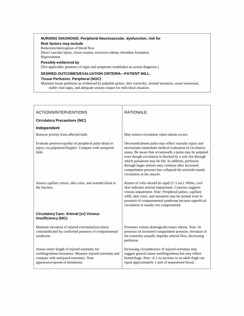
NURSING DIAGNOSIS: Peripheral Neurovascular, dysfunction, risk for
Risk factors may include
Reduction/interruption of blood flow
Direct vascular injury, tissue trauma, excessive edema, thrombus formation
Hypovolemia
Possibly evidenced by
[Not applicable; presence of signs and symptoms establishes an actual diagnosis.]
DESIRED OUTCOMES/EVALUATION CRITERIA—PATIENT WILL:
Tissue Perfusion: Peripheral (NOC)
Maintain tissue perfusion as evidenced by palpable pulses, skin warm/dry, normal sensation, usual sensorium,
stable vital signs, and adequate urinary output for individual situation.
ACTIONS/INTERVENTIONS
Circulatory Precautions (NIC)
Independent
Remove jewelry from affected limb.
Evaluate presence/quality of peripheral pulse distal to
injury via palpation/Doppler. Compare with uninjured
limb.
Assess capillary return, skin color, and warmth distal to
the fracture.
Circulatory Care: Arterial [or] Venous
Insufficiency (NIC)
Maintain elevation of injured extremity(ies) unless
contraindicated by confirmed presence of compartmental
syndrome.
Assess entire length of injured extremity for
swelling/edema formation. Measure injured extremity and
compare with uninjured extremity. Note
appearance/spread of hematoma.
RATIONALE
May restrict circulation when edema occurs.
Decreased/absent pulse may reflect vascular injury and
necessitates immediate medical evaluation of circulatory
status. Be aware that occasionally a pulse may be palpated
even though circulation is blocked by a soft clot through
which pulsations may be felt. In addition, perfusion
through larger arteries may continue after increased
compartment pressure has collapsed the arteriole/venule
circulation in the muscle.
Return of color should be rapid (3–5 sec). White, cool
skin indicates arterial impairment. Cyanosis suggests
venous impairment. Note: Peripheral pulses, capillary
refill, skin color, and sensation may be normal even in
presence of compartmental syndrome because superficial
circulation is usually not compromised.
Promotes venous drainage/decreases edema. Note: In
presence of increased compartment pressure, elevation of
the extremity actually impedes arterial flow, decreasing
perfusion.
Increasing circumference of injured extremity may
suggest general tissue swelling/edema but may reflect
hemorrhage. Note: A 1-in increase in an adult thigh can
equal approximately 1 unit of sequestered blood.

ACTIONS/INTERVENTIONS
Circulatory Care: Arterial [or] Venous
Insufficiency (NIC)
Independent
Note reports of pain extreme for type of injury or
increasing pain on passive movement of extremity,
development of paresthesia, muscle tension/tenderness
with erythema, and change in pulse quality distal to
injury. Do not elevate extremity. Report symptoms to
physician at once.
Investigate sudden signs of limb ischemia, e.g., decreased
skin temperature, pallor, and increased pain.
Encourage patient to routinely exercise digits/joints distal
to injury. Ambulate as soon as possible.
Investigate tenderness, swelling, pain on dorsiflexion of
foot (positive Homans’ sign).
Monitor vital signs. Note signs of general pallor/cyanosis,
cool skin, changes in mentation.
Test stools/gastric aspirant for occult blood. Note
continued bleeding at trauma/injection site(s) and oozing
from mucous membranes.
Pressure Management (NIC)
Perform neurovascular assessments, noting changes in
motor/sensory function. Ask patient to localize pain/
discomfort.
Test sensation of peroneal nerve by pinch/pinprick in the
dorsal web between the first and second toe, and assess
ability to dorsiflex toes if indicated.
Assess tissues around cast edges for rough places/pressure
points. Investigate reports of ―burning sensation‖ under
cast.
Monitor position/location of supporting ring of splints or
sling.
RATIONALE
Continued bleeding/edema formation within a muscle
enclosed by tight fascia can result in impaired blood flow
and ischemic myositis or compartmental syndrome,
necessitating emergency interventions to relieve
pressure/restore circulation. Note: This condition
constitutes a medical emergency and requires immediate
intervention.
Fracture dislocations of joints (especially the knee) may
cause damage to adjacent arteries, with resulting loss of
distal blood flow.
Enhances circulation and reduces pooling of blood,
especially in the lower extremities.
There is an increased potential for thrombophlebitis and
pulmonary emboli in patients immobile for several days.
Note: The absence of a positive Homans’ sign is not a
reliable indicator in many people, especially the elderly
because they often have reduced pain sensation.
Inadequate circulating volume compromises systemic
tissue perfusion.
Increased incidence of gastric bleeding accompanies
fractures/trauma and may be related to stress or
occasionally reflects a clotting disorder requiring further
evaluation.
Impaired feeling, numbness, tingling, increased/diffuse
pain occur when circulation to nerves is inadequate or
nerves are damaged.
Length and position of peroneal nerve increase risk of its
injury in the presence of leg fracture,
edema/compartmental syndrome, or malposition of
traction apparatus.
These factors may be the cause of or be indicative of
tissue pressure/ischemia, leading to breakdown/necrosis.
Traction apparatus can cause pressure on vessels/nerves,
particularly in the axilla and groin, resulting in ischemia
and possible permanent nerve damage.
ACTIONS/INTERVENTIONS
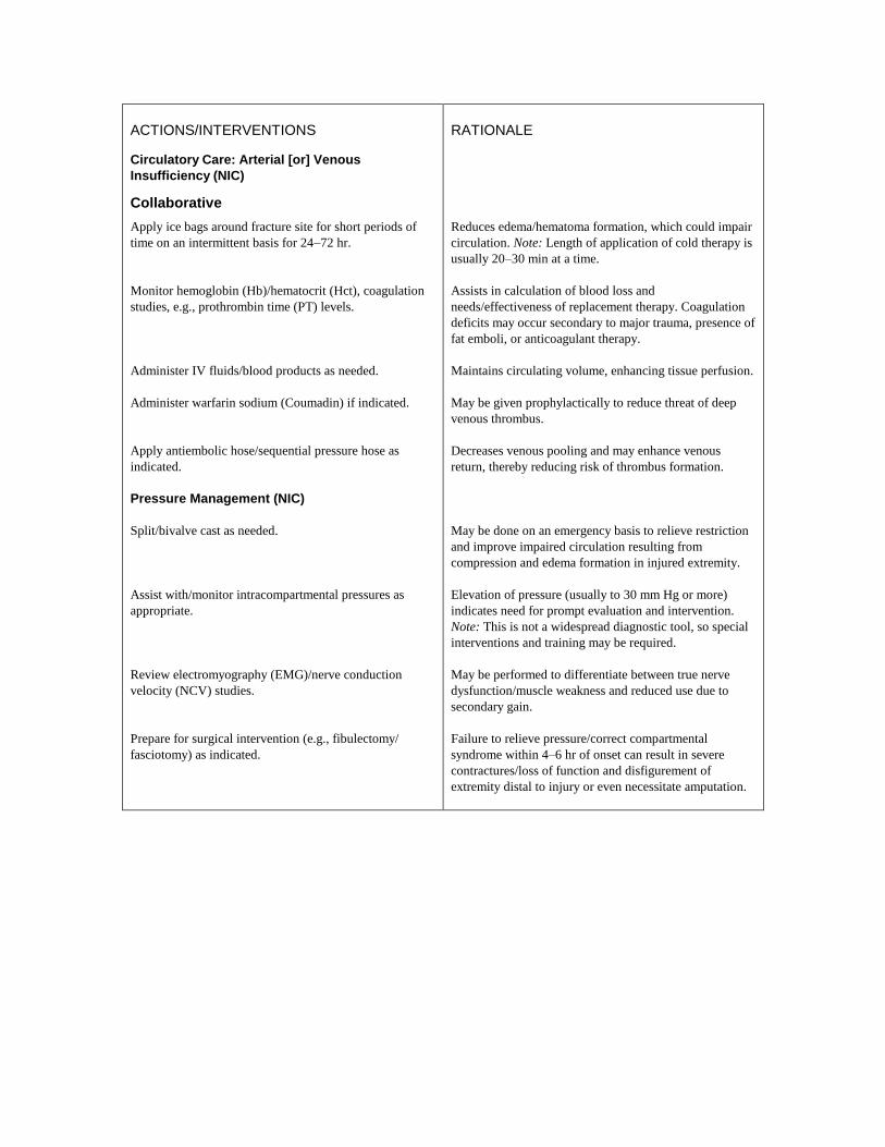
Circulatory Care: Arterial [or] Venous
Insufficiency (NIC)
Collaborative
Apply ice bags around fracture site for short periods of
time on an intermittent basis for 24–72 hr.
Monitor hemoglobin (Hb)/hematocrit (Hct), coagulation
studies, e.g., prothrombin time (PT) levels.
Administer IV fluids/blood products as needed.
Administer warfarin sodium (Coumadin) if indicated.
Apply antiembolic hose/sequential pressure hose as
indicated.
Pressure Management (NIC)
Split/bivalve cast as needed.
Assist with/monitor intracompartmental pressures as
appropriate.
Review electromyography (EMG)/nerve conduction
velocity (NCV) studies.
Prepare for surgical intervention (e.g., fibulectomy/
fasciotomy) as indicated.
RATIONALE
Reduces edema/hematoma formation, which could impair
circulation. Note: Length of application of cold therapy is
usually 20–30 min at a time.
Assists in calculation of blood loss and
needs/effectiveness of replacement therapy. Coagulation
deficits may occur secondary to major trauma, presence of
fat emboli, or anticoagulant therapy.
Maintains circulating volume, enhancing tissue perfusion.
May be given prophylactically to reduce threat of deep
venous thrombus.
Decreases venous pooling and may enhance venous
return, thereby reducing risk of thrombus formation.
May be done on an emergency basis to relieve restriction
and improve impaired circulation resulting from
compression and edema formation in injured extremity.
Elevation of pressure (usually to 30 mm Hg or more)
indicates need for prompt evaluation and intervention.
Note: This is not a widespread diagnostic tool, so special
interventions and training may be required.
May be performed to differentiate between true nerve
dysfunction/muscle weakness and reduced use due to
secondary gain.
Failure to relieve pressure/correct compartmental
syndrome within 4–6 hr of onset can result in severe
contractures/loss of function and disfigurement of
extremity distal to injury or even necessitate amputation.

NURSING DIAGNOSIS: Gas Exchange, risk for impaired
Risk factors may include
Altered blood flow; blood/fat emboli
Alveolar/capillary membrane changes: interstitial, pulmonary edema, congestion
Possibly evidenced by
[Not applicable; presence of signs and symptoms establishes an actual diagnosis.]
DESIRED OUTCOMES/EVALUATION CRITERIA—PATIENT WILL:
Respiratory Status: Gas Exchange (NOC)
Maintain adequate respiratory function, as evidenced by absence of dyspnea/cyanosis; respiratory rate and
arterial blood gases (ABGs) within patient’s normal range.
ACTIONS/INTERVENTIONS
Respiratory Monitoring (NIC)
Independent
Monitor respiratory rate and effort. Note stridor, use of
accessory muscles, retractions, development of central
cyanosis.
Auscultate breath sounds, noting development of unequal,
hyperresonant sounds; also note presence of crackles/
rhonchi/wheezes and inspiratory crowing or croupy
sounds.
Handle injured tissues/bones gently, especially during
first several days.
Instruct and assist with deep-breathing and coughing
exercises. Reposition frequently.
Note increasing restlessness, confusion, lethargy, stupor.
Observe sputum for signs of blood.
Inspect skin for petechiae above nipple line; in axilla,
spreading to abdomen/trunk; buccal mucosa, hard palate;
conjunctival sacs and retina.
RATIONALE
Tachypnea, dyspnea, and changes in mentation are early
signs of respiratory insufficiency and may be the only
indicator of developing pulmonary emboli in the early
stage. Remaining signs/symptoms reflect advanced
respiratory distress/impending failure.
Changes in/presence of adventitious breath sounds
reflects developing respiratory complications, e.g.,
atelectasis, pneumonia, emboli, adult respiratory distress
syndrome (ARDS). Inspiratory crowing reflects upper
airway edema and is suggestive of fat emboli.
This may prevent the development of fat emboli (usually
seen in first 12–72 hr), which are closely associated with
fractures, especially of the long bones and pelvis.
Promotes alveolar ventilation and perfusion.
Repositioning promotes drainage of secretions and
decreases congestion in dependent lung areas.
Impaired gas exchange/presence of pulmonary emboli can
cause deterioration in patient’s level of consciousness as
hypoxemia/acidosis develops.
Hemoptysis may occur with pulmonary emboli.
This is the most characteristic sign of fat emboli, which
may appear within 2–3 days after injury.

ACTIONS/INTERVENTIONS
Respiratory Monitoring (NIC)
Collaborative
Assist with incentive spirometry.
Administer supplemental oxygen if indicated.
Monitor laboratory studies, e.g.:
Serial ABGs;
Hb, calcium, erythrocyte sedimentation rate (ESR),
serum lipase, fat screen, platelets, as appropriate.
Administer medications as indicated:
Low-molecular-weight heparin or heparinoids, e.g.,
enoxaparin (Lovenox), dalteparin (Fragmin),
ardeparin (Normiflo);
Corticosteroids.
RATIONALE
Increases available O2 for optimal tissue oxygenation.
Decreased Pao2 and increased Paco2 indicate impaired gas
exchange/developing failure.
Maximizes ventilation/oxygenation and minimizes
atelectasis.
Anemia, hypocalcemia, elevated ESR and lipase levels,
fat globules in blood/urine/sputum, and decreased platelet
count (thrombocytopenia) are often associated with fat
emboli.
Used for prevention of thromboembolic phenomena,
including deep vein thrombosis and pulmonary emboli.
Steroids have been used with some success to
prevent/treat fat embolus.
NURSING DIAGNOSIS: Mobility, impaired physical
May be related to
Neuromuscular skeletal impairment; pain/discomfort; restrictive therapies (limb immobilization)
Psychological immobility
Possibly evidenced by
Inability to move purposefully within the physical environment, imposed restrictions
Reluctance to attempt movement; limited ROM
Decreased muscle strength/control
DESIRED OUTCOMES/EVALUATION CRITERIA—PATIENT WILL:
Mobility Level (NOC)
Regain/maintain mobility at the highest possible level.
Maintain position of function.
Increase strength/function of affected and compensatory body parts.
Demonstrate techniques that enable resumption of activities.

ACTIONS/INTERVENTIONS
Bed Rest Care (NIC)
Independent
Assess degree of immobility produced by injury/treatment
and note patient’s perception of immobility.
Encourage participation in diversional/recreational
activities. Maintain stimulating environment, e.g., radio,
TV, newspapers, personal possessions/pictures, clock,
calendar, visits from family/friends.
Instruct patient in/assist with active/passive ROM
exercises of affected and unaffected extremities.
Encourage use of isometric exercises starting with the
unaffected limb.
Provide footboard, wrist splints, trochanter/hand rolls as
appropriate.
Place in supine position periodically if possible, when
traction is used to stabilize lower limb fractures.
Instruct in/encourage use of trapeze and ―post position‖
for lower limb fractures.
Assist with/encourage self-care activities (e.g., bathing,
shaving).
Provide/assist with mobility by means of wheelchair,
walker, crutches, canes as soon as possible. Instruct in
safe use of mobility aids.
Monitor blood pressure (BP) with resumption of activity.
Note reports of dizziness.
RATIONALE
Patient may be restricted by self-view/self-perception out
of proportion with actual physical limitations, requiring
information/interventions to promote progress toward
wellness.
Provides opportunity for release of energy, refocuses
attention, enhances patient’s sense of
self-control/self-worth, and aids in reducing social
isolation.
Increases blood flow to muscles and bone to improve
muscle tone, maintain joint mobility; prevent
contractures/atrophy and calcium resorption from disuse.
Isometrics contract muscles without bending joints or
moving limbs and help maintain muscle strength and
mass. Note: These exercises are contraindicated while
acute bleeding/edema is present.
Useful in maintaining functional position of extremities,
hands/feet, and preventing complications (e.g.,
contractures/footdrop).
Reduces risk of flexion contracture of hip.
Facilitates movement during hygiene/skin care and linen
changes; reduces discomfort of remaining flat in bed.
―Post position‖ involves placing the uninjured foot flat on
the bed with the knee bent while grasping the trapeze and
lifting the body off the bed.
Improves muscle strength and circulation, enhances
patient control in situation, and promotes self-directed
wellness.
Early mobility reduces complications of bed rest (e.g.,
phlebitis) and promotes healing and normalization of
organ function. Learning the correct way to use aids is
important to maintain optimal mobility and patient safety.
Postural hypotension is a common problem following
prolonged bed rest and may require specific interventions
(e.g., tilt table with gradual elevation to upright position).

ACTIONS/INTERVENTIONS
Bed Rest Care (NIC)
Independent
Reposition periodically and encourage
coughing/deep-breathing exercises.
Auscultate bowel sounds. Monitor elimination habits and
provide for regular bowel routine. Place on bedside
commode, if feasible, or use fracture pan. Provide
privacy.
Encourage increased fluid intake to 2000–3000 mL/day
(within cardiac tolerance), including acid/ash juices.
Provide diet high in proteins, carbohydrates, vitamins,
and minerals, limiting protein content until after first
bowel movement.
Increase the amount of roughage/fiber in the diet. Limit
gas-forming foods.
Collaborative
Consult with physical/occupational therapist and/or
rehabilitation specialist.
Initiate bowel program (stool softeners, enemas,
laxatives) as indicated.
Refer to psychiatric clinical nurse specialist/therapist as
indicated.
RATIONALE
Prevents/reduces incidence of skin and respiratory
complications (e.g., decubitus, atelectasis, pneumonia).
Bed rest, use of analgesics, and changes in dietary habits
can slow peristalsis and produce constipation. Nursing
measures that facilitate elimination may prevent/limit
complications. Fracture pan limits flexion of hips and
lessens pressure on lumbar region/lower extremity cast.
Keeps the body well hydrated, decreasing risk of urinary
infection, stone formation, and constipation.
In the presence of musculoskeletal injuries, nutrients
required for healing are rapidly depleted, often resulting
in a weight loss of as much as 20/30 lb during skeletal
traction. This can have a profound effect on muscle mass,
tone, and strength. Note: Protein foods increase contents
in small bowel, resulting in gas formation and
constipation. Therefore, gastrointestinal (GI) function
should be fully restored before protein foods are
increased.
Adding bulk to stool helps prevent constipation.
Gas-forming foods may cause abdominal distension,
especially in presence of decreased intestinal motility.
Useful in creating individualized activity/exercise
program. Patient may require long-term assistance with
movement, strengthening, and weight-bearing activities,
as well as use of adjuncts, e.g., walkers, crutches, canes;
elevated toilet seats; pickup sticks/reachers; special eating
utensils.
Done to promote regular bowel evacuation.
Patient/SO may require more intensive treatment to deal
with reality of current condition/prognosis, prolonged
immobility, perceived loss of control.

NURSING DIAGNOSIS: Skin/Tissue Integrity, impaired: actual/risk for
May be related to
Puncture injury; compound fracture; surgical repair; insertion of traction pins, wires, screws
Altered sensation, circulation; accumulation of excretions/secretions
Physical immobilization
Possibly evidenced by (actual)
Reports of itching, pain, numbness, pressure in affected/surrounding area
Disruption of skin surface; invasion of body structures; destruction of skin layers/tissues
DESIRED OUTCOMES/EVALUATION CRITERIA—PATIENT WILL:
Tissue Integrity: Skin &Mucous Membranes (NOC)
Verbalize relief of discomfort.
Demonstrate behaviors/techniques to prevent skin breakdown/facilitate healing as indicated.
Achieve timely wound/lesion healing if present.
ACTIONS/INTERVENTIONS
Skin Surveillance (NIC)
Independent
Examine the skin for open wounds, foreign bodies,
rashes, bleeding, discoloration, duskiness, blanching.
Massage skin and bony prominences. Keep the bed linens
dry and free of wrinkles. Place water pads/other padding
under elbows/heels as indicated.
Reposition frequently. Encourage use of trapeze if
possible.
Assess position of splint ring of traction device.
Cast Care: Wet (NIC)
Plaster cast application and skin care:
Cleanse skin with soap and water. Rub gently with
alcohol and/or dust with small amount of a zinc or
stearate powder;
Cut a length of stockinette to cover the area and
extend several inches beyond the cast;
Use palm of hand to apply, hold, or move cast and
support on pillows after application;
RATIONALE
Provides information regarding skin circulation and
problems that may be caused by application and/or
restriction of cast/splint or traction apparatus, or edema
formation that may require further medical intervention.
Reduces pressure on susceptible areas and risk of
abrasions/skin breakdown.
Lessens constant pressure on same areas and minimizes
risk of skin breakdown. Use of trapeze may reduce risk of
abrasions to elbows/heels.
Improper positioning may cause skin injury/breakdown.
Provides a dry, clean area for cast application. Note:
Excess powder may cake when it comes in contact with
water/perspiration.
Useful for padding bony prominences, finishing cast
edges, and protecting the skin.
Prevents indentations/flattening over bony prominences
and weight-bearing areas (e.g., back of heels), which
would cause abrasions/tissue trauma. An improperly
shaped or dried cast is irritating to the underlying skin
and may lead to circulatory impairment.

ACTIONS/INTERVENTIONS
Cast Care: Wet (NIC)
Independent
Trim excess plaster from edges of cast as soon as
casting is completed;
Promote cast drying by removing bed linen,
exposing to circulating air;
Observe for potential pressure areas, especially at the
edges of and under the splint/cast;
Pad (petal) the edges of the cast with waterproof
tape;
Cleanse excess plaster from skin while still wet, if
possible;
Protect cast and skin in perineal area. Provide
frequent perineal care;
Instruct patient/SO to avoid inserting objects inside
casts;
Massage the skin around the cast edges with alcohol;
Turn frequently to include the uninvolved side, back,
and prone positions (as tolerated) with patient’s feet
over the end of the mattress.
Traction/Immobilization Care (NIC)
Skin traction application and skin care:
Cleanse the skin with warm, soapy water;
Apply tincture of benzoin;
Apply commercial skin traction tapes (or make some
with strips of moleskin/adhesive tape) lengthwise on
opposite sides of the affected limb;
Extend the tapes beyond the length of the limb;
Mark the line where the tapes extend beyond the
extremity;
Place protective padding under the leg and over bony
prominences;
RATIONALE
Uneven plaster is irritating to the skin and may result in
abrasions.
Prevents skin breakdown caused by prolonged moisture
trapped under cast.
Pressure can cause ulcerations, necrosis, and/or nerve
palsies. These problems may be painless when nerve
damage is present.
Provides an effective barrier to cast flaking and moisture.
Helps prevent breakdown of cast material at edges and
reduces skin irritation/excoriation.
Dry plaster may flake into completed cast and cause skin
damage.
Prevents tissue breakdown and infection by fecal
contamination.
―Scratching an itch‖ may cause tissue injury.
Has a drying effect, which toughens the skin. Creams and
lotions are not recommended because excessive oils can
seal cast perimeter, not allowing the cast to ―breathe.‖
Powders are not recommended because of potential for
excessive accumulation inside the cast.
Minimizes pressure on feet and around cast edges.
Reduces level of contaminants on skin.
―Toughens‖ the skin for application of skin traction.
Traction tapes encircling a limb may compromise
circulation.
Traction is inserted in line with the free ends of the tape.
Allows for quick assessment of slippage.
Minimizes pressure on these areas.

ACTIONS/INTERVENTIONS
Traction/Immobilization Care (NIC)
Independent
Wrap the limb circumference, including tapes and
padding, with elastic bandages, being careful to wrap
snugly but not too tightly;
Palpate taped tissues daily and document any
tenderness or pain;
Remove skin traction every 24 hr, per protocol;
inspect and give skin care.
Skeletal traction/fixation application and skin care:
Bend wire ends or cover ends of wires/pins with
rubber or cork protectors or needle caps;
Pad slings/frame with sheepskin, foam.
Pressure Management (NIC)
Collaborative
Provide foam mattress, sheepskins, flotation pads, or air
mattress as indicated.
Monovalve, bivalve, or cut a window in the cast, per
protocol.
RATIONALE
Provides for appropriate traction pull without
compromising circulation.
If area under tapes is tender, suspect skin irritation, and
prepare to remove the bandage system.
Maintains skin integrity.
Prevents injury to other body parts.
Prevents excessive pressure on skin and promotes
moisture evaporation that reduces risk of excoriation.
Because of immobilization of body parts, bony
prominences other than those affected by the casting may
suffer from decreased circulation.
Allows the release of pressure and provides access for
wound/skin care.
NURSING DIAGNOSIS: Infection, risk for
Risk factors may include
Inadequate primary defenses: broken skin, traumatized tissues; environmental exposure
Invasive procedures, skeletal traction
Possibly evidenced by
[Not applicable; presence of signs and symptoms establishes an actual diagnosis.]
DESIRED OUTCOMES/EVALUATION CRITERIA—PATIENT WILL:
Infection Status (NOC)
Achieve timely wound healing, be free of purulent drainage or erythema, and be afebrile.

ACTIONS/INTERVENTIONS
Infection Prevention (NIC)
Independent
Inspect the skin for preexisting irritation or breaks in
continuity.
Assess pin sites/skin areas, noting reports of increased
pain/burning sensation or presence of edema, erythema,
foul odor, or drainage.
Provide sterile pin/wound care according to protocol, and
exercise meticulous handwashing.
Instruct patient not to touch the insertion sites.
Line perineal cast edges with plastic wrap.
Observe wounds for formation of bullae, crepitation,
bronze discoloration of skin, frothy/fruity-smelling
drainage.
Assess muscle tone, reflexes, and ability to speak.
Monitor vital signs. Note presence of chills, fever,
malaise, changes in mentation.
Investigate abrupt onset of pain/limitation of movement
with localized edema/erythema in injured extremity.
Institute prescribed isolation procedures.
Collaborative
Monitor laboratory/diagnostic studies, e.g.:
Complete blood count (CBC);
ESR;
Cultures and sensitivity of wound/serum/bone;
Radioisotope scans.
Administer medications as indicated, e.g.:
IV/topical antibiotics;
RATIONALE
Pins or wires should not be inserted through skin
infections, rashes, or abrasions (may lead to bone
infection).
May indicate onset of local infection/tissue necrosis,
which can lead to osteomyelitis.
May prevent cross-contamination and possibility of
infection.
Minimizes opportunity for contamination.
Damp, soiled casts can promote growth of bacteria.
Signs suggestive of gas gangrene infection.
Muscle rigidity, tonic spasms of jaw muscles, and
dysphagia reflect development of tetanus.
Hypotension, confusion may be seen with gas gangrene;
tachycardia and chills/fever reflect developing sepsis.
May indicate development of osteomyelitis.
Presence of purulent drainage requires wound/linen
precautions to prevent cross-contamination.
Anemia may be noted with osteomyelitis; leukocytosis is
usually present with infective processes.
Elevated in osteomyelitis.
Identifies infective organism and effective antimicrobial
agent(s).
Hot spots signify increased areas of vascularity, indicative
of osteomyelitis.
Wide-spectrum antibiotics may be used prophylactically
or may be geared toward a specific microorganism.

ACTIONS/INTERVENTIONS
Infection Prevention (NIC)
Independent
Tetanus toxoid.
Provide wound/bone irrigations and apply warm/moist
soaks as indicated.
Assist with procedures, e.g., incision/drainage, placement
of drains, hyperbaric oxygen therapy.
Prepare for surgery, as indicated.
RATIONALE
Given prophylactically because the possibility of tetanus
exists with any open wound. Note: Risk increases when
injury/wound(s) occur in ―field conditions‖ (outdoor/rural
areas, work environment).
Local debridement/cleansing of wounds reduces
microorganisms and incidence of systemic infection.
Continuous antimicrobial drip into bone may be necessary
to treat osteomyelitis, especially if blood supply to bone is
compromised.
Numerous procedures may be carried out in treatment of
local infections, osteomyelitis, gas gangrene.
Sequestrectomy (removal of necrotic bone) is necessary to
facilitate healing and prevent extension of infectious
process.
NURSING DIAGNOSIS: Knowledge, deficient [Learning Need] regarding condition, prognosis,
treatment, self-care, and discharge needs
May be related to
Lack of exposure/recall
Information misinterpretation/unfamiliarity with information resources
Possibly evidenced by
Questions/request for information, statement of misconception
Inaccurate follow-through of instructions, development of preventable complications
DESIRED OUTCOMES/EVALUATION CRITERIA—PATIENT WILL:
Knowledge: Treatment Regimen (NOC)
Verbalize understanding of condition, prognosis, and potential complications.
Correctly perform necessary procedures and explain reasons for actions.

ACTIONS/INTERVENTIONS
Teaching: Disease Process (NIC)
Independent
Review pathology, prognosis, and future expectations.
Discuss dietary needs.
Discuss individual drug regimen as appropriate.
Reinforce methods of mobility and ambulation as
instructed by physical therapist when indicated.
Suggest use of a backpack.
List activities patient can perform independently and
those that require assistance.
Identify available community services, e.g., rehabilitation
teams, home nursing/homemaker services.
Encourage patient to continue active exercises for the
joints above and below the fracture.
Discuss importance of clinical and therapy follow-up
appointments.
Review proper pin/wound care.
Recommend cleaning external fixator regularly.
RATIONALE
Provides knowledge base from which patient can make
informed choices. Note: Internal fixation devices can
ultimately compromise the bone’s strength, and
intramedullary nails/rods or plates may be removed at a
future date.
A low-fat diet with adequate quality protein and rich in
calcium promotes healing and general well-being.
Proper use of pain medication and antiplatelet agents can
reduce risk of complications. Long-term use of
alendronate (Fosamax) may reduce risk of stress fractures.
Note: Fosamax should be taken on an empty stomach
with plain water because absorption of drug may be
altered by food and some medications (e.g., antacids,
calcium supplements).
Most fractures require casts, splints, or braces during the
healing process. Further damage and delay in healing
could occur secondary to improper use of ambulatory
devices.
Provides place to carry necessary articles and leaves
hands free to manipulate crutches; may prevent undue
muscle fatigue when one arm is casted.
Organizes activities around need and who is available to
provide help.
Provides assistance to facilitate self-care and support
independence. Promotes optimal self-care and recovery.
Prevents joint stiffness, contractures, and muscle wasting,
promoting earlier return to independence in activities of
daily living (ADLs).
Fracture healing may take as long as a year for
completion, and patient cooperation with the medical
regimen facilitates proper union of bone. Physical therapy
(PT)/occupational therapy (OT) may be indicated for
exercises to maintain/strengthen muscles and improve
function. Additional modalities such as low-intensity
ultrasound may be used to stimulate healing of
lower-forearm or lower-leg fractures.
Reduces risk of bone/tissue trauma and infection, which
can progress to osteomyelitis.
Keeping device free of dust/contaminants reduces risk of
infection.

ACTIONS/INTERVENTIONS
Teaching: Disease Process (NIC)
Independent
Identify signs/symptoms requiring medical evaluation,
e.g., severe pain, fever/chills, foul odors; changes in
sensation, swelling, burning, numbness, tingling, skin
discoloration, paralysis, white/cool toes or fingertips;
warm spots, soft areas, cracks in cast.
Discuss care of ―green‖ or wet cast.
Suggest the use of a blow-dryer to dry small areas of
dampened casts.
Demonstrate use of plastic bags to cover plaster cast
during wet weather or while bathing. Clean soiled cast
with a slightly dampened cloth and some scouring
powder.
Emphasize importance of not adjusting clamps/nuts of
external fixator.
Recommend use of adaptive clothing.
Suggest ways to cover toes, if appropriate, e.g.,
stockinette or soft socks.
Discuss postcast removal instructions:
Instruct patient to continue exercises as permitted;
Inform patient that the skin under the cast is commonly
mottled and covered with scales or crusts of dead skin;
Wash the skin gently with soap, povidone-iodine
(Betadine), or pHisoDerm, and water. Lubricate with a
protective emollient;
Inform patient that muscles may appear flabby and
atrophied (less muscle mass). Recommend supporting the
joint above and below the affected part and the use of
mobility aids, e.g., elastic bandages, splints, braces,
crutches, walkers, or canes;
Elevate the extremity as needed.
RATIONALE
Prompt intervention may reduce severity of complications
such as infection/impaired circulation. Note: Some
darkening of the skin (vascular congestion) may occur
normally when walking on the casted extremity or using
casted arm; however, this should resolve with rest and
elevation.
Promotes proper curing to prevent cast deformities and
associated misalignment/skin irritation. Note: Placing a
―cooling‖ cast directly on rubber or plastic pillows traps
heat and increases drying time.
Cautious use can hasten drying.
Protects from moisture, which softens the plaster and
weakens the cast. Note: Fiberglass casts are being used
more frequently because they are not affected by
moisture. In addition, their light weight may enhance
patient participation in desired activities.
Tampering may alter compression and misalign fracture.
Facilitates dressing/grooming activities.
Helps maintain warmth/protect from injury.
Reduces stiffness and improves strength and function of
affected extremity.
It will be several weeks before normal appearance returns.
New skin is extremely tender because it has been
protected beneath a cast.
Muscle strength will be reduced and new or different
aches and pains may occur for awhile secondary to loss
of support.
Swelling and edema tend to occur after cast removal.
POTENTIAL CONSIDERATIONS following acute hospitalization (dependent on patient’s age,
physical condition/presence of complications, personal resources, and life responsibilities)
In addition to surgical considerations:

Trauma, risk for—loss of skeletal integrity, weakness, balancing difficulties, reduced muscle coordination, lack of
safety precautions, history of previous trauma.
Mobility, impaired physical—neuromuscular skeletal impairment; pain/discomfort, restrictive therapies (limb
immobilization); psychological immobility.
Self-Care deficit—musculoskeletal impairment, decreased strength/endurance, pain.
Infection, risk for—inadequate primary defenses: broken skin, traumatized tissues; environmental exposure; invasive
procedures, skeletal traction.