Embed Size (px)
Citation preview
Ausr. N.Z. J . Surg. (1996) 66, 110-1 17
SURGICAL HISTORY
FREDERIC WOOD JONES: IT WAS A LARGE NERVE AND NOT AN ARTERY
B. E. CHRISTOPHERS
MANCHESTER In early September of 1909, Wood Jones joined the staff of the Manchester University as Lecturer in Anatomy under Professor Grafton Elliot Smith. This was 5 years after he had graduated in medicine and science from the Medical College of the London Hospital. Prior to joining the staff at the Manchester University, he had spent only 7 months as an academic. About 3 months after arriving in Manchester, Wood Jones wrote an article concerning the groove on the first rib, commonly referred to at that time, at least in the English speaking anatomical world, as the groove for the subclavian artery. At a meeting in Basle in 1895 the German Anatomical Society had adopted a system of anatomical nomen- clature known as the Base1 Nomina Anatomica or briefly the B.N.A. In this system the groove for the subclavian artery was named the ‘sulcus subclaviae’; anglicized to ‘subclavian groove’. Wood Jones submitted the article about this groove to the journal, AnatomischerAngeiger and it was published in the February 1910 issue of that journal.
Before the publication of this article, the standard teaching was that the subclavian artery occupied the ‘sulcus subclaviae’ and that the artery ips0 fact0 crossed the rib obliquely. In various text books of anatomy prior to 1910 sighted by the author, the ‘sulcus subclaviae’ is labelled on illustrations of the first rib as the groove for the subclavian artery. This is the case in the first edition of a Manual of Anatomy by Buchanan;‘ but in the text of this manual the occupants of the ‘sulcus subclaviae’ are given as, ‘[the groove] is occupied by the third part of the subclavian artery and the trunks of the brachial plexus of nerves’.
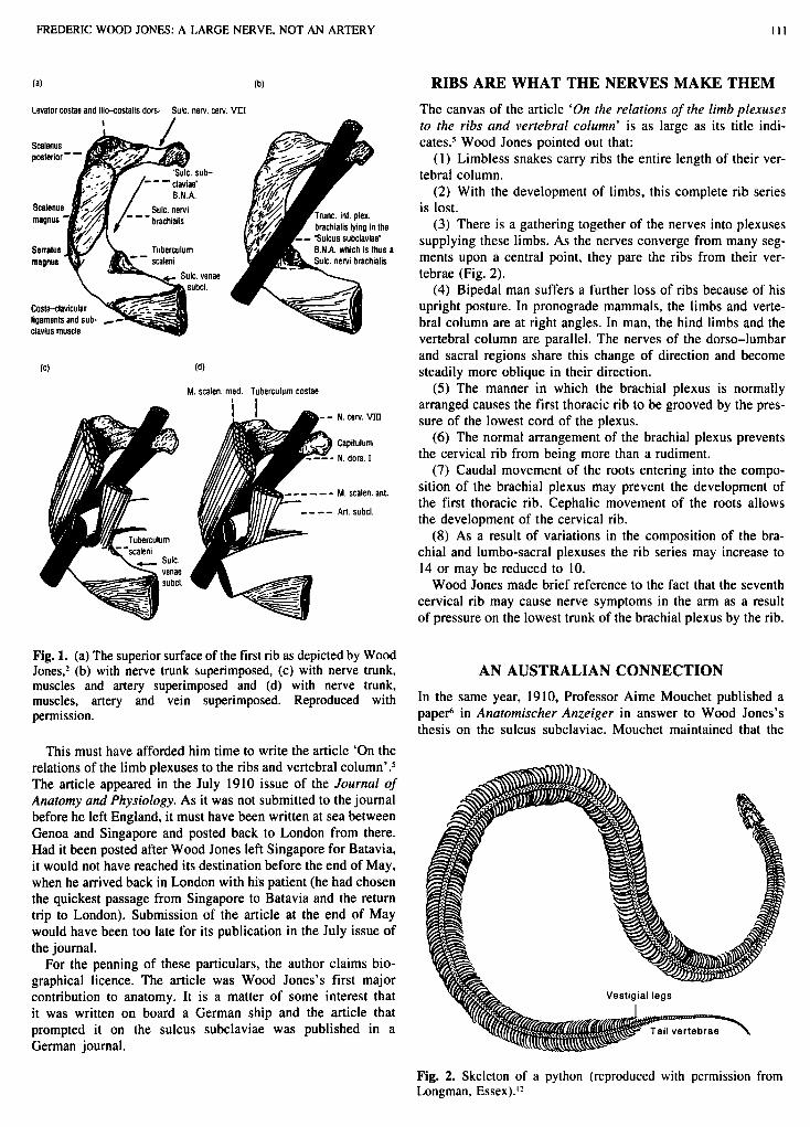
‘On the Real Significance of the “Sulcus Subclaviae” BNA and the Markings on the First Rib was the somewhat sum- monsing title of Wood Jones’s article.2 Here he pointed out that the groove, which was supposed to be formed by the artery and to lodge it, was directed obliquely across the rib; whereas the artery crossed the rib at right angles. A series of four illustra- tions appearing in the article effectively portray his point of view (Fig. I ) . Wood Jones was of the opinion that it was the manner in which the dissection of the region was usually carried out that was probably responsible for the teaching that the groove was caused by and lodged the subclavian artery:
When the arm of a dead subject is forcibly raised from the side, the resisting nerves are stretched and pulled upwards, and the lowest trunk of the brachial plexus, when dissected out, appears to lie altogether above the subclavian artery. But this position of the parts is quite unnatural, and is only brought about by disturbing the relations of the structure during dissection.
Correspondence: Dr B. E. Christophers, 377 Church Street, Richmond, Vic. 3 12 I , Australia.
Accepted for publication 26 April 1995.
How did Wood Jones happen upon this discovery? He may have read Buchanan’s description of the first rib referred to above. Two other possibilities were canvassed by him in this article as to how he came upon this finding.
( I ) A dissection by him of a cadaver whose arms were in the resting adducted position.
(2) A study by him of an illustration in Poirrier’s Traite d’Anatomie Humaine where the subclavian artery was shown lying in a position as Wood Jones described. In the illustration Wood Jones pointed out that ‘unfortunately the nerve trunk was not continued in the drawing so as to occupy the empty groove’.
In summary, Wood Jones’s view on the ‘sulcus subclaviae’ as presented in this paper was that the lowest trunk of the brachial plexus was responsible for and occupied this groove when the arm is adducted. It was while he was still in Manchester that he became aware of the important implications of his find that it was a large nerve and not an artery that lay in most intimate contact with the uppermost rib. He realized that the whole ques- tion of the relationship of the large nerves to the skeleton was involved and more immediately that here lay a ready explanation for the cause of the syndrome known as cervical rib.
A PRODUCTIVE JOURNEY It was in the midst of this study that he received an urgent plea for help from Cocos-Keeling Island. His future father-in-law, George Clunies Ross, was seriously ill and suffering pain. It was clear that he needed medical treatment not available on the atoll. Wood Jones left immediately to bring him back to England.
In order to hasten Clunies Ross’s return to England, he was taken from COCOS to Batavia by yacht so that Wood Jones could pick him up from there. Accompanying Wood Jones on his outward journey was Clunies Ross’s eldest son, Sydney, who was in England when the call for help was received. The two travelled overland to Genoa and here joined the steam ship Kronprinz Ludwig which was German from Captain to cabin boy. This took them to Singapore. From there they continued their journey to Batavia on a Dutch ship.
Wood Jones found an idiosyncratic means of keeping to himself on board the Kronprinz Ludwig. He shared the senti- ments of the character ‘Z’ in Bernard Shaw’s play Village W ~ o i n g , ~ who loathed ‘deck games especially quoits’:
I did not undertake the voyage as a pleasure trip and, after the first day out, decided that I would pass the time in reading, watch- ing the sea and sea birds and in growing another beard. The fact that I was in the early and unpleasant stage of growing a beard gave rise to the belief among the passengers that I was not quite right in the head and that Sydney Ross was in charge of me while I was travelling in order to regain my mental health. I had no objection to this, since it secured my immunity from the tiresome round of dances, sports and competitions that seemed to be regarded as the essential accompaniments of sea travel?
FREDERIC WOOD JONES: A LARGE NERVE, NOT AN ARTERY 1 1 1
(a) (b)
Levator costae and ilio-costalis dorsi Sulc. new. cerv. VIII I /
ubcl
Costa-clavicul lipaments and c!avlus muscle
r Tuberculum
‘rune. inf. plex. ~rachlalis iyinp In the
IS a ,uIc. nervi brachialls
(d)
M. scalen. med. Tuberculum costae
N. cerv. WI
Capitulum N. dors. 1
M. scalen. ant.
Art. subcl.
RIBS ARE WHAT THE NERVES MAKE THEM The canvas of the article ‘On the relations of the limb plexuses to rhe ribs and vertebral column’ is as large as its title indi- cates5 Wood Jones pointed out that:
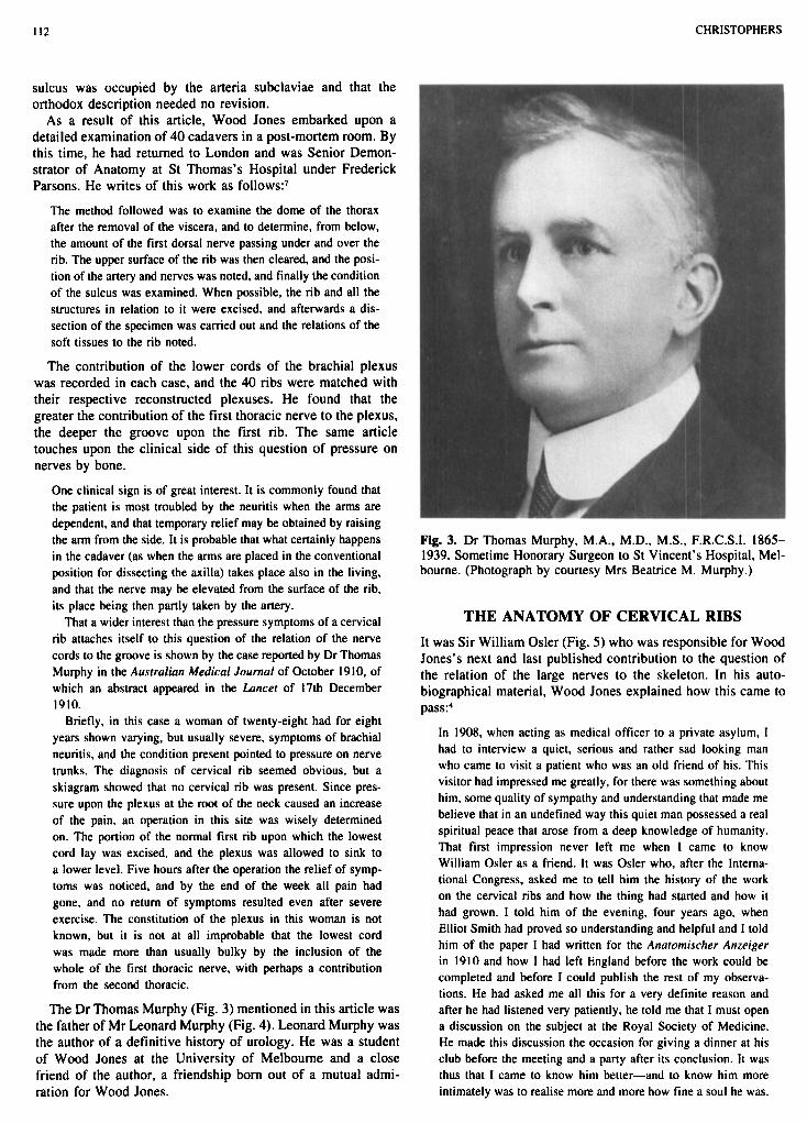
( I ) Limbless snakes carry ribs the entire length of their ver- tebral column.
(2) With the development of limbs, this complete rib series is lost.
(3) There is a gathering together of the nerves into plexuses supplying these limbs. As the nerves converge from many seg- ments upon a central point, they pare the ribs from their ver- tebrae (Fig. 2).
(4) Bipedal man suffers a further loss of ribs because of his upright posture. In pronograde mammals, the limbs and verte- bral column are at right angles. In man, the hind limbs and the vertebral column are parallel. The nerves of the dorso-lumbar and sacral regions share this change of direction and become steadily more oblique in their direction.
( 5 ) The manner in which the brachial plexus is normally arranged causes the first thoracic rib to be grooved by the pres- sure of the lowest cord of the plexus.
(6) The normal arrangement of the brachial plexus prevents the cervical rib from being more than a rudiment.
(7) Caudal movement of the roots entering into the compo- sition of the brachial plexus may prevent the development of the first thoracic rib. Cephalic movement of the roots allows the development of the cervical rib.
(8) As a result of variations in the composition of the bra- chial and lumbo-sacral plexuses the rib series may increase to 14 or may be reduced to 10.
Wood Jones made brief reference to the fact that the seventh cervical rib may cause nerve symptoms in the arm as a result of pressure on the lowest trunk of the brachial plexus by the rib.
Fig. 1. (a) The superior surface of the first rib as depicted by Wood Jones,* (b) with nerve trunk superimposed, (c) with nerve trunk, AN AUSTRALIAN CONNECTION muscles and artery superimposed and (d) with nerve trunk, muscles, artery and vein superimposed. Reproduced with In the same year, 1910, Professor Aime Mouchet published a permission. papeF in Anatomischer Anzeiger in answer to Wood Jones’s
thesis on the sulcus subclaviae. Mouchet maintained that the This must have afforded him time to write the article ‘On the
relations of the limb plexuses to the ribs and vertebral The article appeared in the July 1910 issue of the Journal of Anatomy and Physiology. As it was not submitted to the journal before he left England, it must have been written at sea between Genoa and Singapore and posted back to London from there. Had it been posted after Wood Jones left Singapore for Batavia, it would not have reached its destination before the end of May, when he arrived back in London with his patient (he had chosen the quickest passage from Singapore to Batavia and the return trip to London). Submission of the article at the end of May would have been too late for its publication in the July issue of the journal.
For the penning of these particulars, the author claims bio- graphical licence. The article was Wood Jones’s first major contribution to anatomy. It is a matter of some interest that it was written on board a German ship and the article that prompted it on the sulcus subclaviae was published in a German journal.
Vestigial legs
Fig. 2. Skeleton of a python (reproduced with permission from Longman, Essex).”
112 CHRISTOPHERS
sulcus was occupied by the arteria subclaviae and that the orthodox description needed no revision.
As a result of this article, Wood Jones embarked upon a detailed examination of 40 cadavers in a post-mortem room. By this time, he had returned to London and was Senior Demon- strator of Anatomy at St Thomas's Hospital under Frederick Parsons. He writes of this work as follows:'
The method followed was to examine the dome of the thorax after the removal of the viscera, and to determine, from below, the amount of the first dorsal nerve passing under and over the rib. The upper surface of the rib was then cleared, and the posi- tion of the artery and nerves was noted, and finally the condition of the sulcus was examined. When possible, the rib and all the structures in relation to it were excised, and afterwards a dis- section of the specimen was carried out and the relations of the soft tissues to the rib noted.
The contribution of the lower cords of the brachial plexus was recorded in each case, and the 40 ribs were matched with their respective reconstructed plexuses. He found that the greater the contribution of the first thoracic nerve to the plexus, the deeper the groove upon the first rib. The same article touches upon the clinical side of this question of pressure on nerves by bone.
One clinical sign is of great interest. It is commonly found that the patient is most troubled by the neuritis when the arms are dependent, and that temporary relief may be obtained by raising the arm from the side. It is probable that what certainly happens in the cadaver (as when the arms are placed in the conventional position for dissecting the axilla) takes place also in the living, and that the nerve may be elevated from the surface of the rib. its place being then partly taken by the artery.
That a wider interest than the pressure symptoms of a cervical rib attaches itself to this question of the relation of the nerve cords to the groove is shown by the case reported by Dr Thomas Murphy in the Australian Medical Journal of October 1910. of which an abstract appeared in the Lancer of 17th December 1910.
Briefly, in this case a woman of twenty-eight had for eight years shown varying, but usually severe, symptoms of brachial neuritis, and the condition present pointed to pressure on nerve trunks. The diagnosis of cervical rib seemed obvious, but a skiagram showed that no cervical rib was present. Since pres- sure upon the plexus at the root of the neck caused an increase of the pain, an operation in this site was wisely determined on. The portion of the normal first rib upon which the lowest cord lay was excised, and the plexus was allowed to sink to a lower level. Five hours after the operation the relief of symp- toms was noticed, and by the end of the week all pain had gone, and no return of symptoms resulted even after severe exercise. The constitution of the plexus in this woman is not known, but it is not at all improbable that the lowest cord was made more than usually bulky by the inclusion of the whole of the first thoracic nerve, with perhaps a contribution from the second thoracic.
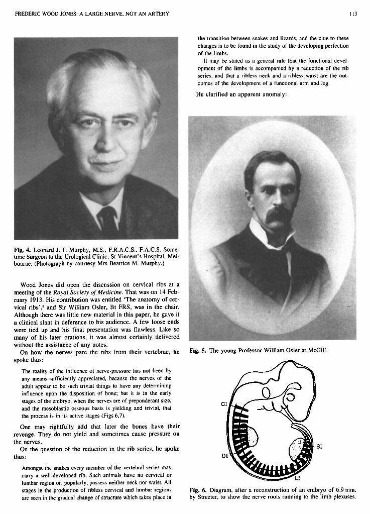
The Dr Thomas Murphy (Fig. 3) mentioned in this article was the father of Mr Leonard Murphy (Fig. 4). Leonard Murphy was the author of a definitive history of urology. He was a student of Wood Jones at the University of Melbourne and a close friend of the author, a friendship born out of a mutual admi- ration for Wood Jones.
Fig. 3. Dr Thomas Murphy, M.A., M.D., M.S., F.R.C.S.I. 1865- 1939. Sometime Honorary Surgeon to St Vincent's Hospital, Mel- bourne. (Photograph by courtesy Mrs Beatrice M. Murphy.)
THE ANATOMY OF CERVICAL RIBS It was Sir William Osler (Fig. 5 ) who was responsible for Wood Jones's next and last published contribution to the question of the relation of the large nerves to the skeleton. In his auto- biographical material, Wood Jones explained how this came to pass4
In 1908, when acting as medical officer to a private asylum, I had to interview a quiet, serious and rather sad looking man who came to visit a patient who was an old friend of his. This visitor had impressed me greatly, for there was something about him. some quality of sympathy and understanding that made me believe that in an undefined way this quiet man possessed a real spiritual peace that arose from a deep knowledge of humanity. That first impression never left me when I came to know William Osler as a friend. It was Osler who, after the Interna- tional Congress, asked me to tell him the history of the work on the cervical ribs and how the thing had started and how it had grown. I told him of the evening, four years ago, when Elliot Smith had proved so understanding and helpful and I told him of the paper I had written for the Anatomischer Anzeiger in 1910 and how I had left England before the work could be completed and before I could publish the rest of my observa- tions. He had asked me all this for a very definite reason and after he had listened very patiently, he told me that I must open a discussion on the subject at the Royal Society of Medicine. He made this discussion the occasion for giving a dinner at his club before the meeting and a party after its conclusion. It was thus that I came to know him better-and to know him more intimately was to realise more and more how fine a soul he was.
FREDERIC WOOD JONES: A LARGE NERVE, NOT AN ARTERY I13
the transition between snakes and lizards, and the clue to these changes is to be found in the study of the developing perfection of the limbs.
It may be stated as a general rule that the functional devel- opment of the limbs is accompanied by a reduction of the rib series, and that a ribless neck and a ribless waist are the out- comes of the development of a functional arm and leg.
He clarified an apparent anomaly:
Fig. 4. Leonard J. T. Murphy, M.S., F.R.A.C.S., F.A.C.S. Some- time Surgeon to the Urological Clinic, St Vincent’s Hospital, Mel- bourne. (Photograph by courtesy Mrs Beatrice M. Murphy.)
Wood Jones did open the discussion on cervical ribs at a meeting of the Royal Society of Medicine. That was on 14 Feb- ruary 1913. His contribution was entitled ‘The anatomy of cer- vical ribs’: and Sir William Osler, Bt FRS, was in the chair. Although there was little new material in this paper, he gave it a clinical slant in deference to his audience. A few loose ends were tied up and his final presentation was flawless. Like so many of his later orations, it was almost certainly delivered without the assistance of any notes.
On how the nerves pare the ribs from their vertebrae, he spoke thus:
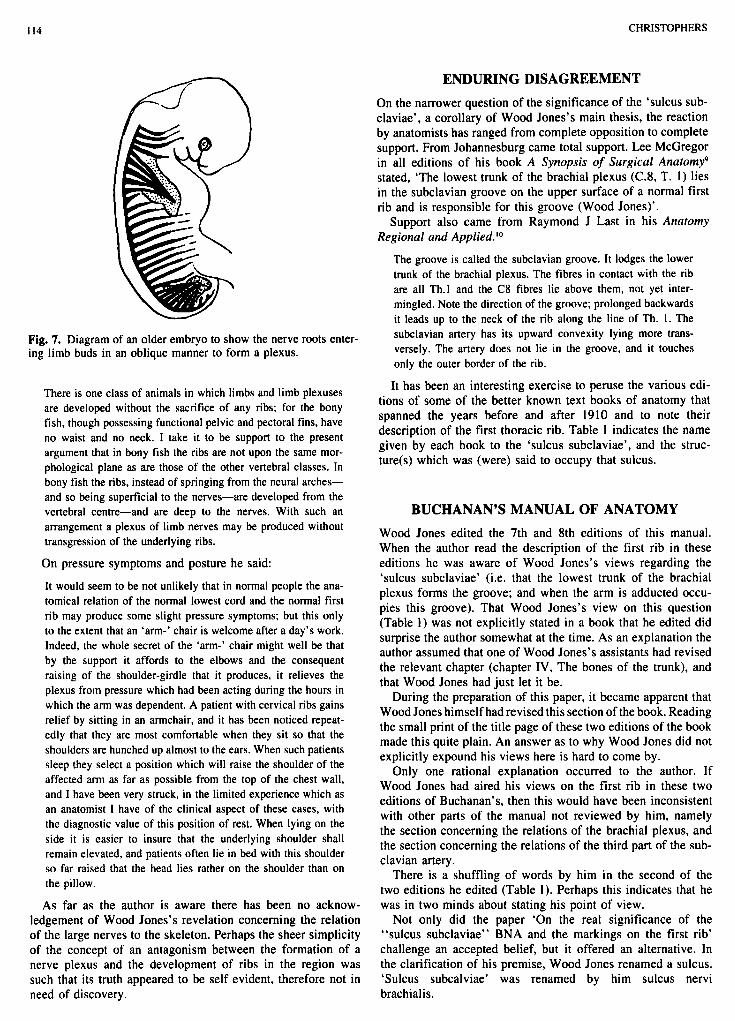
The reality of the influence of nerve-pressure has not been by any means sufficiently appreciated, because the nerves of the adult appear to be such trivial things to have any determining influence upon the disposition of bone; but it is in the early stages of the embryo, when the nerves are of preponderant size, and the mesoblastic osseous basis is yielding and trivial, that the process is in its active stages (Figs 6.7).
One may rightfully add that later the bones have their revenge. They do not yield and sometimes cause pressure on the nerves.
On the question of the reduction in the rib series, he spoke thus:
Amongst the snakes every member of the vertebral series may carry a well-developed rib. Such animals have no cervical or lumbar region or, popularly, possess neither neck nor waist. All
Fig. 5. The young Professor William Osler at McGill.
stages in the production of ribless cervical and lumbar regions are seen in the gradual change of structure which takes place in
Fig. 6. Diagram, after a reconstruction of an embryo of 6.9 mm, by Streeter, to show the nerve roots running to the limb plexuses.
I I4 CHRISTOPHERS
ENDURING DISAGREEMENT On the narrower question of the significance of the ‘sulcus sub- claviae’, a corollary of Wood Jones’s main thesis, the reaction by anatomists has ranged from complete opposition to complete support. From Johannesburg came total support. Lee McGregor in all editions of his book A Synopsis of Surgical Anatomy9 stated, ‘The lowest trunk of the brachial plexus (C.8, T. 1) lies in the subclavian groove on the upper surface of a normal first rib and is responsible for this groove (Wood Jones)’.
Support also came from Raymond J Last in his Anatomy Regional and Applied.‘O
The groove is called the subclavian groove. It lodges the lower trunk of the brachial plexus. The fibres in contact with the rib are all Th.1 and the C8 fibres lie above them, not yet inter- mingled. Note the direction of the groove; prolonged backwards it leads up to the neck of the rib along the line of Th. 1. The subclavian artery has its upward convexity lying more trans- versely. The artery does not lie in the groove, and it touches only the outer border of the rib.
It has been an interesting exercise to peruse the various edi- tions of some of the better known text books of anatomy that spanned the years before and after 1910 and to note their description of the first thoracic rib. Table 1 indicates the name given by each book to the ‘sulcus subclaviae’, and the struc- ture(s) which was (were) said to occupy that sulcus.
Fig. 7. Diagram of an older embryo to show the nerve roots enter- ing limb buds in an oblique manner to form a plexus.
There is one class of animals in which limbs and limb plexuses are developed without the sacrifice of any ribs; for the bony fish, though possessing functional pelvic and pectoral fins, have no waist and no neck. I take it to be support to the present argument that in bony fish the ribs are not upon the same mor- phological plane as are those of the other vertebral classes. In bony fish the ribs, instead of springing from the neural arches- and so being superficial to the nerves-are developed from the vertebral centre-and are deep to the nerves. With such an arrangement a plexus of limb nerves may be produced without transgression of the underlying ribs.
On pressure symptoms and posture he said:
It would seem to be not unlikely that in normal people the ana- tomical relation of the normal lowest cord and the normal first rib may produce some slight pressure symptoms; but this only to the extent that an ‘arm-’ chair is welcome after a day’s work. Indeed, the whole secret of the ‘arm-’ chair might well be that by the support it affords to the elbows and the consequent raising of the shoulder-girdle that it produces, it relieves the plexus from pressure which had been acting during the hours in which the arm was dependent. A patient with cervical ribs gains relief by sitting in an armchair, and it has been noticed repeat- edly that they are most comfortable when they sit so that the shoulders are hunched up almost to the ears. When such patients sleep they select a position which will raise the shoulder of the affected arm as far as possible from the top of the chest wall, and I have been very struck, in the limited experience which as an anatomist I have of the clinical aspect of these cases, with the diagnostic value of this position of rest. When lying on the side it is easier to insure that the underlying shoulder shall remain elevated, and patients often lie in bed with this shoulder so far raised that the head lies rather on the shoulder than on the pillow.
As far as the author is aware there has been no acknow- ledgement of Wood Jones’s revelation concerning the relation of the large nerves to the skeleton. Perhaps the sheer simplicity of the concept of an antagonism between the formation of a nerve plexus and the development of ribs in the region was such that its truth appeared to be self evident, therefore not in need of discovery.
BUCHANAN’S MANUAL OF ANATOMY Wood Jones edited the 7th and 8th editions of this manual. When the author read the description of the first rib in these editions he was aware of Wood Jones’s views regarding the ‘sulcus subclaviae’ (i.e. that the lowest trunk of the brachial plexus forms the groove; and when the arm is adducted occu- pies this groove). That Wood Jones’s view on this question (Table 1) was not explicitly stated in a book that he edited did surprise the author somewhat at the time. As an explanation the author assumed that one of Wood Jones’s assistants had revised the relevant chapter (chapter IV, The bones of the trunk), and that Wood Jones had just let it be.
During the preparation of this paper, it became apparent that Wood Jones himself had revised this section of the book. Reading the small print of the title page of these two editions of the book made this quite plain. An answer as to why Wood Jones did not explicitly expound his views here is hard to come by.
Only one rational explanation occurred to the author. If Wood Jones had aired his views on the first rib in these two editions of Buchanan’s, then this would have been inconsistent with other parts of the manual not reviewed by him, namely the section concerning the relations of the brachial plexus, and the section concerning the relations of the third part of the sub- clavian artery.
There is a shuffling of words by him in the second of the two editions he edited (Table 1). Perhaps this indicates that he was in two minds about stating his point of view.
Not only did the paper ‘On the real significance of the “sulcus subclaviae” BNA and the markings on the first rib’ challenge an accepted belief, but it offered an alternative. In the clarification of his premise, Wood Jones renamed a sulcus. ‘Sulcus subcalviae’ was renamed by him sulcus nervi brachialis.
FREDERIC WOOD JONES: A LARGE NERVE, NOT AN ARTERY 1 I5
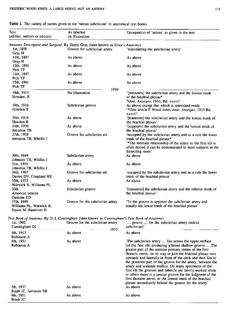
Table 1. The variety of names given to the ‘sulcus subclaviae’ in anatomical text books
Text As labelled (edition, authors or editors) on illustration
Occupant(s) of ‘sulcus’ as given in the text
Anatomy Descriptive and Surgical. By Henry Gray (later known as Gray’s Anatomy) 1st. 1858 Groove for subclavian artery ‘transmitting the subclavian artery’ Gray H 1 Ith, I887 Gray H 12th, 1890 Pick TP 14th, 1897 Pick TP 15th. 1901 Pick TP
As above
As above
As above
As above
18th, 1913 Howden, R
19th, 1916 Howden R
20th. 1918 Howden R 24th, 1930 Johnston TB 27th, 1938 Johnston TB, Whillis J
30th, 1949 Johnston TB, Whillis J 31st, 1954 Johnston TB, Whillis J 34th, 1967 Davies DV, Coupland RE 35th, 1973 Whwick R, Williams PL 30th American edition Carmine CD
No illustration
Subclavian groove
As above
As above
Groove for subclavian art.
Subclavian artery
As above
Groove for subclavian art.
As above
Subclavian groove
As above
As above
As above
As above
‘[transmits] the subclavian artery and the lowest trunk of the brachial plexus* *(Ant. Anzeiger, 1910, Bd. xxxvi)’ As above except that which is asterisked reads: ‘*(See article F Wood Jones Anat. Anzeiger. 1910 Bd. xxxvi)’ ‘[transmits] the subclavian artery and the lowest trunk of the brachial plexus’ ‘[supports] the subclavian artery and the lowest trunk of the brachial plexus’ ‘occupied by the subclavian artery and as a rule the lower trunk of the brachial plexus*’ ‘*The intimate relationship of the artery to the first rib is often denied it can be demonstrated in most subjects in the dissecting room’ As above
1910
As above
‘occupied by the subclavian artery and as a rule the lower trunk of the brachial plexus’ As above
‘[transmits] the subclavian artery and the inferior trunk of the brachial plexus’
37th. 1989 Williams PL, Warwick R, Dyson M, Bannister H
Groove for the subclavian artery ‘To the groove is apposed the subclavian artery and usually the lower trunk of the brachial plexus’
Text Book of Anatomy. By D. J. Cunningham (later known as Cunningham’s Text Book of Anatomy) 1st. 1902 Cunningham DJ 4th, 1913 As above As above Robinson A 6th. 1931 As above Robinson A
Groove for the subclavian artery
1910
‘... groove ... for the subclavian artery (sulcus subclaviae)’
‘The subclavian artery . . . lies across the upper surface [of the first rib] producing a broad shallow groove ... The greater part of the anterior primary ramus of the first thoracic nerve, on its way to join the brachial plexus runs upwards and laterally in front of the neck and then lies in the posterior part of the groove for the artery, between the artery and scalenus medius. On many specimens of the first rib the grooves and tubercle are faintly marked while in others there is a special groove for the lodgment of the first thoracic nerve, or the lowest trunk of the brachial plexus immediately behind the groove for the artery’
7th. 1937 As above As above Brash JC, Jamieson EB 9th; 1951 As above As above Brash JC

I I6 CHRISTOPHERS
Table 1. Cont.
Text (edition, authors or editors)
As labelled on illustration
Occupant(s) of ‘sulcus’ as given in the text
1 Ith, 1972 Romanes GJ
12th. 1981 Romanes GJ
As above
As above
‘... is grooved obliquely by the subclavian artery.’ ‘... The arterial groove also contains most of the ventral ramus of the first thoracic nerve’ As above
Human Anatomy. By Henry Moms (later known as Morris’s Human Anatomy) 3rd, 1902 Morris H 4th. 1907 As above As above Moms H, McMurrich 5th. 1914 As above Jackson CM 8th, 1925 As above As above Jackson CM
Groove for subclavian artery ‘the groove ... is for the subclavian artery’
1910 ‘the groove ... is for the subclavian artery and a nerve trunk passing to the brachial plexus’
Manual of Anatomy. Ist, 1906 Buchanan AM 3rd, 1916
By Alexander McGregor Buchanan (later known as Buchanan’s Manual of Anatomy) Groove for the subclavian artery
1910 As above As above
Buchanan AM 4th, 1919 As above As above Barclay-Smith E, Frazer JE, Wood Jones F, Parsons FG, Wright W 5th, 1925 As above Barclay-Smith E, Frazer JE, Parsons FG, Wright W 6th. 1937 As above As above Frazer JE 7th. 1946 Wood Jones F plexus and subclavian artery As above assisted by Patterson EL, Mottershead S, Barlow TE, Wilde FR, Dobson J 8th. 1950 As above Editor and assistants as above
‘... is occupied by the third part of the subclavian artery and the trunks of the brachial plexus of nerves’
‘... is occupied by the third part of the subclavian artery and the lower trunk of the brachial plexus of nerves’
Groove for lowest trunk of brachial
‘... is occupied by the lower trunk of the brachial plexus of nerves and the third part of the subclavian artery’
THE HISTORY OF ANATOMY Anatomical eponyms embody a rich history of anatomical dis- covery. Wood Jones was mindful of this, and regretted their passing. In the foreword to Dobson’s Anatomical Eponyms he wrote:“
At no time in its history has the Science of Human Anatomy been in so great a danger of losing its best traditions as it is to-day. The policy of eliminating all eponyms from anatomical nomen- clature has deprived the student of his main incentive to learn the history and of the great masters of his subject. For student and teacher alike, Human Anatomy is in danger of becoming a subject that, reduced to its barest utilitarian limits, is to be learned and taught merely as a part of essential academic routine. A subject that has lost its traditions is like to lose its soul.
Dobson’s Anatomical Eponyms is a work undertaken for an ideal-the ideal of restoring that human interest which stimu- lated former generations of students in their study of Human Anatomy. But it has achieved far more than the satisfying of an ideal, for i t has provided an indispensable work of reference for all those who care to follow in the footsteps of the great masters
of Anatomy in seeking to add to our knowledge of the structure and function of the human body.
It is a strange business that although the sulcus upon the seventh cervical rib is universally recognized as being caused by the lowest trunk of the brachial plexus, the similar sulcus on the first thoracic rib is still by many attributed to the sub- clavian artery.
A good case could be argued for accepting Wood Jones’s proposal made 85 years ago: that the ‘sulcus subclaviae’ be known as the sulcus nervi brachialis or, in contemporary terminology, the brachial nerve groove. It could also be argued that it be known eponymously as Wood Jones’s sulcus.
ACKNOWLEDGEMENTS Locating early editions of the standard text books of anatomy proved somewhat difficult. With the assistance of Ms Clare Hutt of the Wellcome Institute for the History of Medicine, London; Ms Jane Oliver of the Gordon Craig Library, Royal
FREDERIC WOOD JONES: A LARGE NERVE, NOT AN ARTERY 117
College of Surgeons, Melbourne; Ms Dorothea Rouse, Life Sci- ences Librarian, Browless Medical Library, University of Mel- bourne; Mr Ian Lyle, Chief Librarian, Royal College of Surgeons of England; Dr Anthony Shannos; Ms Ann McIntyre; and Mr John Mitchell, this problem was overcome. The author wishes to thank all of these people.
Acknowledgement is due to Gustav Fischer Verlag Jena GmbH (publisher of Anatomischer Anzeiger), Cambridge Uni- versity Press (for material in references 5 and 7 from the Journal of Anatomy, formerly the Journal of Anatomy and Physiology), Academic Press London and Baillitre Tindall, and the Royal Society of Medicine (London) for permission to reproduce material from their publications.
The Royal College of Surgeons of England readily granted permission to use material from their library.
Dr Allen Christophers word processed the manuscript and with Mrs A Kirsner made many helpful suggestions regarding the final presentation of the manuscript.
REFERENCES 1. Buchanan AM. Manual of Anatomy. London: Baillitre Tindall
and Cox, 1906-1907.
2. Wood Jones F. On the real significance of the 'sulcus subcla- viae' B.N.A. and the markings of the first rib. Anatomischer Anzeiger 1910; 36: 25-8.
3. Shaw GB. Village Wooing. London: Constable and Company Limited, 1934.
4. Wood Jones F. Autobiographical material (circa 1952). Orig- inal manuscript lodged in the Wood Jones Collection in the Library of the Royal College of Surgeons of England.
5 . Wood Jones F. On the relations of the limb plexuses to the ribs and vertebral column. J Anatomy and Physiology 1910; 44: 377-93.
6. Mouchet A. Sur la gouttiere arterielle de la Iere cote. (sulcus subclaviae, B.N.A.). Anatomischer Anzeiger 1910; 36: 591-5.
7. Wood Jones F. Variations of the first rib, associated with changes in the constitution of the brachial plexus. J Anatomy and Physiology 191 1; 45: 249-55.
8. Wood Jones F. The anatomy of cervical ribs. Proceedings of the Royal Society of Medicine 1912-13; 6 (part 1, clinical section): 95-1 13.
9. McGregor AL. A Synopsis of Surgical Anatomy, 2nd edn. Bristol: John Wright & Sons Ltd, 1934.
10. Last RJ. Anatomy Regional and Applied, 7th edn. Edinburgh: Churchill Livingstone, 1984.
11. Wood Jones F. Foreword to Dobson J. Anatomical Eponyms, London: Baillitre Tindall & Cox, 1946.
12. Rogers E. Looking at Vertebrates. Melbourne: Longman, 1986; 80.


![Gallbladder · Latin vesica fellea; vesica biliaris Gray's subject #250 1197 [1] System Digestive system (GI Tract) Artery Cystic artery Vein Cystic vein Nerve Celiac ganglia, vagus[2]](https://img.pdfslide.net/doc/110x75/5c8da86009d3f219388ce415/gallbladder-latin-vesica-fellea-vesica-biliaris-grays-subject-250-1197-1.jpg)