Embed Size (px)
Citation preview

Free-living and symbiotic characterisation of carbon utilisation
mutants in
Rhizobium tropici CIAT899
Talitha H. Rogers
2016
Submitted in partial requirement for the degree of
Bachelor in Science with Honours in Molecular Biology
School of Veterinary and Life Sciences
Murdoch University

I declare this thesis is my own account of my research and contains as its main content
work which has not been previously submitted for a degree at any tertiary education
institution.
Talitha H. Rogers

iii
Acknowledgements
I would like to take the opportunity to show my appreciation to the many people
who have assisted and supported me throughout my honours project.
Firstly, to my primary supervisor, Dr Jason Terpolilli, for providing me the
support, the opportunity, and the environment I required to be able grow as a
research student. His patience and understanding; enduring long
conversations, regular coffee runs, endless questions, and my pointless
ramblings, was a constant source of reassurance, particularly in times of self-
doubt. I enjoyed (almost) every day of my project, owing largely to his input.
To my supervisor, Dr Wayne Reeve, I would like to extend my gratitude for his
guidance and advice throughout the year, and for allowing me to use the
mTn5-GNm mini transposon for the random mutagenesis component of my
project. This particular aspect of my study allowed me to produce the most
significant results of my research, and gave me the “win” I needed, when
things didn’t go to plan.
I wish to acknowledge and thank ALOSCA, for awarding me the 2015
ALOSCA Honours Scholarship.
To Tim Haskett, for mentoring me in those first few months in the lab, when
very little made sense, and even less worked as expected. His company,
friendship and encouragement eased the transition from undergraduate
studies to honours, making the process feel a lot less intimidating. He also
taught me some excellent experimental short cuts.

iv
To all of the wonderful friends I have made on “Floor 3” of the Biological
Sciences Building, both within and outside of the CRS, for helping to create
such a warm and inviting atmosphere. You all helped to make it a pleasure to
return each day, and to put in the work required to get results. Special
mentions go to Rachel, for letting me play Harry Belafonte loudly in the lab; to
Garth, for the never-ending supply of mint leaves; and to Tayler, my office
buddy, for always being ready to offer much needed emotional support, and
making sure a good quality animal meme was never far from sight.
To my family and friends outside of university, whether it was someone armed
and ready with coffees, better-than-you bears, a gym companion, an X-files
reference, a late-night library study buddy, or someone to play on circus
equipment with - you have all individually made this a “do-able” thing.
To my partner Paul, for always standing by me, through all of my crazy
endeavours, and never doubting me one step of the way. I would never have
dreamed I could have come this far if it weren’t for his unwavering support and
confidence in me.
Finally, a last mention to my dogs, Gravey and Sonic. For all the cuddles.

v
Abstract
The reduction of atmospheric nitrogen (N2) to ammonia (N2 fixation) by
bacteria (rhizobia) living symbiotically within legume root nodules contributes
approximately 50% of the biosphere’s available nitrogen. This association
provides the differentiated microsymbiont (the bacteroid) within the root nodule
of the host legume a source of carbon in the form of malate. While studies
suggest the oxidation of malate through the TCA cycle is essential for effective
symbiotic N2 fixation in rhizobia, exactly how bacteroids balance their own
biosynthetic requirements with the need to supply ATP and reductant to drive
N2 fixation is currently unclear. However, this picture of bacteroid metabolism
is limited to the detailed analysis of very few strains. Developing clearer
explanations of how bacteroids power N2 fixation therefore requires analysis
of a wider range of rhizobia, with particular focus on well-characterised strains
with full genome sequences available that are also highly effective in N2
fixation on a legume host.
Rhizobium tropici CIAT899 is a highly effective N2-fixing microsymbiont of the
grain legume Phaseolus vulgaris (common bean). R. tropici CIAT899 is the
commercial inoculant for P. vulgaris production in many parts of the world and
the genome of this strain was recently fully sequenced. Despite its significant
role in agriculture, very little is known about the metabolism of R. tropici
CIAT899 in symbiotic and free-living conditions, making it an ideal strain for
further investigations

vi
The symbiotic and free-living metabolism of R. tropici CIAT899 was
investigated using a combination of site-directed and random mutagenesis
approaches. For the site directed approach, inactivation vectors pk19icdA and
pJQ200SksucA were successfully created to target genes encoding the TCA
cycle enzymes isocitrate dehydrogenase (icdA) and the E1 component of 2-
oxogluarate dehydrogenase (sucA), respectively. However, isolation of icdA
or sucA mutants was not achieved. In the case of icdA, the pk19icdA construct
failed to suicide in CIAT899, making selection of CIAT899 icdA mutants
impractical. For pJQ200SKsucA, although transconjugants were successfully
isolated, subsequent analysis revealed the vector had most likely integrated
non-specifically into the CIAT899 genome, with all transconjugants carrying
intact copies of sucA.
For the random mutagenesis approach, a total of 3,200 CIAT899 mutants
carrying the mTn5-GNm were screened for growth defects on minimal media
with glucose, succinate, arabinose or pyruvate as sole carbon sources. From
this, 20 mutants were isolated which were further categorised into six different
classes based on their confirmed growth phenotypes. To assess the effect of
these mutations on symbiotic performance, 13 mutants were selected and
inoculated onto P. vulgaris in a glasshouse trial. Two strains, M2A2 and M4A1
produced significantly reduced mean shoot dry weights compared to wild-type
CIAT899 on this host. Nested PCR and subsequent Sanger sequencing
revealed the M4A1 mutation to be in the pyrB gene (RTCIAT899_CH07120),
encoding the putative aspartate carbamoyltransferase catalysing an integral
step pyrimidine biosynthesis. For M2A2, sequencing revealed the insertion to
be in phaC (RTCIAT899_RS07985). The phaC gene encodes poly-β-

vii
hydroxybutyrate (PHB) synthase (polymerase) catalysing the final step in the
synthesis of this polymer.
This is the first study to report a pyrB mutation in a rhizobial species and a
phaC mutation resulting in a dramatic reduction in N2 fixation on P. vulgaris.
While further work is required to rule-out the involvement of secondary
mutations or polar-effects on downstream genes, the results presented here
are likely to form the basis of future studies on bacteroid metabolism. In
particular, the phaC mutant, if confirmed, may help explain how bacteroids
balance their biosynthetic and N2 fixation requirements, leading to a better
understanding of the metabolic determinants of symbiotic N2 fixation.

Contents
Acknowledgements .................................................................................................. iii
Abstract ................................................................................................................... v
Abbreviations ........................................................................................................... x
Chapter 1: Introduction and Literature Review ......................................................... 1
1.1 Symbiotic Nitrogen Fixation ............................................................................ 1
1.2 Development of N2-fixing symbioses .............................................................. 1
1.3 Metabolism of N2-Fixing Bacteroids ................................................................ 5
Nitrogenase Complex ....................................................................................... 5
Bacteroid Dicarboxylate Metabolism ................................................................. 6
The TCA cycle in N2-fixing bacteroids ............................................................... 9
TCA Cycle Mutants ......................................................................................... 13
1.4 Other routes for malate metabolism in bacteroids......................................... 21
1.5 Conclusions on bacteroid metabolism .......................................................... 26
1.6 Creating mutations in metabolic enzymes .................................................... 28
1.7 Aims ............................................................................................................. 30
Chapter 2: Materials and Methods ......................................................................... 31
2.1 Bacterial Strains and plasmids ..................................................................... 31
2.2 Bioinformatic analysis of rhizobial genomes ................................................. 32
2.3 General Molecular Methods ......................................................................... 33
2.4 Construction of the pk19icdA suicide vector ................................................. 36
2.5 Construction of the pJQ200SKsucA suicide vector ....................................... 38
2.6 Conjugative transfer of the pK19icdA or pJQ200SKsucA vector into CIAT899
........................................................................................................................... 41
2.7 Random mutagenesis of CIAT899 ................................................................ 42
2.8 Symbiotic performance of CIAT899 mTn5-GNm mutants with Phaseolus
vulgaris .............................................................................................................. 43
2.9 Plant Harvest and nodule sampling .............................................................. 44

2.10 Identification of insertion location of the GNm-mTn5 in M2A2 and M4A1 ... 45
Chapter 3: Results ................................................................................................. 48
3.1 Identification of icdA and sucA in R. tropici CIAT899 for site-directed
mutagenesis ....................................................................................................... 48
3.2 Construction of the of the pk19icdA vector ................................................... 49
3.3 Construction of the pJQ200SKsucA vector ................................................... 53
3.4 Conjugal transfer of the pk19icdA plasmid ................................................... 56
3.5 Conjugal transfer of pJQ200SKsucA plasmid ............................................... 56
3.6 Random Mutagenesis of CIAT899 ................................................................ 57
3.7 Symbiotic phenotype of CIAT899 carbon utilisation mutants when inoculated
onto Phaseolus vulgaris ..................................................................................... 61
3.8 Nodulation .................................................................................................... 64
3.9 Nodule Sections of M2A2, M4A1 and CIAT899 ............................................ 66
3.10 Location and identification of mTn5-GNm insertion in M2A2 and M4A1
genomes ............................................................................................................ 69
........................................................................................................................... 70
3.11 Sequencing analysis of strain M2A2 ........................................................... 71
3.12 Sequence analysis of strain M4A1 ............................................................. 74
Chapter 4: Discussion ............................................................................................ 77
4.1 Mobilisation of pk19icdA into CIAT899 by insertional mutagenesis .............. 77
4.2 Mobilisation of pJQ200SKsucA into CIAT899 by insertional mutagenesis .... 78
4.3 Random mutagenesis of CIAT899 with mTn5-GNm ..................................... 80
4.4 Glasshouse trials with Phaseolus vulgaris .................................................... 83
4.5 Inactivation of pyrB in CIAT899 .................................................................... 84
4.6 Inactivation of phaC in CIAT899 ................................................................... 85
Conclusions and future directions....................................................................... 88

x
Abbreviations
ALA 5-Aminolevulinic acid hydrochloride
AM3 Antibiotic medium no. 3
Amp Ampicillin
AMS Acid minimal salts
Cm Chloramphenicol
COG Clusters of orthologous groups
Gm Gentamicin
LB Lysogeny broth
mTn minitransposon
Nt Nitrofurantoin
Nx Naladixic acid
PHA Polyhydroxyalkanoate
PHB Poly-β-hydroxybutyrate
Rf Rifampicin
Rlp Rhizobium leguminosarum bv. phaseoli
Rlv Rhizobium leguminosarum bv. viciae
Sm Streptomycin
Sp Spectinomycin
TCA Tricarboxylic acid
TY Tryptone yeast

1
Chapter 1: Introduction and Literature Review
1.1 Symbiotic Nitrogen Fixation
The reduction of atmospheric nitrogen (N2) to ammonia (N2 fixation) by
bacteria (rhizobia) living symbiotically within legume root nodules contributes
approximately 50% of the biosphere’s available nitrogen (Galloway & Gruber,
2008; Olivares et al., 2013). Symbiotic N2-fixing rhizobia comprise a
phylogenetically diverse group of bacteria, consisting of both alpha and beta-
proteobacteria, with the former describing most organisms in the genera
Rhizobium, Ensifer (formerly Sinorhizobium), Mesorhizobium, Bradyrhizobium
or Azorhizobium (Graham, 2008). The common, unifying characteristic of
rhizobia is their ability to form symbiotic relationships with legume hosts, with
a large proportion of these strains sharing the ability of N2 fixation within the
bacteroid, the mature bacterial partner housed within the nodule. In return for
a source of fixed N, the plant provides the bacterial partner (the bacteroid) with
a supply of carbon in the form of C4-dicarboxylic acids (Lodwig & Poole, 2003).
1.2 Development of N2-fixing symbioses
The development of N2-fixing symbioses occurs in three major events: 1)
intracellular infection of the host plant cells by the bacterial microsymbiont; 2)
nodule organogenesis; and 3) initiation of N2 fixation. While events one and
two occur simultaneously, event three can only occur once the nodule has
undergone organogenesis and is dependent on successful bacterial infection
by the rhizobia (Maunoury et al., 2008). Importantly, while three different
modes of root infection by rhizobia are known (crack root, epidermal or root
hair infection mode), this review will focus on root hair infection only, as this is

2
the mechanism employed by most of the major crop and pasture legumes
(Sprent, 2009).
Initial recognition between the plant host and rhizobia begins with the release
of exudates from the plant root into the rhizosphere (i.e. the region of soil
directly surrounding the plant’s root system). These root exudates contain a
range of molecules including flavonoids, chalcones and betaines, which can
be taken-up by rhizobia within the rhizosphere or those attached to legume
root hairs, and which are known to act as inducers of Nod Factor synthesis
(Figure 1.1). Within the bacterium, these compounds can bind to and activate
NodD proteins. NodD proteins are transcriptional regulators directing the
transcription of a suite of nodulation genes (nod, noe and nol) which in turn
encode enzymes that are involved in the synthesis and secretion of rhizobial
Nod factor. Nod factors are lipochitooligosacchride signalling molecules that
are secreted from rhizobia and bind to receptors on the legume root, triggering
nodule organogenesis and root hair deformation in the plant (Murray, 2011).
Root hair infection involves the initial entrapment of the rhizobia attached to
the root hair by root hair curling, followed by an invagination of the root hair,
allowing the bacteria to penetrate the developing infection thread by bacterial
growth and cell division. The infection thread elongates, growing towards the
nodule primordium between the plant cortical cells and rhizobia continue to
proceed down the infection thread by binary fission. Once the thread reaches
the nodule primordium, the rhizobia exit the infection thread by budding-off into
a host nodule cell and forming an infection droplet. The infection droplet
consists of a rhizobial cell surrounded by a portion of plant cell membrane
derived from the infection thread. The rhizobia inside the infection droplet then

3
begin to differentiate into their N2-fixing bacteroid form, at which point the
infection droplet is referred to as a symbiosome (Murray, 2011).
Figure 1.1 – Initial recognition, infection thread development and nodule organogenesis in
symbiotic N2 fixation by root hair infection. Flavonoids released by the host plant trigger the
synthesis and secretion of rhizobial Nod Factors (NF). NFs subsequently bind to NF receptors,
at the root cell surface which induces root hair curling and the formation of an infection thread.
Rhizobia adhered to the root hair undergo cell division and begin to occupy the growing
infection thread. The infection thread continues to extend through the root hair toward the
cortical cells, where it ramifies in the nodule primordium. Rhizobia are finally released into
nodule cells. Figure taken from Deakin and Broughton (2009).
Nodulating plants are categorised into two broad groups, determinate or
indeterminate, based on the types of nodules they form. Indeterminate
nodules (such as those formed on Medicago spp., Pisum sativum and
Trifolium spp.) are cylindrical and can be branched in shape with a gradient
of developmental stages of infecting rhizobia, which are categorised into 5
different zones: Zone (I), the growing tip of the nodule or meristem; Zone (II),
consisting of plant cells which are infected by the bacteria exiting the infection
thread; Zone (III), a zone occupied by N2-fixing bacteroids; Zone (IV), a zone
of older, senescing bacteroids; and Zone (V), a space where bacteria from
4
older parts of the infection thread are capable of reinfecting the senesced
nodule tissue, where it lives as a saprophyte off the decaying plant material
(Timmers et al., 2000; Vasse et al., 1990). The bacteroids of indeterminate
nodules may also undergo endoreduplication (i.e. increase in DNA content
without cell division), leading to increased DNA content, cell enlargement and
loss of viability (Mergaert et al., 2006). In addition, symbiosomes in
indeterminate nodules usually contain of a single bacteroid per symbiosome
(Lodwig et al., 2005; Melino et al., 2012; Mergaert et al., 2003; Vasse et al.,
1990).
In contrast, determinate nodules (such as those formed on Glycine max and
Phaseolus vulgaris) originate from plant cell divisions from the outer to middle
cortex of the root, they lack a persistent meristem and tend to be spherical in
shape. They do not show any gradient of development and instead have a
swollen appearance, which allows for the accommodation of continually
invading bacteria. N2 fixation occurs simultaneously throughout the infected
plant cells, resulting in a single homologous infection zone (Terpolilli et al.,
2012). Bacteroids found in these nodule types are rod shaped, similar in size
to their free-living counterparts, and do not appear to exhibit endoreduplication
(Mergaert et al., 2006). Also, symbiosomes in determinate nodules usually
contain mutliple bacteroids per symbiosome (Lodwig et al., 2005; Udvardi et
al., 1990). Regardless of the nodule type, the rhizobia are released into a
membrane-bound symbiosome, with the symbiosomal membrane being
responsible for controlling the exchange of material between the host and the
microsymbiont (Sprent, 2009).
5
1.3 Metabolism of N2-Fixing Bacteroids
Nitrogenase Complex
Following bacteroid differentiation comes the induction of bacteroid genes
responsible for the reduction of N2 to NH3 (N2 fixation). A wide range of genes
are known to be essential to this process, which are beyond the scope of this
review [see (Fischer, 1994; Terpolilli et al., 2012)]. However, the expression
of nifHDK is particularly important to N2 fixation. Together, these genes encode
the molybdenum-iron-nitrogenase enzyme complex, which catalyzes, under
ideal conditions, the reduction of N2 to NH3 in accordance with the following
stoichiometry:
N2 + 16 ATP + 8e- + 8 H+ 2 NH3 + H2 + 16 ADP + 16 Pi
Thus, N2 fixation through nitrogenase requires the input of 16 moles of ATP
and 8 moles of electrons to reduce one mole of N2. Moreover, although
symbiotic N2 fixation is O2-dependent, it occurs under microaerobic conditions
in the legume root nodules, necessitated by the extreme O2-sensitivity of
nitrogenase (Hill, 1988). Genes of the nitrogenase complex are not expressed
until the nodule provides the newly released rhizobia with a microaerobic
environment by first reducing O2 concentrations in infection zones to
approximately 5-10 nM (Appleby, 1984), compared with aerobic growth in
broth culture at 250 µM (Kaminski et al., 1998). An essential component of the
O2-regulatory system within the nodule is leghemoglobin, an O2 carrier
molecule which aids in buffering O2 concentrations, and gives N2-fixing
nodules their characteristic pink colouring (Minchin et al., 2008). Given these
parameters, it is clear that understanding how bacteroids balance the

6
competing requirements of high energy demand against low O2 tension is an
important part of developing a fully descriptive model of symbiotic N2 fixation.
Before describing the metabolism of N2-fixing bacteroids, it is important to
briefly outline the pathways in operation during free-living growth of these
organisms. Although some strains of rhizobia are capable of utilising
alternative electron acceptors for free-living growth [e.g. nitrate respiration in
Bradyrhizobium sp. (Polcyn & Luciński, 2003)] respiration for most strains is
O2-dependent and non-fermentative (Poole et al., 2008).
All free-living rhizobia analysed to date lack an intact Emden-Meyerhof-Parnas
pathway, due to the absence of a functional phosphofructokinase (Poole et al.,
2008). Instead, hexoses can be metabolised via the alternative Entner-
Doudoroff and pentose phosphate pathways. Metabolic flux analysis of
Sinorhizobium meliloti strain DSMZ 1981, has revealed that the Entner-
Doudoroff pathway is utilised almost exclusively for free-living sugar
catabolism, while the pentose phosphate pathway appears to fulfil a purely
biosynthetic role (Fuhrer et al., 2005). In line with their aerobic respiration, all
free-living rhizobia analysed appear to possess a fully functional tricarboxylic
acid (TCA) cycle and are able to utilise many TCA cycle intermediates as sole
carbon sources for growth (Copeland et al., 1989; Finan et al., 1988; Finan et
al., 1991). Indeed, their ability to metabolise TCA cycle intermediates such as
malate, fumarate and succinate is crucial to their N2-fixing life-style.
Bacteroid Dicarboxylate Metabolism
Within the nodule, the provision of carbon to the bacteroid is ultimately derived
from plant photosynthate, transported to the nodule via the plant phloem in the

7
form of sucrose (Lodwig & Poole, 2003). In infected nodule cells, sucrose is
cleaved by sucrose synthase to UDP-glucose and fructose, which feed into
the classical glycolytic pathway of the infected nodule cell, producing
oxaloacetate and the dicarboxylates malate, fumarate and succinate (Figure
1.2) (Vance, 2008). Labelling studies, reviewed by Lodwig and Poole (2003)
show that fumarate, succinate and malate are labelled in the plant cytosol
when nodules were exposed to 14CO2. This label was then rapidly incorporated
into the bacteroid, predominately as malate, supporting the role of malate
being the primary source of carbon in the bacteroid.
Figure 1.2: Diagram of a single symbiosome in an infected cell from indeterminate nodule
(e.g. Medicago, Pisum, or Trifolium). The bacteroid (coloured orange) is surrounded by a
plant-derived symbiososomal membrane. Provision of carbon to the bacteroid is initially
derived from plant photosynthate in the form of sucrose. This is transported to the nodule via
the plant phloem where it is cleaved into its constituent hexoses which in turn enters the
glycolytic pathway of the infected nodule cell, producing malate. Although the identity of the
dicarboxylate transporter in the plant-derived symbiosomal membrane is yet to be identified,

8
malate is transported across the bacteroid membrane via the Dct system where it is oxidised
to power N2 fixation. Electrons and ATP are channelled to nitrogenase, reducing N2 to NH3.
NH3 is then secreted to the host plant cell, assimilated by glutamate synthase (GS) and
translocated. In temperate species, such as Medicago, Pisum or Trifolium, primary products
containing fixed-N, which are translocated from the nodule into the xylem are the amides,
asparagine and glutamine. However, in tropical species, such as Glycine and Phaseolus,
primary products are metabolised in the nodule to produce the urides, allontoin and allontoic
acid, which are then subsequently transported into the xylem stream (Vance, 2008). Figure
from Udvardi and Poole (2013).
Dct System
Malate uptake by bacteroids is mediated via the dicarboxylate transport (Dct)
system. This transport system consists of three genes, dctA, dctB and dctD
(Yurgel & Kahn, 2004). The dctA gene encodes a membrane transporter, while
dctBD encode a two-component sensor-regulator system. In free-living
conditions, DctB and DctD are constitutively expressed at low levels. In the
presence of C4-dicarboxylates in the periplasm, DctB phosphorylates DctD,
which then binds specifically and cooperatively to tandem activator
sequences, located upstream of the dctA start region, which in turn activates
the transcription of dctA (Yurgel & Kahn, 2004). The DctA protein belongs to
the glutamate transporter family which, in addition to being responsible for
bacterial dicarboxylate transport, also includes glutamate, aspartate and
neutral amino acid transporters found in both bacteria and eukaryotic
organisms (Yurgel & Kahn, 2005).
While the Dct system was initially identified due to its ability to transport C4-
dicarboxlyates (including malate, fumarate and succinate) in S. meliloti 1021,
it appears to have a broad substrate specificity, capable of transporting
additional compounds such as D-lactate, 2-methylsuccinate, 2,2-or 2,3-

9
dimethylsuccinate, acetoacetate, 2-oxoglutarate (α-ketoglutarate),
mercaptosuccinate, and itaconate (Yurgel & Kahn, 2004). However there is
little evidence supporting a symbiotic role for these carbon sources in
bacteroid metabolism and their uptake by this system may primarily be due to
the structural similarities they share with malate, fumarate and succinate.
Studies have shown that dctA mutants form bacteroids, however these
bacteroids fail to fix N2 (i.e. exhibit a Fix- phenotype) and are unable to
transport dicarboxylates (Finan et al., 1983). Interestingly, dctA mutants in S.
meliloti, which partially block dicarboxylate uptake, appear to accumulate
significantly more starch (a plant carbon storage polymer) in uninfected nodule
cells than in the dctA null mutants (which are completely inhibited in their
dicarboxylate uptake), suggesting that the plant may sense this partial
inhibition and attempt to send more carbon to the nodule in response (Yurgel
& Kahn, 2004). Thus, bacteroid N2 fixation is dependent on the provision of
dicarboxylates from the host legume.
The TCA cycle in N2-fixing bacteroids
As N2 fixation occurs under microaerobic conditions, a major route for malate
metabolism in N2-fixing bacteroids would presumably be through the TCA
cycle. It is important to note that the TCA cycle can serve two primary purposes
in aerobic metabolism; 1) to provide biosynthetic precursors, and 2) to provide
a source of reduced electron carriers for oxidative phosphorylation. Most
studies focusing on model strains suggest that a complete TCA cycle is
essential to N2 fixation. However, at least one strain appears to be capable of
undergoing N2 fixation in the absence of a fully functional TCA cycle, which
will be discussed below.

10
Figure 1.3: Summary of central metabolic pathways in bacteroids, modified from Terpolilli et
al. (2012). Bacteroids acquire carbon as dicarboxylates from the plant, primarily as malate.
Pathways in grey have not been confirmed to normally operate in bacteroids. ACN, aconitase;
CS, citrate synthase; ENO, enolase; FUM, fumarase; GAT, γ-aminobutyrate
aminotransferase; GDC, glutamate decarboxylase; GDH, glutamate dehydrogenase;
GOGAT, glutamate synthase; ICL, isocitrate lyase; ICDH, isocitrate dehydrogenase; MDH,
malate dehydrogenase; ME, malic enzyme; MS, malate synthase; OGDH, 2-oxoglutarate
dehydrogenase; PDH, pyruvate dehydrogenase; PEPCK, phosphoenolpyruvate
carboxykinase; POD, pyruvate orthophosphate dikinase; PYK, pyruvate kinase; SCS, succinyl
CoA synthetase; SDH, succinyl CoA dehydrogenase; SSDH, succinic semialdehyde
dehydrogenase. 2-oxoglutarate undergoes transamination via glutamate dehydrogenase, to
glutamate, which is converted to succinic semialdehyde via GABA, feeding into PHB synthesis
via γ-hydroxybutyrate and β-hydroxybutyrate (Lodwig & Poole, 2003).

11
Table 1.1 – Summary table of metabolic mutants in key pathways and enzymes relating to
bacteroid metabolism, which have been described in detail in this review. Note that this table
does not detail all characterised metabolic mutants that have been isolated in rhizobia.
Gene
(s) Product
Carbon
Utilization
Symbiotic
phenotype Organism Reference
Dicarboxylate Transport
? Membrane
transporter/sensor-
regulator system
Succinate-
Fumarate-
Malate-
Fix- R. leguminosarum (Finan et al., 1983)
? Membrane
transporter/sensor-
regulator system
Succinate-
Fumarate-
Malate-
Fix- R. trifollii (Ronson et al., 1981)
? Membrane
transporter/sensor-
regulator system
Succinate- Fix- R. leguminosarum (Arwas et al., 1985)
Malic Enzyme
dme
NAD+-dependent
malic enzyme
- Fix- S. meliloti 1021 (Driscoll & Finan, 1993;
Driscoll & Finan, 1997)
tme NADP+-dependent
malic enzyme
- Fix+ S. meliloti 1021 (Driscoll & Finan, 1996;
Driscoll & Finan, 1997)
dme
NAD+-dependent
malic enzyme
- Fix+ R. leguminosarum
3841
(Mulley et al., 2010)
TCA Cycle
gltA Citrate Synthase Succinate-
Malate-
Aspartate-
Pyruvate-
Glucose-
Glutamate-
Fix- S. meliloti 1021 (Mortimer et al., 1999)
acnA
*spontaneous
mutation in gltA
Aconitase
*Citrate Synthase
Succinate-
Malate-
Aspartate-
Pyruvate-
Glucose-
Glutamate-
Fix- S. meliloti 1021 (Koziol et al., 2009)
icdA Isocitrate
Dehydrogenase
Glutamate-
Other utilization phenotypes not described
Fix-- S. meliloti 1021 (McDermott & Kahn,
1992)
icdA Isocitrate
Dehydrogenase
Acetate-
Glutamate-
Fix+ B. japonicum USD110 (Shah & Emerich, 2006)
sucA E1 Component of 2-
oxoglutarate
Dehydrogenase
Acetate- Fix+ B. japonicum USD110 (Green & Emerich,
1997a; Green &
Emerich, 1997b)
sucA E1 Component of 2-
oxoglutarate
Dehydrogenase
Not
described
Fix- R. leguminosarum
3841
(Walshaw et al., 1997)
Continued on next page.

12
Table 1.0 - Continued
PHB synthesis
phaC PHB synthase Severely
reduced on
TY and all
carbon
sources
Fix+ R. leguminosarum bv.
viciae A34
(Lodwig et al., 2005)
pbhC PHB Synthase Severly
reduced on
D-HB and
acetoacetat
e
Fix- on M.
truncatula
S. meliloti 1021 (Charles et al., 1997)
phaC PHB Synthase Reduced on
pyruvate
and glucose
Fix++ R. elti CFN42 (Noel et al., 1984)
Malic Enzyme
Malic enzyme plays an important role in dicarboxylate metabolism in N2-fixing
bacteroids, allowing for the operation of the TCA cycle by providing a route for
the production of acetyl CoA from malate. Malic enzyme oxidises malate to
pyruvate, which may be further oxidatively decarboxylated to acetyl CoA by
pyruvate dehydrogenase. There are two forms of malic enzyme in rhizobia;
NAD+-dependent (Dme) and NADP+-dependent (Tme). Copeland et al. (1989)
showed that Dme activity was stimulated in the presence of dicarboxylates,
malate and succinate, in B. japonicum USDA110, while the activity of Tme was
not affected. Further investigation through mutations made in both forms of
malic enzyme in S. meliloti 1021 revealed that Dme, but not Tme, was
essential for N2 fixation (Driscoll & Finan, 1993). While Dme (but not Tme) also
plays a similar symbiotic role in pea bacteroids formed by Rhizobium
leguminosarum bv. viciae¸ this strain is able to utilise a second pathway of
acetyl CoA production. This second pathway converts oxaloacetate to
phosphoenolpyruvate (PEP) by PEP carboxykinase and PEP to pyruvate by

13
pyruvate kinase (Mulley et al., 2010). Interestingly, the Dme pathway appears
capable of supporting higher rates of N2 fixation as measured by acetylene
reduction.
TCA Cycle Mutants
The role of the TCA cycle in N2 fixation has been analysed in a number of
different strains. While earlier studies [such as studies by Duncan et al. (1979)
and Gardiol et al. (1982)], report mutations in TCA cycle enzymes, these
studies predate current molecular techniques, so that the location and nature
of the genetic lesion leading to the observed phenotype remains unclear.
Thus, these studies have been excluded from the review that follows.
Citrate Synthase
In the first step of the TCA cycle, citrate is produced from a condensation
reaction between oxaloacetate and acetyl CoA, catalysed by the enzyme
citrate synthase. In S. meliloti strain 104A14, mutation of gltA, encoding citrate
synthase, resulted in a mutant unable to grow on succinate, malate, aspartate,
pyruvate or glucose as sole carbon sources (Mortimer et al., 1999). However,
the gltA mutant was able to grow on both 2-oxoglutarate and arabinose as a
sole carbon sources, albeit at a severely reduced rate when compared with
the wild-type strain. While complete genome sequencing was not conducted,
Mortimer et al (1999) were able to restore growth of the gltA mutant to wild-
type rates by introducing a plasmid carrying gltA, indicating that gltA was likely
to encode the only functional copy of the citrate synthase gene in the genome.
Although a monosaccharide, arabinose is metabolised to the TCA
intermediate 2-oxogluturate in rhizobia (Dilworth et al., 1986). Thus, the ability

14
of S. meliloti gltA to grow on both 2-oxoglutarate and arabinose is likely due to
these carbon sources entering the TCA cycle after the block at the level of
citrate synthase.
Interestingly, although gltA grew well on arabinose, it failed to grow when
media was supplemented with arabinose in combination with succinate or
malate. Given that growth was supported by arabinose plus mannitol, and that
mannitol appears to use an alternative uptake system to that required for C4-
dicarboxylates in S. meliloti (I. Oresnik, pers. Comm.), it is possible that the
gltA mutant may in fact harbour a secondary mutation affecting the Dct system,
thereby inhibiting the uptake of dicarboxylates completely.
Symbiotically, Medicago sativa inoculated with gltA mutant strain resembled
the uninoculated controls, which were yellow and much smaller than those
inoculated with the parent strain. At the nodule level, while the wild-type strain
elicited pink presumably effective nodules on M. sativa, the gltA mutant formed
white nodules that appeared similar in shape and size to those induced by the
wild-type strain. On microscopic examination, the nodules formed by the gltA
mutant were devoid of bacteroids. Therefore, the cause of the gltA mutant of
S. meliloti being deficient in N2 fixation appears to be a result of being
compromised in infection (i.e. does not exit infection threads), as bacteroids
have failed to form. This indicates that gltA in S. meliloti is essential to the
infection process on M. sativa.
Aconitase
Citrate is next converted to isocitrate by the dehydration/rehydration reaction
catalysed by aconitase. Koziol et al. (2009) attempted to construct an

15
aconitase mutant strain in S. meliloti 1021, however reported that it was not
possible without inducing secondary mutations. They reviewed previous
mutations made in isocitrate dehydrogenase (icdA) which induced a
secondary mutation in gltA, encoding citrate synthase, to overcome growth
retardation (McDermott & Kahn, 1992), and suggested that the accumulation
of lethal levels of intracellular citrate might explain the inability to isolate an
aconitase mutant of S. meliloti. To test this hypothesis, Koziol et al. (2009)
attempted to isolate an aconitase mutant of S. meliloti in a gltA (citrate
synthase) null background. They found that a stable aconitase mutant was
maintained in the gltA background which displayed glutamate auxotrophy,
similar to that of other gltA mutants unable to synthesize 2-oxoglutarate.
The mutant strain had similar growth phenotypes and carbon utilisation as the
gltA mutant reported by Mortimer et al. (1999), with the exception of growth
being equal to that of the wild-type when media was supplemented with
arabinose. While it displayed characteristics of glutamate auxotrophy when
grown on mannitol minimal media with NH4Cl, growth was rescued when
arabinose, glutamate and to a lesser extent 2-oxoglutarate were supplied
separately as carbon sources. The mutant strain also produced white nodules
when inoculated on M. sativa. While this suggests that the mutant was not
capable of fixing N2, nodule sections were not investigated for the presence of
bacteroids and therefore it is not certain whether the Fix- phenotype was a
result of the TCA cycle mutations affecting bacteroid metabolism, or
compromising infection of the root nodule.

16
Isocitrate Dehydrogenase
Isocitrate is next oxidatively decarboxylated to 2-oxoglutarate (α-
ketoglutarate) by isocitrate dehydrogenase. Two independent isocitrate
dehydrogenase mutants were isolated in S. meliloti strain 104A14 (McDermott
& Kahn, 1992), which differed in the position of the transposon insertion in icdA
(McDermott & Kahn, 1992). Each mutant strain displayed two distinct colony
morphologies; a small, slow-growing, and a large, fast-growing colony type,
with both types displaying glutamate auxotrophy. While enzyme assays
revealed cells from both colony types were devoid of isocitrate dehydrogenase
activity, cell extracts of small colonies displayed wild-type citrate synthase
activity, while cells of large colonies had no detectable citrate synthase activity.
When complemented with a plasmid carrying a copy of the native icdA gene,
the small colonies were relieved of their glutamate auxotrophy and icdA activity
was restored. Complementation of icdA in cells of large colony types also
restored icdA activity, however it did not relieve glutamate auxotrophy. Thus,
the small colony phenotype was due to mutation in icdA, while the large colony
phenotype was likely due to a secondary mutation in gltA (i.e. large colony
cells were mutated in icdA and gltA). However, large colony cells were not
complemented with a copy of the native gltA gene to confirm this genotype
(McDermott & Kahn, 1992). Thus, while it is possible that phenotypes
observed for fast-growing colony types were due to a secondary mutation in
gltA, further investigation would be required to confirm this.
Symbiotic phenotypes of M. sativa plants inoculated with the icdA mutant
showed white nodules that were likely ineffective, although similar in
morphology to that of the nodules formed when inoculated with the wild-type

17
strain. However, plants were approximately a quarter of the size of those
inoculated with the wild-type strain at 5 weeks. Analysis of strains isolated from
nodules of plants inoculated with either the icdA or icdA gltA strains revealed
that both mutations were stable, although icdA gltA strains formed irregular
callus-like spherical structures on M. sativa roots, highly dissimilar to those
formed by the icdA mutant or wild-type S. meliloti (McDermott & Kahn, 1992).
The small, icdA mutant colony types were capable of forming nodules
containing viable cells, but were ineffective for N2 fixation. It is possible that
this mutation demonstrates a relationship between TCA cycle activity and N2
fixation, given the strain’s ability to carry-out normal function until the point of
N2 fixation. While no isocitrate dehydrogenase activity was detected, without
assessing carbon utilisation in free-living types, it is difficult to assess whether
the strain is functioning without a complete TCA cycle, or if it is capable of
utilising an alternative pathway to facilitate its metabolic requirements.
Large, icdA gltA mutant colony types were unable to form normal shaped
nodules, and while nodule isolates were confirmed to be infected with the
correct strain, they appeared to carry substantially less bacteria compared to
the icdA mutant and the wild-type (McDermott & Kahn, 1992). It would appear
as though the rhizobia are capable infecting M. sativa roots however it results
in aberrant nodule organogenesis. Without further investigation into nodule
occupation and infection patterns via light micrographs, it is difficult to
conclude on the role that mutating both icdA and gltA has on symbiosis. It is
possible that an inability of the icdA gltA mutant to metabolise carbon
substrates available in the rhizophere or infection threads results in an inability
to undergo normal nodulation, symbiotic and bacteroid development.

18
In contrast, the icdA mutant of Bradyrhizobium japonicum USDA110, while it
similarly displayed glutamate auxotrophy, paradoxically had slightly increased
mean generation times compared with the wild-type strain when grown in
defined medium supplemented with arabinose, pyruvate, succinate or malate
as sole carbon sources, and glutamate as the nitrogen source, but was
severely impaired when grown on acetate (Shah & Emerich, 2006).
Complementation with a plasmid containing the full length of the icdA gene
relieved glutamate auxotrophy, confirming that the phenotype was due to a
mutation in the gene coding for isocitrate dehydrogenase and not a polar effect
on genes downstream, nor a secondary mutation. Most notably, the icdA
mutant is also able to fix N2 at wild type rates on Glycine max (Shah & Emerich,
2006). Light micrographs revealed USDA110 had normal nodule morphology,
with uniform infection in the central region of the nodule during early
development. Conversely, unusual infection patterns were noted in the early
development of bacteroids infected with the icdA mutant (18 days post
inoculation), showing infection in the peripheral cells of the nodule and a
central region of uninfected cells. At 31 days post inoculation, nodules infected
with icdA showed similar morphology to the wild-type, with the exception of a
“patchy” infection pattern, resembling that which was similarly noted in a B.
japonicum sucA null mutant (see below).
When compared with nodules infected with USDA110, the icdA mutant had a
greater accumulation of starch granules in uninfected nodule cells at 18, 22
and 31 days post-inoculation and a slight increase in poly-β-hyroxybutyrate
(PHB) accumulation in bacteroids (Discussed further in Section 1.4).

19
Moreover, bacteroids of the mutant strain had comparable rates of acetylene
reduction to that of the wild-type strain (Shah & Emerich, 2006).
The symbiotic and free-living results for the B. japonicum USDA110 icdA
mutant strongly indicate that alternative pathways of bacteroid and
dicarboxylate metabolism may exist in this strain. Enzyme assays of
bacteroids isolated from nodules infected with USDA110 and the icdA mutant
separately, showed that isocitrate lyase activity was negligible in both strains,
making a glyoxylate bypass an unlikely explanation for the observed
phenotypes (Shah & Emerich, 2006).
An alternative possibility is that icdA bacteroids could source sufficient
glutamate from the plant during symbiosis and metabolize it via a γ-
aminobutyric acid pathway (Figure 1.3). In this pathway, glutamate is first
decarboxylated to γ-aminobutyric acid (GABA) by a glutamate decarboxylase,
and then converted to succinic semialdehyde by γ-aminobutyrate
aminotransferase. Succinic semialdehyde may then be oxidised to succinate
by succinic semialdehyde dehydrogenase, which can enter the TCA cycle after
the block in isocitrate dehydrogenase activity (Shah & Emerich, 2006).
However, this pathway remains solely a hypothesis as the genes encoding 2-
oxoglutarate decarboxylase are yet to be identified and confirmed in rhizobia.
Thus, unless an intermediate of this pathway is directly supplied to USDA110
bacteroids or an alternative pathway can be found, it is currently not possible
to adequately explain the symbiotic and free-living phenotype of this icdA
mutant.

20
2-oxoglutarate dehydrogenase
Earlier studies conducted by Green and Emerich (1997a) isolated a B.
japonicum USDA110 2-oxogluturate dehydrogenase (sucA) mutant which, like
the icdA mutant, could also grow on malate and succinate separately, when
supplied as sole carbon sources, in the absence of 2-oxoglutarate
dehydrogenase activity. Growth on acetate and pyruvate however, was
extremely poor. Similarly, bacteroids infected with the sucA mutant also
produced a Fix+ symbiotic phenotype on G. max (Green & Emerich, 1997a;
Green & Emerich, 1997b). Plants inoculated with the sucA mutant had delayed
nodulation, with nodule colour ranging from off-white to tan. Light micrographs
showed fewer infected cells compared to the wild-type, with uninfected and
infected cells showing large accumulations of starch granules. By 32 days
post-inoculation, nodules infected by the mutant had attained a normal
appearance, although there were still fewer infected cells per nodule
compared with USDA110. Starch granules were no longer present in infected
cells of sucA infected mature nodules, but remained in uninfected cells,
although the amount of starch observed was greater in the sucA mutant than
in uninfected cells of the wild-type (Green & Emerich, 1997b). While nodules
infected with the sucA mutant contained fewer bacteroids compared to
USDA110, extrapolation of acetylene reduction measurements inferred that
mutant bacteroids, on an individual basis, were capable of fixing N2 at near
wild-type rates (Green & Emerich, 1997b).
While isocitrate lyase activity was not measured in bacteroids of the sucA
mutant, given isocitrate lyase was negligible when measured in the bacteroids
of UDSA110 and the icdA mutant, it is likely that similar levels of activity could

21
be expected for sucA bacteroids, and that a glyoxylate bypass is not
responsible for the symbiotic phenotype observed. As hypothesized for icdA
mutants, it is possible that a GABA shunt is active in USDA110, allowing for a
bypass of the sucA lesion utilising succinic semialdehyde to feed into the TCA
cycle at the point of succinate. However, as previously stated, genes encoding
2-oxoglutarate decarboxylase are yet to be identified in rhizobia.
Conversely, a sucA mutant of R. leguminosarum bv. viciae 3841, also capable
of utilising succinate when provided as a sole carbon source and lacking in 2-
oxoglutarate dehydrogenase activity (Walshaw et al., 1997), produced a Fix-
phenotype when inoculated on pea host plants (Terpolilli et al., 2016). Electron
micrographs of occupied nodule cells showed that the sucA mutant of Rlv3841
was deficient in infection however, bacteroid development appeared to be
arrested, as rhizobia had not swollen and infection droplets appeared to be
senescent (J. Terpolilli, unpublished results).
Together, these contrasting results suggest that the symbiotic requirements
for N2 fixation differs between strains, and that further investigation into the
metabolic requirements within the bacteroid of a range of strains is necessary
to draw further conclusions on the link between carbon metabolism and
nitrogenase activity.
1.4 Other routes for malate metabolism in bacteroids
While the TCA cycle might be the major route for the metabolism of plant-
derived malate in N2-fixing bacteroids in most rhizobia analysed to date, other
biosynthetic pathways which divert carbon away from its complete oxidation in

22
the TCA cycle such as poly-β-hydroxybutyrate (PHB) synthesis and
gluconeogenesis could also have important symbiotic roles.
Poly-β-hydroxybutyrate synthesis
Poly-β-hydroxybutyrate (PHB) is classed as a PHA (polyhydroalkanoate acid)
with hydroxybutyric acid monomers (Tombolini et al., 1995). This polymer has
been noted to accumulate as a carbon storage molecule in bacteroids of
several strains of rhizobia, most notably in those forming symbiotic interactions
on G. max (soybean) and P. vulgaris (common bean) (Bergersen & Turner,
1990; Bergersen et al., 1991; Lodwig et al., 2005). Biochemically, PHB is
formed from the condensation of two molecules of acetyl CoA, catalysed by β-
ketothiolase, forming acetoacetyl CoA. The acetoacetyl CoA is then reduced
to hydroxybutyryl CoA by an NADPH-dependant acetoacetyl CoA reductase,
forming D-β-hydrobutyryl CoA, which is subsequently polymerised into PHB
by a PHA synthase (Lodwig & Poole, 2003). PHA synthases can be grouped
into four classes based on their substrate specificity and subunit composition.
PHA synthase classes I and II comprise enzymes of only a single type of
subunit (PhaC), whereas classes III and IV have two, differing subunits (PhaC
and PhaE, and PhaC and PhaR, respectively). Each class preferentially
utilises (R)-3-hydroxy fatty acids of differing carbon chain lengths. The P.
vulgaris nodulating strain, Rhizobium tropici CIAT899, falls into class I and can
be compared structurally to R. etli and S. meliloti, both of which have been
characterised in the review by Rehm (2003). An example of a strain which
carries more than one PHA synthase can be seen in Rlv3841, which has both
a class I and class III PHA synthase (Terpolilli et al., 2016).

23
Large accumulations of PHB are evident in mature N2-fixing bacteroids formed
in the root nodules of some legumes, most notably G. max and P. vulgaris,
with soybean bacteroids capable of accumulating over 50% of bacteroid dry
weight as PHB (Sprent, 2009). In contrast, bacteroids of other legume species,
such as Medicago spp. or P. sativum do not appear to store such large
quantities of PHB, although small quantities have been noted in Rlv3841
bacteroids on P. sativum (Terpolilli et al., 2016). Rhizobia appear to be broadly
capable of synthesising PHB as undifferentiated bacteria or in infection
threads (Lodwig et al., 2005; Trainer et al., 2010).
Several studies have investigated the role of PHB in symbiosis. Lodwig et al.
(2005) compared the effect of a PHB synthase (phaC) mutation in two different
strains of rhizobia: R. leguminosarum bv. viciae A34 (nodulates P. sativum
and does not produce detectable quantities of PHB in bacteroids) and R.
leguminosarum bv. phaseoli 4292 (nodulates P. vulgaris and produces visible
quantities of PHB in bacteroids). In both mutant strains, N2 fixation was
unaffected on their respective hosts. Conversely, phaC mutants of R. etli have
been shown to have increased N2 fixation when inoculated onto P. vulgaris L.
(Cevallos et al., 1996).
Wang et al. (2007) describe a mutant strain defective in PHB synthase (phbC),
in S. meliloti 1021. Interestingly, when inoculated onto M. sativa, the mutant
strain had reduced shoot dry weights, while acetylene reduction activity and
dry nodule weights were comparable to the wild-type. In contrast, when the
mutant was inoculated onto M. truncatula, it had significantly reduced
acetylene reduction activity, shoot dry weights, and nodule dry weights. Thus,

24
blocking PHB synthesis had significant detrimental effects on N2 fixation on
both hosts.
Analysis of PHB synthase mutants across a range of rhizobial strains reveals
strain and host-specific requirements for PHB synthesis in N2 fixation. It is
therefore difficult to clearly define a universal role for PHB in bacteroid
metabolism. The function of PHB in bacteroid metabolism, and its link to N2
fixation, has been the topic of much debate for many years, and the factors
governing accumulation and degradation are still not yet fully understood
(Udvardi & Poole, 2013). One hypothesis is that is acts as a carbon storage
polymer and redox sink, allowing microaerobic respiring bacteroids to store
excess carbon or electrons that cannot be oxidised in the TCA cycle. This
would link PHB tightly with the fine redox balance that is required within the
bacteroid, allowing high ATP production, while maintaining a low-02
environment, avoiding the inactivation of nitrogenase (Terpolilli et al., 2016).
An alternative hypothesis suggests that PHB accumulation may directly
compete with nitrogenase for carbon. If this were true, it would be expected
that a mutation perturbing PHB synthesis would result in an increase in N2
fixation, as was seen in phaC mutants of R. etli (Cevallos et al., 1996). A third
possibility is that rhizobia may horde PHB as an energy resource, in
preparation for continued life outside the nodule. If so, a physiological
mechanism to control this function is yet to be identified (Denison, 2000; Kiers
et al., 2008). In order to accurately define the function of PHB synthesis and
accumulation, a greater understanding of the potential link between N2 fixation,
PHB synthesis and central metabolism is required.

25
Gluconeogenesis
While bacteroids have an absolute requirement for dicarboxylates to power N2
fixation, they would presumably also require sugars for other biosynthetic
purposes, including aromatic amino acid, nucleotide and phospholipid
biosynthesis. Studies on sugar uptake by bacteroids from nodules of P.
sativum and G. max have revealed that bacteroids do not actively take-up
sugars (Hudman & Glenn, 1980; Udvardi et al., 1990). In this case,
gluconeogenesis might be considered to be integral to bacteroid metabolism.
Mutants in S. melilotii strain 1021 in three general sugar metabolism enzymes,
enolase, glyceraldehyde-3-phosphate dehydrogenase and 3-
phosphoglycerate kinase, but which are also involved in gluconeogenesis,
produced a Fix- phenotype on M. sativa host plants (Finan et al., 1991).
Nodules produced by each mutant strain were small and white, with light
microscopy revealing that few bacteria had been released into host cells.
Similar reduced N2 fixation phenotypes were observed on Aeschynomene
indica infected with Bradyrhizobium sp. ORS278, mutated in enolase and
phosphoglycerate kinase, where N2 fixation was reduced dramatically to only
5% of the wild-type rate, when measured via acetylene reduction (Bonaldi et
al., 2010). While these enzymes are important steps in the gluconeogenic
pathway, they also play a role in both sugar catabolism and synthesis and
therefore do not unequivocally indicate a role for sugar synthesis in symbiosis.
The first unique enzyme of the gluconeogeneic pathway is
phosphoenolpyruvate carboxykinase (PEPCK). There have been varying
results from PEPCK mutants across different strains, with some showing
reduced N2 fixation, and others appearing to be unaffected when measured

26
via acetylene reduction (Bonaldi et al., 2010; Finan et al., 1991; Mulley et al.,
2010; Tatè et al., 2004). While Mulley et al. (2010) describes Rlv3841 PEPCK
mutants that increase N2 fixation on P. sativum, Finan et al. (1991) describes
PEPCK mutants, in S. meliloti 1021, as having reduced N2 fixation on alfalfa.
Contrasting this, R. etli PEPCK mutants (nodulating P. vulgaris) were defective
in infection, where only few nodule-like structures were induced, and infection
threads were limited to the invaded root-hair, with no infection observed in the
central region of the nodule (Tatè et al., 2004). These results suggest that the
requirement for sugars to be supplied from the host differs between different
rhizobia strains, and particular rhizobia-host associations. In some strains,
sugar synthesis appears essential to N2 fixation, while in others it is not. It
may be that some rhizobia have alternative gluconeogenic pathways in
operation that allow a bypass to the PEPCK lesions. Alternatively, although
symbiosomes appear to not actively transport sugars, bacteroids may be able
to acquire sufficient sugars from the plant by passive diffusion alone, or they
may have a negligible requirement for sugars.
1.5 Conclusions on bacteroid metabolism
The way that bacteroids achieve the delicate balance to provide nitrogenase
with sufficient ATP and electrons to power N2 fixation whilst complying with the
enzyme’s O2 sensitivities has been a subject of much debate. It has been
demonstrated through multiple studies that bacteroids have a requirement for
dicarboxylates to fuel N2 fixation and that pathways associated with the
metabolism of malate to pyruvate are essential for bacteroid metabolism.

27
While the importance of bacteroid malic enzyme in the initial metabolism of
malate to pyruvate is clear, the subsequent fate of this carbon is problematic.
Many studies reviewed here have indicated that mutations made in TCA cycle
enzymes result in a Fix- phenotype on host plants, however some of these
studies do not report on nodule occupancy or specific rates of N2 fixation of
the mutant strains. This makes interpreting these results difficult, as it is not
clear whether the phenotype is due to direct inhibition of N2 fixation, or a
perturbation of nodule infection or symbiotic development. Thus, it is
particularly relevant to identify at what stage mutations affect symbiosis
through microscopic examination of nodule morphology and bacteroid
occupancy, as mutations which affect factors such as infection and symbiotic
development can strongly influence a Fix- or reduced-fixing phenotype.
Our present knowledge of bacteroid metabolism is limited to the analysis of
very few strains of rhizobia. While this has been successful in revealing key
attributes of bacteroid metabolism, clearly many questions remain
unanswered. Moreover, the B. japonicum icdA and sucA (Green & Emerich,
1997a; Green & Emerich, 1997b; Shah & Emerich, 2006) mutants suggest that
the idea of a fully functional TCA cycle being essential to N2 fixation is clearly
not universal and emphasises that our picture of bacteroid metabolism is far
from complete. To develop clearer explanations of how bacteroids power N2
fixation therefore requires analysis of a wider range of rhizobia, with particular
focus on well-characterised strains with full genome sequences available that
are also highly effective in N2 fixation on a legume host.
Rhizobium tropici CIAT899, originating from Columbian soils (Graham et al.,
1982; Graham, 2008), has been widely used as a commercial inoculant,
28
particularly across South America and Africa (Hungria et al., 2003; Ormeño-
Orrillo et al., 2012). Success of CIAT899 in these regions, and in areas with
similar soil profiles, is due to its high acid-tolerance, competitiveness, and
effectiveness on host P. vulgaris (Graham, 1992; Graham & Vance, 2003).
The full genome sequence for CIAT899 has been annotated (Ormeño-Orrillo
et al., 2012), yet very little is known about its metabolism. While several studies
have made metabolic mutants in other P. vulgaris nodulating strains, CIAT899
is yet to be characterised in relation to bacteroid metabolism in symbiotic and
free-living conditions, making it an ideal strain for further investigation.
1.6 Creating mutations in metabolic enzymes
One way to identify and characterise enzymes important to CIAT899
metabolism is to create mutants and observe the free-living and symbiotic
phenotypes of the mutant compared to wild-type CIAT899. There are two
broad approaches to mutagenesis; (1) site directed mutagenesis, or (2)
random mutagenesis. In site directed mutagenesis, a region of DNA
corresponding to a target gene or genomic sequence is cloned into a small
plasmid, carrying antibiotic markers (e.g. pK19mob) and often counter-
selectable markers (e.g. pJQ200SK) to aid in transformant and transconjugant
selection (Quandt & Hynes, 1993a; Schafer et al., 1994). The region cloned
can be mutated, for example by insertion of an antibiotic resistance cassette,
resulting in the plasmid carrying an interrupted and therefore mutated copy of
the target DNA, as well as an antibiotic marker to aid in selection. The resulting
plasmid is referred to as an inactivation vector and is then mobilised into the
recipient organism. Homologous recombination between the cloned region in
the vector, flanking the antibiotic cassette with the target sequence in the host

29
genome, leads to an exchange of the native copy of the target region with the
interrupted version on the vector - this is referred to as double crossover
inactivation. To be successful, the plasmid chosen must be a suicide vector
(i.e. the plasmid must fail to replicate in the recipient cell) to ensure that the
donating plasmid is lost in progeny cells, while the mutation persists in the
chromosome of the recipient. It is also possible to insertionally inactivate a
target gene without mutating the region cloned into the inactivation vector. In
this approach, homologous recombination between the cloned region and the
target sequence in the host genome results in the entire plasmid integrating
into the host genome. In this way, homologous recombination by single
crossover yields a disrupted version of the target gene (Snyder, 2013).
Alternatively, random mutagenesis relies on a transposable element, such as
a minitransposon (mTn), inserting in a non-specific manner into the genome
of the recipient cell. The mTn usually carries an antibiotic marker and/or a
reporter gene and is delivered into the recipient cell on a suicide vector, which
expresses the transposase functions required to mobilise the mTn [such as
mTn5-GNm (Reeve et al., 1999)]. As the location of the insertion of the mTn
is non-specific, it is necessary to produce a library of mTn mutants and to
develop a strategy by which the library may subsequently be screened for
mutants with a desired phenotype.
30
1.7 Aims
Rhizobium tropici CIAT899 is a commercial inoculant for Phaseolus vulgaris
and is a highly effective N2-fixing microsymbiont on this host. However, no
studies have characterised the metabolism of this organism in free-living and
symbiotic conditions. Therefore, this study will use a combination of site-
directed and random mutagenesis approaches to investigate free-living
carbon utilisation and N2 fixation of CIAT899.
The aims of this study are:
1) To create site-directed mutations in R. tropici CIAT899, in genes
encoding the TCA cycles enzymes isocitrate dehydrogenase and 2-
oxoglutarate dehydrogenase, with the use of suicide vectors
pK19mob and pJQ200SK, respectively.
2) To create random mutations in R. tropici CIAT899, with mTn5-GNm,
selecting for carbon utilisation phenotypes in free-living, laboratory
grown cultures, on a range of different carbon sources.
3) To analyse the symbiotic phenotype of CIAT899 mutants on P.
vulgaris.
31
Chapter 2: Materials and Methods
2.1 Bacterial Strains and plasmids
Bacterial strains and plasmid used in this study are listed in Table 2.1.
Escherichia coli strains were routinely grown in LB (Bertani, 1952) or AM3
media (Oxoid), and incubated at 37°C. Rhizobia were cultured in TY (rich)
(Beringer, 1974) or AMS (defined) media at 28°C with a final concentration of
agar in solid media of 1.5% (w/v). All media were adjusted to a pH of 7.0. For
liquid cultures, incubation was performed on a gyratory shaker at 250 rpm.
Where appropriate, media were supplemented with antibiotics at the following
concentrations (µg mL-1): nitrofurantoin (5) chloramphenicol (20), naladixic
acid (75), streptomycin (200), kanamycin (100), gentamycin (10) and
neomycin (250) for Rhizobium tropici; streptomycin (100) for E. coli DH10B;
ampicillin (100), 5-aminolevulinic acid or ALA (60) for E. coli ST18. All
antibiotics were purchased through Sigma-Aldrich (St. Louis, USA).

32
Table 2.1 – Bacterial strains and plasmids used in this study
Organism Characteristic Source/Reference R. tropici
CIAT899 Wild-type strain, CmR, NxR (Martinez-Romero et al., 1991)
R. leguminosarum bv. phaseoli
8002 Wild-type strain NtR (Lamb et al., 1982)
E. coli
ST18 S17 λpir ∆hemA (Thoma & Schobert, 2009)
DH5α
F- endA1 hsdR17 supE44 thi-1 recA1 gyrA96 relA1 Δ(argF-lacZYA)
Bioline
DH10B F- mcrA ∆(mrr-hsdRMS-mcrBC) φ80lacZ∆M15 ∆lacX74 recA1 endA1 araD139 ∆ (ara, leu)7697 galU galK λ- rpsL nupG /pMON14272 / pMON7124
Invitrogen Life Technologies
MT616 MT607(pKR600); RP4 Helper strain (Finan et al., 1986)
MUE487 BW20767 (pUT::mTn5-GNm); Ap100 Km100 Reeve et al. (1999)
Plasmids
pk19mob pk19mob pUC19 derivative, lacZ mob, NmR (Schafer et al., 1994)
pJQ200SK pACYC derivative, P15A origin of replication, GmR
(Quandt & Hynes, 1993b)
pJET1.2/Blunt PCR product cloning vector; ApR Thermo-Fischer
pHP45 Vector containing the Ω-spectinomycin/streptomycin cassette Spr Smr Apr
(Prentki & Krisch, 1984)
pCRS589 pUT : : mTn5-Nm; ApR KmR (Reeve et al., 1999)
2.2 Bioinformatic analysis of rhizobial genomes
All genome sequences were obtained through the integrated microbial
genomes (IMG) (Markowitz et al., 2012) or National Centre for Biotechnology
(NCBI) databases (Benson et al., 2009; Sayers et al., 2009) via Geneious
version 9.0.5 (http://www.geneious.com/), Kearse et al. (2012). Gene
identification and functions were confirmed and based on protein motif data

33
from COG (clusters of orthologous groups), and locus tags provided on the
IMG database. Comparative analyses were performed using the comparative
analysis tools provided on the IMG and NCBI websites, using the BLASTN
and BLASTP algorithms. Similarities between sequence alignments of over
80% were required to be deemed satisfactory for identification of orthologous
genes. Locus and COG tags for each gene and their corresponding strain are
as listed in Table 2.2.
Table 2.2 – Locus tags for genes identified and interrogated in this study from their
corresponding strain
Strain Gene Locus Tag
B. japonicum icdA blr747
B. japonicum sucA bll0452
R. tropici icdA RTCIAT899_CH09915
R. tropici sucA RTCIAT899_CH16825
R. tropici phaC RTCIAT899_RS07985
R. leguminosarum bv. phaseoli icdA Rleg18DRAFT_1435
R. leguminosarum bv. phaseoli sucA Rleg18DRAFT_3260
*N.B. Rhizobium leguminosarum bv. phaseoli 8002 is incorrectly labelled as Rhizobium legumonisarum bv. phaseoli 4292 on the IMG database. For the purpose of this thesis, it will be referred to as Rhizobium leguminosarum bv. phaseoli 8002.
2.3 General Molecular Methods
Agarose gel electrophoresis was performed with 1.2% (w/v) agarose in 1 x
TAE buffer (40mM Tris-acetate, 1mM EDTA) as describe by Sambrook and
McCallum (2000). Gels were post-stained using Ethidium Bromide (10 mg/ML)

34
(Sigma-Aldrich). Amplified and purified DNA was quantified by measuring
absorbance on a Nanodrop 2000c (Thermo-Fisher Scientific) and calculating
the ratio in absorbance (A) as A260nm/ A280nm and A230nm/A260nm. Values for
A260nm/A280nm of > 1.8 and A230nm/A260nm of 2.0-2.2 were considered sufficiently
pure for downstream processing (Sambrook & Russell, 2001). Restriction
enzymes EcoRV (cat # R3195S), SmaI (cat # R0141S) and BglII (cat #
R0144S) were sourced from New England Biolabs (USA). Restriction
enzymes BamHI (cat # R6021) and HindIII (#R6045) were were sourced from
Promega (USA). Custom oligonucleotide primers were designed using
Geneious® 9.0.5, based on sequences obtained through the NCBI database
and ordered through Integrated DNA Technologies (Singapore) (Table 2.3).
Standard PCR cycling conditions for this study were as follows:
PCR 1: 98°C, 30 s (x 1); 98°C, 10 s, 68°C, 20 s, 72°C, 20 s (x 5); 98°C, 10 s,
72°C, 30 s (x 30); 72°C, 5 min (x1).
PCR 2: 94°C, 2 min (x 1); 94°C, 30 s, 57°C, 20 s, 70°C, 30 s (x 30); 70°C, 3
min (x 1).
PCR 3: 95°C, 5 min (x 1); 95°C, 1 min, 57°C, 1 min, 72°C, 2 min 30 s (x 30);
72°C, 5 min (x 1).

35
Table 2.3 – Table of oligonucleotides used in this study
Name Oligonucleotide (5’ – 3’ orientation) Reference/ Source
1 Fo 8002 icdA AAAAAGCTTGGATGAAGCCCGCGTCAAG This study
2 Ro 8002 icdA TTTGGATCCCGATCAGGCGGTGTTCGTAG This study
3 2OGDH Insert Fo V1 AAATCCTCGAGCGCACCTATTGCTC This study
4 2OGDH Insert Ro V1 GCC GGA GCA CAT GAC GAC GC This study
5 pK19icdA trans screen F AACGCCATCAAGAAG This study
6 pK19icdA trans screen R GCCGGACCACTTGAG This study
7 DXO Screen 2OG 899 F GCCGCGACTTTCAAACGAAA This study
8 DXO screen 2OG 899 R AACCGGCAATGCAAACAAGTT This study
9 SXO Ro 2OG TGAGGCGCTAAATGA This study
10 pJQ200SK F AGCGGATAACAATTTCACACAGG Tim Haskett
11 pJQ200SK R ACTCACTATAGGGCGAATTGGG Tim Haskett
12 SP7 GAAGTAATCGCAACATCCGCA This study
13 SP8 GCTTGTGGAACTTCATGCGG This study
14 IF3Cass2OG TAG CGC CTC AAA TAG ATC CTG TTC A This study
15 pJET 2.1 Sequence Pimer F CGACTCACTATAGGGAGAGCGGC Thermo Scientific
16 pJET 2.1 Sequence Pimer R AAGAACATCGATTTTCCATGGCAG Thermo Scientific
17 CEKG4 GGCCACGCGTCGACTAGTAC Chun et al. (1997)
18 CEKGRNB1 GGCCACGCGTCGACTAGTACNNNNNNNNNNGCGCGC Wayne Reeve
19 CEKGRNB2 GGCCACGCGTCGACTAGTACNNNNNNNNNNGCCGCC George Mwenda
20 CEKGRNB3* GGCCACGCGTCGACTAGTACNNNNNNNNNYICCGCC George Mwenda
21 CEKGRNB4* GGCCACGCGTCGACTAGTACNNNNNNBBBBNCCGCC George Mwenda
22 GUS 134 CTTGTAACGCG TTTCCCAC Wayne Reeve
23 WIL3 GAATGCCCACAGGCCGTCGAG Wilson et al. (1995)
24 mTn5 F ACGTGGCAAAGGATTCGATAAC This study
25 mTn5 R TTCATGACGACCAAAGCCAGT This study
26 M2A2 F CTCCAGGACATAGCTGCCG This study
27 M2A2 R AGCAAGCGGGACAAGAATGA This study
28 M4A1 F AAGCAGGATATTGGAGCGGG This study
29 M4A1 R ATTGCATGGGGAACAAACGG This study

36
2.4 Construction of the pk19icdA suicide vector
2.4.1 Preparation of template genomic DNA
Rlp8002 and CIAT899 were cultured in TY broth, supplemented with
appropriate antibiotics, at 28°C for four days. Cells were harvested via
centrifugation and genomic DNA was extracted using Prepman Ultra Sample
Preparation Reagent (Thermo Fisher Scientific, cat # 4318930) in accordance
with the manufacturer’s recommendations.
Primers 1 & 2 (Table 2.3) were designed to amplify a 515 bp fragment within
the icdA gene of Rlp8002 with 5’ tails encoding BamHI and HindIII restriction
sites. PCR reactions were prepared in 25 µL volumes, containing 2.5 units of
Phusion® High Fidelity DNA Polymerase (NEB, Cat #M0530S), 10 ng of
template genomic DNA, 0.25 µM of each primer, 0.75 µL of 100% DMSO. PCR
cycling conditions were as described in Section 2.3 for PCR 1 (Section 2.3).
PCR products were electrophoresed and the molecular weight of amplicons
determined by comparison to a 1 kb ladder (Axygen, Fisher Biotec, cat # M-
DNA-1kb). Amplified DNA was then purified using the FavorPrep™ GEL/PCR
Purification Kit (Favorgen Cat #FAPDE001). A 30 µL aliquot of the gel-purified
PCR product was digested, using 20 units of BamHI and 20 units of HindIII
restriction enzymes, with 1 x Buffer E (Promega, USA), in a 50 µL total
reaction. The reactions were incubated for 1 h in a 37°C water bath, then heat-
treated in a heating block at 75°C for 15 min. Digested DNA was purified once
more, using the FavorPrep™ GEL/PCR Purification Kit.

37
2.4.2 Preparation of pk19mob DNA and ligation with Rlp8002 icdA
fragment
E. coli strain DH10B carrying the pk19mob plasmid was grown in LB broth
overnight, and cells harvested via centrifugation (5,000 x g, 5 min). Plasmid
DNA was extracted using the QIAPrep Spin Miniprep Kit (Qiagen Cat #27104),
according to the manufacturer’s instructions and 10 µL of purified plasmid DNA
was digested using 20 units of BamHI and 20 units of HindIII restriction
enzymes, 1 x Buffer E (Promega, USA), in a total reaction volume of 500 µL.
The reaction was incubated for 1 h in a 37°C water bath, then heat-treated in
a heating block at 75°C for 15 min.
The ligation reaction for the construction of the pk19icdA 8002 construct was
prepared in a 3:1 insert:vector ratio reaction. A 1.6 µL (0.06 pmoles/20 ng)
aliquot of digested template DNA was added to 0.6 µL (0.02 pmoles/50ng) of
digested pk19mob plasmid DNA, along with 1 x NEB T4 DNA Ligase Buffer
(50 mM Tris-HCl, 10 mM MgCl2, 1 mM ATP, 10 mM DTT), and 200 units of
NEB T4 DNA Ligase (NEB Cat #M0202S). The reaction was incubated at room
temperature for 10 min, heat-inactivated for 10 min at 65°C and then chilled
on ice in preparation for transformation into DH5α competent cells.
2.4.3 Transformation into E. coli
Ligated DNA was transformed into chemically competent E. coli ST18 cells or
DH5α cells, in accordance with the protocol described by Sambrook and
Russell (2001). A 100 µL aliquot of transformation reaction was subsequently
spread onto LB, supplemented with ALA (for ST-18 transformations only) and
kanamycin (50 µg mL-1) and incubated overnight at 37°C. Colonies were
38
confirmed for the presence of the pK19icdA vector by colony PCR, using
primers 10 and 11 (Table 2.3) designed on the plasmid backbone flanking the
icdA cloned region, in accordance with their corresponding cycling conditions.
PCR reactions were prepared in 10 µL total volumes, containing 1 x GoTaq®
Green Mastermix (Promega Cat. #M7122), 0.5 µM of each primer. PCR
cycling conditions were as described in Section 2.3 for PCR 2.
2.5 Construction of the pJQ200SKsucA suicide vector
2.5.1 Preparation of Genomic Template DNA
Due to the high degree of homology between the sucA gene in Rlp8002 and
CIAT899, the construct was modelled on the Rlp8002 sucA gene. This was
based on the availability and location of restriction enzymes within the Rlp8002
sucA gene being most suited to the cloning approach. A 2 kb region was
selected and amplified from the Rlp8002 genome using primers 3 & 4 (Table
2.3), with the forward primer occurring 637 bp downstream from the beginning
of the gene, and the reverse primer located 342 bp upstream from the end of
the gene. This region was specifically selected for the EcoRV restriction site,
occurring 1.25 kb downstream from the beginning of the amplified region.
Rlp8002 was cultured for 4 days in TY broth after which cells were harvested
via centrifugation (5,000 x g, 5 min) and genomic DNA and PCR reactions
were prepared as described in section 2.4.1, with PCR cycling conditions
described in Section 2.3 for PCR 1. Amplified DNA was purified using the
FavorPrep™ GEL/PCR Purification Kit.

39
2.5.2 Ligation of amplified sucA region with pJET2.1
The amplified sucA region was ligated into the blunt end, linearized pJET2.1
vector using the ThermoFisher CloneJET PCR Cloning Kit (Cat. # K1231), in
accordance with the manufacturer’s recommendations. Ligation
reactions were then chemically transformed into DH10B as described in
section 2.4.3. Transformants were screened for the ligation of the
pJET2.1 blunt vector with the amplified sucA region with primers 15 & 16
(Table 2.3), with binding sites on the pJET2.1 backbone, flanking the
multiple cloning site. PCR reactions were prepared in 10 µL total volumes,
containing 1 x GoTaq® Green Mastermix (Promega Cat. #M7122) and 0.5 µM
of each primer. PCR cycling conditions were as described in Section 2.3 for
PCR 2. The successful clone was named pJETsucA.
2.5.3 Insertion of omega-spectinomycin/streptomycin cassette (Ω-
Sp/Sm) into pJETsucA
E. coli carrying separately the pHP45-Ω-Sp/Sm or pJETsucA were grown
overnight in LB broth at 37°C. Cells were harvested via centrifugation (5,000
x g, 5 min) and plasmid was extracted as described in section 2.4.1. For
pHP45-Ω-Sp/Sm, a 4 µL aliquot of plasmid DNA was digested using 10 units
of SmaI, 1 x Cutsmart Buffer (50 mM Potassium Acetate, 20 mM Tris-acetate,
10 mM Magnesium Acetate, 100 µg/mL BSA) in a final reaction volume of 20
µL, and incubated at 25°C for 1 h. Digested DNA was electrophoresed and the
target ~2.1 kb band corresponding to the excised Ω-Sp/Sm cassette was gel-
extracted using the FavorPrep™ GEL/PCR Purification Kit as previously
described (Section 2.4.1).

40
For purified pJETsucA, a 16 µL aliquot of plasmid DNA was digested using 20
units of EcoRV, 1 x CutSmart Buffer (50 mM Potassium Acetate, 20 mM Tris-
acetate, 10 mM Magnesium Acetate, 100 µg/mL BSA), in a final reaction
volume of 150 µL, and incubated at 37°C for 1 h, followed by incubation of the
reaction at 65°C for 20 min to inactivate the restriction enzyme.
A 1 µL aliquot of EcoRV-digested pJETsucA DNA (180 ng/µL) was combined
with 1 µL of purified Ω-Sp/Sm cassette DNA (10 ng/µL) in a 10 µL ligation
reaction containing 1 x T4 DNA Ligase Buffer (50 mM Tris-HCl, 10 mM MgCl2,
1 mM ATP, 10 mM DTT) (NEB Biolabs Cat #B0202S) and 200 units of T4 DNA
ligase (NEB Biolabs Cat #M0202S). Following transformation into E. coli
(Section 2.4.3), correct insertion of the Ω-Sp/Sm cassette was confirmed
via PCR and gel electrophoresis, using primer set 15 & 16 (Table 2.3),
flanking the overall cloned region and cassette. The successful clone
was named pJETsucAΩ-Sp/Sm.
2.5.4 Subcloning of digested sucA insert into pJQ200SK plasmid
Purified pJQ200SK (Section 2.5.1) plasmid was digested with BamHI to
linearize the plasmid in a reaction containing, 2 µg of plasmid DNA, 10 units
of BamHI, 1 x Buffer E (Promega, USA), and incubated at 37°C for 1 h.
Purified pJETsucAΩ-Sp/Sm (Section 2.5.3) was digested with BglII to release
the sucAΩ-Sp/Sm region, in a reaction containing 12 µL of plasmid DNA (280
ng/µL), 15 units of BglII and 1 x NEB 3.1 Buffer (100 mM NaCl, 50mM Tris-
HCl, 10 mM MgCl2, 100 µg/mL BSA).
A 1 µL aliquot of purified pJQ200SK plasmid DNA was ligated with 3 µL of
sucAΩ-Sp/Sm purified DNA, with reaction conditions as described

41
(Section 2.4.2). The ligation mixture was then transformed in E. coli
DH10B (Section 2.4.3) and after overnight incubation of the transformants,
colonies were screened via colony PCR using primers 10 and 14 (Table 2.3),
to confirm the successful ligation and transformation of the pJQ200SKsucA
vector.
2.6 Conjugative transfer of the pK19icdA or pJQ200SKsucA vector into
CIAT899
The pK19icdA vector was mobilised from E. coli ST18 into CIAT899, via a
biparental mating. Rhizobia were grown in 5 mL of TY broth and harvested at
stationary phase (OD600nm ~ 1.3). E. coli ST18 was grown in 5 mL LB broth,
supplemented with ALA, and harvested at log phase, with an approximate
OD600nm of 0.3. Cultures were concentrated by centrifugation (5,000 x g, 5
min), supernatant was discarded and cells were resuspended in 500 µL of
sterile 0.89% (w/v) saline. A 50 µL aliquot each of ST18 and CIAT899 were
combined and the mixture subsequently spotted on TY agar and incubated
overnight at 28°C. Mating spots were then resuspended in 1 mL solution of
0.89% (w/v) saline with 15% (v/v) glycerol, and aliquots spread onto TY agar
plates, supplemented with Cm, Nx and Nm.
To mobilise the pK19icdA and pJQ200SKsucA suicide vectors, separately,
from E.coli DH5α and DH10B, respectively, triparental matings were
performed with the helper strain E. coli MT616. Methods for cell preparation
and conjugative matings were as described in Section 2.6, with the addition of
a 50 µL aliquot of MT616 resuspended culture to the mating mixture, creating
a final volume of a 150 µL. Mating spots were resuspended in 1 mL of 0.89%

42
(w/v) saline with 15% (v/v) glycerol solution, and spread onto TY agar plates,
supplemented with Cm, Nx and Nm, for pk19icdA; and Gm, Sp, Nx, and Cm
for pJQ200SKsucA.
To confirm single crossover mutations of pJQ200SKsucA in CIAT899,
transconjugants were passaged onto TY agar, supplemented with 5% (w/v)
sucrose. Additionally, colony PCR reactions were performed on isolated
colonies in 10 µL reactions, consisting of 0.5 µM of each primer and 1 x of
GoTaq® Green Mastermix,. Primers sets 7 and 12, and 8 and 13 (Table 2.3)
were used to screen for insertions in sucA, according to cycling conditions
described for PCR 3 (Section 2.3).
2.7 Random mutagenesis of CIAT899
To mobilise the pUT plasmid carrying the mTn5-GNm from E. coli MUE487
into CIAT899, a triparental mating was performed, utilising the helper strain
MT616. CIAT899 and E. coli MUE487 and E. coli MT616 were grown and
concentrated as described previously (Section 2.6) Cells were resuspended in
2 mL of sterile 0.89% (w/v) saline and 1.5 mL aliquots of each culture were
combined to make a final mating volume of 4.5 mL. Forty 50 µL aliquots were
then individually spotted on TY agar and incubated overnight at 28°C.
Individual mating spots were subsequently resuspended into 250 µL solution
of 0.89% (w/v) saline with 15% (v/v) glycerol, and stored at -80°C. Single
mating mixtures were thawed progressively and 10 µL aliquots spread onto
TY agar, supplemented with Cm, Nx, and Km.
To select for potential carbon utilisation mutants, isolated colonies were
replica-patched from mating plates onto TY agar plates as a control, then on

43
selective AMS plates supplemented individually with glucose (10 mM),
arabinose (20 mM), succinate (20 mM) and pyruvate (50 mM), each with
NH4Cl (10 mM) as the nitrogen source,. All plates contained Cm, Nx, and Km
for antibiotic selection. A total of 260 mutants were patched from each
individual mating mixture.
Patched colonies that showed a carbon utilisation phenotype, but maintained
growth on TY agar, were re-streaked from the control plate onto all selective
media to confirm their phenotype, and were subsequently inoculated in TY
broth to be stored at -80°C in 0.89% (w/v) saline with 15% (v/v) glycerol.
2.8 Symbiotic performance of CIAT899 mTn5-GNm mutants with
Phaseolus vulgaris
An eight week glasshouse trial was conducted to measure the symbiotic
performance of 13 carbon utilisation mutants on the host plant, P. vulgaris,
compared to wild-type CIAT899 and uninoculated nitrogen-fed (N-fed) and
nitrogen starved (uninoculated) controls. Initially, eight surface-sterilised
seeds were sown per treatment into 3 L free-draining pots, containing steam-
sterilised, washed, fine-grain vermiculite. Seeds were sterilised in 70% (v/v)
ethanol for 30 s, followed by six rinses in sterile water, a further 2 min
immersion in 3% (v/v) hydrogen peroxide, followed by a final six rinses in
sterile water. To make the inoculant mixture, pure cultures of mutant strains
and wild-type CIAT899 were streaked onto TY agar slopes and incubated for
4 days. Slopes were washed with 3 mL of sterile water and a 1 mL aliquot was
then diluted into 50 mL of sterile water to make the final inoculant solution. A
total volume of 10 mL of inoculant was dispersed evenly over each pot for

44
each treatment. Plants were watered with 100 mL Broughton and Dilworth
(1971) nutrient solution, with varying frequency depending on plant age (Table
2.4).
Table 2.4 – Watering regime for glasshouse experiment of Phaseolus vulgaris, using the
Broughton and Dilworth nutrient solution, and 100 mM KNO3 as the nitrogen source.
Time Volume Solution Frequency Treatments 2 days post inoculation
100 mL B& D solution supplemented with 5 mM KNO3
X 1 All treatments
Week Two 100mL B & D solution X 2 All treatments 10 mL 100 mM KNO3 X 2 N-fed Week Three 100mL B & D solution X 3 All treatments 10 mL 100 mM KNO3 X3 N-fed Week Four 100 mL B & D solution X 4 All treatments 10 mL 100 mM KNO3 X 4 N-fed Subsequent weeks
Maintain watering as describe for week four until harvest
2.9 Plant Harvest and nodule sampling
Plants were harvested at eight weeks and nodules were scored for size,
distribution, colour and number (Table 2.5). Nodules were subsequently
sampled for each treatment for crushing and preparation for sectioning.
Nodules to be crushed were sterilised by in 70% (v/v) ethanol for 30 s, then
4% (w/v) hypochlorite for 30 s, followed by six rinses in sterile water. Nodules
were then crushed, resuspended in sterile water and streaked on TY agar.
Nodules to be prepared for sectioning were fixed with 3% (v/v) glutaraldehyde
in 0.025 M phosphate buffer overnight at 4°C. Nodule sections and
subsequent light micrographs were prepared as previously described (Yates
45
et al., 2016). Shoots for each plant were separated from roots and dried
individually at 60°C for four days, prior to weighing.
Table 2.5 – Nodulation scoring key assessing plant nodulation in regards to distribution, size,
number and colour. The scoring key has been developed by George Mwenda (unpublished)
to assess the nodulation of Phaseolus vulgaris.
Nodulation Score Key
Distribution Score Nodule Number Score
*Crown + full laterals 1 >201 1
Crown + partial
laterals 0.75 101-200 0.75
Full laterals 0.5 21-100 0.5
Partial Laterals 0.25 1-20 0.25
Nodule Size** Nodule Colour***
Mainly Large (>75%) 1 Pink 1
Large + Medium 0.75 Pink + White/Green 0.75
Medium + Small 0.5 Pink + Green + White 0.5
Small (>75%) 0.25 Green/White 0.25
2.10 Identification of insertion location of the GNm-mTn5 in M2A2 and
M4A1
The location of the mTn5-GNm insertion in strains M2A2 and M4A1 was
identified by a method modified from the semi-random two-step PCR protocol
of Chun et al. (1997). The modified method involves a three-step PCR
protocol, which consists of three different cycling conditions. Genomic DNA
was extracted from strains M2A2 and M4A1 using the Qiagen DNeasy Blood
and Tissue Kit, in accordance with manufacturer’s directions, specific for
Gram-negative bacteria. PCR cycling conditions were as follows:
PCR 1: 94°C, 2 mins (x 1); 94°C, 30 s 60°C, 30 s, 70°C, 90 s (x 10).
PCR 2: 94°C, 30 s, 42°C, 30 s, 70°C, 90 s (x 1); 94°C, 30 s, 41°C, 30 s, 70°C,
3 min (x 1); 94°C, 30 s, 40°C, 30 s, 70°C, 3 min (x 1); 94°C, 30 s, 39°C, 30 s,

46
70°C, 3 min (x 1); 94°C, 30 s, 38°C, 30 s, 70°C, 3 min (x 1); 94°C, 30 s, 60°C,
30 s, 70°C, 3 min (x 24).
PCR 3: 94°C, 30 s, 60°C, 30 s, 70°C, 90 s (x 30); 70°C; 7 min (x 1)
PCR 1 was performed using 3 µL of purified genomic DNA in a 25 µL reaction
consisting of 0.5 µM of primer 22 (Table 2.3), 1 x GoTaq® Green Mastermix,
and 9 µL of PCR grade water according to the corresponding cycling
conditions.
The product of PCR 1 was diluted 5-fold with PCR grade water and
subsequently used as the template for PCR 2. PCR 2 was carried out using
primer 22 (Table 2.3) paired with, separately, primers 18, 19, 20 and 21 (Table
2.3) in 20 µL reactions consisting of 1 µL of template DNA, 1 x GoTaq® Green
Mastermix and 0.5 µM of each primer, in accordance with described
corresponding cycling conditions.
Products of PCR 2 were diluted 5-fold with PCR grade water, and
subsequently used for PCR 3. PCR3 was performed using 0.5 µM each of
primers 17 & 23 (Table 2.3) in 25 µL reactions consisting of 1 µL of template
DNA and 1 x GoTaq® Green Mastermix in accordance with described cycling
corresponding cycling conditions.
Products of PCR 3 were separated via gel electrophoresis excised and purified
as previously described (Section 2.5.3). Purified amplicons were sequenced
by Sanger technique using primers 17 and 23 (Table 2.3), through the
Australian Genome Research Facility (AGRF).
Sequence data was subsequently compared against the CIAT899 genome
sequence using a BLASTN analysis to identify insertion locations. Additionally,
47
primers where designed to bind to the genomic region flanking the putative
insertion site, to confirm the location of mTn5-GNm via PCR amplification.
Primers 24-29 (Table 2.3) were used for their respective strains, along with
paired primers positioned to read from the mid-way point of the mTn5 cassette.
Reaction mixtures consisted of 12 ng of template DNA, 0.5 µM of forward and
reverse primers and 1 x GoTaq® Green Mastermix. PCR cycling conditions
were as described for PCR 2 (Section 2.3). PCR products were
electrophoresed and the molecular weight of amplicons determined by
comparison to a 1 kb ladder (NEB, cat # N3232L).

48
Chapter 3: Results
3.1 Identification of icdA and sucA in R. tropici CIAT899 for site-directed
mutagenesis
The genes encoding isocitrate dehydrogenase (icdA, locus tag blr747), and
the E1 component of the 2-oxoglutarate dehydrogenase complex (sucA, locus
tag bll0452) have been extensively characterised in B. japonicum strain
USDA110 (Green & Emerich, 1997a; Green & Emerich, 1997b; Shah &
Emerich, 2006). Therefore, the available genome sequence for R. tropici
CIAT899 was interrogated with the nucleotide and protein sequences of
USDA110 icdA and sucA, to identify putative icdA and sucA sequences,
respectively, in CIAT899.
CIAT899 icdA (locus tag RTCIAT899_CH09915) showed 85% identity at the
nucleotide level to icdA from USDA110. Further BLASTN analysis of the
genome of CIAT899 with USDA110 icdA at lower stringency did not yield
additional putative icdA matches. Similarly, BLASTN comparison of the
CIAT899 genome with the icdA sequence did not return any additional
significant hits. Taken together, these data are consistent with CIAT899
containing one copy of the icdA gene.
CIAT899 sucA (locus tag RTCIAT899_CH16825) showed 77% identity at the
nucleotide level to sucA from USDA110. Further BLASTN analysis of the
CIAT899 genome with USDA110 sucA at lower stringencies did not yield
additional likely putative genes encoding sucA. Interrogation of the CIAT899
genome with the CIAT899 sucA sequence did not return any additional

49
significant hits. Together, these data support that CIAT899 contains only one
copy of the sucA gene.
Originally, inactivation of icdA and sucA in the bean-nodulating strain R.
leguminosarum bv. phaseoli 4292 was also planned. Using the same BLASTN
analysis against the USDA110 genome described above, putative icdA and
sucA regions (locus tags Rleg18DRAFT_1435 and Rleg18DRAFT_3260,
respectively) were identified in Rlp4292. However, initial trials of the Rlp4292
accession stored within the Genomic Encyclopaedia of Bacteria and Archaea
(GEBA) collection revealed it was sensitive to rifampicin. Rlp4292 is a
rifampicin resistant derivative of the parent strain Rlp8002 (Johnston et al.,
1982), thus the accession within the GEBA collection could not be Rlp4292,
but instead was the parent strain, Rlp8002 (from here on, the strain will be
referred to as Rlp8002). Without rifampicin resistance, it would be extremely
difficult to select for Rlp8002 transconjugants, as this strain was sensitive to a
wide range of antibiotics (Rf, Cm, Gm, Nx, Km, Sm, and Sp). Thus, although
vectors were constructed to target these genes for mutagenesis (Sections 3.2
and 3.3), inactivation of these genes in Rlp8002 was not further pursued.
3.2 Construction of the of the pk19icdA vector
While testing of the antibiotic profile of Rlp8002 was underway, cloning of icdA
was commenced firstly from the Rlp8002 icdA region. Comparison of
sequences of icdA from Rlp8002 and CIAT899 revealed 99% identity between
these two strains. Therefore, although the antibiotic profiling results (Section
3.1) precluded the inactivation of icdA in Rlp8002, the almost perfect identity

50
between the two genes meant that the construct could still be used to target
CIAT899 icdA for inactivation.
To construct the inactivation vector for icdA, a 515 bp intragenic region was
PCR amplified from Rlp8002 icdA, and cloned into BamHI and HindIII-digested
plasmid pk19mob (Figure 3.1). Screening of the resultant vector, pk19icdA,
via PCR amplification of purified plasmid DNA from kanamycin-resistant E. coli
with primers flanking the insertion site, revealed a 678 bp band after gel
electrophoresis (Figure 3.2), consistent with the successful cloning of the 515
bp-icdA fragment into pk19mob. Sanger sequencing of pk19icdA plasmid DNA
showed 100% identity to the sequence of the cloned region compared to the
in silico prediction of the construct, confirming that pk19icdA construct had
been successfully created.

51
Figure 3.1 – Map of the inactivation vector pk19icdA containing a 515 bp intragenic fragment
amplified from Rlp8002 genome. The map details the presence and relative locations of the
origin of replication (oriV), origin of transfer (oriT), and gene encoding kanamycin resistance
(nptII). Note that regions are not shown to scale.
.

52
Figure 3.2 – Image of electrophoresed PCR products amplified from purified plasmid DNA of
the putative pk19icdA inactivation vector and pk19mob empty vector. PCR was primed with
primers 1 & 2 (Table 2.2). Lane 1, contained the 1kb Molecular weight marker. Template DNA
was as follows: Lane 2 pk19icdA, Lane 3 pk19mob empty vector. Image shows a band size
of 678 bp in lane 2 pk19icdA, with no product of comparable size in lane 3. Lane 3 does
contain a band approximately < 100 bp in size, consistent with primer-dimer products.
Molecular weights (bp) represented in lane 1 are as follows (top to bottom): 10,000; 8,000;
6,000; 5,000; 4,000; 3,000; 2,000; 1,500; 1,000; 750 and 500 bp respectively.
1 2 3
53
3.3 Construction of the pJQ200SKsucA vector
Initial inspection of the putative sucA from CIAT899 revealed a large number
of restriction sites within the gene. In particular, BglII and BamHI were present,
which were the same sites necessary to clone the region into the target cloning
vector pJQ200SK. This meant that the CIAT899 sucA region could not be
cloned into this vector. However, these restriction sites were absent in the
Rlp8002 sucA region, which shared 87% identity with CIAT899 sucA. Owing
to this high percentage identity and the ease of cloning into pJQ200SK,
Rlp8002 sucA was selected as the template for construction of the sucA
inactivation vector.
To construct the pJQ200SKsucA vector (Figure 3.3), a 2 kb intragenic region
was PCR amplified from Rpl8002 sucA, cloned into pJET, and the resulting
construct digested with EcoRV, to insert an omega
spectinomycin/streptomycin (Ω-Sp/Sm) cassette. The interrupted region was
subsequently cloned into BamHI-digested pJQ200SK plasmid. PCR
amplification of purified plasmid DNA, using primers flanking the vector cloning
site, revealed a 4.1 kb band following gel electrophoresis (Figure 3.3),
consistent with the successful cloning of the 2 kb sucA region, containing a 2.1
kb Ω-Sp/Sm cassette. Sanger sequencing of pJQ200SKsucA plasmid DNA
extracted from gentamicin-resistant E. coli, showed 100% identity compared
to the in silico prediction of the construct, confirming that an interrupted region
of sucA had been successfully cloned into pJQ200SK.

54
Figure 3.3 – Plasmid map of the inactivation vector, pJQ200SKsucA, containing a 2 kb cloned
intragenic region, amplified from the Rpl8002 genome, with a subsequent ~2.1 kb cassette
insertion. The map details the presence and relative locations of the origin of replication (oriV),
the origin of transfer (oriT), the counter-selectable marker encoding levansucrase (sacB)
leading to sucrose sensitivity, traJ, involved in conjugal transfer mating, and aacC1, encoding
aminoglycoside acetyltransferase for gentamicin resistance. Note that regions are not shown
to scale.

55
Figure 3.4 – Image of electrophoresed PCR products amplified from purified plasmid DNA of
the putative pJQ200SKsucA inactivation vector. PCR was primed with primers 10 & 11 (Table
2.2). Lane 1 and 7, contained the 1kb Molecular weight marker. Template DNA was as follows:
Lanes 2-6 pJQ200SKsucA vector. Image shows a band size of 4.1 kb in lanes 2-6
pJQ200SKsucA. Molecular weights (bp) represented in lanes 1 and 7 are as follows (top to
bottom): 10,000; 8,000; 6,000; 5,000; 4,000; 3,000; 2,000; 1,500; 1,000; 750 and 500 bp
respectively.
1 2 3 4 5 6 7

56
3.4 Conjugal transfer of the pk19icdA plasmid
E. coli ST-18 carrying pK19icdA and control vector pK19mob were mated with
CIAT899 and the resultant mix plated onto selective media (TY with Cm, Nx,
Nm and no added ALA). While there was confluent growth of CIAT899 on the
pK19icdA mating plates, equally confluent growth was observed of CIAT899
on the pk19mob control plates. Matings were repeated twice and results were
consistent each time.
To ascertain whether growth was a result of ST-18 overcoming antibiotic
selection, pK19icdA and pK19mob were both transformed into commercially
competent E. coli DH5α cells and the resultant mating mixture spread onto
selective media (TY with Cm, Nx and Nm). Equal amounts of confluent growth
were observed on both pk19icdA and pk19mob control plates. These results
are consistent with the pK19mob vector not suiciding in CIAT899 and instead,
persisting as an extrachromosomal replicon. As such, it was not possible to
use the pk19icdA construct to isolate a single crossover icdA mutant of
CIAT899.
3.5 Conjugal transfer of pJQ200SKsucA plasmid
When CIAT899 and E. coli DH10B strain, carrying the pJQ200SKsucA vector,
were mated and spread onto media selecting for a double crossover mutation
(TY with Sp, Cm and Nx), no transconjugants were produced. Matings were
repeated four times, with fresh media, antibiotics and cultures, yet no double
crossover mutants were found. However, when matings were subsequently
spread onto media selecting for a single crossover insertion (TY with Sp, Cm,
Nx and Gm), four colonies were produced on selective media spread with a

57
large 500 µL aliquot of the mating mix, with no colonies produced by spreading
of the same volume of mating mixture carrying the empty vector control. Each
colony was restreaked on selective media and confirmed to exhibit a growth
phenotype consistent with a single-crossover insertion (SucroseS, GmR, SpR).
Individual colonies were selected from representative passage plates and
screened via colony PCR, with two different sets of primers; each set designed
to read from inside the Ω-Sp/Sm and outside the intragenic fragment in the
CIAT899 genome, to account for the two potential insertion orientations.
Analysis of PCR products by gel electrophoresis revealed all four colonies
screened produced amplicons of comparable size to that of the wild-type sucA
gene in CIAT899, indicating that mutants carried a native, uninterrupted sucA
region only. These results, along with the mutants carrying the correct
selectable markers for a single crossover mutation, suggest that the vector
had inserted in a non-target region of the CIAT899 genome, and while
CIAT899 transconjugants were generated, PCR-screening could not identify
them as sucA mutants. Additionally, given that only four colonies were isolated
on mating plates spread with 500 µL volume, it is likely that the rate of insertion
was extremely low.
3.6 Random Mutagenesis of CIAT899
An alternative approach to investigate bacteroid central metabolism using a
random mutagenesis strategy was devised to complement the site-directed
approach already described. CIAT899 was selected as the strain of interest as
its resistance to Nx and Cm would be valuable markers for selection of
transconjugants. A mini-transposon, mTn5-GNm, encoding a promoterless

58
gusA reporter gene, fused to nptII (neomycin phosphotransferase, encoding
resistance to kanamycin/neomycin), was mobilised into CIAT899 and a library
of random mutants was produced. From this library, the carbon source
utilisation phenotype of 3,200 individual colonies was screened by patching of
colonies separately onto TY agar and AMS minimal media, supplemented with
either glucose, arabinose, succinate or pyruvate as the sole carbon source
(and NH4Cl as the N-source) and compared to the growth of the wild-type,
CIAT899. Putative carbon utilisation mutants were further passaged onto TY
agar and AMS minimal media supplemented with the same carbon sources to
confirm their growth phenotype. From these experiments, a total of 20
colonies were confirmed to have a carbon utilisation phenotype which differed
markedly from that of CIAT899 (Table 3.1).
Random mutants were classed into six different categories based on their
carbon utilisation phenotype. Type I mutants showed growth consistent with
being pyruvate utilisation mutants, having equal growth to the wild-type on TY,
glucose, arabinose and succinate. Type II mutants were unable to utilise
succinate, having either equal or reduced growth on arabinose and pyruvate,
yet equal growth to the wild-type on TY and glucose. Type III mutants showed
growth consistent with being arabinose utilisation mutants, with growth equal
to the wild-type on TY, glucose, succinate and pyruvate. Type IV mutants were
unable to utilise pyruvate, succinate or arabinose, and had either reduced or
equal growth to the wild-type on TY and glucose, while type V mutants were
unable to grow on any of the supplemented carbon sources, yet maintained
equal growth to the wild-type on TY. Type VI mutants had overall reduced

59
growth compared to the wild-type on all supplemented carbon sources, but
once again, maintained equal growth to the wild-type on TY.

60
Table 3.1 – Table of CIAT899 mutants and their carbon utilisation phenotypes on minimal
media. CIAT899 mutant strains carrying the mTn5-GNm mini transposon, and their individual
carbon utilisation phenotypes when grown on AMS minimal media supplemented with NH4Cl
(10 mM) as a N-source, and separately with either glucose (10 mM), arabinose (20 mM),
succinate (20 mM), or pyruvate (50 mM), as the sole carbon sources are shown. All strains,
including those with a reduced growth phenotype on minimal media, grew at wild-type rates
on TY. Growth was scored as either equal to wild type (+ +), reduced compared to wildtype
(+), or as absence of growth (-). Mutant strains that were inoculated onto host plant, P. vulgaris
(Section 3.7), in glasshouse trials are indicated (*).
Type Mutant TY Agar Glucose Arabinose Succinate Pyruvate
Wildtype 899 + + + + + + + + + +
Type I
MC36* + + + + + + + + -
M2A3* + + + + + + + + -
MC34* + + + + + + + + -
Type II
MC321 + + + + + + - + +
MC322-1 + + + + + - +
Type III
M8A3* + + + + - + + + +
Type IV
M2A2* + + - - -
M8A1* + + + - - -
M8A2* + + + + - - -
Type V
MC31* + + - - - -
MC33-2* + + - - - -
MC315* + + - - - -
MC316 + + - - - -
MC322-2 + + - - - -
MC322-3 + + - - - -
MC326 + + - - - -
Type VI
M4A1* + + + + + +
MC311* + + + + + +
MC314* + + + + + +
MC324 + + + + + +
To determine whether type V strains were potentially biosynthetic auxotrophs,
or instead carried mutations in genes encoding enzymes of phase II of the
Entner-Doudoroff pathway (known to be common to glucose-oxidation and

61
gluconeogenic pathways in rhizobia), these strains were selected for further
analysis. Strains were streaked onto media containing both 10 mM glucose
and 10 mM succinate, with 10mM NH4Cl as the N-source.
Growth of type V mutants was not restored on AMS supplemented with
glucose and succinate, consistent with these mutants not being perturbed in
phase II of the Entner-Doudoroff pathway.
3.7 Symbiotic phenotype of CIAT899 carbon utilisation mutants when
inoculated onto Phaseolus vulgaris
Of the 20 mutants isolated (Table 3.1), 13 were selected for assessment of
their symbiotic phenotype with the host plant, Phaseolus vulgaris, relative to
wild-type CIAT899. Mutants were patched progressively and therefore, due to
limitations in the size of the glasshouse trials that could be conducted, the
initial first 13 mutants isolated were chosen for further investigation. Plants
were harvested 56 days post-inoculation, shoots were excised, dried and
weighed and nodulation assessed. Upon harvest, N-fed and uninoculated
control plants were observed to be devoid of nodules. Nodule crushes from
treatments inoculated with each strain showed that 11 mutants retained their
antibiotic markers (i.e. CmR, NxR and NmR), while one strain (MC311), carried
wild-type markers only (i.e. CmR, NxR, but NmS). MC311 was likely to be a
treatment that had inadvertently not been inoculated, with resulting nodulation
likely due to late infection by the wild-type strain. Strain MC314 did not carry
wild-type or mutant markers however did produce nodules on P. vulgaris.
Average dry weights per plant for MC314 suggest that it did not fix with the
host plant (0.428 g ± 0.002) and, given the lack of appropriate antibiotic

62
markers, it is likely that P. vulgaris in this treatment were nodulated by an
alternative strain, incapable of N2 fixation with this host. Given this, MC314
and MC311 were excluded from statistical analysis of mean shoot dry weight
yield.
Figure 3.5 –Mean shoot dry weights per plant for each strain, including wild-type, CIAT899,
N-fed and uninoculated (uninoc) controls. Mean shoot dry weights of P. vulgaris inoculated
with M4A1 and M2A2 (shown in orange) were statistically different compared to the CIAT899
when analysed using a Tukey’s Post-Hoc test (p<0.05). Although mean shoot dry weights of
P. vulgaris inoculated with strains M8A2, M8A3, MC31 and MC322.2 were reduced compared
to CIAT899, these differences were not statistically significant (p> 0.05).
N-fed, CIAT899 and uninoculated controls plants had mean shoot dry weights
of 1.32 g, 0.80 g and 0.46 g, respectively (Figure 3.5). Analysis of the overall
mean shoot dry weight data revealed that strains M4A1 (Type VI phenotype)
and M2A2 (Type IV phenotype) yielded dry matter significantly different to wild-
type CIAT899 (p-values of 0.022 and 0.014, respectively, Tukey’s Post-Hoc at
0
0.2
0.4
0.6
0.8
1
1.2
1.4
1.6
Mean s
hoot
dry
weig
ht
(mg/p
lant)
Strain

63
a 0.05 significance level) and equal to uninoculated controls (p > 0.05). All
other mutants analysed yielded shoot dry weights that were not significantly
different to CIAT899, including strain M8A2 and MC34, which produced p-
values of 0.051 and 0.07 (at 0.05 significance level), respectively.
Figure 3.6 - Symbiotic phenotypes of CIAT899 and mutant strains M2A2 and M4A1 inoculated
on P. vulgaris. (a) The symbiotic phenotype of P. vulgaris inoculated with mutant strain M2A2
compared with the wild-type, CIAT899. (b) P. vulgaris inoculated with strain M4A1 compared
with the wild-type CIAT899.
b
a b
M2A2 CIAT899 M4A1 CIAT899

64
3.8 Nodulation
Visual inspection of nodulation of P. vulgaris inoculated with wild-type
CIAT899, showed pink nodules, which ranged in size from medium to large
(Figure 3.7a). Counts were between 100-200 nodules per plant, distributed
within the crown region and throughout the full lateral roots. Nodules were
scored for size, distribution, colour and number for each plant per treatment,
adding to a total score out of four, designated to each strain (Figure 3.8). In
general, nodulation characteristics were consistent within each treatment
however, varied greatly across different strains. There appeared to be a
general trend between nodule score and mean shoot dry weight, with higher
shoot dry weights appearing to correlate with higher nodule scores.
Assessment of strain M2A2 revealed nodules that were slightly pink in colour
(Figure 3.7b). This colouration was considerably less intense than observed in
nodules formed on P. vulgaris roots inoculated with wild-type CIAT899 (Figure
3.7a). Nodules were well formed with counts of approximately 100-200
nodules per plant, distributed throughout the lateral roots, but absent from the
crown. Conversely, M4A1 treatments showed white, small nodules only, with
similar counts of 100-200 per plant and distribution throughout lateral roots,
with an absence of nodulation in the crown region (Figure 3.7c).

65
Figure 3.7 – Nodulation of (a) wild-type CIAT899, mutant strains (b) M2A2 and (c) M4A1.
Nodulation of M2A2 treatments show well-formed nodules, slightly smaller and with a lighter
pink colouration than those of CIAT899. In contrast, nodules on M4A1 treatments appear
small and white. Nodule counts of both mutant strains were 100-200 nodules per plant, and
both had a distribution throughout lateral and partial roots, with an absence of crown
nodulation
Figure 3.8 - Total nodulation score and standard error of strains based mean scores
designated to nodule size, distribution, colour and number. Strains were scored for each
category and summed to equal a total score out of 4.
0
1
2
3
4
No
du
le S
co
re
Strain
a b c
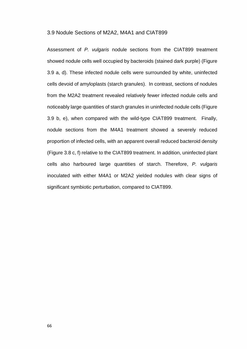
66
3.9 Nodule Sections of M2A2, M4A1 and CIAT899
Assessment of P. vulgaris nodule sections from the CIAT899 treatment
showed nodule cells well occupied by bacteroids (stained dark purple) (Figure
3.9 a, d). These infected nodule cells were surrounded by white, uninfected
cells devoid of amyloplasts (starch granules). In contrast, sections of nodules
from the M2A2 treatment revealed relatively fewer infected nodule cells and
noticeably large quantities of starch granules in uninfected nodule cells (Figure
3.9 b, e), when compared with the wild-type CIAT899 treatment. Finally,
nodule sections from the M4A1 treatment showed a severely reduced
proportion of infected cells, with an apparent overall reduced bacteroid density
(Figure 3.8 c, f) relative to the CIAT899 treatment. In addition, uninfected plant
cells also harboured large quantities of starch. Therefore, P. vulgaris
inoculated with either M4A1 or M2A2 yielded nodules with clear signs of
significant symbiotic perturbation, compared to CIAT899.

67
Figure 3.9 – Nodule Sections of CIAT899 (a and d), M2A2 (b and e) and M4A1 (c and f) –
40x magnification (a, b, c) and 100x magnification (d, e, f). An example of an infected nodule
cell is indicated by the black arrow (d), showing the stained purple colouration in comparison
to surrounding unstained, uninfected cells, easily distinguishable in nodules infected with wild-
type strain CIAT899. Surrounding uninfected cells did not appear to have accumulated starch
granules in CIAT899 (a, d). M2A2 (b, e) shows an overall decrease in infected cells within the
nodule compared with the wildtype (a, d). There is evidence of large accumulations of starch
granules in non-infected nodule cells, consistent with a block in carbon flow within the
bacteroid, as indicated by red arrows (e). M4A1 (c, f) shows a significant decrease in infected
a
b
c
d
e
f

68
cells, when compared with CIAT899 (a, d). Similarly to M2A2, M4A1 appeared to have
accumulated large amounts of starch granules in non-infected cells, as indicated by red arrows
(f), consistent with a block in carbon flow within the bacteroid.

69
3.10 Location and identification of mTn5-GNm insertion in M2A2 and
M4A1 genomes
P. vulgaris inoculated with M2A2 and M4A1 revealed significantly reduced
shoot dry weights and divergent nodule morphology compared with plants
inoculated with CIAT899. To determine the site of insertion of mTn5-GNm in
the M2A2 and M4A1 genomes, a three step, nested PCR protocol was
conducted, using a combination of primers targeting the 3` end of gusA and
those binding non-specifically within the genome. This approach yielded
amplicons approximately 450 bp and 250 bp in size, for M2A2 and M4A1,
respectively, following gel electrophoresis (Figure 3.6). Both amplicons were
excised and gel-purified for sequencing.

70
Figure 3.10 – Image of electrophoresed PCR products amplified from purified genomic DNA
of strains M2A2 and M4A1. PCR amplicons were produced from a 3-step PCR protocol using
non-specific primers 18 & 22 (Table 2.2), binding within the CIAT899 genome, and primers
specific to the gusA cassette of the mTn5-GNm. Lane 1 contained the 1kb molecular weight
marker. Template DNA was as follows: Lane 2 M2A2, lane 3 M4A1. Image shows bands of
approximately 450 bp (lane 2) and 250 bp (lane 3) for M2A2 and M4A1, respectively.
Molecular weights represented in lane 1 are as follows:(top to bottom)10,000; 8.000; 6,000;
5,000; 4,000; 3,000; 1,500; 1,000; and 500 bp respectively.
1 2 3

71
3.11 Sequencing analysis of strain M2A2
Sequence data generated from M2A2 was compared to the CIAT899 genome
(using the BLASTN algorithm). Results revealed a 223 bp region sharing 100%
identity to phaC (RTCIAT899_RS07985), coding for a putative Class I poly-β-
hydroxybutyrate synthase (polymerase). Using this sequence information, the
mTn5-GNm insertion site was mapped 227 bp downstream of phaC start
codon, reading in the opposite orientation of the gene. Further analysis of the
genome confirmed that CIAT899 does not appear to harbour any additional
putative PHB synthases. phaC from CIAT899 shares 78% similarity with phaC
from Rlp8002 (Rleg18DRAFT_1006), along with a high similarity to several
other R. leguminosarum species, as well as R. leguminosarum bv. trifolii and
R. etli, suggesting that it is highly conserved.
To confirm the site of insertion of mTn5-GNm, primer pairs were designed to
amplify regions flanking both sides of the insertion site in M2A2, with one
primer of each pair reading from inside the minitransposon, and the second
located at either the start or end codon of the gene. Visualisation of the PCR
products by gel electrophoresis revealed band sizes of 2263 and 2591 bp,
confirming the presence of the mTn5-GNm insertion, and location of the
insertion within the phaC gene to be consistent with the in silico prediction of
the insertion created, based on sequence data.
DNA co-ordinates for the mTn5-GNm insertion within the phaC gene were 227
bp downstream of the start codon, and 1615 bp upstream of the stop codon of
phaC. Sequence analysis suggested the location of the PHA synthase
catalytic domain to be from 83-611 bp within the phaC gene, with the

72
conserved phaC N-terminus location being within 124-296 bp nested within
the PHA synthase domain. Based on these co-ordinates, along with sequence
data, it appears that the mTn5-GNm has interrupted the phaC catalytic
domain, likely rendering the gene non-functional.

73
Figure 3.11 – Image of electrophoresed PCR products amplified from purified genomic DNA
of strain M2A2. PCR amplicons were produced by primers 24-27 (Table 2.2), reading from
within the mTn5-GNm, and flanking the insertion, and from the start and end codons of the
gene. Lane 5 contained the molecular weight marker. Lanes 2 and 3 contained template DNA.
Image shows bands of approximately 2.263kb (lane 1) and 2.591kb (lane 2), respectively.
Molecular weights represented in 1 kb ladder, from top to bottom, are 10,000; 8.000; 6,000;
5,000; 4,000; 3,000; 1,500; 1,000; and 500 bp respectively.
1 2 3 4 5

74
3.12 Sequence analysis of strain M4A1
Sequence data generated from M4A1 was compared to the CIAT899 genome,
using a BLASTN algorithm. Results revealed a 61 bp region with 94% identity
to the pyrB gene, coding for aspartate carbamoyltransferase
(RTCIAT899_CH07120), located 17 bp downstream from the start of the gene,
reading in the opposite direction of pyrB. Further analysis of the genome
confirmed that CIAT899 does not appear to harbour any additional putative
aspartate carbamoyltransferases. CIAT899 pyrB shares 88% similarity to pyrB
Rlp8002 Rleg18DRAFT_0735, and 89% similarity to R. leguminosarum bv.
trifolii WSM1325 Rleg_1391, suggesting the gene is highly conserved.
To confirm the site of insertion of mTn5-GNm, primer pairs were designed to
amplify regions flanking both sides of the insertion site in M4A1, with one
primer of each pair reading from inside the minitransposon, and the second
located at either the start or end codon of the gene. Visualisation of the PCR
products by gel electrophoresis revealed band sizes of 1, 164 and 2, 501 bp,
confirming the presence of the mTn5-GNm insertion, and location of the
insertion within the pyrB gene to be consistent with the in silico prediction of
the insertion created based on sequence data.
Co-ordinates for the mTn5-GNm insertion were 78 bp downstream of the start
of the pyrB gene, through to 879 bp upstream of the end of pyrB. Catalytic
binding domains within pyrB, coding for carbamoyl-P binding and
aspartate/ornithine binding, were located at intervals, 22-450 bp and 472-909
bp, respectively. These co-oridinates along with the site of the mTn5-GNm
insertion suggest that the pyrB carbamoyl-P catalytic domain has been

75
interrupted likely resulting in complete inhibition of aspartate
carbamoyltransferase activity.
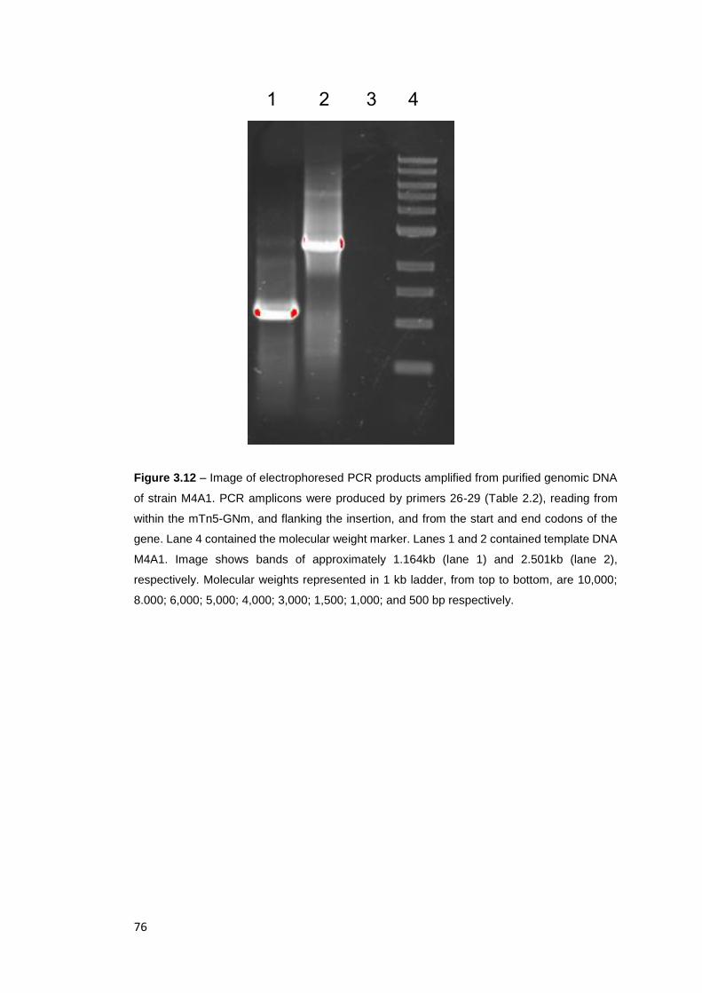
76
Figure 3.12 – Image of electrophoresed PCR products amplified from purified genomic DNA
of strain M4A1. PCR amplicons were produced by primers 26-29 (Table 2.2), reading from
within the mTn5-GNm, and flanking the insertion, and from the start and end codons of the
gene. Lane 4 contained the molecular weight marker. Lanes 1 and 2 contained template DNA
M4A1. Image shows bands of approximately 1.164kb (lane 1) and 2.501kb (lane 2),
respectively. Molecular weights represented in 1 kb ladder, from top to bottom, are 10,000;
8.000; 6,000; 5,000; 4,000; 3,000; 1,500; 1,000; and 500 bp respectively.
1 2 3 4

77
Chapter 4: Discussion
Site-directed mutations were attempted in Rhizobium tropici CIAT899 in genes
encoding TCA cycle enzymes isocitrate dehydrogenase and the E1
component of 2-oxoglutarate dehydrogenase with constructs pK19icdA and
pJQ200SKsucA, respectively. Concurrent random mutagenesis experiments
were performed in CIAT899, utilising the mTn5-GNm and selecting for carbon
utilisation phenotypes in free-living cultures. Isolated transconjugants were
replica-patched on minimal media supplemented with glucose, arabinose,
succinate or pyruvate separately, and classified according to their growth
phenotype on these carbon sources. Confirmed CIAT899 carbon utilisation
mutants were inoculated onto Phaseolus vulgaris in a glasshouse trial, to
investigate their symbiotic phenotypes. Two strains that showed statistically
different mean shoot dry weights when compared to wild-type controls were
further analysed and the site of mTn5-GNm insertion determined.
4.1 Mobilisation of pk19icdA into CIAT899 by insertional mutagenesis
Isolation of an icdA mutant of CIAT899 was not possible as results indicated
that pK19mob empty vector controls did not suicide when mobilised into this
host. In order for a plasmid to be useful for site-directed gene inactivation, it
must not replicate in the target strain (Snyder, 2013). Plasmid replication is
controlled by host proteins, which must recognise the plasmid’s origin of
replication (oriV). While this site encodes several proteins associated with
replication, it relies on the host cell for many others, such as the DnaA initiator
protein, the integration host factor (IHF), and the factor for inversion stimulation
(FIS) (del Solar et al., 1998). A suiciding plasmid is characterised as not being
78
capable of using replication proteins offered by the host cell, and hence, it fails
to persist as it’s ultimately lost from the cell during binary fission (Snyder,
2013).
The pk19mob plasmid carries a ColE1 origin of replication, and while it has
been successfully used as a suicide plasmid in other rhizobial strains (Mulley
et al., 2010), this origin appears to be recognised by CIAT899 replication
proteins. In order to attempt site-directed inactivation of icdA in CIAT899 in
future, it would be necessary to select an alternative plasmid with a different
origin of replication. Alternative plasmids carrying p15A replication origins
which have been shown to be successful as inactivation vectors, such as
pMQ150 and pMQ118, have been described by Shanks et al. (2009). These
plasmids possess a different oriV region to the pk19mob plasmid, along with
counter selectable markers, allowing for the screening for the loss of the
plasmid in transconjugants.
4.2 Mobilisation of pJQ200SKsucA into CIAT899 by insertional
mutagenesis
Conjugal transfer of the pJQ200SKsucA vector into CIAT899 was successful
and sucrose sensitive, gentamycin and spectinomycin resistant single
crossover mutants were isolated. However, PCR screening of these mutants
showed they all contained intact copies of sucA, suggesting that
pJQ200SksucA may have integrated non-specifically into the CIAT899
chromosome.
A sucA mutant, was successfully isolated in Rlv3841 by Walshaw et al. (1997)
via random mutagenesis with Tn5. Enzyme assays revealed the mutant to be

79
devoid of 2-oxoglutarate dehydrogenase activity. In addition, with glucose as
the sole carbon source, sucA mutants had a mean generation time of 721 min,
compared to 270 min for wild-type Rlv3841. In contrast, with succinate as the
sole carbon source, mean generation times of the sucA mutant and wild-type
were 305 and 220 min, respectively. Thus, the sucA mutation in Rlv3841
affects growth on glucose much more severely than growth on succinate.
While oxidation of glucose would necessitate the operation of the
decarboxylation arm of the TCA cycle (including 2-oxoglutarate
dehydrogenase), succinate enters the cycle after this point, thus its oxidation
presumably supports better growth.
Green and Emerich (1997a) isolated a sucA mutant by random mutagenesis
in B. japonicum USDA110, which conversely showed increased mean
generation times compared with the wild-type strain when grown on a range
of carbon sources (including glucose and succinate). These contrasting results
to the Rlv3841 sucA mutant suggest that mutations in sucA produce
remarkably different strain-specific growth phenotypes, and that while B.
japonicum appears to not have an absolute requirement for 2-oxoglutarate
dehydrogenase, growth was still affected in both strains by a lack of this
enzyme activity.
While previous studies have successfully isolated sucA mutants, each has
been obtained through random mutagenesis, with a site-directed sucA mutant
yet to be characterised. It is possible that, due to the critical role of 2-
oxoglutarate dehydrogenase in the TCA cycle, as well as amino acid
metabolism, site-directed mutants could be extremely difficult to isolate, with

80
mutation rates possibly being very low. It is likely that integration of the
pJQ200SKsucA vector observed was via non-specific integration, consistent
with CIAT899 sucA mutants being difficult to obtain due to the gene being
essential to cell growth. Given that the growth rate of the Rlv3841 sucA mutant
was considerably greater with succinate rather than glucose as the sole
carbon source, it is possible that in future inactivation attempts, screening
transconjugants on succinate may yield successful mutants.
4.3 Random mutagenesis of CIAT899 with mTn5-GNm
Isolated CIAT899 mTn5-GNm mutants were classified into six classes, based
on their carbon utilisation phenotypes. Growth of Type I mutants was equal to
wild-type CIAT899 on all media tested except for pyruvate. Given that growth
of Type I mutants was supported by glucose and gluconeogenic substrates
(arabinose and succinate), it is unlikely that these mutants are defective in
central metabolism. Mutation of mctP, encoding a monocarboxylic transport
permease in Rlv3841, yielded growth phenotypes on minimal media
supplemented separately with either glucose, succinate or pyruvate,
analogous to that of Type I mutants described in this thesis (Hosie et al., 2002).
Thus, it is possible that Type I mutants may be defective in pyruvate uptake.
Screening these mutants by PCR to locate the sites of insertion and measuring
rates of pyruvate uptake is required to further investigate the cause of this
growth phenotype.
Type II mutants were unable to grow on succinate as the sole carbon source,
yet retained wild-type growth on TY, glucose, arabinose and pyruvate. Two
likely causes of this phenotype are defects in pyruvate kinase activity or

81
dicarboxylate uptake. In rhizobia, oxidation of glucose (via the Entner
Doudoroff Pathway) (Fuhrer et al., 2005) produces pyruvate without the action
of pyruvate kinase. However, succinate oxidation would be predicted to
require this enzyme to produce acetyl CoA to fuel the TCA cycle. Indeed,
Mulley et al. (2010) reported pykA mutants of Rlv3841 to grow as well as the
wild-type strain on glucose, pyruvate, and succinate, suggesting it is more
likely that Type II mutants in this study are defective in dicarboxylate uptake.
The uptake of succinate, a C4-dicarboxylate, is known to be facilitated by the
Dct system in rhizobia (Lodwig & Poole, 2003) with mutations in any of the
three genes constituting the system (dctA, dctB, or dctD), abolishing growth
on C4-dicarboxylates, while growth on glucose and pyruvate is unaffected
(Arwas et al., 1985; Finan et al., 1988; Lodwig & Poole, 2003). Therefore,
transport assays examining C4-dicarboxylate uptake are required to further
characterise the Type II mutants isolated in this work.
Type III mutants, unable to utilise arabinose as a sole carbon source, but
maintaining wild-type growth rates on TY, glucose, pyruvate and succinate,
are most likely to be mutated in a gene within the arabinose catabolic pathway,
which feeds into the TCA cycle at 2-oxoglutarate. The oxidation of arabinose
is also known to produce pyruvate in B. japonicum 61A76, and Bradyrhizobium
32HI, via L-2-keto-3-deoxyarabinonate (KDA) aldose (Duncan, 1979).
However, KDA aldose was only found to be present at low activites in S.
meliloti, and was not detectable in R. leguminosarum bv. trifolii, R.
leguminosarum bv. phaseoli (Dilworth et al., 1986). Given these findings, it is
possible that KDA aldose may also not be active, or only active at low levels,
in CIAT899, and that arabinose is metabolised via 2-oxoglutarate

82
semialdehyde dehydrogenase to 2-oxoglutarate (Duncan, 1979). If this is true,
a mutation at any point in the arabinose catabolic pathway may result in an
inability to utilise arabinose as a sole carbon source, while growth on glucose,
succinate and pyruvate would likely be unaffected.
Growth of Type IV mutants were perturbed on gluconeogenic substrates
succinate, pyruvate, and arabinose, while growth was equal to, or slightly
reduced, compared to the wild-type, on TY and glucose. This may be
suggestive of a mutation in genes associated with gluconeogenesis, or
overflow pathways however, given that the possibilities are quite broad, further
screening via PCR, and growth trials on a wider selection of carbon sources
needs to be performed in order to draw clearer conclusions on the genetic and
metabolic cause of this phenotype.
Type V mutants were unable to utilise glucose, arabinose, succinate or
pyruvate as sole carbon sources, while maintaining wild-type growth rates on
TY. When streaked on minimal media supplemented with dual carbon sources,
glucose and succinate, growth inhibition could not be relieved. This is
indicative of type V mutants being auxotrophs, lacking the ability on minimal
media to synthesise a product integral to growth such as amino acids, pentose
sugars, nucleotides or any number of other critical biosynthetic intermediates
or products. Nested PCR and subsequent sequencing of mutants to identify
sites of insertion would likely be the most time-efficient way of uncovering the
genetic cause of this phenotype.
Phenotypes observed for type VI mutants which exhibited reduced growth on
all carbon sources tested, could potentially be related to a mutation that

83
reduces, but does not abolish enzyme activity. These mutations are difficult to
characterise based on carbon utilisation, as there are many possible insertions
that could result in a reduced growth phenotype. Further screening via PCR to
locate the site of insertion in each mutant is required to fully ascertain the
cause of this growth phenotype.
Although these are possible explanations for the growth phenotypes observed
in random CIAT899 mutants, there are likely other possible explanations.
Therefore, in order to accurately interpret carbon utilisation profiles in the
mutant strains, nested PCR screening to locate mTn5-GNm insertions within
each strain is required, with sequence analysis and subsequent enzyme
assays performed.
4.4 Glasshouse trials with Phaseolus vulgaris
An analysis of the 13 strains inoculated onto P. vulgaris revealed that two
strains, M4A1 and M2A2, produced significantly different mean shoot dry
weights to that of wild-type CIAT899. Of the non-significant strains, statistical
analysis using a Tukey’s Post-Hoc test showed p-values of 0.051 and 0.07 at
a 0.05 alpha level, for strains M8A2 and MC34, respectively. Given the minimal
degree of divergence from the alpha level, repeat glasshouse trials on these
strains to assess their N2-fixing capabilities would be recommended. Due to
time restrictions, only strains that showed a statistical difference compared to
the wild-type were sequenced.
In combination with dry weights, a quantitative measurement of nitrogenase
activity in all mutant strains should be conducted using acetylene reduction
assays, and total N2 content measurements from plant biomasses (Yates et

84
al., 2016). Additionally, as all random CIAT899 mutants isolated possessed a
carbon utilisation growth phenotype (Table 3.1) in free-living cultures, further
screening of the symbiotic phenotype for the remaining seven mutants would
also be recommended.
4.5 Inactivation of pyrB in CIAT899
Sequence analysis of M4A1 revealed the insertion of mTn5-GNm in the pyrB
gene of CIAT899, coding for a putative aspartate carbamoyltransferase.
Currently, this is first study to characterise a pyrB mutant in a species of
rhizobia. Aspartate carbamoyltransferase catalyses the transfer of the
carbamoyl moiety from carbamoyl phosphate to L- aspartate, which is a critical
step in UMP biosynthesis (McMurry & Begley, 2005).
Given the role of pyrB in nucleotide biosynthesis, it is possible that an inhibition
in UMP synthesis could give rise to the reduced growth rate, as observed in
free-living cultures. Moreover, nodule sections revealed a reduction in the
number of infected host cells, consistent with an overall reduction in nodule
infection. Whether this is due to a reduced rate of bacterial growth in the plant
rhizosphere or in infection threads, or another defect in infection droplet
formation requires further exploration. A quantitative measurement of cell
counts per bacteroid, and acetylene reduction assays as well as the
measurement of rhizosphere populations are required to further explain the
basis of this phenotype. Moreover, conducting enzyme assays to measure the
activity of aspartate carbamoyltransferase would similarly be informative to
quantitatively ascertain the degree to which the mutation has perturbed
enzyme activity. Finally, complementation of the pyrB mutation with an intact

85
copy of the gene expressed on a broad host range vector, such as pJP2 (Prell
et al., 2002) is required to rule-out the involvement of secondary mutations in
the observed phenotype.
4.6 Inactivation of phaC in CIAT899
Sequence analysis of strain M2A2 revealed the insertion of the mTn5-GNm
within the phaC gene, encoding a putative poly-β-hydroxybutyrate (PHB)
synthase, suggesting that the free-living and symbiotic phenotype exhibited by
this strain could be due to perturbation in the synthesis of this polymer.
In free-living conditions, M2A2 was unable to utilise arabinose, succinate or
pyruvate as a sole carbon source and grew much slower on glucose and TY
when compared with wild-type CIAT899. Similar phenotypes have been
observed in previously characterised phaC mutants in other P. vulgaris-
nodulating strains such as R. leguminosarum bv. phaseoli 4292 and R. etli
(Dunn et al., 2002; Lodwig et al., 2005)
PHB is a lipid-like polymer, known to accumulate in large quantities in some
rhizobial species, including R. leguminosarum bv. phaseoli, R. leguminosarum
bv. viciae, B. japonicum, and R. etli (Lodwig & Poole, 2003; Terpolilli et al.,
2016), and acts as carbon storage compound, or a sink for reducing
equivalents (Tombolini et al., 1995). Wältermann and Steinbüchel (2005)
report that an accumulation of at least one type of storage lipid
[triacylglycerols, wax esters, or poly(hydroxyalkanoates)] occurs in nearly all
prokaryotes. This storage mechanism appears to occur in free-living bacterial
cells, in response to an excess of carbon and reductant, when growth is limited
due to a nutritional deficit elsewhere, such as in N2 or phosphorus (Rehm,

86
2003; Wältermann & Steinbüchel, 2005). While the extent to which PHB
accumulates in free-living cells of CIAT899 is not known, it is possible that it
plays an important role in balancing carbon and reductant for growth in defined
media.
Glasshouse trials revealed M2A2 to have mean shoot dry weights not different
to uninoculated controls, but with nodules that exhibited a light pink
colouration, suggesting the mutant was capable of some nitrogenase activity.
In contrast, a phaC mutant of R. leguminosarum bv. phaseoli 4292 was
unaffected in N2 fixation (Lodwig et al., 2005), while an R. etli phaC mutant
actually showed an increase in N2 fixation as measured by acetylene reduction
and plant N2 content (Dunn et al., 2002).Thus, it appears from the available
evidence that the importance of PHB synthesis to N2 fixation is strain-specific.
Nodules of P. vulgaris inoculated with M2A2 observed via light microscopy
showed starch granules in uninfected nodule cells which were absent in
nodules from CIAT899-inoculated plants. Starch is a glucose storage polymer
in plants and an increase in starch deposits in nodules has been previously
noted when nodules are occupied by mutant strains which fix little or no N2
(Yurgel & Kahn, 2005). Although the cause of this increased starch
accumulation has yet to be demonstrated, it is possible that carbon that cannot
be metabolised by bacteroids perturbed in N2 fixation, is instead stored by the
host plant as starch. If this is true, it suggests that PHB accumulation is
essential to effective N2 fixation in CIAT899, and that an increase in starch
deposits in M2A2 infected nodules could be indicative of a block in carbon flux
within the bacteroid.
87
Lodwig et al. (2005) review the possibility that PHB accumulation in bacteroids
may be important to maintaining redox balance, as it appears to do in free-
living cells. PHB content was reported to be inversely correlated to nitrogenase
activity in R. leguminosarum bv. phaseoli and R. lupini, with both showing daily
fluctuations in their PHB content (Kretovich et al., 1977). Additionally, it has
been shown that B. japonicum bacteroids degrade PHB when supplies of
carbon to the nodule are perturbed, and are still able to fix N2 when no
substrate is provided, suggesting the mobilisation and use of endogenous
reserve products (Bergersen & Turner, 1990; Bergersen et al., 1991).
It has also been suggested that lipogenesis in bacteroids (which includes the
synthesis of glycerolipids as well as PHB) may be intricately linked to N2
fixation, with the existence of lipid sinks being an essential part of the
energisation of nitrogenase and associated redox balance (Terpolilli et al.,
2016). If this is true, the existence of multiple carbon storage and redox sinks
(PHB, glycerolipids and glycogen) in different strains may vary in their relative
function and importance, which may explain the variation of PHB accumulation
across different rhizobia strains.
It is clear that more research is required in order to understand the possible
role of PHB synthesis in N2-fixing bacteroids. The mechanisms by which the
bacteroid achieves a fine balance in carbon flux, allowing for the production of
reductant to power the nitrogenase complex, while simultaneously meeting the
energy demands of normal cellular metabolism, all in a low oxygen tension
environment, is still unclear. The common hypothesis remains that PHB acts
as an overflow pathway, to aid in the powering of nitrogenase by allowing
sufficient carbon to move through the TCA cycle, producing reductant, without
88
causing oxidative stress and inhibition of nitrogenase activity. However,
whether this pathway plays an important role in N2 fixation appears to be strain
specific, with phaC mutants of different strains showing a range of disparate,
symbiotic phenotypes.
For phaC mutant M2A2, acetylene reduction assays need to be performed to
gain quantitative measurements of nitrogenase activity in the bacteroid, to
ascertain whether the strain is truly a Fix- phenotype on P. vulgaris.
Additionally, electron micrographs are needed to observe PHB stores in the
M2A2 bacteroid, compared with wild-type CIAT899. Finally, complementation
of the mutation, with an intact copy of the phaC gene expressed on a broad
host range vector, would be required to confirm that the free-living and
symbiotic phenotypes observed in M2A2 were not the result of a secondary
mutation. Complete characterisation of this strain is necessary, as it is the first
time a Fix-, or even Fix reduced, phaC mutant has been described in any
Rhizobium species.
Conclusions and future directions
Isolation and characterisation of carbon utilisation mutants of CIAT899 has
revealed 20 strains with free-living phenotypes different to those of the wild-
type strain. Only 13 of these strains could be screened in the time-frame,
therefore the remaining seven require further screening from their symbiotic
phenotype in glasshouse trials on P. vulgaris.
Two of the 13 strains (M2A2 and M4A1) exhibit strong symbiotic phenotypes
which require further work to confidently ascribe their phenotype to the

89
inactivation of pyrB and phaC, respectively. Further replica-patching could
also be performed, which could potentially reveal additional novel mutants.
Our understanding of bacteroid metabolism is limited to a total of some 50
characterised metabolic mutants representing only 4 genera of rhizobia
(Terpolilli et al., 2012). As there are currently some 17 genera of
phylogenetically distinct rhizobia (ICSP, 2016) it is evident that our knowledge
of bacteroid metabolism is limited to a very small sample size.
In order to draw conclusions on the relationship between bacteroid metabolism
and how effectively a strain can fix N2 on a given host, a clearer understanding
of carbon flux in a wider variety of strains and genera needs to be established.
Characterising the mechanisms which underpin carbon flow between the TCA
cycle, nitrogenase, and overflow pathways, across a more diverse selection of
strains, may be the key to understanding the metabolic determinants of
effective N2 fixation.

90
References
Appleby, C. (1984). Leghaemoglobin and Rhizobium Respiration. Annual Review of Plant Physiology 35, 443-478.
Arwas, R., McKay, I. A. & Rowney, F. R. P. (1985). Properties of Organic Acid Utilization Mutants of Rhizobium leguminosarum strain 300. Journal of General Microbiology 131, 2059-2066.
Benson, D., Karsch-Mizrachi, I., Lipman, D. J., Ostell, J. & Sayers, E. (2009).Genbank. In Nucleic acids research, pp. D26-31: Epub.
Bergersen, F. & Turner, G. (1990). Bacteroids from Soybean Root Nodules: Accumulation of Poly-β -hydroxybutyrate During Supply of Malate and Succinate in Relation to N2Fixation in Flow-Chamber Reactions. Proceedings of the Royal Society of London Series B, Biological Sciences 240, 39-59.
Bergersen, F., Peoples, M. & Turner, G. (1991). A Role for poly- β- hydroxybutyrate in Bacteroids of Soybean Root Nodules. Proceedings of the Royal Society of London Series B: Biological Sciences 245, 59-64.
Beringer, J. (1974). R. Factor Transfer in Rhizobium leguminosarum. Journal of General Microbiology 84, 188-198.
Bertani, G. (1952). Studies on Lysogenesis. I. The Mode of Phage Liberation by Lysogenic Escherichia coli. Journal of Bacteriology 62, 293-300.
Bonaldi, K., Gourion, B., Fardoux, J. & other authors (2010). Large-scale Transposon Mutagenesis of Photosynthetic Bradyrhizobium sp. strain ORS278 Reveals New Genetic Loci Putatively Important for Nod-Independent Symbiosis with Aeschynomene indica. Molecular plant-microbe interactions : MPMI 23, 760-770.
Broughton, W. & Dilworth, M. (1971). Control of Leghaemoglobin Synthesis in Snake Beans. Biochemical Journal 125, 1075-1080.
Cevallos, M., Encarnación, S., Leija, A., Mora, Y. & Mora, J. (1996). Genetic and Physiological Characterization of a Rhizobium etli Mutant Strain Unable to Synthesize Poly-beta-hydroxybutyrate. Journal of Bacteriology 178, 1646-1654.

91
Charles, T., Cai, C. & Aneja, P. (1997). Megaplasmid and Chromosomal Loci for the PHB Degradation Pathway in Rhizobium (Sinorhizobium) meliloti. Genetics 146, 1211-1220.
Chun, K., Edenberg, H., Kelley, M. & Goebl, M. (1997). Rapid Amplification of Uncharacterized Transposon-Tagged DNA Sequences from Genomic DNA. Yeast 13, 233-240.
Copeland, L., Quinnell, R. & Day, D. (1989). Malic Enzyme Activity in Bacteroids from Soybean Nodules. Journal of General Microbiology 135, 2005-2011.
Deakin, W. & Broughton, W. (2009). Symbiotic Use of Pathogenic Strategies: Rhizobial Protein Secretion Systems. Nature Reviews Microbiology 7, 312-320.
del Solar, G., Giraldo, R., Ruiz-Echevarría, M., Espinosa, M. & Díaz-Orejas, R. (1998). Replication and Control of Circular Bacterial Plasmids. Microbiology and Molecular Biology Reviews 62, 434-464.
Denison, R. F. (2000). Legume Sanctions and the Evolution of Symbiotic Cooperation by Rhizobia. The American Naturalist 156, 567-576.
Dilworth, M., Arwas, R., McKay, I., Saroso, S. & Glenn, A. (1986). Pentose Metabolism in Rhizobium leguminosarum MNF300 and in Cowpea Rhizobium NGR234. 132, 2733 - 2742.
Driscoll, B. & Finan, T. (1993). NAD(+)-dependent Malic Enzyme of Rhizobium meliloti is Required for Symbiotic Nitrogen Fixation. Molecular microbiology 7, 865-873.
Driscoll, B. & Finan, T. (1996). NADP+ -Dependent Malic Enzyme of Rhizobium meliloti. Journal of Bacteriology 178, 2224-2231.
Driscoll, B. & Finan, T. (1997). Properties of NAD+- and NADP+-Dependent Malic Enzymes of Rhizobium (Sinorhizobium) meliloti and Differential Expression of Their Genes in Nitrogen-Fixing Bacteroids. Microbiology 143, 489-498.
Duncan, M. (1979). L-Arabinose Metabolism in Rhizobia. Microbiology 113, 177-179.
Duncan, M. & D., F. (1979). Alpha-Ketoglutarate Dehydrogenase Mutant of Rhizobium meliloti. Journal of Bacteriology 137, 415-419.

92
Dunn, M., Araíza, G., Encarnación, S., María del Carmen, V. & Mora, J. (2002). Effect of aniA (Carbon Flux Regulator) and phaC (Poly-β-Hydroxybutyrate Synthase) Mutations on Pyruvate Metabolism in Rhizobium etli. Journal of Bacteriology 184, 2296-2299.
Finan, M., Kunkel, B., De Vos, G. & Signer, E. (1986). Second Symbiotic Megaplasmid in Rhizobium meliloti Carrying Exopolysaccharide and Thiamine Synthesis Genes. Journal of Bacteriology 164, 66-72.
Finan, M., Oresnik, I. & Bottacin, A. (1988). Mutants of Rhizobium meliloti Defective in Succinate Metabolism. Journal of Bacteriology 170, 3396-3403.
Finan, T., Wood, J. & Jordan, D. (1983). Symbiotic Properties of C4-dicarboxylic Acid Transport Mutants of Rhizobium leguminosarum. Journal of Bacteriology 154, 1403-1413.
Finan, T., McWhinnie, E., Driscoll, B. & Watson, R. (1991). Complex Symbiotic Phenotypes Results from Gluconeogenic Mutations in Rhizobium meliloti. Molecular plant-microbe interactions : MPMI 4, 386-392.
Fischer, H. (1994). Genetic Regulation of Nitrogen Fixation in Rhizobia. Microbiological reviews 58, 352-386.
Fuhrer, T., Fischer, E. & Sauer, U. (2005). Experimental Identification and Quantification of Glucose Metabolism in Seven Bacterial Species. Journal of Bacteriology 187, 1581-1590.
Galloway, J. & Gruber, N. (2008). An Earth-system Perspective of the Global Nitrogen Cycle. Nature 451, 293-296.
Gardiol, A., Arias, A., C., C. & Martinez-Drets, G. (1982). Succinate Dehydrogenase Mutant of Rhizobium meliloti. Journal of Bacteriology 151, 1621-1623.
Graham, P., Viteri, S., Mackie, F., Vargas, A. & Palacios, A. (1982). Variation in Acid Soil Tolerance Among Strains of Rhizobium phaseoli. Field Crops Research 5, 121-128.
Graham, P. (1992). Stress Tolerance in Rhizobium and Bradyrhizobium, and Nodulation Under Adverse Soil Conditions. Canadian Journal of Microbiology 38, 475-484.

93
Graham, P. & Vance, C. (2003). Legumes: Importance and Constraints to Greater Use. Plant Physiology 131, 872-877.
Graham, P. (2008). Ecology of Root-Nodule Bacteria of Legumes. In Nitrogen-Fixing Leguminous Symbioses, pp. 23-43. Edited by M. Dilworth, E. James, J. Sprent & W. Newton. The Netherlands: Springer.
Green, L. & Emerich, D. (1997a). Bradyrhizobium japonicum Does Not Require Alpha-Ketoglutarate Dehydrogenase for Growth on Succinate or Malate. Journal of Bacteriology 179, 194-201.
Green, L. & Emerich, D. (1997b). The Formation of Nitrogen-Fixing Bacteroids Is Delayed but Not Abolished in Soybean Infected by an alpha-Ketoglutarate Dehydrogenase-Deficient Mutant of Bradyrhizobium japonicum. Plant Physiology 114, 1359-1368.
Hill, S. (1988). How is nitrogenase regulated by oxygen? FEMS Microbiology Reviews 4, 111-129.
Hosie, A., Allaway, D. & Poole, P. (2002). A Monocarboxylate Permease of Rhizobium leguminosarum Is the First Member of a New Subfamily of Transporters. Journal of Bacteriology 184, 5436-5448.
Hudman, J. & Glenn, A. (1980). Glucose Uptake by Free-Living and Bacteroid Forms of Rhizobium leguminosarum. Archives of Microbiology 128, 72-77.
Hungria, M., Campo, R. & Mendes, I. (2003). Benefits of Inoculation of the Common Bean (Phaseolus vulgaris) Crop with Efficient and Competitive Rhizobium tropici Strains. Biology and Fertility of Soils 39, 88-93.
ICSP (2016). ICSP Subcommittee on the Taxonomy of Rhizobium and Agrobacterium - Diversity, Phylogeny and Systematics. Available "http://edzna.ccg.unam.mx/rhizobial-taxonomy/node/4" (Accessed: 24/10/2016).
Johnston, A., Hombrecher, G., Brewin, N. & Cooper, M. (1982). Two Transmissible Plasmids in Rhizobium leguminosarum strain 300. Jounrnal of Microbiology 128, 85-93.
Kaminski, P., Batut, J. & Boistard, P. (1998). A Survey of Symbiotic Nitrogen Fixation by Rhizobia. In The Rhizobiaceae: Molecular Biology of Model Plant-Associated Bacteria, pp. 429-460. Edited by H. Spaink, A.

94
Kondorosi, P. Hooykaas & J. Dodrecht. The Netherlands: Kluwer Academic Publishers.
Kearse, M., Moir, R., Wilson, A. & other authors (2012). Geneious Basic: An Integrated and Extendable Desktop Software Platform for the Organization and Analysis of Sequence Data. Bioinformatics 28, 1647-1649.
Kiers, E., West, S. & Denison, R. (2008). Maintaining Cooperation in the Legume-Rhizobia Symbiosis: Identifying Selection Pressures and Mechanisms. In Nitrogen-fixing Leguminous Symbioses, pp. 70. Edited by M. Dilworth, E. James, J. Sprent & W. Newton. The Netherlands: Springer.
Koziol, U., Hannibal, L., Rodríguez, M., Fabiano, E., Kahn, M. & Noya, F. (2009). Deletion of Citrate Synthase Restores Growth of Sinorhizobium meliloti 1021 Aconitase Mutants. Journal of Bacteriology 191, 7581-7586.
Kretovich, W., Romanov, V., Yushkova, L., Shramko, V. & Fedulova, N. (1977). Nitrogen Fixation and Poly-β-hydroxybutyric Acid Content in Bacteroids of Rhizobium lupini and Rhizobium leguminosarum. Plant and Soil 48, 291-302.
Lamb, J. W., Hombrecher, G. & Johnston, A. (1982). Plasmid-Determined Nodulation and Nitrogen Fixation Abilities in Rhizobium phaseoli. Molecular and General Genetics 186, 449-452.
Lodwig, E. & Poole, P. (2003). Metabolism of Rhizobium Bacteroids. Critical Reviews in Plant Sciences 22, 37-78.
Lodwig, E., Leonard, M., Marroqui, S., Wheeler, T., Findlay, K., Downie, J. & Poole, P. (2005). Role of Polyhydroxybutyrate and Glycogen as Carbon Storage Compounds in Pea and Bean Bacteroids. Molecular Plant-Microbe Interactions 18, 67-74.
Markowitz, V., Chen, I., Palaniappan, K., Chu, K., Szeto, E., Grechkin, Y., Ratner, A., Jacon, B. & others (2012).IMG: The Integrated Microbial Genomes Database and Comparative Analysis System.: Nuceic Acids Research.
Martinez-Romero, E., Segovia, L., Mercante, F., Franco, A., Graham, P. & Pardo, M. (1991). Rhizobium tropici, A Novel Species Nodulating Phaseoulus vulgaris L. Beans and Leucaena sp. Trees. International Journal of Systematic Bacteriology 41, 417-426.

95
Maunoury, N., Kondorosi, A., Kondorosi, E. & Mergaert, P. (2008). Cell Biology of Nodule Infection and Development. In Nitrogen-Fixing Leguminous Symbioses, pp. 153-180. Edited by E. J. Michael Dilworth, Janet Sprent, William Newton. The Netherlands: Springer.
McDermott, T. & Kahn, M. (1992). Cloning and Mutagenesis of the Rhizobium meliloti Isocitrate Dehydrogenase Gene. Journal of bacteriology 174, 4790-4797.
McMurry, J. & Begley, T. (2005). The Organic Chemistry of Biological Pathways: Roberts and Company Publishers.
Melino, V., Drew, E., Ballard, R., Reeve, W., Thomson, G., White, R. & O'Hara, G. (2012). Identifying Abnormalities in Symbiotic Development Between Trifolium spp. and Rhizobium leguminosarum bv. trifolii Leading to Sub-Optimal and Ineffective Nodule Phenotypes. Annals of botany 110, 1559-1572.
Mergaert, P., Kelemen, Z., Vaubert, D., Kondorosi, A. & Kondorosi, E. (2003). A Novel Family in Medicago truncatula Consisting of More than 300 Nodule-Specific Genes Coding for Small, Secreted Polypeptides with Conserved Cysteine Motifs. Plant Physiology 132, 161-173.
Mergaert, P., Uchiumi, T., Alunni, B. & other authors (2006). Eukaryotic Control on Bacterial Cell Cycle and Differentiation in the Rhizobium-legume Symbiosis. Proceedings of the National Academy of Sciences of the United States of America 103, 5230-5235.
Minchin, F., James, E. & Becana, M. (2008). Oxygen Diffussion, Production of Reactive Oxygen and Nitrogen Species, and Antioxidants in Legume Nodules. In Nitrogen-fixing Leguminous Symbioses, pp. 321-353. Edited by E. J. Michael Dilworth, Janet Sprent, William Newton. The Netherlands: Springer.
Mortimer, M., McDermott, T., York, G., Walker, G. & Kahn, M. (1999). Citrate Synthase Mutants of Sinorhizobium meliloti Are Ineffective and Have Altered Cell Surface Polysaccharides. Journal of Bacteriology 181, 7608-7613.
Mulley, G., Lopez-Gomez, M., Zhang, Y., Terpolilli, J., Prell, J., Finan, T. & Poole, P. (2010). Pyruvate Is Synthesized by Two Pathways in Pea Bacteroids with Different Efficiencies for Nitrogen Fixation. Journal of Bacteriology 192, 4944-4953.

96
Murray, J. (2011). Invasion by Invitation: Rhizobial Infection in Legumes. Molecular plant-microbe interactions : MPMI 24, 631-639.
Noel, K., Sanchez, A., Fernandez, L., Leemans, J. & Cevallos, M. (1984). Rhizobium phaseoli Symbiotic Mutants with Transposon Tn5 Insertions. Journal of Bacteriology 158, 148-155.
Olivares, J., Bedmar, E. & Sanjuán, J. (2013). Biological Nitrogen Fixation in the Context of Global Change. Molecular plant-microbe interactions : MPMI 26, 486-494.
Ormeño-Orrillo, E., Menna, P., Almeida, L. & other authors (2012). Genomic Basis of Broad Host Range and Environmental Adaptability of Rhizobium tropici CIAT 899 and Rhizobium sp. PRF 81 which are Used in Inoculants for Common Bean (Phaseolus vulgaris L.). BMC Genomics 13, 735-735.
Polcyn, W. & Luciński, R. (2003). Aerobic and Anaerobic Nitrate and Nitrite Reduction in Free-Living Cells of Bradyrhizobium sp. ( Lupinus). FEMS Microbiology Letters 226, 331-337.
Poole, P., Hynes, M., Johnston, A., Tiwari, R., Reeve, W. & Downie, J. (2008). Physiology of Root-Nodule Bacteria. In Nitrogen-fixing Leguminous Symbioses, pp. 241-276. Edited by E. J. Michael Dilworth, Janet Sprent, William Newton. The Netherlands: Springer.
Prell, J., Boesten, B., Poole, P. & Priefer, U. (2002). The Rhizobium leguminosarum bv. viciae VF39 Gamma-aminobutyrate (GABA) Aminotransferase Gene (gabT) is Induced by GABA and Highly Expressed in Bacteroids. Microbiology 148, 615-623.
Prentki, P. & Krisch, H. (1984). In Vitro Insertional Mutagenesis with a Selectable DNA Fragment. Gene 29, 303-313.
Quandt, J. & Hynes, M. (1993a). Versatile Suicide Vectors which allow Direct Selection for Gene Replacement in Gram-Negative Bacteria. Gene 127, 15-21.
Quandt, J. & Hynes, M. (1993b). Versatile Suicide Vectors which Allow Direct Selection for Gene Replacement in Gram-Negative Bacteria. Gene 127, 15-21.
Reeve, W., Tiwari, R., Worsley, P., Dilworth, M., Glenn, A. & Howieson, J. (1999). Constructs for Insertional Mutagenesis, Transcriptional Signal

97
Localization and Gene Regulation Studies in Root Nodule and Other Bacteria. Jounrnal of Microbiology 145, 1307-1316.
Rehm, B. (2003). Polyester Synthases: Natural Catalysts for Plastics. Biochemical Journal 376, 15-33.
Ronson, C., Lyttleton, P. & Robertson, J. (1981). C(4)-Dicarboxylate Transport Mutants of Rhizobium trifolii Form Ineffective Nodules on Trifolium repens. Proceedings of the National Academy of Sciences of the United States of America 78, 4284-4288.
Sambrook, J. & Russell, D. (2001). Molecular Cloning: A Laboratory Manual. Cold Spring Harbor, N.Y: Cold Spring Harbor Laboratory.
Sayers, E., Barrett, T., Benson, D. & other authors (2009).Database resources of the National Center for Biotechnology Information. In Nucleic acids research, pp. D5-15: Epub.
Schafer, A., Tauch, A., Jager, W., Kalinowski, J., Thierbach, G. & Puhler, A. (1994). Small mobilizable Multipurpose Cloning Vectors Derived from the Escherichia coli Plasmids pK18 and pK19: Selection of Defined Deletions in the Chromosome of Corynebacterium glutamicum Gene 145, 69-73.
Shah, R. & Emerich, D. (2006). Isocitrate Dehydrogenase of Bradyrhizobium japonicum Is Not Required for Symbiotic Nitrogen Fixation with Soybean. Journal of Bacteriology 188, 7600-7608.
Shanks, R., Kadouri, D., MacEachran, D. & O’Toole, G. (2009). New Yeast Recombineering Tools for Bacteria. Plasmid 62, 88-97.
Snyder, L. (2013). Molecular Genetics of Bacteria. Washington, DC: ASM Press.
Sprent, J. I. (2009). Legume Nodulation: A Global Perspective. West Sussex, UK: Wiley-Blackwell.
Tatè, R., Ferraioli, S., Filosa, S., Cermola, M., Riccio, A., Iaccarino, M. & Patriarca, E. (2004). Glutamine Utilization by Rhizobium etli. Molecular plant-microbe interactions : MPMI 17, 720-728.
Terpolilli, J., Hood, G. & Poole, P. (2012). What Determines the Efficiency of N(2)-fixing Rhizobium-Legume Symbioses? Advances in Microbial Physiology 60, 326-389.

98
Terpolilli, J., Masakapalli, S., Karunakaran, R., Webb, I., Green, R., Watmough, N., Kruger, N., Ratcliffe, R. & Poole, P. (2016). Lipogenesis and Redox Balance in Nitrogen-Fixing Pea Bacteroids. Journal of Bacteriology 198, 2864-2875.
Thoma, S. & Schobert, M. (2009). An Improved Escherichia coli Donor Strain for Diparental Mating. FEMS microbiology letters 294, 127-132.
Timmers, A., Soupène, E., Auriac, M., de Billy, F., Vasse, J., Boistard, P. & Truchet, G. (2000). Saprophytic Intracellular Rhizobia in Alfalfa Nodules. Molecular plant-microbe interactions : MPMI 13, 1204-1213.
Tombolini, R., Povolo, S., Buson, A., Squartini, A. & Nuti, M. (1995). Poly-β-hydroxybutyrate (PHB) biosynthetic genes in Rhizobium meliloti 41. Microbiology 141, 2553-2559.
Trainer, M., Capstick, D., Zachertowska, A., Lam, K., Clark, S. & Charles, T. (2010). Identification and Characterization of the Intracellular Poly-3-hydroxybutyrate Depolymerase Enzyme PhaZ of Sinorhizobium meliloti. BMC Microbiology 10, 92-92.
Udvardi, M., Yang, L., Young , S. & Day, D. (1990). Sugar and Amino Acid Transport Across Symbiotic Membranes from Soybean Nodules. Molecular Plant-Microbe Interactions 3, 334-340.
Udvardi, M. & Poole, P. (2013). Transport and Metabolism in Legume-Rhizobia Symbioses. Annual review of plant biology 64, 781-805.
Vance, C. (2008). Carbon Nitrogen Metabolism in Legume Nodules. In Nitrogen-fixing Leguminous Symbioses, pp. 293-315. Edited by M. Dilworth, E. James, J. Sprent & W. Newton. The Netherlands: Springer.
Vasse, J., de Billy, F., Camut, S. & Truchet, G. (1990). Correlation Between Ultrastructural Differentiation of Bacteroids and Nitrogen Fixation in Alfalfa Nodules. Journal of Bacteriology 172, 4295-4306.
Walshaw, D., Wilkinson, A., Mundy, M., Smith, M. & Poole, P. (1997). Regulation of the TCA Cycle and the General Amino Acid Permease by Overflow Metabolism in Rhizobium leguminosarum. Microbiology 143, 2209-2221.

99
Wältermann, M. & Steinbüchel, A. (2005). Neutral Lipid Bodies in Prokaryotes: Recent Insights into Structure, Formation, and Relationship to Eukaryotic Lipid Depots. Journal of bacteriology 187, 3607-3619.
Wilson, K., Sessitsch, A., Corbo, J., Giller, K., Akkermans, A. & Jefferson, R. (1995). β-Glucuronidase (GUS) Transposons for Ecological and Genetic Studies of Rhizobia and Other Gram-Negative Bacteria. Microbiology 141, 1691-1705.
Yates, R., Howieson, J., Hungria, M., Bala, A., O’Hara, G. & Terpolilli, J. (2016). Authentification of Rhizobia and Assessment of the Legume Symbiosis in Controlled Plant Growth Systems. In Working with rhizobia, pp. 73-108. Edited by J. Howieson & M. Dilworth. Canberra: Australian Centre for International Agricultural Research.
Yurgel, S. & Kahn, M. (2004). Dicarboxylate Transport by Rhizobia. FEMS Microbiology Reviews 28, 489-501.
Yurgel, S. & Kahn, M. (2005). Sinorhizobium meliloti dctA Mutants with Partial Ability To Transport Dicarboxylic Acids. Journal of Bacteriology 187, 1161-1172.


![Briefpaper Time-varyingfeedbackforregulationofnormal ...flyingv.ucsd.edu/papers/PDF/266.pdfV +). |+ ≤ +. (− −) +. + [− (,) + (− + ())(− −)],](https://img.pdfslide.net/doc/110x75/60f6a2f91441fd540243b29d/briefpaper-time-varyingfeedbackforregulationofnormal-v-a-a-a.jpg)