Embed Size (px)
Citation preview
Freely Available Online
www.openaccesspub.org | JDOI CC-license DOI : 10.14302/issn.2473-1005.jdoi-16-1159 Vol-1 Issue 3 Pg. no.- 1
Maxillary constriction with skeletal class II malocclusion - A comprehensive treatment approach.
A. ARIF YEZDANI, MDS, FWFO1, Jabeen Fathima, BDS2
1. Sree Balaji Dental College and Hospital.
2. Yezdani Dental and Orthodontic Center.
Abstract
This case report reiterates the fact that a bilateral posterior crossbite with severe skeletal class II malocclusion in
the growth period could be effectively treated by a comprehensive approach with a rapid palatal expansion
appliance followed by fixed appliance therapy. A 14-year-old boy presented with a severe skeletal Class II
malocclusion with an orthognathic maxilla, retrognathic mandible and a high mandibular plane angle with an
Angles’ Class II division 1 subdivision malocclusion with maxillary constriction, increased overjet, deep bite and
severe crowding of maxillary and mandibular incisors. A banded rapid palatal expansion appliance was initially
given to correct the bilateral posterior cross bite and subsequently maxillary and mandibular first premolars were
extracted and Roths’ Pre adjusted edgewise appliance therapy (0.022 x 0.28-inch slot) was strapped up to
correct the severe tooth size-arch length discrepancy. The patient’s soft tissue profile and dentofacial esthetics
improved dramatically with increased self-confidence and enthused self-esteem.
J o u r n a l D e n t a l a n d O r a l I m p l a n t sJ o u r n a l D e n t a l a n d O r a l I m p l a n t s ISSN NO: 2473-1005
RESEARCH ARTICLE DOI : 10.14302/issn.2473-1005.jdoi-16-1159
Corresponding author: A. ARIF YEZDANI, MDS, FWFO, Professor and HOD, Dept. of Orthodontics and
Dentofacial Orthopaedics, Sree Balaji Dental College and Hospital, Narayanapuram, Pallikaranai,
Chennai-600100, E-mail: [email protected]
Key words: bilateral maxillary posterior cross-bite, banded rapid palatal expansion appliance, Roth’s
preadjusted edgewise appliance therapy.
Received Jun 08, 2016; Accepted Jul 20, 2016; Published Jul 30, 2016;
Freely Available Online
www.openaccesspub.org | JDOI CC-license DOI : 10.14302/issn.2473-1005.jdoi-16-1159 Vol-1 Issue 3 Pg. no.- 2
Introduction
Growth period of an individual is an ideal period
for the treatment of desired orthopaedic effects of
skeletal jaw discrepancies. It has been reported that a
deficiency in maxillary arch width is associated with
Class II malocclusion. Skeletal maxillary constriction
treated with rapid palatal expansion appliance has been
widely reported in literature, however, other possible
indications of this technique have also been proposed in
addition to its prime objective of correction of posterior
cross bite.1, 2
Haas3 opined that all Class II division 1 and
Class division 2 patients present mandibular functional
retrusion and that with Class II division 1 group the
retrusion was due to constriction of maxillary dental
arch, especially between the canines. It has been
reported that skeletal Class II malocclusion with
retrognathic mandible have benefited immensely with
spontaneous forward positioning of the mandible
facilitated by the widening of the constricted maxilla.4, 5
This case report illustrates the treatment of a skeletal
Class II malocclusion with an orthognathic maxilla and
retrognathic mandible with Angles’ Class II division 1
subdivision malocclusion with maxillary constriction and
severe maxillary and mandibular tooth size-arch length
discrepancy. Initial treatment with rapid palatal
expansion appliance to correct the maxillary constriction
to promote spontaneous forward positioning of the
mandible and subsequent orthodontic treatment with
fixed appliance therapy resulted in a remarkable change
in the patient’s dentofacial esthetics.
CASE REPORT
Diagnosis and etiology
A 14-year-old male presented with severe
crowding and forward placement of the maxillary and
mandibular incisors. Inherited growth pattern could
have been the most possible etiological factor.
Extraoral assessment. (Fig 1a-c).
The patient had a leptoprosopic face, convex
profile, posterior divergence, incompetent lips, clinical
Fig.1a. Pre-treatment
extra-oral-Frontal
Fig.1b. Pre-treatment
extra-oral-Profile
Fig.1c. Pre-treatment
extra-oral-Smiling
Freely Available Online
www.openaccesspub.org | JDOI CC-license DOI : 10.14302/issn.2473-1005.jdoi-16-1159 Vol-1 Issue 3 Pg. no.- 3
high mandibular plane angle, complete maxillary incisor
display on smiling with no signs of temporomandibular
joint dysfunction.
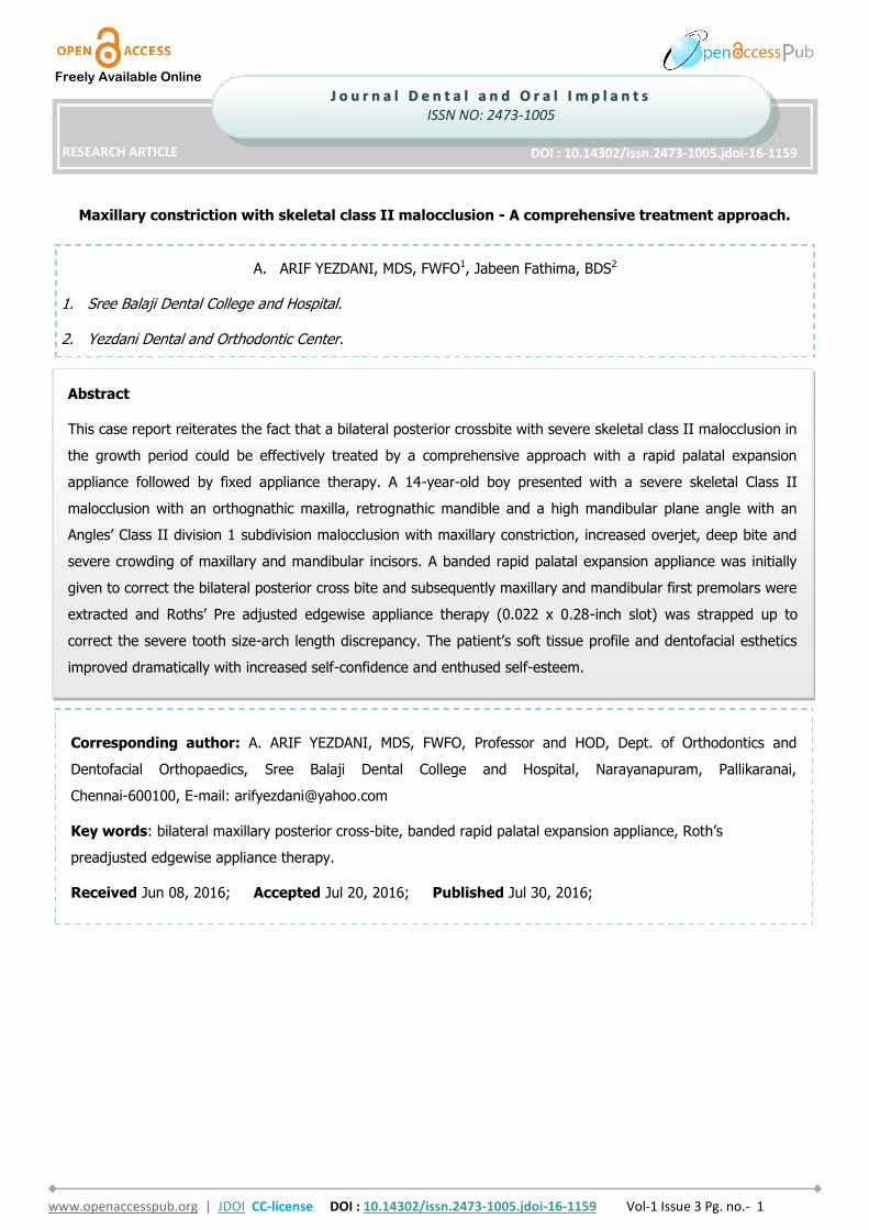
Intraoral assessment. (Fig 1d-h).
The maxillary arch was V-shaped with severely
proclined and rotated maxillary incisors with a palatally
placed 12. The mandibular arch was U-shaped with
severe crowding of mandibular incisors, with 43 partially
erupted and buccally placed with transpositioned 42 and
43, with 42 mesiolingually rotated, 33 distolingually
rotated and 34 distolingually rotated and buccally
placed.
Severe increase in overjet and deep bite were
both observed. The maxillary midline coincided with the
skeletal midline but the mandibular midline was shifted
to the right side by 1 mm. Bilateral maxillary posterior
crossbite was also observed. On right side the molar
relation was Class I and on the left side it was Class II.
The canine relation was Class II on the left side and the
curve of Spee was increased.
Radiographic assessment. The panoramic
radiograph confirmed the presence of all permanent
Fig.1d. Pre-treatment intra-oral-
Frontal
Fig.1e. Pre-treatment intra-oral-
Right
Fig.1f. Pre-treatment intra-oral-
Left
Fig.1g. Pre-treatment intra-oral-
Upper occlusal
Fig.1h. Pre-treatment intra-oral-
Lower occlusal
Fig.2. Pre-treatment panoramic radio-
graph
Freely Available Online
www.openaccesspub.org | JDOI CC-license DOI : 10.14302/issn.2473-1005.jdoi-16-1159 Vol-1 Issue 3 Pg. no.- 4
teeth with the presence of 18, 28, 38 and 48 tooth
germs with normal alveolar bone levels. (Fig 2).
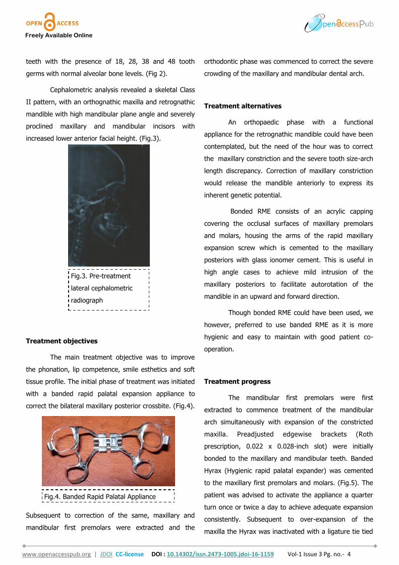
Cephalometric analysis revealed a skeletal Class
II pattern, with an orthognathic maxilla and retrognathic
mandible with high mandibular plane angle and severely
proclined maxillary and mandibular incisors with
increased lower anterior facial height. (Fig.3).
Treatment objectives
The main treatment objective was to improve
the phonation, lip competence, smile esthetics and soft
tissue profile. The initial phase of treatment was initiated
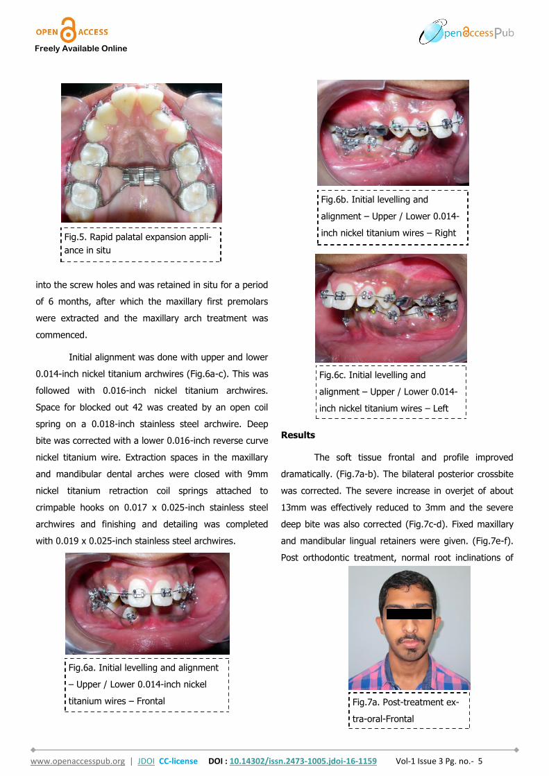
with a banded rapid palatal expansion appliance to
correct the bilateral maxillary posterior crossbite. (Fig.4).
Subsequent to correction of the same, maxillary and
mandibular first premolars were extracted and the
orthodontic phase was commenced to correct the severe
crowding of the maxillary and mandibular dental arch.
Treatment alternatives
An orthopaedic phase with a functional
appliance for the retrognathic mandible could have been
contemplated, but the need of the hour was to correct
the maxillary constriction and the severe tooth size-arch
length discrepancy. Correction of maxillary constriction
would release the mandible anteriorly to express its
inherent genetic potential.
Bonded RME consists of an acrylic capping
covering the occlusal surfaces of maxillary premolars
and molars, housing the arms of the rapid maxillary
expansion screw which is cemented to the maxillary
posteriors with glass ionomer cement. This is useful in
high angle cases to achieve mild intrusion of the
maxillary posteriors to facilitate autorotation of the
mandible in an upward and forward direction.
Though bonded RME could have been used, we
however, preferred to use banded RME as it is more
hygienic and easy to maintain with good patient co-
operation.
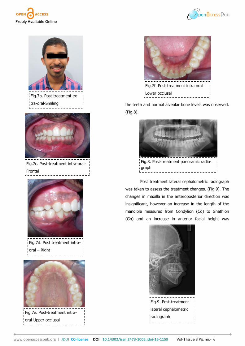
Treatment progress
The mandibular first premolars were first
extracted to commence treatment of the mandibular
arch simultaneously with expansion of the constricted
maxilla. Preadjusted edgewise brackets (Roth
prescription, 0.022 x 0.028-inch slot) were initially
bonded to the maxillary and mandibular teeth. Banded
Hyrax (Hygienic rapid palatal expander) was cemented
to the maxillary first premolars and molars. (Fig.5). The
patient was advised to activate the appliance a quarter
turn once or twice a day to achieve adequate expansion
consistently. Subsequent to over-expansion of the
maxilla the Hyrax was inactivated with a ligature tie tied
Fig.3. Pre-treatment
lateral cephalometric
radiograph
Fig.4. Banded Rapid Palatal Appliance
Freely Available Online
www.openaccesspub.org | JDOI CC-license DOI : 10.14302/issn.2473-1005.jdoi-16-1159 Vol-1 Issue 3 Pg. no.- 5
into the screw holes and was retained in situ for a period
of 6 months, after which the maxillary first premolars
were extracted and the maxillary arch treatment was
commenced.
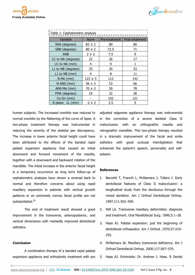
Initial alignment was done with upper and lower
0.014-inch nickel titanium archwires (Fig.6a-c). This was
followed with 0.016-inch nickel titanium archwires.
Space for blocked out 42 was created by an open coil
spring on a 0.018-inch stainless steel archwire. Deep
bite was corrected with a lower 0.016-inch reverse curve
nickel titanium wire. Extraction spaces in the maxillary
and mandibular dental arches were closed with 9mm
nickel titanium retraction coil springs attached to
crimpable hooks on 0.017 x 0.025-inch stainless steel
archwires and finishing and detailing was completed
with 0.019 x 0.025-inch stainless steel archwires.
Results
The soft tissue frontal and profile improved
dramatically. (Fig.7a-b). The bilateral posterior crossbite
was corrected. The severe increase in overjet of about
13mm was effectively reduced to 3mm and the severe
deep bite was also corrected (Fig.7c-d). Fixed maxillary
and mandibular lingual retainers were given. (Fig.7e-f).
Post orthodontic treatment, normal root inclinations of
Fig.6a. Initial levelling and alignment
– Upper / Lower 0.014-inch nickel
titanium wires – Frontal
Fig.7a. Post-treatment ex-
tra-oral-Frontal
Fig.5. Rapid palatal expansion appli-
ance in situ
Fig.6b. Initial levelling and
alignment – Upper / Lower 0.014-
inch nickel titanium wires – Right
Fig.6c. Initial levelling and
alignment – Upper / Lower 0.014-
inch nickel titanium wires – Left
Freely Available Online
www.openaccesspub.org | JDOI CC-license DOI : 10.14302/issn.2473-1005.jdoi-16-1159 Vol-1 Issue 3 Pg. no.- 6
the teeth and normal alveolar bone levels was observed.
(Fig.8).
Post treatment lateral cephalometric radiograph
was taken to assess the treatment changes. (Fig.9). The
changes in maxilla in the anteroposterior direction was
insignificant, however an increase in the length of the
mandible measured from Condylion (Co) to Gnathion
(Gn) and an increase in anterior facial height was
Fig.7e. Post-treatment intra-
oral-Upper occlusal
Fig.7f. Post-treatment intra oral-
Lower occlusal
Fig.9. Post-treatment
lateral cephalometric
radiograph
Fig.7d. Post treatment intra-
oral – Right
Fig.7c. Post-treatment intra-oral-
Frontal
Fig.7b. Post-treatment ex-
tra-oral-Smiling
Fig.8. Post-treatment panoramic radio-
graph
Fig.7b. Post-treatment ex-
tra-oral-Smiling

Freely Available Online
www.openaccesspub.org | JDOI CC-license DOI : 10.14302/issn.2473-1005.jdoi-16-1159 Vol-1 Issue 3 Pg. no.- 7
observed. Maxillary incisors were retracted dramatically
by 8mm and the mandibular incisors were proclined by
3mm to camouflage the skeletal class II malocclusion.
(Table 1). Effective expansion of the maxillary
constriction as an end-of treatment goal was achieved.
(Fig.10a-b).
Discussion
Bilateral posterior cross bite with severe skeletal
Class II malocclusion is a routinely encountered
malocclusion in an orthodontic practice. The probable
causes for skeletal posterior crossbite could be
hereditary, airway obstruction, non-nutritive sucking
habits, lowered tongue position, and early loss of
primary teeth. The skeletal malocclusion in the case
reported had an orthognathic maxilla and retrognathic
mandible with severe maxillary constriction. Some
authors are of the opinion that functional appliances
have no real effect on mandibular length 6, 7 while others
believe that mandibular growth can be increased with
functional appliance treatment 8-10. However, Wendling11
was of the opinion that a spontaneous correction of
some Class II malocclusions is favoured with initial rapid
palatal expansion. It has been reported in the literature
that in the Class II, division 1 group, the retrusion was
due to constriction of the maxillary dental arch and that
in such cases, it is important to expand the maxillary
arch to obtain a permanent orthopedic effect on the
maxilla by releasing the mandible to move anteriorly.
Literature reports too point to a strong correlation of
deficient maxillary arch width and Class II malocclu-
sion.12, 13 In compliance with this tenet banded Hyrax
was used to expand the maxilla to permit spontaneous
forward positioning of the mandible. The rapid maxillary
expansion appliance widens the intermaxillary suture
and increases basal bone width 14- 17 as also causes an
increase in dental arch perimeter18 as had been
observed in the treated case too. Post treatment
cephalometric analysis revealed an increase in length of
the mandible with no orthopaedic functional device
given to jump the mandible. This could most probably
have been attributed to the widening of the constricted
maxilla with the banded rapid maxillary expansion
appliance that promoted the mandible to express normal
growth in an anterior direction. However mandibular
growth was not sufficient enough to correct the severe
anteroposterior skeletal discrepancy but the dentoalveo-
lar compensation was maximized. The increased overjet
of 13 mm was reduced to 3mm at the end of treatment
most probably attributed to retroclination of the
maxillary anteriors and proclination of the mandibular
incisors. The mesial movement of the mandibular arch
observed in this patient was favourable for the reduction
in the increased overjet in compliance to the observation
by Voudouris et al 19 who too found mesial movement of
the mandibular arch in both experimental animals and
Fig.10b. Post expansion -
maxillary arch
Fig.10a. Pre expansion - maxillary
arch
Freely Available Online
www.openaccesspub.org | JDOI CC-license DOI : 10.14302/issn.2473-1005.jdoi-16-1159 Vol-1 Issue 3 Pg. no.- 8
human subjects. The increased overbite was reduced to
normal overbite by the flattening of the curve of Spee. A
two-phase treatment therapy was instrumental in
reducing the severity of the skeletal jaw discrepancy.
The increase in lower anterior facial height could have
been attributed to the effects of the banded rapid
palatal expansion appliance that caused an initial
downward and forward movement of the maxilla,
together with a downward and backward rotation of the
mandible. The initial increase in the anterior facial height
is a temporary occurrence as long term follow-up of
cephalometric analyses have shown a reversal back to
normal and therefore concerns about using rapid
maxillary expansion in patients with vertical growth
patterns or an extremely convex facial profile are not
substantiated.20
The end of treatment result showed a good
improvement in the transverse, anteroposterior, and
vertical dimensions with markedly improved dentofacial
esthetics.
Conclusion
A combination therapy of a banded rapid palatal
expansion appliance and orthodontic treatment with pre
adjusted edgewise appliance therapy was instrumental
in the correction of a severe skeletal Class II
malocclusion with an orthognathic maxilla and
retrognathic mandible. This two-phase therapy resulted
in a dramatic improvement of the facial and smile
esthetics with good occlusal interdigitation that
enhanced the patient’s speech, personality and self-
esteem.
References
1. Baccetti T, Franchi L, McNamara J, Tollaro I. Early
dentofacial features of Class II malocclusion: a
longitudinal study from the deciduous through the
mixed dentition. Am J Orthod Dentofacial Orthop.
1997;111:502–509.
2. Will LA. Transverse maxillary deformities: diagnosis
and treatment. Oral Maxillofacial Surg. 1996;5:1–28.
3. Haas AJ. Palatal expansion: just the beginning of
dentofacial orthopedics. Am J Orthod. 1970;57:219–
255.
4. McNamara JA. Maxillary transverse deficiency. Am J
Orthod Dentofacial Orthop. 2000;117:567–570.
5. Haas AJ. Entrevista: Dr. Andrew J. Haas. R Dental
Variable Norm Pre-treatment Post-treatment
SNA (degrees) 82 ± 2 80 80
SNB (degrees) 80 ± 2 72.5 71
ANB 2 ± 2 7.5 9
U1 to NA (degrees) 22 26 17
U1 to NA (mm) 4 9 1
L1 to NB (degrees) 25 30 33
L1 to NB (mm) 4 8 11
N-Me (mm) 123 ± 5 112 142
N-ANS (mm) 56 ± 3 53 66
ANS-Me (mm) 70 ± 3 59 78
FMA (degrees) 25 32 36
Co-Gn (mm) - 102 121
E-plane LL (mm) -2 ± 2 2.5 5
Table 1: Cephalometric analysis

Freely Available Online
www.openaccesspub.org | JDOI CC-license DOI : 10.14302/issn.2473-1005.jdoi-16-1159 Vol-1 Issue 3 Pg. no.- 9
Press Ortodon Ortop Facial. 2001;6:1–10.
6. Janson I. Skeletal and dentoalveolar changes in
patients treated with a bionator during prepubertal
and pubertal growth. In: McNamara JA Jr, Ribbens
KA, Howe RP, editors. Clinical alteration of the
growing face. Monograph 14. Craniofacial Growth
Series. Ann Arbor: Center for Human Growth and
Development; University of Michigan; 1983.
7. Vargervik K, Harvold EP. Response to activator
treatment in Class II malocclusions. Am J Orthod
1985;88:242-51.
8. Luder HU. Skeletal profile changes related to two
patterns of activator effects. Am J Orthod
1982;81:390-6.
9. McNamara JA Jr, Bookstein FL, Shaughnessy TG.
Skeletal and dental changes following functional
regulator therapy on Class II patients. Am J Orthod
1985;88:91-110.
10. Mamandras AH, Allen LP. Mandibular response to
orthodontic treatment with the bionator appliance.
Am J Orthod Dentofacial Orthop 1990;97:113-20.
11. Wendling LK. Short-Term Skeletal and Dental Effects
of the Acrylic Splint Rapid Maxillary Expansion
Appliance [master’s thesis]. Ann Arbor, Mich: The
University of Michigan; 1997.
12. Kirjavainen M, Kirjavainen T, Haavikko K. Changes in
dental arch dimensions by use of an orthopedic
cervical headgear in Class II correction. Am J Orthod
Dentofacial Orthop. 1997;111: 59–66.
13. Lima RMA, Lima AL. Case report: long-term outcome
of Class II division 1 malocclusion treated with rapid
palatal expansion and cervical traction. Angle
Orthod. 2000;70:89–94.
14. Haas AJ. Rapid expansion of the maxillary dental
arch and nasal cavity by opening the midpalatal
suture. Angle Orthod. 1961;31(2):73–90.
15. Starnbach H, Bayne D, Cleall J, Subtelny JD.
Facioskeletal and dental changes resulting from
rapid maxillary expansion. Angle Orthod. 1966;36
(2):152–164.
16. Wertz RA. Skeletal and dental changes accompany-
ing rapid midpalatal suture opening. Am J Orthod.
1970;58(1):41–66. .
17. Garib DG, Henriques JFC, Janson G, Freitas MR,
Coelho RA. Rapid maxillary expansion—tooth tissue-
borne versus tooth-borne expanders: a computed
tomography evaluation of dentoskeletal effects.
Angle Orthod. 2005;75(4):548–557.
18. Adkins MD, Nanda RS, Currier GF. Arch perimeter
changes on rapid palatal expansion. Am J Orthod
Dentofacial Or-thop. 1990;97(3):194–199.
19. Voudouris JC, Woodside DG, Altuna G, Angelopoulos
G, Bourque PJ, Lacouture CY. Condyle-fossa
modifications and muscle interactions during Herbst
treatment, part 2. Results and conclusions. Am J
Orthod Dentofacial Orthop 2003;124:13-29.
20. Garib DG, Henriques FC, Carvalho PEG; Gomes SC.
Longitudinal Effects of Rapid Maxillary Expansion-A
Retrospective Cephalometric Study. Angle
Orthodontist 2007;77:442-448.