Embed Size (px)
Citation preview

1
Frequently ordered laboratory tests
3
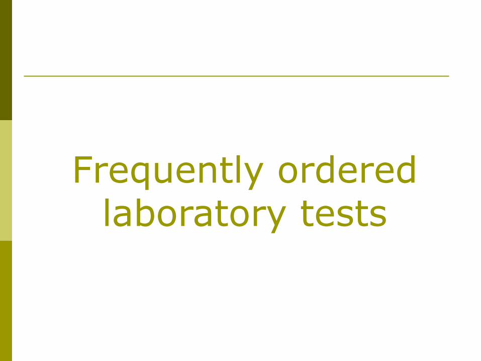
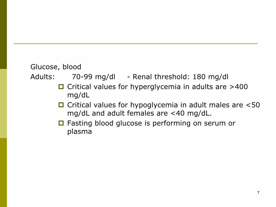
Glucose, blood
Adults: 70-99 mg/dl - Renal threshold: 180 mg/dl
Critical values for hyperglycemia in adults are >400 mg/dL
Critical values for hypoglycemia in adult males are <50 mg/dL and adult females are <40 mg/dL.
Fasting blood glucose is performing on serum or plasma
4
Glucose increased by:
Acromegaly
Adenoma of pancreas
Brain trauma
Cushing’s syndrome
Diabetes
Eclampsia
Hyperthyroidism
Liver disease
Malnutrition
MI
Obesity
Pancreatitis
Pheochromocytoma
Stress (insulin resistance)
Drugs that increase blood glucose:
Corticosteroids
Diuretics
Epinephrine
Glucagon
Nicotine
Glucose decreased by:
Addison’s disease
Exercise
Hepatic necrosis
Islet cell carcinoma
Malabsorption
Pancreatic cancer
Pituitary hypofunction
Post gastrectomy
Sepsis
Stress
Drugs that ay decrease blood glucose:
Alcohol
Amphetamines
Beta-blockers
Ginseng
Glucasol (agestroemia speciosi L. etc)
Gymnema Sylvestre ext.
Insulin
Momordica charantia (bitter melon)
Glucose, blood
5
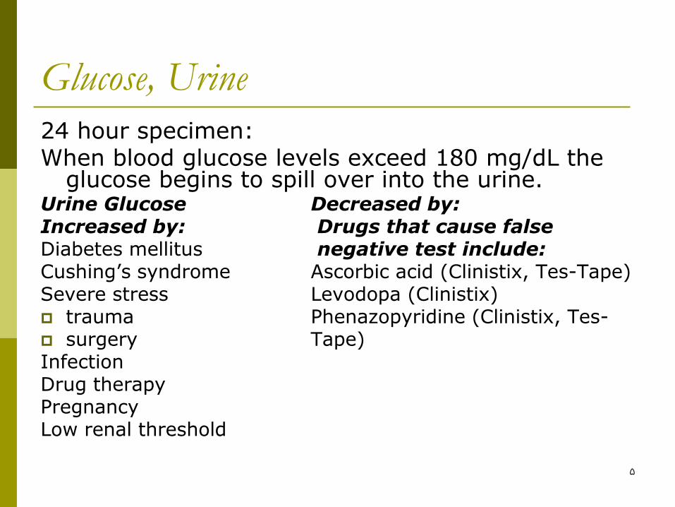
Glucose, Urine
24 hour specimen: When blood glucose levels exceed 180 mg/dL the
glucose begins to spill over into the urine. Urine Glucose Decreased by: Increased by: Drugs that cause false Diabetes mellitus negative test include: Cushing’s syndrome Ascorbic acid (Clinistix, Tes-Tape) Severe stress Levodopa (Clinistix) trauma Phenazopyridine (Clinistix, Tes- surgery Tape) Infection Drug therapy Pregnancy Low renal threshold
6
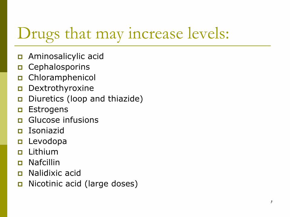
Drugs that may increase levels:
Aminosalicylic acid
Cephalosporins
Chloramphenicol
Dextrothyroxine
Diuretics (loop and thiazide)
Estrogens
Glucose infusions
Isoniazid
Levodopa
Lithium
Nafcillin
Nalidixic acid
Nicotinic acid (large doses)
7
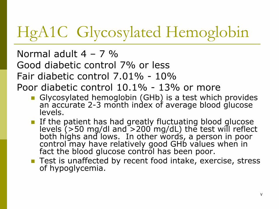
HgA1C Glycosylated Hemoglobin
Normal adult 4 – 7 % Good diabetic control 7% or less Fair diabetic control 7.01% - 10% Poor diabetic control 10.1% - 13% or more
Glycosylated hemoglobin (GHb) is a test which provides an accurate 2-3 month index of average blood glucose levels.
If the patient has had greatly fluctuating blood glucose levels (>50 mg/dl and >200 mg/dL) the test will reflect both highs and lows. In other words, a person in poor control may have relatively good GHb values when in fact the blood glucose control has been poor.
Test is unaffected by recent food intake, exercise, stress of hypoglycemia.
8
HgA1C Glycosylated Hemoglobin, cont’d
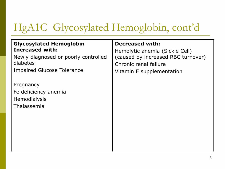
Glycosylated Hemoglobin Increased with:
Newly diagnosed or poorly controlled diabetes
Impaired Glucose Tolerance
Pregnancy
Fe deficiency anemia
Hemodialysis
Thalassemia
Decreased with:
Hemolytic anemia (Sickle Cell) (caused by increased RBC turnover)
Chronic renal failure
Vitamin E supplementation

9
Sodium, Serum Adults: 136-145 mEq/L Sodium is the major cation in extracellular space Intracellular sodium is approximately 5mEq/L…. Aldosterone and antidiuretic hormone help regulate sodium
balance Symptoms of hypernatremia: Dehydration thirst, agitation, restlessness, hyper-reflexes and
seizures. Symptoms of hyponatremia Muscle cramps, muscle twitching, headache, dizziness, lethargy,
confusion, convulsions, stupor and coma. The changes in the central nervous system are due to fluid shifts from the extracellular spaces to the intracellular spaces, causing cells to swell.
Elevated blood glucose levels give falsely low serum sodium values. Use this formula to correct serum sodium values. Na= glucose x 2 + Na 6

10
Drugs that may increase sodium Drugs that may decrease sodium
methyldopa diuretics
antibiotics vasopressin (ADH)
corticosteroids
oral contraceptive agents
Sodium Increased by Sodium decreased by
Excessive intake Decreased intake
Dehydration Severe diarrhea
Excessive sweating Vomiting
Diabetes insipidus Hypergllycemia
Hypercalcemic nephroopathy Hyperproteinemia
Hypokalemic nephropathy Addison’s disease
Cushing’s disease Cirrhosis with ascites
Primary aldosteronism Excessive non-electrolyte IVs
Sodium, Serum, Cont’d
11
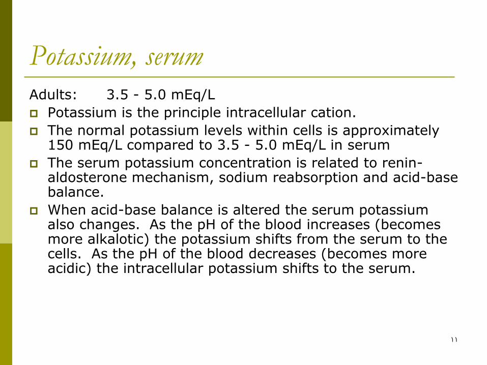
Potassium, serum
Adults: 3.5 - 5.0 mEq/L
Potassium is the principle intracellular cation.
The normal potassium levels within cells is approximately 150 mEq/L compared to 3.5 - 5.0 mEq/L in serum
The serum potassium concentration is related to renin-aldosterone mechanism, sodium reabsorption and acid-base balance.
When acid-base balance is altered the serum potassium also changes. As the pH of the blood increases (becomes more alkalotic) the potassium shifts from the serum to the cells. As the pH of the blood decreases (becomes more acidic) the intracellular potassium shifts to the serum.

12
Potassium, serum cont’d
Symptoms of hypokalemia:
Muscle weakness, cramps, hyporeflexia, paresthesias, decreased bowel motility, hypotension, cardiac arrhythmia, drowsiness, lethargy and coma. Serum potassium below 3.5 mEq/L is often seen with a serum pH above 7.45, decreased serum bicarbonate level and possibly elevated blood glucose.
Symptoms of hyperkalemia:
include confusion, irritability, nausea, vomiting, intestinal colic, paresthesia abdominal cramps and muscle paralysis.
13
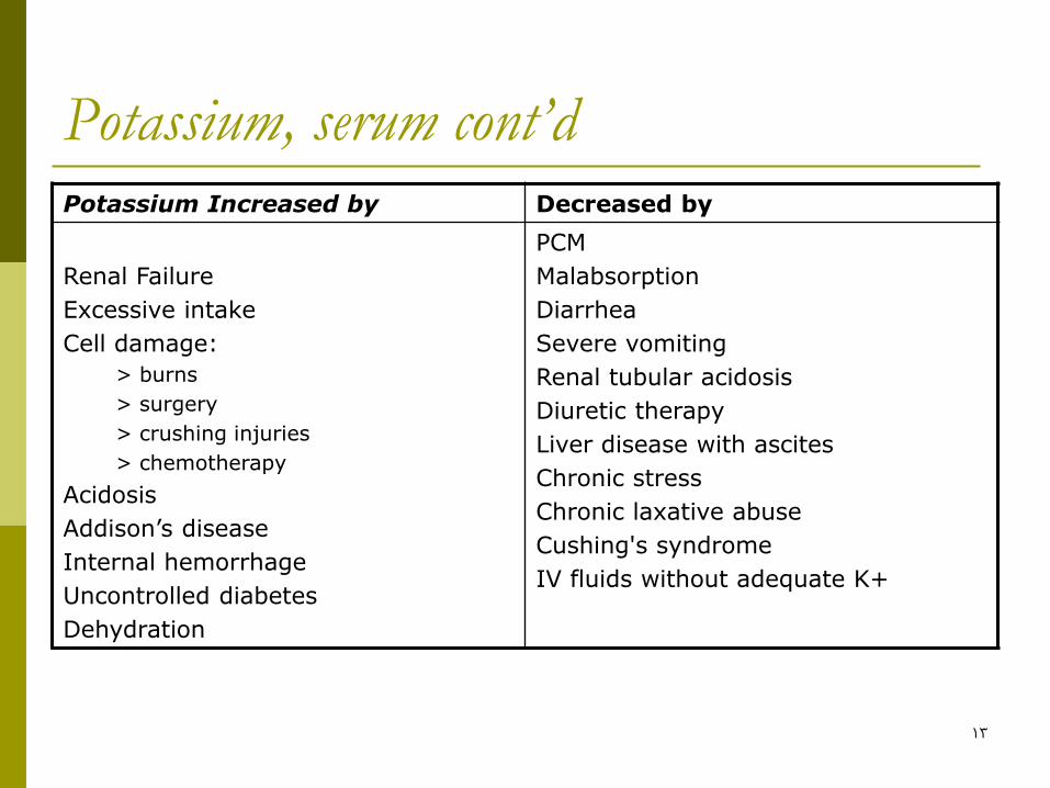
Potassium, serum cont’d Potassium Increased by Decreased by
Renal Failure
Excessive intake
Cell damage:
> burns
> surgery
> crushing injuries
> chemotherapy
Acidosis
Addison’s disease
Internal hemorrhage
Uncontrolled diabetes
Dehydration
PCM
Malabsorption
Diarrhea
Severe vomiting
Renal tubular acidosis
Diuretic therapy
Liver disease with ascites
Chronic stress
Chronic laxative abuse
Cushing's syndrome
IV fluids without adequate K+
14
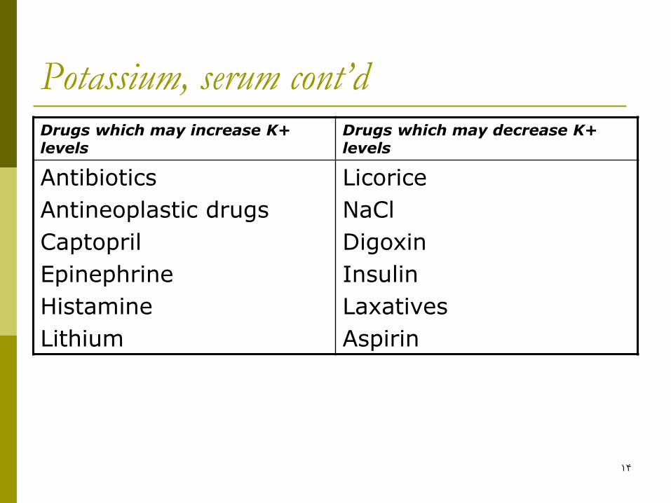
Potassium, serum cont’d Drugs which may increase K+ levels
Drugs which may decrease K+ levels
Antibiotics
Antineoplastic drugs
Captopril
Epinephrine
Histamine
Lithium
Licorice
NaCl
Digoxin
Insulin
Laxatives
Aspirin
15
Chloride, Cont’d
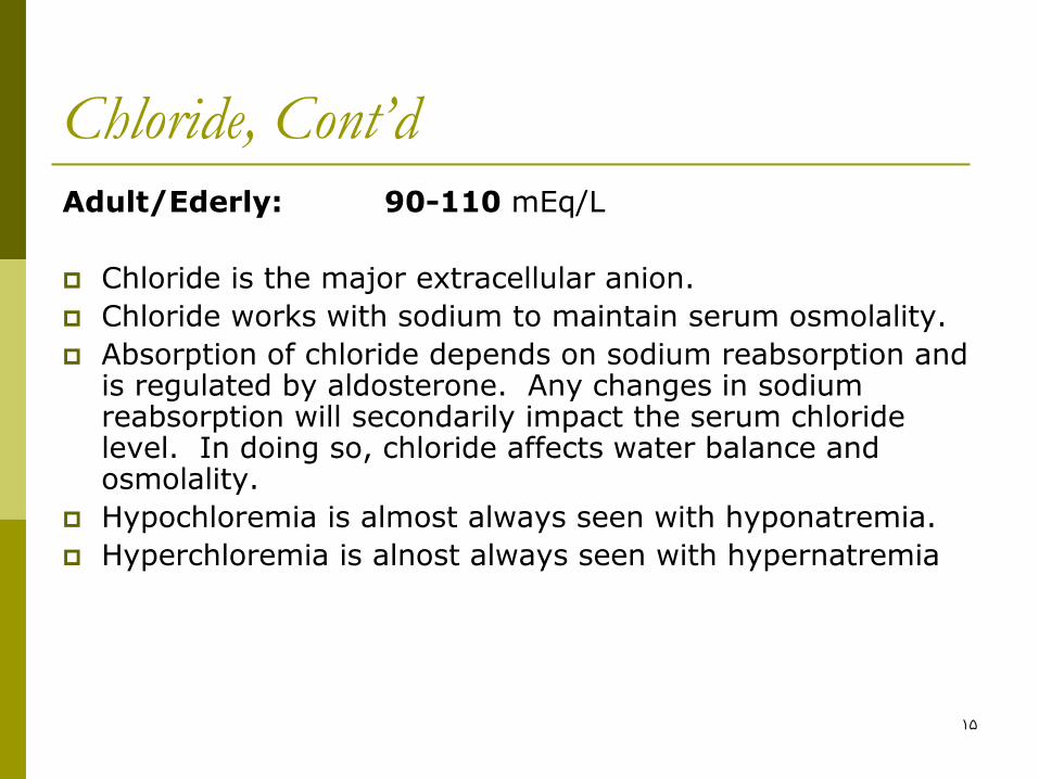
Adult/Ederly: 90-110 mEq/L
Chloride is the major extracellular anion.
Chloride works with sodium to maintain serum osmolality.
Absorption of chloride depends on sodium reabsorption and is regulated by aldosterone. Any changes in sodium reabsorption will secondarily impact the serum chloride level. In doing so, chloride affects water balance and osmolality.
Hypochloremia is almost always seen with hyponatremia.
Hyperchloremia is alnost always seen with hypernatremia
16
Chloride, Cont’d
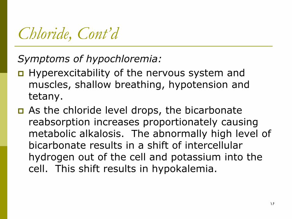
Symptoms of hypochloremia:
Hyperexcitability of the nervous system and muscles, shallow breathing, hypotension and tetany.
As the chloride level drops, the bicarbonate reabsorption increases proportionately causing metabolic alkalosis. The abnormally high level of bicarbonate results in a shift of intercellular hydrogen out of the cell and potassium into the cell. This shift results in hypokalemia.
17
Chloride, Cont’d
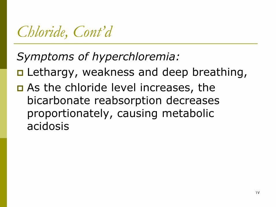
Symptoms of hyperchloremia:
Lethargy, weakness and deep breathing,
As the chloride level increases, the bicarbonate reabsorption decreases proportionately, causing metabolic acidosis

18
Chloride, Cont’d Chloride Increased by Decreased by
Dehydration
Excessive infusion of saline
Cushing’s syndrome
Eclampsia
Anemia
Hypernatremia
Multiple myeloma
Metabolic acidosis
Hyperventilation
Overhydration
Congestive heart failure
Vomiting
Addison’s disease
Hypokalemia
Hyponatremia
Diuretic therapy
Metabolic alkalosis
Burns
Emphysema
19
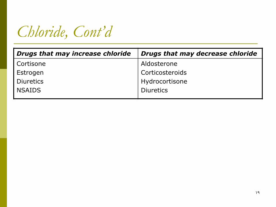
Chloride, Cont’d Drugs that may increase chloride Drugs that may decrease chloride
Cortisone
Estrogen
Diuretics
NSAIDS
Aldosterone
Corticosteroids
Hydrocortisone
Diuretics
20
Carbon Dioxide
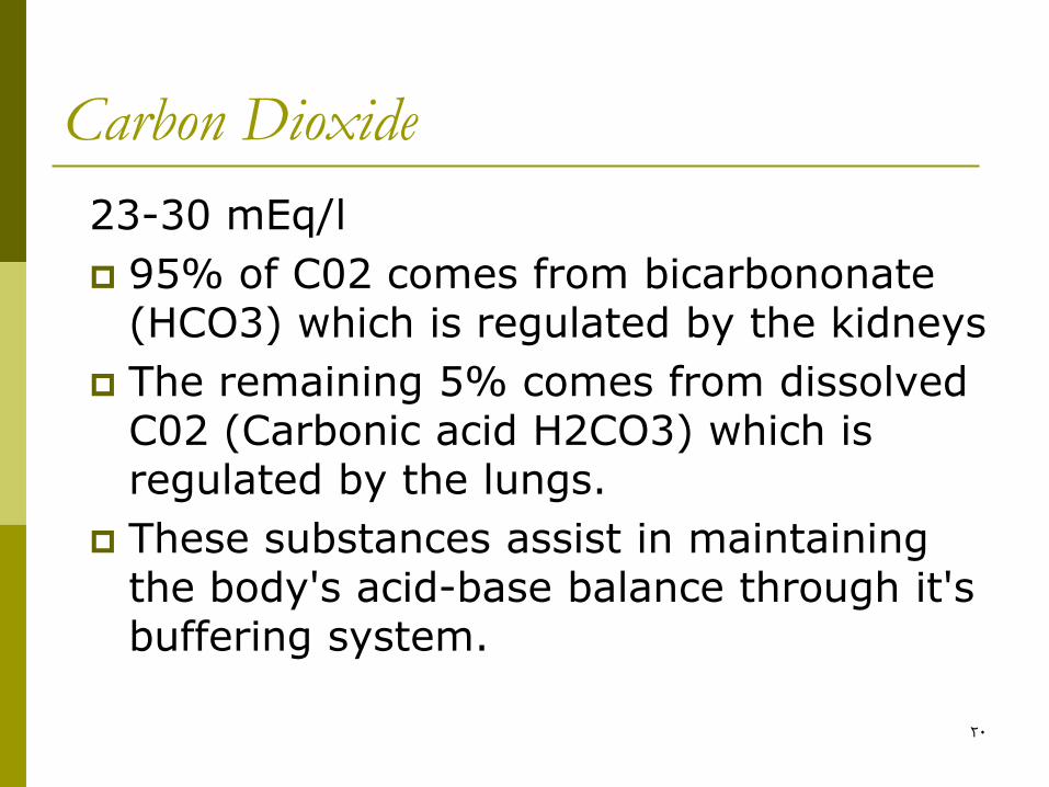
23-30 mEq/l
95% of C02 comes from bicarbononate (HCO3) which is regulated by the kidneys
The remaining 5% comes from dissolved C02 (Carbonic acid H2CO3) which is regulated by the lungs.
These substances assist in maintaining the body's acid-base balance through it's buffering system.
21
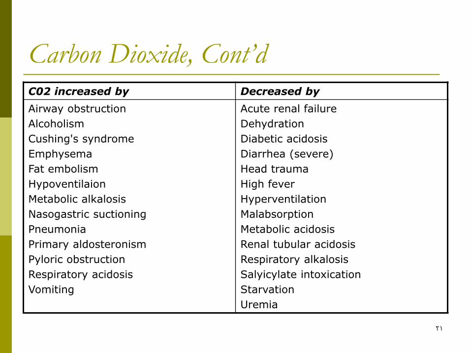
Carbon Dioxide, Cont’d C02 increased by Decreased by
Airway obstruction
Alcoholism
Cushing's syndrome
Emphysema
Fat embolism
Hypoventilaion
Metabolic alkalosis
Nasogastric suctioning
Pneumonia
Primary aldosteronism
Pyloric obstruction
Respiratory acidosis
Vomiting
Acute renal failure
Dehydration
Diabetic acidosis
Diarrhea (severe)
Head trauma
High fever
Hyperventilation
Malabsorption
Metabolic acidosis
Renal tubular acidosis
Respiratory alkalosis
Salyicylate intoxication
Starvation
Uremia
22
Carbon Dioxide, Cont’d
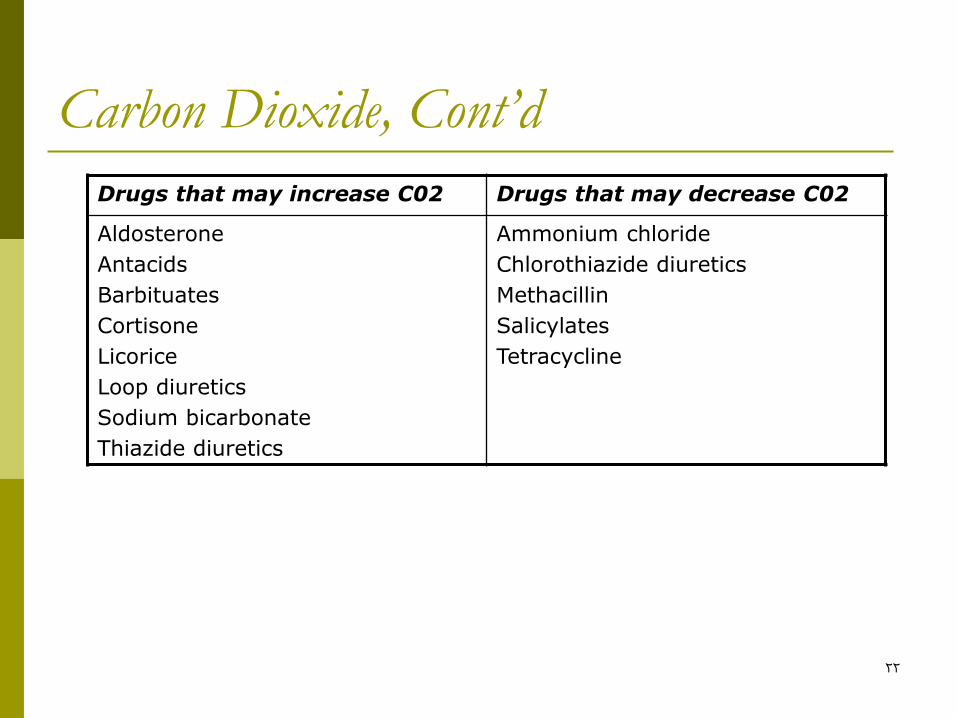
Drugs that may increase C02 Drugs that may decrease C02
Aldosterone
Antacids
Barbituates
Cortisone
Licorice
Loop diuretics
Sodium bicarbonate
Thiazide diuretics
Ammonium chloride
Chlorothiazide diuretics
Methacillin
Salicylates
Tetracycline
23
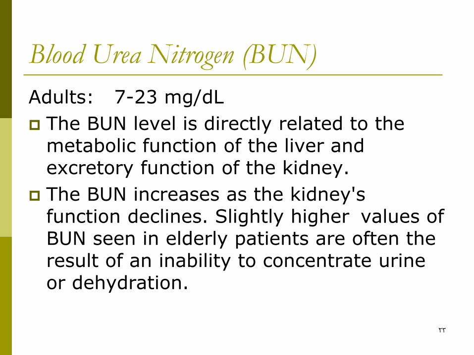
Blood Urea Nitrogen (BUN)
Adults: 7-23 mg/dL
The BUN level is directly related to the metabolic function of the liver and excretory function of the kidney.
The BUN increases as the kidney's function declines. Slightly higher values of BUN seen in elderly patients are often the result of an inability to concentrate urine or dehydration.
24
Blood Urea Nitrogen (BUN) Cont’d
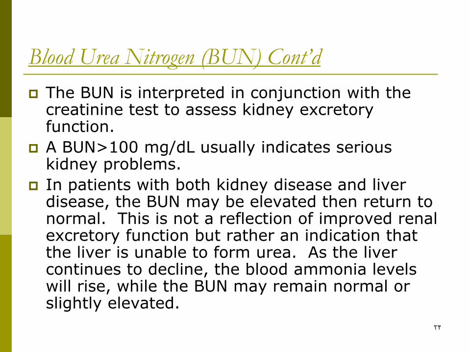
The BUN is interpreted in conjunction with the creatinine test to assess kidney excretory function.
A BUN>100 mg/dL usually indicates serious kidney problems.
In patients with both kidney disease and liver disease, the BUN may be elevated then return to normal. This is not a reflection of improved renal excretory function but rather an indication that the liver is unable to form urea. As the liver continues to decline, the blood ammonia levels will rise, while the BUN may remain normal or slightly elevated.
25
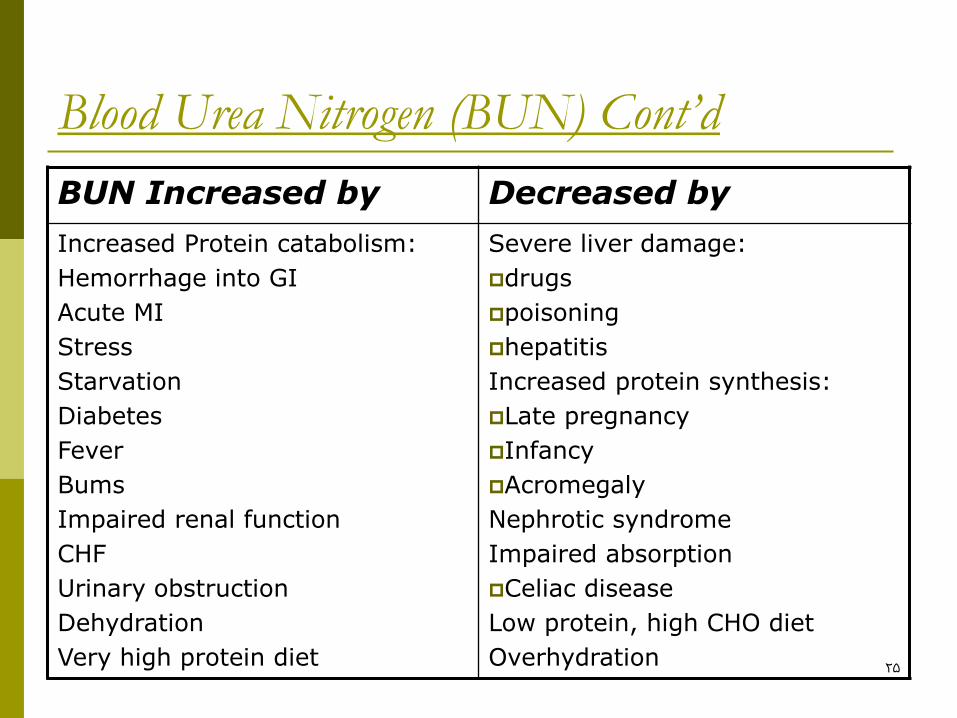
Blood Urea Nitrogen (BUN) Cont’d
BUN Increased by Decreased by
Increased Protein catabolism:
Hemorrhage into GI
Acute MI
Stress
Starvation
Diabetes
Fever
Bums
Impaired renal function
CHF
Urinary obstruction
Dehydration
Very high protein diet
Severe liver damage:
drugs
poisoning
hepatitis
Increased protein synthesis:
Late pregnancy
Infancy
Acromegaly
Nephrotic syndrome
Impaired absorption
Celiac disease
Low protein, high CHO diet
Overhydration
26
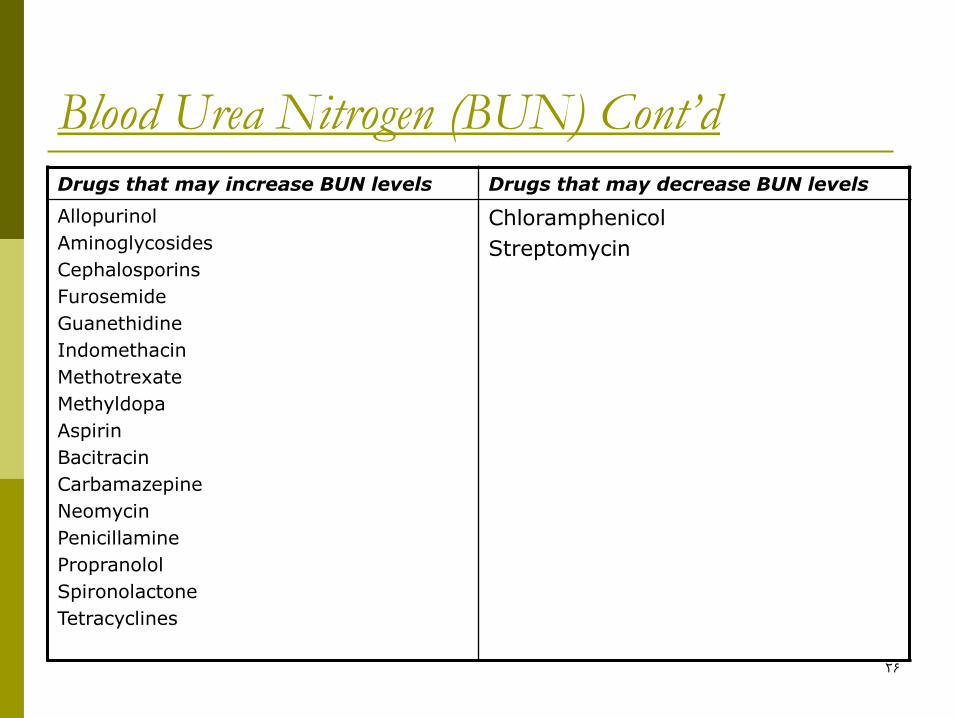
Blood Urea Nitrogen (BUN) Cont’d
Drugs that may increase BUN levels Drugs that may decrease BUN levels
Allopurinol
Aminoglycosides
Cephalosporins
Furosemide
Guanethidine
Indomethacin
Methotrexate
Methyldopa
Aspirin
Bacitracin
Carbamazepine
Neomycin
Penicillamine
Propranolol
Spironolactone
Tetracyclines
Chloramphenicol
Streptomycin

27
Creatinine
Adult males: 0.9 - 1.3 mg/dL;
Adult females: 0.6 - 1.1 mg/dL;
Creatinine is a by-product of the metabolism of muscle creatine phosphate to form ATP.
Critical values >4 mg/dL indicate serious impairment in renal function. However, a normal creatinine does not always mean unimpaired renal function.
Unlike the BUN, creatinine is not affected by malnutrition or hepatic function.

28
Creatinine, Cont’d Creatinine increased by: Decreased by:
Acromegaly
CHF Debilitation
Diabetic nephropathy Muscular dystrophy
Impaired renal function
Nephritis
Reduced renal blood flow:
Shock Drugs that may decrease Creatinine:
Dehydration
Congestive heart failure Diuretics
Rapid muscle loss Marijuana
Rhabdomyolysis
Surgery
Trauma
Urinary tract obstruction
Uremia
29
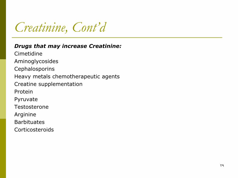
Creatinine, Cont’d Drugs that may increase Creatinine:
Cimetidine
Aminoglycosides
Cephalosporins
Heavy metals chemotherapeutic agents
Creatine supplementation
Protein
Pyruvate
Testosterone
Arginine
Barbituates
Corticosteroids
30
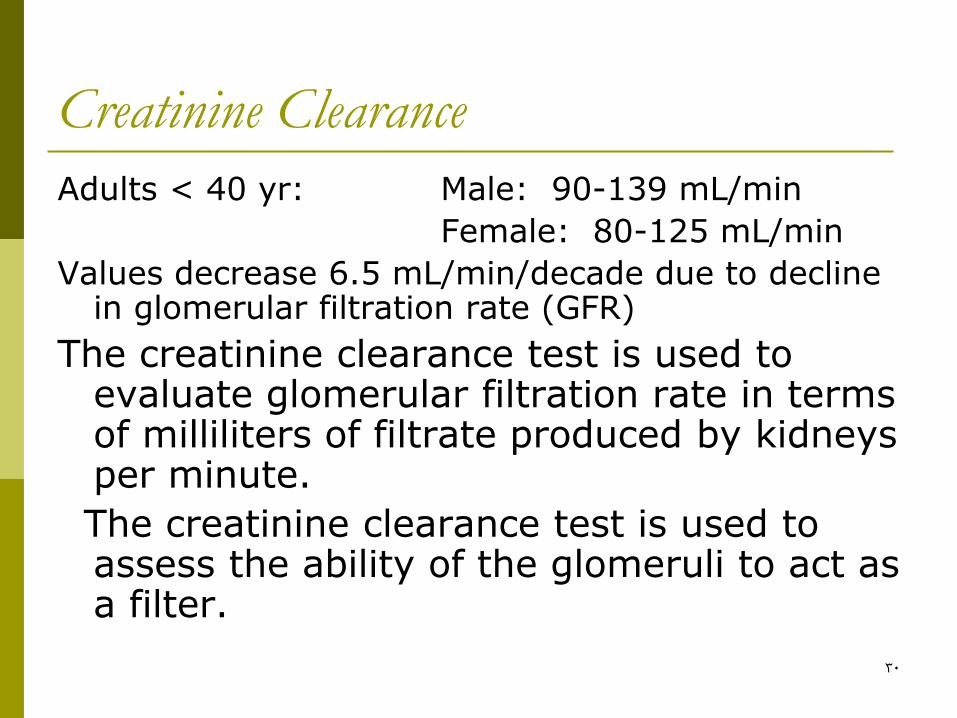
Creatinine Clearance
Adults < 40 yr: Male: 90-139 mL/min
Female: 80-125 mL/min
Values decrease 6.5 mL/min/decade due to decline in glomerular filtration rate (GFR)
The creatinine clearance test is used to evaluate glomerular filtration rate in terms of milliliters of filtrate produced by kidneys per minute.
The creatinine clearance test is used to assess the ability of the glomeruli to act as a filter.
31
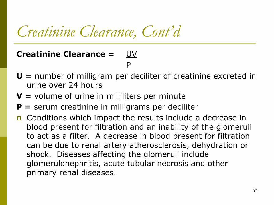
Creatinine Clearance, Cont’d
Creatinine Clearance = UV
P
U = number of milligram per deciliter of creatinine excreted in urine over 24 hours
V = volume of urine in milliliters per minute
P = serum creatinine in milligrams per deciliter
Conditions which impact the results include a decrease in blood present for filtration and an inability of the glomeruli to act as a filter. A decrease in blood present for filtration can be due to renal artery atherosclerosis, dehydration or shock. Diseases affecting the glomeruli include glomerulonephritis, acute tubular necrosis and other primary renal diseases.

32
Creatinine Clearance, Cont’d Creatinine Clearance Increased by: Decreased by:
Exercise Impaired kidney function
Pregnancy Renal artery atherosclerosis
Glomerulonephritis
Drugs that may increase levels: Acute tubular necrosis
Aminoglycosides: Dehydration
Gentamicin Congestive heart failure
Cimetidine Cirrhosis with ascites
Heavy-metal chemotherapeutic agents: Shock
Cisplatin
Nephrotoxic drugs:
Cephalosporins
Cefoxitin
33
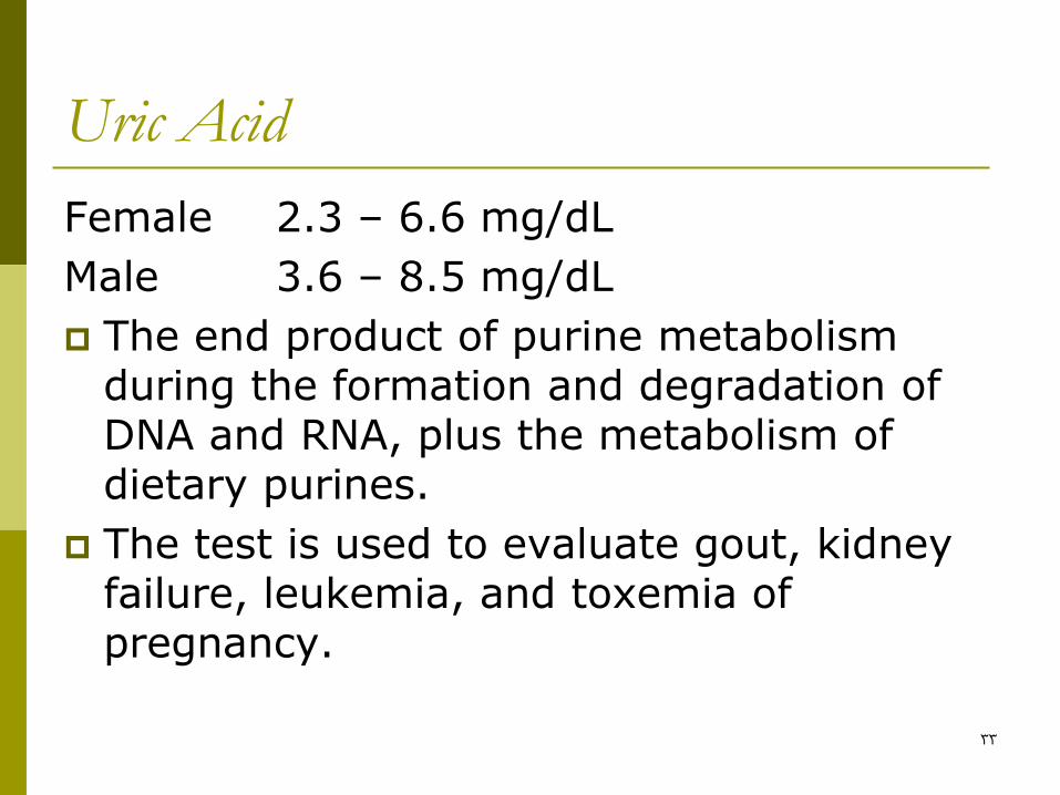
Uric Acid
Female 2.3 – 6.6 mg/dL
Male 3.6 – 8.5 mg/dL
The end product of purine metabolism during the formation and degradation of DNA and RNA, plus the metabolism of dietary purines.
The test is used to evaluate gout, kidney failure, leukemia, and toxemia of pregnancy.

34
Uric Acid, Cont’d Uric Acid Increased by Decreased by
Acute leukemia
Alcoholism
Anemia
Congestive heart failure
Dehydration
Down syndrome
Eclampsia
Fasting
Glomerulonephritis
Gouty arthritis
Hypothyroidism’
Hypoparathyroidism
Infectious mono
Lead poisoning
Lymphomas
Malnutrition
Neoplasms
Nephritis
Polycythemia vera
Radiation
Renal failure
Shock
Uremia
Acromegaly
Celiac disease
Hodgkin’s disease
Liver disease
Neoploasms
Renal tubular defects
SIADH
Wilson’s disease
Xanthinuria
35
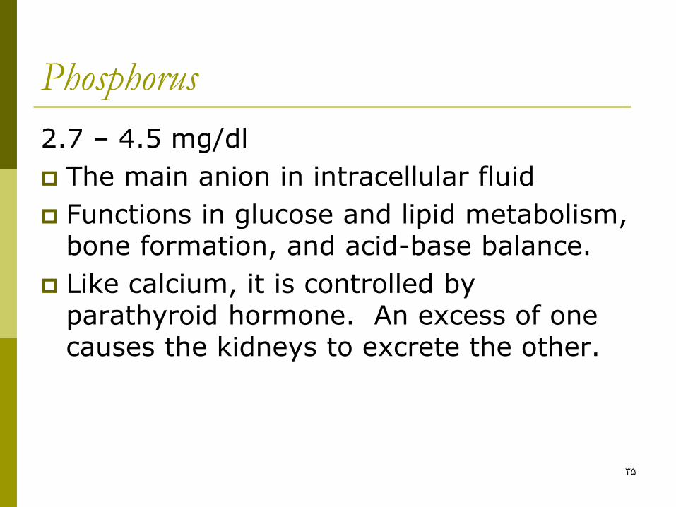
Phosphorus
2.7 – 4.5 mg/dl
The main anion in intracellular fluid
Functions in glucose and lipid metabolism, bone formation, and acid-base balance.
Like calcium, it is controlled by parathyroid hormone. An excess of one causes the kidneys to excrete the other.
36
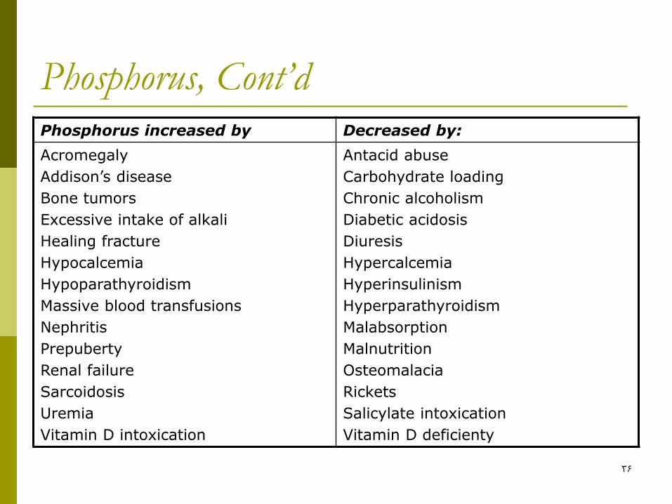
Phosphorus, Cont’d Phosphorus increased by Decreased by:
Acromegaly
Addison’s disease
Bone tumors
Excessive intake of alkali
Healing fracture
Hypocalcemia
Hypoparathyroidism
Massive blood transfusions
Nephritis
Prepuberty
Renal failure
Sarcoidosis
Uremia
Vitamin D intoxication
Antacid abuse
Carbohydrate loading
Chronic alcoholism
Diabetic acidosis
Diuresis
Hypercalcemia
Hyperinsulinism
Hyperparathyroidism
Malabsorption
Malnutrition
Osteomalacia
Rickets
Salicylate intoxication
Vitamin D deficienty

37
Phosphorus, Cont’d
Drugs that may increase Phosphorus
Drugs that may decrease phorphorus
Heparin
Phenytoin
Methicillin sodium
Phosphate enemas
Anabolic steroids
Androgens
Antacids
Diuretics
Epinephrine
Glucagon
Glucose
Insulin
Mannitol
Salicylates
38
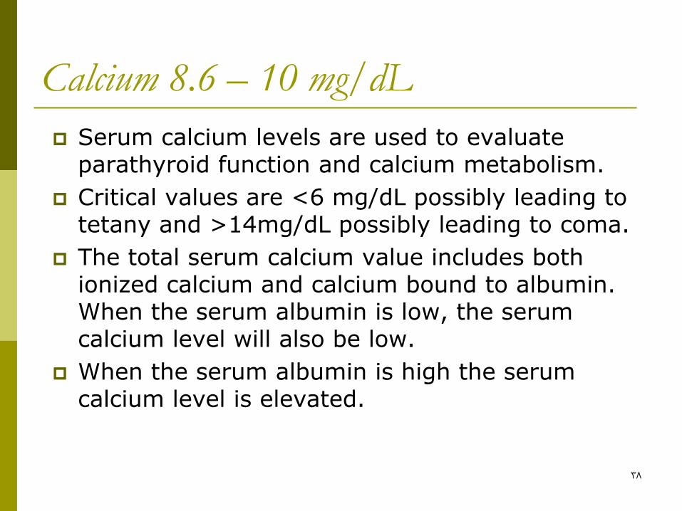
Calcium 8.6 – 10 mg/dL
Serum calcium levels are used to evaluate parathyroid function and calcium metabolism.
Critical values are <6 mg/dL possibly leading to tetany and >14mg/dL possibly leading to coma.
The total serum calcium value includes both ionized calcium and calcium bound to albumin. When the serum albumin is low, the serum calcium level will also be low.
When the serum albumin is high the serum calcium level is elevated.
39
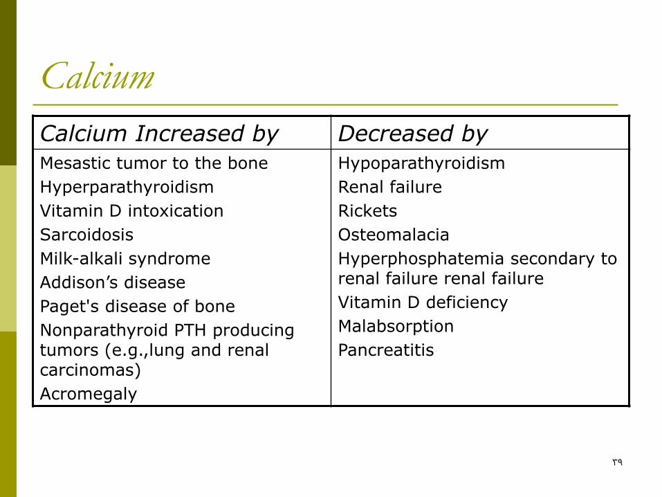
Calcium
Calcium Increased by Decreased by
Mesastic tumor to the bone
Hyperparathyroidism
Vitamin D intoxication
Sarcoidosis
Milk-alkali syndrome
Addison’s disease
Paget's disease of bone
Nonparathyroid PTH producing tumors (e.g.,lung and renal carcinomas)
Acromegaly
Hypoparathyroidism
Renal failure
Rickets
Osteomalacia
Hyperphosphatemia secondary to renal failure renal failure
Vitamin D deficiency
Malabsorption
Pancreatitis
40
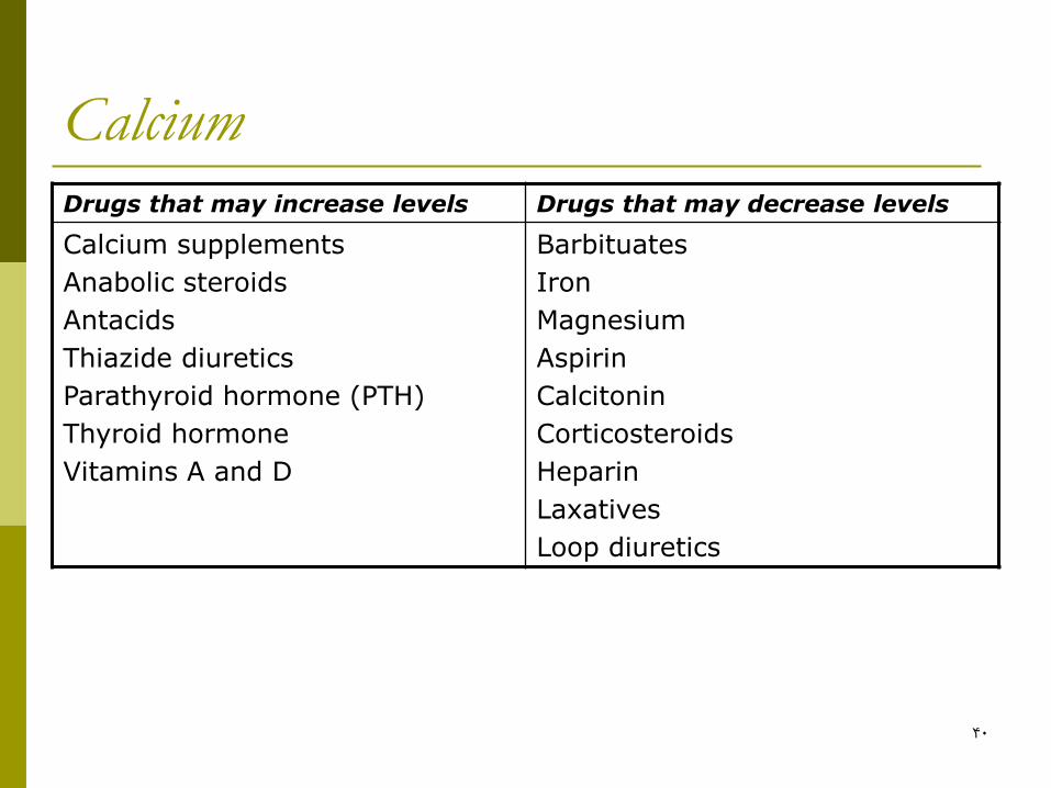
Calcium Drugs that may increase levels Drugs that may decrease levels
Calcium supplements
Anabolic steroids
Antacids
Thiazide diuretics
Parathyroid hormone (PTH)
Thyroid hormone
Vitamins A and D
Barbituates
Iron
Magnesium
Aspirin
Calcitonin
Corticosteroids
Heparin
Laxatives
Loop diuretics
41
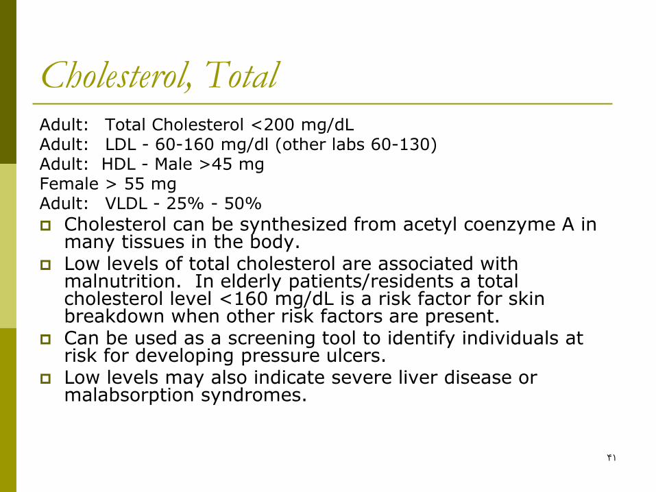
Cholesterol, Total Adult: Total Cholesterol <200 mg/dL Adult: LDL - 60-160 mg/dl (other labs 60-130) Adult: HDL - Male >45 mg Female > 55 mg Adult: VLDL - 25% - 50%
Cholesterol can be synthesized from acetyl coenzyme A in many tissues in the body.
Low levels of total cholesterol are associated with malnutrition. In elderly patients/residents a total cholesterol level <160 mg/dL is a risk factor for skin breakdown when other risk factors are present.
Can be used as a screening tool to identify individuals at risk for developing pressure ulcers.
Low levels may also indicate severe liver disease or malabsorption syndromes.

42
Cholesterol, Total
Cholesterol Increased by: Decreased by
Diabetes
High calorie diet
Hypothyroidism
Hypertension
Nephrotic syndrome
Pregnancy
Stress
Biliary cirrhosis
Malabsorption
PCM
Hyperthyroidism
Pemicious aneniia
Hemolytic anemia
Sepsis
Stress
Liver disease
AIDS
43
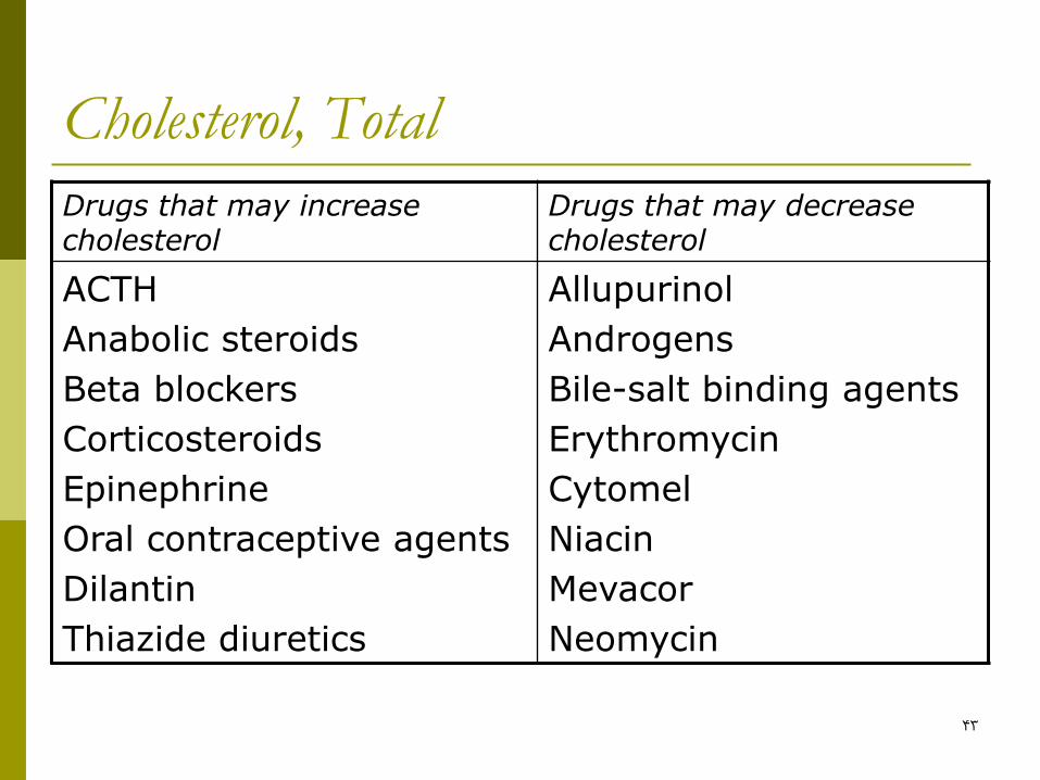
Cholesterol, Total
Drugs that may increase cholesterol
Drugs that may decrease cholesterol
ACTH
Anabolic steroids
Beta blockers
Corticosteroids
Epinephrine
Oral contraceptive agents
Dilantin
Thiazide diuretics
Allupurinol
Androgens
Bile-salt binding agents
Erythromycin
Cytomel
Niacin
Mevacor
Neomycin

44
Triglycerides Adult: Males 40-100 mg/dl
Females 35-100 mg/dl
Triglycerides increased by: Decreased by:
MI Malabsorption
High (excessive) calorie intake PCM
High CHO diet or High Fat Diet Hyperthyroidism
Poorly controlled diabetes AIDS
Nephrotic syndrome Drugs that may decreased levels:
Alcoholic cirrhosis Ascorbic acid
Alcohol Clofibrate
Drugs that may increase levels: Cholesterol
Cholestyramine
Estrogen
Oral contraceptive agents
45
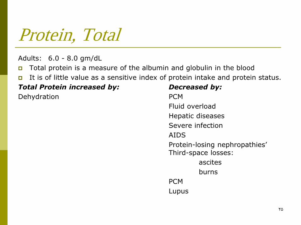
Protein, Total Adults: 6.0 - 8.0 gm/dL
Total protein is a measure of the albumin and globulin in the blood
It is of little value as a sensitive index of protein intake and protein status.
Total Protein increased by: Decreased by:
Dehydration PCM
Fluid overload
Hepatic diseases
Severe infection
AIDS
Protein-losing nephropathies’ Third-space losses:
ascites
burns
PCM
Lupus

46
Albumin Adults: 4.0 - 6.0 gm/dL (labs vary)
Degree of depletion
Mild: 3.0-3.5, gm/dL moderate: 2.1-3.0 gm/dL severe: <2.1 gm/dL
Albumin is synthesized in the liver at a rate of 8-14 grams/day. It makes up approximately 60% of the total protein.
Albumin provides approximately 80% of colloidal osmotic pressure of the
Albumin also serves as a carrier of metals, ions, fatty acids, amino acids, metabolites, bilirubin, enzymes, hormones and drugs.
Albumin level is dependent upon hepatocyte function. With age and declining liver function the liver can lose its ability to synthesize albumin.
Because the half-fife of albumin is 12-21 days, significant changes in liver n specific to albumin synthesis may go undetected until after that point.
Severely malnourished individuals have greatly decreased levels of serum albumin. However, albumin is a poor indicator of early malnutrition because of its long half-life.
When albumin levels are low the serum calcium level is also low.
When the serum albumin is elevated the serum calcium is elevated.
47
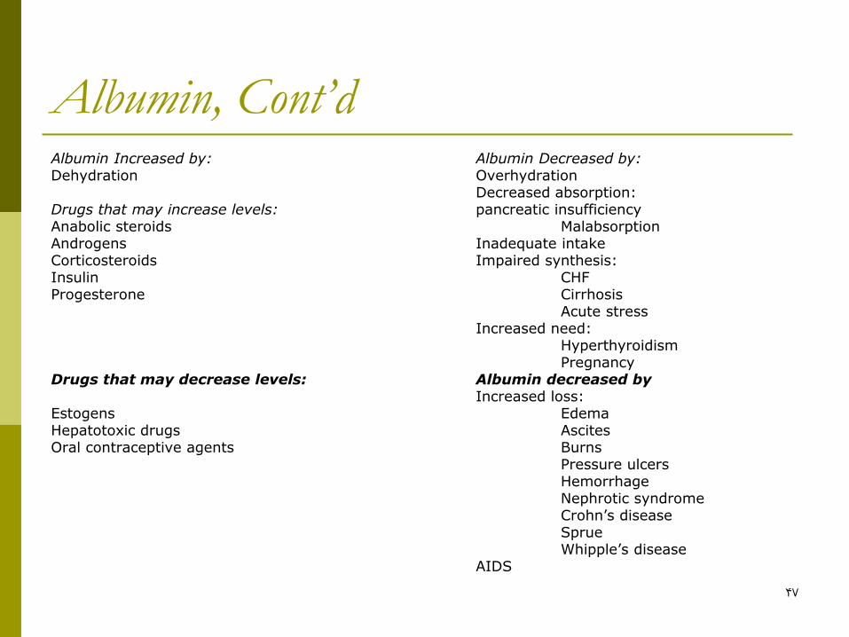
Albumin, Cont’d Albumin Increased by: Albumin Decreased by: Dehydration Overhydration Decreased absorption: Drugs that may increase levels: pancreatic insufficiency Anabolic steroids Malabsorption Androgens Inadequate intake Corticosteroids Impaired synthesis: Insulin CHF Progesterone Cirrhosis Acute stress Increased need: Hyperthyroidism Pregnancy Drugs that may decrease levels: Albumin decreased by Increased loss: Estogens Edema Hepatotoxic drugs Ascites Oral contraceptive agents Burns Pressure ulcers Hemorrhage Nephrotic syndrome Crohn’s disease Sprue Whipple’s disease AIDS
48
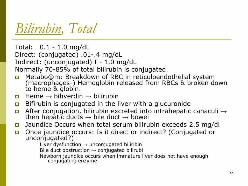
Bilirubin, Total Total: 0.1 - 1.0 mg/dL Direct: (conjugated) .01-.4 mg/dL Indirect: (unconjugated) I - 1.0 mg/dL Normally 70-85% of total bilirubin is conjugated. Metabo@m: Breakdown of RBC in reticuloendothelial system
(macrophages-) Hemoglobin released from RBCs & broken down to heme & globin.
Heme → bihverdin → bilirubin
Bifirubin is conjugated in the liver with a glucuronide After conjugation, bilirubin excreted into intrahepatic canaculi →
then hepatic ducts → bile duct → bowel
Jaundice Occurs when total serum bilirubin exceeds 2.5 mg/dl Once jaundice occurs: Is it direct or indirect? (Conjugated or
unconjugated?) Liver dysfunction → unconjugated biliribin Bile duct obstruction → conjugated bilirubi Newborn jaundice occurs when immature liver does not have enough
conjugating enzyme

49
Lactic Dehydrogenase (LDH)
Adult/Elderly: 110-210 U/L
Isoenzymes in adult/elderly: LDH-1: 17%-27% heart & blood vessels
LDH-2: 27% -37% reticuloendothelial system
LDH-3: 18% - 25% lungs
LDH-4: 3%- 8% kidney, placenta, pancreas
LDH-5: 00/o - 5% liver and striated muscle
50
Lactic Dehydrogenase (LDH), Cont’d
Lactic dehydrogenase (LDH) is an enzyme found in heart, liver, skeletal muscle, brain, red blood cells and lung cells.
It rises within 24 to 72 hours after a myocardial infarction, peaks in 3 to 4 days and returns to normal in about 14 days.
Lactic dehydrogenase is used with creatinine phosphokinase MB isoenzyme and aspartate ammotransferase to diagnose a myocardial infarction.
LDH-2 levels are normally higher than the other isoenzymes. When LDH-1 is elevated above LDH-2 it suggests a possible myocardial Actions If the laboratory only assesses for LDH-1 look for a level greater than 40% to suggest a myocardial infarction.

51
Lactic Dehydrogenase (LDH), Cont’d
LDH Increased by:
Myocardial Actions pulmonary infarction, hepatic diseases, hemolytic anemias, skeletal muscle disease and injury, renal parenchymal diseases, intestinal ischemia infarction, cerebrovascular accident, neoplastic states, infectious mononucleosis, heat stroke, pancreatitis, collagen diseases, fracture, muscular dystrophy, shock, hypotension
Drugs that may increase levels:
Alcohol anesthetics, aspirin, clofibrate, fluorides, mithramycin, narcotics, procainamide

52
Creatinine Phosphokinase (CPK)
Total CPK
Adult/Elderly: Male 12 - 17 U/mL
Female: 10 - 55 U/mL
Isoenzymes
CPK-MM - 100% skeletal muscle
CPK-MB - 0% specific for myocardial cells
CPK-BB - 0% brain & lung
53
Creatinine Phosphokinase (CPK), Cont’d Creatinine phosphokinase (CPK) is an enzyme found in the heart,
muscle, skeletal muscle and brain. Elevations in serum creatinine phosphokinase are always due to damaged muscle cells. The CPK levels often rise within 6 hours after trauma, peak at 18 hours and return to normal in 2 to 3 days if no additional injury occurs.
Elevations in creatinine phosphokinase are often associated with a myocardial infarction. The CPK-MB appears to be specific for myocardial cells. The CPK-MB levels rise 3 to 6 hours after a myocardial infarction, peak at 12 to 24 hours and return to normal in 12 to 48 hours. If additional myocardial damage occurs, the CPK-MB will begin to rise again. CPK-MB levels usually do not rise with chest pain caused by angina, pulmonary embolism or congestive heart failure.
Elevations in the CPK-BB are associated with a cerebrovascular accident or pulmonary infarction.
When the CPK level increases due to an elevation in CPK-MM it is likely due to injury or stress to the skeletal muscle.
54
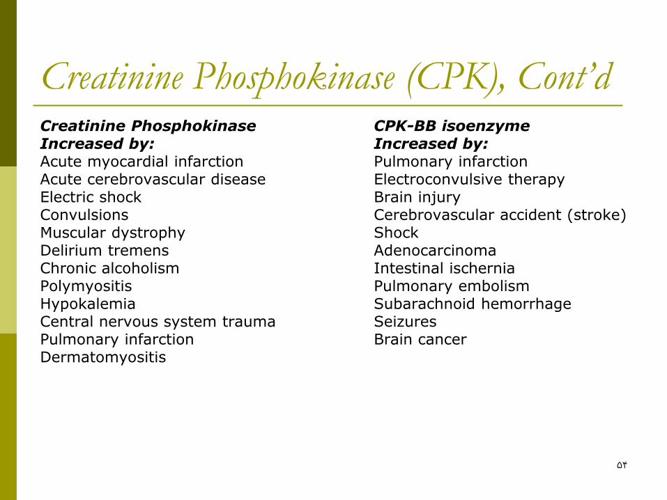
Creatinine Phosphokinase (CPK), Cont’d Creatinine Phosphokinase CPK-BB isoenzyme Increased by: Increased by: Acute myocardial infarction Pulmonary infarction Acute cerebrovascular disease Electroconvulsive therapy Electric shock Brain injury Convulsions Cerebrovascular accident (stroke) Muscular dystrophy Shock Delirium tremens Adenocarcinoma Chronic alcoholism Intestinal ischernia Polymyositis Pulmonary embolism Hypokalemia Subarachnoid hemorrhage Central nervous system trauma Seizures Pulmonary infarction Brain cancer Dermatomyositis
55
Troponins
Cardiac troponin T (cTnT) <0,2 ng/mL
Cardiac troponin I (ctnl) <0.03 ng/mL
New biochemical markers for heart disease
They become elevated quicker and stay elevated longer than CPK-MB
Increased levels = myocardial injury or myocardial infarction
Severe skeletal muscle may cause false elevation of cTnT.

56
Lymphocytes (20-40%)
Lymphocytes fight chronic bacterial infections and acute viral infections.
Lymphocytes are either T-cells or B-cells. T-cells are primarily involved with cellular type
immune reaction i.e. fungal and viral infections and transplant rejection The T-cell secretions stimulate the B-cell response.
B-cells participate in antibody production or humoral immunity. When the humoral immunity is activated the B-cells specific to the antigen differentiate into plasma or memory cells. These cells synthesize and release antibodies or immunoglobulins. The antibody binds to the antigen and inactivates it.
57
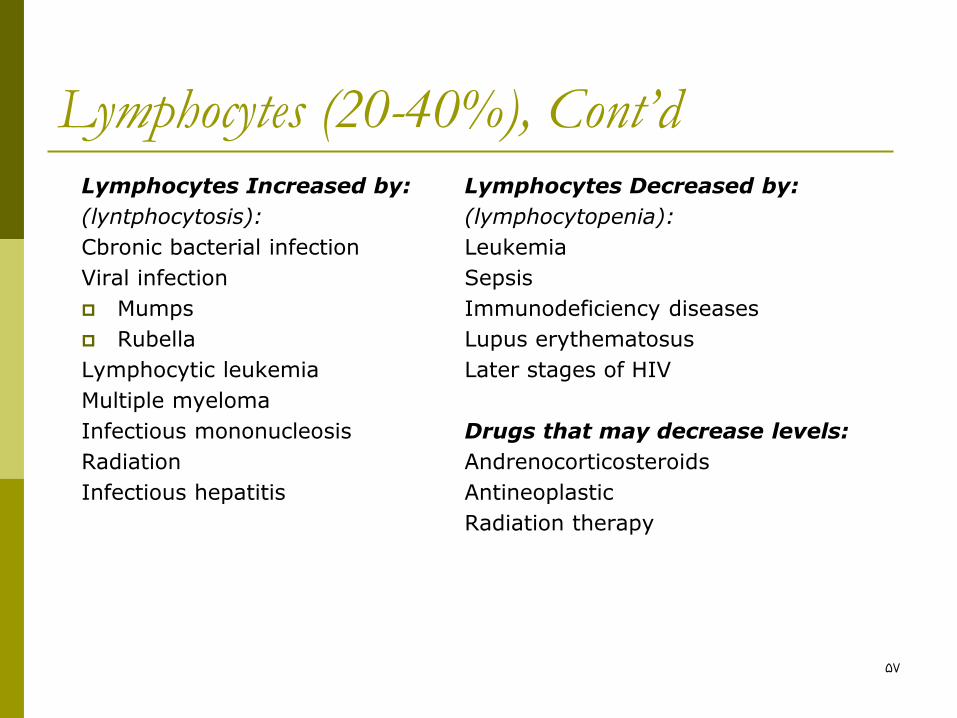
Lymphocytes (20-40%), Cont’d Lymphocytes Increased by: Lymphocytes Decreased by:
(lyntphocytosis): (lymphocytopenia):
Cbronic bacterial infection Leukemia
Viral infection Sepsis
Mumps Immunodeficiency diseases
Rubella Lupus erythematosus
Lymphocytic leukemia Later stages of HIV
Multiple myeloma
Infectious mononucleosis Drugs that may decrease levels:
Radiation Andrenocorticosteroids
Infectious hepatitis Antineoplastic
Radiation therapy
58
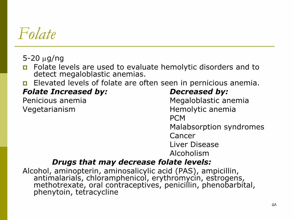
Folate
5-20 g/ng Folate levels are used to evaluate hemolytic disorders and to
detect megaloblastic anemias. Elevated levels of folate are often seen in pernicious anemia. Folate Increased by: Decreased by: Penicious anemia Megaloblastic anemia Vegetarianism Hemolytic anemia PCM Malabsorption syndromes Cancer Liver Disease Alcoholism Drugs that may decrease folate levels: Alcohol, aminopterin, aminosalicylic acid (PAS), ampicillin,
antimalarials, chloramphenicol, erythromycin, estrogens, methotrexate, oral contraceptives, penicillin, phenobarbital, phenytoin, tetracycline
59
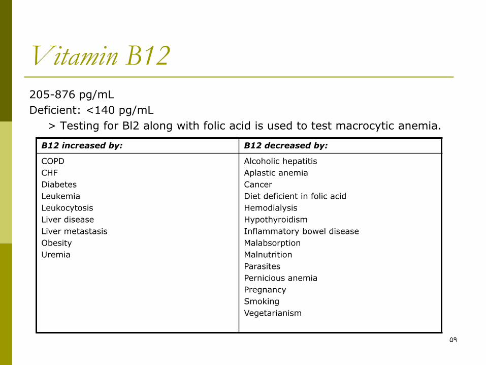
Vitamin B12 205-876 pg/mL
Deficient: <140 pg/mL
> Testing for Bl2 along with folic acid is used to test macrocytic anemia.
B12 increased by: B12 decreased by:
COPD
CHF
Diabetes
Leukemia
Leukocytosis
Liver disease
Liver metastasis
Obesity
Uremia
Alcoholic hepatitis
Aplastic anemia
Cancer
Diet deficient in folic acid
Hemodialysis
Hypothyroidism
Inflammatory bowel disease
Malabsorption
Malnutrition
Parasites
Pernicious anemia
Pregnancy
Smoking
Vegetarianism
60
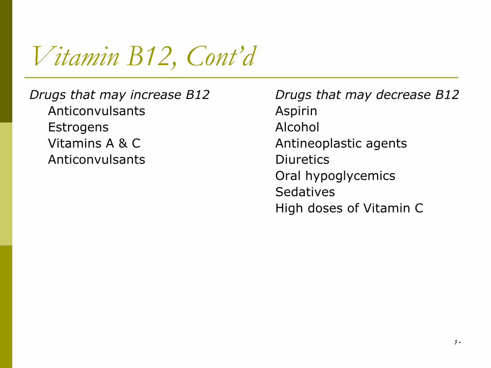
Vitamin B12, Cont’d Drugs that may increase B12 Drugs that may decrease B12
Anticonvulsants Aspirin
Estrogens Alcohol
Vitamins A & C Antineoplastic agents
Anticonvulsants Diuretics
Oral hypoglycemics
Sedatives
High doses of Vitamin C
61
C-Reactive Protein
Adults: <0.8 mg/dL
C-reactive protein (CRP) is an acute-phase protein which increases in infectious diseases and inflammatory disorders. It is an abnormal protein synthesized by the liver during an acute inflammatory process. An increase in CRP is associated with decreased levels of albumin and pre-albumin. A positive test indicates an acute inflammatory reaction however, it does not indicate the cause of the reaction. Antigens, immune complexes, bacteria, fungi and trauma can initiate the synthesis of CRP.
This test is also used to monitor patients with a myocardial infarction. The level of CRP follows a similar pattern to creatine kinase MB isoenzyme, however the peak occurs one to three days later. If CRP levels do not return to less than 0.8 mg/dL, this suggests damage to heart tissue. Levels are not elevated with angina pectoris.
62
C-Reactive Protein, Cont’d CRP can be used postoperatively to identify wound infections.
Generally, CRP increases four to six hours after surgery and will return to <0.8 mg/dL in three days. Elevated levels suggest complications.
CRP Increased by:
Acute rheumatic fever, Rheumatoid arthritis, Acute myocardial infarction, Postoperative wound infections, Pulmonary Actions Kidney or bone marrow transplant rejection, Malignant diseases, Bacterial infections (tuberculosis,), Viral infections, Crohn’s disease, Reiter's syndrome, Vasculitis syndromes, Lupus erythematosus, Tissue necrosis or trauma
Drugs that may increase CRP levels: Drugs that may decrease levels:
oral contraceptives nonsteroidal anti-inflammatories
salicylates
steroids
63
Pre-albumin Adults: 19-43 mg/dl
Deficiency: mild: 10-15 mg/dl
moderate: 5-10 mg/dl
severe: <5 mg/dl
Pre-albumin (PAB) is in a class of proteins having rapid turnover with short half-lives (2-3 days). Also called transthyretin and thyroxine-binding pre-albumin.
PAB is a transport protein synthesized by the liver and transported in serum as a complex of retinol-binding protein and vitamin A.
PAB is a sensitive indicator of change affecting protein synthesis and catabolism. Frequently ordered to monitor TPN.
Levels are reduced in hepatobiliary disease because of impaired synthesis.
Serum PAB increases when more than 60% of Basal Energy Expenditure (BEE) needs are met and decreases when less than 45% are met or when there is no oral intake.
A reasonable goal of a reseeding program is to increase serum PAB by at least 2.0-3.0 mg/dL in one week.
64
Pre-albumin, Cont’d
PAB Increased by: Decreased by
Dehydration Malnutrition
Hodgkin’s disease Liver damage
Chronic kidney disease Inflammation
Nephrotic syndrome Post-surgery
Bums
Overhydration
Zinc deficiency
65
Ferritin
Males: 12-300 ng/ml
Females: 10-150 ng/ml
Ferritin is one of the major iron storage proteins.
Levels of ferritin in the upper ranges are believed to correlate to increased risk for cardiovascular disease.
66
Ferritin, Cont’d
Ferritin Increased by: Decreased by:
Hemochromatosis Iron deficiency anemia
Hemosiderosis Severe protein deficiency
Megaloblastic anemia Hemodialysis
Hemolytic anemia
Alcoholic/inflammatory liver disease
Hodgkin’s disease
Breast cancer
67
Retinol-Binding Protein
2.6 – 7.6 mg/dl
A low molecular weight protein that responds to both protein and calorie restrictions.
More affected by energy restriction than protein deficiency.
Not affected by the acute inflammatory response like pre-albumin, albumin, or C-reactive protein.
RBP is a more reliable indicator of nutritional status following surgery or injury. RBP has a 10 hour half life and appears to be a better indicator of both short term and long term changes in protein status than albumin, pre-albumin or transferrin.
RBP is not a reliable indicator of protein status in advanced chronic renal disease or in advanced liver disorders.
68
Insulin-like Growth Factor (IGF-1) [Somatomedin C]
42-110 ng/ml
Levels are dependent on levels of GH.
Non-pituitary causes of reduced levels include protein-calorie malnutrition, severe chronic illness, liver disease, and hypothyroidism.
Restoration is more dependent on protein than on energy. (In contrast, Prealbumin is more dependent on energy than on protein.)
More sensitive and specific than albumin, pre-albumin, transfeff4 or retinol-binding protein.
69
Magnesium
1.3 – 2.1 mEq/L
Most magnesium exists within cells. Half is in bone
Most is bound to ATP, making this electrolyte important in all metabolic processes
Neuromuscular tissue depends on magnesium, and it is especially important to monitor magnesium levels in cardiac patients.
Increased by: Renal insufficiency, diabetes, Addison’s disease, Hypothyroidism, Magnesium containing antacids
Decreased by: Malnutrition, malabsorption, Hypoparathyroidism, Renal disease, Diabetic acidosis, stress, alcohol, diuretics, antibiotics
Symptoms of deficiency: apathy, forgetfulness, insomnia, depression, cardiac arrhythmia, muscle spasms, twitching, a tendency to startle easily
70
Magnesium, Cont’d
Symptoms of excess: lethargy, flushing, hypotension, respiratory depression, bradycardia
Most frequently observed abnormal analyte in pediatric patients
75% of diabetic patients are deficient
30% of patients in intensive care have low serum levels
Hypomagnesemia is a serious risk factor for hypertension, cardiac arrhythmia stroke & heart failure

71
Lipid Panel, Cont’d
HDL Increased by: HDL Decreased by:
Frequent exercise High alcohol intake
Liver disease Smoking
Moderate alcohol intake AIDS
LDL & VLDL Increased by: LDL & VLDL Decreased by:
Excessive calories Malnutrition
High saturated fat diet Malabsorption
Drugs that may increase both IML & LDL:
Aspirin, oral contraceptive agents, steroids, sulfonamides
72
Homocysteine
7-22 mol/L
Elevated levels are associated with an increased risk for heart disease that is independent of lipid levels.
Homocysteine is an intermediate metabolite of the methionine pathway. When methionine is released from dietary protein it condenses with adenosine triphosphate (ATP) to form S-adenosylmethionine (SAM). SAM acts as a methyl donor and homocysteine is formed. Homocysteine can either be remethylated to form methionine or catabolized in the transsulfuration pathway. The remethylation of homocysteine to methionine requires both vitamin B12 and folic acid. When B12 is deficient in the diet, the folic acid becomes trapped in an unusable form as methyltetrahydrofolate. Megaloblastic anemia results. Large doses of folic acid can override the pathway and mask a B12 deficiency.

73
Homocysteine, Cont’d
Serum homocysteine will rise when there is a B12 and or a folic acid deficiency.
The threshold for rising levels of homocysteine appears to be serum folate <12.5 nmol/L and/or serum B12 <225 pmol/L. The etiology of the disease process is unclear. Also, elevated levels of homocysteine are associated with risk for end stage renal failure. Normal levels and "at risk" levels are not consistent in the research literature and remain under research investigation.
74
Homocysteine, Cont’d
Increased levels of Homocysteine
Associated with:
Renal failure Drugs that may increase levels:
Folate deficiency Methotrexate
B12 deficiency Dilantin
CHD Nitrous oxide
Smoking
75
Vitamin K
Deficiency:
Prolonged prothrombin time
Small hemorrhagic spots
Impaired bone formation
76
Protein C
70% - 140%
A fibrin inhibitor that prevents clotting from occurring indiscriminately. It is produced by the liver, vitamin K is required for its production. Test is done to evaluate patients with severe thrombosis.
Values decreased: cirrhosis, vitamin K deficiency, anticoagulant drugs.
77
Protein S
70%-140%
A cofactor which enhances the anticoagulant effect of Protein C
Produced in the liver.
Vitamin K required for production.
78
Zinc
Deficiency is widespread.
Diuretics can lead to deficiency.
Elderly subjects have high incidence of deficiency.