Embed Size (px)
Citation preview

[Frontiers in Bioscience 10, 1440-1461 May 1, 2005]
1440
ESTROGEN RECEPTOR REGULATION OF QUINONE REDUCTASE IN BREAST CANCER: IMPLICATIONSFOR ESTROGEN-INDUCED BREAST TUMOR GROWTH AND THE THERAPEUTIC USES OF TAMOXIFEN
Monica M. Montano 1, Nicole R. Bianco 1, Huayun Deng 1, Bryan M. Wittmann 1, Laura C. Chaplin 1 and Benita S.Katzenellenbogen 2
1 Department of Pharmacology, Case Western Reserve University, Cleveland, OH 44122, 2 Department of Molecular andIntegrative Physiology, University of Illinois and College of Medicine, Urbana, IL 61801
TABLE OF CONTENTS
1. Abstract2. Introduction
2.1. Estrogen receptor (ER) bioactivity in breast cancer cells2.2. ER alpha and beta2.3. Antiestrogens and the regulation of ER bioactivity in breast cancer cells2.4. Detoxifying enzymes and chemoprotection against cancer2.5. Regulation of the expression of detoxifying enzymes2.6. Metabolism of estrogens2.7. Role of estrogen metabolites in carcinogenesis
3. Regulation of NQO1 by the ER3.1. Induction of NQO1 activity in breast cancer cells by antiestrogens3.2. Induction of EpRE enhancer activity by antiestrogens in breast cancer cells3.3. Regulation of NQO1 gene transcriptional activity by ER-alpha and ER-beta3.4. Analysis of the interaction of ER-alpha and ER-beta with the EpRE3.5. Identification of a novel protein factor, hPMC2, involved in the regulation of NQO1 activity by the ER
4. Functional implications of antiestrogen regulation of NQO1: protection against estrogen- induced DNA damage4.1. Physiological levels of estrogens induce DNA damage independent of E and dependent on estrogen metabolism4.2. Role of NQO1 and ER-beta in antiestrogen inhibition of estrogen-induced DNA damage
5. Perspective6. Acknowledgements7. References
1. ABSTRACT
Antiestrogens have found widespread use in thetreatment of breast cancer (reviewed in ref. 1). There is alsointerest in the use of tamoxifen as a preventive agent forbreast cancer. However, the mechanism for the antitumoreffects of antiestrogens is relatively unknown. For the mostpart it is thought that the basis for the anticancer action ofantiestrogens is the inhibition of estradiol (E2)-stimulatedtumor growth. We have observed however that antiestrogenscan activate detoxifying enzymes, like quinone reductase(NQO1), which protect cells against the toxic and tumor-promoting effects of carcinogens (2). Studies characterizingthe molecular mechanisms behind the regulation of NQO1by the Estrogen Receptor (ER) support an important role ofthe ER in pathways regulating antioxidant defenses.Moreover these findings represent a novel mechanismthrough which antiestrogens function. The activation ofNQO1 may contribute to the beneficial anticancer andantioxidant activity of antiestrogens in breast cancer andpossibly other estrogen target tissues. It is possible that thebasis for some of the anticancer action of antiestrogens isthat the induction of NQO1 inhibits the genotoxic effectsinduced by the oxidative metabolism of estrogens.
2. INTRODUCTION
2.1. Estrogen receptor bioactivity in breast cancer cellsThe estrogen receptor (ER) protein is essential
for mediating the actions of estrogen in target tissues. Thebinding of estrogen initiates a process of receptor activationthat includes the high affinity binding of ER to specificDNA sequences, termed estrogen response elements(EREs). The interaction of ER with EREs results in themodulation of specific gene expression, through which thephysiological actions of estrogens are manifested (figure1A; reviewed in ref. 3). Estrogens acting via the ERdramatically escalate proliferative and metastatic activity inthese tumor cells, in part via the induction of growthfactors, proteases, and basement membrane receptors(reviewed in ref. 4, 5). However the relative role of theinduction of these genes on the proliferative effects ofestrogens remains unknown.
The regulatory actions of estrogens on geneexpression, which are generally stimulatory, can beinhibited by potent synthetic ER antagonists (1) termedantiestrogens (AEs). Tamoxifen is an antiestrogen that iswidely used in the treatment of breast cancer (1). Theantiproliferative actions of tamoxifen and othertriphenylethylene derivatives at submicromolar concentrationsin estrogen-dependent breast carcinoma cells are believedto be mediated by high affinity binding to the ER.
2.2. Estrogen receptor alpha and betaThe human ER-alpha is a 67 kDa protein comprising

Estrogen Receptor Regulation of Quinone Reductase
1441
Figure 1. (A) Mechanism of Estrogen Receptor (ER)action, (B) Functional domains of the ER, (C) EstrogenReceptor ER-alpha and ER-beta.
of 595 amino acids. Deletional and mutational analyseshave led to the identification of conserved domainsinvolved in DNA binding, ligand binding, dimerization,protein binding, and transcriptional activation (reviewed inref. 3). The A/B domain contains transactivation function 1(figure 1B). A highly conserved DNA binding domaincomprises domain C. The hinge domain appears to beimportant in receptor dimerization, and along with carboxyterminal region of domain C is thought to contain thenuclear localization signal. The E domain is functionallycomplex and is involved in ligand binding, dimerization,ligand-dependent transactivation, and interaction withcoactivators and corepressors. A role in estrogen andantiestrogen discrimination has been proposed for the Fdomain (6).
Another subtype of the ER, ER-beta has beencloned (figure 1C, ref. 7-9). Human ER-beta sharesregions of homology such as the DNA binding domain,while the N-terminal A/B domain, hinge region, theligand binding domain, and the F domain are distinctbetween ER-alpha and ER-beta. ER-beta exhibits lower
transcriptional response to E2 and TOT when compared toER-alpha in the context of several estrogen responseelement (ERE)-containing gene reporter constructs (7-9).However, ER-beta has been reported to show increasedTOT agonism from reporter constructs containing an AP1site (9).
ER-alpha is predominantly expressed in thebreast, uterus, and vagina (reviewed in ref.10). Reportsindicate a positive correlation between ER-alpha and ER-beta in human breast cancers (reviewed in ref. 11 and 12).Studies correlating ER-beta protein expression with breasttumor grade predict good prognosis for ER-beta positivetumors. ER-beta status also appears to be a good predictorof response to tamoxifen (13-16). However, thesignificance of ER-beta in breast cancer development andtreatment of breast cancer is not clear.
2.3. Antiestrogens and the regulation of estrogenreceptor bioactivity in breast cancer cells
Tamoxifen is presumed to exert its antitumor andchemoprotective effects by competitively antagonizing thebinding of estradiol (E2) to ER, which ultimately leads tothe inhibition of gene transcription and protein synthesis.There is also interest in antiestrogen effects exerted throughmechanisms that may not involve inhibition of E2-stimulated activities. For example, tamoxifen is also beingassessed as a preventive agent for breast cancer and forother potential benefits, such as protection againstcardiovascular disease and osteoporosis (17, 18). It hasbeen proposed that tamoxifen may function as ananticancer drug by acting as an antioxidant (19-21).However, the basis for the antioxidant capabilities oftamoxifen has not been well characterized.
Despite its clear efficacy in the short-termtreatment of breast cancer, clinically tamoxifen resistanceremains a problem (1). Although ca. 75% of ER-positivetumors respond well to tamoxifen therapy, almost all breasttumors eventually become refractory to the beneficialgrowth suppressive effects of tamoxifen. Another cause forconcern is that although tamoxifen acts primarily as achemopreventive agent in mammary carcinogenesis, it caninduce uterine and liver cell proliferation (22). Thus thedevelopment of adjuvant therapy for women undergoingtamoxifen therapy is of clinical importance.
2.4. Detoxifying enzymes and chemoprotection againstcancer
Detoxifying enzymes such asNAD(P)H:(quinone-acceptor) oxidoreductase [quinonereductase (NQO1)], glutathione S-transferases (GSTs),epoxide hydrolase, and UDP-glucuronosyltransferases areinduced in cells by electrophilic compounds and phenolicantioxidants (reviewed in ref. 23 and 24). These widelydistributed enzymes detoxify electrophilic quinones,thereby protecting cells against the toxic and neoplasticeffects of carcinogens (figure 2). The detoxificationmechanism for quinones involves two electron reduction,which bypasses semiquinone generation and therebydiminishes the subsequent production of reactive oxygenspecies.

Estrogen Receptor Regulation of Quinone Reductase
1442
Figure 2. Major reactive oxygen species pathways and antioxidant defenses.
In addition to detoxification of exogenousquinones NQO1 may also play a role as an antioxidant byreducing endogenous quinones and protect cellularmembranes against oxidative DNA damage (reviewed inref. 25). NQO1 can catalyze the reduction of ubiquinoneanalogs (coenzyme Q) to their ubiquinol forms. Theproduct of Vitamin E (a-tocopherol ) oxidation, a-tocopherolquinone, is reduced to a-tocopherolhydroquinone, in a reaction catalyzed by NQO1.This process is able to protect cells against lipidperodixation.
Depending on the properties of the hydroquinonegenerated after reduction by NQO1, two electron reductionof a quinone may also represent a bioactivation step.Hydroquinones may autooxidize, leading to the generationof reactive oxygen species and toxicity. Alternativelyhydroquinones may undergo rearrangement to producereactive alkylating agents. This process is termedbioreductive alkylation (26, 27) and bioreductive alkylatingagents are of clinical interest for their anticancer effects ortheir ability to induce DNA damage and toxicity in tumorcells. Thus, NQO1 has attracted a lot of attention due to itsability to metabolize chemotherapeutic quinones and otherbioreductive antitumor compounds in many cells includingmammary tumor cells (28-30). Moreover, the levels ofdetoxifying enzymes like NQO1 and GST have been shownto be elevated in a number of tumor types including breast,
liver, and prostate when compared to normal cells of thesame origin (31-33). These observations may provide abasis for targeting these tumor types for treatment withchemotherapeutic quinones.
The non-protein thiol, glutathione (gamma-glutamyl-cysteinyl-glycine, GSH) is a predominant cellularantioxidant. As such GSH serves critical functions in themaintenance of cellular redox balance, provides protectionagainst reactive oxygen species (ROS), and is involved inthe detoxification of xenobiotics. GSH detoxifies eitherthrough direct reactions with reactive intermediates or viaenzymatic conjugation reactions catalyzed by GSTs (34)Exposure of cells to a number of xenobiotic agents resultsin a significant increase in the total intracellular GSHcontent, secondary to transcriptional up-regulation of thegenes encoding the two protein subunits (catalytic andregulatory) of gamma-glutamylcysteine synthetase (GCS),the rate-limiting enzyme in its de novo synthesis (34).
GSTs are important in the detoxification ofxenobiotics, catalyzing the nucleophilic attack by the thiolgroup of GSH on the xenobiotic (35). Since they catalyzethe inactivation of several known carcinogens, theseenzymes can provide a defense against carcinogenesis. Onthe other hand, elevation of GST levels in solid tumorsappears to be a major factor in the development ofresistance to treatment with cytotoxic agents (36). GSTs are

Estrogen Receptor Regulation of Quinone Reductase
1443
Figure 3. Metabolism of Estrogens.
grouped into at least six different gene families based onsequence similarity and substrate specificity; and theseisozymes differ in their ability to confer resistance toparticular anti-cancer drugs (35). This property may explainthe high levels of GST- Pi in many solid tumors and drugresistant cell lines, where no obvious catalytic role forGST-Pi (35, 36).
2.5. Regulation of the expression of detoxifying enzymesDetermination of the molecular mechanisms
involved in the induction of detoxifying enzymes isimportant in devising strategies for chemoprotectionagainst cancer. It has been reported that the induction ofgenes NQO1, GCSH, and GST is mediated through anelectrophile (or antioxidant) response element (EpRE orARE) (37-40). An EpRE/ARE has been identified in the 5'-flanking region of the human and rat NQO1 genes and therat GST gene (41, 42). A short core sequence, 5'TGACnnnGC-3', which is present in the EpRE of bothNQO1 and GST genes, is required to control geneexpression in response to phenolic antioxidants (tert-butylhydroquinone and hydroquinone), and metabolizableplanar aromatic compounds leading to enzyme induction bythese compounds (38, 42).
The human NQO1 gene EpRE core sequence(TGACTCAGC) contains a TPA response element or TRE(TGACTCA, ref. 43). The TRE is the binding site for theAP1 family of transcription factors, which includes c-junand c-fos. However the EpRE sequence of the rat NQO1gene does not contain a high affinity recognition site for in
vitro translated c-jun and c-fos (44). Furthermore, theEpRE of the rat GST Ya gene forms a complex withprotein factors in HepG2 cell nuclear extracts that is notcompeted with the TRE (45). This finding suggests thatthere are factors in addition to the AP-1 proteins thatregulate expression via EpRE sequences. It has beenreported that the transcription factors, Nrf1 and Nrf2, arepart of the complex that binds to the EpRE andoverexpression of these factors in certain cell linesincreased EpRE-mediated expression of the NQO1 gene(46).
The transcriptional up-regulation of the two GCSsubunit genes involves the EpRE but distinct combinationsof trans-acting factors, contributing to differentialregulation in response to specific inducing agents (47, 48).Transcription is hypothesized to involve dimerictranscription factors composed of small Maf proteins andvarious other bZIP family members, including Nrf1 andNrf2 (47, 48). The AP1 transcription factors, Fos and Jun,have been implicated in the transcriptional regulation of theGST-Pi (49).
2.6. Metabolism of estrogensIn both normal and breast cancer cells estrogens
may be oxidized by extrahepatic cytochrome P450enzymes (mainly CYP1A1 and CYP1B1) to hydroxy-catecholestrogens and further oxidized to the semiquinoneand quinone form (figure 3; ref. 50-52). This is potentiallyharmful given that the quinone can bind to DNA formingDNA adducts; and redox cycling, involving quinone and

Estrogen Receptor Regulation of Quinone Reductase
1444
Figure 4. Antiestrogens, but not estrogen, induceNAD(P)H:Quinone Oxidoreductase enzyme activity inMCF-7 breast cancer cells. Effect of different compoundson NQO1 activity in MCF-7 cells. Cells were treated withdifferent concentrations of estradiol (E2), trans-hydroxytamoxifen (TOT), tamoxifen (TAM), ICI182,780(ICI) or tert-butylhydroquinone (TBHQ), or 12-O-tetradecanoylphorbol-13-acetate (TPA). Cells wereharvested after 4 days of treatment. Cytosolic extracts wereassayed for NQO1 activity as described in "Materials andMethods." Values are the means ± S.E. from three separateexperiments. Reprinted with permission from the NationalAcademy of Sciences, U.S.A. (2).
unstable semiquinone interconversions form causeshydroxyl radical formation which can lead to hydroxylatedbases (eg. 8-hydroxydeoxyguanine, 8-OHdG) andpermanent mutation if not repaired (53-55). Glutathione-S-transferase (GST) detoxifies these quinones by conjugationwith GSH, and catechol O-methyltransferase (COMT)detoxifies the hydroxy-catecholestrogens by methylation(53). The estrogens and catecholestrogens can also bedetoxified by conjugation to glucuronides and sulfates incertain tissues (50). The ability of NQO1 to detoxifyquinone-catecholestrogens by reduction of the reactivequinone-catecholestrogen back to the hydroxy-catecholestrogen has been shown for two syntheticestrogens, diethylstilbestrol (DES) (56) and 4-hydroxyequilenin-o-quinone (57). In human breastepithelial cells E2 is primarily converted to 4-hydroxyestradiol (4-OHE2), (53). This is important becauseCOMT is less effective at inactivating 4-OHE2 than 2-hydroxyestradiol (2-OHE2) (58), and 4-OHE2 are oxidizedto form predominantly depurinating adducts and consideredto be more mutagenic than 2-OH-E2 (53).
2.7. Role of estrogen metabolites in carcinogenesisRecent studies indicate that estrogen itself may
be a genotoxic mutagen capable of causing chromosomalmutations in animals and cell culture (53, 59, 60], leadingto tumorigenesis in various animal models (53, 61).Estrogens also cause MCF10 breast epithelial cells, thatlack both ER-alpha and ER-beta, to take on a transformedphenotype (59). This neoplastic transformation isaccompanied by genomic alterations, as exhibited by Lossof Heterozygosity and Microsatellite Instability at specificloci in chromosomes 11, 13 and 17. These loci have beenreported to be affected in several types of breast lesions.
Further support for a role of estrogen metabolites incarcinogenesis is the observation that ERs in humanmammary epithelium are localized in cells distinct fromthose expressing proliferation markers (62). There aresynthetic estrogens that strongly activate ER but havedecreased catechol estrogen formation and are poorlycarcinogenic (53). Thus, initiation may be due not only toER-mediated proliferation, but also DNA damage due to acombination of estrogen metabolism and preexistinglesions. Once initiated, the ER may then confer a selectiveadvantage to these premalignant cells.
Increases in estrogen metabolism relative toconjugation were observed in breast carcinomas (63). Inline with the protective role of antioxidative stressenzymes, polymorphisms in the NQO1 and GST-Pi locihave been associated with increased risk for breast cancerand other types of cancer (64-71). Epigenetic silencingthrough promoter hypermethylation of GST-pi gene isassociated with breast, prostate and renal cancer (72-73).
3. REGULATION OF NQO1 BY THE ESTROGENRECEPTOR
3.1. Induction of NQO1 activity in breast cancer cells byantiestrogens
Through the technique of differential RNAdisplay, we observed NQO1 to be a species that waspresent at a much higher level in an MCF7 breast cancercell subline that had been grown long-term (over 6 months)in the presence of high (10-6 M) levels of the antiestrogentrans-hydroxytamoxifen (TOT), as compared to theparental MCF7 cells grown in the absence of TOT (2).Differential expression was verified using Northern blotanalyses which indicate that NQO1 RNA was present at 8-times greater abundance in the long-term TOT-maintainedcells than in the parental MCF7 cells. A similar increase inNQO1 mRNA expression can be induced in parental MCF7cells transiently exposed to the antiestrogens TOT and ICI182,780 (74). No increase in NQO1 mRNA levels wasevident in cells treated with E2; E2 even appeared to slightlyreduce the control level of NQO1 mRNA in the cells. Wetherefore examined the ability of antiestrogen to evokeincreases in NQO1 enzymatic activity when parental MCF7cells were treated with antiestrogens.
As shown in figure 4 a 3- to 4-fold increase inNQO1 activity was observed upon treatment with theantiestrogens TOT, tamoxifen, and ICI182,780. Theinduction of NQO1 activity by antiestrogens occurred in adose-dependent manner, and maximal stimulation wasobtained with a relatively low concentration of theantiestrogens (10-8 M for TOT and ICI, ca. 10-7 M forTAM). The concentrations of antiestrogen required forstimulation of NQO1 activity were substantially lower thanthose needed for stimulation by a previously identifiedpotent inducer of NQO1 activity in other systems, tert-butylhydroquinone (TBHQ). Of note, no increase in NQO1activity was observed in the presence of E2 or the tumorpromoter phorbol ester TPA. We have also observed thatthe antiestrogen-induced increase in NQO1 activity wasfully inhibited by E2 (2). These observations suggest that

Estrogen Receptor Regulation of Quinone Reductase
1445
the modulation of NQO1 activity by antiestrogens ismediated by the ER. In contrast, E2 did not affect theinduction of NQO1 by TBHQ.
3.2. Induction of EpRE enhancer activity byantiestrogens in breast cancer cells
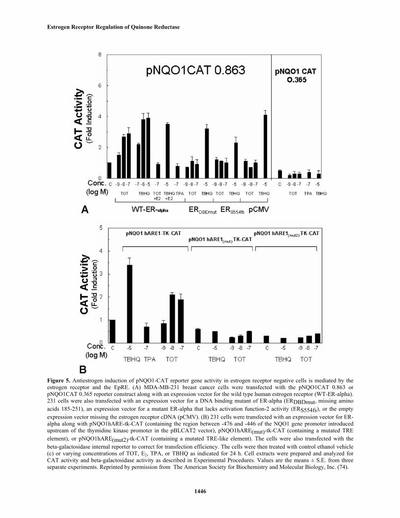
Studies toward understanding the molecularmechanisms for the induction of detoxifying enzymes byantiestrogens were also conducted in view of theirimportance in devising strategies for chemoprotectionagainst cancer. To examine the possible transcriptionalregulation of the NQO1 gene by antiestrogen, a reporterconstruct which contains the 863 bp 5'-flanking andpromoter regions of the human NQO1 gene upstream of thechloramphenicol acetyltransferase (CAT) gene (figure 5A)was introduced into an ER-negative breast cancer cell line,MDA-MB-231 cells (74). In cells cotransfected with anexpression vector for the wild type ER-alpha TOT inducedan increase in pNQO1-CAT 0.863 reporter activity. E2inhibited the TOT-mediated, not TBHQ-mediated, increasein NQO1 transcriptional activity. No increase in CATactivity was evident in cells after treatment with TPA, aknown inducer of AP1 activity (75). Cells that were co-transfected with the control expression vector lacking theER cDNA (pCMV5), with an expression vector for a mutantER-alpha that has impaired activation function-2 (AF-2)activity (S554fs), or with an expression vector for a mutantER-alpha that has impaired DNA-binding ability (DBDmut)did not show activation from pNQO1-CAT 0.863 in responseto antiestrogens, although the effect of TBHQ was stillobserved. Thus antiestrogen-mediated activation of NQO1gene promoter activity requires a transcriptionally active ER-alpha. Treatment with either TOT or TBHQ did not inducetranscription from a deletion mutant of the pNQO1-CAT0.863 reporter construct, denoted pNQO1-CAT-0.365, thatlacks the -366 to -863 bp portion of the 5' flanking region.As expected (44), this truncated construct has reduced basalpromoter activity. Our results indicate that the regionbetween -0.863 and -0.365 kb of the NQO1 gene isessential for induction by both TOT and TBHQ.
Sequence analysis of the 5'-flanking sequence ofthe human NQO1 gene indicates the presence of a singlecopy of the EpRE motif at position -467 to -437 (40, 43).The EpRE motif contains a TRE and a TRE-like element.The EpRE-containing region was introduced upstream of aheterologous promoter, thymidine kinase, and the CATgene (40). Although this EpRE-containing constructshowed significant basal CAT activity in 231 cells, TOTand TBHQ were able to induce transcription 2.2-fold and3.4-fold, respectively, over basal levels (left portion offigure 5B). Mutation of the perfect TRE element (middleportion of figure 5B) reduced basal CAT activity andeliminated induction by TBHQ and TOT. Mutation of theTRE-like element (right portion of figure 5B) also reducedbasal activity and eliminated the TBHQ- and TOT-inducedincrease in NQO1 gene transcriptional activity. TPA, whichincreases AP1 activity, did not induce an increase in EpRE-mediated CAT gene expression of the wild type(unmutated) gene construct in these cells. This observationdoes not appear to be due to the absence of AP1 activitybecause we have previously shown that TPA induced TRE-
mediated CAT reporter gene activity in 231 cells (2).
In contrast to the observations made withantiestrogens, TBHQ-mediated induction of NQO1 genetranscriptional activity did not require the ER and occurredequally well in the presence of functionally inactive ER orin the absence of ER altogether. However the DNAelements required for antiestrogen-mediated induction ofNQO1 gene transcriptional activity that we identifiedthrough deletion and mutational studies mapped identicallywith the elements required for TBHQ-mediated induction.Our experiments indicate that NQO1 can be activated by ER-independent and ER-dependent pathways. Activation byantiestrogens depends on the presence of a functional ER andrequires much lower doses of compound (10-10 – 10-8 M) thanstimulation by known activators of NQO1 such as TBHQ (10-
6 – 10-5 M). While the signals between these two pathwaysmay be integrated at some level, the ER-dependent pathway –which may involve induction or regulation of some proteinthat regulates the EpRE – must be different in part from theelectrophile-dependent pathway utilized by TBHQ, where theER is not at all required for NQO1 stimulation.
3.3. Regulation of NQO1 gene transcriptional activityby ER-alpha and ER-beta
Although ER-beta exhibits lower transcriptionalresponse to E2 and TOT when compared to ER-alpha in thecontext of several estrogen response element (ERE)-containing gene reporter constructs (7-9), ER-beta has beenreported to show increased TOT agonism from reporterconstructs containing an AP1 site (9). We thus examinedligand activation of ER-alpha and ER-beta from an EpRE,as compared to activation from an ERE (74).
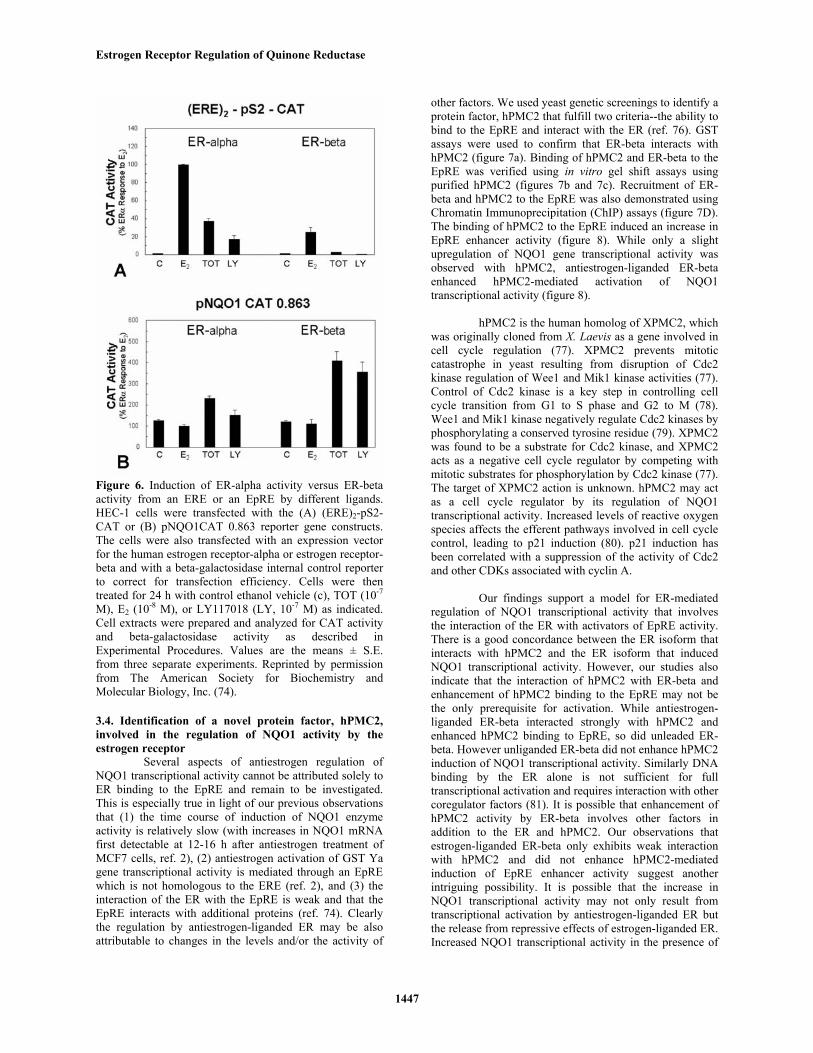
With a reporter construct containing EREsupstream of the promoter for the estrogen-regulated pS2gene (figure 6), ER-alpha and ER-beta both showedstimulation by E2 and as expected, ER-beta showed a lowertranscriptional response. Interestingly, ER-alpha wasweakly stimulated by the antiestrogen TOT and by anotherantiestrogen, LY 117018 (LY) at this ERE-containing geneconstruct, whereas the antiestrogens failed to elicit ER-betatranscriptional activity at this ERE-containing gene. Thetranscriptional response of ER-alpha and ER-beta to theseligands was very different in the context of the NQO1 genepromoter construct pNQO1-CAT 0.863. Of note, althoughantiestrogens stimulated NQO1 activity via both ER-alphaand ER-beta, the magnitude of increase in NQO1 genetranscriptional activity in response to TOT or LY washigher with ER-beta than with ER-alpha and no stimulationby E2 was observed via ER-alpha or ER-beta at an EpRE.
It is clear that ligand activation of ERtranscriptional activity is highly dependent on the nature ofthe response element. Antiestrogen ligands which inducevery little activity from ER-beta at an ERE, can inducesignificant activity from ER-beta at an EpRE and, aspreviously reported, from a TRE (9). It has been proposedthat ER-beta regulates transcription from an AP1 site by adirect interaction with Fos and Jun. However our studiessuggest that transcriptional activation of the NQO1 gene byTOT is not mediated through Fos and Jun.
Estrogen Receptor Regulation of Quinone Reductase
1446
Figure 5. Antiestrogen induction of pNQO1-CAT reporter gene activity in estrogen receptor negative cells is mediated by theestrogen receptor and the EpRE. (A) MDA-MB-231 breast cancer cells were transfected with the pNQO1CAT 0.863 orpNQO1CAT 0.365 reporter construct along with an expression vector for the wild type human estrogen receptor (WT-ER-alpha).231 cells were also transfected with an expression vector for a DNA binding mutant of ER-alpha (ERDBDmut, missing aminoacids 185-251), an expression vector for a mutant ER-alpha that lacks activation function-2 activity (ERS554fs), or the emptyexpression vector missing the estrogen receptor cDNA (pCMV). (B) 231 cells were transfected with an expression vector for ER-alpha along with pNQO1hARE-tk-CAT (containing the region between -476 and -446 of the NQO1 gene promoter introducedupstream of the thymidine kinase promoter in the pBLCAT2 vector), pNQO1hARE(mut)-tk-CAT (containing a mutated TREelement), or pNQO1hARE(mut2)-tk-CAT (containing a mutated TRE-like element). The cells were also transfected with thebeta-galactosidase internal reporter to correct for transfection efficiency. The cells were then treated with control ethanol vehicle(c) or varying concentrations of TOT, E2, TPA, or TBHQ as indicated for 24 h. Cell extracts were prepared and analyzed forCAT activity and beta-galactosidase activity as described in Experimental Procedures. Values are the means ± S.E. from threeseparate experiments. Reprinted by permission from The American Society for Biochemistry and Molecular Biology, Inc. (74).
Estrogen Receptor Regulation of Quinone Reductase
1447
Figure 6. Induction of ER-alpha activity versus ER-betaactivity from an ERE or an EpRE by different ligands.HEC-1 cells were transfected with the (A) (ERE)2-pS2-CAT or (B) pNQO1CAT 0.863 reporter gene constructs.The cells were also transfected with an expression vectorfor the human estrogen receptor-alpha or estrogen receptor-beta and with a beta-galactosidase internal control reporterto correct for transfection efficiency. Cells were thentreated for 24 h with control ethanol vehicle (c), TOT (10-7
M), E2 (10-8 M), or LY117018 (LY, 10-7 M) as indicated.Cell extracts were prepared and analyzed for CAT activityand beta-galactosidase activity as described inExperimental Procedures. Values are the means ± S.E.from three separate experiments. Reprinted by permissionfrom The American Society for Biochemistry andMolecular Biology, Inc. (74).
3.4. Identification of a novel protein factor, hPMC2,involved in the regulation of NQO1 activity by theestrogen receptor
Several aspects of antiestrogen regulation ofNQO1 transcriptional activity cannot be attributed solely toER binding to the EpRE and remain to be investigated.This is especially true in light of our previous observationsthat (1) the time course of induction of NQO1 enzymeactivity is relatively slow (with increases in NQO1 mRNAfirst detectable at 12-16 h after antiestrogen treatment ofMCF7 cells, ref. 2), (2) antiestrogen activation of GST Yagene transcriptional activity is mediated through an EpREwhich is not homologous to the ERE (ref. 2), and (3) theinteraction of the ER with the EpRE is weak and that theEpRE interacts with additional proteins (ref. 74). Clearlythe regulation by antiestrogen-liganded ER may be alsoattributable to changes in the levels and/or the activity of
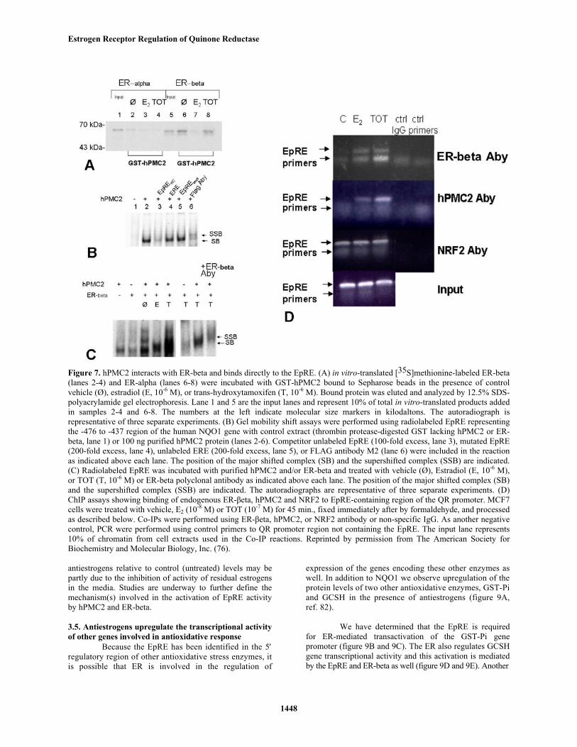
other factors. We used yeast genetic screenings to identify aprotein factor, hPMC2 that fulfill two criteria--the ability tobind to the EpRE and interact with the ER (ref. 76). GSTassays were used to confirm that ER-beta interacts withhPMC2 (figure 7a). Binding of hPMC2 and ER-beta to theEpRE was verified using in vitro gel shift assays usingpurified hPMC2 (figures 7b and 7c). Recruitment of ER-beta and hPMC2 to the EpRE was also demonstrated usingChromatin Immunoprecipitation (ChIP) assays (figure 7D).The binding of hPMC2 to the EpRE induced an increase inEpRE enhancer activity (figure 8). While only a slightupregulation of NQO1 gene transcriptional activity wasobserved with hPMC2, antiestrogen-liganded ER-betaenhanced hPMC2-mediated activation of NQO1transcriptional activity (figure 8).
hPMC2 is the human homolog of XPMC2, whichwas originally cloned from X. Laevis as a gene involved incell cycle regulation (77). XPMC2 prevents mitoticcatastrophe in yeast resulting from disruption of Cdc2kinase regulation of Wee1 and Mik1 kinase activities (77).Control of Cdc2 kinase is a key step in controlling cellcycle transition from G1 to S phase and G2 to M (78).Wee1 and Mik1 kinase negatively regulate Cdc2 kinases byphosphorylating a conserved tyrosine residue (79). XPMC2was found to be a substrate for Cdc2 kinase, and XPMC2acts as a negative cell cycle regulator by competing withmitotic substrates for phosphorylation by Cdc2 kinase (77).The target of XPMC2 action is unknown. hPMC2 may actas a cell cycle regulator by its regulation of NQO1transcriptional activity. Increased levels of reactive oxygenspecies affects the efferent pathways involved in cell cyclecontrol, leading to p21 induction (80). p21 induction hasbeen correlated with a suppression of the activity of Cdc2and other CDKs associated with cyclin A.
Our findings support a model for ER-mediatedregulation of NQO1 transcriptional activity that involvesthe interaction of the ER with activators of EpRE activity.There is a good concordance between the ER isoform thatinteracts with hPMC2 and the ER isoform that inducedNQO1 transcriptional activity. However, our studies alsoindicate that the interaction of hPMC2 with ER-beta andenhancement of hPMC2 binding to the EpRE may not bethe only prerequisite for activation. While antiestrogen-liganded ER-beta interacted strongly with hPMC2 andenhanced hPMC2 binding to EpRE, so did unleaded ER-beta. However unliganded ER-beta did not enhance hPMC2induction of NQO1 transcriptional activity. Similarly DNAbinding by the ER alone is not sufficient for fulltranscriptional activation and requires interaction with othercoregulator factors (81). It is possible that enhancement ofhPMC2 activity by ER-beta involves other factors inaddition to the ER and hPMC2. Our observations thatestrogen-liganded ER-beta only exhibits weak interactionwith hPMC2 and did not enhance hPMC2-mediatedinduction of EpRE enhancer activity suggest anotherintriguing possibility. It is possible that the increase inNQO1 transcriptional activity may not only result fromtranscriptional activation by antiestrogen-liganded ER butthe release from repressive effects of estrogen-liganded ER.Increased NQO1 transcriptional activity in the presence of
Estrogen Receptor Regulation of Quinone Reductase
1448
Figure 7. hPMC2 interacts with ER-beta and binds directly to the EpRE. (A) in vitro-translated [35S]methionine-labeled ER-beta(lanes 2-4) and ER-alpha (lanes 6-8) were incubated with GST-hPMC2 bound to Sepharose beads in the presence of controlvehicle (Ø), estradiol (E, 10-6 M), or trans-hydroxytamoxifen (T, 10-6 M). Bound protein was eluted and analyzed by 12.5% SDS-polyacrylamide gel electrophoresis. Lane 1 and 5 are the input lanes and represent 10% of total in vitro-translated products addedin samples 2-4 and 6-8. The numbers at the left indicate molecular size markers in kilodaltons. The autoradiograph isrepresentative of three separate experiments. (B) Gel mobility shift assays were performed using radiolabeled EpRE representingthe -476 to -437 region of the human NQO1 gene with control extract (thrombin protease-digested GST lacking hPMC2 or ER-beta, lane 1) or 100 ng purified hPMC2 protein (lanes 2-6). Competitor unlabeled EpRE (100-fold excess, lane 3), mutated EpRE(200-fold excess, lane 4), unlabeled ERE (200-fold excess, lane 5), or FLAG antibody M2 (lane 6) were included in the reactionas indicated above each lane. The position of the major shifted complex (SB) and the supershifted complex (SSB) are indicated.(C) Radiolabeled EpRE was incubated with purified hPMC2 and/or ER-beta and treated with vehicle (Ø), Estradiol (E, 10-6 M),or TOT (T, 10-6 M) or ER-beta polyclonal antibody as indicated above each lane. The position of the major shifted complex (SB)and the supershifted complex (SSB) are indicated. The autoradiographs are representative of three separate experiments. (D)ChIP assays showing binding of endogenous ER-βeta, hPMC2 and NRF2 to EpRE-containing region of the QR promoter. MCF7cells were treated with vehicle, E2 (10-8 M) or TOT (10-7 M) for 45 min., fixed immediately after by formaldehyde, and processedas described below. Co-IPs were performed using ER-βeta, hPMC2, or NRF2 antibody or non-specific IgG. As another negativecontrol, PCR were performed using control primers to QR promoter region not containing the EpRE. The input lane represents10% of chromatin from cell extracts used in the Co-IP reactions. Reprinted by permission from The American Society forBiochemistry and Molecular Biology, Inc. (76).
antiestrogens relative to control (untreated) levels may bepartly due to the inhibition of activity of residual estrogensin the media. Studies are underway to further define themechanism(s) involved in the activation of EpRE activityby hPMC2 and ER-beta.
3.5. Antiestrogens upregulate the transcriptional activityof other genes involved in antioxidative response
Because the EpRE has been identified in the 5′regulatory region of other antioxidative stress enzymes, itis possible that ER is involved in the regulation of
expression of the genes encoding these other enzymes aswell. In addition to NQO1 we observe upregulation of theprotein levels of two other antioxidative enzymes, GST-Piand GCSH in the presence of antiestrogens (figure 9A,ref. 82).
We have determined that the EpRE is requiredfor ER-mediated transactivation of the GST-Pi genepromoter (figure 9B and 9C). The ER also regulates GCSHgene transcriptional activity and this activation is mediatedby the EpRE and ER-beta as well (figure 9D and 9E). Another

Estrogen Receptor Regulation of Quinone Reductase
1449
Figure 8. Enhancement of EpRE enhancer activity byhPMC2 and modulation by ER-beta (A) MDA-MB-231cells were transfected with expression vectors for hPMC2or ER-beta using the indicated ng amounts along withpNQO1hARE-tk-CAT (containing the region between -476and -446 of the NQO1 gene promoter introduced upstreamof the thymidine kinase promoter in the pBLCAT2 vector,upper panel). Cells were treated with TBHQ (10-5 M), TOT(10-7 M), or E2 (10-8 M) for 24 h as indicated. (B) MDA-MB-231 cells were transfected with pNQO1hARE(mut)-tk-CAT (containing a mutated TRE element, lower panel) orpNQO1hARE(mut2)-tk-CAT (containing a mutated TRE-like element, lower panel) along with 100 ng of hPMC2expression vector and 25 ng of ER-beta expression vector.Cells transfected with ER-beta expression vector were alsotreated with TOT (10-7 M) for 24 h. Cells were alsotransfected with the beta-galactosidase internal reporter tocorrect for transfection efficiency. Cell extracts wereprepared and analyzed for CAT activity and beta-galactosidase activity as described in the "ExperimentalProcedures". Values are the means ± S.E. from threeseparate experiments. Reprinted by permission from TheAmerican Society for Biochemistry and MolecularBiology, Inc. (76).
group has reported that tamoxifen induces an increase inthe mRNA levels of other phase II detoxification enzymesin rat liver (83). The induction of NQO1 genetranscriptional activity by antiestrogens was also evident inmore than one cell context (2, 74). These findings raise theintriguing possibility that antiestrogens might regulate, in
several cellular contexts, the activity of numerous proteinsthat contain EpREs in their regulatory regions, and therebyafford substantial chemoprotective benefit to ER-containing cells.
4. FUNCTIONAL IMPLICATIONS OF ANTIESTROGENREGULATION OF NQO1: PROTECTION AGAINSTESTROGEN- INDUCED DNA DAMAGE
4.1. Physiological levels of E2 induces DNA damageindependent of ER and dependent on E2 metabolism
It has been reported that metabolites of estrogen,termed catecholestrogens, can form DNA adducts andcause oxidative DNA damage (50-52) We hypothesizedthat NQO1 inhibits estrogen-induced DNA damage bydetoxification of reactive catecholestrogens. We used 8-hydroxydeoxyguanine (8-OHdG) as a marker for oxidativedamage because it is one of the most common oxidizedbases and has demonstrated mutagenic potential (84). 8-OHdG lesions result in mutational frequencies of 1-5%(mainly G:C to T:A transitions) (85), and may also beprognostic in that both normal and malignant breast tissuefrom breast cancer patients was shown to have higherlevels of 8-OHdG than control subjects (86, 87). Althoughprevious studies have shown estrogen-induced oxidativeDNA damage in cultured breast cells, typically highconcentrations of estrogen have been used (88, 89). Also,these studies have used in vitro methods such as HPLC-ECto determine oxidative damage to DNA that has producedcontroversial results (84). Thus, to determine the effect ofphysiological concentrations of estrogen, we treated breastcells with physiological doses of E2 (10-10 M - 10-8 M) andthen measured the oxidative DNA marker 8-OHdG byquantitative immunocytochemistry (90). This is apreviously established method for quantifying 8-OHdGlevels in cells (91) and unlike methods that involve priorisolation of DNA, this method does not create artificialoxidative modification during the procedure. This methodalso allowed us to quantify 8-OHdG immunoreactivity percell rather than total 8-OHdG of a cell population.
We observed that physiological concentrations ofE2 cause oxidative DNA damage (as measured by levels of8-hydroxydeoxyguanine) in ER positive MCF7 breast cancercells, MDA-MB-231 breast cancer cells (ER-alphanegative/ER-beta positive) and nontumorigenic MCF10Abreast epithelial cells (very low ER) (figure 10). As a control,we combined E2 with the antioxidant N-acetylcysteine(NAC). As expected, NAC reduced the 8-OHdG induced byE2 (figure 10A). NAC alone had no effect on basal levels.
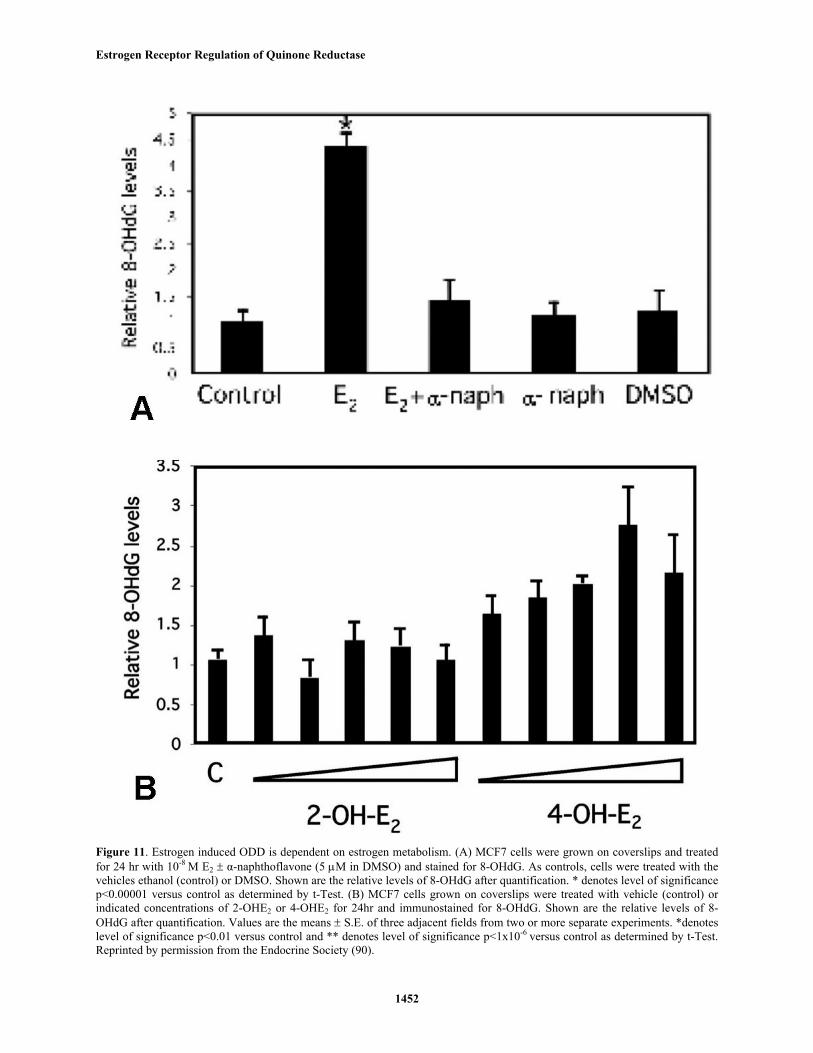
While the increase in 8-OHdG is independent ofER-mediated proliferation, our results suggest that it isdependent upon estrogen metabolism. Treating the cells withthe estrogen metabolism inhibitor alpha-naphthoflavone,which inhibits CYP1A and 1B, blocked the E2 effect (figure11A). alpha-Naphthoflavone or its solvent DMSO had noeffects on basal levels of 8-OHdG. In breast epithelial cells,E2 is metabolized to the hyroxylated catechols, with theformation of the more carcinogenic 4-OHE2 being favoredover 2-OHE2 (53, 58). Consistent with this, we see increased8-OH-dG only with 4-OHE2 (figure 11B).

Estrogen Receptor Regulation of Quinone Reductase
1450
Figure 9. Increased protein expression of GST-Pi and GCSH in the presence of trans-hydroxytamoxifen and ER-mediatedupregulation of GST-Pi and GCSH gene promoter activity can be localized to the EpRE. (A) Western blot analyses of GST-Pi and GCSH protein levels in MCF7 cells in the absence (c) or presence of E2 (10-8 M), or TOT (trans-hydroxytamoxifen,10-7 M). Western blots were probed with GST-Pi or GCSH antibody, and visualized using horseradish peroxidase-conjugated secondary antibody. The lower figures show the blot probed with cytokeratin 18 to show equal loading. Theimmunoblots are representative of 3 separate experiments. MDA-MB-231 cells were transfected with the (B) GST-Pi genepromoter reporter construct (C) wild type or mutant -83/-33-GST-Pi gene promoter reporter constructs (D) GCSH genepromoter reporter constructs (E) wild type or mutant 22 bp EpRE containing region (at -3.1 kb) of the GCSH promoteralong with control expression vector or expression vector for ER-alpha or ER-βeta. Cells were also transfected with PRL-SV40/Luc internal control reporter to correct for transfection efficiency. Cells were then treated for 24 h with controlethanol vehicle (c), E2 (10-8 M) or TOT (10-7M) as indicated. Data are expressed as the ratio of firefly to Renilla luciferaseactivity. Values are the means ± S.E. from three or more separate experiments. Reprinted by permission from MacMillanPublishers Ltd. (42).
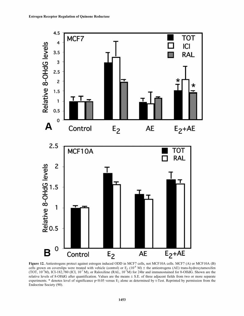
To determine the functional significance ofantiestrogen-mediated induction of NQO1, we monitored theeffect of antiestrogens and NQO1 on estrogen-inducedoxidative damage, as NQO1 may be able to detoxify the
reactive catecholestrogen quinones (50-52). We see inhibitionof E2-induced 8-OHdG with pharmacological concentrationsof TOT and ICI 182,780 but only in cells containing high ER,confirming a probable ER-mediated effect (figure 12).

Estrogen Receptor Regulation of Quinone Reductase
1451
Figure 10. Estrogen induces ODD in breast epithelial cells (A) MCF7 breast cancer cells were grown on coverslips and treatedwith vehicle (control) or increasing doses of E2 for 24 hr. As a control for oxidative damage, cells were treated with NAC (300µM) ± E2. The cells were then immunostained for 8-OHdG using the 1F7 monoclonal antibody (1:100; Trevigen, Gaithersburg,MD). Shown are the relative levels of 8-OHdG after quantification. Values are the means ±S.E. of three adjacent fields fromthree or more separate experiments. *denotes level of significance p=0.01 versus control and ** denotes level of significancep<1x10⁻⁶ versus control as determined by t-Test. (B) Same experiments using MCF10A or MDA-MB-231N cells treated with E2(10-8 M). ** denotes p≤0.001, * denotes p≤ 0.005 versus control as determined by t-Test. Reprinted by permission from theEndocrine Society (90).
Estrogen Receptor Regulation of Quinone Reductase
1452
Figure 11. Estrogen induced ODD is dependent on estrogen metabolism. (A) MCF7 cells were grown on coverslips and treatedfor 24 hr with 10-8 M E2 ± α-naphthoflavone (5 µM in DMSO) and stained for 8-OHdG. As controls, cells were treated with thevehicles ethanol (control) or DMSO. Shown are the relative levels of 8-OHdG after quantification. * denotes level of significancep<0.00001 versus control as determined by t-Test. (B) MCF7 cells grown on coverslips were treated with vehicle (control) orindicated concentrations of 2-OHE2 or 4-OHE2 for 24hr and immunostained for 8-OHdG. Shown are the relative levels of 8-OHdG after quantification. Values are the means ± S.E. of three adjacent fields from two or more separate experiments. *denoteslevel of significance p<0.01 versus control and ** denotes level of significance p<1x10⁻⁶
versus control as determined by t-Test.Reprinted by permission from the Endocrine Society (90).
Estrogen Receptor Regulation of Quinone Reductase
1453
Figure 12. Antiestrogens protect against estrogen induced ODD in MCF7 cells, not MCF10A cells. MCF7 (A) or MCF10A (B)cells grown on coverslips were treated with vehicle (control) or E2 (10-8 M) ± the antiestrogens (AE) trans-hydroxytamoxifen(TOT, 10-7M), ICI-182,780 (ICI, 10-7 M), or Raloxifene (RAL, 10-7M) for 24hr and immunostained for 8-OHdG. Shown are therelative levels of 8-OHdG after quantification. Values are the means ± S.E. of three adjacent fields from two or more separateexperiments. * denotes level of significance p<0.05 versus E2 alone as determined by t-Test. Reprinted by permission from theEndocrine Society (90).
Estrogen Receptor Regulation of Quinone Reductase
1454
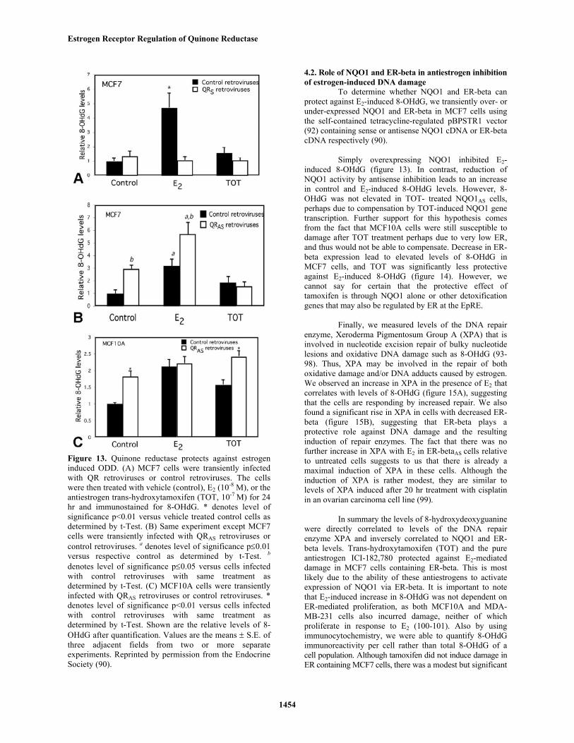
Figure 13. Quinone reductase protects against estrogeninduced ODD. (A) MCF7 cells were transiently infectedwith QR retroviruses or control retroviruses. The cellswere then treated with vehicle (control), E2 (10-8 M), or theantiestrogen trans-hydroxytamoxifen (TOT, 10-7 M) for 24hr and immunostained for 8-OHdG. * denotes level ofsignificance p<0.01 versus vehicle treated control cells asdetermined by t-Test. (B) Same experiment except MCF7cells were transiently infected with QRAS retroviruses orcontrol retroviruses. a denotes level of significance p≤0.01versus respective control as determined by t-Test. b
denotes level of significance p≤0.05 versus cells infectedwith control retroviruses with same treatment asdetermined by t-Test. (C) MCF10A cells were transientlyinfected with QRAS retroviruses or control retroviruses. *denotes level of significance p<0.01 versus cells infectedwith control retroviruses with same treatment asdetermined by t-Test. Shown are the relative levels of 8-OHdG after quantification. Values are the means ± S.E. ofthree adjacent fields from two or more separateexperiments. Reprinted by permission from the EndocrineSociety (90).
4.2. Role of NQO1 and ER-beta in antiestrogen inhibitionof estrogen-induced DNA damage
To determine whether NQO1 and ER-beta canprotect against E2-induced 8-OHdG, we transiently over- orunder-expressed NQO1 and ER-beta in MCF7 cells usingthe self-contained tetracycline-regulated pBPSTR1 vector(92) containing sense or antisense NQO1 cDNA or ER-betacDNA respectively (90).
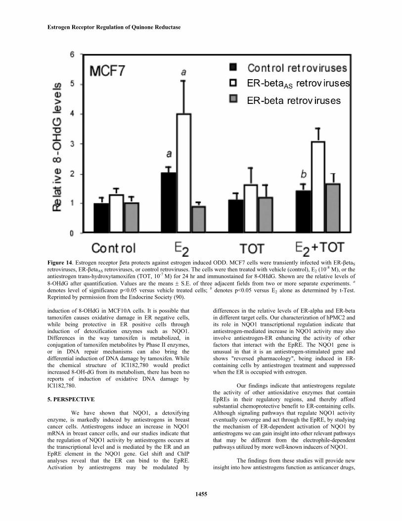
Simply overexpressing NQO1 inhibited E2-induced 8-OHdG (figure 13). In contrast, reduction ofNQO1 activity by antisense inhibition leads to an increasein control and E2-induced 8-OHdG levels. However, 8-OHdG was not elevated in TOT- treated NQO1AS cells,perhaps due to compensation by TOT-induced NQO1 genetranscription. Further support for this hypothesis comesfrom the fact that MCF10A cells were still susceptible todamage after TOT treatment perhaps due to very low ER,and thus would not be able to compensate. Decrease in ER-beta expression lead to elevated levels of 8-OHdG inMCF7 cells, and TOT was significantly less protectiveagainst E2-induced 8-OHdG (figure 14). However, wecannot say for certain that the protective effect oftamoxifen is through NQO1 alone or other detoxificationgenes that may also be regulated by ER at the EpRE.
Finally, we measured levels of the DNA repairenzyme, Xeroderma Pigmentosum Group A (XPA) that isinvolved in nucleotide excision repair of bulky nucleotidelesions and oxidative DNA damage such as 8-OHdG (93-98). Thus, XPA may be involved in the repair of bothoxidative damage and/or DNA adducts caused by estrogen.We observed an increase in XPA in the presence of E2 thatcorrelates with levels of 8-OHdG (figure 15A), suggestingthat the cells are responding by increased repair. We alsofound a significant rise in XPA in cells with decreased ER-beta (figure 15B), suggesting that ER-beta plays aprotective role against DNA damage and the resultinginduction of repair enzymes. The fact that there was nofurther increase in XPA with E2 in ER-betaAS cells relativeto untreated cells suggests to us that there is already amaximal induction of XPA in these cells. Although theinduction of XPA is rather modest, they are similar tolevels of XPA induced after 20 hr treatment with cisplatinin an ovarian carcinoma cell line (99).
In summary the levels of 8-hydroxydeoxyguaninewere directly correlated to levels of the DNA repairenzyme XPA and inversely correlated to NQO1 and ER-beta levels. Trans-hydroxytamoxifen (TOT) and the pureantiestrogen ICI-182,780 protected against E2-mediateddamage in MCF7 cells containing ER-beta. This is mostlikely due to the ability of these antiestrogens to activateexpression of NQO1 via ER-beta. It is important to notethat E2-induced increase in 8-OHdG was not dependent onER-mediated proliferation, as both MCF10A and MDA-MB-231 cells also incurred damage, neither of whichproliferate in response to E2 (100-101). Also by usingimmunocytochemistry, we were able to quantify 8-OHdGimmunoreactivity per cell rather than total 8-OHdG of acell population. Although tamoxifen did not induce damage inER containing MCF7 cells, there was a modest but significant
Estrogen Receptor Regulation of Quinone Reductase
1455
Figure 14. Estrogen receptor βeta protects against estrogen induced ODD. MCF7 cells were transiently infected with ER-βetaSretroviruses, ER-βetaAS retroviruses, or control retroviruses. The cells were then treated with vehicle (control), E2 (10-8 M), or theantiestrogen trans-hydroxytamoxifen (TOT, 10-7 M) for 24 hr and immunostained for 8-OHdG. Shown are the relative levels of8-OHdG after quantification. Values are the means ± S.E. of three adjacent fields from two or more separate experiments. a
denotes level of significance p<0.05 versus vehicle treated cells; b denotes p<0.05 versus E2 alone as determined by t-Test.Reprinted by permission from the Endocrine Society (90).
induction of 8-OHdG in MCF10A cells. It is possible thattamoxifen causes oxidative damage in ER negative cells,while being protective in ER positive cells throughinduction of detoxification enzymes such as NQO1.Differences in the way tamoxifen is metabolized, inconjugation of tamoxifen metabolites by Phase II enzymes,or in DNA repair mechanisms can also bring thedifferential induction of DNA damage by tamoxifen. Whilethe chemical structure of ICI182,780 would predictincreased 8-OH-dG from its metabolism, there has been noreports of induction of oxidative DNA damage byICI182,780.
5. PERSPECTIVE
We have shown that NQO1, a detoxifyingenzyme, is markedly induced by antiestrogens in breastcancer cells. Antiestrogens induce an increase in NQO1mRNA in breast cancer cells, and our studies indicate thatthe regulation of NQO1 activity by antiestrogens occurs atthe transcriptional level and is mediated by the ER and anEpRE element in the NQO1 gene. Gel shift and ChIPanalyses reveal that the ER can bind to the EpRE.Activation by antiestrogens may be modulated by
differences in the relative levels of ER-alpha and ER-betain different target cells. Our characterization of hPMC2 andits role in NQO1 transcriptional regulation indicate thatantiestrogen-mediated increase in NQO1 activity may alsoinvolve antiestrogen-ER enhancing the activity of otherfactors that interact with the EpRE. The NQO1 gene isunusual in that it is an antiestrogen-stimulated gene andshows "reversed pharmacology", being induced in ER-containing cells by antiestrogen treatment and suppressedwhen the ER is occupied with estrogen.
Our findings indicate that antiestrogens regulatethe activity of other antioxidative enzymes that containEpREs in their regulatory regions, and thereby affordsubstantial chemoprotective benefit to ER-containing cells.Although signaling pathways that regulate NQO1 activityeventually converge and act through the EpRE, by studyingthe mechanism of ER-dependent activation of NQO1 byantiestrogens we can gain insight into other relevant pathwaysthat may be different from the electrophile-dependentpathways utilized by more well-known inducers of NQO1.
The findings from these studies will provide newinsight into how antiestrogens function as anticancer drugs,

Estrogen Receptor Regulation of Quinone Reductase
1456
Figure 15. Estrogen and decreased levels of ER-βeta lead to elevated XPA levels in breast epithelial cells. (A) MCF7 or MCF10Acells grown on coverslips were treated with vehicle (control), E2 (10-8 M), or the antiestrogen trans-hydroxytamoxifen (TOT, 10-7
M) for 24hr. The cells were then immunostained for XPA and quantified. Values are the means ± S.E. of three adjacent fields fromthree or more separate experiments. * denotes level of significance p≤0.05 versus respective vehicle treated cells as determined byt-Test. (B) MCF7 cells were transiently infected with ER-βetaS retroviruses, ER-βetaAS retroviruses or control retroviruses. Thecells were then treated with vehicle (control), E2 (10-8 M), and/or the antiestrogen trans-hydroxytamoxifen (TOT, 10-7 M) for 24 hrand immunostained for 8-OHdG. Shown are the relative levels of 8-OHdG after quantification. Values are the means ± S.E. ofthree adjacent fields from two or more separate experiments. a denotes level of significance p<0.05 versus respective vehicletreated cells; b denotes p≤0.05 versus E2 alone; c denotes level of significance p<0.05 versus cells infected with control retroviruseswith same treatment as determined by t-Test. Reprinted by permission from the Endocrine Society (90).
Estrogen Receptor Regulation of Quinone Reductase
1457
which may derive not only from the already well-knownrepression of estrogen-stimulated activities, but also fromthe activation of detoxifying enzymes. NQO1 reduces thesecondary toxicity of certain cytotoxic agents that aremediated by semiquinone redox cycling. Indeed themetabolism of estrogens can be one of these processes thatinduce genotoxic effects. We observed that upregulation ofNQO1, either by overexpression or induction by TOT, canprotect breast cells against oxidative DNA damage causedby estrogen metabolites, representing a possible novelmechanism of tamoxifen prevention against breast cancer.It remains to be examined if NQO1 and ER can also inhibitanother type of DNA damage, DNA adducts formed byreactive catechol estrogens.
The activation of NQO1 activity by antiestrogensmay thus contribute to the beneficial antioxidant activity ofantiestrogens in breast cancer and possibly other estrogentarget tissues. The ability of antiestrogens such astamoxifen to increase NQO1 levels and activity also hasimportant potential implications for increasing the potencyof certain chemotherapeutic agents that are activated byquinone reduction. Although regulation of the effectivenessof cancer chemotherapeutic agents is complex, antiestrogentreatment might also enhance the sensitivity of tumor cellsto agents that are activated by quinone reduction, such asmitomycin C and aziridylbenzoquinones (26-30). This maycontribute to the beneficial effects of antiestrogens incancer therapy and in chemoprevention. Antiestrogens liketamoxifen are being employed extensively in boththerapeutic trials and in chemoprevention trials; thusstudies on possible effects of antiestrogens on the relativeefficacy of chemotherapeutic agents are of clinicalrelevance. By examining the effect of antiestrogens on thesensitivity of breast cancer cells to bioreductivechemotherapeutic agents that are activated by NQO1, thesestudies may contribute toward a more effective selection ofadjuvant chemotherapy for the treatment of breast cancer.
6. ACKNOWLEDGEMENTS
This work was supported by National Institute ofHealth Grants CA80959 to M.M.M. and CA18119 to B.S.K.
7. REFERENCES
1. Gradishar, W. J. and V. C. Jordan: Advances inendocrine therapy for the treatment and prevention ofbreast cancer. Cancer Chemother Biol Response Modif 21,211-222 (2003)
2. Montano, M. M. and B. S. Katzenellenbogen: Thequinone reductase gene: a unique estrogen receptor-regulated gene that is activated by antiestrogens. Proc NatlAcad Sci 94, 2581-2586 (1997)
3. Barkhem, T., S. Nilsson and J. A. Gustafsson: Molecularmechanisms, physiological consequences andpharmacological implications of estrogen receptor action.Am J Pharmacogenomics 4, 19-28 (2004)
4. Dickson, R. B. and M. E. Lippman: Estrogenic
regulation of growth and polypeptide growth factorsecretion in human breast carcinoma. Endocrine Rev 8, 29-43 (1987)
5. Katzenellenbogen, B. S: Estrogen receptors: bioactivitiesand interactions with cell signaling pathways. Biol Reprod54, 287-293 (1996)
6. Montano, M. M., V. Mueller, A. Trobaugh and B. S.Katzenellenbogen: The carboxy-terminal F domain of thehuman estrogen receptor: role in transcriptional activity ofthe receptor and the effectiveness of antiestrogens asestrogen antagonists. Molecular Endocrinology 9, 814-825(1995)
7. Mosselman, S., J. Polman and R. Dijkema: ER beta:identification and characterization of a novel humanestrogen receptor. FEBS Lett 392, 49-53 (1996)
8. Ogawa, S., S. Inoue, T. Watanabe, H. Hiroi, A. Orimo,T. Hosoi, Y. Ouchi and M. Muramatsu: The completeprimary structure of human estrogen receptor beta (hERbeta) and its heterodimerization with ER alpha in vivo andin vitro. Biochem Biophys Res Commun 243, 122-126(1998)
9. Paech, K., P. Webb, G. G. Kuiper, S. Nilsson, J.Gustafsson, P. J. Kushner and T. S. Scanlan: Differentialligand activation of estrogen receptors ERalpha and ERbetaat AP1 sites. Science 277, 1508-1510 (1997)
10. Enmark, E. and J. A. Gustafsson: Oestrogen receptors -an overview. J Intern Med 246, 133-138 (1999)
11. Pavao, M. and A. M. Traish: Estrogen receptorantibodies: specificity and utility in detection, localizationand analypises of estrogen receptor alpha and beta. Steroids66, 1-16 (2001)
12. Mann, S., R. Laucirica R, N. Carlson, P. S. Younes, N.Ali, A. Younes, Y. Li and M. Younes: Estrogen receptorbeta expression in invasive breast cancer. Hum Pathol 32,113-118. (2001)
13. Omoto, Y., S. Inoue, S. Ogawa, T. Toyama, H.Yamashita, M. Muramatsu, S.Kobayashi and H. Iwase:Clinical value of the wild-type estrogen receptor betaexpression in breast cancer. Cancer Lett 163, 207-212(2001)
14. Palmieri, C., G .J. Cheng, S. Saji, M. Zelada-Hedman,A. Warri, Z. Weihua, S.Van S. Noorden S, T. Wahlstrom,R. C. Coombes, M. Warner and J. A. Gustafsson: Estrogenreceptor beta in breast cancer. Endocr Relat Cancer 9, 1-13(2002)
15. Speirs, V., P. J. Carder, S. Lane, D. Dodwell, M. R.Lansdown and A. M. Hanby: Oestrogen receptor beta: whatit means for patients with breast cancer. Lancet Oncol 5,174-181 (2004)
16. Nakopoulou, L., A. C. Lazaris, E. G. Panayotopoulou,
Estrogen Receptor Regulation of Quinone Reductase
1458
I. Giannopoulou, N. Givalos, S. Markaki and A.Keramopoulos: The favourable prognostic value ofoestrogen receptor beta immunohistochemical expressionin breast cancer. J Clin Pathol 57, 523-528 (2004)
17. McDonald, C. C. and H. J. Stewart: Fatal myocardialinfarction in the Scottish adjuvant tamoxifen trial. TheScottish Breast Cancer Committee Br Med J 303, 435-437(1991)
18. Love, R. R., M. D. Richard, R. B. Mazess, H. S.Barden, S. Epstein, P. A. Newcomb, V. C. Jordan, P. P.Carbone and D. L. DeMets: Effects of tamoxifen on bonemineral density in postmenopausal women with breastcancer. N Engl J Med 326, 852-856 (1992)
19. Bhimani, R. S., W. Troll, D. Grunberger, D and K.Frenkel: Inhibition of oxidative stress in HeLa cells bychemopreventive agents. Cancer Res 53, 4528-4533 (1993)
20. Ahotupa, M., P. Hirsimaki, R. Parssinen and E.Mantyla: Alterations of drug metabolizing and antioxidantenzyme activities during tamoxifen-inducedhepatocarcinogenesis in the rat. Carcinogenesis 15, 863-868 (1994)
21. Wiseman, H: Tamoxifen: new membrane-mediatedmechanisms of action and therapeutic advances. TrendsPharmacolog Sci 15, 83-89 (1994)
22. Wolf, D. M. and V. C. Jordan: Gynecologiccomplications associated with long-term adjuvanttamoxifen therapy for breast cancer. Gynecol Oncol 45,118-128 (1992)
23. Talalay, P: Mechanisms of induction of enzymes thatprotect against chemical carcinogenesis. Adv Enzyme Reg28, 237-250 (1989)
24. Prestera, T., Y. Zhang, S. R. Spencer, C .A. Wilczakand P. Talalay: The electrophile counterattack response:protection against neoplasia and toxicity. Adv Enzyme Reg33, 281-296 (1993)
25. Talalay, P. and A. T. Dinkova-Kostova: Role ofnicotinamide quinone oxidoreductase 1 (NQO1) inprotection against toxicity of electrophiles and reactiveoxygen intermediates. Methods Enzymol 382, 355-6 (2004)
26. Lin, A. J., L. A. Cosby, C. W. Shansky and A. C.Sartorelli: Potential bioreductive alkylating agents. 1.Benzoquinone derivatives. J Med Chem 15, 1247-1252(1972)
27. Sartorelli, A. C., W. F. Hodnick, M. F. Belcourt, M.Tomasz, B. Haffty, J. J. Fischer and S. Rockwell:Mitomycin C: a prototype bioreductive agent. OncologyRes 6, 501-508 (1994)
28. Siegel, D., N. W. Gibson, P. C. Preusch and D. Ross:Metabolism of diaziquone by NAD(P)H:(quinone acceptor)oxidoreductase (DT-diaphorase): role in diaziquone-
induced DNA damage and cytotoxicity in human coloncarcinoma cells. Cancer Res 50, 7293-7300 (1990)
29. Siegel, D., N. W. Gibson, P. C. Preusch and D. Ross:Metabolism of mitomycin C by DT-diaphorase: role inmitomycin C-induced DNA damage and cytotoxicity inhuman colon carcinoma cells. Cancer Res 50, 7483-7489(1990)
30. Ross, D., D. Siegel, H. Beall, A. S. Prakash, R. T.Mulcahy and N. W. Gibson: DT-diaphorase in activationand detoxification of quinones. Bioreductive activation ofmitomycin C. Cancer Metastasis Rev 12, 83-101 (1993)
31. Schlager, J. J. and G. Powis: CytosolicNAD(P)H:(quinone-acceptor) oxidoreductase in humannormal and tumor tissue: effects of cigarette smoking andalcohol. Int J Cancer 45, 403-409 (1990)
32. Cresteil, T. and A. K. Jaiswal: High levels ofexpression of the NAD(P)H:quinone oxidoreductase(NQO1) gene in tumor cells compared to normal cells ofthe same origin. Biochem Pharmacol 42, 1021-1027 (1991)
33. Mekhail-Ishak, K., N. Hudson, M. S. Tsao and G.Batist: Implications for therapy of drug-metabolizingenzymes in human colon cancer. Cancer Res 49, 4866-4869 (1989)
34. Griffith, O. W. and R. T. Mulcahy: The enzymes ofglutathione synthesis: gamma-glutamylcysteine synthetase.Adv Enzymol Relat Areas Mol Biol 73, 209-267 (1999)
35. Strange, R. C., M. A. Spiteri, S. Ramachandran and A.A. Fryer: Glutathione-S-transferase family of enzymes.Mutat Res 482, 21-26 (2001)
36. Tew, K. D: Glutathione-associated enzymes inanticancer drug resistance. Cancer Res 54 4313-4320(1994)
37. Friling, R. S., A. Bensimon, Y. Tichauer and V. Daniel:Xenobiotic-inducible expression of murine glutathione S-transferase Ya subunit gene is controlled by anelectrophile-responsive element. Proc Natl Acad Sci 87,6258-6262 (1990)
38. Rushmore, T. H., M. R. Morton and C. B. Pickett: Theantioxidant responsive element. Activation by oxidativestress and identification of the DNA consensus sequencerequired for functional activity. J Biol Chem 266, 11632-11639 (1991)
39. Prestera, T. and P. Talalay: Electrophile and antioxidantregulation of enzymes that detoxify carcinogens. Proc NatlAcad Sci 92, 8965-8969 (1995)
40. Xie, T., M. Belinsky, Y. Xu and A. K. Jaiswal: ARE-and TRE-mediated regulation of gene expression. J BiolChem 270, 6894-6900 (1995)
41. Favreau, L. V. and C. B. Pickett: Transcriptional
Estrogen Receptor Regulation of Quinone Reductase
1459
regulation of the rat NAD(P)H:quinone reductase gene.Identification of regulatory elements controlling basal levelexpression and inducible expression by planar aromaticcompounds and phenolic antioxidants. J Biol Chem 266,4556-4561 (1991)
42. Rushmore, T. H., R. G. King, K. E. Paulson and C. B.Pickett: Regulation of glutathione S-transferase Ya subunitgene expression: identification of a unique xenobiotic-responsive element controlling inducible expression byplanar aromatic compounds. Proc Natl Acad Sci 87, 3826-3830 (1990)
43. Jaiswal, A. K: Human NAD(P)H:quinoneoxidoreductase (NQO1) gene structure and induction bydioxin. Biochemistry 30, 10647-10653 (1991)
44. Favreau, L. V. and C. B. Pickett: Transcriptionalregulation of the rat NAD(P)H:quinone reductase gene.Characterization of a DNA-protein interaction at theantioxidant responsive element and induction by 12-O-tetradecanoylphorbol 13-acetate. J Biol Chem 268, 19875-19881 (1993)
45. Nguyen, T. and C. B. Pickett: Regulation of ratglutathione S-transferase Ya subunit gene expression.DNA-protein interaction at the antioxidant responsiveelement. J Biol Chem 267, 13535-13539 (1992)
46. Venugopal, R. and A. K. Jaiswal: Nrf1 and Nrf2positively and c-Fos and Fra1 negatively regulate thehuman antioxidant response element-mediated expressionof NAD(P)H:quinone oxidoreductase1 gene. Proc NatlAcad Sci 93, 14960-14965 (1996)
47. Wild, A. C. and R. T. Mulcahy: Regulation of gamma-glutamylcysteine synthetase subunit gene expression:insights into transcriptional control of antioxidant defenses.Free Radic Res 32, 281-301 (2000)
48. Wild, A. C., H. R. Moinova and R. T. Mulcahy:Regulation of gamma-glutamylcysteine synthetase subunitgene expression by the transcription factor Nrf2. J BiolChem 274 33627-33636 (1999)
49. Jhaveri, M. S. and C. S. Morrow: Contribution ofproximal promoter elements to the regulation of basal anddifferential glutathione S-transferase P1 gene expression inhuman breast cancer cells. Biochim Biophys Acta 1396,179-190 (1998)
50. Jefcoate, C. R., J. G. Liehr, R. J. Santen, T. R. Sutter, J.D. Yager, W. Yue, S. J. Santner, R. Tekmal, L. Demers, R.Pauley, F. Naftolin, G. Mor and L. Berstein: Tissue-specific synthesis and oxidative metabolism of estrogens. JNatl Cancer Inst Monogr 27, 95-112 (2000)
51. Zhu, B. T. and A. H. Conney: Functional role ofestrogen metabolism in target cells: review andperspectives. Carcinogenesis 19, 1-27 (1998)
52. Spink, D. C., B. C. Spink, J. Q. Cao, J. A. DePasquale,
B. T. Pentecost, M. J. Fasco, Y Li and T. R. Sutter:Differential expression of CYP1A1 and CYP1B1 in humanbreast epithelial cells and breast tumor cells.Carcinogenesis 19, 291-298 (1998)
53. Liehr, J. G: Is estradiol a genotoxic mutageniccarcinogen? Endocr Rev 21, 40-54 (2000)
54. Liehr, J. G: Dual role of oestrogens as hormones andpro-carcinogens: tumour initiation by metabolic activationof oestrogens. Eur J Cancer Prev 6, 3-10 (1997)
55. Thibodeau, P. A. and B. Paquette: DNA damageinduced by catecholestrogens in the presence of copper (II):Generation of reactive oxygen species and enhancement byNADH. Free Rad Biol Med 27, 1367-1377. (1999)
56. Roy, D. and J. G. Liehr: Temporary decrease in renalquinone reductase activity induced by chronicadministration of estradiol to male Syrian hamsters.Increased superoxide formation by redox cycling ofestrogen. J Biol Chem 263, 3646-3651 (1998)
57. Shen, L., E. Pisha, Z. Huang, J. M. Pezzuto, E. Krol, Z.Alam, R. B. van Breemen and J. L. Bolton: Bioreductiveactivation of catechol estrogen-ortho-quinones:aromatization of the B ring in 4-hydroxyequileninmarkedly alters quinoid formation and reactivity.Carcinogenesis 18, 1093-1101 (1997)
58. Roy, D., J. Weisz and J. G. Liehr: The O-methylation of4-hydroxyestradiol is inhibited by 2-hydroxyestradiol:implications for estrogen-induced carcinogenesis.Carcinogenesis 11, 459-462 (1990)
59. Russo, J., Y. F. Hu, Q. Tahin, D. Mihaila, C. Slater, M.H. Laree and I. H. Russo: Carcinogenicity of estrogens inhuman breast epithelial cells. APMIS 109, 39-52 (2001)
60. Cavalieri, E., K. Frenkel, J. G. Liehr, E. Rogan and D.Roy: Estrogens as endogenous genotoxic agents--DNAadducts and mutations. J Natl Cancer Inst Monogr 27, 75-93 (2000)
61. Newbold, R. R. and J. G. Liehr: Induction of uterineadenocarcinoma in CD-1 mice by catechol estrogens.Cancer Res 60, 235-237 (2000)
62. Russo, J., X. Ao, C. Grill and I. H. Russo: Pattern ofdistribution of cells positive for estrogen receptor alpha andprogesterone receptor in relation to proliferating cells in themammary gland. Breast Cancer Res Treat 53, 217-227(1999)
63. Rogan, E. G., A. F. Badawi, P. D. Devanesan, J. L.Meza, J. A. Edney, W. W. West, S. M. Higginbotham andE. L. Cavalieri: Relative imbalances in estrogenmetabolism and conjugation in breast tissue of women withcarcinoma: potential biomarkers of susceptibility to cancer.Carcinogenesis 24, 697-702 (2003)
64. Siegelmann-Danieli, N. and K. H. Buetow: Significance
Estrogen Receptor Regulation of Quinone Reductase
1460
of genetic variation at the glutathione S-transferase M1 andNAD(P)H:quinone oxidoreductase 1 detoxification genesin breast cancer development. Oncology 62, 39-45 (2003)
65. Rebbeck, T. R., A. K. Godwin and K. H. Buetow:Variability in loss of constitutional heterozygosity acrossloci and among individuals: association with candidategenes in ductal breast carcinoma. Mol Carcinogenesis 17,117-125 (1996)
66. Maugard, C. M., J. Charrier, A. Pitard, L. Campion, O.Akande, F. Pleasants and F. Ali-Osman: Geneticpolymorphism at the glutathione S-transferase (GST) P1locus is a breast cancer risk modifier. Int J Cancer 91, 334-339 (2001)
67. Sarbia, M., M. Bitzer, D. Siegel, D. Ross, W. A.Schulz, R. B. Zotz, S. Kiel, H. Geddert, Y. Kandemir, A.Walter, R. Willers and H. E. Gabbert: Association betweenNAD(P)H: quinone oxidoreductase 1 (NQ01) inactivatingC609T polymorphism and adenocarcinoma of the uppergastrointestinal tract. Int Jcancer 107, 381-386 (2003)
68. Smith, M. T., Y. Wang, C. F. Skibola, D. J. Slater, L.Lo Nigro, P. C. Nowell, B. J. Lange and C. A. Felix: LowNAD(P)H:quinone oxidoreductase activity is associatedwith increased risk of leukemia with MLL translocations ininfants and children. Blood 100, 4590-3459 (2002)
69. Goode, E. L., A. M. Dunning, B. Kuschel, C. S. Healey,N. E. Day, B. A. Ponder, D. F. Easton and P. P. Pharoah:Effect of germ-line genetic variation on breast cancersurvival in a population-based study. Cancer Res 62, 3052-3057 (2002)
70. Mitrunen, K., and A. Hirvonen: Molecularepidemiology of sporadic breast cancer. The role ofpolymorphic genes involved in oestrogen biosynthesis andmetabolism. Mutat Res 544, 9-41 (2003)
71. Hamajima, N., K. Matsuo, H. Iwata, M. Shinoda, Y.Yamamur, T. Kato, S. Hatooka, T. Mitsudomi, M. Suyama,Y. Kagami, M. Ogur, M. Ando, Y. Sugimura and K.Tajima: NAD(P)H: quinone oxidoreductase 1 (NQO1)C609T polymorphism and the risk of eight cancers forJapanese. Int J Clin Oncol 7, 103-108 (2002)
72. Esteller, M., P. G. Corn, J. M. Urena, E. Gabrielson, S.B. Baylin and J. G. Herma: Inactivation of glutathione S-transferase P1 gene by promoter hypermethylation inhuman neoplasia. Cancer Res 58, 4515-4518 (1998)
73. Singal, R., J. van Wert and M. Bashambu: Cytosinemethylation represses glutathione S-transferase P1(GSTP1) gene expression in human prostate cancer cells.Cancer Res 61, 4820-4826 (2001)
74. Montano, M. M., A. K. Jaiswal and B. S.Katzenellenbogen: Transcriptional regulation of thehuman quinone reductase gene by antiestrogen-liganded estrogen receptor alpha estrogen receptor betavia the electrophile/antioxidant response element. J
Biol Chem 273, 25443-25449 (1998)
75. Piette, J., S. Hirai and M. Yaniv: Constitutive synthesisof activator protein 1 transcription factor after viraltransformation of mouse fibroblasts. Proc Natl Acad Sci 85,3401-3405 (1988)
76. Montano, M. M., B. M. Wittmann and N. R. Bianco:Identification and characterization of a novel factor thatregulates quinone reductase gene transcriptional Activity. JBiol Chem 275, 34306-34313 (2000)
77. Su, J. Y. and J. L. Maller: Cloning and expression of aXenopus gene that prevents mitotic catastrophe in fissionyeast. Mol Gen Genet 246, 387-396 (1995)
78. King, R. W., P. K. Jackson and M. W. Kirschner:Mitosis in transition. Cell 79, 563-571 (1994)
79. Lundgren, K., N. Walworth, R. Booher, M. Dembski,M. Kirschner and D. Beach: mik1 and wee1 cooperate inthe inhibitory tyrosine phosphorylation of cdc2. Cell 64,1111-1122 (1991)
80. Qiu, X., H. J. Forman, A. H. Schonthal and E. Cadenas:Induction of p21 mediated by reactive oxygen speciesformed during the metabolism of aziridinylbenzoquinonesby HCT116 cells. J Biol Chem 271, 31915-31921 (1996)
81. Reese, J. C. and B. S. Katzenellenbogen: Examinationof the DNA-binding ability of estrogen receptor in wholecells: implications for hormone-independent transactivationand the actions of antiestrogens. Mol Cell Biol 12, 4531-4538 (1992)
82. Montano, M. M., H. Deng, M. Liu, X. Sun, and R.Singal: Transcriptional regulation by the estrogen receptorof antioxidative stress enzymes and its functionalimplications. Oncogene 23, 2442-2453
83. Hellriegel, E. T., G. A. Matwyshyn, P. Fei, K. H.Dragnew, R. W. Nims, R. Lubet and A. T. Kong:Regulation of gene expression of various phase I and phaseII drug-metabolizing enzymes by tamoxifen in rat liver.Biochem Pharmacol 52, 1561-1568 (1996)
84. Helbock, H. J., K. B. Beckman and B. N. Ames: 8-Hydroxydeoxyguanosine and 8-hydroxyguanine asbiomarkers of oxidative DNA damage. Methods Enzymol300, 156-166 (1999)
85. Shibutani, S. and A. P. Grollman: Miscoding duringDNA synthesis on damaged DNA templates catalysed bymammalian cell extracts. Cancer Lett 83, 315-322 (1994)
86. Li, D., W. Zhan, J. Zhu, P. Chang, A. Sahin, E.Singletary, M. Bondy, T. Hazra, S. Mitra, S. S. Lau, J. Shenand J. DiGiovanni: Oxidative DNA damage and 8-hydroxy-2-deoxyguanosine DNA glycosylase/apurinic lyase inhuman breast cancer. Mol Carcinog 31, 214-223 (2001)
87. Musarrat, J., J. Arezina-Wilson and A. A. Wani:
Estrogen Receptor Regulation of Quinone Reductase
1461
Prognostic and aetiological relevance of 8-hydroxyguanosine in human breast carcinogenesis. Eur JCancer 32A, 1209-1214 (1996)
88. Lavigne, J. A., J. E. Goodman, T. Fonong, S. Odwin, P.He, D. W. Roberts and J. D. Yager: The effects of catechol-O-methyltransferase inhibition on estrogen metabolite andoxidative DNA damage levels in estradiol-treated MCF-7cells. Cancer Res 61, 7488-7494 (2001)
89. Hiraku, Y., N. Yamashita, M. Nishiguchi and S.Kawanishi: Catechol estrogens induce oxidative DNAdamage and estradiol enhances cell proliferation. Int JCancer 92, 333-337 (2001)
90. Bianco, N. R., G. Perry, M. A. Smith, D. J. Templetonand M. M. Montano: Functional implications ofantiestrogen induction of quinone reductase: inhibition ofestrogen-induced DNA damage. Molecular Endocrinology17, 1344-1355 (2003)
91. Nunomura, A., G. Perry, G. Aliev, K. Hirai, A. Takeda,E. K. Balraj, P. K. Jones, H. Ghanbari, T. Wataya, S.Shimohama, S. Chiba, C. S. Atwood, R. B. Petersen and M.A. Smith: Oxidative damage is the earliest event inAlzheimer disease. J Neuropathol Exp Neurol 60, 759-767(2001)
92. Paulus, W., I. Baur, F. M. Boyce, X. O. Breakfield andS. A. Reeves: Self-contained, tetracline regulated retroviralvector system for gene delivery to mammalian cells. J Virol70, 62-67 (1996)
93. Croteau, D. L. and V. A. Bohr: Repair of oxidativedamage to nuclear and mitochondrial DNA in mammaliancells. J Biol Chem 272, 25409-25412. (1997)
94. Reardon, J. T., T. Bessho, H. C. Kung, P. H. Bolton andA. Sancar: In vitro repair of oxidative DNA damage byhuman nucleotide excision repair system: possibleexplanation for neurodegeneration in xerodermapigmentosum patients. Proc Natl Acad Sci 94, 9463-9468(1997)
95. Jaiswal, M., L. J. Lipinski, V. A. Bohr and S. J. Mazur:Efficient in vitro repair of 7-hydro-8-oxodeoxyguanosineby human cell extracts: involvement of multiple pathways.Nucleic Acids Res 26, 2184-2191 (1998)
96. Driggers, W. J., V. I. Grishko, S. P. LeDoux and G. L.Wilson: Defective repair of oxidative damage in themitochondrial DNA of a xeroderma pigmentosum group Acell line. Cancer Res 56, 1262-1266 (1996)
97. Lipinski, L. J., N. Hoehr, S. J. Mazur, G. L. Dianov, S.Senturker, M. Dizdaroglu and V. A. Bohr Repair ofoxidative DNA base lesions induced by fluorescent light isdefective in xeroderma pigmentosum group A cells.Nucleic Acids Res 27, 3153-3158 (1999)
98. Klein, J. C., M. J. Bleeker, C. P. Saris, H. C. Roelen, H.F. Brugghe, H. van den Elst, G. A. van der Marel, J. H. van
Boom, J. G. Westra and E. Kriek: Repair and replication ofplasmids with site-specific 8-oxodG and 8- AAFdGresidues in normal and repair-deficient human cells.Nucleic Acids Res 20, 4437-4443 (1992)
99. Hector, S., W. Bolanowska-Higdon, J. Zdanowicz, S.Hitt and L. Pendyala: In vitro studies on the mechanisms ofoxaliplatin resistance. Cancer Chemother Pharmacol 48,398-406. (2001)
100. Pilat, M. J., J. K. Cristman and S. C. Brooks:Characterization of the estrogen receptor transfectedMCF10A breast cell line 139B6. Breast Cancer Res Treat37, 253-266 (1996)
101. Garcia, M., D. Derocq, G. Freiss and H. Rochefort:Activation of estrogen receptor transfected into a receptor-negative breast cancer cell line decreases the metastatic andinvasive potential of the cells. Proc Natl Acad Sci 89,11538-11542 (1996)
Abbreviations: ER: estrogen receptor, E2: 17 beta-estradiol, NQO1: quinone reductase, TBHQ: tert-butylhydroquinone, EpRE: electrophile response element,TOT: trans-hydroxytamoxifen, GST-Pi: glutathione S-transferases Pi, GCSh: gamma-glutamylcysteine synthetaseheavy subunit, 8-OHdG: 8-hydroxydeoxyguanine, NAC:N-acetylcysteine, 2- or 4-OH-E2: 2- or 4-hydroxyestradiol
Key Words: Estrogens, Estrogen receptor, Antiestrogens,Quinone reductase, Review
Send correspondence to: Dr Monica M Montano, CaseWestern Reserve University School of Medicine,Department of Pharmacology, H G Wood Bldg W307,2109 Adelbert Road, Cleveland, OH 44122, Tel: 216-368-3378, Fax: 216-368-3395, E-mail: [email protected]
http://www.bioscience.org/current/vol10.htm