Embed Size (px)
Citation preview
www.advhealthmat.dewww.MaterialsViews.com
FULL
PAPER
© 2013 WILEY-VCH Verlag GmbH & Co. KGaA, Weinheim88 wileyonlinelibrary.com
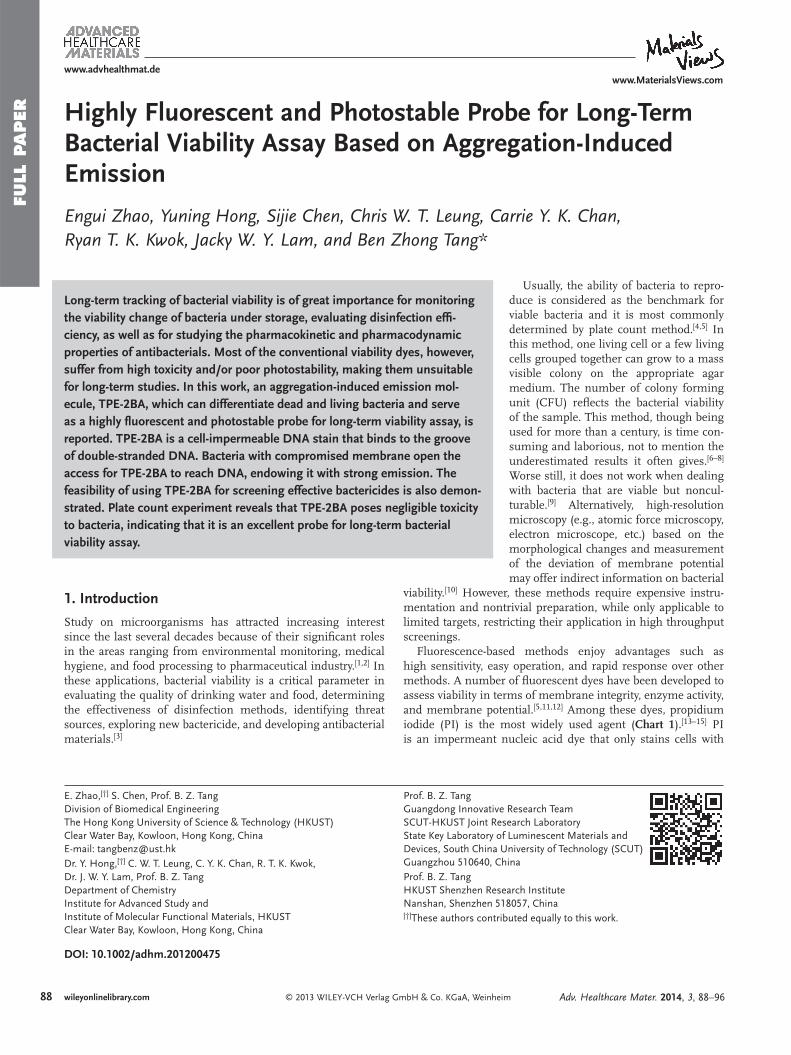
Highly Fluorescent and Photostable Probe for Long-Term Bacterial Viability Assay Based on Aggregation-Induced Emission
Engui Zhao , Yuning Hong , Sijie Chen , Chris W. T. Leung , Carrie Y. K. Chan , Ryan T. K. Kwok , Jacky W. Y. Lam , and Ben Zhong Tang *
DOI: 10.1002/adhm.201200475
E. Zhao,[†] S. Chen, Prof. B. Z. TangDivision of Biomedical EngineeringThe Hong Kong University of Science & Technology (HKUST)Clear Water Bay, Kowloon, Hong Kong, China E-mail: [email protected] Dr. Y. Hong,[†] C. W. T. Leung, C. Y. K. Chan, R. T. K. Kwok, Dr. J. W. Y. Lam, Prof. B. Z. TangDepartment of ChemistryInstitute for Advanced Study and Institute of Molecular Functional Materials, HKUSTClear Water Bay, Kowloon, Hong Kong, China
Long-term tracking of bacterial viability is of great importance for monitoring the viability change of bacteria under storage, evaluating disinfection effi -ciency, as well as for studying the pharmacokinetic and pharmacodynamic properties of antibacterials. Most of the conventional viability dyes, however, suffer from high toxicity and/or poor photostability, making them unsuitable for long-term studies. In this work, an aggregation-induced emission mol-ecule, TPE-2BA, which can differentiate dead and living bacteria and serve as a highly fl uorescent and photostable probe for long-term viability assay, is reported. TPE-2BA is a cell-impermeable DNA stain that binds to the groove of double-stranded DNA. Bacteria with compromised membrane open the access for TPE-2BA to reach DNA, endowing it with strong emission. The feasibility of using TPE-2BA for screening effective bactericides is also demon-strated. Plate count experiment reveals that TPE-2BA poses negligible toxicity to bacteria, indicating that it is an excellent probe for long-term bacterial viability assay.
Usually, the ability of bacteria to repro-duce is considered as the benchmark for viable bacteria and it is most commonly determined by plate count method. [ 4 , 5 ] In this method, one living cell or a few living cells grouped together can grow to a mass visible colony on the appropriate agar medium. The number of colony forming unit (CFU) refl ects the bacterial viability of the sample. This method, though being used for more than a century, is time con-suming and laborious, not to mention the underestimated results it often gives. [ 6 – 8 ] Worse still, it does not work when dealing with bacteria that are viable but noncul-turable. [ 9 ] Alternatively, high-resolution microscopy (e.g., atomic force microscopy, electron microscope, etc.) based on the morphological changes and measurement of the deviation of membrane potential may offer indirect information on bacterial
viability. [ 10 ] However, these methods require expensive instru-mentation and nontrivial preparation, while only applicable to limited targets, restricting their application in high throughput screenings.
Fluorescence-based methods enjoy advantages such as high sensitivity, easy operation, and rapid response over other methods. A number of fl uorescent dyes have been developed to assess viability in terms of membrane integrity, enzyme activity, and membrane potential. [ 5 , 11 , 12 ] Among these dyes, propidium iodide (PI) is the most widely used agent ( Chart 1 ). [ 13– 15 ] PI is an impermeant nucleic acid dye that only stains cells with
1. Introduction
Study on microorganisms has attracted increasing interest since the last several decades because of their signifi cant roles in the areas ranging from environmental monitoring, medical hygiene, and food processing to pharmaceutical industry. [ 1 , 2 ] In these applications, bacterial viability is a critical parameter in evaluating the quality of drinking water and food, determining the effectiveness of disinfection methods, identifying threat sources, exploring new bactericide, and developing antibacterial materials. [ 3 ]
Prof. B. Z. TangGuangdong Innovative Research TeamSCUT-HKUST Joint Research LaboratoryState Key Laboratory of Luminescent Materials and Devices, South China University of Technology (SCUT)Guangzhou 510640, China Prof. B. Z. TangHKUST Shenzhen Research InstituteNanshan, Shenzhen 518057, China [†]These authors contributed equally to this work.
Adv. Healthcare Mater. 2014, 3, 88–96
www.MaterialsViews.com
FULL P
APER
www.advhealthmat.de
89wileyonlinelibrary.com© 2013 WILEY-VCH Verlag GmbH & Co. KGaA, Weinheim
compromised membrane, and the enhanced fl uorescence can thus indicate cell death. The use of PI, however, poses several problems. PI is structurally a derivative of ethidium bromide, a notorious strong mutagen and carcinogen, implying its poten-tial harm to human body. Photostability and cytotoxicity are also concerns that limit its applications: characterization of the stained cells has to be fi nished shortly after staining. [ 16 ] This calls for the development of new probes with high sensitivity and selectivity, superior photostability, and good biocompat-ibility for long-term bacterial viability assays.
We have previously discovered a group of unique lumino-gens which are nonluminescent when molecularly dissolved but highly fl uorescent when aggregate. [ 17 ] We termed aggrega-tion-induced emission (AIE) for this novel phenomenon and proposed restriction of intramolecular motions as the main cause for the AIE phenomenon. [ 18 , 19 ] In the past decade, the AIE luminogens have been successfully employed in a variety of biological applications such as for long-term cell tracking, [ 20 ] nonself-quenching DNA labeling, [ 21 ] inhibition of amyloid fi brillation, [ 22 ] monitoring of cell apoptosis, [ 23 ] etc. [ 24 – 26 ] The AIE luminogens exhibit high quantum effi ciency, good biocom-patibility, and appreciable photostability, which motivate us to further exploit its application in the thriving area of bacteria studies.
Recently, we have reported an AIE-active molecule, 4,4 ′ -(1,2-diphenylethene-1,2-diyl)bis(4,1-phenylene)diboronic
acid (TPE-2BA), [ 27 ] which can specifi cally recognize D -glucose through hydrogen bonding with cis -diol moiety of the sugar. In this work, we unveil another important function of TPE-2BA as a fl uorescent probe for differentiating living and dead bacteria. Compared with commercial viability probe, this dye is highly emissive, photostable and very friendly to bacteria, making them well-suited for long-term bacterial viability tracking. To our best knowledge, this is the fi rst study reported for using AIE luminogens in bacteria studies.
2. Results and Discussion
2.1. Differentiation of Dead/Live Bacteria
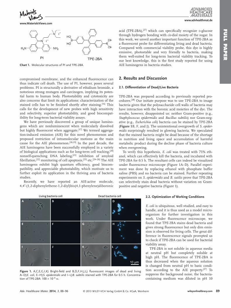
TPE-2BA was prepared according to previously reported pro-cedures. [ 28 ] Our initiate purpose was to use TPE-2BA to image bacteria given that the polysaccharide cell walls of bacteria may have interaction with the boronic acid moieties of the dye. The results, however, disappointed us: neither Gram-positive (e.g., Staphylococcus epidermidis and Bacillus subtilis ) nor Gram-neg-ative (e.g., Escherichia coli ) bacteria can be stained by TPE-2BA ( Figure 1 B, F, and J). The unintentional overgrowth of S. epider-midis surprisingly resulted in glowing bacteria. We speculated that the stained bacteria might be dead because of the shortage in nutrition and living space and accumulation of harmful metabolic product during the decline phase of bacteria culture when overgrowing.
To verify this hypothesis, E. coli was treated with 75% eth-anol, which can effectively kill the bacteria, and incubated with TPE-2BA for 0.5 h. The resultant cells can indeed be visualized under fl uorescence microscope (Figure 1 A–D). Parallel experi-ment was done by replacing ethanol with phosphate buffer saline (PBS) and no bacteria can be stained. Further repeating experiments on S. epidermidis and B. sutilis prove that TPE-2BA can selectively stain dead bacteria without variation on Gram-positive and negative bacteria (Figure 1 ).
2.2. Optimization of Working Conditions
E. coli is ubiquitous, well studied, and easy to handle, and it is thus used as a model micro-organism for further investigation in this work. Under fl uorescence microscope, we found that TPE-2BA stains dead bacteria and gives strong fl uorescence but only dim emis-sion is observed for living cells. The great dif-ference in fl uorescence signals prompted us to check if TPE-2BA can be used for bacterial viability assay.
TPE-2BA is not soluble in aqueous media at neutral pH but completely soluble at high pH. The fl uorescence of TPE-2BA is thus decreased when the aqueous solution is changed from neutral pH to basic condi-tion according to the AIE property. [ 27 ] To suppress the background noise, the bacteria-containing medium was diluted in pH 10
Figure 1 . A,C,E,G,I,K) Bright-fi eld and B,D,F,H,J,L) fl uorescent images of dead and living A − D) E. coli , E − H) S. epidermidis and I − L) B. subtilis stained with TPE-2BA for 0.5 h. Concentra-tion of TPE-2BA: 100 × 10 − 6 M .
Chart 1. Molecular structures of PI and TPE-2BA.
Adv. Healthcare Mater. 2014, 3, 88–96
www.MaterialsViews.com
FULL
PAPER
www.advhealthmat.de
90 wileyonlinelibrary.com © 2013 WILEY-VCH Verlag GmbH & Co. KGaA, Weinheim
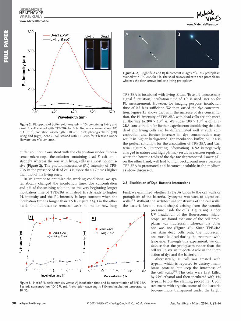
buffer solution. Consistent with the observation under fl uores-cence microscope, the solution containing dead E. coli emits strongly, whereas the one with living cells is almost nonemis-sive ( Figure 2 ). The photoluminescence (PL) intensity of TPE-2BA in the presence of dead cells is more than 12 times higher than that of the living ones.
In an attempt to optimize the working conditions, we sys-tematically changed the incubation time, dye concentration and pH of the staining solution. At the very beginning longer incubation time of TPE-2BA with dead E. coli leads to higher PL intensity and the PL intensity is kept constant when the incubation time is longer than 1.5 h ( Figure 3 A). On the other hand, the fl uorescence remains weak no matter how long
TPE-2BA is incubated with living E. coli . To avoid unnecessary signal fl uctuation, incubation time of 3 h is used later on for PL measurement. However, for imaging purpose, incubation time of 0.5 h is suffi cient. We then varied the dye concentra-tion. Figure 3 B shows that with the increase of dye concentra-tion, the PL intensity of TPE-2BA with dead cells are enhanced all the way to 200 × 10 − 6 M . We chose 100 × 10 − 6 M of TPE-2BA concentration for further experiments considering that the dead and living cells can be differentiated well at such con-centration and further increase in dye concentration may result in higher background. For incubation buffer, pH 7.4 is the perfect condition for the association of TPE-2BA and bac-teria (Figure S1, Supporting Information). DNA is negatively charged in nature and high pH may result in electron repulsion when the boronic acids of the dye are deprotonated. Lower pH, on the other hand, will lead to high background noise because TPE-2BA is protonated and becomes insoluble in the medium as above discussed.
2.3. Elucidation of Dye–Bacteria Interactions
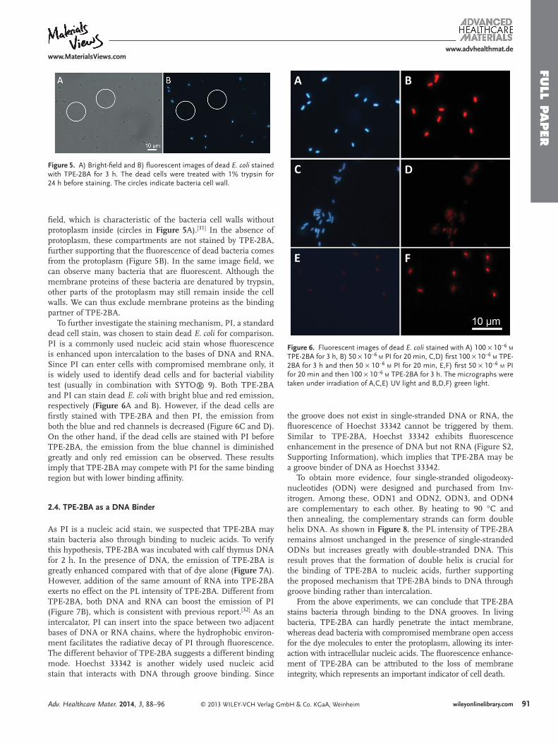
First, we examined whether TPE-2BA binds to the cell walls or protoplasm of the bacteria. Lysozyme was used to digest cell walls. [ 29 ] Without the architectural constraints of the cell walls, the bacteria become round-shaped arising from the osmotic
pressure inside the cells ( Figure 4 A). Under UV irradiation of the fl uorescence micro-scope, we found that one of the cell proto-plasm was fl uorescent, whereas the other one was not (Figure 4 B). Since TPE-2BA can stain dead cells only, the fl uorescent one must be dead during the treatment with lysozyme. Through this experiment, we can deduce that the protoplasm rather than the cell wall plays an important role in the inter-action of dye and the bacterium.
Alternatively, E. coli was treated with trypsin, which is reported to destroy mem-brane proteins but keep the intactness of the cell walls. [ 30 ] The cells were fi rst killed by 75% ethanol and then incubated with 1% trypsin before the staining procedure. Upon treatment with trypsin, some of the bacteria become more transparent under the bright
Figure 2 . PL spectra of buffer solutions (pH = 10) containing living and dead E. coli stained with TPE-2BA for 3 h. Bacteria concentration: 10 7 CFU mL − 1 ; excitation wavelength: 310 nm. Inset: photographs of (left) living and (right) dead E. coli stained with TPE-2BA for 3 h taken under illumination of a UV lamp.
Figure 3 . Plot of PL peak intensity versus A) incubation time and B) concentration of TPE-2BA. Bacteria concentration: 10 7 CFU mL − 1 ; excitation wavelength: 310 nm; incubation temperature: 30 ° C.
Figure 4 . A) Bright-fi eld and B) fl uorescent images of E. coli protoplasm stained with TPE-2BA for 3 h. The solid arrows indicate dead protoplasm, whereas the dash arrows indicate living protoplasm.
Adv. Healthcare Mater. 2014, 3, 88–96
www.MaterialsViews.com
FULL P
APER
www.advhealthmat.de
91wileyonlinelibrary.com© 2013 WILEY-VCH Verlag GmbH & Co. KGaA, Weinheim
fi eld, which is characteristic of the bacteria cell walls without protoplasm inside (circles in Figure 5 A). [ 31 ] In the absence of protoplasm, these compartments are not stained by TPE-2BA, further supporting that the fl uorescence of dead bacteria comes from the protoplasm (Figure 5 B). In the same image fi eld, we can observe many bacteria that are fl uorescent. Although the membrane proteins of these bacteria are denatured by trypsin, other parts of the protoplasm may still remain inside the cell walls. We can thus exclude membrane proteins as the binding partner of TPE-2BA.
To further investigate the staining mechanism, PI, a standard dead cell stain, was chosen to stain dead E. coli for comparison. PI is a commonly used nucleic acid stain whose fl uorescence is enhanced upon intercalation to the bases of DNA and RNA. Since PI can enter cells with compromised membrane only, it is widely used to identify dead cells and for bacterial viability test (usually in combination with SYTO® 9). Both TPE-2BA and PI can stain dead E. coli with bright blue and red emission, respectively ( Figure 6 A and B). However, if the dead cells are fi rstly stained with TPE-2BA and then PI, the emission from both the blue and red channels is decreased (Figure 6 C and D). On the other hand, if the dead cells are stained with PI before TPE-2BA, the emission from the blue channel is diminished greatly and only red emission can be observed. These results imply that TPE-2BA may compete with PI for the same binding region but with lower binding affi nity.
2.4. TPE-2BA as a DNA Binder
As PI is a nucleic acid stain, we suspected that TPE-2BA may stain bacteria also through binding to nucleic acids. To verify this hypothesis, TPE-2BA was incubated with calf thymus DNA for 2 h. In the presence of DNA, the emission of TPE-2BA is greatly enhanced compared with that of dye alone ( Figure 7 A). However, addition of the same amount of RNA into TPE-2BA exerts no effect on the PL intensity of TPE-2BA. Different from TPE-2BA, both DNA and RNA can boost the emission of PI (Figure 7 B), which is consistent with previous report. [ 32 ] As an intercalator, PI can insert into the space between two adjacent bases of DNA or RNA chains, where the hydrophobic environ-ment facilitates the radiative decay of PI through fl uorescence. The different behavior of TPE-2BA suggests a different binding mode. Hoechst 33342 is another widely used nucleic acid stain that interacts with DNA through groove binding. Since
the groove does not exist in single-stranded DNA or RNA, the fl uorescence of Hoechst 33342 cannot be triggered by them. Similar to TPE-2BA, Hoechst 33342 exhibits fl uorescence enhancement in the presence of DNA but not RNA (Figure S2, Supporting Information), which implies that TPE-2BA may be a groove binder of DNA as Hoechst 33342.
To obtain more evidence, four single-stranded oligodeoxy-nucleotides (ODN) were designed and purchased from Inv-itrogen. Among these, ODN1 and ODN2, ODN3, and ODN4 are complementary to each other. By heating to 90 ° C and then annealing, the complementary strands can form double helix DNA. As shown in Figure 8 , the PL intensity of TPE-2BA remains almost unchanged in the presence of single-stranded ODNs but increases greatly with double-stranded DNA. This result proves that the formation of double helix is crucial for the binding of TPE-2BA to nucleic acids, further supporting the proposed mechanism that TPE-2BA binds to DNA through groove binding rather than intercalation.
From the above experiments, we can conclude that TPE-2BA stains bacteria through binding to the DNA grooves. In living bacteria, TPE-2BA can hardly penetrate the intact membrane, whereas dead bacteria with compromised membrane open access for the dye molecules to enter the protoplasm, allowing its inter-action with intracellular nucleic acids. The fl uorescence enhance-ment of TPE-2BA can be attributed to the loss of membrane integrity, which represents an important indicator of cell death.
Figure 6 . Fluorescent images of dead E. coli stained with A) 100 × 10 − 6 M TPE-2BA for 3 h, B) 50 × 10 − 6 M PI for 20 min, C,D) fi rst 100 × 10 − 6 M TPE-2BA for 3 h and then 50 × 10 − 6 M PI for 20 min, E,F) fi rst 50 × 10 − 6 M PI for 20 min and then 100 × 10 − 6 M TPE-2BA for 3 h. The micrographs were taken under irradiation of A,C,E) UV light and B,D,F) green light.
Figure 5 . A) Bright-fi eld and B) fl uorescent images of dead E. coli stained with TPE-2BA for 3 h. The dead cells were treated with 1% trypsin for 24 h before staining. The circles indicate bacteria cell wall.
Adv. Healthcare Mater. 2014, 3, 88–96
www.MaterialsViews.com
FULL
PAPER
www.advhealthmat.de
92 wileyonlinelibrary.com © 2013 WILEY-VCH Verlag GmbH & Co. KGaA, Weinheim
2.5. TPE-2BA for Long-Term Viability Assay
The above experiments demonstrate that TPE-2BA can stain only dead bacteria which have the compromised membrane for the dye molecules to enter and approach DNA. We here cos-tained E. coli with TPE-2BA and SYTO® ″ 9 for multicorlor imaging of living and dead bacteria. SYTO® 9 is a commercial membrane permeable green dye which can stain both living and dead cells. Figure 9 shows the living and dead bacteria mixtures incubated with TPE-2BA for 3 h, followed by staining with SYTO 9 for 15 min. The blue channels collect the emis-sion from the dead bacteria (Figure 9 A) stained by TPE-2BA, whereas all the cells are visualized in the green channels of the
emission from SYTO® 9 (Figure 9 B). From the merged image, we can easily differentiate dead and living bacteria: the living bacteria emit stronger green light and the dead bacteria emit blue light (Figure 9 D).
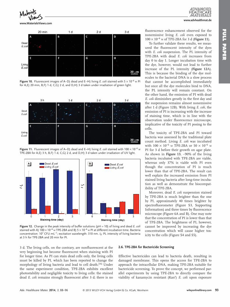
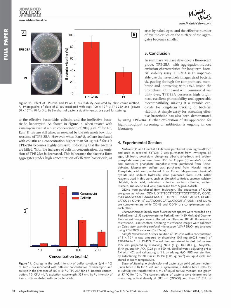
Since TPE-2BA can be applied for bacterial viability assay, we tested whether it can be used for the long-term assay, con-sidering the good photostablity of the AIE fl uorogens. [ 33 – 35 ] PI is used for side-by-side comparison. As shown in Figure 10 , although the discrimination of dead and living E. coli with PI can be achieved in 20 min of staining time, the emission of dead cells is fading away in 1 d and completely disappears after
Figure 7 . PL spectra of A) TPE-2BA in pH 10 buffer solution and B) PI in pH 7.4 buffer solution without and with 10 μ g mL − 1 of calf thymus DNA (type I) and RNA from torula yeast (type VI). Excitation wavelength: 310 nm for TPE-2BA and 535 nm for PI.
Figure 8 . Change in the peak intensity of TPE-2BA in the presence of different ODN after 1 h incubation. Concentration of ODN: 1 × 10 − 6 M ; excitation wavelength: 355 nm. I 0 : PL intensity of TPE-2BA alone.
Figure 9 . Confocal images of dead and living E. coli cells mixture stained with A) TPE-2BA and B) SYTO® 9. Excitation wavelength: 405 nm for TPE-2BA and 488 nm for Syto 9. Emission ranges: A) 452–488 and B) 523–575 nm. C) Bright-fi eld images of the bacteria. D) Image merged from panels A, B, and C.
Adv. Healthcare Mater. 2014, 3, 88–96
www.MaterialsViews.com
FULL P
APER
www.advhealthmat.de
93wileyonlinelibrary.com© 2013 WILEY-VCH Verlag GmbH & Co. KGaA, Weinheim
fl uorescence enhancement observed for the nonemissive living E. coli even exposed to 100 × 10 − 6 M of TPE-2BA for 3 d ( Figure 11 ).
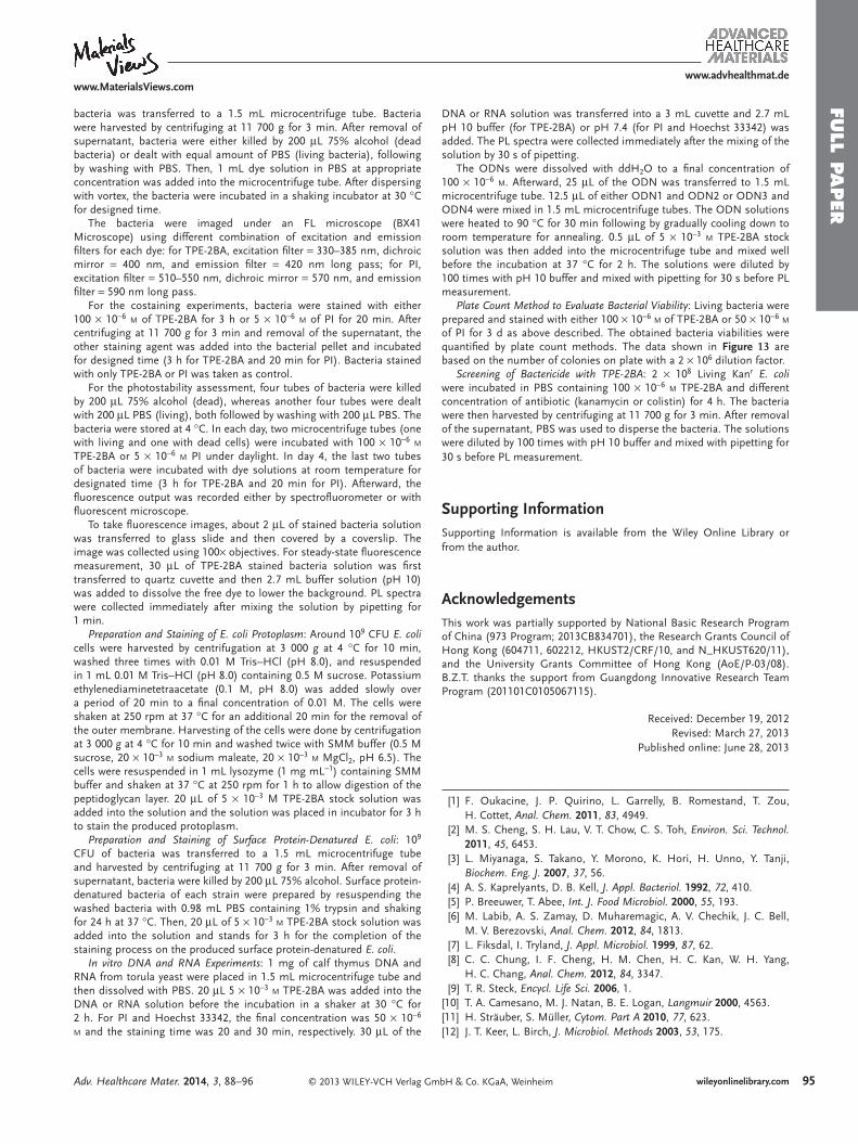
To further validate these results, we meas-ured the fl uorescent intensity of the dyes with E. coli suspension. The PL intensity of TPE-2BA with dead E. coli increases from day 0 to day 1. Longer incubation time with the dye, however, would not lead to further increase of the PL intensity ( Figure 12 A). This is because the binding of the dye mol-ecules to the bacterial DNA is a slow process that cannot be accomplished immediately but once all the dye molecules bind to DNA, the PL intensity will remain constant. On the other hand, the emission of PI with dead E. coli diminishes greatly in the fi rst day and the suspension remains almost nonemissive after 1 d (Figure 12 B). With living E. coli , the emission of PI is increasing with the increase of staining time, which is in line with the observation under fl uorescence microscope, implicative of the toxicity of PI posing to the cells.
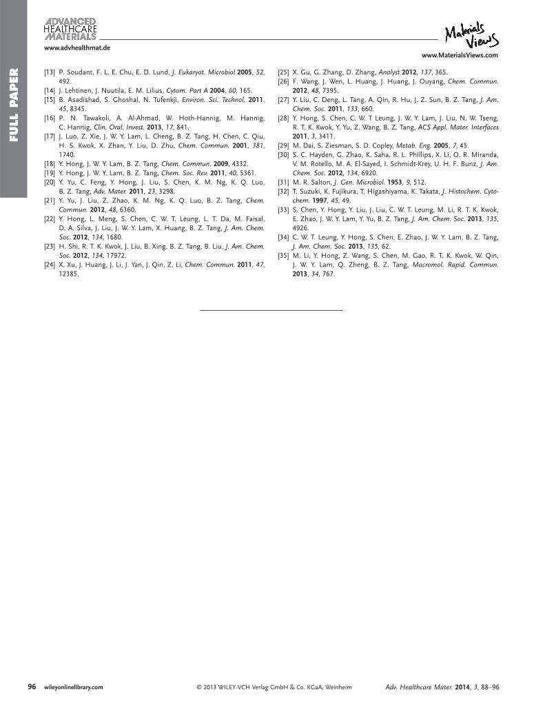
The toxicity of TPE-2BA and PI toward bacteria was assessed by the traditional plate count method. Living E. coli was incubated with 100 × 10 − 6 M TPE-2BA or 50 × 10 − 6 M PI for 3 d before their growth on agar plate. As shown in Figure 13 , ∼ 90% of the living bacteria incubated with TPE-2BA are viable, whereas only 37% is viable with PI even though the concentration of PI is much lower than that of TPE-2BA. The result can well explain the increased emission from PI stained living bacteria after long-time incuba-tion as well as demonstrate the biocompat-ibility of TPE-2BA.
Moreover, dead E. coli suspension stained by TPE-2BA is much brighter than the one by PI, approximately 40 times brighter by spectrofl uorometer (Figure S3, Supporting Information) and three times by fl uorescence microscope (Figure 6 A and B). One may note that the concentration of PI is lower than that of TPE-2BA. The brightness of PI, however, cannot be improved by increasing the dye concentration which will cause higher tox-icity to the cells (Figure S4 and S5).
2.6. TPE-2BA for Bactericide Screening
Effective bactericides can lead to bacteria death, resulting in damaged membrane. This opens the access for TPE-2BA to approach the intracellular DNA, making TPE-2BA suitable for bactericide screening. To prove the concept, we performed par-allel experiments by using TPE-2BA to directly compare the viability of kanamycin resistant (Kan r ) E. coli upon exposure
3 d. The living cells, on the contrary, are nonfl uorescent at the very beginning but become fl uorescent when staining with PI for longer time. As PI can stain dead cells only, the living cells must be killed by PI, which has been reported to change the morphology of living bacteria and lead to cell death. [ 16 ] Under the same experiment condition, TPE-2BA exhibits excellent photostability and negligible toxicity to living cells: the stained dead E. coli remains strongly fl uorescent after 3 d; there is no
Figure 10 . Fluorescent images of A − D) dead and E − H) living E. coli stained with 5 × 10 − 6 M PI for A,E) 20 min, B,F) 1 d, C,G) 2 d, and D,H) 3 d taken under irradiation of green light.
Figure 11 . Fluorescent images of A − D) dead and E − H) living E. coli stained with 100 × 10 − 6 M TPE-2BA for A,E) 3 h, B,F) 1 d, C,G) 2 d, and D,H) 3 d taken under irradiation of UV light.
Figure 12 . Change in the peak intensity of buffer solutions (pH = 10) of living and dead E. coli stained with A) 100 × 10 − 6 M TPE-2BA and B) 5 × 10 − 6 M PI at different incubation time. Bacteria concentration: 10 7 CFU mL − 1 ; excitation wavelength: 310 nm. I 0 : PL intensity of living bacteria at 3 h for TPE-2BA and 20 min for PI.
Adv. Healthcare Mater. 2014, 3, 88–96
www.MaterialsViews.com
FULL
PAPER
www.advhealthmat.de
94 wileyonlinelibrary.com © 2013 WILEY-VCH Verlag GmbH & Co. KGaA, Weinheim
seen by naked eyes, and the effective number of dye molecules on the surface of the aggre-gates becomes smaller.
3. Conclusion
In summary, we have developed a fl uorescent probe, TPE-2BA, with aggregation-induced emission characteristics for long-term bacte-rial viability assay. TPE-2BA is an imperme-able dye that selectively images dead bacteria via passing through the compromised mem-brane and interacting with DNA inside the protoplasm. Compared with commercial via-bility dyes, TPE-2BA possesses high bright-ness, excellent photostability, and appreciable biocompatibility, making it a suitable can-didate for long-term tracking of bacterial viability. A simple assay for screening effec-tive bactericide has also been demonstrated
by using TPE-2BA. Further exploration of its application for high-throughput screening of antibiotics is ongoing in our laboratory.
4. Experimental Section Materials : PI and Hoechst 33342 were purchased from Sigma–Aldrich
and used as received. SYTO® 9 was purchased from Invitrogen. LB agar, LB broth, potassium phosphate dibasic anhydrous and sodium phosphate were purchased from USB Co. Copper (II) sulfate-5 hydrate and potassium phosphate monobasic were purchased from Reidel-deHaën. Magnesium sulfate was purchased from Nacalai teque. Phosphate acid was purchased from Fisher. Magnesium chloride-6 hydrate and sodium hydroxide were purchased from BDH. Other reagents used in this work, such as dimethyl sulfoxide, sucrose, calcium chloride, boric acid, potassium chloride, sodium chloride, sodium maleate, and acetic acid were purchased from Sigma–Aldrich.
ODNs were purchased from Invitrogen. The sequences of ODNs are given as follows: ODN1: 5 ′ -TTTGCTTTGCTTTGCTTTGC-3 ′ ; ODN2: 5 ′ -GCAAAGCAAAGCAAAGCAAA-3 ′ ; ODN3: 5 ′ -ATGCATGCATGCATG-CATGC-3 ′ ; ODN4: 5 ′ -GCATGCATGCATGCATGCAT-3 ′ . ODN1 and ODN2 are complementary while ODN3 and ODN4 are complementary with each other.
Characterization : Steady-state fl uorescence spectra were recorded on a PerkinElmer LS 55 spectrometer or PerkinElmer 1420 Multiabel Counter. Fluorescent images were collected on Olympus BX 41 fl uorescence microscope. Laser confocal scanning microscope images were collected on Zeiss laser scanning confocal microscope (LSM7 DUO) and analyzed using ZEN 2009 software (Carl Zeiss).
Sample Preparations : A stock solution of TPE-2BA with a concentration of 5 × 10 − 3 M was prepared by dissolving 10.5 mg (0.025 mmol) of TPE-2BA in 5 mL DMSO. The solution was stored in dark before use. PBS was prepared by dissolving NaCl (8 g), KCl (0.2 g), Na 2 HPO 4 (1.44 g), and KH 2 PO 4 (0.24 g) in 800 mL distilled water, adjusting pH to 7.4 with HCl, and calibrating to 1 L by adding H 2 O. PBS was sterilized by autoclaving for 20 min at 15 Psi (1.05 kg cm − 2 ) on liquid cycle and stored at room temperature.
Bacterial Staining : A single colony of bacteria on solid culture medium [Luria broth (LB) for E. coli and S. epidermidis , nutrition broth (NB) for B. subtilis ] was transferred to 5 mL of liquid culture medium and grown at 37 ° C for 10 h. The concentrations of bacteria were determined by measuring optical density at 600 nm (OD600) and then 10 9 CFU of
to the effective bactericide, colistin, and the ineffective bacte-ricide, kanamycin. As shown in Figure 14 , when treated with kanamycin even at a high concentration of 200 μ g mL − 1 for 4 h, Kan r E. coli are still alive, as revealed by the extremely low fl uo-rescence of TPE-2BA. However, when Kan r E. coli are incubated with colistin at a concentration higher than 50 μ g mL − 1 for 4 h TPE-2BA becomes highly emissive, indicating that the bacteria are killed. With the increase of colistin concentration, the emis-sion of TPE-2BA is decreased. This is because the bacteria form aggregates under high concentration of effective bactericide, as
Figure 13 . Effect of TPE-2BA and PI on E. coli viability evaluated by plate count method. A) Photographs of plate of E. coli incubated with (up) 100 × 10 − 6 M TPE-2BA and (down) 50 × 10 − 6 M PI for 3 d. B) Bar chart of bacteria viability versus dye used for staining.
Figure 14 . Change in the peak intensity of buffer solutions (pH = 10) of Kan r E.coli incubated with different concentration of kanamycin and colistin in the presence of 100 × 10 − 6 M TPE-2BA for 4 h. Bacteria concen-tration: 10 7 CFU mL − 1 ; excitation wavelength: 355 nm. I 0 : PL intensity of Kan r E. coli incubated with no bacteriacide.
Adv. Healthcare Mater. 2014, 3, 88–96
www.MaterialsViews.com
FULL P
APER
www.advhealthmat.de
95wileyonlinelibrary.com© 2013 WILEY-VCH Verlag GmbH & Co. KGaA, Weinheim
DNA or RNA solution was transferred into a 3 mL cuvette and 2.7 mL pH 10 buffer (for TPE-2BA) or pH 7.4 (for PI and Hoechst 33342) was added. The PL spectra were collected immediately after the mixing of the solution by 30 s of pipetting.
The ODNs were dissolved with ddH 2 O to a fi nal concentration of 100 × 10 − 6 M . Afterward, 25 μ L of the ODN was transferred to 1.5 mL microcentrifuge tube. 12.5 μ L of either ODN1 and ODN2 or ODN3 and ODN4 were mixed in 1.5 mL microcentrifuge tubes. The ODN solutions were heated to 90 ° C for 30 min following by gradually cooling down to room temperature for annealing. 0.5 μ L of 5 × 10 − 3 M TPE-2BA stock solution was then added into the microcentrifuge tube and mixed well before the incubation at 37 ° C for 2 h. The solutions were diluted by 100 times with pH 10 buffer and mixed with pipetting for 30 s before PL measurement.
Plate Count Method to Evaluate Bacterial Viability : Living bacteria were prepared and stained with either 100 × 10 − 6 M of TPE-2BA or 50 × 10 − 6 M of PI for 3 d as above described. The obtained bacteria viabilities were quantifi ed by plate count methods. The data shown in Figure 13 are based on the number of colonies on plate with a 2 × 10 6 dilution factor.
Screening of Bactericide with TPE-2BA : 2 × 10 8 Living Kan r E. coli were incubated in PBS containing 100 × 10 − 6 M TPE-2BA and different concentration of antibiotic (kanamycin or colistin) for 4 h. The bacteria were then harvested by centrifuging at 11 700 g for 3 min. After removal of the supernatant, PBS was used to disperse the bacteria. The solutions were diluted by 100 times with pH 10 buffer and mixed with pipetting for 30 s before PL measurement.
Supporting Information Supporting Information is available from the Wiley Online Library or from the author.
Acknowledgements This work was partially supported by National Basic Research Program of China (973 Program; 2013CB834701), the Research Grants Council of Hong Kong (604711, 602212, HKUST2/CRF/10, and N_HKUST620/11), and the University Grants Committee of Hong Kong (AoE/P-03/08). B.Z.T. thanks the support from Guangdong Innovative Research Team Program (201101C0105067115).
Received: December 19, 2012 Revised: March 27, 2013
Published online: June 28, 2013
bacteria was transferred to a 1.5 mL microcentrifuge tube. Bacteria were harvested by centrifuging at 11 700 g for 3 min. After removal of supernatant, bacteria were either killed by 200 μ L 75% alcohol (dead bacteria) or dealt with equal amount of PBS (living bacteria), following by washing with PBS. Then, 1 mL dye solution in PBS at appropriate concentration was added into the microcentrifuge tube. After dispersing with vortex, the bacteria were incubated in a shaking incubator at 30 ° C for designed time.
The bacteria were imaged under an FL microscope (BX41 Microscope) using different combination of excitation and emission fi lters for each dye: for TPE-2BA, excitation fi lter = 330–385 nm, dichroic mirror = 400 nm, and emission fi lter = 420 nm long pass; for PI, excitation fi lter = 510–550 nm, dichroic mirror = 570 nm, and emission fi lter = 590 nm long pass.
For the costaining experiments, bacteria were stained with either 100 × 10 − 6 M of TPE-2BA for 3 h or 5 × 10 − 6 M of PI for 20 min. After centrifuging at 11 700 g for 3 min and removal of the supernatant, the other staining agent was added into the bacterial pellet and incubated for designed time (3 h for TPE-2BA and 20 min for PI). Bacteria stained with only TPE-2BA or PI was taken as control.
For the photostability assessment, four tubes of bacteria were killed by 200 μ L 75% alcohol (dead), whereas another four tubes were dealt with 200 μ L PBS (living), both followed by washing with 200 μ L PBS. The bacteria were stored at 4 ° C. In each day, two microcentrifuge tubes (one with living and one with dead cells) were incubated with 100 × 10 − 6 M TPE-2BA or 5 × 10 − 6 M PI under daylight. In day 4, the last two tubes of bacteria were incubated with dye solutions at room temperature for designated time (3 h for TPE-2BA and 20 min for PI). Afterward, the fl uorescence output was recorded either by spectrofl uorometer or with fl uorescent microscope.
To take fl uorescence images, about 2 μ L of stained bacteria solution was transferred to glass slide and then covered by a coverslip. The image was collected using 100 × objectives. For steady-state fl uorescence measurement, 30 μ L of TPE-2BA stained bacteria solution was fi rst transferred to quartz cuvette and then 2.7 mL buffer solution (pH 10) was added to dissolve the free dye to lower the background. PL spectra were collected immediately after mixing the solution by pipetting for 1 min.
Preparation and Staining of E. coli Protoplasm : Around 10 9 CFU E. coli cells were harvested by centrifugation at 3 000 g at 4 ° C for 10 min, washed three times with 0.01 M Tris–HCl (pH 8.0), and resuspended in 1 mL 0.01 M Tris–HCl (pH 8.0) containing 0.5 M sucrose. Potassium ethylenediaminetetraacetate (0.1 M, pH 8.0) was added slowly over a period of 20 min to a fi nal concentration of 0.01 M. The cells were shaken at 250 rpm at 37 ° C for an additional 20 min for the removal of the outer membrane. Harvesting of the cells were done by centrifugation at 3 000 g at 4 ° C for 10 min and washed twice with SMM buffer (0.5 M sucrose, 20 × 10 − 3 M sodium maleate, 20 × 10 − 3 M MgCl 2 , pH 6.5). The cells were resuspended in 1 mL lysozyme (1 mg mL − 1 ) containing SMM buffer and shaken at 37 ° C at 250 rpm for 1 h to allow digestion of the peptidoglycan layer. 20 μ L of 5 × 10 − 3 M TPE-2BA stock solution was added into the solution and the solution was placed in incubator for 3 h to stain the produced protoplasm.
Preparation and Staining of Surface Protein-Denatured E. coli : 10 9 CFU of bacteria was transferred to a 1.5 mL microcentrifuge tube and harvested by centrifuging at 11 700 g for 3 min. After removal of supernatant, bacteria were killed by 200 μ L 75% alcohol. Surface protein-denatured bacteria of each strain were prepared by resuspending the washed bacteria with 0.98 mL PBS containing 1% trypsin and shaking for 24 h at 37 ° C. Then, 20 μ L of 5 × 10 − 3 M TPE-2BA stock solution was added into the solution and stands for 3 h for the completion of the staining process on the produced surface protein-denatured E. coli .
In vitro DNA and RNA Experiments : 1 mg of calf thymus DNA and RNA from torula yeast were placed in 1.5 mL microcentrifuge tube and then dissolved with PBS. 20 μ L 5 × 10 − 3 M TPE-2BA was added into the DNA or RNA solution before the incubation in a shaker at 30 ° C for 2 h. For PI and Hoechst 33342, the fi nal concentration was 50 × 10 − 6 M and the staining time was 20 and 30 min, respectively. 30 μ L of the
[ 1 ] F. Oukacine , J. P. Quirino , L. Garrelly , B. Romestand , T. Zou , H. Cottet , Anal. Chem. 2011 , 83 , 4949 .
[ 2 ] M. S. Cheng , S. H. Lau , V. T. Chow , C. S. Toh , Environ. Sci. Technol. 2011 , 45 , 6453 .
[ 3 ] L. Miyanaga , S. Takano , Y. Morono , K. Hori , H. Unno , Y. Tanji , Biochem. Eng. J. 2007 , 37 , 56 .
[ 4 ] A. S. Kaprelyants , D. B. Kell , J. Appl. Bacteriol. 1992 , 72 , 410 . [ 5 ] P. Breeuwer , T. Abee , Int. J. Food Microbiol. 2000 , 55 , 193 . [ 6 ] M. Labib , A. S. Zamay , D. Muharemagic , A. V. Chechik , J. C. Bell ,
M. V. Berezovski , Anal. Chem. 2012 , 84 , 1813 . [ 7 ] L. Fiksdal , I. Tryland , J. Appl. Microbiol. 1999 , 87 , 62 . [ 8 ] C. C. Chung , I. F. Cheng , H. M. Chen , H. C. Kan , W. H. Yang ,
H. C. Chang , Anal. Chem. 2012 , 84 , 3347 . [ 9 ] T. R. Steck , Encycl. Life Sci. 2006 , 1 . [ 10 ] T. A. Camesano , M. J. Natan , B. E. Logan , Langmuir 2000 , 4563 . [ 11 ] H. Sträuber , S. Müller , Cytom. Part A 2010 , 77 , 623 . [ 12 ] J. T. Keer , L. Birch , J. Microbiol. Methods 2003 , 53 , 175 .
Adv. Healthcare Mater. 2014, 3, 88–96
www.MaterialsViews.com
FULL
PAPER
www.advhealthmat.de
96 wileyonlinelibrary.com © 2013 WILEY-VCH Verlag GmbH & Co. KGaA, Weinheim
[ 25 ] X. Gu , G. Zhang , D. Zhang , Analyst 2012 , 137 , 365 . [ 26 ] F. Wang , J. Wen , L. Huang , J. Huang , J. Ouyang , Chem. Commun.
2012 , 48 , 7395 . [ 27 ] Y. Liu , C. Deng , L. Tang , A. Qin , R. Hu , J. Z. Sun , B. Z. Tang , J. Am.
Chem. Soc. 2011 , 133 , 660 . [ 28 ] Y. Hong , S. Chen , C. W. T Leung , J. W. Y. Lam , J. Liu , N. W. Tseng ,
R. T. K. Kwok , Y. Yu , Z. Wang , B. Z. Tang , ACS Appl. Mater. Interfaces 2011 , 3 , 3411 .
[ 29 ] M. Dai , S. Ziesman , S. D. Copley , Metab. Eng. 2005 , 7 , 45 . [ 30 ] S. C. Hayden , G. Zhao , K. Saha , R. L. Phillips , X. Li , O. R. Miranda ,
V. M. Rotello , M. A. El-Sayed , I. Schmidt-Krey , U. H. F. Bunz , J. Am. Chem. Soc. 2012 , 134 , 6920 .
[ 31 ] M. R. Salton , J. Gen. Microbiol. 1953 , 9 , 512 . [ 32 ] T. Suzuki , K. Fujikura , T. Higashiyama , K. Takata , J. Histochem. Cyto-
chem. 1997 , 45 , 49 . [ 33 ] S. Chen , Y. Hong , Y. Liu , J. Liu , C. W. T. Leung , M. Li , R. T. K. Kwok ,
E. Zhao , J. W. Y. Lam , Y. Yu , B. Z. Tang , J. Am. Chem. Soc. 2013 , 135 , 4926 .
[ 34 ] C. W. T. Leung , Y. Hong , S. Chen , E. Zhao , J. W. Y. Lam , B. Z. Tang , J. Am. Chem. Soc. 2013 , 135 , 62 .
[ 35 ] M. Li , Y. Hong , Z. Wang , S. Chen , M. Gao , R. T. K. Kwok , W. Qin , J. W. Y. Lam , Q. Zheng , B. Z. Tang , Macromol. Rapid. Commun. 2013 , 34 , 767 .
[ 13 ] P. Soudant , F. L. E. Chu , E. D. Lund , J. Eukaryot. Microbiol 2005 , 52 , 492 .
[ 14 ] J. Lehtinen , J. Nuutila , E. M. Lilius , Cytom. Part A 2004 , 60 , 165 . [ 15 ] B. Asadishad , S. Ghoshal , N. Tufenkji , Environ. Sci. Technol. 2011 ,
45 , 8345 . [ 16 ] P. N. Tawakoli , A. Al-Ahmad , W. Hoth-Hannig , M. Hannig ,
C. Hannig , Clin. Oral. Invest. 20 13 , 17 , 841 . [ 17 ] J. Luo , Z. Xie , J. W. Y. Lam , L. Cheng , B. Z. Tang , H. Chen , C. Qiu ,
H. S. Kwok , X. Zhan , Y. Liu , D. Zhu , Chem. Commun. 2001 , 381 , 1740 .
[ 18 ] Y. Hong , J. W. Y. Lam , B. Z. Tang , Chem. Commun. 2009 , 4332 . [ 19 ] Y. Hong , J. W. Y. Lam , B. Z. Tang , Chem. Soc. Rev. 2011 , 40 , 5361 . [ 20 ] Y. Yu , C. Feng , Y. Hong , J. Liu , S. Chen , K. M. Ng , K. Q. Luo ,
B. Z. Tang , Adv. Mater. 2011 , 23 , 3298 . [ 21 ] Y. Yu , J. Liu , Z. Zhao , K. M. Ng , K. Q. Luo , B. Z. Tang , Chem.
Commun. 2012 , 48 , 6360 . [ 22 ] Y. Hong , L. Meng , S. Chen , C. W. T. Leung , L. T. Da , M. Faisal ,
D. A. Silva , J. Liu , J. W. Y. Lam , X. Huang , B. Z. Tang , J. Am. Chem. Soc. 2012 , 134 , 1680 .
[ 23 ] H. Shi , R. T. K. Kwok , J. Liu , B. Xing , B. Z. Tang , B. Liu , J. Am. Chem. Soc. 2012 , 134 , 17972 .
[ 24 ] X. Xu , J. Huang , J. Li , J. Yan , J. Qin , Z. Li , Chem. Commun. 2011 , 47 , 1 2385 .
Adv. Healthcare Mater. 2014, 3, 88–96