Embed Size (px)
Citation preview

Functional Analyses of Natural Variation in Sp1 Binding Sites of aTATA-Less Promoter
Jeff A. Segal,1,* J. Lynn Barnett, 2 Douglas L. Crawford2
1 Department of Organismal Biology and Anatomy, University of Chicago, Chicago, IL, USA2 Division of Molecular Biology and Biochemistry, 5007 Rockhill Rd., University of Missouri, Kansas City, MO 64110, USA
Received: 12 February 1999 / Accepted: 2 June 1999
Abstract. Within the lactate dehydrogenase-B (LdhB)proximal promoter is a region with multiple in vivo foot-printed sites that resembles the binding site for the tran-scription factor SP1. Like many sequences that regulatetranscription rate, these Sp1 binding sites are well con-served among species of the teleost fishFundulus.Theonly exception is in the northern population ofF. het-eroclitus,where there are many changes in the Sp1 bind-ing sites. These changes affect footprinting patterns,measures of promoter strength, and are associated withthe adaptive increase inLdh-B transcription rates. Re-ported here is data that demonstrates thatFundulushe-patocyctes have an SP1-like protein; in comparison tohuman SP1 protein, it has similar specificity and size anda greater affinity for the consensus Sp1 site. ThisFun-dulushepatocyte SP1-like protein as well as the humanSP1 protein binds theLdh-B Sp1 sites. Sequence varia-tion in the northern Sp1 region eliminates the “preferred”Sp1 binding site, yet these northern Sp1 sites have sig-nificantly greater affinity for the SP1 protein than eitherthe Sp1 sites from southernF. heteroclitus(∼ 1.6-fold) orthe consensus Sp1 site (GGGCGG;∼ 1.8-fold). Further-more, theLdh-B Sp1 sites also bind non-SP1 proteins,and the extent of binding is affected by the sequencevariation in the proximal promoter. These data suggestthat natural variation in Sp1 sites affect binding of tran-
scription factors and may effect a modest change in tran-scription rates.
Key Words: Binding affinities — phylogeny — evo-lutionary adaptation — transcription
Introduction
Sequence variation is a feature of almost all species (Li1997) and knowledge of how this variation affects anorganism’s biochemical, physiological, and cellularfunction will be necessary to interpret the significance ofthe increasing number of genomic sequences being de-scribed. For example, natural sequence variation in pro-moter regions could produce important phenotypic dif-ferences. The unexpected diversity in proximal promotersequences has led to the suggestion that these proximalregions contribute to differential regulation of transcrip-tion or subtle changes in transcription rate (O’Shea-Greenfield and Smale 1992; Zenzie-Gregory et al.1993a, 1993b; Ernst and Smale 1995; Verrijzer et al.1995; Goodrich et al. 1996). Typically, proximal pro-moters have a TATA sequence that binds TFIID, an es-sential factor for all mRNA transcription (Roeder 1991;Smale 1994). Nevertheless, many promoters lack aTATA sequence (e.g., approximately 50% of transcribedgenes inDrosophila [Arkhipova, 1995]). These TATA-less promoters require Sp1 binding sites (consensus se-quence: GGGCGG; Dynan and Tjian 1983) for signifi-cant activity (Azizkhan et al. 1993), and the degree of
*Present address:Lilly Research Laboratories, CNS Discover, Bldg.48, Drop Code 0510, Indianapolis, IN 46285-0510, USACorrespondence to:D.L. Crawford;e-mail: [email protected]
J Mol Evol (1999) 49:736–749
© Springer-Verlag New York Inc. 1999

activation from Sp1 tends to be stronger in the context ofTATA-less promoters than TATA-containing promoters(Colgan and Manley 1995). Activation of TATA-lesspromoters by SP1 may involve SP1 recruitment of TFIIDto TATA-less promoters (Pugh and Tjian 1991; Wiley etal. 1992). Furthermore, in many of these promoters, SP1binding is intimately involved in the determination of thetranscription start site or sites (Jolliff et al. 1991; Kollmaret al. 1994; Lu et al. 1994; Boam et al. 1995). Thesefacts, together with the observation that a disproportion-ate number of the TATA-less promoters described are inconstitutively regulated genes (Azizkhan et al. 1993),suggest that variation in SP1 binding is one means bywhich constitutive transcription is modified.
For lactate dehydrogenase-B (Ldh-B), the constitutivetranscription rate is twofold greater in hepatocytes fromnorthern versus southern populations of the teleost fishFundulus heteroclitus(Crawford and Powers 1992). Thisdifference in transcription rate results in a greater LDH-B4 enzyme concentration that compensates for the coldertemperatures experienced by the northern population(Crawford and Powers 1989, 1992; Segal and Crawford1994). The variation inLdh-B expression is adaptive(i.e., evolved by natural selection; Pierce and Crawford1997a) and thus is biologically important because naturalselection acts only on traits that affect longevity, repro-ductive fitness, or survival (Endler 1986).
Much of the adaptive variation inLdh-Bexpression isassociated with sequence variation in the proximal pro-moter. This sequence variation affects in vivo and invitro footprinting patterns and levels of gene expressionin transient cell transfection (Segal et al. 1996; Crawfordet al. 1999b). Specifically, proximal promoters isolatedfrom northern individuals consistently have greater pro-moter strength than promoters isolated from southernindividuals. TheLdh-Bgene proximal promoter (Fig. 1)is TATA-less with multiple transcriptional start sites, Inr(initiation of transcription) sequences associated with thestart sites and a cluster of binding sites similar to thoserecognized by the ubiquitous SP1 protein (i.e., Sp1 sitesthat match at least five of the six nucleotides of theconsensus Sp1 site, Segal et al. 1996). This Sp1 regionconsists of multiple overlapping perfect and imperfectSp1 sites that promote transcription in transient transfec-tion assays (Segal et al. 1996). Similar to many transcrip-tional regulatory elements (e.g., Gumucio et al. 1994,1996; Hu et al. 1995) the Sp1 binding sites are highlyconserved among species ofFundulus(Fig. 1). However,a comparison between populations ofF. heteroclitusre-veals significant sequence variation: the Sp1 sites fromnorthern populations (northern Sp1 sites) have manyunique derived nucleotides that are not found in the Sp1sites from southern populations (southern Sp1 sites) orother species (Fig. 1; Crawford et al. 1999b). Establishedphylogenies ofFundulusthat have been generated frommorphological traits, protein isoforms, and DNA se-
quences have all groupedF. heteroclituspopulations intoone species and divided the other taxa into much olderspecies that diverged several million years ago (Wiley1986; Cashner et al. 1992; Bernardi and Powers 1995;Crawford et al. 1999b). However, the phylogenetic treeestablished using the sequences from Sp1 regions isstrikingly different: only the northernF. heteroclituspopulation is a distinct group, and the southern popula-tion and the other species all group together. This dis-parity indicates that the Sp1 sites withinF. heteroclitushave recently accumulated many more substitutions incomparison to homologous Sp1 sites among much olderspecies. Importantly, the pattern of nucleotide substitu-tions in the Sp1 sites (and other protein:DNA bindingregions in the proximal promoter) is nonrandom and in-dicates that theLdh-Bproximal promoter has evolved bynatural selection, specifically directional selection(Crawford et al. 1999b). Thus, the natural sequencevariation in theLdh-B proximal promoter appears to bebiologically important.
To better understand how the natural sequence varia-tion of the Sp1 region affects transcription, we examinedthe nature of the transcription factors that bind to thisregion of theLdh-Bproximal promoter and analyzed thefunctional consequences of this variation. The data dem-onstrate that (1) expression of an SP1 protein is neces-sary for transcription from theLdh-Bproximal promoter,(2) purified human SP1 protein binds to theLdh-B Sp1sites, (3) there is aFundulusSP1–like protein in hepa-tocyte nuclear extracts from both populations that bindsto the Sp1 sites, (4) there are non-SP1 proteins in thesenuclear extracts that bind these sequences, (5) theFun-dulusSP1–like proteins have greater affinity for the con-sensus Sp1 site than the human SP1, and (6) the variationin Sp1 sites results in different Kd’s for the human Sp1protein and differential binding ofFundulus non-SP1factors. These data demonstrate that the naturally occur-ring variation in Sp1 sites alters the binding of transcrip-tion factors and thus may contribute to the difference inconstitutiveLdh-B transcription rate in vivo.
Materials and Methods
Organisms
Funduluswere captured from wild populations (northern and southernF. heteroclitus:Wiscassett, ME, and Sapelo Is. Dock, GA, respec-tively; F. grandis.Panacea, FL;F. diaphanus,Green Banks, NJ) andacclimated. The acclimation regime was initiated by a 2-week feedingperiod, a 6-week pseudo-winter (6°C, 16:8 light:dark cycle), followedby minimum of 6 weeks at 20°C, 14:10 light:dark cycle. During thistime all species came into reproductive condition and spawned. Thereproductive tissues were in regression in all species when assayed.This acclimation regime is sufficient to remove environmentally in-duced physiological difference (Pierce and Crawford 1997b).
737

Plasmids
The Ldh-B proximal promoter is depicted in Fig. 1A. Two proximalpromoters with Sp1 sites, TCC repeats and Inrs (STI) were designed,which represent either the typical northern or southern proximal pro-moter (see Crawford et al. 1999b). These proximal promoters wereligated into a luciferase reporter plasmid (pGL3, Promega). The SP1expression vector (pPacSp1; Courey and Tjian 1988; Kadonaga et al.1988) was provided by Dr. R. Tjian (University of California, Berke-ley).
Transfections
Drosophilacell lines were used to determine the role of SP1 inLdh-Bproximal promoter activity (SL2 ATCC, Rockville, MD; Fig. 2). Thesecell lines lack SP1 proteins (Courey and Tjian 1988; Hagen et al. 1994).Transfections were performed as in Segal et al. (1996). Promoter ac-tivity is expressed as net relative light units (luciferase activity minusthe negative control) normalized for transfection activity (net B-galatosidase activity, B-galatosidase activity minus the negative con-trol). Promoter activity was further standardized by dividing by a con-stant: the mean activity for southern STI proximal promoter. This doesnot alter the statistical variance and makes it simpler to compare rela-tive activity in different cell lines.
Oligonucleotides
The consensus Sp1 double-stranded oligonucleotide was purchasedfrom Promega (sequence delineated in Fig. 1D) and labeled directly.All other single-stranded oligonucleotides were purchased from Op-eron (sequences indicated in Fig. 1D: SN, SS1, and SS2; Fig. 6: SNgaand SNgc; Results section: Sp1N) and purified by polyacrylamide gelelectrophoresis. Equimolar quantities of complementary strands wereannealed by heating to 90°C for 10 min followed by slow cooling to25°C overnight.
For mobility shift assays, all oligonucleotides were end-labeledwith T4 polynucleotide kinase (PNK; New England Biolabs) in a 10mlreaction containing 2.5 pmol of double-stranded annealed oligonucleo-tide, 1.0 ml of PNK 10× buffer (New England Biolabs), 1.0ml ofgamma P32 ATP (3,000 Ci/mmol; Dupont), 5.0ml of H2O, and 1mlPNK. The reaction was incubated at 37°C for 20 min and stopped bythe addition of 1.0ml of 0.5 M EDTA. The volume was brought up to100ml with the addition of 1X TE, and the unincorporated radionucleo-tide was removed by applying it to a G25 column equilibrated with 1XTE.
Mobility Shift Assay
All binding reactions were performed in a final volume of 10ml andcontained 2ml of 5X binding buffer (5XBB: 20% glycerol, 5 mMMgCl2, 2.5 mM EDTA, 2.5 mM DTT, 250 mM NaCl, 50 mM Tris-HClpH 7.5, and 0.25mg/ml poly dI-dC) and 1ml 5 mg/ml BSA. Experi-ments typically used either 0.5 footprinting units of purified human Sp1protein (rhSP1, expressed in HeLa cells; Promega) or approximately1–3mg of northern or southern hepatocyte nuclear extract protein (de-scribed below) per reaction. The protein, binding buffer, BSA, and H2Oto 9 ml was incubated on ice for 10 min before the addition of 25 fmolof freshly labeled oligonucleotide. After the addition of labeled oligo-nucleotide, the reaction was incubated for 20 more min on ice, followedby the addition of 1ml of 10X loading dye (250 mM Tris-HCl pH 7.5,0.2% bromophenol blue, 0.2% xylene cyanol, and 40% glycerol). Forexperiments that included an excess unlabeled competitor oligonucleo-tide, the competitor was added before the addition of labeled oligo-
nucleotide. For supershift experiments, antibody was incubated withprotein, binding buffer, and BSA for 30 min instead of 10 min beforethe addition of labeled oligonucleotide. All reactions were separated byelectrophoresis at 4°C in a 4% (80:1 acrylamide to bisacrylamide)nondenaturing gel with 0.5X TBE and 0.5X TBE running buffer. Thegel was subsequently dried and subjected to autoradiography overnightwith an intensifying screen.
Nuclear Extracts
Hepatocyte nuclear extracts were prepared from livers of northern andsouthernF. heteroclitusaccording to Gorski et al. (1986) with modi-fications. All the procedures were carried out in a cold room; all buff-ers, solutions, and rotors were cooled to 4°C before use. Livers fromthree to five northern or two to four southernF. heteroclitusindividuals(typically 500 mg of tissue) were excised and homogenized with fourstrokes of a motor-driven Teflon-glass homogenizer in 5.5 ml of ho-mogenization buffer (2.0M sucrose, 10 mM Hepes, pH 7.6, 0.5 mMspermidine, 0.15 mM spermine, 1.0 mM EDTA, and 15 mM KCl)supplemented just before use with 1% nonfat milk, 1 mM DTT, and 1Xprotease inhibitor mix (0.5 mM PMSF, 2mg/ml aprotinin, 2mg/mlleupeptin, and 2mg/ml pepstatin). This homogenate was filteredthrough glass wool to remove solid material and mixed with an equalvolume of homogenization buffer. The sample was then layered over 4ml of sucrose cushion (same as homogenization buffer, but with 2.2Msucrose and without the lowfat milk) and centrifuged for 45 min at24,600 rpm in a SW41 rotor (75,000g; Beckman). Each nuclear pelletwas resuspended by gentle pipetting in 2.5 ml of nuclear lysis buffer(10 mM Hepes, pH 7.6, 100 mM KCl, 0.1 mM EDTA, 10% glycerol,and 3 mM MgCl2), supplemented just prior to use with 1 mM DTT and1X protease inhibitor mix. 375ml of 3 M (NH4)2SO4 was added to eachsample, followed by gentle rocking for 30 min at 4°C. The sampleswere then centrifuged for 60 min at 35,000 rpm in a 70.1 Ti rotor(90,000g; Beckman) to pellet the chromatin. Two ml of the superna-tant were transferred to a new tube, and 0.66 g of solid (NH4)2SO4
(0.33 g/ml) was slowly dissolved into the sample. After 45 min at 4°C,the sample was centrifuged for 60 min at 24,600 rpm in the SW41 rotor(75,000g) to pellet the protein precipitate. Each protein pellet wasresuspended at 4°C in 60ml of nuclear dialysis buffer (25 mM Hepes,pH 7.6, 0.1 mM EDTA, 40 mM KCl, and 10% glycerol), supplementedjust prior to use with 1 mM DTT and 1X protease inhibitor mix. Thesamples were then dialyzed twice for 90 min at 4°C against 150 ml ofnuclear dialysis buffer supplemented with 1 mM DTT. The extractswere then centrifuged in a microcentrifuge (16,000g) at 4°C for 5 minto clarify. Ten microliters of the supernatant were removed for proteinassay determination, and then an equal volume of 5 mg/ml BSA (innuclear dialysis buffer supplemented with 1 mM DTT and 1X proteaseinhibitor mix) was added to the remaining 50ml. Aliquots were quick-frozen in liquid nitrogen.
Binding Affinities
Mobility shift assays were used to separate bound from free oligo.Mobility shifts, used for the nonlinear analyses of quantitative bindingrates, were done as described above. For each experiment, nine differ-ent amounts of oligonucleotides were used: 500 fm, 400 fm, 335 fm,270 fm, 205 fm, 140 fm, 75 fm, 50 fm, and 30 fm; in a 10-ml reaction,yielding 50 to 3 nM of oligonucleotides. Binding reactions containedeither 0.013 footprinting units of rhSP1 or 0.5mg of northern or south-ern nuclear extract. In all cases, these conditions gave a range of proteinto oligonucleotide ratio that approached protein binding saturation atthe highest oligonucleotide concentration. After electrophoresis as de-scribed above, the gel was dried and the radioactivity corresponding tobound and free oligonucleotide was quantified with a phosphorimager(Molecular Dynamics). Nonlinear regression analyses of binding af-
738
finities were generated from the data for each experimental gel and theKd determined with the use of GraphPad Prism 2.0 software (Graph-Pad). Each oligonucleotide:protein interaction was tested in at leastthree separate experiments, and the mean Kd was calculated from theseseparate measures. Statistical significance of differences between dif-ferent oligonucleotide Kds was determined by analysis of variance(ANOVA) using Minitab.
Results
Sequence variation in Sp1 region
The structure of theF. heteroclitus Ldh-Bproximal pro-moter and the variation in Sp1 sites are shown in Fig. 1.Previous work has demonstrated that theLdh-BSp1 sitescontribute significantly to promoter activity (Segal et al.1996). These Sp1 sites consist of a number of heteroge-neous sequences that match at least five of the sixnucleotides for the canonical Sp1 binding site(GGGCGG; Dynan and Tjian 1983). All of these se-quences are the reverse complement relative to the cod-ing strand. However, in either orientation Sp1 sites willbind SP1 protein and activate transcription (Kadonaga etal. 1987). The Sp1 sequence region can be subdividedinto two general subregions: the 58 GA region (GG-GAGG/CCTCCC) and the 38 GC region. In the GC sub-region from the southern population ofF. heteroclitusand in otherFundulusspecies there are perfect (six of sixnucleotide match) Sp1 sites. In contrast, from northernpopulations ofF. heteroclitusthere are no “perfect” Sp1binding sites because of a substitution of a G for a C (#5,Fig. 1B) that changes GGGCGG to GGCCGG. Withinthe GA subregion, all northern sequences have four po-tential Sp1 binding sites (i.e., five of six match; see Fig.1D) while southernF. heteroclitusand otherFundulusspecies have only two or three potential Sp1 bindingsites. This difference is due to a unique insertion in thenorthern population of an extra C (#1, Fig. 1) or deletionsin this region (#4 CCT, Fig. 1). The other major differ-ence specific to the northern population is the deletion ofGGCCA (Fig. 1) that eliminates a Sp1 site (five of sixmatch) and decreases the distance to the TCC repeat (theputative TFIID binding site; Segal et al. 1996).
SP1 Protein Activates Transcription
The DrosophilaSL2 cell line (lacking SP1 protein) wasused to determine if SP1 transactivation is required forLdh-Bproximal promoter function.Ldh-BSTI proximalpromoters (Sp1, TCC, and Inr, Fig. 1A) were transfectedwith or without a human SP1 expression vector(pPacSp1). Promoter strength of a STI proximal pro-moter is highly correlated with the amount of human SP1expression vector that is contransfected (r2 4 0.969; Fig.2A). Moreover, expression of SP1 (by cotransfection ofpPacSp1) with northern or southern STI promoters sig-
nificantly increases promoter strength (Fig. 2B). For thenorthern promoter, this increase in expression waslargely dependent on the presence of the Sp1 sites (com-pare STI north with TI north in the presence of SP1protein). Surprisingly, this was not the case for the south-ern proximal promoter: SP1 expression increases boththe southern TI and STI promoter activities equally.These results suggest that the southern Sp1 sites are notimportant for mediating Sp1 transactivation. However,transfection into fish cell lines has demonstrated thatboth northern and southern promoter activities are criti-cally dependent on the Sp1 sites (Segal et al. 1996). Theonly difference between the northern and southern TIpromoters is that the northern promoter has a single T toC transition among the TCC repeats (Crawford et al.1999b). A possible explanation for these disparate resultsis that significant overexpression of exogenous SP1 pro-tein in the SL2 cell lines allows for SP1 binding andtransactivation from southern TI promoters, whereas theT to C transition in the northern promoters precludes this.
Northern and Southern Sp1 Regions Bind a HumanRecombinant SP1 Protein
To investigate whether SP1 proteins interacts with theLdh-BSp1 sites and whether sequence variation betweennorthern and southern promoter types influences suchinteractions, oligonucleotides were designed for use inmobility shift assays (Fig. 1D). Three oligonucleotideswere designed based on theLdh-B Sp1 sites found innorthern or southern individuals: a northern sequence(SN) and two southern sequences (SS1 and SS2; Fig.1D). These threeLdh-Boligonucleotides reflect the typeand extent of variability found in natural populations(Fig. 1B; Crawford et al. 1999b). Additionally, an oligo-nucleotide that contains a single, perfect Sp1 site wasutilized (Sp1; Promega).
As a first approach to determine whether SP1 binds totheLdh-BSp1 sites, mobility shift assays were done witha recombinant human SP1 protein (rhSP1 expressed inHeLa cells; Promega) in the presence of 0.05 mg/ml polydI-dC. The results from this experiment demonstrate thatrhSP1 binds specifically to the Sp1, SN, SS1, and SS2oligonucleotides (Fig. 3). Specificity of binding is dem-onstrated by the reduction of mobility shift with 30- to40-fold excess of Sp1 consensus oligo. The Sp1 oligobinds a single rhSP1 whereas the threeLdh-B promoterregions appear to be capable of binding more than onerhSP1 protein (compare lane 2 to lanes 8, 14, and 20, Fig.3). This pattern reflects the greater number of Sp1 bind-ing sites that occur in these oligonucleotides.
Northern and Southern Hepatocyte Nuclear ExtractsContain an SP1-Like Protein
Whether the Sp1 sites bind SP1 protein in vivo is depen-dent initially on whetherFundulushepatocytes contain
739

Fig. 1. TheFundulus Ldh-Bproximal promoter and natural sequencevariation for Sp1 region.A TheLdh-Bproximal promoter has Sp1 sites,is TATA-less, and has TCC repeats instead of a TATA box, and hastwo initiators (Inr). The two clusters of transcriptional start sites areindicated by large (major start sites) and small (minor start sites) ar-rows.B Sp1 region from three species ofFundulus(F. heteroclitus, F.grandis,andF. diaphanus). N and S refer to the northern and southernpopulations ofF. heteroclitus,respectively. Fg and Fd refer to theothertwo species (F. grandisandF. diaphanus,respectively). Numbersand lines under sequences are traits that are consistently different for
the maximum parsimony tree below (Fig. 1C, i.e., consistency index,CI, 4 1). Numbers 2 and 4 refer to deletions of the nucleotides CCT.Number 6 refers to the deletions of nucleotides GGCCA. Sequencescontain four extra nucleotides at both 58 and 38 end that do not bindprotein in vivo. Sequences were aligned with Clustal W in MacVector6.0 (Oxford Molecular) and subsequently altered to minimize the num-ber of gaps.C Evolutionary relationships for Sp1 sites (above, 1B).Maximum parsimony tree for Sp1 region (PAUP 3.1; Swofford 1989)and rooted withF. diaphanus.Tree has significant structure (g40.824057;p > 0.01; Huelsenbeck and Hillis 1992), and branch leading to
740

SP1 or a SP1-like protein (SP1-like: a SP1 protein withsimilar specificity and transcriptional activation). Hu-mans, rats, and mice all express SP1 protein ubiqui-tously, however,Drosophila does not have such a pro-tein. The extent to which other vertebrates, such as fish,have SP1 proteins is unknown. To address this question,hepatocyte nuclear extracts were made from the livers ofnorthern or southernF. heteroclitusand used in mobilityshift assays with the consensus Sp1 oligonucleotide (Fig.4). There is a protein present in both northern and south-ern hepatocyte nuclear extracts that binds specifically tothe consensus Sp1 site (lanes 2 and 12, Fig. 4). Thespecificity of binding is demonstrated by the ability ofexcess unlabeled Sp1 oligonucleotide to outcompete theshifted band (lanes 3–5 and 13–15, Fig. 4) and the in-ability of (1) a nonspecific unlabeled oligonucleotide(containing the CREB transcription factor binding site;lanes 9 and 19, Fig. 4); (2) a single-stranded oligonucleo-tide (lanes 10 and 20, Fig. 4); and (3) over 500-fold massexcess of herring sperm DNA or poly dI-dC to outcom-pete the shifted band. Furthermore, an excess of unla-beled SN, SS1, or SS2 is capable of outcompeting thisshifted band (lanes 6–8 and 16–18, Fig. 4). The shiftedoligo is composed of two bands of nearly identical mo-bility and is comparable to the pattern seen in mobilityshifts with mammalian extract preparations. These twobands are typical of mobility shifts using cellularly de-rived SP1 due to post-translational modifications of theprotein (Jackson and Tjian 1988; Jackson et al. 1993).Neither the northern nor the southern SP1-like proteininteracts with the human Sp1-antibody (targeted toamino acids 436 to 454 of the human SP1 protein, SantaCruz Biotechnology; data not shown). However, this pat-tern of two closely migrating bands, the absolute mobil-ity of the doublet (migrating with a mobility identical torhSP1) and the specificity of binding to the Sp1 consen-sus sequence together strongly indicate that this factor isa FundulusSP1 or SP1-like protein.
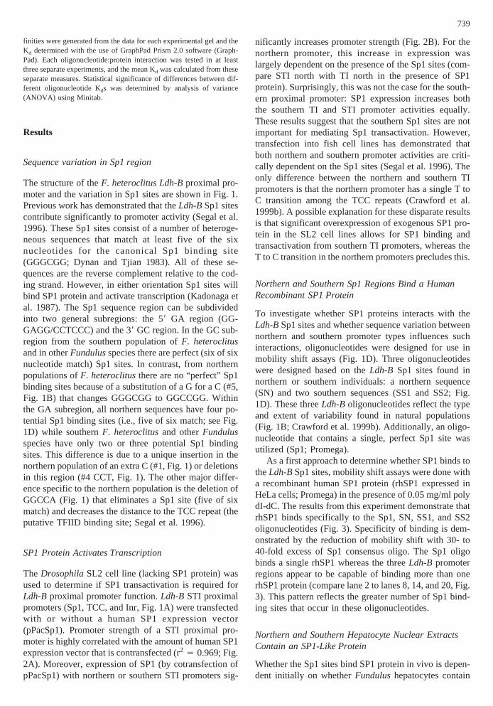
TheFundulusSP1-like protein binds to SN, SS1, andSS2, however, non-SP1 factors bind as well. The effec-tive competition of SN, SS1, and SS2 against the Sp1oligo (lanes 6–8 and 16–18, Fig. 4) indicates that theseoligos can bind theFundulusSP1-like protein. To di-rectly determine the interactions that occur betweenthese oligos and the SP1-like protein in the northern andsouthern nuclear extracts, mobility shifts with theLdh-BSp1 oligos and theFundulusextracts were performedwith various unlabeled competitors (Fig. 5). It is appar-
ent that several complexes form between SN and thenorthern extract (lane 2, Fig. 5A). The slowest migratingband represents the bound SP1-like protein as increasingconcentrations of the unlabeled Sp1 oligonucleotide spe-cifically outcompetes just this band (lanes 4–6, Fig. 5A).Additionally, the band is a doublet and migrates at the
Fig. 2. Promoter strength is dependent on SP1 expression. Promoterstrength (measured as the amount of luciferase expressed relative to theamount of cotransfected CMVb-Gal) was determined inDrosophilacells that lack SP1 expression (Courey and Tjian 1988; Hagen et al.1994).A Relative promoter strength with different amounts (0, 0.25, 5,1, and 2mg per flask) of human SP1 expression vector (pPacSp1) wascotransfected with STI-luciferase (Sp1, TCC repeat and Inr, Fig. 1A)and CMV b-gal. For each concentration of pPacSp1 cotransfected,there are three transfections measured in triplicate. Means for all threetransfections are displayed.B STI and TI (lacking Sp1 like sequences)proximal promoters transfected intoDrosophila cell lines with andwithout human SP1 expression vector. N and S refer to northern orsouthern proximal promoters, respectively.
<
Fig. 1. Continued. F. heteroclitusnorth has 100% bootstrap support(n 4 1,000). Alldeletions or insertions, regardless of size, are scored asa single character in the phylogenetic analysis. Listed between nodes,“Substitutions” are changes that have a CI of 1.0. Numbers refer tochanges underlined in the sequence alignment (Fig. 1B). Changes withCI < 1.0 that affect tree topology are not listed.D denotes a deletion orgap. Notice, except forF. heteroclitusnorth, none of the other taxa canbe separated from each other.D Oligos used in gel shift assays. Thesequences of the northern (SN oligonucleotide) and two southern
(SS1 and SS2 oligonucleotides)Sp1 regions are indicated. Sequencesmatching six of the six nucleotides of the consensus Sp1 site are indi-cated by horizontal lines with a shaded oval, while sequences matchingfive of the six nucleotides of the core consensus Sp1 site are indicatedby horizontal lines. Vertical lines are sites with deleted sequencesoutlined in boxes (insertion #1 or deletion #6, relative to northernsequence). Shaded boxes, #5, are substitutions of G for C (S→ N) thatremoves the consensus Sp1 site. Deletions #2 and #3 within the south-ern Sp1 sites are not shown.
741

same mobility as the SP1 complex presented in Fig. 4.Interestingly, there are several faster migrating bandsthat are not outcompeted with the unlabeled Sp1 oligo-nucleotide. These complexes are specific protein:DNAinteractions as they are outcompeted with unlabeled SN(as well as SS1 and SS2), but are not outcompeted withan unlabeled Sp1 oligo, nonspecific competitor, a single-stranded oligonucleotide (compare lanes 3, 7, and 8 tolanes 4–6, 9, and 10, Fig. 5A), poly dI-dC, or with ge-nomic DNA. The results of mobility shifts with SN andthe southern extract (lanes 12–20, Fig. 5A) are virtuallyidentical to that seen with the northern extract (lanes
2–10, Fig. 5A). Thus, the SN oligonucleotide binds theFundulusSP1-like protein as well as additional non-SP1factors that are present in both northern and southernnuclear extracts.
Mobility shifts performed with the SS1 and SS2 oli-gonucleotides and northern and southern extracts revealthat similar multiple interactions occur (lanes 2, 5, 9 and12, Fig. 5B). In all cases, the slowest migrating band isthe SP1-specific complex (the only complex outcom-peted by unlabeled Sp1 oligonucleotide; lanes 4, 7, 11,and 14, Fig. 5B), and the faster migrating complexes arethe result of bound non-SP1 factors. Importantly, thedegree of non-SP1 binding relative to SP1 protein bind-ing is different among the threeLdh-Boligonucleotides.The interaction between the non-SP1 factors and SN arethe strongest followed by SS1, then SS2. This can beseen by comparing the intensity of the SP1-specific bandto non-SP1 bands for SN (lanes 2 and 12, Fig. 5A) tothose for SS1 and SS2 (lanes 2, 5, 9, and 12, Fig. 5B).The SS2 oligonucleotide binds very little non-SP1 factorfrom either extract. Thus, both theFundulusSP1-likeprotein and non-SP1 factors bind to the southern Sp1sites; however, sequence variation between the northernand southern oligonucleotides results in different degreesof non-SP1 factor binding.
Non-SP1 Factors Bind to the 58 GA Subregion
TheLdh-BSp1 sites have two general subregions: the 58GA region and the 38 GC region. To begin to localizewhere SP1-like and non-SP1 factors are binding, twooligonucleotides were designed that correspond either tothe GA-region of SN (SNga, Fig. 6A) or to the GC-region of SN (SNgc, Fig. 6A). These oligonucleotideswere used in mobility shift assays as labeled probes andas unlabeled competitor (Fig. 6B). The SNga oligo-nucleotide binds both the SP1-like protein and the non-SP1 factors, whereas the SNgc oligonucleotide binds
Fig. 3. Mobility shift analysis demonstrating the binding of rhSP1 toSp1 sites from the northern and southernLdh-B promoters. Recombi-nant purified SP1 protein binds and alters the mobility of labeled Sp1,SN, SS1, and SS2 oligonucleotides. SP1-oligo complexes were reducedby competing with unlabeled Sp1 oligonucleotide at increasing
amounts of (0-, 5-, 10-, 20-, 40-fold molar excess; lanes 2–6, 8–12,14–18, and 20–24). + indicates the presence of rhSP1 protein. Ø is anegative control consisting of labeled oligonucleotide in the absence ofrhSP1 protein (lanes 1, 7, 13, and 19). The shifted band(s) correspond-ing to bound rhSP1 are indicated by the arrow.
Fig. 4. Northern and southern populations have an SP1-like proteinin hepatocyte nuclear extracts. Nuclear extracts from northern (lanes2–10) or southern (lanes 12–20) populations specifically bind and alterthe mobility of the labeled Sp1 oligonucleotide. Unlabeled competitoroligonucleotides were: Sp1 (10-, 25-, 50-fold molar excess; lanes 3, 4,5 and 13, 14, 15), SN (50-fold molar excess; lanes 6 and 16), SS1(50-fold molar excess; lanes 7 and 17), SS2 (50-fold molar excess;lanes 8 and 18), ns (a nonspecific binding site [CREB oligonucleotide];50-fold molar excess; lanes 9 and 19), and ss (a nonspecific single-stranded oligonucleotide; 50-fold molar excess; lanes 10 and 20).Negative controls, Ø, consisting of labeled oligonucleotide in the ab-sence of northern or southern extract (lanes 1 and 11) are included. −indicates lack of competitor. The shifted band(s) corresponding tobound northern or southern SP1-like proteins are indicated by the ar-row.
742
only the SP1-like protein (compare lanes 1 and 2, Fig.6B). This is further demonstrated by using these oligo-nucleotides as unlabeled competitors against the full-length labeled SN oligonucleotide. The SNga oligo-nucleotide outcompetes all the SN complexes (lanes 4
and 5, Fig. 6B). However, the SNgc oligonucleotide out-competes only the SP1-like complex (lanes 6 and 7, Fig.6B), which is similar to the competition with the con-sensus Sp1 oligonucleotide (lane 8, Fig. 6B).
The Northern and Southern Sp1 Sites Have DifferentAffinities for rhSP1
Disassociation constants (Kd) were determined to inves-tigate whether the sequence variability in the Sp1 sitesaffects the affinity of SP1 protein binding. The multipleinteractions that occur betweenF. heteroclitusnuclearextracts and the Sp1 sites precluded an accurate deter-mination of the Kd between theFundulusSP1-like pro-tein and theLdh-BSp1 sites. Instead, the effect ofLdh-Bsequence variability was addressed by measuring the af-finity of rhSP1 to north or southLdh-B Sp1 sites. Todetermine if there was a difference between north andsouthFundulusSP1 proteins, Kds were determined forthese proteins binding to the consensus Sp1 site.
The mobility shift assay was used to separate boundand free oligonucleotide. In each lane the shifted bandrepresents a 1:1 stoichiometry of protein to oligo (i.e.,only a single protein is bound per oligo; Fig. 7A). Thebound and free oligo were quantified and, along withnonlinear regression analysis, were used to determine Kd
of the differentLdh-BSp1 sites for the rhSP1 protein. Arepresentative mobility shift autoradiogram is shown(Fig. 7A) along with a plot of bound versus total oligo-nucleotide (Fig. 7B). The nonlinear regression of boundversus total is used to calculate the Kd. In this way,relative affinities were generated for the binding ofrhSP1 to the oligonucleotides of interest (Fig. 7C). Eacholigonucleotide was measured at least three times, andthe reported values are the average of these separateexperiments for the indicated oligonucleotide.
The consensus Sp1 oligonucleotide had the lowestaffinity for rhSP1 (i.e., highest Kd; Kd 4 9.88 nm),followed by SS2 (Kd 4 8.67 nm), SS1 (Kd 4 7.0 nm),and SN with the highest affinity (Kd 4 5.51 nm). Theaffinity of the SN oligonucleotide for rhSP1 was signifi-cantly greater than both Sp1 and SS2 (approximatelytwofold greater affinity;p 4 0.01 and 0.05, respectively,after using Fisher PLSD to correct for multiple compari-son). Although SN had a higher affinity than SS1, thisdifference was not significant (p 4 0.32), primarily dueto the greater variance for the SS1 (see standard error ofthe mean, SEM, in Fig. 7C). Thus, sequence variabilityin the Ldh-B Sp1 sites (SN, SS1, and SS2) results indifferent affinities for the rhSP1 protein.
To determine if the affinity difference between the SNand Sp1 oligonucleotides was due to multiple sites in theSN oligonucleotide, a new oligonucleotide was con-structed containing multiple consensus Sp1 sites. Thisoligonucleotide was named Sp1N and consisted of the SNoligonucleotide with GC sequences replacing all the GA
Fig. 5. FundulusSP-1 like Protein.Fundulusnuclear extracts haveSP1-like proteins that bind the northern and southern Sp1 sites andcontain additional non-SP1 factors.A Nuclear extracts from northern(lanes 2–10) and southern (lanes 12–20)Funduluspopulations bind andalter the mobility of a labeled SN oligonucleotide. The specificity ofbinding was determined by competing with unlabeled competitors.Competitor oligonucleotides were: SN (50-fold molar excess; lanes 3and 13); Sp1 (10, 25, 50-fold molar excess; lanes 4, 5, 6 and 14, 15, 16),SS1 (50-fold molar excess; lanes 7 and 17), SS2 (50-fold molar excess;lanes 8 and 18), NS (a nonspecific binding site [CREB oligonucleo-tide]; 50-fold molar excess; lanes 9 and 19), and SS (a nonspecificsingle-stranded oligonucleotide; 50-fold molar excess; lanes 10 and20). Negative controls, Ø, consisting of labeled oligonucleotide in theabsence of northern or southern extracts (lanes 1 and 11) are included.− indicates no competitor. The shifted band(s) corresponding to boundnorthern or southern SP1-like protein are indicated by the arrow, whileshifted bands corresponding to bound, non-SP1 factor are indicated bythe { symbol.B Labeled SS1 or SS2 oligonucleotide was incubatedwith either northern nuclear extract (lanes 2, 3, 4 and 9, 10, 11) orsouthern nuclear extract (lanes 5, 6, 7 and 12, 13, 14). Unlabeledcompetitor oligonucleotides were: SS1 (50-fold molar excess, lanes 3and 6); SS2 (50-fold molar excess, lanes 10 and 13); and Sp1 (50-foldmolar excess, lanes 4, 7, 11, and 14). Negative controls consisting oflabeled oligonucleotide in the absence of northern or southern extract(lanes 1 and 8) are included. The shifted band(s) corresponding tobound northern or southern SP1-like protein are indicated by the arrow,while shifted bands corresponding to bound, non-SP1 factor are indi-cated by the { symbol.
743

sequences (Sp1N sequence: 58 CCGCTCGAGTCCCC-G C C C G C C C G C C C G C C C G C C G G A G C C C G -GCCCCATCC 38). These replacements create an oligowith five consensus Sp1 binding sites. Analyses of theSp1N indicates that this oligonucleotide had a Kd 4 9.77nm for rhSP1. This corresponds to a significantly loweraffinity than SN (p 4 0.01 after correction for multiplecomparisons). In fact, the Sp1N oligonucleotide has alower affinity than any of the otherFundulusSp1 oligo-nucleotides tested and is essentially the same as the con-sensus Sp1 oligo.
To determine the relative affinities of theFundulusnorthern and southern SP1-like proteins, Kds were deter-mined with the consensus Sp1 oligonucleotide and north-ern or southern nuclear extracts. There is only a singleprotein:DNA interaction when using theseFundulusnuclear extracts with the consensus Sp1 oligo nucleotide(Fig. 4). A comparison of the Kds of rhSP1 and northernor southern SP1-like protein for the Sp1 oligonucleotiderevealed that theFundulusSP1-like proteins have a con-siderably higher affinity than rhSP1 (approximately two-fold higher;p 4 0.005, Fig. 7C). Although the northernSP1-like protein has a greater affinity (lower Kd) than thesouthern SP1-like protein for the Sp1 oligonucleotide,they are not significantly difference (p 4 0.06, Fig. 7C).
Discussion
The hallmark of most promoter sequences is their con-servation among different species (Azizkhan et al. 1993;Gumucio et al. 1993, 1996). Consistent with this, thereare not many differences inLdh-BSp1 sites amongFun-dulusspecies that diverged millions of years ago. How-ever, this in not the case for the northern population ofF.heteroclituswhere there are many changes inLdh-BSp1sites: three unique fixed differences that cause the north-ern Sp1 region to segregate phylogenetically from thesouthern population and all other species (Fig. 1C).These changes eliminate a consensus Sp1 binding siteand increase the number of GA Sp1-like binding motifs.Nonetheless, these Sp1 sites from both populations bindprotein in vivo, in vitro and affect transcription in threedifferent fish cell lines (Segal et al. 1996; Crawford et al.1999b). Additionally, these changes are associated withthe observed difference inLdh-B gene expression: thenorthern population expresses moreLdh-B than theseother taxa. Importantly, these sequence changes (to-gether with sequence differences in the other functionalregions in the proximal promoter) are the result of direc-tional selection (Crawford et al. 1999b). Thus, the rela-tively large number of changes in theLdh-B Sp1 region
Fig. 6. Protein interacts with GA and GCsubregions. SP1-like protein binds both GC and GAsubregions, but non-SP1 proteins only bind to theGA subregion.A The sequences for oligonucleotidesSNga and SNgc are indicated and their relationshipto the original SN oligonucleotide. Bold sequencesare derived from SN. Nonbolded sequences are thesame in both SNga and SNgc.B Protein:DNAinteractions using northern nuclear extract and SNoligo or subregions of SN: SNga and SNgc. LabeledSNga (lane 1), SNgc (lane 2), or SN (lanes 3–8)were incubated with northern nuclear extracts.Unlabeled competitor oligonucleotides were: SNga(10-, 50-fold molar excess; lanes 4 and 5,respectively); SNgc (10-, 50-fold molar excess; lanes6 and 7, respectively); and Sp1 (50-fold molarexcess; lane 8). − indicates lack of competitor. Theshifted band(s) corresponding to bound SP1-likeprotein are indicated by the arrow, while shiftedbands corresponding to bound, non-SP1 factor areindicated by the { symbol.
744

from northern populations ofF. heteroclitusare not dueto random genetic effects and most likely effect a changein transcription.
Functional Consequence of LDH-B Sequence Variation
The sequence variation in theLdh-B Sp1 sites affectstranscription in hepatoma, cardiac, and embryonic fishcell lines (Segal et al. 1996; Crawford et al. 1999a). Thislikely reflects binding and transactivation of aFundulusSP1-like factor because: (1)Ldh-B proximal promoteractivity is dependant on expression of SP1 (Fig. 2); (2)Ldh-B Sp1 sites bind rhSP1 (Fig. 3); and (3)Fundulushepatocyte nuclear extracts express a protein that bindsto the Ldh-B Sp1 sites similarly to rhSP1 (Fig. 4; seefurther discussion below:Fundulus SP1 Protein). TheLdh-B Sp1 sequence variation affects rhSP1 binding af-finity (Fig. 7C) and the apparent binding of non-SP1proteins (compare Fig. 5A to 5B). Thus, these differ-ences between northern and southern sequences both
within and between cell lines demonstrate that sequencedivergence in this region is capable of effecting modestdifferences in transcription factor binding. The implica-tion is that the natural sequence variation in the Sp1 sitesproduce these observed differences between populationsin the Ldh-B transcription rate. Studies on other genesalso suggest that Sp1 sequence variation may play a rolein modulating transcription. For example, a single mu-tation in the long terminal repeat (LTR) of the Moloneymurine sarcoma virus (Mo-MSV) creates a perfect Sp1site that allows for efficient transcription in embryoniccarcinoma cells that other wise does not occur (Princeand Rigby 1991). Additionally, sequence variation in aproximal Sp1 site of the human low-density lipoproteinreceptor gene promoter has been linked to familial hy-percholesterolemia (Koivisto et al. 1994). These studiesindicate that naturally occurring modifications of proxi-mal Sp1 sites may be an important mechanism for chang-ing transcription rate.
The sequence variation in theLdh-BSp1 sites affects
Fig. 7. Analysis of binding affinity.A A representative mobility shiftwith increasing amounts of labeled SS2 oligonucleotide (lanes 1–9 with3, 5, 7, 14, 20.5, 27, 33.5, 40, and 50 nM of oligo respectively) and aconstant amount of purified recombinant human SP1 (rhSP1; 0.013footprinting units). A negative control consisting of 50 nM labeledoligonucleotide in the absence of rhSP1 (lane 10) is included.B Bind-ing curves for SS2 oligo with rhSP1. Amounts of bound oligonucleo-tide were quantified using Phosphorimager. The amount of bound ver-sus total oligonucleotide per lane is plotted.C Disassociation constants(Kd) for different oligos with rhSP1 or nuclear extracts with the con-sensus Sp1 oligo. Oligos are shown in Fig. 1. Protein refers either to
nuclear extract from northern or southern populations ofF. heteroclitusor rhSP1.n refers to the number of experimental determinations. Kdsare in nm with standard error of mean in parentheses. Nonlinear analy-ses of binding affinities were generated from the data for each experi-mental gel, and the Kd determined with the use of GraphPad Prism 2.0software (GraphPad). Each oligonucleotide:protein interaction wastested in at least three separate experiments and the mean Kd wascalculated from these separate measures. Statistical significance of dif-ferences between different oligonucleotide Kds were determined byanalysis of variance (ANOVA, with correction for multiple compari-sons) using Minitab 10.1.
745

SP1 protein binding affinities. The dissociation constant(Kd) for rhSP1 binding to the Sp1 oligo is approximately9.9 nm (Fig. 7C). This is similar to the reported valuesfor Kds of SP1, which range from 0.1 nm to 10 nm(reviewed in Sogawa et al., [1993]). The rhSP1 Kd re-ported here is at the higher range (thus lower affinity) ofthese values, but is comparable to that determined byKriwacki et al. (1992). Differences among these studiesare likely to be due to both different assay systems (i.e.,mobility shift versus footprinting) and different assayconditions (i.e., salt concentration in binding buffer, tem-perature of binding reaction, etc.). The results presentedhere indicate that the northern Sp1 (SN) has an approxi-mate twofold higher affinity than either the consensusSp1 or southern (SS2) sequences (Fig. 7C). These resultswere unexpected because the northern sequences lack theconsensus Sp1 site (GGGCGG) and the southern se-quences and the Sp1 oligo all contain it (Fig. 1; Letovskyand Dynan 1989; Kutoh et al. 1993; Chang et al. 1996).Additionally, the order of Kds amongFundulus se-quences (SN, SS1, SS2; by decreasing affinity) corre-lates with the number of GA motifs (GGGAGG). Thus,northern Sp1 sites have a greater affinity for SP1 byvirtue of two unique changes: the substitution of a G fora C in the consensus Sp1 site (#5, Fig. 1) and the inser-tion of a C, which creates an additional GA motif (#1,Fig. 1).
It may be that the Kds reported here reflect binding tothe GA motifs. TheFundulusSP1 protein does bind tothe GA motifs of the sequence as well as the GC motifs(Fig. 6). Similarly, mammalian SP1 is known to bind tovariations of the canonical sequence in other promoters(Jones et al. 1986; Evans et al. 1988; Kriwacki et al.1992). The binding of SP1 to the GA motif is transcrip-tionally important in the promoters of the rat transform-ing growth factor-a gene, the human c-myc gene, and theinsulin-like growth factor binding protein-2 gene (Chenet al. 1992; DesJardins and Hay 1993; Kutoh et al. 1993).Kriwacki et al. (1992) report that a GA sequence bindsSP1 protein with high affinity; however, several otherstudies have indicated that the GA sequence binds SP1protein with a lower affinity than the GC sequence(Letovsky and Dynan 1989; Kutoh et al. 1993; Chang etal. 1996). Importantly, these studies have measured bind-ing to single binding sites, whereas in our study theLdh-B oligonucleotides each contain multiple bindingsites. Synergy in binding cannot explain the results herebecause in these assays, only one protein molecule isbound per DNA molecule (despite the presence of mul-tiple sites per DNA molecule). Furthermore, Pascal andTjian (1991) determined that SP1 protein does not bindsynergistically, although they did detect synergy in trans-activation. It may be that the presence of multiple sitesincreases stability of the protein: DNA complex or thatmultiple sites allow for sliding of the protein from site tosite. However, when the GA motifs in the SN oligo were
converted to consensus Sp1 sites (creating the Sp1N oli-gonucleotide), this Sp1N also had a significantly loweraffinity than the SN oligonucleotide. It is interesting tospeculate that, although a single GA motif has a loweraffinity for SP1 than a single consensus Sp1 site(Letovsky and Dynan 1989; Kutoh et al. 1993; Chang etal. 1996), multiple overlapping GA motifs may result inan apparent higher affinity than either a single or mul-tiple consensus Sp1 sites. This is supported by the factthat Kds correlate with the total number of GA motifs inthe Fundulus Ldh-Bnorthern and southern sequences.Regardless of the mechanism, it is clear that sequencevariability in theLdh-B Sp1 sites results in significantlydifferent affinities for the rhSP1 protein.
In addition to binding SP1 protein, theLdh-B Sp1sites bind several non-SP1 factors fromFundulushepa-tocyte nuclear extracts (Fig. 5). Many naturally occurringSp1 binding regions bind non-SP1 factors (e.g., ETF[Kageyama et al. 1989], Krox-24 and Krox-20 [Faisstand Meyer 1992], BTEB [Imataka et al. 1992], GCF[Kageyama et al. 1989], Wnt-1 [St-Arnaud and Moir1993], and MAZ [Bossone et al. 1992]). These resultsindicate that non-SP1 factors binding to Sp1 sites cancontribute to differential transcriptional regulation. Forthe Ldh-B Sp1 region there is variation in the extent ofnon-SP1 binding to theLdh-Bnorthern and southern se-quences (Fig. 5), with SN binding the most, followed bySS1 and SS2. This non-SP1 binding was localized ex-clusively to the GA motif region of the oligonucleotides(Fig. 6). An examination of these GA sequences amongoligonucleotides (Fig. 1 and 3) reveals four GA motifs inSN because of a single C insertion (#1 Fig. 1C), while thesouthern sequences contain either three in SS1 or, be-cause of variation in the length of GA region, only two inSS2. Thus, the extent of non-SP1 binding is correlatedwith sequence variation in the region of binding.
The FundulusSP1-like protein also binds to the GAmotif of these sequences (Fig. 6). The data presentedhere does not distinguish whether SP1 and non-SP1 fac-tors bind in a mutually exclusive manner, cooperatively,or regardless of the other. However, the regulation ofmany genes is achieved in part by interactions betweenSP1 and other factors binding to overlapping sites. TheWilms tumor protein WT1 represses the colony-stimulating factor-1 gene when WT1 outcompetes eitherSP1 or SP3 for their respective overlapping binding sites(Harrington et al. 1993). Similarly, gastrin gene tran-scription is repressed in islet cells when AT-binding fac-tor binds to its binding site adjacent to a Sp1 bindingsites (Chung et al. 1995), putatively by interfering withSP1 transactivation. Upregulation of platelet-derivedgrowth factorb-chain (PDGF-b) expression is achievedwhen the early-growth-response gene product (Egr-1)binds to its binding site in the PDGF-b proximal pro-moter and displaces SP1 bound to overlapping sequences(Khachigian et al. 1996). Finally, rat growth hormonepromoter activity is dependent on the integrity of over-
746

lapping pituitary-specific transcription factor (PIT-1) andSp1 binding sites, even though both proteins cannot bebound at the same time (Schaufele et al. 1990). Theseexamples demonstrate the wide variety of regulatorymechanisms involving SP1 and non-SP1 factors bindingto overlapping or adjacent sequences. If similar interac-tions occur in theFundulus Ldh-Bproximal promoter,the different extent of non-SP1 factor binding betweennorthern and southern sequences would affect transcrip-tion rate. Furthermore, the differential strength of north-ern and southern promoter sequences across differentfish cell lines (Segal et al. 1996; Crawford et al. 1999b)may be directly related to the degree or type of non-SP1factor expressed in those cell types.
Fundulus Sp1-Like Protein
Whether theFundulusnuclear protein that binds Sp1 siteis homologous to mammalian SP1 protein remains to bedetermined. Nonetheless, it is similar to mammalian SP1in that (1) it binds specifically to the Sp1 consensussequence (GGGCGG); (2) when bound to the Sp1 con-sensus sequence it appears as two closely migratingbands (presumably due to post-translational modifica-tions); and (3) the absolute mobility of this doublets isequivalent to the human SP1. ThisFundulusprotein hasa higher affinity to the consensus Sp1 sequence than therhSP1 protein (expressed and purified from HeLa cells).Additionally, theFundulusnuclear protein binds in vivoat the same position as rhSP1 protein bind in vitro(Crawford et al. 1999b).
The affinity of the northern and southernFundulusSP1-like protein for the consensus Sp1 site are similar toeach other and approximately twice that of rhSP1 protein(5.2 and 6.5 versus 9.9 nm, Fig. 7C). The high affinity ofthe FundulusSP1-like protein to the consensus Sp1 se-quence strongly suggests that this nonmammalian verte-brate has an SP1 homologue. However, an antibodyraised against human SP1 (Santa Cruz Biotechnology)does not cross-react with theseFundulusSP1-like pro-teins or any otherFundulusnuclear proteins. The anti-body does interact with a low molecular weight cyto-plasmic protein fromFundulus (data not shown). Theantibody used here is polyclonal and directed toward theepitope consisting of amino acid residues 436–454 of thehuman SP1 protein. This epitope is distinct from theCOOH-terminal zinc fingers that are exclusively respon-sible for sequence specific binding (Kadonaga et al.1988). Furthermore, these amino acids do not appear tobe responsible for transcriptional activation, but may beimportant for the affinity of SP1 binding to its specificbinding sequence (Kadonaga et al. 1988). Amino acidreplacements in this region could occur without signifi-cantly altering SP1 protein function (i.e., binding speci-ficity and transactivational properties). Thus, the differ-ences in the amino acid sequence of theFundulusSP1-like proteins that prevent antibody recognition may
well be responsible for the higher affinity. An alternativeexplanation for the higher affinity of theFundulusSP1-like proteins is that they have undergone post-translational modification that is not present in the rhSP1expressed in HeLa cells. Glycosolation of mammalianSP1 does not appear to have any effect on binding af-finity (Jackson and Tjian 1988); however, phosphoryla-tion has been reported to affect the binding affinity ofboth mouse and rat SP1 (Leggett et al. 1995; Daniel et al.1996). An alternative explanation is that there is noFun-dulus homologue to SP1. Without primary sequence oftheFundulusprotein that binds Sp1 sites it is uncertain ifit is SP1, another member of the SP1 family or a uniqueGC binding protein. Regardless of the eventual designa-tion of this Fundulusprotein, it nonetheless binds Sp1sites with a greater affinity than rhSP1 and should besimilarly affected by the sequence variation. More ex-tensive characterization of theFundulusSP1-like proteinshould provide information about this matter and con-tribute to our understanding of the evolution of the SP1protein.
Conclusion
The molecular mechanisms responsible for variation inconstitutive transcription rates and the functional signifi-cance of intraspecific sequence diversity in proximalpromoters are largely unknown. The results presentedhere demonstrate that naturally occurring divergence inSp1 binding sites of theF. heteroclitus Ldh-Bproximalpromoter has functional consequences for the extent andaffinity with which specific proteins (both SP1 and non-SP1) bind and is correlated with differential transcriptionrate in several cell lines. These data suggest that theteleost fishFundulushas a nuclear protein that binds toSp1 sites with similar specificity and greater affinitiesthan human SP1 protein. Together, these data demon-strate that natural variation in Sp1 sites may be an im-portant mechanism for the subtle modulation of tran-scription rate.
Acknowledgment. This research benefited from helpful discussionswith Drs. Mark Q. Martindale, Steve Hand, Radovan Zak, and twoanonymous reviewers. Kevin Kolell assisted in the analysis of SP1binding affinities and Margie Oleksiak was responsible for westernanalyses and cross-reactivity of SP1 antibodies. This research was sup-ported by IBN 9419781 to DLC. JAS was supported by NIH predoc-toral training grant GM 07183.
References
Arkhipova IR (1995) Promoter elements inDrosophila melanogasterrevealed by sequence analysis. Genetics 139:1359–1369
Azizkhan JC, Jensen DE, Pierce AJ, Wade M (1993) Transcriptionfrom TATA-less promoters: dihydrofolate reductase as a model.Crit Rev Eukar Gene Exp 3:229–254
Bernardi G, Powers DA (1995) Phylogenetic relationships among ninespecies from the genusFundulus (Cyprinodontiformes, Funduli-dae) inferred from sequences of the cytochrome B. Copeia 1995:469–473
747

Boam DS, Davidson I, Chambon P (1995) A TATA-less promotercontaining binding sites for ubiquitous transcription factors medi-ates cell type-specific regulation of the gene for transcription en-hancer factor-1 (TEF-1). J Biol Chem 270:19487–19494
Bossone SA, Asselin C, Patel AJ, Marcu KB (1992) MAZ, a zinc fingerprotein, binds to c-MYC and C2 gene sequences regulating tran-scriptional initiation and termination. Proc Natl Acad Sci USA89:7452–7456
Cashner RC, Rogers JS, Grady JM (1992) Phylogenetic studies of thegenusFundulus.In: Mayden RL (ed) Systematic, historical ecologyand North American freshwater fishes. Stanford University Press,Stanford, CA, pp 421–437
Chang R, Yang E, Chamblis D, Kumar A, Wise J, Mehta KD (1996) Invivo role of the Sp1 site neighboring sterol-responsive element-1 incontrolling low-density lipoprotein receptor gene expression. Bio-chem Biophys Res Commun 218:733–739
Chen X, Azizkhan JC, Lee DC (1992) The binding of transcriptionfactor Sp1 to multiple sites is required for maximal expression fromthe rat transforming growth factor alpha promoter. Oncogene 7:1805–1815
Chung KC, Huang D, Chen Y, Short S, Short ML, Zhang Z, JungmannRA (1995) Identification of a silencer module which selectivelyrepresses cyclic AMP-responsive element-dependent gene expres-sion. Mol Cell Biol 15:6139–6149
Colgan J, Manley JL (1995) Cooperation between core promoter ele-ments influences transcriptional activity in vivo. Proc Natl Acad SciUSA 92:1955–1959
Courey AJ, Tjian R (1988) Analysis of Sp1 in vivo reveals multipletranscriptional domains, including a novel glutamine-rich activa-tion motif. Cell 55:887–898
Crawford DL, Powers DA (1989) Molecular basis of evolutionary ad-aptation at the lactate dehydrogenase-B locus in the fishFundulusheteroclitus.Proc Natl Acad Sci USA 86:9365–9369
Crawford DL, Powers DA (1992) Evolutionary adaptation to differentthermal environments via transcriptional regulation. Mol Biol Evol9:806–813
Crawford DL, Pierce VA, Segal JA (1999a) Evolutionary physiologyof closely related taxa: analyses of enzyme expression. Am Zool32:389–400
Crawford DL, Segal JA, Barnett JL (1999b) Evolutionary analysis ofTATA-less proximal promoter function. Mol Biol Evol 16:194–207
Daniel S, Zhang SY, Depaoliroach AA, Kim KH (1996) Dephosphory-lation of Sp1 by protein phosphatase 1 is involved in the glucose-mediated activation of the acetyl-coa carboxylase gene. J BiolChem 271:14692–14697
DesJardins E, Hay N (1993) Repeated CT elements bound by zincfinger proteins control the absolute and relative activities of the twoprincipal human c-myc promoters. Mol Cell Biol 13:5710–5724
Dynan WS, Tjian R (1983) The promoter-specific transcription factorSp1 binds to upstream sequences in the SV40 early promoter. Cell35:79–87
Endler JA (1986) Natural selection in the wild. Princeton UniversityPress, Princeton, NJ
Ernst P, Smale ST (1995) Combinatorial regulation of transcription II:the immunoglobulin mu heavy chain gene. Immunity 2:427–438
Evans T, DeChiara T, Efstratiadis A (1988) A promoter of the ratinsulin-like growth factor II gene consists of minimal control ele-ments. J Mol Biol 199:61–81
Faisst S, Meyer S (1992) Compilation of vertebrate-encoded transcrip-tion factors. Nucleic Acids Res 20:3–26
Goodrich JA, Cutler G, Tjian R (1996) Contacts in context: promoterspecificity and macromolecular interactions in transcription. Cell84:825–830
Gorski K, Carneiro M, Schibler U (1986) Tissue-specific in vitro tran-scription from the mouse albumin promoter. Cell 47:767–776
Gumucio DL, Shelton DA, Bailey WJ, Slightom JL, Goodman M(1993) Phylogenetic footprinting reveals unexpected complexity in
trans factor binding upstream from the epsilon-globin gene. ProcNatl Acad Sci USA 90:6018–6022
Gumucio DL, Shelton DA, Blanchard-McQuate K, Gray T, Tarle S,Heilstedt-Williamson H, Slightom JL, Collins F, Goodman M(1994) Differential phylogenetic footprinting as a means to identifybase changes responsible for recruitment of the anthropoid gammagene to a fetal expression pattern. J Biol Chem 269:15371–15380
Gumucio DL, Shelton DA, Zhu W, Millinoff D, Gray T, Bock JH,Slightom JL, Goodman M (1996) Evolutionary strategies for theelucidation of cis and trans factors that regulate the developmentalswitching programs of the beta-like globin genes. Mol PhylogenEvol 5:18–32
Hagen G, Muller S, Beato M, Suske G (1994) Sp1-mediated transcrip-tional activation is repressed by Sp3. EMBO J 13:3843–3851
Harrington MA, Konicek B, Song A, Xia XL, Fredericks WJ, RauscherFJ (1993) Inhibition of colony-stimulating factor-1 promoter activ-ity by the product of the Wilms’ tumor locus. J Biol Chem 268:21271–21275
Hu J, Qazzaz H, Brennan MD (1995) A transcriptional role for con-served footprinting sequences within the larval promoter of aDro-sophilaalcohol dehydrogenase gene. J Mol Biol 249:259–269
Huelsenbeck JP, Hillis DM (1992) Signal, noise, reliability in molecu-lar phylogenetic analyses. J Heredity 83:189–195
Imataka H, Sogawa K, Yasumoto K, Kikuchi Y, Sasano K, KobayashiA, Hayami M, Fujii-Kuriyama Y (1992) Two regulatory proteinsthat bind to the basic transcription element (BTE), a GC box se-quence in the promoter region of the rat P-4501A1 gene. EMBO J11:3663–3671
Jackson SP, Tjian R (1988) O-glycosylation of eukaryotic transcriptionfactors: implications for mechanisms of transcriptional regulation.Cell 55:125–133
Jackson S, Gottlieb T, Hartley K (1993) Phosphorylation of transcrip-tion factor Sp1 by the DNA-dependent protein kinase. Adv SecondMess Phosphop Res 28:279–286
Jolliff K, Li Y, Johnson LF (1991) Multiple protein-DNA interactionsin the TATAA-less mouse thymidylate synthase promoter. NucAcids Res 19:2267–2274
Jones KA, Kadonaga JT, Luciw PA, Tjian R (1986) Activation of theAIDS retrovirus promoter by the cellular transcription factor, Sp1.Science 232:755–759
Kadonaga JT, Carner KR, Masiarz FR, Tjian R (1987) Isolation ofcDNA encoding transcription factor Sp1 and functional analysis ofthe DNA binding domain. Cell 51:1079–1090
Kadonaga JT, Courey AJ, Ladika J, Tjian R (1988) Distinct regions ofSp1 modulate DNA binding and transcriptional activation. Science242:1566–1570
Kageyama R, Merlino GT, Pastan I (1989) Nuclear factor ETF spe-cifically stimulates transcription from promoters without a TATAbox. J Biol Chem 264:15508–15514
Khachigian LM, Lindner V, Williams AJ, Collins T (1996) Egr-1-induced endothelial gene expression: a common theme in vascularinjury. Science 271:1427–1431
Koivisto UM, Palvimo J, Janne OA, Kontula K (1994) A single-basesubstitution in the proximal Sp1 site of the human low densitylipoprotein receptor promoter as a cause of heterozygous familialhypercholesterolemia. Proc Natl Acad Sci USA 91:10526–10530
Kollmar R, Sukow KA, Sponagle SK, Farnham PJ (1994) Start siteselection at the TATA-less carbamoyl-phosphate synthase (gluta-mine-hydrolyzing)/aspartate carbamoyltransferase/dihydroorotasepromoter. J Biol Chem 269:2252–2257
Kriwacki RW, Schultz SC, Steitz TA, Caradonna JP (1992) Sequence-specific recognition of DNA by zinc-finger peptides derived fromthe transcription factor Sp1. Proc Natl Acad Sci USA 89:9759–9763
Kutoh E, Margot JB, Schwander J (1993) Genomic structure and regu-lation of the promoter of the rat insulin-like growth factor bindingprotein-2 gene. Mol Endocrin 7:1205–1216
Leggett RW, Armstrong SA, Barry D, Mueller CR (1995) Sp1 is phos-
748

phorylated and its DNA binding activity down-regulated upon ter-minal differentiation of the liver. J Biol Chem 270:25879–25884
Letovsky J, Dynan WS (1989) Measurement of the binding of tran-scription factor Sp1 to a single GC box recognition sequence. NucAcids Res 17:2639–2653
Li W-H (1997) Molecural evolution. Sinauer Associates, Sunderland,MA
Lu J, Lee W, Jiang C, Keller EB (1994) Start site selection by Sp1 inthe TATA-less human Ha-ras promoter. J Biol Chem 269:5391–5402
O’Shea-Greenfield A, Smale ST (1992) Roles of TATA and initiatorelements in determining the start site location and direction of RNApolymerase II transcription. J Biol Chem 267:6450
Pascal E, Tjian R (1991) Different activation domains of Sp1 governformation of multimers and mediate transcriptional synergism.Genes & Develop 5:1646–1656
Pierce VA, Crawford DL (1997a) Phylogenetic analysis of glycolyticenzyme expression. Science 275:256–259
Pierce VA, Crawford DL (1997b) Phylogenetic analysis of thermalacclimation of the glycolytic enzymes in the genusFundulus.Physiol Zool 70:597–609
Prince VE, Rigby PW (1991) Derivatives of Moloney murine sarcomavirus capable of being transcribed in embryonal carcinoma stemcells have gained a functional Sp1 binding site. J Virol 65:1803–1811
Pugh BF, Tjian R (1991) Transcription from a TATA-less promoterrequires a multisubunit TFIID complex. Genes & Develop 5:1935–1945
Roeder RG (1991) The complexities of eukaryotic transcription initia-tion: regulation of preinitiation complex assembly. Trends BiochemSci 16:402–408
Schaufele F, West BL, Reudelhuber TL (1990) Overlapping Pit-1 andSp1 binding sites are both essential to full rat growth hormone genepromoter activity despite mutually exclusive Pit-1 and Sp1 binding.J Biol Chem 265:17189–17196
Segal JA, Crawford DL (1994) LDH-B enzyme expression: the mecha-
nisms of altered gene expression in acclimation and evolutionaryadaptation. Am J Physiol 267:R1150–R1153
Segal JA, Schulte PM, Powers DA, Crawford DL (1996) Descriptiveand functional characterization of variation in theFundulus hetero-clitus Ldh-Bproximal promoter. J Exp Zool 275:355–364
Smale ST (1994) Core promoter architecture for eukaryotic protein-coding genes. In: Conaway RC, Conaway JW (eds) Transcription:mechanisms and regulation. Raven Press, New York, NY, pp 63–81
Sogawa K, Kikuchi Y, Imataka H, Fujii-Kuriyama Y (1993) Compari-son of DNA-binding properties between BTEB and Sp1. J Biochem114:605–609
St-Arnaud R, Moir JM (1993) Wnt-1-inducing factor-1: a novel G/Cbox-binding transcription factor regulating the expression of Wnt-1during neuroectodermal differentiation. Mol Cell Biol 13:1590–1598
Swofford DL (1989) PAUP: phylogenetic analysis using parsimony,version 3.0b. Illinois Natural History Survey, Champaign, IL
Verrijzer CP, Chen JL, Yokomori K, Tjian R (1995) Binding of TAFsto core elements directs promoter selectivity by RNA polymeraseII. Cell 81:1115–1125
Wiley EO (1986) A study of the evolutionary relationships ofFundulustopminnows (Teleostei: Fundulidae). Am Zool 26:121–130
Wiley SR, Kraus RJ, Mertz JE (1992) Functional binding of the“TATA” box binding component of transcription factor TFIID tothe −30 region of TATA-less promoters. Proc Natl Acad Sci USA89:5814–5818
Zenzie-Gregory B, Khachi A, Garraway IP, Smale ST (1993a) Mecha-nism of initiator-mediated transcription: evidence for a functionalinteraction between the TATA-binding protein and DNA in theabsence of a specific recognition sequence. Mol Cell Biol 13:3841–3849
Zenzie-Gregory B, Sheridan P, Jones KA, Smale ST (1993b) HIV-1core promoter lacks a simple initiator element but contains a bi-partite activator at the transcription start site. J Biol Chem 268:15823–15832
749
![Research Paper The Circadian Clock Gene Bmal1 Controls ... · embryonic factor) and DBP (D-site binding protein) as well as E4BP4 (E4 promoter-binding protein 4)] ... influencing](https://img.pdfslide.net/doc/110x75/604a8989f9ae380cc8298336/research-paper-the-circadian-clock-gene-bmal1-controls-embryonic-factor-and.jpg)


![Hypomethylation and increased expression of the putative ... · CDX2 and SP1 to the ELMO3 promoter activates the gene [8], DNA methylation studies of the ELMO3 promoter have not been](https://img.pdfslide.net/doc/110x75/5f1654f382374021d140b765/hypomethylation-and-increased-expression-of-the-putative-cdx2-and-sp1-to-the.jpg)