Embed Size (px)
Citation preview
Ch 4:
Functional
Anatomy of
Prokaryotic and
Eukaryotic Cells
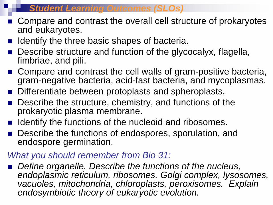
Student Learning Outcomes (SLOs)
Compare and contrast the overall cell structure of prokaryotes and eukaryotes.
Identify the three basic shapes of bacteria.
Describe structure and function of the glycocalyx, flagella, fimbriae, and pili.
Compare and contrast the cell walls of gram-positive bacteria, gram-negative bacteria, acid-fast bacteria, and mycoplasmas.
Differentiate between protoplasts and spheroplasts.
Describe the structure, chemistry, and functions of the prokaryotic plasma membrane.
Identify the functions of the nucleoid and ribosomes.
Describe the functions of endospores, sporulation, and endospore germination.
What you should remember from Bio 31:
Define organelle. Describe the functions of the nucleus, endoplasmic reticulum, ribosomes, Golgi complex, lysosomes, vacuoles, mitochondria, chloroplasts, peroxisomes. Explain endosymbiotic theory of eukaryotic evolution.
What is the main feature that distinguishes
prokaryotes from eukaryotes?
How would you be able to identify streptococci
through a microscope?
Why are bacterial capsules medically important?
How do bacteria move?
Why are drugs that target cell wall synthesis useful?
What happens to protoplasts and spheroplasts when
put into hyper- or hyptonic solutions?
Where is the DNA located in a prokaryotic cell?
Under what conditions do endospores form?
SLOs cont.: Check Your Understanding
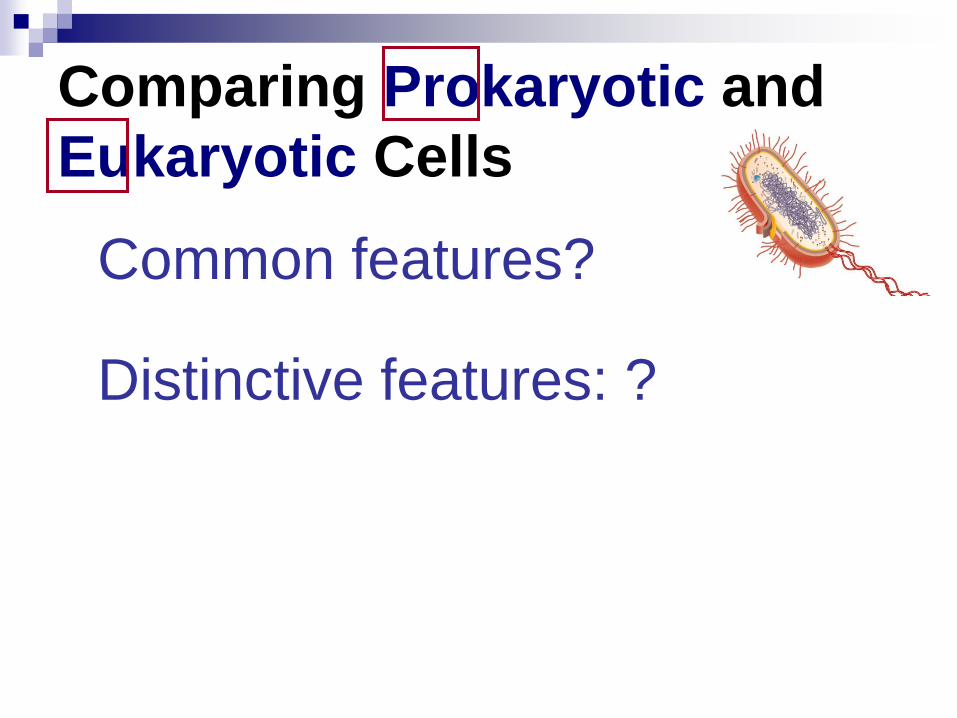
Comparing Prokaryotic and
Eukaryotic Cells
Common features?
Distinctive features: ?
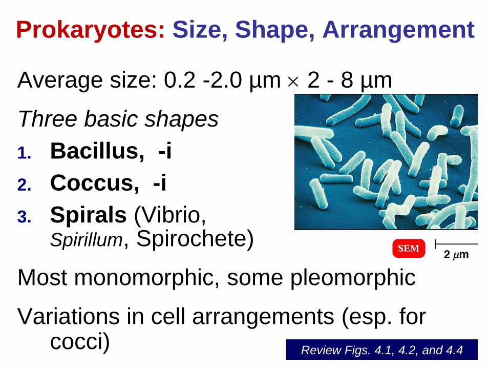
Prokaryotes: Size, Shape, Arrangement
Average size: 0.2 -2.0 µm 2 - 8 µm
Three basic shapes
1. Bacillus, -i
2. Coccus, -i
3. Spirals (Vibrio, Spirillum, Spirochete)
Most monomorphic, some pleomorphic
Variations in cell arrangements (esp. for cocci)
Review Figs. 4.1, 4.2, and 4.4
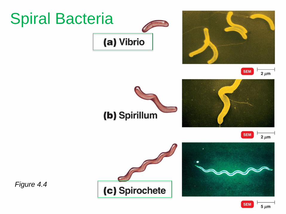
Figure 4.4
Spiral Bacteria
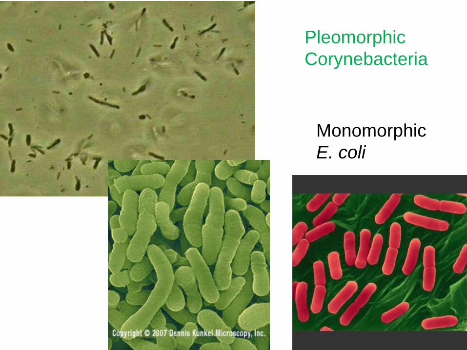
Pleomorphic
Corynebacteria
Monomorphic
E. coli
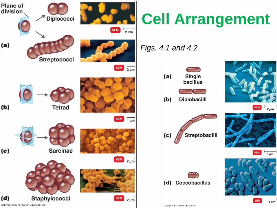
Cell Arrangement
Figs. 4.1 and 4.2
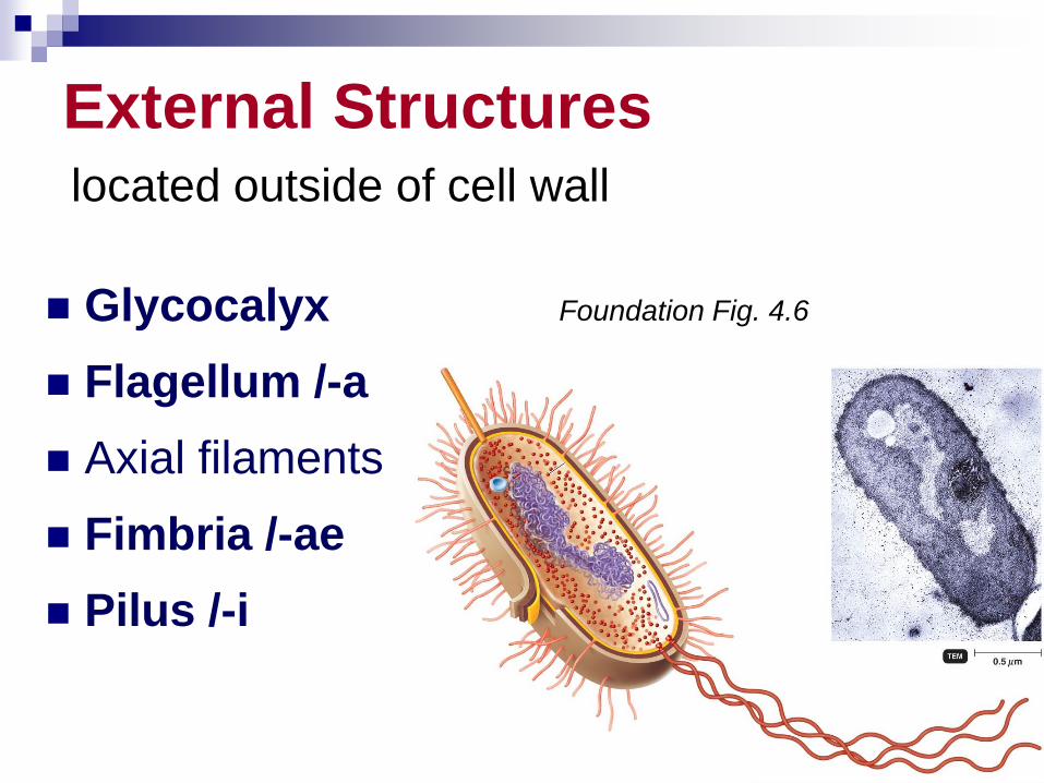
External Structures located outside of cell wall
Glycocalyx
Flagellum /-a
Axial filaments
Fimbria /-ae
Pilus /-i
Foundation Fig. 4.6
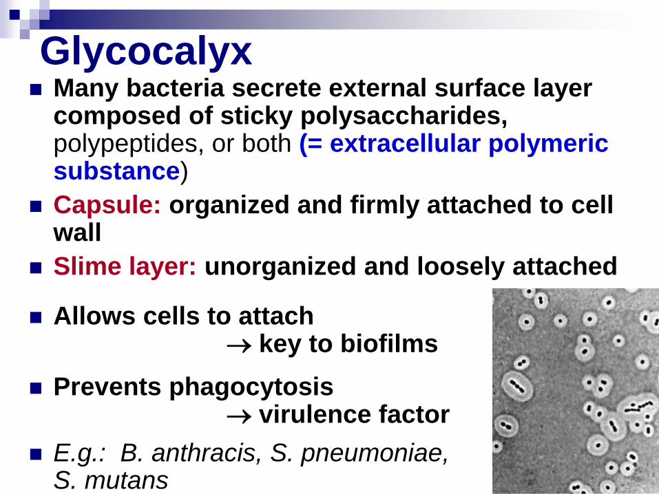
Glycocalyx Many bacteria secrete external surface layer
composed of sticky polysaccharides, polypeptides, or both (= extracellular polymeric substance)
Capsule: organized and firmly attached to cell wall
Slime layer: unorganized and loosely attached
Allows cells to attach key to biofilms
Prevents phagocytosis virulence factor
E.g.: B. anthracis, S. pneumoniae, S. mutans
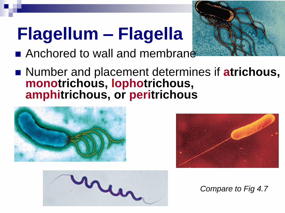
Flagellum – Flagella Anchored to wall and membrane
Number and placement determines if atrichous, monotrichous, lophotrichous, amphitrichous, or peritrichous
Compare to Fig 4.7
___________
_______
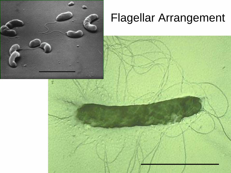
Flagellar Arrangement
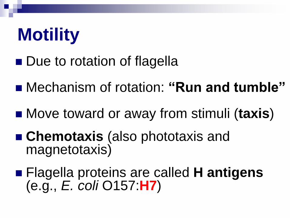
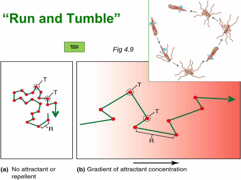
Motility
Due to rotation of flagella
Mechanism of rotation: “Run and tumble”
Move toward or away from stimuli (taxis)
Chemotaxis (also phototaxis and magnetotaxis)
Flagella proteins are called H antigens (e.g., E. coli O157:H7)
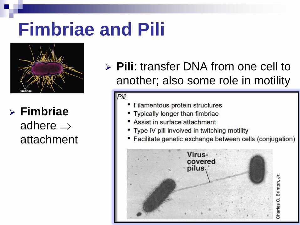
Fimbriae and Pili
Fimbriae
adhere
attachment
Pili: transfer DNA from one cell to
another; also some role in motility
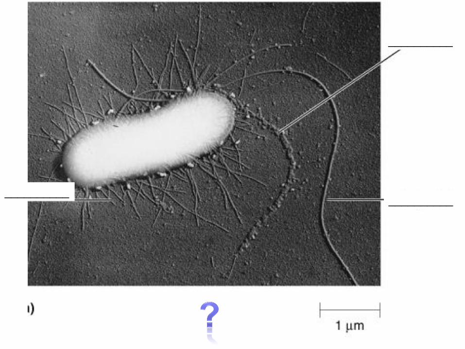
__________ __________
__________
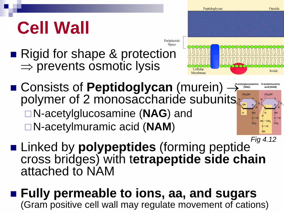
Cell Wall
Rigid for shape & protection prevents osmotic lysis
Consists of Peptidoglycan (murein) polymer of 2 monosaccharide subunits N-acetylglucosamine (NAG) and
N-acetylmuramic acid (NAM)
Linked by polypeptides (forming peptide cross bridges) with tetrapeptide side chain attached to NAM
Fully permeable to ions, aa, and sugars (Gram positive cell wall may regulate movement of cations)
Fig 4.12
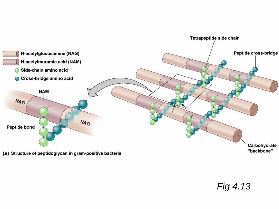
Fig 4.13
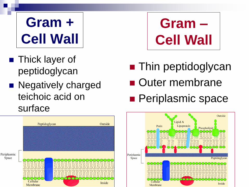
Gram +
Cell Wall
Thick layer of
peptidoglycan
Negatively charged
teichoic acid on
surface
Thin peptidoglycan
Outer membrane
Periplasmic space
Gram –
Cell Wall
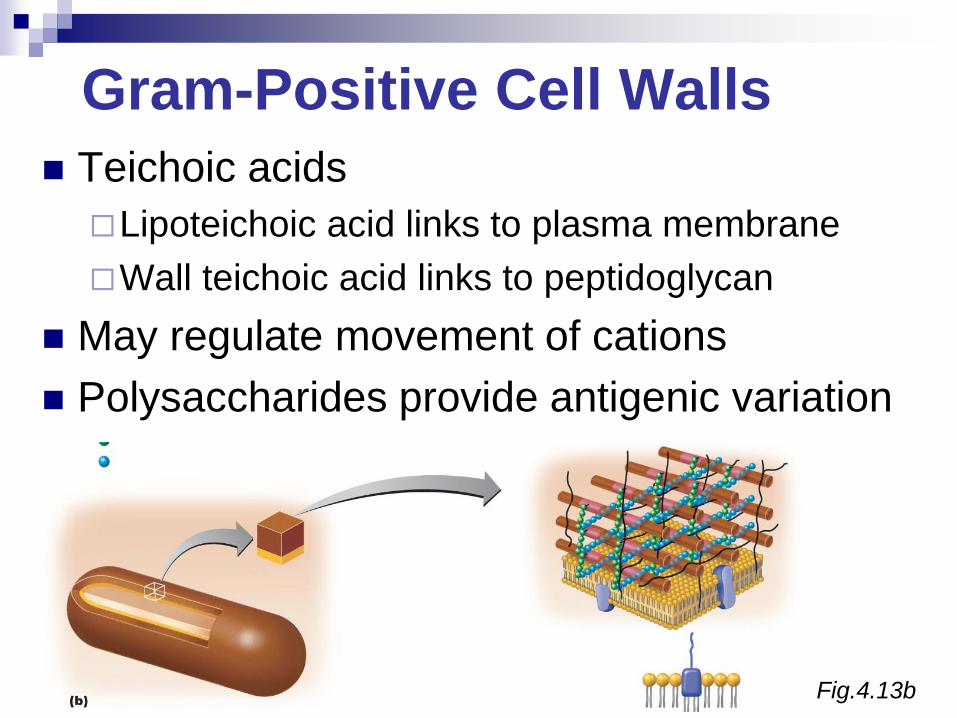
Fig.4.13b
Gram-Positive Cell Walls
Teichoic acids
Lipoteichoic acid links to plasma membrane
Wall teichoic acid links to peptidoglycan
May regulate movement of cations
Polysaccharides provide antigenic variation
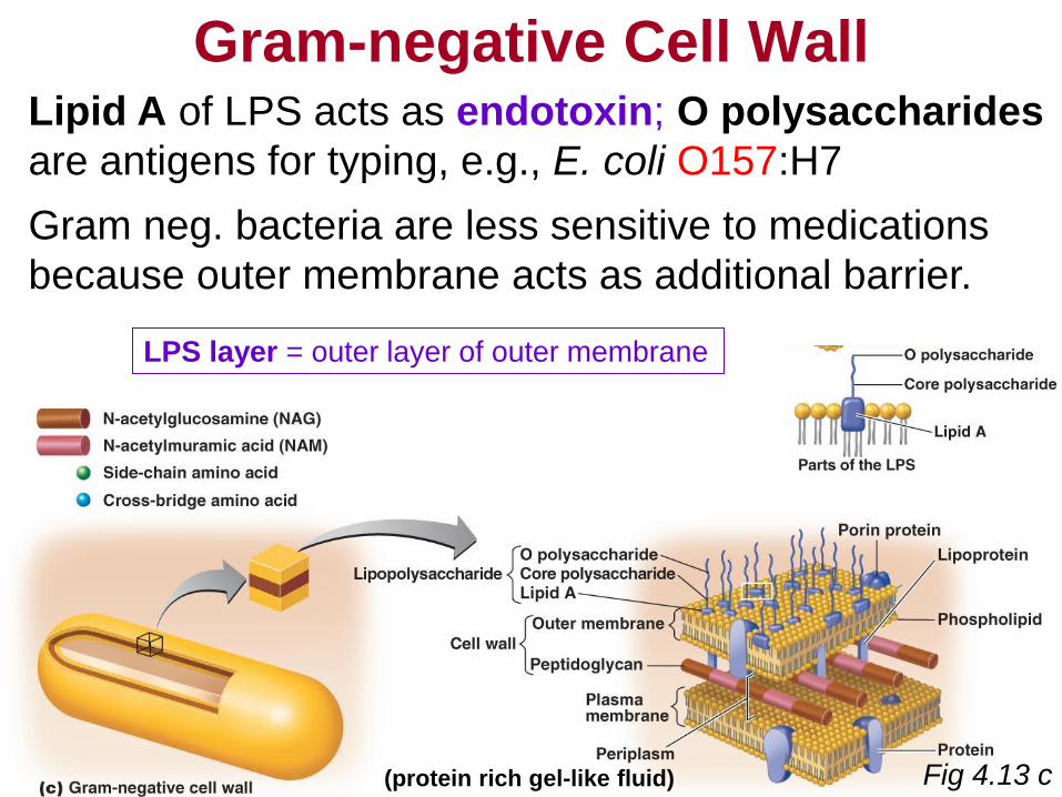
Gram-negative Cell Wall Lipid A of LPS acts as endotoxin; O polysaccharides
are antigens for typing, e.g., E. coli O157:H7
Gram neg. bacteria are less sensitive to medications
because outer membrane acts as additional barrier.
LPS layer = outer layer of outer membrane
(protein rich gel-like fluid) Fig 4.13 c
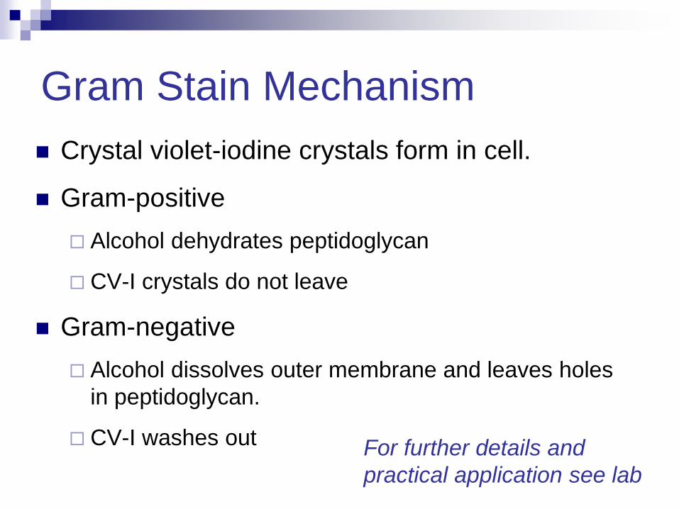
Gram Stain Mechanism
Crystal violet-iodine crystals form in cell.
Gram-positive
Alcohol dehydrates peptidoglycan
CV-I crystals do not leave
Gram-negative
Alcohol dissolves outer membrane and leaves holes
in peptidoglycan.
CV-I washes out
For further details and
practical application see lab
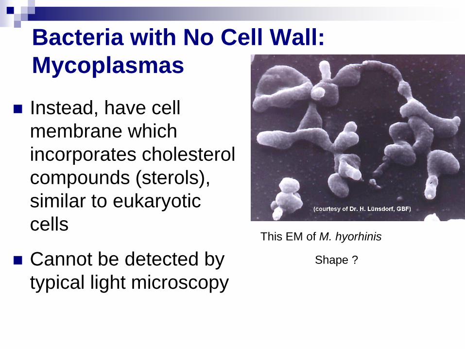
Bacteria with No Cell Wall:
Mycoplasmas
Instead, have cell
membrane which
incorporates cholesterol
compounds (sterols),
similar to eukaryotic
cells
Cannot be detected by
typical light microscopy
This EM of M. hyorhinis
Shape ?
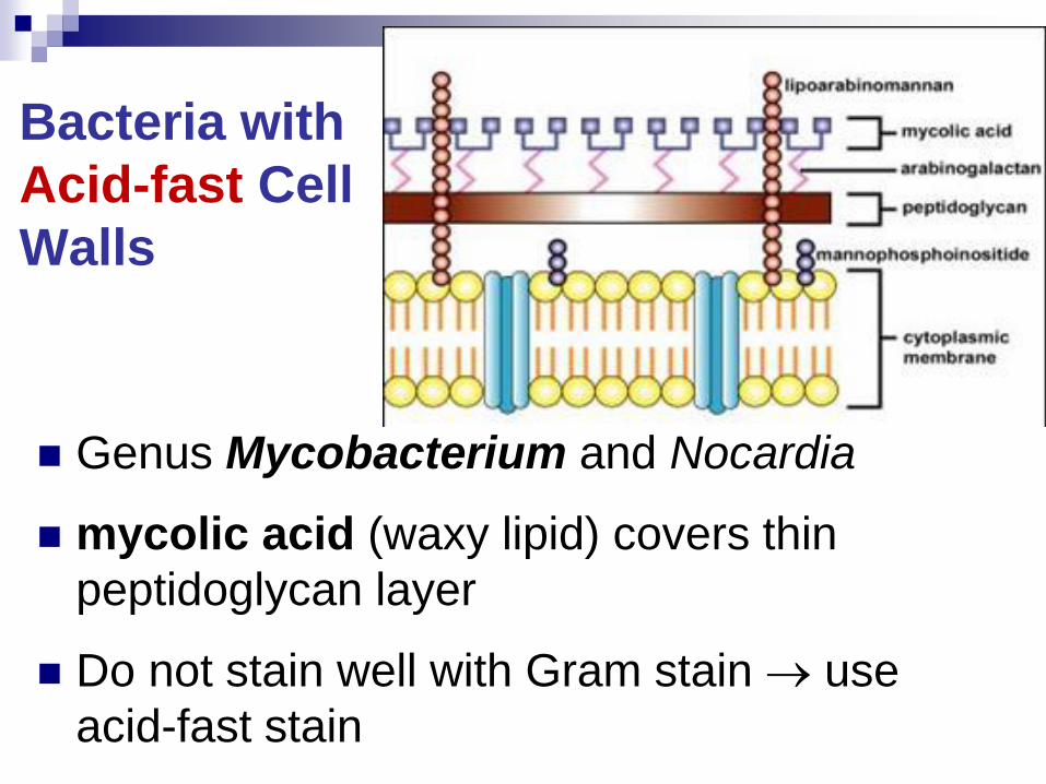
Bacteria with
Acid-fast Cell
Walls
Genus Mycobacterium and Nocardia
mycolic acid (waxy lipid) covers thin
peptidoglycan layer
Do not stain well with Gram stain use
acid-fast stain
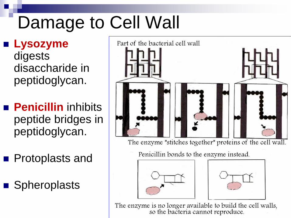
Damage to Cell Wall Lysozyme
digests disaccharide in peptidoglycan.
Penicillin inhibits peptide bridges in peptidoglycan.
Protoplasts and
Spheroplasts
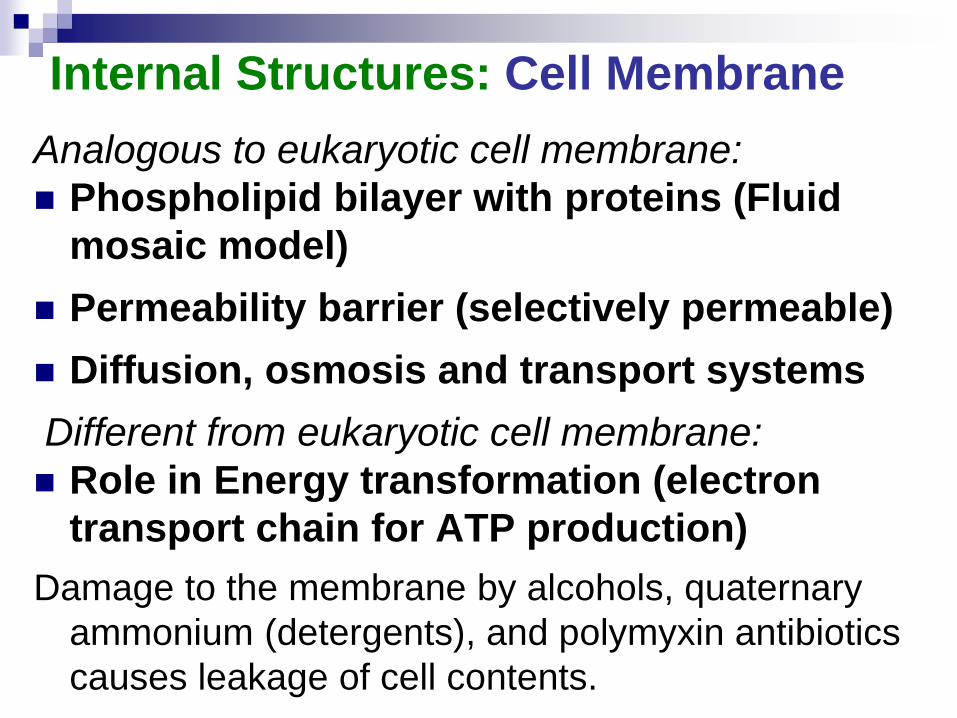
Internal Structures: Cell Membrane
Analogous to eukaryotic cell membrane:
Phospholipid bilayer with proteins (Fluid
mosaic model)
Permeability barrier (selectively permeable)
Diffusion, osmosis and transport systems
Different from eukaryotic cell membrane:
Role in Energy transformation (electron
transport chain for ATP production)
Damage to the membrane by alcohols, quaternary
ammonium (detergents), and polymyxin antibiotics
causes leakage of cell contents.
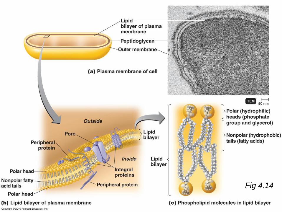
Fig 4.14
Movement of Materials across
Membranes
See Bio 31!
Review on your own if necessary (pages 92 – 94)
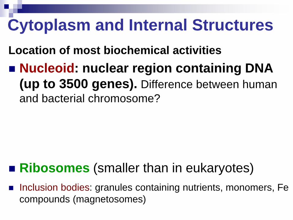
Cytoplasm and Internal Structures
Location of most biochemical activities
Nucleoid: nuclear region containing DNA
(up to 3500 genes). Difference between human
and bacterial chromosome?
Ribosomes (smaller than in eukaryotes)
Inclusion bodies: granules containing nutrients, monomers, Fe
compounds (magnetosomes)
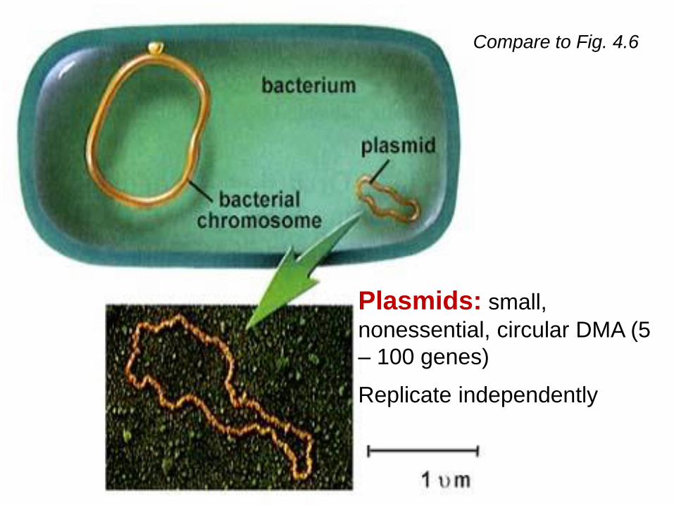
Compare to Fig. 4.6
Plasmids: small,
nonessential, circular DMA (5
– 100 genes)
Replicate independently
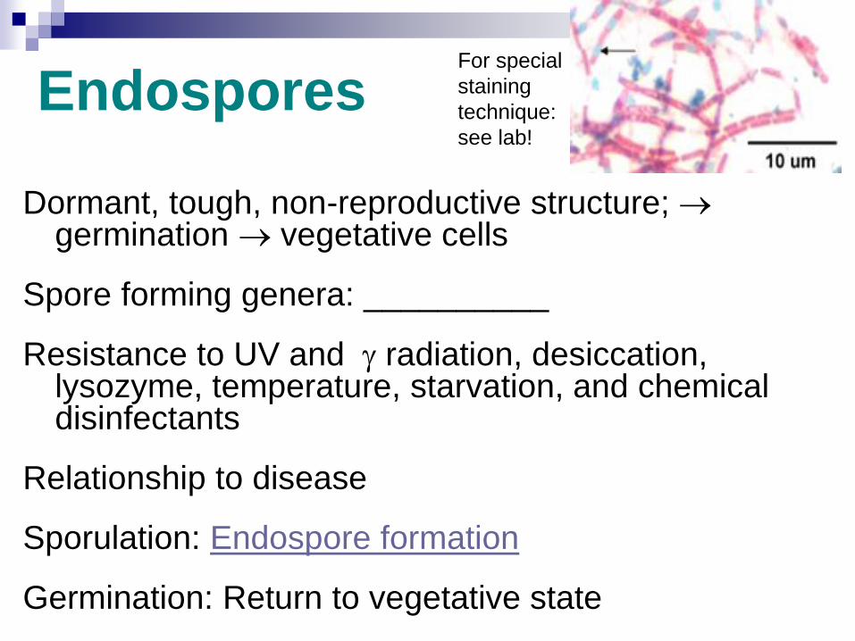
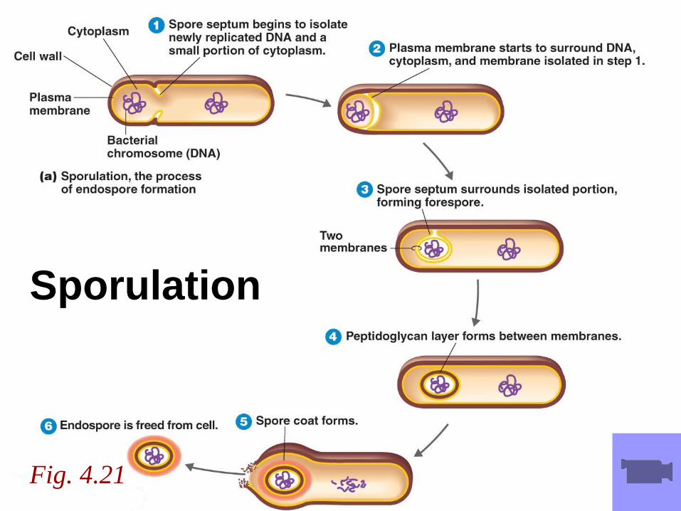
Endospores
Dormant, tough, non-reproductive structure; germination vegetative cells
Spore forming genera: __________
Resistance to UV and radiation, desiccation, lysozyme, temperature, starvation, and chemical disinfectants
Relationship to disease
Sporulation: Endospore formation
Germination: Return to vegetative state
For special
staining
technique:
see lab!
The Eukaryotic Cell
See BIO 30!
Review on your own if necessary (pages 94 – 102)
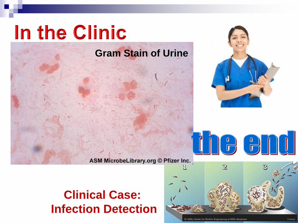
Clinical Case:
Infection Detection
Gram Stain of Urine