Embed Size (px)
Citation preview
www.MaterialsViews.com
2067© 2016 Wiley-VCH Verlag GmbH & Co. KGaA, Weinheim www.small-journal.com
Functional Nanoparticles Activate a Decellularized Liver Scaffold for Blood Detoxifi cation Fen Xu , Tianyi Kang , Jie Deng , Junli Liu , Xiaolei Chen , Yuan Wang , Liang Ouyang , Ting Du , Hong Tang , Xiaoping Xu , Shaochen Chen , Yanan Du , Yujun Shi , Zhiyong Qian , Yuquan Wei , Hongxin Deng , * and Maling Gou *
DOI: 10.1002/smll.201503320
Extracorporeal devices have great promise for cleansing the body of virulence factors that are caused by venomous injuries, bacterial infections, and biological weaponry. The clinically used extracorporeal devices, such as artifi cial liver-support systems that are mainly based on dialysis or electrostatic interaction, are limited to remove a target toxin. Here, a liver-mimetic device is shown that consists of decellularized liver scaffold (DLS) populated with polydiacetylene (PDA) nanoparticles. DLS has the gross shape and 3D architecture of a liver, and the PDA nanoparticles selectively capture and neutralize the pore-forming toxins (PFTs). This device can effi ciently and target-orientedly remove PFTs in human blood ex vivo without changing blood components or activating complement factors, showing potential application in antidotal therapy. This work provides a proof-of-principle for blood detoxifi cation by a nanoparticle-activated DLS, and can lead to the development of future medical devices for antidotal therapy.
Biomimetics
Dr. F. Xu, Dr. T. Kang, Dr. J. Deng, Dr. J. Liu, Dr. X. Chen, Y. Wang, Dr. L. Ouyang, T. Du, Prof. Z. Qian, Prof. Y. Wei, Prof. H. Deng, Prof. M. Gou State Key Laboratory of Biotherapy and Cancer Center West China Hospital Sichuan University and Collaborative Innovation Center for Biotherapy Chengdu 610041 , ChinaE-mail: [email protected]; [email protected]
Dr. F. Xu State Key Laboratory of Cardiovascular Disease Fuwai Hospital National Center for Cardiovascular Disease Chinese Academy of Medical Sciences and PekingUnion Medical College Beijing 100037 , China
Prof. H. Tang Center of Infectious Diseases West China Hospital Sichuan University Chengdu 610041 , China
Prof. X. Xu West China School of Pharmacy Sichuan University Chengdu 610041 , P. R. China
Prof. S. Chen Department of NanoEngineering University of California San Diego , CA 92093 , USA
Prof. Y. Du Department of Biomedical Engineering School of Medicine Tsinghua University Beijing 100084 , China
Prof. Y. Shi Laboratory of Pathology West China Hospital Sichuan University Key Laboratory of Transplant Engineering and Immunology NHFPC, West China Hospital Sichuan University Chengdu 610041 , China
1. Introduction
Extracorporeal detoxifi cation devices offer clinical ways
to cleanse the blood of virulence factors. [ 1–3 ] Pore-forming
toxins (PFTs), one of the most common protein toxins in
nature, underlie the virulence mechanisms in animal bites/
sting and bacterial infections, [ 4–8 ] Over 80 families of PFTs
have been identifi ed, displaying diverse molecular structures,
small 2016, 12, No. 15, 2067–2076
full paperswww.MaterialsViews.com
2068 www.small-journal.com © 2016 Wiley-VCH Verlag GmbH & Co. KGaA, Weinheim
different molecular weight, and distinctive epitopic targets. [ 9 ]
Currently, there is no clinical detoxifi cation device that can
targetedly remove PFTs from blood, despite the existed
liver-support systems which remove toxins by dialysis or
ionic adsorption column can benefi t patients who suffer from
PFTs.
Nanoparticles can be rationally designed to bind and
neutralize target toxins, owing to their inherent small size
and fl exibility in design and preparation. [ 10–15 ] All PFTs
have the same capacity to disrupt cells by forming pores in
cellular membranes and altering their permeability. Recent
advancements have spurred the development of an action
mechanism-targeted detoxifi cation of PFTs by nanoparti-
cles that function as a toxin decoy to attract and trap PFTs,
which allows one nanoparticle to capture and neutralize dif-
ferent PFTs. [ 16,17 ] Therefore, a fl uidic system that is integrated
with retrievable nanoparticles has potential application in
PFTs detoxifi cation by targetedly removing PFTs from the
bloodstream.
The liver is an important apparatus of our body which
has the function of detox, in which the liver-specifi c micro-
structure facilitates hepatocytes to effi ciently detoxify the
bloodstream. [ 18,19 ] This inspired the design of a liver-mimetic
fl uidic device for detoxifi cation. And it is of great interest to
integrate nanoparticles into a liver-like fl uidic systems for the
effi cient removial of toxins. Herein, we performed an attempt
to use polydiacetylene (PDA) nanoparticles to “recellularize”
an animal decellularized liver scaffold (DLS), to construct a
liver-mimetic device for human blood detoxifi cation. PDA
nanoparticles were derived from self-assembly of 10,12-pen-
tacosadiynoic acid (PCDA). The nanoparticle surface is made
of a microvasculature π-conjugated polymer with alternating
double- and triple-bond groups in the main polymer chain.
The cell membrane-mimic surface functions to attract, cap-
ture, and neutralize toxins owing to the interactions between
PDA and PFTs. [ 2 ] DLS preserves the gross shape and 3D
architecture of a liver which is a naturally precise microfl u-
idic system. Our results indicate that the liver-mimetic device
(named as PDA-DLS device) can effi ciently remove PFTs
toxins in human blood ex vivo, and does not affect blood
components and complement factors. This work might inspire
many breakthroughs in the development of extracorporeal
device that can effi ciently remove target toxins from blood-
stream for future clinical use.
2. Results
2.1. Preparation of a Nanoparticle-Activated Liver-Mimetic Device
The PDA-DLS device is schematically presented in
Figure 1 . To generate this device, we used a decellularization
technology to prepare the DLS, followed by seeding with
PDA nanoparticles. By portal vein perfusion with sodium
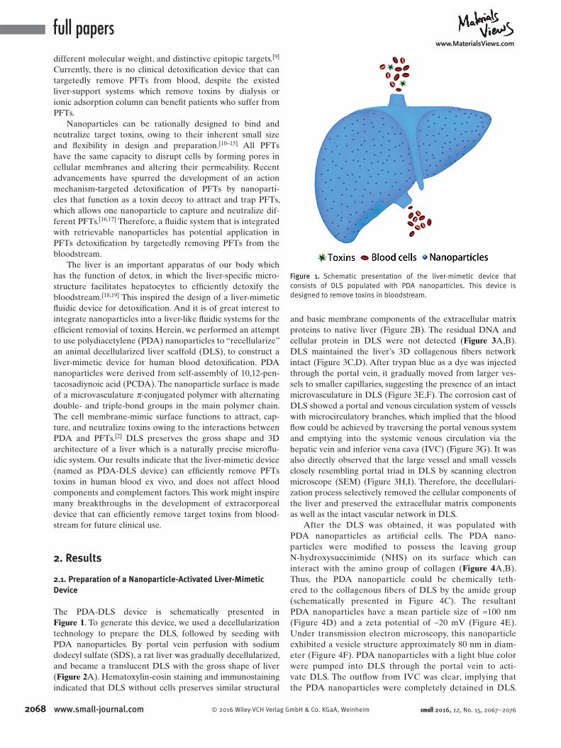
dodecyl sulfate (SDS), a rat liver was gradually decellularized,
and became a translucent DLS with the gross shape of liver
( Figure 2 A). Hematoxylin-eosin staining and immunostaining
indicated that DLS without cells preserves similar structural
and basic membrane components of the extracellular matrix
proteins to native liver (Figure 2 B). The residual DNA and
cellular protein in DLS were not detected ( Figure 3 A,B).
DLS maintained the liver’s 3D collagenous fi bers network
intact (Figure 3 C,D). After trypan blue as a dye was injected
through the portal vein, it gradually moved from larger ves-
sels to smaller capillaries, suggesting the presence of an intact
microvasculature in DLS (Figure 3 E,F). The corrosion cast of
DLS showed a portal and venous circulation system of vessels
with microcirculatory branches, which implied that the blood
fl ow could be achieved by traversing the portal venous system
and emptying into the systemic venous circulation via the
hepatic vein and inferior vena cava (IVC) (Figure 3 G). It was
also directly observed that the large vessel and small vessels
closely resembling portal triad in DLS by scanning electron
microscope (SEM) (Figure 3 H,I). Therefore, the decellulari-
zation process selectively removed the cellular components of
the liver and preserved the extracellular matrix components
as well as the intact vascular network in DLS.
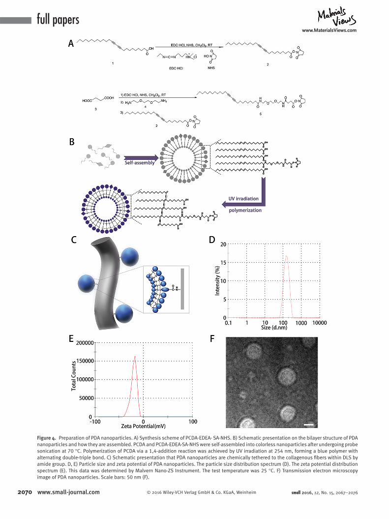
After the DLS was obtained, it was populated with
PDA nanoparticles as artifi cial cells. The PDA nano-
particles were modifi ed to possess the leaving group
N-hydroxysuccinimide (NHS) on its surface which can
interact with the amino group of collagen ( Figure 4 A,B).
Thus, the PDA nanoparticle could be chemically teth-
ered to the collagenous fi bers of DLS by the amide group
(schematically presented in Figure 4 C). The resultant
PDA nanoparticles have a mean particle size of ≈100 nm
(Figure 4 D) and a zeta potential of −20 mV (Figure 4 E).
Under transmission electron microscopy, this nanoparticle
exhibited a vesicle structure approximately 80 nm in diam-
eter (Figure 4 F). PDA nanoparticles with a light blue color
were pumped into DLS through the portal vein to acti-
vate DLS. The outfl ow from IVC was clear, implying that
the PDA nanoparticles were completely detained in DLS.
small 2016, 12, No. 15, 2067–2076
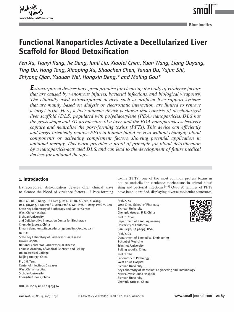
Figure 1. Schematic presentation of the liver-mimetic device that consists of DLS populated with PDA nanoparticles. This device is designed to remove toxins in bloodstream.
www.MaterialsViews.com
2069© 2016 Wiley-VCH Verlag GmbH & Co. KGaA, Weinheim www.small-journal.com
It was also observed that unmodifi ed PDA nanoparticles
could not be effi ciently trapped in the DLS (Supplemen-
tary video, Supporting Information). Furthermore, the
colorimetric readings of the outfl ows indicated that 100%
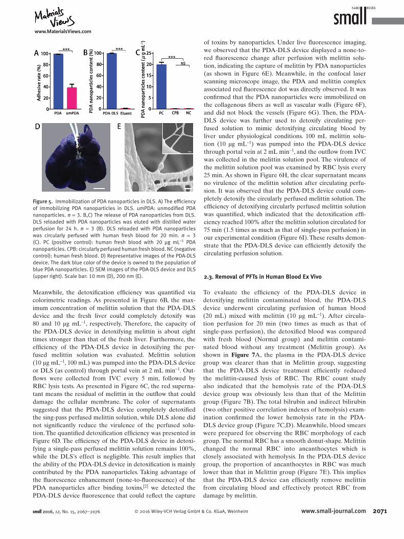
PDA nanoparticles were detained in DLS ( Figure 5 A).
Thus, the chemical modifi cation of PDA nanoparticles is
necessary for effi ciently immobilizing PDA nanoparticles
into DLS. Furthermore, when PDA-DLS was eluted with
distilled water or human fresh blood, the immobilized
PDA nanoparticles would not be released (Figure 5 B, C).
By perfusion of DLS with 50 mg of PDA nanoparticles,
a PDA-DLS device was created. The appearance of this
device was shown in Figure 5 D. Under transmission elec-
tron microscopy, it was observed that PDA nanoparticles
were tethered to the surface of collagenous fi bers within
the PDA-DLS device (Figure 5 E).
2.2. Removal of PFTs in Solution In Vitro
The ability of the PDA-DLS device to detoxify PFTs was
tested by red blood cells (RBC) lysis test. Melittin was used
as the model PFT. The capacity of the PDA-DLS device in
detoxifying the melittin solution was compared with that
of the fresh liver. The PDA-DLS device and the fresh liver
(as control) were processed into pieces with the volume of
100 mm 3 , respectively, followed by incubation of each piece
with melittin solutions (200 µL) of different concentrations.
After incubation for 60 min, the samples were centrifuged,
and the supernatants were added to rat RBC solution under-
going RBC lysis test. The centrifuged RBC solution in each
test was shown in Figure 6 A. The red color of the superna-
tant means hemolysis. And the PDA-DLS device was more
effi cient in detoxifying melittin than that of the fresh liver.
small 2016, 12, No. 15, 2067–2076
Figure 2. Decellularization of rat liver. A) Representative images of a fresh liver during the decellularization process at 0 h, 1 h, 3 h, 6 h, and 12h. B) Comparison of the fresh liver (top) and DLS (bottom). Left to right: H&E, collagen I (red), collagen IV (red), elastin (red), fi bronectin (red), and laminin (red) staining. Sections were counterstained with DAPI (blue). Scale bars: 10 mm (A), 100 μm (B).
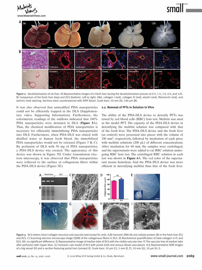
Figure 3. DLS retains intact collagen structure and vascular bed except for cells. A,B) Genomic DNA (A) and cellular protein (B) in the fresh liver (FL) and DLS. C) Scanning electron microscope image (SEM) of the collagenous fi bers in DLS. D) Biochemical quantifi cation of total collagen in FL and DLS. NS: no signifi cant difference. E) Representative image of median lobe of DLS with the visible vascular tree. F) The vascular tree of median lobe after perfusion with trypan blue. G) Corrosion cast model of DLS with portal (red) and venous (blue) vasculature. H,I) Representative SEM images of a big vessel (H) and a section featuring portal tract (arrows) (I). Scale bars: 10 μm (C), 5 mm (E, F), 10 mm (G), 10 μm (H, I).
full paperswww.MaterialsViews.com
2070 www.small-journal.com © 2016 Wiley-VCH Verlag GmbH & Co. KGaA, Weinheim small 2016, 12, No. 15, 2067–2076
Figure 4. Preparation of PDA nanoparticles. A) Synthesis scheme of PCDA-EDEA- SA-NHS. B) Schematic presentation on the bilayer structure of PDA nanoparticles and how they are assembled. PCDA and PCDA-EDEA-SA-NHS were self-assembled into colorless nanoparticles after undergoing probe sonication at 70 °C. Polymerization of PCDA via a 1,4-addition reaction was achieved by UV irradiation at 254 nm, forming a blue polymer with alternating double-triple bond. C) Schematic presentation that PDA nanoparticles are chemically tethered to the collagenous fi bers within DLS by amide group. D, E) Particle size and zeta potential of PDA nanoparticles. The particle size distribution spectrum (D). The zeta potential distribution spectrum (E). This data was determined by Malvern Nano-ZS Instrument. The test temperature was 25 °C. F) Transmission electron microscopy image of PDA nanoparticles. Scale bars: 50 nm (F).
www.MaterialsViews.com
2071© 2016 Wiley-VCH Verlag GmbH & Co. KGaA, Weinheim www.small-journal.com
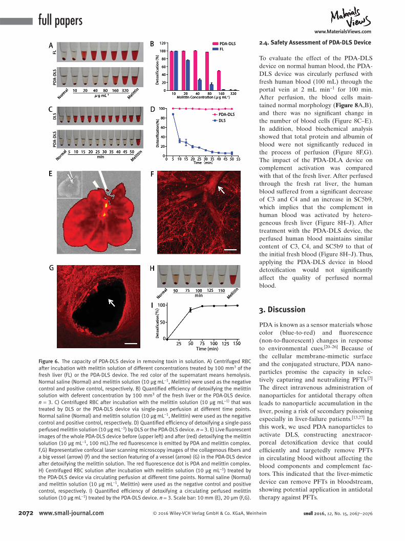
Meanwhile, the detoxifi cation effi ciency was quantifi ed via
colorimetric readings. As presented in Figure 6 B, the max-
imum concentration of melittin solution that the PDA-DLS
device and the fresh liver could completely detoxify was
80 and 10 µg mL −1 , respectively. Therefore, the capacity of
the PDA-DLS device in detoxifying melittin is about eight
times stronger than that of the fresh liver. Furthermore, the
effi ciency of the PDA-DLS device in detoxifying the per-
fused melittin solution was evaluated. Melittin solution
(10 µg mL −1 , 100 mL) was pumped into the PDA-DLS device
or DLS (as control) through portal vein at 2 mL min −1 . Out-
fl ows were collected from IVC every 5 min, followed by
RBC lysis tests. As presented in Figure 6 C, the red superna-
tant means the residual of melittin in the outfl ow that could
damage the cellular membrane. The color of supernatants
suggested that the PDA-DLS device completely detoxifi ed
the sing-pass perfused melittin solution, while DLS alone did
not signifi cantly reduce the virulence of the perfused solu-
tion. The quantifi ed detoxifi cation effi ciency was presented in
Figure 6 D. The effi ciency of the PDA-DLS device in detoxi-
fying a single-pass perfused melittin solution remains 100%,
while the DLS’s effect is negligible. This result implies that
the ability of the PDA-DLS device in detoxifi cation is mainly
contributed by the PDA nanoparticles. Taking advantage of
the fl uorescence enhancement (none-to-fl uorescence) of the
PDA nanoparticles after binding toxins, [ 2 ] we detected the
PDA-DLS device fl uorescence that could refl ect the capture
of toxins by nanoparticles. Under live fl uorescence imaging,
we observed that the PDA-DLS device displayed a none-to-
red fl uorescence change after perfusion with melittin solu-
tion, indicating the capture of melittin by PDA nanoparticles
(as shown in Figure 6 E). Meanwhile, in the confocal laser
scanning microscope image, the PDA and melittin complex
associated red fl uorescence dot was directly observed. It was
confi rmed that the PDA nanoparticles were immobilized on
the collagenous fi bers as well as vascular walls (Figure 6 F),
and did not block the vessels (Figure 6 G). Then, the PDA-
DLS device was further used to detoxify circulating per-
fused solution to mimic detoxifying circulating blood by
liver under physiological conditions. 100 mL melittin solu-
tion (10 µg mL −1 ) was pumped into the PDA-DLS device
through portal vein at 2 mL min −1 , and the outfl ow from IVC
was collected in the melittin solution pool. The virulence of
the melittin solution pool was examined by RBC lysis every
25 min. As shown in Figure 6 H, the clear supernatant means
no virulence of the melittin solution after circulating perfu-
sion. It was observed that the PDA-DLS device could com-
pletely detoxify the circularly perfused melittin solution. The
effi ciency of detoxifying circularly perfused melittin solution
was quantifi ed, which indicated that the detoxifi cation effi -
ciency reached 100% after the melittin solution circulated for
75 min (1.5 times as much as that of single-pass perfusion) in
our experimental condition (Figure 6 I). These results demon-
strate that the PDA-DLS device can effi ciently detoxify the
circulating perfusion solution.
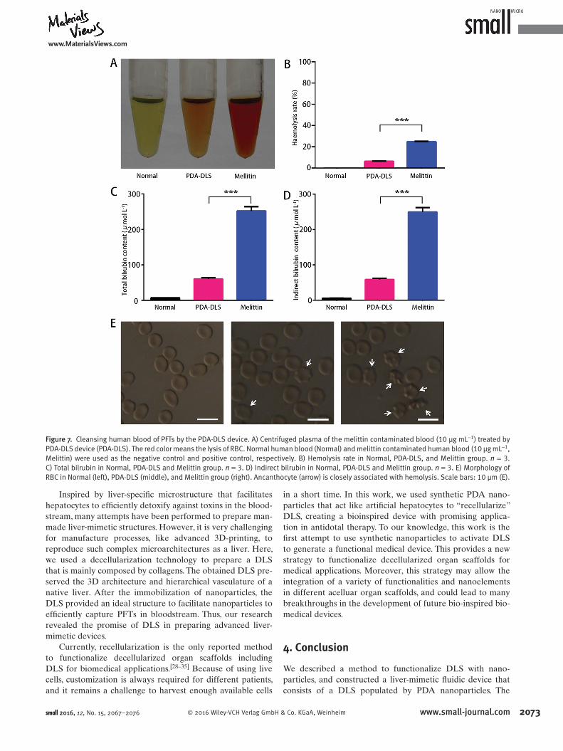
2.3. Removal of PFTs in Human Blood Ex Vivo
To evaluate the effi ciency of the PDA-DLS device in
detoxifying melittin contaminated blood, the PDA-DLS
device underwent circulating perfusion of human blood
(20 mL) mixed with melittin (10 µg mL −1 ). After circula-
tion perfusion for 20 min (two times as much as that of
single-pass perfusion), the detoxifi ed blood was compared
with fresh blood (Normal group) and melittin contami-
nated blood without any treatment (Melittin group). As
shown in Figure 7 A, the plasma in the PDA-DLS device
group was clearer than that in Melittin group, suggesting
that the PDA-DLS device treatment effi ciently reduced
the melittin-caused lysis of RBC. The RBC count study
also indicated that the hemolysis rate of the PDA-DLS
device group was obviously less than that of the Melittin
group (Figure 7 B). The total bilrubin and indirect bilirubin
(two other positive correlation indexes of hemolysis) exam-
ination confi rmed the lower hemolysis rate in the PDA-
DLS device group (Figure 7 C,D). Meanwhile, blood smears
were prepared for observing the RBC morphology of each
group. The normal RBC has a smooth donut-shape. Melittin
changed the normal RBC into ancanthocytes which is
closely associated with hemolysis. In the PDA-DLS device
group, the proportion of ancanthocytes in RBC was much
lower than that in Melittin group (Figure 7 E). This implies
that the PDA-DLS device can effi ciently remove melittin
from circulating blood and effectively protect RBC from
damage by melittin.
small 2016, 12, No. 15, 2067–2076
Figure 5. Immobilization of PDA nanoparticles in DLS. A) The effi ciency of immobilizing PDA nanoparticles in DLS. umPDA: unmodifi ed PDA nanoparticles. n = 3. B,C) The release of PDA nanoparticles from DLS. DLS reloaded with PDA nanoparticles was eluted with distilled water perfusion for 24 h. n = 3 (B). DLS reloaded with PDA nanoparticles was circularly perfused with human fresh blood for 20 min. n = 3 (C). PC (positive control): human fresh blood with 20 μg mL −1 PDA nanoparticles. CPB: circularly perfused human fresh blood. NC (negative control): human fresh blood. D) Representative images of the PDA-DLS device. The dark blue color of the device is owned to the population of blue PDA nanoparticles. E) SEM images of the PDA-DLS device and DLS (upper right). Scale bar: 10 mm (D), 200 nm (E).
full paperswww.MaterialsViews.com
2072 www.small-journal.com © 2016 Wiley-VCH Verlag GmbH & Co. KGaA, Weinheim
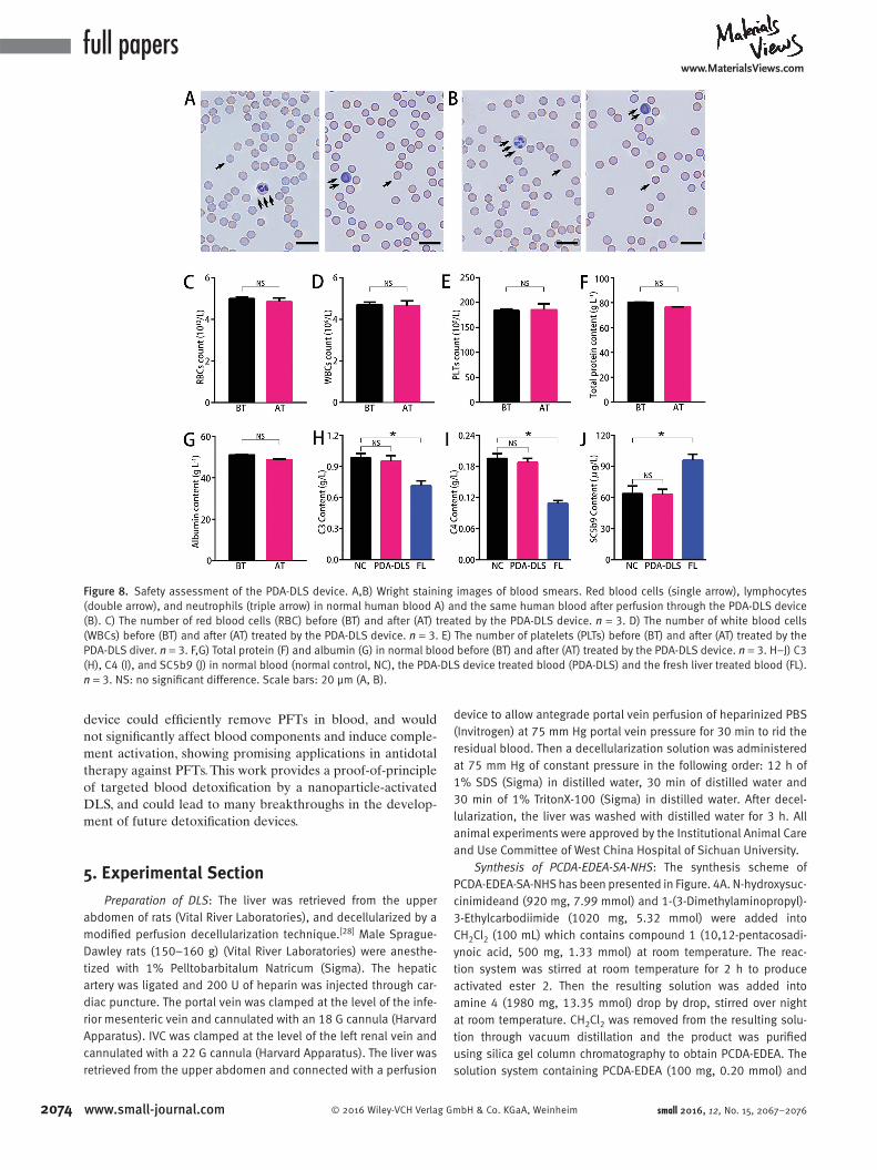
2.4. Safety Assessment of PDA-DLS Device
To evaluate the effect of the PDA-DLS
device on normal human blood, the PDA-
DLS device was circularly perfused with
fresh human blood (100 mL) through the
portal vein at 2 mL min −1 for 100 min.
After perfusion, the blood cells main-
tained normal morphology ( Figure 8 A,B),
and there was no signifi cant change in
the number of blood cells (Figure 8 C–E).
In addition, blood biochemical analysis
showed that total protein and albumin of
blood were not signifi cantly reduced in
the process of perfusion (Figure 8 F,G).
The impact of the PDA-DLA device on
complement activation was compared
with that of the fresh liver. After perfused
through the fresh rat liver, the human
blood suffered from a signifi cant decrease
of C3 and C4 and an increase in SC5b9,
which implies that the complement in
human blood was activated by hetero-
geneous fresh liver (Figure 8 H–J). After
treatment with the PDA-DLS device, the
perfused human blood maintains similar
content of C3, C4, and SC5b9 to that of
the initial fresh blood (Figure 8 H–J). Thus,
applying the PDA-DLS device in blood
detoxifi cation would not signifi cantly
affect the quality of perfused normal
blood.
3. Discussion
PDA is known as a sensor materials whose
color (blue-to-red) and fl uorescence
(non-to-fl uorescent) changes in response
to environmental cues. [ 20–26 ] Because of
the cellular membrane-mimetic surface
and the conjugated structure, PDA nano-
particles promise the capacity in selec-
tively capturing and neutralizing PFTs. [ 2 ]
The direct intravenous administration of
nanoparticles for antidotal therapy often
leads to nanoparticle accumulation in the
liver, posing a risk of secondary poisoning
especially in liver-failure patients. [ 13,27 ] In
this work, we used PDA nanoparticles to
activate DLS, constructing anextracor-
poreal detoxifi cation device that could
effi ciently and targetedly remove PFTs
in circulating blood without affecting the
blood components and complement fac-
tors. This indicated that the liver-mimetic
device can remove PFTs in bloodstream,
showing potential application in antidotal
therapy against PFTs.
small 2016, 12, No. 15, 2067–2076
Figure 6. The capacity of PDA-DLS device in removing toxin in solution. A) Centrifuged RBC after incubation with melittin solution of different concentrations treated by 100 mm 3 of the fresh liver (FL) or the PDA-DLS device. The red color of the supernatant means hemolysis. Normal saline (Normal) and melittin solution (10 μg mL −1 , Melittin) were used as the negative control and positive control, respectively. B) Quantifi ed effi ciency of detoxifying the melittin solution with deferent concentration by 100 mm 3 of the fresh liver or the PDA-DLS device. n = 3. C) Centrifuged RBC after incubation with the melittin solution (10 μg mL −1) that was treated by DLS or the PDA-DLS device via single-pass perfusion at different time points. Normal saline (Normal) and melittin solution (10 μg mL −1 , Melittin) were used as the negative control and positive control, respectively. D) Quantifi ed effi ciency of detoxifying a single-pass perfused melittin solution (10 μg mL −1 ) by DLS or the PDA-DLS device. n = 3. E) Live fl uorescent images of the whole PDA-DLS device before (upper left) and after (red) detoxifying the melittin solution (10 μg mL −1 , 100 mL).The red fl uorescence is emitted by PDA and melittin complex. F,G) Representative confocal laser scanning microscopy images of the collagenous fi bers and a big vessel (arrow) (F) and the section featuring of a vessel (arrow) (G) in the PDA-DLS device after detoxifying the melittin solution. The red fl uorescence dot is PDA and melittin complex. H) Centrifuged RBC solution after incubation with melittin solution (10 μg mL −1 ) treated by the PDA-DLS device via circulating perfusion at different time points. Normal saline (Normal) and melittin solution (10 μg mL −1 , Melittin) were used as the negative control and positive control, respectively. I) Quantifi ed effi ciency of detoxifying a circulating perfused melittin solution (10 μg mL −1 ) treated by the PDA-DLS device. n = 3. Scale bar: 10 mm (E), 20 μm (F,G).
www.MaterialsViews.com
2073© 2016 Wiley-VCH Verlag GmbH & Co. KGaA, Weinheim www.small-journal.com
Inspired by liver-specifi c microstructure that facilitates
hepatocytes to effi ciently detoxify against toxins in the blood-
stream, many attempts have been performed to prepare man-
made liver-mimetic structures. However, it is very challenging
for manufacture processes, like advanced 3D-printing, to
reproduce such complex microarchitectures as a liver. Here,
we used a decellularization technology to prepare a DLS
that is mainly composed by collagens. The obtained DLS pre-
served the 3D architecture and hierarchical vasculature of a
native liver. After the immobilization of nanoparticles, the
DLS provided an ideal structure to facilitate nanoparticles to
effi ciently capture PFTs in bloodstream. Thus, our research
revealed the promise of DLS in preparing advanced liver-
mimetic devices.
Currently, recellularization is the only reported method
to functionalize decellularized organ scaffolds including
DLS for biomedical applications. [ 28–35 ] Because of using live
cells, customization is always required for different patients,
and it remains a challenge to harvest enough available cells
in a short time. In this work, we used synthetic PDA nano-
particles that act like artifi cial hepatocytes to “recellularize”
DLS, creating a bioinspired device with promising applica-
tion in antidotal therapy. To our knowledge, this work is the
fi rst attempt to use synthetic nanoparticles to activate DLS
to generate a functional medical device. This provides a new
strategy to functionalize decellularized organ scaffolds for
medical applications. Moreover, this strategy may allow the
integration of a variety of functionalities and nanoelements
in different acelluar organ scaffolds, and could lead to many
breakthroughs in the development of future bio-inspired bio-
medical devices.
4. Conclusion
We described a method to functionalize DLS with nano-
particles, and constructed a liver-mimetic fl uidic device that
consists of a DLS populated by PDA nanoparticles. The
small 2016, 12, No. 15, 2067–2076
Figure 7. Cleansing human blood of PFTs by the PDA-DLS device. A) Centrifuged plasma of the melittin contaminated blood (10 μg mL −1 ) treated by PDA-DLS device (PDA-DLS). The red color means the lysis of RBC. Normal human blood (Normal) and melittin contaminated human blood (10 μg mL −1 , Melittin) were used as the negative control and positive control, respectively. B) Hemolysis rate in Normal, PDA-DLS, and Melittin group. n = 3.C) Total bilrubin in Normal, PDA-DLS and Melittin group. n = 3. D) Indirect bilrubin in Normal, PDA-DLS and Melittin group. n = 3. E) Morphology of RBC in Normal (left), PDA-DLS (middle), and Melittin group (right). Ancanthocyte (arrow) is closely associated with hemolysis. Scale bars: 10 μm (E).
full paperswww.MaterialsViews.com
2074 www.small-journal.com © 2016 Wiley-VCH Verlag GmbH & Co. KGaA, Weinheim small 2016, 12, No. 15, 2067–2076
device could effi ciently remove PFTs in blood, and would
not signifi cantly affect blood components and induce comple-
ment activation, showing promising applications in antidotal
therapy against PFTs. This work provides a proof-of-principle
of targeted blood detoxifi cation by a nanoparticle-activated
DLS, and could lead to many breakthroughs in the develop-
ment of future detoxifi cation devices.
5. Experimental Section
Preparation of DLS : The liver was retrieved from the upper abdomen of rats (Vital River Laboratories), and decellularized by a modifi ed perfusion decellularization technique. [ 28 ] Male Sprague-Dawley rats (150–160 g) (Vital River Laboratories) were anesthe-tized with 1% Pelltobarbitalum Natricum (Sigma). The hepatic artery was ligated and 200 U of heparin was injected through car-diac puncture. The portal vein was clamped at the level of the infe-rior mesenteric vein and cannulated with an 18 G cannula (Harvard Apparatus). IVC was clamped at the level of the left renal vein and cannulated with a 22 G cannula (Harvard Apparatus). The liver was retrieved from the upper abdomen and connected with a perfusion
device to allow antegrade portal vein perfusion of heparinized PBS (Invitrogen) at 75 mm Hg portal vein pressure for 30 min to rid the residual blood. Then a decellularization solution was administered at 75 mm Hg of constant pressure in the following order: 12 h of 1% SDS (Sigma) in distilled water, 30 min of distilled water and 30 min of 1% TritonX-100 (Sigma) in distilled water. After decel-lularization, the liver was washed with distilled water for 3 h. All animal experiments were approved by the Institutional Animal Care and Use Committee of West China Hospital of Sichuan University.
Synthesis of PCDA-EDEA-SA-NHS : The synthesis scheme of PCDA-EDEA-SA-NHS has been presented in Figure. 4 A. N-hydroxysuc-cinimideand (920 mg, 7.99 mmol) and 1-(3-Dimethylaminopropyl)-3-Ethylcarbodiimide (1020 mg, 5.32 mmol) were added into CH 2 Cl 2 (100 mL) which contains compound 1 (10,12-pentacosadi-ynoic acid, 500 mg, 1.33 mmol) at room temperature. The reac-tion system was stirred at room temperature for 2 h to produce activated ester 2. Then the resulting solution was added into amine 4 (1980 mg, 13.35 mmol) drop by drop, stirred over night at room temperature. CH 2 Cl 2 was removed from the resulting solu-tion through vacuum distillation and the product was purifi ed using silica gel column chromatography to obtain PCDA-EDEA. The solution system containing PCDA-EDEA (100 mg, 0.20 mmol) and
Figure 8. Safety assessment of the PDA-DLS device. A,B) Wright staining images of blood smears. Red blood cells (single arrow), lymphocytes (double arrow), and neutrophils (triple arrow) in normal human blood A) and the same human blood after perfusion through the PDA-DLS device (B). C) The number of red blood cells (RBC) before (BT) and after (AT) treated by the PDA-DLS device. n = 3. D) The number of white blood cells (WBCs) before (BT) and after (AT) treated by the PDA-DLS device. n = 3. E) The number of platelets (PLTs) before (BT) and after (AT) treated by the PDA-DLS diver. n = 3. F,G) Total protein (F) and albumin (G) in normal blood before (BT) and after (AT) treated by the PDA-DLS device. n = 3. H–J) C3 (H), C4 (I), and SC5b9 (J) in normal blood (normal control, NC), the PDA-DLS device treated blood (PDA-DLS) and the fresh liver treated blood (FL). n = 3. NS: no signifi cant difference. Scale bars: 20 μm (A, B).
www.MaterialsViews.com
2075© 2016 Wiley-VCH Verlag GmbH & Co. KGaA, Weinheim www.small-journal.comsmall 2016, 12, No. 15, 2067–2076
activated acid 3(230 mg, 1.95 mmol) was stirred over night at room temperature. The white solid end-product 5 (PCDA-EDEA-SA-NHS) was obtained after column purifi cation. The yield was 15%.
Preparation of PDA Nanoparticles : The mixture containing PCDA (190 mg) and PCDA-EDEA-SA (10 mg) were added into hot water (100 mL, 70 °C), followed by probe sonication for 10 min. Then, the solution was stored at 4 °C overnight, and irradiated by UV at 254 nm for 10 min to produce blue PDA nanoparticles. When unmodifi ed PDA nanoparticle was prepared, the PCDA-EDEA-SA was not applied.
Preparation of the PDA-DLS Device : To immobilize PDA nano-particles in DLS, DLS was connected to an ALC-M Perfusion device (Shanghai Alcott Biotech CO.) via the portal vein cannulation. The IVC was left open for outlet. PDA nanoparticles (1 mg mL −1 , 50 mL) were pumped into DLS via the portal vein at 2 mL min −1 , and unmodifi ed PDA nanoparticles were used as the control. This process was shown in the Supplementary video of the Supporting Information. The content of PDA nanoparticles in outfl ow was mon-itored by measuring the absorbance (A E ) of outfl ow at 640 nm. The absorbance of distilled water is A 0 , while that of initial PDA solu-tion is A 100% . The adhesive rate was calculated by Equation ( 1)
( ) ( )
( )
= − −
× − ×−
Adhesive rate % 100 A A
A A 100
E 0%
100% 0%1
(1)
To study the release of PDA nanoparticles from DLS, DLS reloaded with PDA nanoparticles was eluted with distilled water perfusion at 2 mL min −1 for 24 h. The content of released PDA nanoparticles from DLS was detected by measuring the absorb-ance (A E ) of the elutant at 640 nm. The absorbance of distilled water is A 0 , while that of initial PDA solution is A 100% . The content of PDA nanoparticles in the elutant and DLS was calculated by Equations ( 2) and ( 3) , respectively,
( ) ( ) ( )= − × − ×−Content % A A A A 100E 0% 100% 0%
1
(2)
( ) ( )
( )
= − −
× − ×−
Content % 100 A A
A A 100
E 0%
100% 0%1
(3)
The generated PDA-DLS device was then blocked by pumping glycine solution (10 g L −1 , 20 mL) at 2 mL min −1 for 10 min.
To further confi rm PDA nanoparticles would not be released to circulating blood from PDA-DLS device, human fresh blood (50 mL) was circularly perfused through the device at 2 mL min −1 . After 20 min, 1 mL blood was taken out and then centrifuged at 800 g for 5 min. Controls consisted of human fresh blood (negative) and human fresh blood with 20 μg mL −1 PDA nanoparticles (positive), respectively. All supernatant plasma was collected and measured the absorbance (AE) at 640 nm. To quantify PDA nanoparticles in the supernatant plasma (Cp), a standard curve was simultaneously performed by measuring AE of a series of plasma with 0, 10, 20, 30, 40, 50 μg mL −1 PDA nanoparticles, respectively.
Detoxifi cation Using the PDA-DLS Device : To assess the ability of PDA-DLS device to detoxify melittin, the PDA-DLS device and the fresh liver (as control) were processed into pieces with the volume of 100 mm 3 , followed by incubation of each piece with
melittin solutions (200 μL) of different concentrations. After incu-bation for 60 min at 37 °C, the samples were centrifuged, and the supernatants were added to rat RBCs’ solution (4% v/v, 100 μL) and incubated for 30 min at 37 °C, separately. Samples were then centrifuged at 800 g for 5 min and the release of hemoglobin was measured by colorimetric reading. Controls for 0% and 100% neu-tralization of haemolytic activity consisted of RBCs incubated with 5 μg mL −1 melittin (A 0% ) and a RBC suspension with normal saline (A 100% ), respectively. The percentage of neutralization was calcu-lated according to Equation ( 4) :
( )
( )
= − −
× − ×−
Neutralization A A
A A
sample(%) 100
100
100%
0% 100%1
(4)
To detoxify the single-pass perfused melittin solution, the PDA-DLS device and DLS (as control) were connected to a perfusion device via portal vein cannulation. Melittin solution (10 μg mL −1 , 100 mL) was pumped into the PDA-DLS device or DLS through the portal vein at 2 mL min −1 , and fl owed out from IVC to create a single-pass perfusion. In the process of perfusion, the outfl ow was collected every 5 min. Each outfl ow (100 μL) was added to RBCs’ solution (4% v/v, 100 μL) for RBCs’ lysis tests, and the detoxifi ca-tion effi ciency was calculated using Equation ( 4) .
To detoxify the circular perfused melittin solution, the PDA-DLS device was connected to the perfusion device via portal vein cannulation. Melittin solution (10 μg mL −1 , 100 mL) was pumped into the PDA-DLS device through the portal vein at 2 mL min −1 , and the outfl ow from IVC was collected by the melittin solution pool to create a circulation perfusion. The circulation lasted for 150 min. In the meantime, the melittin solution (100 μL) was taken out from the melittin solution pool every 25 min and added to the RBCs’ solution (4% v/v, 100 μL) for RBCs’ lysis tests. Then, the detoxifi -cation effi ciency was calculated as Equation ( 4) .
To detoxify melittin contaminated blood, the PDA-DLS device was connected to the perfusion device via portal vein cannulation. After mixing with melittin (10 μg mL −1 ), the human blood (20 mL) from a healthy donator was pumped into the PDA-DLS device immediately at 2 mL min −1 for circular perfusion. The donor was aware of his blood use in our research and fi rmed a written con-sent, as from the declaration of Helsinki. The circulation continued for 20 min. Meanwhile, the same blood with or without melittin (10 μg mL −1 ) served as Melittin group and Normal group to eval-uate the quality of detoxifi ed blood.
Analysis of the Impact of PDA-DLS Device on Normal Human Blood : The PDA-DLS device and the fresh liver (as control) were connected to the perfusion device via portal vein cannulation. The human blood (100 mL) from the healthy donor was pumped into the PDA-DLS device or the fresh liver at 2 mL min −1 for circulating perfusion. The donor was aware of his blood use in our research and fi rmed a written consent, as from the declaration of Helsinki. After circulating for 100 min, the blood was used for cellular mor-phology observation, complete blood count, blood biochemical analysis, and immune quantitation.
Determination of Collagen Content : DLS was cut into small pieces and put into centrifuge tubes. Then the samples were lyophilized and acid hydrolyzed with 6 M HCl at 105 °C for 12 h. The collagen content was measured indirectly through the meas-urement of hydroxyl proline content according to Jamall et al. [ 36 ]
full paperswww.MaterialsViews.com
2076 www.small-journal.com © 2016 Wiley-VCH Verlag GmbH & Co. KGaA, Weinheim small 2016, 12, No. 15, 2067–2076
Then the results were normalized to the weight of fresh liver (6.97 ± 0.21 g).
Histological Analysis : Fresh liver and DLS were frozen, and processed for staining with hematoxylin and eosin. Additional samples were incubated with rabbit polyclonal anti-collagen I, rabbit polyclonal anti-collagen IV, rabbit polyclonal anti-laminin β1, rabbit polyclonal anti-fi bronectin, and rabbit polyclonal anti-elastin (Abcam). Secondary antibodies were anti-rabbit IgG PE conjugate (Santa Cruz). Counter staining was performed with DAPI (Sigma). Samples were imaged using Olympus BX51TF camera.
Scanning Electron Microscopy Imaging : DLS was fi xed with 2% paraformaldehyde and 2.5% glutaraldehyde in 0.2 M sodium phosphate buffer at room temperature for 24 h. The fi xed samples were washed three times with deionized water. The samples were dehydrated with a series of ethanol solutions of increasing con-centration, 70%, 80%, 90%, and 100%. The samples were dried in Emitech K850X Critical Point Dryer and sputter coated with chro-mium. The samples were visualized using a JEOL 7500F fi eld emis-sion scanning electron microscope. To observe the location of PDA nanoparticles in the PDA-DLS device, the PDA-DLS device and DLS (as control) were fi xed by liquid nitrogen fl ash freezing, and dried in Boyikang FD-1C-50 lyophilizer. The samples were then sputter coated with gold and visualized using a Hitachi S-4800 fi eld emis-sion scanning electron microscope.
Confocal Laser Scanning Microscope Imaging : After perfused with a melittin solution (10 μg mL −1 , 100 mL), the PDA-DLS device was processed to pieces with the size of 5 × 3 × 1 mm. Then, the samples were sealed with nonfl uorescence mounting agent in con-cave slide, and underwent 3D imaging using a Nikon A1R confocal laser scanning microscope.
Supporting Information
Supporting Information is available from the Wiley Online Library or from the author.
Acknowledgements
F.X., T.K., and J.D. contributed equally to this work. This work was supported by the 863 project (2015AA020303, 2014AA020509, and 2012AA021004), National Natural Science Foundation of China (81422025, 81572990, and 81301907).
[1] J. H. Kang , M. Super , C. W. Yung , R. M. Cooper , K. Domansky , A. R. Graveline , T. Mammoto , J. B. Berthet , H. Tobin , M. J. Cartwright , A. L. Watters , M. Rottman , A. Waterhouse , A. Mammoto , N. Gamini , M. J. Rodas , A. Kole , A. Jiang , T. M. Valentin , A. Diaz , K. Takahashi , D. E. Ingber , Nat. Med. 2014 , 20 , 1211.
[2] M. Gou , X. Qu , W. Zhu , M. Xiang , J. Yang , K. Zhang , Y. Wei , S. Chen , Nat. Commun. 2014 , 5 , 3774 .
[3] B. Struecker , N. Raschzok , I. M. Sauer , Nat. Rev. Gastroenterol. Hepatol. 2014 , 11 , 166 .
[4] M. W. Parker , S. C. Feil , Prog. Biophys. Mol. Biol. 2005 , 88 , 91 . [5] M. Michalek , F. D. Sönnichsen , R. Wechselberger , A. J. Dingley ,
C. Hung , A. Kopp , H. Wienk , M. Simanski , R. Herbst , I. Lorenzen ,
F. Marciano-Cabral , C. Gelhaus , T. Gutsmann , A. Tholey , J. Grötzinger , M. Leippe , Nat. Chem. Biol. 2013 , 9 , 37 .
[6] F. C. Los , T. M. Randis , R. V. Aroian , A. J. Ratner , Microbiol. Mol. Biol. Rev. 2013 , 77 , 173 .
[7] B. S. Gold , R. C. Dart , R. A. Barish , N. Engl. J. Med. 2002 , 347 , 1804 .
[8] C. E. Dempsey , Biochim. Biophys. Acta. 1990 , 1031 , 143 . [9] H. Bayley , Nature 2009 , 459 , 651 .
[10] K. Yoshimatsu , H. Koide , Y. Hoshino , K. J. Shea , Nat. Protoc. 2015 , 10 , 595 .
[11] V. Forster , J. C. Leroux , Sci. Transl. Med. 2015 , 7 , 290ps14 . [12] Y. Liu , J. Du , M. Yan , M. Y. Lau , J. Hu , H. Han , O. O. Yang , S. Liang ,
W. Wei , H. Wang , J. Li , X. Zhu , L. Shi , W. Chen , C. Ji , Y. Lu , Nat. Nanotechnol. 2013 , 8 , 187 .
[13] Y. Hoshino , H. Koide , K. Furuya , W. W. Haberaecker , S. H. Lee , T. Kodama , H. Kanazawa , N. Oku , K. J. Shea , Proc. Natl. Acad. Sci. USA 2012 , 109 , 33 .
[14] L. M. Graham , T. M. Nguyen , S. B. Lee , Nanomedicine (London, U. K.) 2011 , 6 , 921 .
[15] J. C. Leroux , Nat. Nanotechnol. 2007 , 2 , 679 . [16] C. M. Hu , R. H. Fang , J. Copp , B. T. Luk , L. Zhang , Nat. Nano-
technol. 2013 , 8 , 336 . [17] C. M. Hu , R. H. Fang , B. T. Luk , L. Zhang , Nat. Nanotechnol. 2013 ,
8 , 933 . [18] B. Struecker , N. Raschzok , I. M. Sauer , Nat. Rev. Gastroenterol.
Hepatol. 2014 , 11 , 166 . [19] D. E. Malarkey , K. Johnson , L. Ryan , G. Boorman , R. R. Maronpot ,
Toxicol. Pathol. 2005 , 33 , 27 . [20] Y. Lu , Y. Yang , A. Sellinger , M. Lu , J. Huang , H. Fan , R. Haddad ,
G. Lopez , A. R. Burns , D. Y. Sasaki , J. Shelnutt , C. J. Brinker , Nature 2001 , 410 , 913 .
[21] J. Lee , H. T. Chang , H. An , S. Ahn , J. Shim , J. M. Kim , Nat. Commun. 2013 , 4 , 2461 .
[22] X. Wang , X. Sun , P. Hu , J. Zhang , L. Wang , W. Feng , S. Lei , B. Yang , W. Cao , Adv. Funct. Mater. 2013 , 23 , 6044 .
[23] H. Peng , X. Sun , F. Cai , X. Chen , Y. Zhu , G. Liao , D. Chen , Q. Li , Y. Lu , Y. Zhu , Q. Jia , Nat. Nanotechnol. 2009 , 4 , 738 .
[24] X. Chen , G. Zhou , X. Peng , J. Yoon , Chem. Soc. Rev. 2012 , 41 , 4610 . [25] O. Yarimaga , J. Jaworski , B. Yoon , J. M. Kim , Chem. Commun.
2012 , 48 , 2469 . [26] R. Jelinek , M. Ritenberg , RSC Adv. 2013 , 3 , 21192 . [27] A. Nel , T. Xia , L. Madler , N. Li , Science 2006 , 311 , 622 . [28] B. E. Uygun , A. Soto-Gutierrez , H. Yagi , M. L. Izamis ,
M. A. Guzzardi , C. Shulman , J. Milwid , N. Kobayashi , A. Tilles , F. Berthiaume , M. Hertl , Y. Nahmias , M. L. Yarmush , K. Uygun , Nat. Med. 2010 , 16 , 814 .
[29] S. F. Badylak , D. Taylor , K. Uygun , Annu. Rev. Biomed. Eng. 2011 , 13 , 27 .
[30] J. E. Arenas-Herrera , I. K. Ko , A. Atala , J. J. Yoo , Biomed. Mater. 2013 , 8 , 014106 .
[31] H. C. Ott , T. S. Matthiesen , S. K. Goh , L. D. Black , S. M. Kren , T. I. Netoff , D. A. Taylor , Nat. Med. 2008 , 14 , 213 .
[32] T. H. Petersen , E. A. Calle , L. Zhao , E. J. Lee , L. Gui , M. B. Raredon , K. Gavrilov , T. Yi , Z. W. Zhuang , C. Breuer , E. Herzog , L. E. Niklason , Science 2010 , 329 , 538 .
[33] H. C. Ott , B. Clippinger , C. Conrad , C. Schuetz , I. Pomerantseva , L. Ikonomou , D. Kotton , J. P. Vacanti , Nat. Med. 2010 , 16 , 927 .
[34] J. J. Song , J. P. Guyette , S. E. Gilpin , G. Gonzalez , J. P. Vacanti , H. C. Ott , Nat. Med. 2013 , 19 , 646 .
[35] P. Macchiarini , P. Jungebluth , T. Go , M. A. Asnaghi , L. E. Rees , T. A. Cogan , A. Dodson , J. Martorell , S. Bellini , P. P. Parnigotto , S. C. Dickinson , A. P. Hollander , S. Mantero , M. T. Conconi , M. A. Birchall , Lancet 2008 , 372 , 2023 .
[36] I. S. Jamall , V. N. Finelli , S. S. Que Hee , Anal. Biochem. 1981 , 112 , 70 .
Received: November 2, 2015 Revised: January 5, 2016 Published online: February 23, 2016