Embed Size (px)
Citation preview
1
David Boyer, MDClinical Professor Ophthalmology
USC/Keck Medical School Retina Vitreous Associates
Los Angeles, Ca
David Boyer, MDClinical Professor Ophthalmology
USC/Keck Medical School Retina Vitreous Associates
Los Angeles, Ca
Future Therapies for Exudative AMD
Future Therapies for Exudative AMD
Financial DisclosureFinancial Disclosure
• Consultant/Advisor: Alcon, Allergan, Bausch&Lomb, Regeneron, Genentech, Neurotech, GSK, Santeen, Pfizer, Allegro
• Consultant/Advisor: Alcon, Allergan, Bausch&Lomb, Regeneron, Genentech, Neurotech, GSK, Santeen, Pfizer, Allegro
Angiogenesis in AMD is a Complex Cascade of events
Griffioen and Molema. Pharmacol Rev. 2000;52:237; Das et al. Prog Retin Eye Res. 2003;22:721;Davis and Senger. Circ Res. 2005;97:1093.
Basementmembranedegradation
Basementmembranedegradation
Tubeformation +remodeling
Tubeformation +remodeling
VEGF
Vascular Endothelial Growth Factor
Endothelial cell activation
Endothelial cell activation
Endothelial cell proliferation,
migration
Endothelial cell proliferation,
migration
Current Paradigm – Block VEGF
Griffioen and Molema. Pharmacol Rev. 2000;52:237; Das et al. Prog Retin Eye Res. 2003;22:721;Davis and Senger. Circ Res. 2005;97:1093.
Basementmembranedegradation
Basementmembranedegradation
Tubeformation +remodeling
Tubeformation +remodeling
VEGF
Vascular Endothelial Growth Factor
Endothelial cell activation
Endothelial cell activation
Endothelial cell proliferation,
migration
Endothelial cell proliferation,
migration
2
UpstreamUpstream
DownstreamDownstream
ExtracellularSpace
ExtracellularSpace
UpstreamUpstream
DownstreamDownstream
ExtracellularSpace
ExtracellularSpace
PegaptanibBevacizumabRanibizumabVEGF Trap
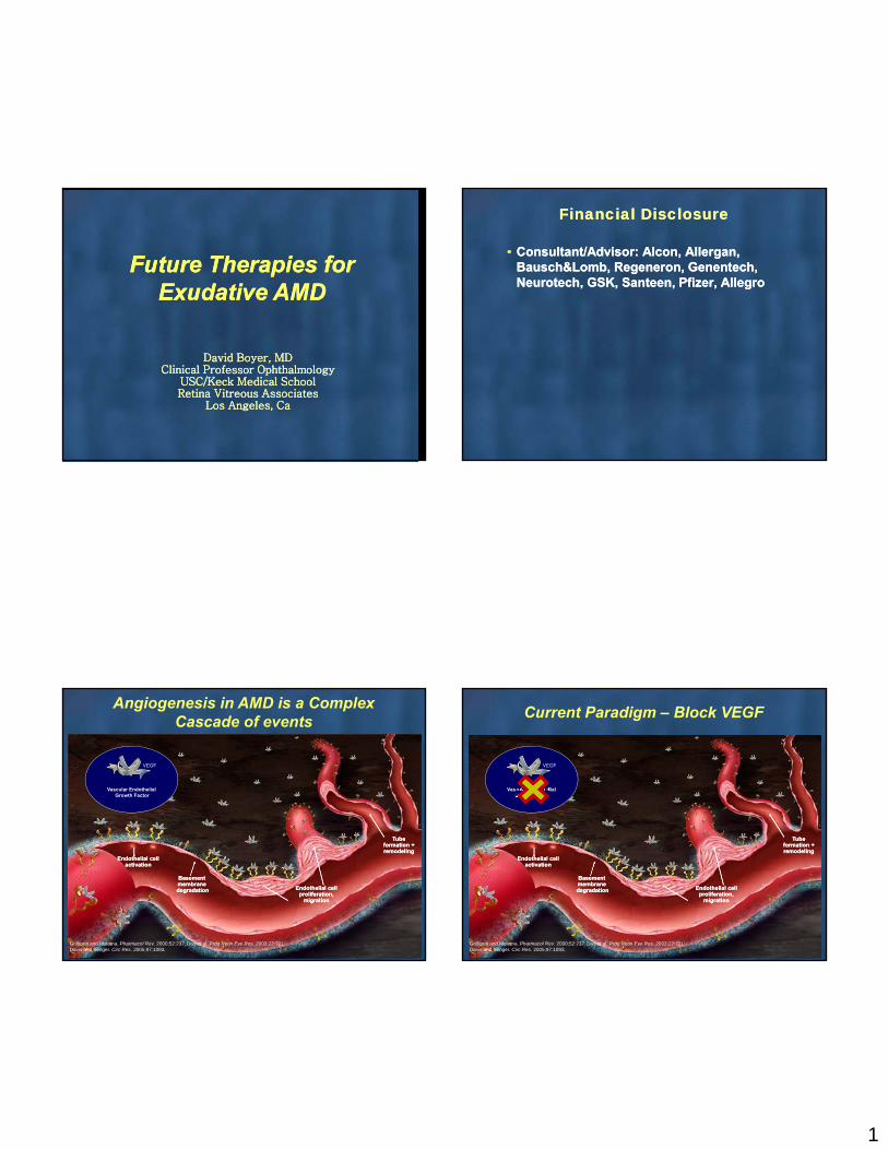
0102030405060708090
100
PDT(n=143)
Ranibizumab 0.5 mg(n=139)
90%‡
% o
f su
bjec
ts 66%
% o
f su
bjec
ts
0102030405060708090
100 90%†
53%
Sham(n=238)
Ranibizumab 0.5 mg(n=240)
*Month 12 was the primary endpoint in both trials; month 24 was a secondary endpoint.†P<0.01 vs sham; ‡P<0.01 vs PDT.
ANCHOR at month 24MARINA at month 24
Fibrovascular Proliferation
RPE Atrophy
Retinal Damage
Anti‐VEGF Non‐Responders
3
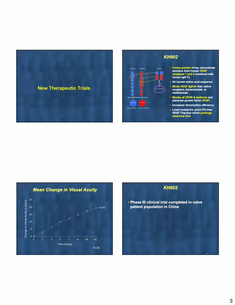
New Therapeutic Trials
Fusion protein of key extracellular domains from human VEGF receptors 1 and 2 combined with human IgG Fc
All human amino acid sequence
Binds VEGF tighter than native receptors, bevacizumab, or ranibizumab
Blocks all VEGF-A isoforms and placental growth factor (PlGF)
Increased dimerization efficiency
Lower isoelectric point (PI) than VEGF Trap-Eye which prolongs clearance time
1
2
3
4
5
6
7
1
2
3
4
5
6
7
Kinase Kinase
Kd 10-30 pM Kd 100-300 pM
VEGFR-1 VEGFR-2 KH902
Fc
Kd ~10 pM
KH902KH902
2
3
2
3
44
+19.61
0
5
10
15
20
25
0 1 3 5 7 14 28 42
Time (Days)
Cha
nge
in V
isua
l Acu
ity (
Lett
ers)
N=28
Mean Change in Visual Acuity Mean Change in Visual Acuity KH902KH902
Phase III clinical trial completed in naïve patient population in China Phase III clinical trial completed in naïve
patient population in China
12
4
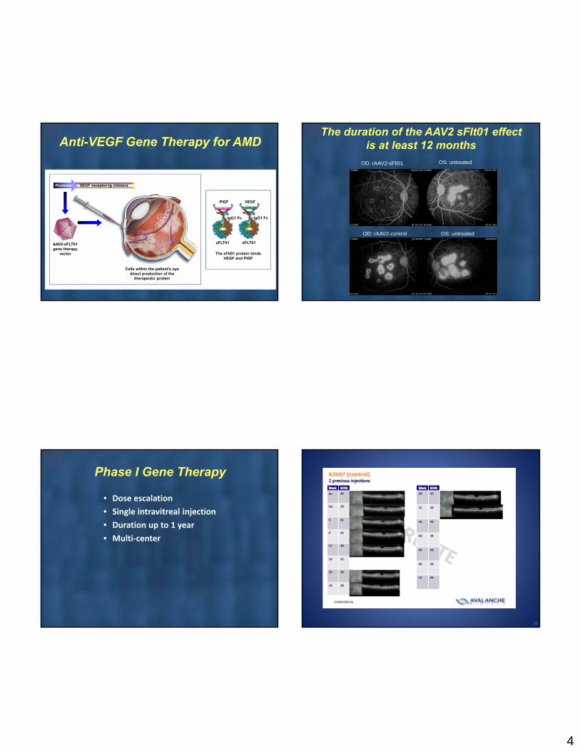
Anti-VEGF Gene Therapy for AMD
AAV2-sFLT01 gene therapy
vector
Cells within the patient’s eye direct production of the
therapeutic protein
VEGF receptor-Ig chimeraPromoter
sFLT01
PlGF
IgG1 Fc
sFLT01
VEGF
IgG1 Fc
The sFlt01 protein binds VEGF and PlGF
The duration of the AAV2 sFlt01 effect is at least 12 months
OS: untreatedOD: rAAV2-control
OD: rAAV2-sFlt01 OS: untreated
Phase I Gene Therapy
• Dose escalation
• Single intravitreal injection
• Duration up to 1 year
• Multi‐center
16

5
Encapsulated Cell Technology Neurotech
• Device contains human RPE cells (ARPE‐19) genetically modified to secrete a drug
• The device is surgically implanted in the vitreous through a tiny scleral incision and is anchored by a single suture through a titanium loop at one end of the device
• The semi‐permeable membrane allows the outward diffusion of drug and other cellular metabolites
• Allows inward diffusion of nutrients necessary to support the cell survival in the vitreous cavity while protecting the contents from host cellular immunologic attacK
• “Immunologically privileged”
• Biologic activity demonstrated in CNTF trial
Encapsulated Cell Technology Neurotech
• Neurotech device (NT‐503) encapsulating VEGF receptor Fc‐fusion protein (VEGFR‐Fc)‐releasing cells.
• This VEGFR‐Fc is 20‐fold more efficient in neutralizing VEGF compared with ranibizumab NT‐503 is confirmed to release VEGFR‐Fc constantly up to 1 year in the rabbit vitreous.
• A phase 1 clinical trial of NT‐503 for neovascular AMD is ongoing outside of the United States.
Patient A2301 –76 Y/O WomanGen 2 Double Implant
19
2 Months50 Letters295 um
40 Letters639 um
46 Letters355 um
Baseline 1 Month 3 Months53 Letters
301 um
4 Months54 Letters
324 um
5 Months52 Letters
314 um
6 Months60 Letters
288 um
8 Months65 Letters
283 um
+25 letter vision gain and ~350µ decrease in central foveal thickness
UpstreamUpstream
DownstreamDownstream
ExtracellularSpace
ExtracellularSpace
6
PI3K
S6
TSC1 TSC2
PIP3
Akt
PT‐308
PS‐473
Rheb GTP
HIF VEGF PathwayAngiogenesisVascular Permeability
Cell ProliferationAngiogenesis
TORC2
TORC1
PDK1
GSK Cyclin D1
P
P
Cell ProliferationAngiogenesisBCL‐2/BAX
Cell SurvivalAnti‐Apoptotic
MMP‐9
Migration
Growth Factor Receptors(GPCR, RTK, Integrin, Cytokine)
Upstream Pathways
MMP‐2
SirolimusEverolimus
SirolimusEverolimus
PF-655PF-655
PI3K
S6
TSC1 TSC2
PIP3
Akt
PT‐308
PS‐473
Rheb GTP
HIF VEGF PathwayAngiogenesisVascular Permeability
Cell ProliferationAngiogenesis
TORC2
TORC1
PDK1
GSK Cyclin D1
P
P
Cell ProliferationAngiogenesisBCL‐2/BAX
Cell SurvivalAnti‐Apoptotic
MMP‐9
Migration
P529X
X
XX
Growth Factor Receptors(GPCR, RTK, Integrin, Cytokine)
X
X
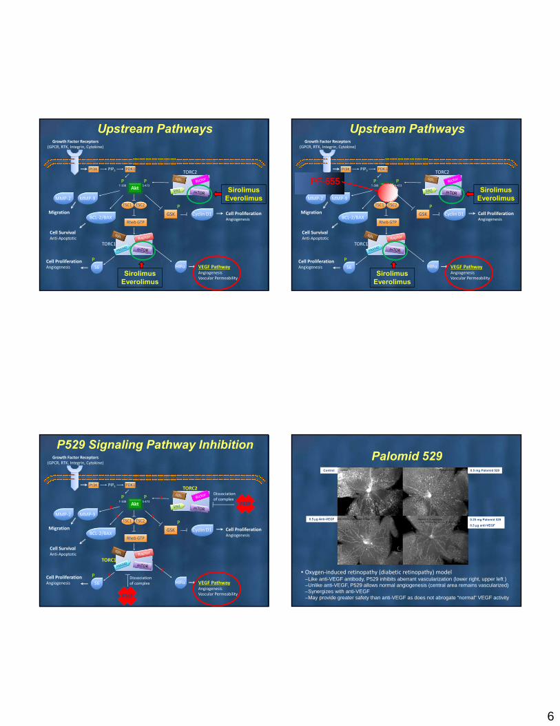
P529
P529 Signaling Pathway Inhibition
MMP‐2
Dissociationof complex
Dissociationof complex
Palomid 529Control 0.5 mg Palomid 529
0.5 g Anti-VEGF 0.25 mg Palomid 529
0.5 g anti-VEGF
• Oxygen‐induced retinopathy (diabetic retinopathy) model–Like anti-VEGF antibody, P529 inhibits aberrant vascularization (lower right, upper left )–Unlike anti-VEGF, P529 allows normal angiogenesis (central area remains vascularized)–Synergizes with anti-VEGF–May provide greater safety than anti-VEGF as does not abrogate “normal” VEGF activity
7
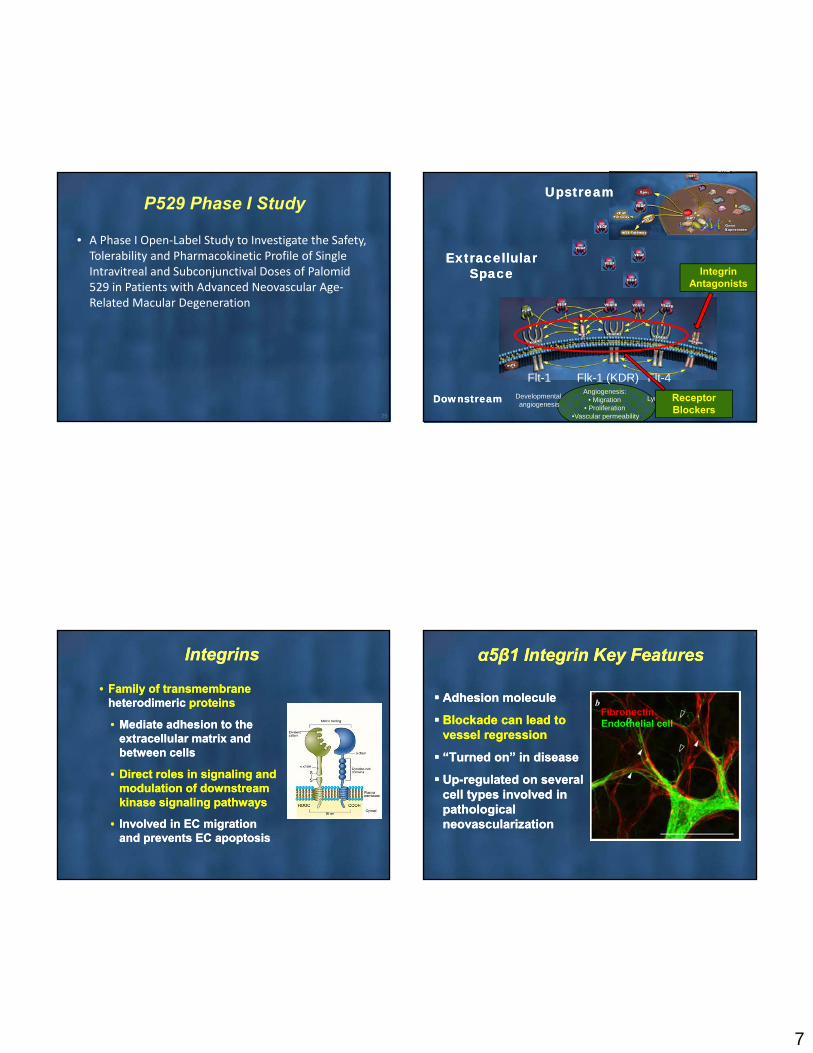
P529 Phase I Study
• A Phase I Open‐Label Study to Investigate the Safety, Tolerability and Pharmacokinetic Profile of Single Intravitreal and Subconjunctival Doses of Palomid 529 in Patients with Advanced Neovascular Age‐Related Macular Degeneration
25
UpstreamUpstream
ExtracellularSpace
ExtracellularSpace
Flt-1 Flk-1 (KDR) Flt-4
Lymphangiogenesis Developmental angiogenesis
Angiogenesis: • Migration
• Proliferation •Vascular permeability
DownstreamDownstream
Integrin Antagonists
Receptor Blockers
IntegrinsIntegrins
• Family of transmembraneheterodimeric proteins
• Mediate adhesion to the extracellular matrix and between cells
• Direct roles in signaling and modulation of downstream kinase signaling pathways
• Involved in EC migration and prevents EC apoptosis
• Family of transmembraneheterodimeric proteins
• Mediate adhesion to the extracellular matrix and between cells
• Direct roles in signaling and modulation of downstream kinase signaling pathways
• Involved in EC migration and prevents EC apoptosis
8
α5β1 Integrin Key Featuresα5β1 Integrin Key Features
Adhesion molecule
Blockade can lead to vessel regression
“Turned on” in disease
Up-regulated on several cell types involved in pathological neovascularization
Adhesion molecule
Blockade can lead to vessel regression
“Turned on” in disease
Up-regulated on several cell types involved in pathological neovascularization
FibronectinEndothelial cell

8
VolociximabVolociximab
• Volociximab is a chimeric (82% human/18%murine) IgG4 monoclonal antibody (Mab) against α5β1 integrin
• Volociximab is a chimeric (82% human/18%murine) IgG4 monoclonal antibody (Mab) against α5β1 integrin
IIA1 M200
Parent 82% human
Control D13 Control D20 Control D27
M200 D13 M200 D20 M200 D27
Inhibition of CNV: FA LeakageInhibition of CNV: FA Leakage
Rmakrishnan V. et al J Exp Ther Oncol. 2006;5(4):273-86
DownstreamDownstream
Tyrosine kinases
VEGF Activation
DownstreamDownstream
Tyrosine kinases
VEGF Activation
•Tyrosine kinases are enzymes that provide a central switch mechanism in signal transduction pathways
•Phosphorylation of proteins
•Involved in cell proliferation, metabolism, survival and apoptosis
•Tyrosine kinases are enzymes that provide a central switch mechanism in signal transduction pathways
•Phosphorylation of proteins
•Involved in cell proliferation, metabolism, survival and apoptosis

9
DownstreamDownstream
Tyrosine kinases
Tyrosine Kinase Inhibitors:
Pazopanib (gtts)Vatalanib (po)
AG013958 (pst)AL39324 (pst)
X-82X-82 is a potential new treatment for wet AMD
It is unique because: It is given orally
It blocks both VEGF and PDGFThis study is designed to evaluate its safety and
preliminary efficacy in patients with wet AMDCompleting a Phase 1/2 trial
Orally administered
X-82 for wet AMDX-82 is able to inhibit pVEGFR, pPDGFR, HUVEC cell growth, and blood
vessel tube formation at very low concentrations (~50nM), much lower than what is required for cancer.
X-82 at 50mg is able to reach and exceed the concentration required for anti-angiogenesis
Average AUC of cancer pts 4,6,9 (5732 ng.hr/ml) is 4.5x that of pts taking 50mg QD (1282 ng.hr/ml)
QOD will evaluate if constant inhibition is necessary for efficacy
The effect of CM082 on HUVEC tube formation induced by VEGF
0
20
40
60
80
100
control VEGF VEGF+Sutent VEGF+CM082
Nu
mber
of
tubes
50nM
100nM
Effect of X-82 on HUVEC tube formation induced by VEGF Concentration (ng/ml) – time (hr) curve of X-82
Oncology Patient with AMD
Started X-82 100 mg tablet on 10-25-12AMD Diagnosed 11-06-12 Symptoms noted at baseline
Official diagnosis: Peripapillary atrophy with peripapillary choroidal neovacular membrane and
subretinal bloodBCVA improved from 20/60 to 20/25
Leakage from peripapillary subretinal neovascular membrane has improved
Subretinal fluid is much less
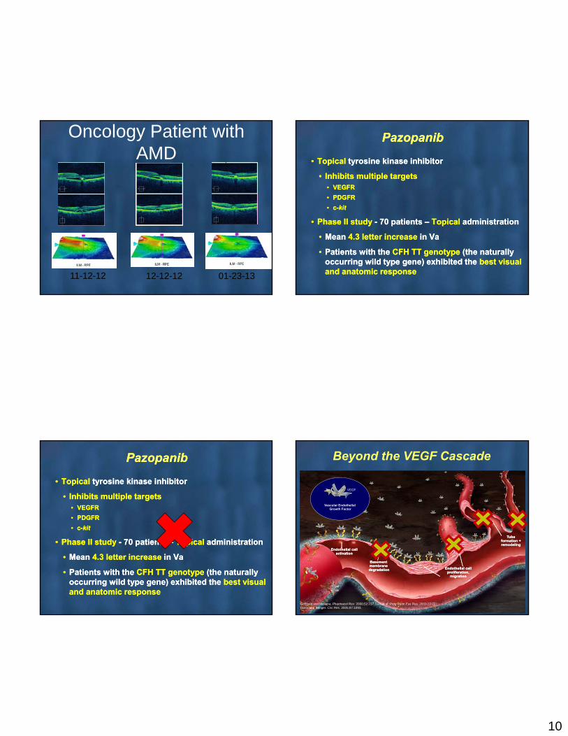
10
Oncology Patient with AMD
11-12-12 12-12-12 01-23-13
PazopanibPazopanib
• Topical tyrosine kinase inhibitor
• Inhibits multiple targets• VEGFR
• PDGFR
• c-kit
• Phase II study - 70 patients – Topical administration
• Mean 4.3 letter increase in Va
• Patients with the CFH TT genotype (the naturally occurring wild type gene) exhibited the best visual and anatomic response
Beyond the VEGF Cascade
Griffioen and Molema. Pharmacol Rev. 2000;52:237; Das et al. Prog Retin Eye Res. 2003;22:721;Davis and Senger. Circ Res. 2005;97:1093.
Basementmembranedegradation
Basementmembranedegradation
Tubeformation +remodeling
Tubeformation +remodeling
VEGF
Vascular Endothelial Growth Factor
Endothelial cell activation
Endothelial cell activation
Endothelial cell proliferation,
migration
Endothelial cell proliferation,
migration
11
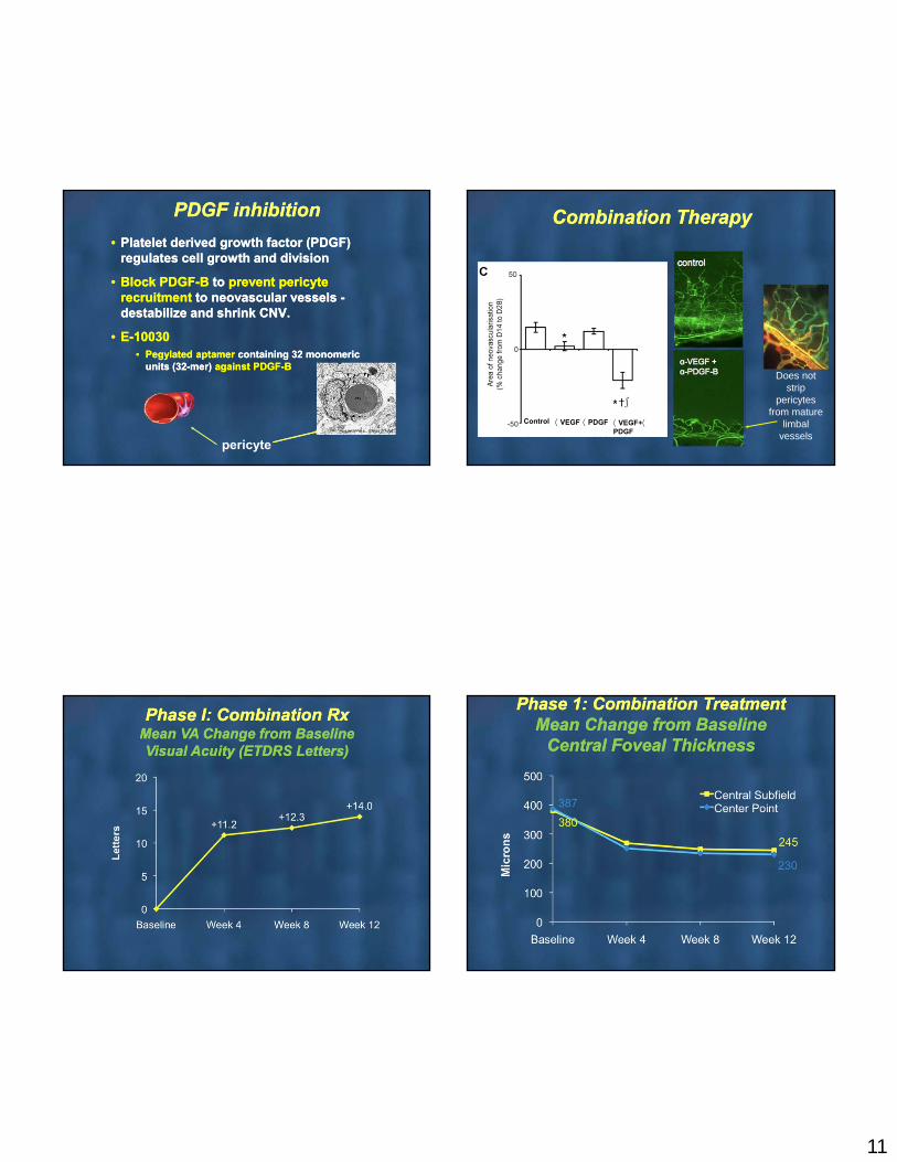
PDGF inhibitionPDGF inhibition
• Platelet derived growth factor (PDGF) regulates cell growth and division
• Block PDGF-B to prevent pericyte recruitment to neovascular vessels -destabilize and shrink CNV.
• E-10030• Pegylated aptamer containing 32 monomeric
units (32-mer) against PDGF-B
• Platelet derived growth factor (PDGF) regulates cell growth and division
• Block PDGF-B to prevent pericyte recruitment to neovascular vessels -destabilize and shrink CNV.
• E-10030• Pegylated aptamer containing 32 monomeric
units (32-mer) against PDGF-B
pericyte
Combination TherapyCombination Therapy
Does not strip
pericytes from mature
limbal vessels
Control VEGF PDGF VEGF+PDGF
Phase I: Combination Rx Mean VA Change from BaselineVisual Acuity (ETDRS Letters)
Phase I: Combination Rx Mean VA Change from BaselineVisual Acuity (ETDRS Letters)
Phase 1: Combination TreatmentMean Change from Baseline
Central Foveal Thickness
Phase 1: Combination TreatmentMean Change from Baseline
Central Foveal Thickness
12
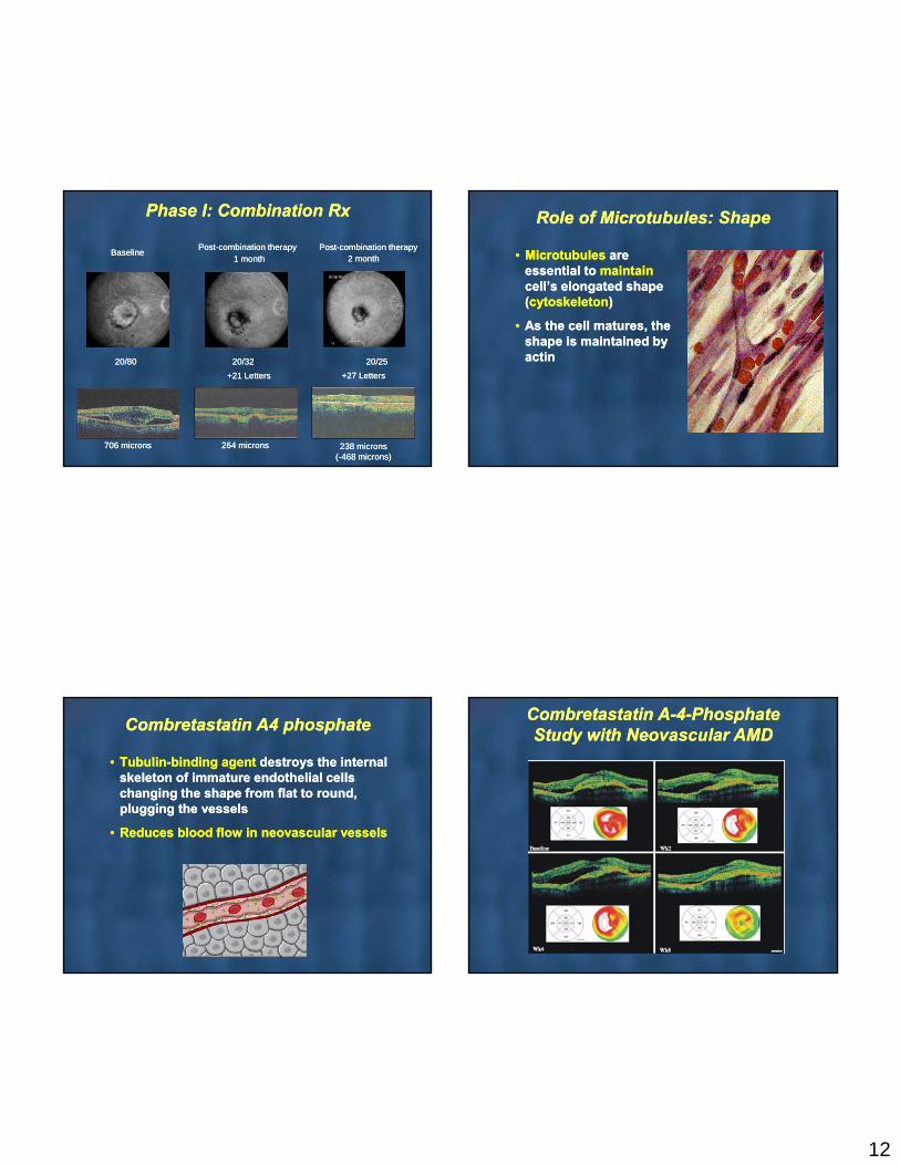
Phase I: Combination RxPhase I: Combination Rx
+21 Letters+21 Letters +27 Letters+27 Letters
BaselineBaselinePost-combination therapyPost-combination therapy
1 month1 monthPost-combination therapyPost-combination therapy
2 month2 month
20/80 20/32 20/2520/80 20/32 20/25
706 microns706 microns 264 microns264 microns 238 microns(-468 microns)238 microns
(-468 microns)
Role of Microtubules: ShapeRole of Microtubules: Shape
• Microtubules are essential to maintain cell’s elongated shape (cytoskeleton)
• As the cell matures, the shape is maintained by actin
• Microtubules are essential to maintain cell’s elongated shape (cytoskeleton)
• As the cell matures, the shape is maintained by actin
Combretastatin A4 phosphateCombretastatin A4 phosphate
• Tubulin-binding agent destroys the internal skeleton of immature endothelial cells changing the shape from flat to round, plugging the vessels
• Reduces blood flow in neovascular vessels
• Tubulin-binding agent destroys the internal skeleton of immature endothelial cells changing the shape from flat to round, plugging the vessels
• Reduces blood flow in neovascular vessels
Combretastatin A-4-Phosphate Study with Neovascular AMD
Combretastatin A-4-Phosphate Study with Neovascular AMD
13
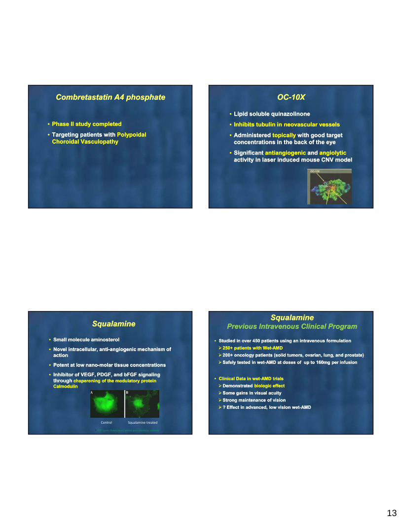
Combretastatin A4 phosphateCombretastatin A4 phosphate
• Phase II study completed
• Targeting patients with Polypoidal Choroidal Vasculopathy
• Phase II study completed
• Targeting patients with Polypoidal Choroidal Vasculopathy
OC-10XOC-10X
• Lipid soluble quinazolinone
• Inhibits tubulin in neovascular vessels
• Administered topically with good target concentrations in the back of the eye
• Significant antiangiogenic and angiolyticactivity in laser induced mouse CNV model
• Lipid soluble quinazolinone
• Inhibits tubulin in neovascular vessels
• Administered topically with good target concentrations in the back of the eye
• Significant antiangiogenic and angiolyticactivity in laser induced mouse CNV model
SqualamineSqualamine
• Small molecule aminosterol
• Novel intracellular, anti-angiogenic mechanism of action
• Potent at low nano-molar tissue concentrations
• Inhibitor of VEGF, PDGF, and bFGF signaling through chaperoning of the modulatory protein Calmodulin
• Small molecule aminosterol
• Novel intracellular, anti-angiogenic mechanism of action
• Potent at low nano-molar tissue concentrations
• Inhibitor of VEGF, PDGF, and bFGF signaling through chaperoning of the modulatory protein Calmodulin
Control Squalamine treated
*FITC (green fluorescence) labeled anti-calmodulin antibody
SqualaminePrevious Intravenous Clinical Program
SqualaminePrevious Intravenous Clinical Program
• Studied in over 450 patients using an intravenous formulation
250+ patients with Wet-AMD
200+ oncology patients (solid tumors, ovarian, lung, and prostate)
Safely tested in wet-AMD at doses of up to 160mg per infusion
• Clinical Data in wet-AMD trials
Demonstrated biologic effect
Some gains in visual acuity
Strong maintenance of vision
? Effect in advanced, low vision wet-AMD

14
SqualaminePrevious Intravenous Clinical Program
SqualaminePrevious Intravenous Clinical Program
• Entered phase III trials for wet-AMD under fast track status and a Special Protocol Assessment (US FDA)
Discontinued due to
Enrollment difficulty of an IV approach
Changing competitive landscape of ranibizumab and bevacizumab Lack of commercial potential for chronic IV infusions
Suboptimal dosing due to relatively short half life when given systemically
SqualamineNew Topical Clinical Program
SqualamineNew Topical Clinical Program
• Preclinical testing
• Safe to ocular tissues
• Anti-angiogenic concentrations achieved in posterior sclera and choroid
• No quantifiable uptake in aqueous humor
• Negligible systemic uptake
• Clinical Trials
Phase II multi-center, randomized, placebo controlled trial
Treatment of wet-AMD
Initiation expected in mid 2012
• hI-con1 is a recombinant protein designed to target Tissue Factor (TF)
• hI-con1 binds very tightly to TF
• TF present on inner surface of CNV but not normal blood vessels
• Binding of hI-con1 to CNV triggers cell-mediated cytotoxicity via Natural Killer cells
• Anticipated result: REGRESSION of CNV
55
hl-con1
• CNV Regression
• Significant number of laser‐generated lesions (76
spots/eye)
• Single IVT dose of hI‐con1 10 days after laser burn
•Measurement of CNV 4 days later
Pig wet AMD Model: hI-con1 Triggers CNV Regression in a Dose-Dependent Manner
• Longer resorption = longer treatment window = higher
opportunity to assess whether or not pre‐formed CNV can be reversed by I‐con1 treatment
RATIONALE
DESIGN
GOAL
hI‐con1 destroys established laser‐induced CNV in a dose‐dependent fashion
Total Delivered Dose (micrograms)
Control 25 50 100 200
% C
NV
10
20
30
40
50
60
70
80
90
60.3
49.3
38.3
13.53.2 10.2
[ 75%]
[ 50%]
[ 25%]
n = 56
n = 100
n = 85
n = 75n = 111
CONCLUSION
15
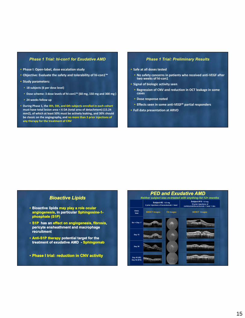
Phase 1 Trial: hI-con1 for Exudative AMD
• Phase I: Open‐label, dose escalation study
• Objective: Evaluate the safety and tolerability of hI‐con1™
• Study parameters:
• 18 subjects (6 per dose level)
• Dose scheme: 3 dose levels of hI‐con1™ (60 mg, 150 mg and 300 mg )
• 24 weeks follow up
• During Phase 1, the 4th, 5th, and 6th subjects enrolled in each cohort must have total lesion area < 6 DA (total area of detachment) (15.24 mm2), of which at least 50% must be actively leaking, and 30% should be classic on the angiography, and no more than 3 prior injections of any therapy for the treatment of CNV
Phase 1 Trial: Preliminary Results
• Safe at all doses tested
• No safety concerns in patients who received anti‐VEGF after two weeks of hI‐con1
• Signal of biologic activity seen
• Regression of CNV and reduction in OCT leakage in some cases
• Dose response noted
• Effects seen in some anti‐VEGF® partial responders
• Full data presentation at ARVO
Bioactive LipidsBioactive Lipids
• Bioactive lipids may play a role ocular angiogenesis, in particular Sphingosine-1-phosphate (S1P)
• S1P has an effect on angiogenesis, fibrosis, pericyte ensheathment and macrophage recruitment
• Anti-S1P therapy potential target for the treatment of exudative AMD - Sphingomab
• Phase I trial: reduction in CNV activity
PED and Exudative AMDNeither subject was re-treated with anything for 12+ months
PED and Exudative AMDNeither subject was re-treated with anything for 12+ months
Subject #2: 0.2 mg
2 prior injections of bevacizumab + laser
Subject #15: 1.8 mg
16 prior injections of ranibizumab/bevacizumab + laser + dex.
Clinic
VisitSDOCT Images FA Images SDOCT Images
Scr = Day -7
Day 15
Day 30
Day 45 (#2)
Day 54 (#15)
16
In vitro VEGF Inhibition Assay
DARPinTarget
DARPin’s
•Novel class of binding proteins
•High affinity and specificity for many targets
MP0112
•VEGF antagonizing DARPin
•IC50 of <10 pM
•Half-life in the eye >6 days
•Stable at RT for >several months
•Produced at 7-8 g/L in bacteria(E Coli)
•Formulation as a liquid
•Well tolerated in phase 1 testing
Designed Ankyrin Repeat Protein
* Molecular Partners and Allergan
DARPin MP0112*
Association for Research and Vision Meeting, May 1-5, 2011 Ft. Lauderdale
Drug activity in the vitreous over time
Days
B
A
0 10 20 30 40 50 60 70 80 90 100 110 120 130 140
10
1
0.1
0.01
0.001
1x10⁻⁴
1x10⁻⁵
1x10⁻⁶
Co
nce
ntr
atio
n (
mM
) MP0112MP0112 high e
MP0112: Possible less frequent dosing
A. PK and stability
Half life: MP0112 >6 days
Half-life: Ranibizumab = 3 days
Stability: MP0112 >>> Ranibizumab
B. Efficacy
IC50 : MP0112 <10 pM
IC50 : Ranibizumab >250 pM
RedosingRanibizumab
RedosingMP0112
Ranibizumabdos
DARPin MP0112
IRay
• X-Ray Beams
• Robotic control with eye stabilization
• Non-invasive
• Outpatient
X-Ray Therapy*
*Oraya Therapeutics
Complement inhibitionComplement inhibition
• Complement protein polymorphisms are associated with drusen formation and all stages of AMD
• Complement inhibition could provide therapeutic benefits at various stages of disease progression
• Choroidal neovascularization
• Transition from dry to wet AMD
• Geographic atrophy
• Drusen formation
• Complement protein polymorphisms are associated with drusen formation and all stages of AMD
• Complement inhibition could provide therapeutic benefits at various stages of disease progression
• Choroidal neovascularization
• Transition from dry to wet AMD
• Geographic atrophy
• Drusen formation
17
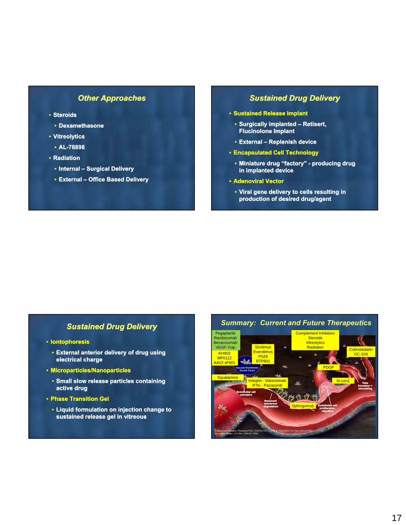
Other ApproachesOther Approaches
• Steroids
• Dexamethasone
• Vitreolytics
• AL-78898
• Radiation
• Internal – Surgical Delivery
• External – Office Based Delivery
• Steroids
• Dexamethasone
• Vitreolytics
• AL-78898
• Radiation
• Internal – Surgical Delivery
• External – Office Based Delivery
Sustained Drug DeliverySustained Drug Delivery
• Sustained Release Implant
• Surgically implanted – Retisert, Flucinolone Implant
• External – Replenish device
• Encapsulated Cell Technology
• Miniature drug “factory” - producing drug in implanted device
• Adenoviral Vector
• Viral gene delivery to cells resulting in production of desired drug/agent
Sustained Drug DeliverySustained Drug Delivery
• Iontophoresis
• External anterior delivery of drug using electrical charge
• Microparticles/Nanoparticles
• Small slow release particles containing active drug
• Phase Transition Gel
• Liquid formulation on injection change to sustained release gel in vitreous
• Iontophoresis
• External anterior delivery of drug using electrical charge
• Microparticles/Nanoparticles
• Small slow release particles containing active drug
• Phase Transition Gel
• Liquid formulation on injection change to sustained release gel in vitreous
Summary: Current and Future Therapeutics
Griffioen and Molema. Pharmacol Rev. 2000;52:237; Das et al. Prog Retin Eye Res. 2003;22:721;Davis and Senger. Circ Res. 2005;97:1093.
Basementmembranedegradation
Basementmembranedegradation
Tubeformation +remodeling
Tubeformation +remodeling
Vascular Endothelial Growth Factor
Endothelial cell activation
Endothelial cell activation
Endothelial cell proliferation,
migration
Endothelial cell proliferation,
migration
PegaptanibRanibizumabBevacizumabVEGF-Trap
KH902MP0112
AAV2 sFlt01
SirolimusEverolimus
P529 RTP801
Integrin - VolociximabRTki - Pazopanib
PDGF
CobretastatinOC-10X
Sphingomab
Complement InhibitorsSteroids
VitreolyticsRadiation
hI-con1Squalamine

18
Thank YouThank You