Embed Size (px)
Citation preview
Cellular/Molecular
GABA, Its Receptors, and GABAergic Inhibition in MouseTaste Buds
Gennady Dvoryanchikov,1* Yijen A. Huang,1* Rene Barro-Soria,1 Nirupa Chaudhari,1,2 and Stephen D. Roper1,2
1Department of Physiology and Biophysics and 2Program in Neuroscience, Miller School of Medicine, University of Miami, Miami, Florida 33136
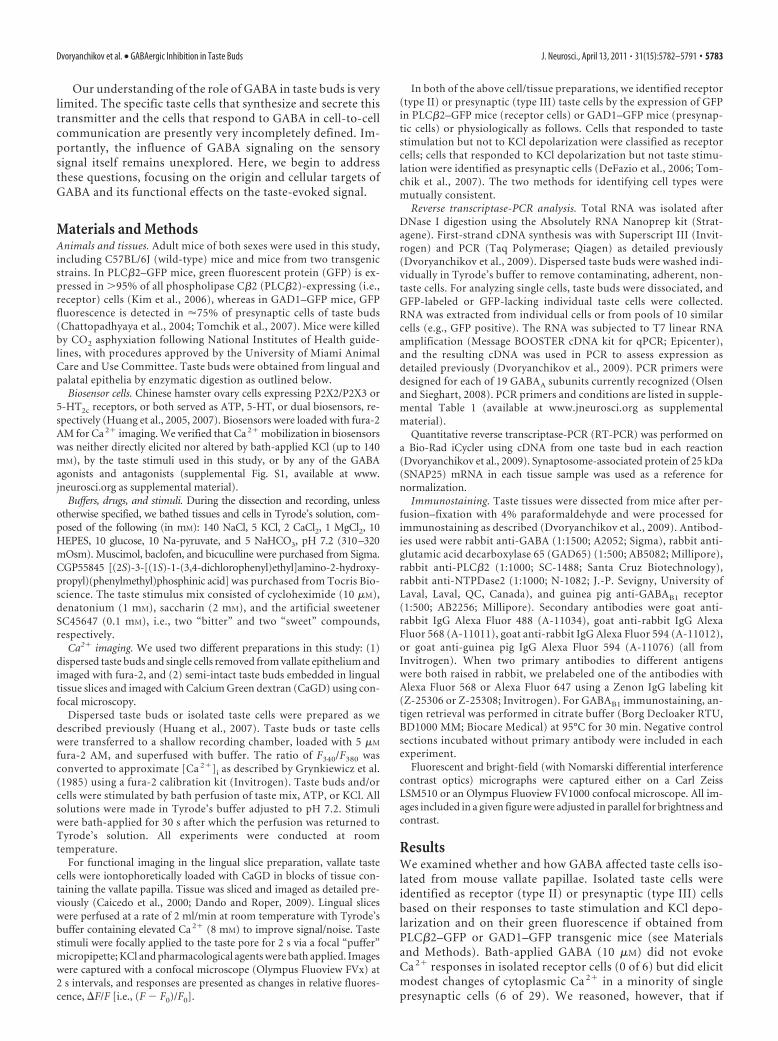
Taste buds consist of at least three principal cell types that have different functions in processing gustatory signals: glial-like (typeI) cells, receptor (type II) cells, and presynaptic (type III) cells. Using a combination of Ca 2� imaging, single-cell reversetranscriptase-PCR and immunostaining, we show that GABA is an inhibitory transmitter in mouse taste buds, acting on GABAA
and GABAB receptors to suppress transmitter (ATP) secretion from receptor cells during taste stimulation. Specifically, receptorcells express GABAA receptor subunits �2, �, and �, as well as GABAB receptors. In contrast, presynaptic cells express the GABAA
�3 subunit and only occasionally GABAB receptors. In keeping with the distinct expression pattern of GABA receptors in presyn-aptic cells, we detected no GABAergic suppression of transmitter release from presynaptic cells. We suggest that GABA may servefunction(s) in taste buds in addition to synaptic inhibition. Finally, we also defined the source of GABA in taste buds: GABA issynthesized by GAD65 in type I taste cells as well as by GAD67 in presynaptic (type III) taste cells and is stored in both those two celltypes. We conclude that GABA is an inhibitory transmitter released during taste stimulation and possibly also during growth anddifferentiation of taste buds.
IntroductionMammalian taste buds contain three morphologically andfunctionally distinct cell types (for review, see Chaudhari andRoper, 2010). Type I cells appear to be supporting or glial-likecells (Bartel et al., 2006; Dvoryanchikov et al., 2009). Some ofthe type I cells may also play a role in salt (Na �) taste (Van-denbeuch et al., 2008; Chandrashekar et al., 2010). Type II(receptor) cells are the primary detectors of sweet, bitter, andumami compounds; they express G-protein-coupled taste re-ceptors and effectors for these taste stimuli (Perez et al., 2002;Zhao et al., 2003; Clapp et al., 2004; DeFazio et al., 2006). TypeIII (presynaptic) cells detect sour tastants. Presynaptic cellsalso are the only taste bud cells showing well-differentiated synapsesand expressing synaptic proteins (Yee et al., 2001; DeFazio etal., 2006).
During taste stimulation and after the primary transduc-tion response, the different types of cells in the taste bud in-teract and process gustatory signals via chemical signalingintrinsic to the taste bud. Taste stimulation triggers receptorcells to secrete ATP and presynaptic cells to release serotonin(5-HT) and norepinephrine (Dvoryanchikov et al., 2007;Huang et al., 2007, 2008; Romanov et al., 2007). ATP appears
to be a transmitter between receptor cells and primary afferentnerve fibers (Finger et al., 2005; Huang et al., 2007; Romanovet al., 2007). Both 5-HT and ATP play critical roles in cell-to-cell signaling within the taste bud, establishing positive andnegative feedback circuits that shape the afferent signal andmay contribute to the coding of sensory information (Roper,2007; Huang et al., 2009). Other transmitters such as gluta-mate and acetylcholine also serve in cell– cell communicationwithin the taste bud (Ogura et al., 2007; Vandenbeuch et al.,2010). Additionally, cholecystokinin and neuropeptide Y mayfunction in this capacity (Herness and Zhao, 2009).
In addition to the above transmitters, there is evidence thatan inhibitory amino acid transmitter, GABA, figures in tastebuds. Early immunocytochemical and autoradiography datarevealed GABA in taste cells and gustatory nerve endings inamphibians and rodents (Jain and Roper, 1991; Obata et al.,1997; Nagai et al., 1998). Electrophysiological recordings fromsensory ganglion cells that innervate taste buds showed thatGABA mainly produces hyperpolarizing responses when ap-plied to the cell body (Koga and Bradley, 2000). This wasinterpreted as a possible role for GABA as an afferent tastetransmitter at the central and/or peripheral sensory endings ofthese ganglion cells. More recently, patch-clamp recordingshave shown that GABA hyperpolarizes cells in rat taste buds(Cao et al., 2009). Those workers proposed that GABA is in-volved in cell-to-cell communication within taste buds. Re-sponses to GABA can be produced via ionotropic (GABAA)and metabotropic (GABAB) receptors. In different cells, re-sponses to GABA may vary depending on the intracellularconcentration of Cl �, the particular receptor subunits ex-pressed, and the signaling pathways within cells.
Received Oct. 7, 2010; revised Dec. 26, 2010; accepted Feb. 10, 2011.This work was supported by National Institutes of Health/National Institute on Deafness and Other Communica-
tion Disorders Grants R01DC7630 (S.D.R.), R01DC374 (S.D.R.), and R01DC6308 (N.C.).*G.D. and Y.A.H. contributed equally to this work.The authors declare no competing financial interests.Correspondence should be addressed to either Stephen D. Roper or Nirupa Chaudhari, Department of
Physiology and Biophysics, Miller School of Medicine, 1600 N.W. 10th Avenue, Miami, FL 33136, E-mail:[email protected] or [email protected].
DOI:10.1523/JNEUROSCI.5559-10.2011Copyright © 2011 the authors 0270-6474/11/315782-10$15.00/0
5782 • The Journal of Neuroscience, April 13, 2011 • 31(15):5782–5791
Our understanding of the role of GABA in taste buds is verylimited. The specific taste cells that synthesize and secrete thistransmitter and the cells that respond to GABA in cell-to-cellcommunication are presently very incompletely defined. Im-portantly, the influence of GABA signaling on the sensorysignal itself remains unexplored. Here, we begin to addressthese questions, focusing on the origin and cellular targets ofGABA and its functional effects on the taste-evoked signal.
Materials and MethodsAnimals and tissues. Adult mice of both sexes were used in this study,including C57BL/6J (wild-type) mice and mice from two transgenicstrains. In PLC�2–GFP mice, green fluorescent protein (GFP) is ex-pressed in �95% of all phospholipase C�2 (PLC�2)-expressing (i.e.,receptor) cells (Kim et al., 2006), whereas in GAD1–GFP mice, GFPfluorescence is detected in �75% of presynaptic cells of taste buds(Chattopadhyaya et al., 2004; Tomchik et al., 2007). Mice were killedby CO2 asphyxiation following National Institutes of Health guide-lines, with procedures approved by the University of Miami AnimalCare and Use Committee. Taste buds were obtained from lingual andpalatal epithelia by enzymatic digestion as outlined below.
Biosensor cells. Chinese hamster ovary cells expressing P2X2/P2X3 or5-HT2c receptors, or both served as ATP, 5-HT, or dual biosensors, re-spectively (Huang et al., 2005, 2007). Biosensors were loaded with fura-2AM for Ca 2� imaging. We verified that Ca 2� mobilization in biosensorswas neither directly elicited nor altered by bath-applied KCl (up to 140mM), by the taste stimuli used in this study, or by any of the GABAagonists and antagonists (supplemental Fig. S1, available at www.jneurosci.org as supplemental material).
Buffers, drugs, and stimuli. During the dissection and recording, unlessotherwise specified, we bathed tissues and cells in Tyrode’s solution, com-posed of the following (in mM): 140 NaCl, 5 KCl, 2 CaCl2, 1 MgCl2, 10HEPES, 10 glucose, 10 Na-pyruvate, and 5 NaHCO3, pH 7.2 (310–320mOsm). Muscimol, baclofen, and bicuculline were purchased from Sigma.CGP55845 [(2S)-3-[(1S)-1-(3,4-dichlorophenyl)ethyl]amino-2-hydroxy-propyl)(phenylmethyl)phosphinic acid] was purchased from Tocris Bio-science. The taste stimulus mix consisted of cycloheximide (10 �M),denatonium (1 mM), saccharin (2 mM), and the artificial sweetenerSC45647 (0.1 mM), i.e., two “bitter” and two “sweet” compounds,respectively.
Ca2� imaging. We used two different preparations in this study: (1)dispersed taste buds and single cells removed from vallate epithelium andimaged with fura-2, and (2) semi-intact taste buds embedded in lingualtissue slices and imaged with Calcium Green dextran (CaGD) using con-focal microscopy.
Dispersed taste buds or isolated taste cells were prepared as wedescribed previously (Huang et al., 2007). Taste buds or taste cellswere transferred to a shallow recording chamber, loaded with 5 �M
fura-2 AM, and superfused with buffer. The ratio of F340/F380 wasconverted to approximate [Ca 2�]i as described by Grynkiewicz et al.(1985) using a fura-2 calibration kit (Invitrogen). Taste buds and/orcells were stimulated by bath perfusion of taste mix, ATP, or KCl. Allsolutions were made in Tyrode’s buffer adjusted to pH 7.2. Stimuliwere bath-applied for 30 s after which the perfusion was returned toTyrode’s solution. All experiments were conducted at roomtemperature.
For functional imaging in the lingual slice preparation, vallate tastecells were iontophoretically loaded with CaGD in blocks of tissue con-taining the vallate papilla. Tissue was sliced and imaged as detailed pre-viously (Caicedo et al., 2000; Dando and Roper, 2009). Lingual sliceswere perfused at a rate of 2 ml/min at room temperature with Tyrode’sbuffer containing elevated Ca 2� (8 mM) to improve signal/noise. Tastestimuli were focally applied to the taste pore for 2 s via a focal “puffer”micropipette; KCl and pharmacological agents were bath applied. Imageswere captured with a confocal microscope (Olympus Fluoview FVx) at2 s intervals, and responses are presented as changes in relative fluores-cence, �F/F [i.e., (F � F0)/F0].
In both of the above cell/tissue preparations, we identified receptor(type II) or presynaptic (type III) taste cells by the expression of GFPin PLC�2–GFP mice (receptor cells) or GAD1–GFP mice (presynap-tic cells) or physiologically as follows. Cells that responded to tastestimulation but not to KCl depolarization were classified as receptorcells; cells that responded to KCl depolarization but not taste stimu-lation were identified as presynaptic cells (DeFazio et al., 2006; Tom-chik et al., 2007). The two methods for identifying cell types weremutually consistent.
Reverse transcriptase-PCR analysis. Total RNA was isolated afterDNase I digestion using the Absolutely RNA Nanoprep kit (Strat-agene). First-strand cDNA synthesis was with Superscript III (Invit-rogen) and PCR (Taq Polymerase; Qiagen) as detailed previously(Dvoryanchikov et al., 2009). Dispersed taste buds were washed indi-vidually in Tyrode’s buffer to remove contaminating, adherent, non-taste cells. For analyzing single cells, taste buds were dissociated, andGFP-labeled or GFP-lacking individual taste cells were collected.RNA was extracted from individual cells or from pools of 10 similarcells (e.g., GFP positive). The RNA was subjected to T7 linear RNAamplification (Message BOOSTER cDNA kit for qPCR; Epicenter),and the resulting cDNA was used in PCR to assess expression asdetailed previously (Dvoryanchikov et al., 2009). PCR primers weredesigned for each of 19 GABAA subunits currently recognized (Olsenand Sieghart, 2008). PCR primers and conditions are listed in supple-mental Table 1 (available at www.jneurosci.org as supplementalmaterial).
Quantitative reverse transcriptase-PCR (RT-PCR) was performed ona Bio-Rad iCycler using cDNA from one taste bud in each reaction(Dvoryanchikov et al., 2009). Synaptosome-associated protein of 25 kDa(SNAP25) mRNA in each tissue sample was used as a reference fornormalization.
Immunostaining. Taste tissues were dissected from mice after per-fusion–fixation with 4% paraformaldehyde and were processed forimmunostaining as described (Dvoryanchikov et al., 2009). Antibod-ies used were rabbit anti-GABA (1:1500; A2052; Sigma), rabbit anti-glutamic acid decarboxylase 65 (GAD65) (1:500; AB5082; Millipore),rabbit anti-PLC�2 (1:1000; SC-1488; Santa Cruz Biotechnology),rabbit anti-NTPDase2 (1:1000; N-1082; J.-P. Sevigny, University ofLaval, Laval, QC, Canada), and guinea pig anti-GABAB1 receptor(1:500; AB2256; Millipore). Secondary antibodies were goat anti-rabbit IgG Alexa Fluor 488 (A-11034), goat anti-rabbit IgG AlexaFluor 568 (A-11011), goat anti-rabbit IgG Alexa Fluor 594 (A-11012),or goat anti-guinea pig IgG Alexa Fluor 594 (A-11076) (all fromInvitrogen). When two primary antibodies to different antigenswere both raised in rabbit, we prelabeled one of the antibodies withAlexa Fluor 568 or Alexa Fluor 647 using a Zenon IgG labeling kit(Z-25306 or Z-25308; Invitrogen). For GABAB1 immunostaining, an-tigen retrieval was performed in citrate buffer (Borg Decloaker RTU,BD1000 MM; Biocare Medical) at 95°C for 30 min. Negative controlsections incubated without primary antibody were included in eachexperiment.
Fluorescent and bright-field (with Nomarski differential interferencecontrast optics) micrographs were captured either on a Carl ZeissLSM510 or an Olympus Fluoview FV1000 confocal microscope. All im-ages included in a given figure were adjusted in parallel for brightness andcontrast.
ResultsWe examined whether and how GABA affected taste cells iso-lated from mouse vallate papillae. Isolated taste cells wereidentified as receptor (type II) or presynaptic (type III) cellsbased on their responses to taste stimulation and KCl depo-larization and on their green fluorescence if obtained fromPLC�2–GFP or GAD1–GFP transgenic mice (see Materialsand Methods). Bath-applied GABA (10 �M) did not evokeCa 2� responses in isolated receptor cells (0 of 6) but did elicitmodest changes of cytoplasmic Ca 2� in a minority of singlepresynaptic cells (6 of 29). We reasoned, however, that if
Dvoryanchikov et al. • GABAergic Inhibition in Taste Buds J. Neurosci., April 13, 2011 • 31(15):5782–5791 • 5783
GABA was an inhibitory neurotransmitter, as is the case else-where in the nervous system and described for rat taste cells byCao et al. (2009), one might not necessarily observe significantGABA-evoked Ca 2� mobilization (as indeed the case, shownbelow). Thus, we turned to other measures of taste bud func-tion to measure the action of GABA.
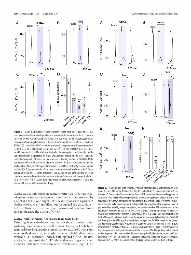
GABA inhibits transmitter release from taste budsUsing highly sensitive biosensors, we have shown previously thatgustatory stimulation elicits 5-HT release from intact taste budsremoved from lingual epithelium (Huang et al., 2005). Using thatsame methodology, we now asked whether GABA alters taste-evoked 5-HT secretion. Indeed, bath-applied GABA (10 �M)markedly suppressed the 5-HT release that was triggered whendispersed taste buds were stimulated with tastants (Fig. 1). To
Figure 2. GABA inhibits taste-evoked ATP release from taste buds. Taste stimulation as inFigure 1 evokes ATP release that is reduced by 10 �M GABA (A), 1 �M muscimol (B), or 1 �M
baclofen (C). Traces in A–C show responses from an ATP biosensor that was closely apposed toan isolated taste bud (3 different experiments). Arrows show application of taste mixture, andgray shaded area shows bath perfusion with agonists. D, E, Inhibition of ATP release by musci-mol or baclofen is eliminated by specific antagonists to the respective GABA receptors. Thus, 10�M bicuculline, a GABAA receptor antagonist, rescues taste-evoked ATP secretion even in thepresence of muscimol (D) and 10 �M CGP55845, a GABAB receptor antagonist, restores ATPrelease that was blocked by baclofen. Lightly shaded areas in D and E show bath application ofthe GABA agonist; and darker shaded area shows perfusion of agonist plus antagonist. Data in Dand E verify that the GABA agonists were indeed acting on specific GABA receptors, not gener-ally depressing taste bud cells. F, Summary of data from several experiments as in A–E. Barsshow means � SEM of ATP biosensor responses, normalized as in Figure 1. Paired Student’s ttest compared each taste-evoked response in the presence of GABAergic drugs to the controlevoked response in the same taste bud/biosensor pair (paired Student’s t test; ns, no significantdifference; **p � 0.01). N, Numbers of taste buds tested; mus, muscimol; bic, bicuculline; bac,baclofen; CGP, CGP55845; be, control before drug application; af, after washout of drugs.
Figure 1. GABA inhibits taste-evoked serotonin release from mouse taste buds. Tastebuds were isolated from vallate papillae and recorded with biosensors to detect release ofserotonin (5-HT). A, Stimulating an isolated taste bud with a bitter–sweet taste mixture(arrows) containing cycloheximide (10 �M), denatonium (1 mM), saccharin (2 mM), andSC45647 (0.1 mM) elicited 5-HT secretion, as shown by the pronounced biosensor response(5-HT-bio). 5-HT secretion was recorded as �[Ca 2�]i in the serotonin biosensor, mea-sured in nanomolar (see Materials and Methods). Repeating the taste stimulation on thesame taste bud in the presence of 10 �M GABA (shaded region, middle trace) showed amarked reduction of 5-HT secretion that was reversed during washout of GABA [GABA didnot directly affect 5-HT biosensors (data not shown)]. Similar results were obtained byapplying the GABAA receptor agonist muscimol (1 �M) (B) or the GABAB receptor agonistbaclofen (C). D, Summary of data from several experiments such as shown in A–C. Taste-evoked serotonin release in the presence of GABA agonists was normalized to serotoninrelease under control conditions for the same taste bud/biosensor pair (paired Student’s ttest; *p � 0.05; **p � 0.01). Bars show mean � SEM. mus, Muscimol (1 �M); bac,baclofen (1 �M); af, after washout of drugs.
5784 • J. Neurosci., April 13, 2011 • 31(15):5782–5791 Dvoryanchikov et al. • GABAergic Inhibition in Taste Buds
classify the GABA receptor types involved,we repeated the experiments with musci-mol and baclofen, agonists at ionotropic(GABAA) or metabotropic (GABAB) re-ceptors, respectively (Fig. 1B,C). Each ag-onist (at 1 �M) significantly inhibited5-HT secretion, suggesting that bothGABAA and GABAB receptors are presentin taste buds (Fig. 1D). There was no sig-nificant difference between the depres-sion produced by GABA, muscimol, andbaclofen (ANOVA with post hoc New-man–Keuls multiple comparison test).
Taste stimulation also triggers ATPsecretion from taste buds (Finger et al.,2005; Huang et al., 2007; Romanov etal., 2007). Thus, we tested whetherGABA inhibits taste-evoked ATP re-lease. We used ATP biosensors to mon-itor ATP release from dispersed tastebuds and found that GABA also signifi-cantly reduced taste-evoked ATP secretion(Fig. 2 A). Furthermore, both muscimoland baclofen were effective inhibitors ofATP secretion (Fig. 2 B, C), reinforcingthe notion that GABAA and GABAB re-ceptors influence transduction and/ortransmission pathways in mouse taste buds.The inhibitory action of muscimol wasblocked by the GABAA-selective antagonistbicuculline (10 �M) (Fig. 2D). Conversely,the action of baclofen was antagonized bythe GABAB selective blocker CGP55845 (10�M) (Fig. 2E). These findings confirm thatGABA, muscimol, and baclofen act specifi-cally on GABAA and GABAB receptors inmouse taste cells and are not depressingtaste cell responses nonselectively.
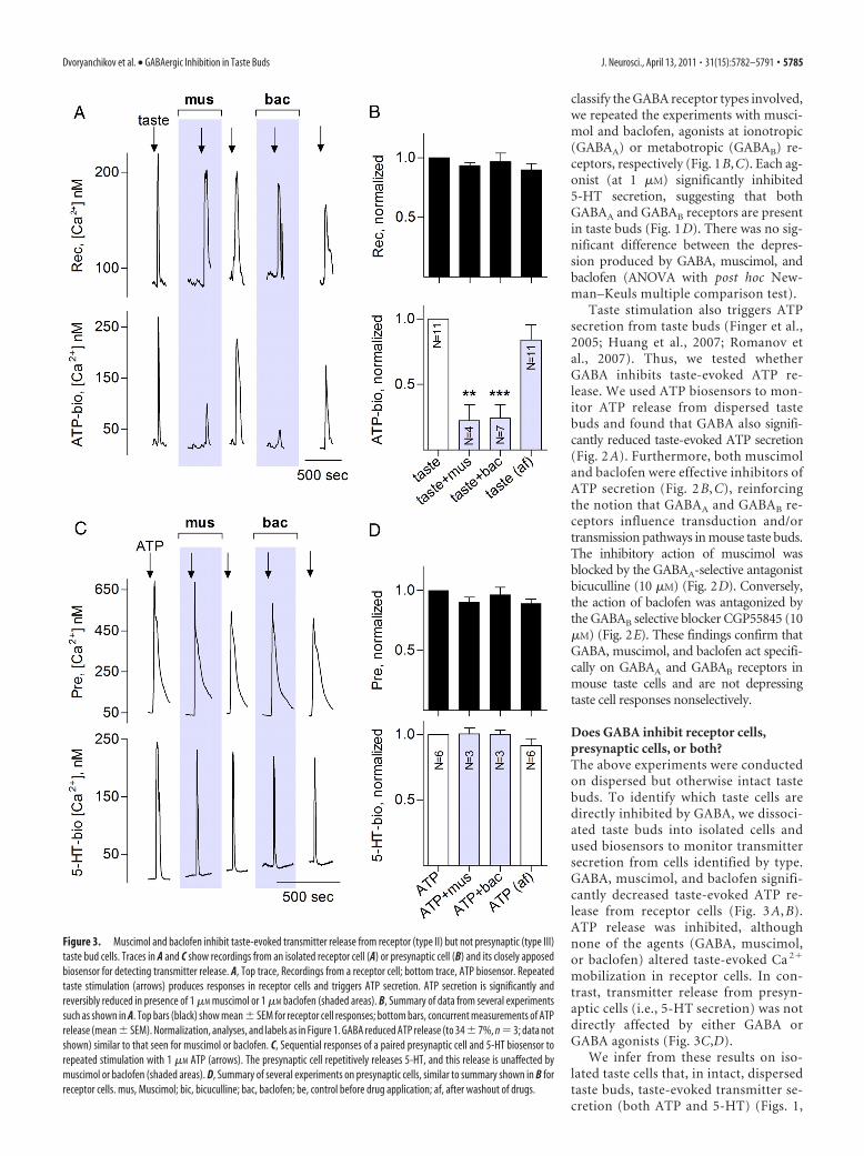
Does GABA inhibit receptor cells,presynaptic cells, or both?The above experiments were conductedon dispersed but otherwise intact tastebuds. To identify which taste cells aredirectly inhibited by GABA, we dissoci-ated taste buds into isolated cells andused biosensors to monitor transmittersecretion from cells identified by type.GABA, muscimol, and baclofen signifi-cantly decreased taste-evoked ATP re-lease from receptor cells (Fig. 3 A, B).ATP release was inhibited, althoughnone of the agents (GABA, muscimol,or baclofen) altered taste-evoked Ca 2�
mobilization in receptor cells. In con-trast, transmitter release from presyn-aptic cells (i.e., 5-HT secretion) was notdirectly affected by either GABA orGABA agonists (Fig. 3C,D).
We infer from these results on iso-lated taste cells that, in intact, dispersedtaste buds, taste-evoked transmitter se-cretion (both ATP and 5-HT) (Figs. 1,
Figure 3. Muscimol and baclofen inhibit taste-evoked transmitter release from receptor (type II) but not presynaptic (type III)taste bud cells. Traces in A and C show recordings from an isolated receptor cell (A) or presynaptic cell (B) and its closely apposedbiosensor for detecting transmitter release. A, Top trace, Recordings from a receptor cell; bottom trace, ATP biosensor. Repeatedtaste stimulation (arrows) produces responses in receptor cells and triggers ATP secretion. ATP secretion is significantly andreversibly reduced in presence of 1 �M muscimol or 1 �M baclofen (shaded areas). B, Summary of data from several experimentssuch as shown in A. Top bars (black) show mean � SEM for receptor cell responses; bottom bars, concurrent measurements of ATPrelease (mean � SEM). Normalization, analyses, and labels as in Figure 1. GABA reduced ATP release (to 34 � 7%, n � 3; data notshown) similar to that seen for muscimol or baclofen. C, Sequential responses of a paired presynaptic cell and 5-HT biosensor torepeated stimulation with 1 �M ATP (arrows). The presynaptic cell repetitively releases 5-HT, and this release is unaffected bymuscimol or baclofen (shaded areas). D, Summary of several experiments on presynaptic cells, similar to summary shown in B forreceptor cells. mus, Muscimol; bic, bicuculline; bac, baclofen; be, control before drug application; af, after washout of drugs.
Dvoryanchikov et al. • GABAergic Inhibition in Taste Buds J. Neurosci., April 13, 2011 • 31(15):5782–5791 • 5785
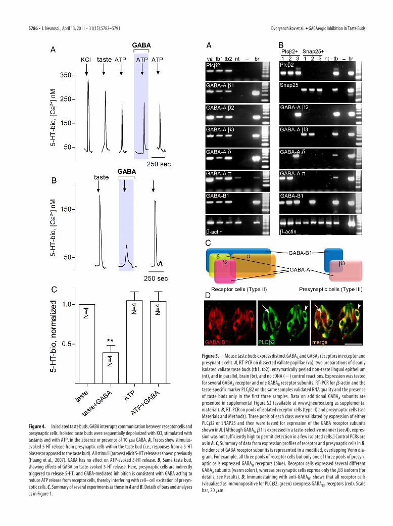
Figure 4. In isolated taste buds, GABA interrupts communication between receptor cells andpresynaptic cells. Isolated taste buds were sequentially depolarized with KCl, stimulated withtastants and with ATP, in the absence or presence of 10 �M GABA. A, Traces show stimulus-evoked 5-HT release from presynaptic cells within the taste bud (i.e., responses from a 5-HTbiosensor apposed to the taste bud). All stimuli (arrows) elicit 5-HT release as shown previously(Huang et al., 2007). GABA has no effect on ATP-evoked 5-HT release. B, Same taste bud,showing effects of GABA on taste-evoked 5-HT release. Here, presynaptic cells are indirectlytriggered to release 5-HT, and GABA-mediated inhibition is consistent with GABA acting toreduce ATP release from receptor cells, thereby interfering with cell– cell excitation of presyn-aptic cells. C, Summary of several experiments as those in A and B. Details of bars and analysesas in Figure 1.
Figure 5. Mouse taste buds express distinct GABAA and GABAB receptors in receptor andpresynaptic cells. A, RT-PCR on dissected vallate papillae (va), two preparations of cleanlyisolated vallate taste buds (tb1, tb2), enzymatically peeled non-taste lingual epithelium(nt), and in parallel, brain (br), and no cDNA (�) control reactions. Expression was testedfor several GABAA receptor and one GABAB receptor subunits. RT-PCR for �-actin and thetaste-specific marker PLC�2 on the same samples validated RNA quality and the presenceof taste buds only in the first three samples. Data on additional GABAA subunits arepresented in supplemental Figure S2 (available at www.jneurosci.org as supplementalmaterial). B, RT-PCR on pools of isolated receptor cells (type II) and presynaptic cells (seeMaterials and Methods). Three pools of each class were validated by expression of eitherPLC�2 or SNAP25 and then were tested for expression of the GABA receptor subunitsshown in A. [Although GABAA �1 is expressed in a taste-selective manner (see A), expres-sion was not sufficiently high to permit detection in a few isolated cells.] Control PCRs areas in A. C, Summary of data from expression profiles of receptor and presynaptic cells in B.Incidence of GABA receptor subunits is represented in a modified, overlapping Venn dia-gram. For example, all three pools of receptor cells but only one of three pools of presyn-aptic cells expressed GABAB receptors (blue). Receptor cells expressed several differentGABAA subunits (warm colors), whereas presynaptic cells express only the �3 isoform (fordetails, see Results). D, Immunostaining with anti-GABAB1 shows that all receptor cells(visualized as immunopositive for PLC�2; green) coexpress GABAB1 receptors (red). Scalebar, 20 �m.
5786 • J. Neurosci., April 13, 2011 • 31(15):5782–5791 Dvoryanchikov et al. • GABAergic Inhibition in Taste Buds
2) is inhibited primarily by the actions of GABA on receptorcells, as follows:
GABA
�
Taste stimulation3 Receptor cells3 ATP secretion
3 Presynaptic cells3 5-HT release
Consistent with this interpretation, GABA did not reduce 5-HTsecretion from taste buds that were stimulated with ATP (Fig.4A), although taste-evoked 5-HT release in the same taste budwas inhibited (Fig. 4B,C).
Taste receptor and presynaptic cells express distinctGABA receptorsAs an independent confirmation of GABAergic mechanisms inmouse taste buds, we used RT-PCR on cleanly isolated taste budsto evaluate the expression of all the known subunits of GABAA
and GABAB receptors. Taste buds abundantly express mRNAs forseveral of GABAA subunits, particularly �1, �2, �3, �, and �, aswell as for an obligate subunit of GABAB receptors, GABAB1 (Fig.5A,D). mRNAs for the remaining GABAA subunits (�1, �2, �3,�4, �5, �6, �1, �2, �3, �1, �2, �3, �, and ) were either undetect-able or appeared at very low levels and inconsistently across tastebud samples (supplemental Fig. S2, available at www.jneurosci.org as supplemental material). GABA receptor subunits were notexpressed to any appreciable level in the non-taste epitheliumadjacent to taste buds (Fig. 5A) (supplemental Fig. S2, available atwww.jneurosci.org as supplemental material).
To assess which taste cell types express this limited set ofGABA receptors, we tested cDNAs from receptor cells (threepools, each pool containing 10 cells) and presynaptic cells (alsothree pools, each with 10 cells). These pools were obtained byharvesting GFP-positive taste cells after isolating and dissociatingtaste buds from PLC�2–GFP mice (receptor cells) or GAD67–GFP mice (presynaptic cells). The homogeneity of each pool ofcells was verified by RT-PCR for two diagnostic genes: PLC�2(for receptor cells) and SNAP25 (for presynaptic cells). Receptorcell pools consistently expressed GABAB1. In addition, GABAA
subunits �2, �, and � were detected in some but not all pools ofreceptor cells. Of the three pools of presynaptic cells, two ex-pressed GABAA �3, and one of these also expressed GABAB1 (Fig.5B). When expression levels for a particular gene are low, themRNA is inconsistently detected by single-cell RT-PCR. Thus,the uneven pattern of expression of GABAA subunits in both celltypes may reflect either low mRNA abundance or heterogeneityin the particular subunits expressed in each cell. GABAB subunits,conversely, were detected in all receptor cell pools, suggestingthat most receptor cells express the metabotropic receptor forGABA. These RT-PCR data on the expression pattern of GABAsubunits are summarized in Figure 5C.
We also performed immunofluorescence on mouse taste tis-sue for the one subunit most consistently detected by RT-PCR,GABAB1. Consistent with our single-cell RT-PCR data, we de-tected GABAB1 in all receptor cells and also at lower intensity inadditional, unidentified cells (Fig. 5D, arrow).
In summary, receptor cells express GABAA and GABAB recep-tor subunits, consistent with the calcium imaging data. GABAB
subunits are prominently expressed in receptor cells but are alsoseen, although less frequently, in presynaptic cells. GABAA sub-units are expressed at lower levels, or in subsets of cells: receptorcells heterogeneously express �2, � and �, whereas presynapticcells express �3 (Fig. 5C).
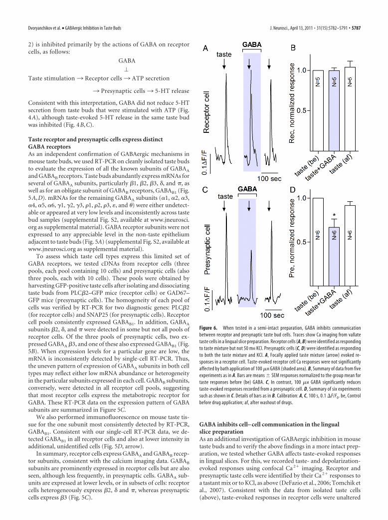
GABA inhibits cell– cell communication in the lingualslice preparationAs an additional investigation of GABAergic inhibition in mousetaste buds and to verify the above findings in a more intact prep-aration, we tested whether GABA affects taste-evoked responsesin lingual slices. For this, we recorded taste- and depolarization-evoked responses using confocal Ca 2� imaging. Receptor andpresynaptic taste cells were identified by their Ca 2� responses toa tastant mix or to KCl, as above (DeFazio et al., 2006; Tomchik etal., 2007). Consistent with the data from isolated taste cells(above), taste-evoked responses in receptor cells were unaltered
Figure 6. When tested in a semi-intact preparation, GABA inhibits communicationbetween receptor and presynaptic taste bud cells. Traces show Ca imaging from vallatetaste cells in a lingual slice preparation. Receptor cells (A, B) were identified as respondingto taste mixture but not 50 mM KCl. Presynaptic cells (C, D) were identified as respondingto both the taste mixture and KCl. A, Focally applied taste mixture (arrow) evoked re-sponses in a receptor cell. Taste-evoked receptor cell Ca responses were not significantlyaffected by bath application of 100 �M GABA (shaded area). B, Summary of data from fiveexperiments as in A. Bars are means � SEM responses normalized to the group mean fortaste responses before (be) GABA. C, In contrast, 100 �M GABA significantly reducestaste-evoked responses recorded from a presynaptic cell. D, Summary of six experimentssuch as shown in C. Details of bars as in B. Calibration: A, C, 100 s, 0.1 �F/F0. be, Controlbefore drug application; af, after washout of drugs.
Dvoryanchikov et al. • GABAergic Inhibition in Taste Buds J. Neurosci., April 13, 2011 • 31(15):5782–5791 • 5787
by bath-applied GABA (100 �M) (Fig. 6A,B). However, Ca 2�
responses in presynaptic cells (i.e., “downstream” of receptorcells) were significantly reduced by GABA (Fig. 6C,D). These datastrongly support the interpretation that GABA inhibits taste-triggered and ATP-mediated communication from receptor topresynaptic cells.
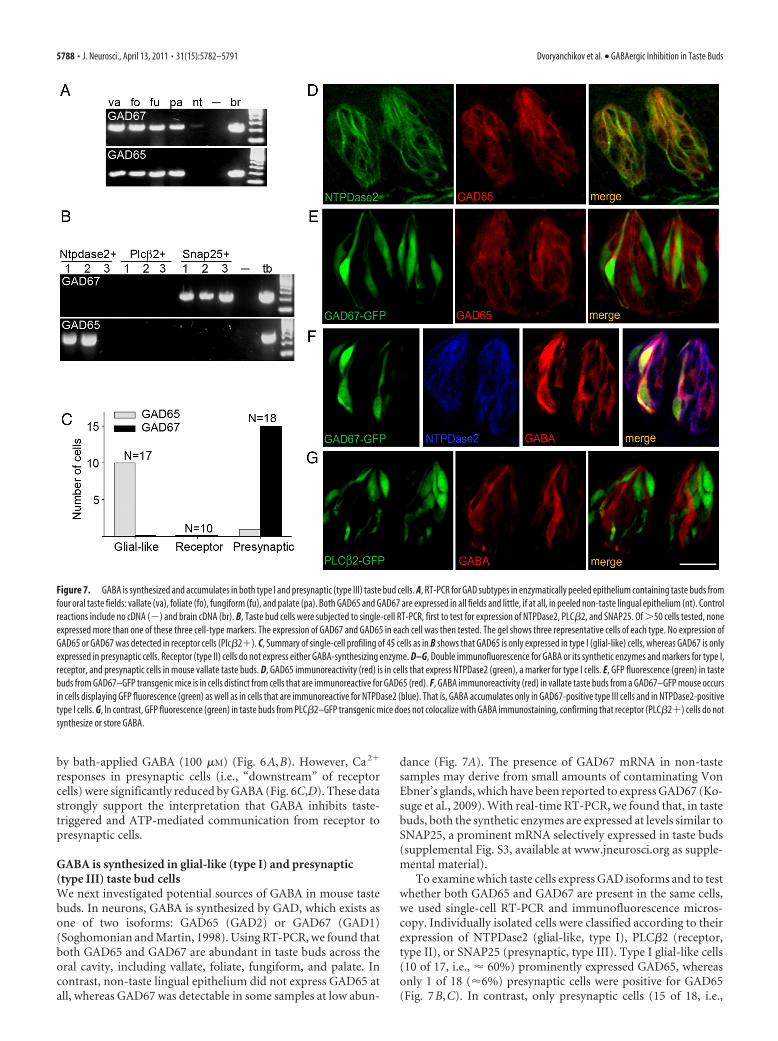
GABA is synthesized in glial-like (type I) and presynaptic(type III) taste bud cellsWe next investigated potential sources of GABA in mouse tastebuds. In neurons, GABA is synthesized by GAD, which exists asone of two isoforms: GAD65 (GAD2) or GAD67 (GAD1)(Soghomonian and Martin, 1998). Using RT-PCR, we found thatboth GAD65 and GAD67 are abundant in taste buds across theoral cavity, including vallate, foliate, fungiform, and palate. Incontrast, non-taste lingual epithelium did not express GAD65 atall, whereas GAD67 was detectable in some samples at low abun-
dance (Fig. 7A). The presence of GAD67 mRNA in non-tastesamples may derive from small amounts of contaminating VonEbner’s glands, which have been reported to express GAD67 (Ko-suge et al., 2009). With real-time RT-PCR, we found that, in tastebuds, both the synthetic enzymes are expressed at levels similar toSNAP25, a prominent mRNA selectively expressed in taste buds(supplemental Fig. S3, available at www.jneurosci.org as supple-mental material).
To examine which taste cells express GAD isoforms and to testwhether both GAD65 and GAD67 are present in the same cells,we used single-cell RT-PCR and immunofluorescence micros-copy. Individually isolated cells were classified according to theirexpression of NTPDase2 (glial-like, type I), PLC�2 (receptor,type II), or SNAP25 (presynaptic, type III). Type I glial-like cells(10 of 17, i.e., � 60%) prominently expressed GAD65, whereasonly 1 of 18 (�6%) presynaptic cells were positive for GAD65(Fig. 7B,C). In contrast, only presynaptic cells (15 of 18, i.e.,
Figure 7. GABA is synthesized and accumulates in both type I and presynaptic (type III) taste bud cells. A, RT-PCR for GAD subtypes in enzymatically peeled epithelium containing taste buds fromfour oral taste fields: vallate (va), foliate (fo), fungiform (fu), and palate (pa). Both GAD65 and GAD67 are expressed in all fields and little, if at all, in peeled non-taste lingual epithelium (nt). Controlreactions include no cDNA (�) and brain cDNA (br). B, Taste bud cells were subjected to single-cell RT-PCR, first to test for expression of NTPDase2, PLC�2, and SNAP25. Of �50 cells tested, noneexpressed more than one of these three cell-type markers. The expression of GAD67 and GAD65 in each cell was then tested. The gel shows three representative cells of each type. No expression ofGAD65 or GAD67 was detected in receptor cells (Plc�2�). C, Summary of single-cell profiling of 45 cells as in B shows that GAD65 is only expressed in type I (glial-like) cells, whereas GAD67 is onlyexpressed in presynaptic cells. Receptor (type II) cells do not express either GABA-synthesizing enzyme. D–G, Double immunofluorescence for GABA or its synthetic enzymes and markers for type I,receptor, and presynaptic cells in mouse vallate taste buds. D, GAD65 immunoreactivity (red) is in cells that express NTPDase2 (green), a marker for type I cells. E, GFP fluorescence (green) in tastebuds from GAD67–GFP transgenic mice is in cells distinct from cells that are immunoreactive for GAD65 (red). F, GABA immunoreactivity (red) in vallate taste buds from a GAD67–GFP mouse occursin cells displaying GFP fluorescence (green) as well as in cells that are immunoreactive for NTPDase2 (blue). That is, GABA accumulates only in GAD67-positive type III cells and in NTPDase2-positivetype I cells. G, In contrast, GFP fluorescence (green) in taste buds from PLC�2–GFP transgenic mice does not colocalize with GABA immunostaining, confirming that receptor (PLC�2�) cells do notsynthesize or store GABA.
5788 • J. Neurosci., April 13, 2011 • 31(15):5782–5791 Dvoryanchikov et al. • GABAergic Inhibition in Taste Buds
83%) expressed GAD67. None of the receptor cells tested ex-pressed either isoform of GAD. These expression patterns ofGAD67 and GAD65 across the three cell types were significantlydifferent from each other ( 2 test, p � 0.0002).
We extended our single-cell RT-PCR results on GAD isoformsusing immunofluorescence microscopy. We have shown previ-ously (DeFazio et al., 2006; Tomchik et al., 2007) that GAD67 isexpressed only in type III presynaptic cells. As shown in Figure7D, GAD65 immunoreactivity substantially colocalized with NT-PDase2, a marker for type I glial-like taste cells. We further ex-amined GAD65 expression in taste buds from two well-characterized strains of transgenic mice, PLC�2–GFP (in whichreceptor cells are labeled) and GAD67–GFP, in which most pre-synaptic cells are labeled. GAD65 was lacking from GFP-positivetaste cells in both GAD67–GFP tissue [i.e., from presynaptic cells(Fig. 7E)] and PLC�2–GFP tissue [i.e., receptor cells (data notshown)]. These observations are fully consistent with the non-overlapping distribution of GAD65 and GAD67 seen in oursingle-cell RT-PCR data (Fig. 7B).
Does GABA accumulate in the cells that express GAD65 orGAD67? Using anti-GABA antibodies, we observed GABA immu-nofluorescence in several cells in vallate, fungiform, and palate tastebuds (Fig. 7 F, G) (supplemental Fig. S4, available at www.
jneurosci.org as supplemental material). Using taste buds inwhich receptor or presynaptic cells are marked with GFP andby immunostaining for cell-type markers, we determined thatGABA-immunoreactive cells were either NTPDase2 expressing(i.e., type I) or GAD67 expressing [i.e., presynaptic cells (Fig. 7F)].Receptor cells (PLC�2-positive or PLC�2–GFP) were never seen toaccumulate GABA (Fig. 7G), consistent with their lack of GABA-synthesizing enzymes. We noted that occasional GAD67–GFP-positive presynaptic cells did not accumulate GABA; we did notexplore this further. In vallate taste buds, there are relatively largenumbers of presynaptic cells (Dvoryanchikov et al., 2007), andGABA may be secreted from either type I or presynaptic cells. Be-cause fungiform and palatal taste buds contain few presynaptic cells(supplemental Fig. S4, available at www.jneurosci.org as supplemen-tal material), type I glial-like cells may constitute the main stores ofGABA in these taste buds.
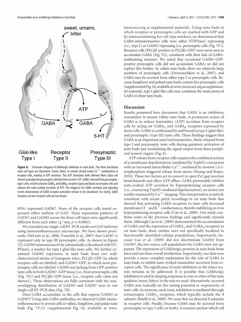
DiscussionResults presented here document that GABA is an inhibitorytransmitter in mouse vallate taste buds. A prominent action ofGABA is to reduce transmitter (ATP) secretion from receptorcells by acting on GABAA and GABAB receptors expressed bythose cells. GABA is synthesized by and found in type I (glial-like)and presynaptic (type III) taste cells. These findings suggest thatGABA is an important taste bud transmitter, likely released fromtype I and presynaptic taste cells during gustatory activation oftaste buds and modulating the signal output from these periph-eral sensory organs (Fig. 8).
ATP release from receptor cells requires the combined actionsof a membrane depolarization (mediated by TrpM5) concurrentwith an increased intracellular Ca 2�, mediated by inositol 1,4,5-trisphosphate-triggered release from stores (Huang and Roper,2010). These two factors act in concert to open Px1 gap junctionhemichannels and allow ATP efflux. GABA presumably inhibitstaste-evoked ATP secretion by hyperpolarizing receptor cells(i.e., countering TrpM5-mediated depolarization), an action notreadily measured by Ca 2� imaging. This interpretation is entirelyconsistent with recent patch recordings in rat taste buds thatshowed that activating GABA receptors in taste cells increasedmembrane Cl� and K� conductances, thereby stabilizing or evenhyperpolarizing receptor cells (Cao et al., 2009). Our study con-firms some of the previous findings and significantly extendsthem. Although Cao et al. (2009) demonstrated the accumulationof GABA and the expression of GABAA and GABAB receptors inrat taste buds, these entities were not specifically localized tofunctionally identified cellular populations. Importantly, be-cause Cao et al. (2009) did not discriminate GAD65 fromGAD67, the two source cell populations for GABA were not ap-parent. The expression of GABAergic proteins in taste buds frommice and rats bear overall similarities. Importantly, our data nowprovide a more complete explanation for the role of GABA intaste buds, to inhibit taste-evoked transmitter secretion from re-ceptor cells. The significance of such inhibition in the intact sys-tem remains to be addressed. It is possible that GABAergicinhibition is used in shaping responses to one or other of the tastequalities (sweet, bitter) in the mix we used. Alternatively, secretedGABA may tonically set the resting potential or responsivity oftaste cells. In neurons, such tonic inhibition is mediated throughextrasynaptic GABAA receptors, which typically include the �subunit (Belelli et al., 2009). We note that we detected � subunitsin receptor cells. Finally, because GABA may be secreted frompresynaptic or type I cells (or both), it remains unclear which cell
Figure 8. Schematic diagram of GABAergic inhibition in taste buds. The three functionaltaste cell types are illustrated. Sweet, bitter, or umami stimuli evoke Ca 2� mobilization inreceptor cells, leading to ATP secretion. This ATP stimulates both afferent fibers (data notshown) and adjacent presynaptic cells that then secrete 5-HT. GABA, released from presynaptic,type I cells, or both activates GABAA and GABAB receptors (gray and black) on receptor cells andreduces the taste-evoked secretion of ATP. The trigger(s) for GABA secretion and signalingevents downstream of GABA receptor activation remain to be elucidated. For clarity, GABAreceptors on non-receptor cells are not shown.
Dvoryanchikov et al. • GABAergic Inhibition in Taste Buds J. Neurosci., April 13, 2011 • 31(15):5782–5791 • 5789
type is responsible for GABA secretion during taste stimulation.Our future experiments will address these questions.
Previous studies (Cao et al., 2009; Starostik et al., 2010) haveexamined GABA receptor subunits in taste tissues of rodents.Our semiquantitative RT-PCR data suggest that expression levelsfor most GABA subunits are relatively or very low in taste cells.This may account for some of the apparent discrepancies acrossdata in these three reports. Furthermore, our comparisons be-tween isolated taste buds and dissected taste papillae (Fig. 5)(supplemental Fig. S2, available at www.jneurosci.org as supple-mental material) suggest that many of the GABA receptor sub-units may be expressed in cells underneath the taste epithelium(e.g., nerves). Nevertheless, we and Starostik et al. (2010) bothfind that GABAA �3 is a relatively abundant subunit in taste buds;all three reports show the presence of GABAB receptors.
We note that muscimol, baclofen, and GABA all producedsimilar levels of inhibition (Figs. 1D, 3B). These results may sug-gest that signals from GABAA and GABAB receptors in taste cellsconverge on a common step that is upstream of the transmitterrelease. Thus, a saturating dose of any one agonist would producemaximum inhibition. Furthermore, receptor cells express bothGABAA and GABAB subunits and ATP secretion from receptorcells is the primary target of GABAergic inhibition (Fig. 3A,B).That is, the taste cells that express GABA receptors prominentlyare the ones that are obviously affected by GABA.
Although both receptor (type II) and presynaptic (type III)cells express GABAA receptors, we only detected GABAergic in-hibition of transmitter secretion from receptor cells. This maysuggest that GABA has additional functions in mouse taste buds.For instance, presynaptic cells express principally GABAA �3 re-ceptors. The �3 subunit has been shown to be important in thedevelopment of the palate (Hagiwara et al., 2003). Indeed, there isby now substantial evidence that GABA acts as a trophic factorand modulates cell proliferation and synaptic formation duringneuronal development (Owens and Kriegstein, 2002). One mightspeculate that the action of GABA on presynaptic cells is relatedto the development and maturation of these taste bud cells andtheir synapses, as is the case in the CNS (Wang and Kriegstein,2009).
Our results in mice, combined with those of Cao et al. (2009)in rats, firmly establish GABA as an inhibitory transmitter in tastebuds. However, the implication of this for how animals discrim-inate sweet, sour, salty, etc. and how taste behavior is affectedby GABAergic mechanisms is unclear. Our data indicate thatGABA is synthesized and stored in specific taste cells: GABA-biosynthetic enzymes are found in type I glial-like cells (GAD65)and presynaptic (type III) cells (GAD67). Although neurons havelong been known to secrete GABA as a transmitter, recent evi-dence indicates that many glia also synthesize and secrete GABA(Jow et al., 2004), and, indeed, GABA is recognized as a signifi-cant “gliotransmitter” (Angulo et al., 2008). Taste preference ex-periments have been conducted on GAD65 knock-out mice(Shimura et al., 2004), but the findings have no straightforwardinterpretation. Namely, GAD65 knock-out mice did not differfrom wild-type mice in taste preferences for sucrose, NaCl, HCl,or quinine when these solutions were presented alone. However,GAD65 knock-out versus wild-type mice responded differentlyto binary taste mixtures, notably sucrose plus quinine. The au-thors concluded that GABA (from GAD65) is not involved inbasic taste discrimination, per se, but is instead involved in signalprocessing for more complex information such as taste mixtures.A major complication in interpreting these data is that knockingout GAD65 also interrupts GABAergic synapses in the CNS, not
just the actions of GABA (including trophic, if any) in taste buds.This may have profound effects on all behaviors, including tastepreference and discrimination ability. Taste behavioral assays onGAD67 knock-out mice have not been conducted; the geneticmutation is lethal at birth. In addition to the genetic studies,pharmacological investigations have revealed that GABAergicdrugs, such as benzodiazepines, do indeed influence taste prefer-ences (Cooper, 1989). Again, there is the caveat that these drugsare inevitably exerting powerful CNS actions. In short, given itsinhibitory effects in taste buds, it is likely that GABA plays adistinct peripheral role in taste reception and signaling (in addi-tion to the aforementioned role in development). However, pin-pointing whether and how GABA is released from type I glial-likecells, presynaptic cells, or both during taste reception has notbeen undertaken, and it remains to be determined what overalleffects this inhibitory transmitter exerts in taste reception.
ReferencesAngulo MC, Le Meur K, Kozlov AS, Charpak S, Audinat E (2008) GABA, a
forgotten gliotransmitter. Prog Neurobiol 86:297–303.Bartel DL, Sullivan SL, Lavoie EG, Sevigny J, Finger TE (2006) Nucleoside
triphosphate diphosphohydrolase-2 is the ecto-ATPase of type I cells intaste buds. J Comp Neurol 497:1–12.
Belelli D, Harrison NL, Maguire J, Macdonald RL, Walker MC, Cope DW(2009) Extrasynaptic GABAA receptors: form, pharmacology, and func-tion. J Neurosci 29:12757–12763.
Caicedo A, Jafri MS, Roper SD (2000) In situ Ca 2� imaging reveals neu-rotransmitter receptors for glutamate in taste receptor cells. J Neurosci20:7978 –7985.
Cao Y, Zhao FL, Kolli T, Hivley R, Herness S (2009) GABA expression in themammalian taste bud functions as a route of inhibitory cell-to-cell com-munication. Proc Natl Acad Sci U S A 106:4006 – 4011.
Chandrashekar J, Kuhn C, Oka Y, Yarmolinsky DA, Hummler E, Ryba NJ,Zuker CS (2010) The cells and peripheral representation of sodium tastein mice. Nature 464:297–301.
Chattopadhyaya B, Di Cristo G, Higashiyama H, Knott GW, Kuhlman SJ,Welker E, Huang ZJ (2004) Experience and activity-dependent matura-tion of perisomatic GABAergic innervation in primary visual cortex dur-ing a postnatal critical period. J Neurosci 24:9598 –9611.
Chaudhari N, Roper SD (2010) The cell biology of taste. J Cell Biol190:285–296.
Clapp TR, Yang R, Stoick CL, Kinnamon SC, Kinnamon JC (2004) Mor-phologic characterization of rat taste receptor cells that express com-ponents of the phospholipase C signaling pathway. J Comp Neurol468:311–321.
Cooper SJ (1989) Benzodiazepine receptor-mediated enhancement and in-hibition of taste reactivity, food choice, and intake. Ann N Y Acad Sci575:321–336; discussion 336 –337.
Dando R, Roper SD (2009) Cell-to-cell communication in intact taste budsthrough ATP signalling from pannexin 1 gap junction hemichannels.J Physiol 587:5899 –5906.
DeFazio RA, Dvoryanchikov G, Maruyama Y, Kim JW, Pereira E, Roper SD,Chaudhari N (2006) Separate populations of receptor cells and presyn-aptic cells in mouse taste buds. J Neurosci 26:3971–3980.
Dvoryanchikov G, Tomchik SM, Chaudhari N (2007) Biogenic amine syn-thesis and uptake in rodent taste buds. J Comp Neurol 505:302–313.
Dvoryanchikov G, Sinclair MS, Perea-Martinez I, Wang T, Chaudhari N(2009) Inward rectifier channel, ROMK, is localized to the apical tips ofglial-like cells in mouse taste buds. J Comp Neurol 517:1–14.
Finger TE, Danilova V, Barrows J, Bartel DL, Vigers AJ, Stone L, Hellekant G,Kinnamon SC (2005) ATP signaling is crucial for communication fromtaste buds to gustatory nerves. Science 310:1495–1499.
Grynkiewicz G, Poenie M, Tsien RY (1985) A new generation of Ca 2� indi-cators with greatly improved fluorescence properties. J Biol Chem260:3440 –3450.
Hagiwara N, Katarova Z, Siracusa LD, Brilliant MH (2003) Nonneuronalexpression of the GABA(A) beta3 subunit gene is required for normalpalate development in mice. Dev Biol 254:93–101.
Herness S, Zhao FL (2009) The neuropeptides CCK and NPY and the
5790 • J. Neurosci., April 13, 2011 • 31(15):5782–5791 Dvoryanchikov et al. • GABAergic Inhibition in Taste Buds
changing view of cell-to-cell communication in the taste bud. PhysiolBehav 97:581–591.
Huang YA, Roper SD (2010) Intracellular Ca 2� and TRPM5-mediatedmembrane depolarization produce ATP secretion from taste receptorcells. J Physiol 588:2343–2350.
Huang YA, Maruyama Y, Roper SD (2008) Norepinephrine is coreleasedwith serotonin in mouse taste buds. J Neurosci 28:13088 –13093.
Huang YA, Dando R, Roper SD (2009) Autocrine and paracrine roles forATP and serotonin in mouse taste buds. J Neurosci 29:13909 –13918.
Huang YJ, Maruyama Y, Lu KS, Pereira E, Plonsky I, Baur JE, Wu D, Roper SD(2005) Mouse taste buds use serotonin as a neurotransmitter. J Neurosci25:843– 847.
Huang YJ, Maruyama Y, Dvoryanchikov G, Pereira E, Chaudhari N, RoperSD (2007) The role of pannexin 1 hemichannels in ATP release andcell-cell communication in mouse taste buds. Proc Natl Acad Sci U S A104:6436 – 6441.
Jain S, Roper SD (1991) Immunocytochemistry of gamma-aminobutyricacid, glutamate, serotonin, and histamine in Necturus taste buds. J CompNeurol 307:675– 682.
Jow F, Chiu D, Lim HK, Novak T, Lin S (2004) Production of GABA bycultured hippocampal glial cells. Neurochem Int 45:273–283.
Kim JW, Roberts C, Maruyama Y, Berg S, Roper S, Chaudhari N (2006)Faithful expression of GFP from the PLCbeta2 promoter in a functionalclass of taste receptor cells. Chem Senses 31:213–219.
Koga T, Bradley RM (2000) Biophysical properties and responses to neu-rotransmitters of petrosal and geniculate ganglion neurons innervatingthe tongue. J Neurophysiol 84:1404 –1413.
Kosuge Y, Kawaguchi M, Sawaki K, Okubo M, Shinomiya T, Sakai T (2009)Immunohistochemical study on GABAergic system in salivary glands.Eur J Pharmacol 610:18 –22.
Nagai T, Delay RJ, Welton J, Roper SD (1998) Uptake and release of neu-rotransmitter candidates, [ 3H]serotonin, [ 3H]glutamate, and[ 3H]gamma-aminobutyric acid, in taste buds of the mudpuppy, Necturusmaculosus. J Comp Neurol 392:199 –208.
Obata H, Shimada K, Sakai N, Saito N (1997) GABAergic neurotransmis-sion in rat taste buds: immunocytochemical study for GABA and GABAtransporter subtypes. Brain Res Mol Brain Res 49:29 –36.
Ogura T, Margolskee RF, Tallini YN, Shui B, Kotlikoff MI, Lin W (2007)Immuno-localization of vesicular acetylcholine transporter in mousetaste cells and adjacent nerve fibers: indication of acetylcholine release.Cell Tissue Res 330:17–28.
Olsen RW, Sieghart W (2008) International Union of Pharmacology. LXX.Subtypes of gamma-aminobutyric acid(A) receptors: classification on thebasis of subunit composition, pharmacology, and function. Update.Pharmacol Rev 60:243–260.
Owens DF, Kriegstein AR (2002) Is there more to GABA than synaptic in-hibition? Nat Rev Neurosci 3:715–727.
Perez CA, Huang L, Rong M, Kozak JA, Preuss AK, Zhang H, Max M, Mar-golskee RF (2002) A transient receptor potential channel expressed intaste receptor cells. Nat Neurosci 5:1169 –1176.
Romanov RA, Rogachevskaja OA, Bystrova MF, Jiang P, Margolskee RF,Kolesnikov SS (2007) Afferent neurotransmission mediated by hemi-channels in mammalian taste cells. EMBO J 26:657– 667.
Roper SD (2007) Signal transduction and information processing in mam-malian taste buds. Pflugers Arch 454:759 –776.
Shimura T, Watanabe U, Yanagawa Y, Yamamoto T (2004) Altered tastefunction in mice deficient in the 65-kDa isoform of glutamate decarbox-ylase. Neurosci Lett 356:171–174.
Soghomonian JJ, Martin DL (1998) Two isoforms of glutamate decarboxyl-ase: why? Trends Pharmacol Sci 19:500 –505.
Starostik MR, Rebello MR, Cotter KA, Kulik A, Medler KF (2010) Expres-sion of GABAergic receptors in mouse taste receptor cells. PLoS One5:e13639.
Tomchik SM, Berg S, Kim JW, Chaudhari N, Roper SD (2007) Breadth oftuning and taste coding in mammalian taste buds. J Neurosci27:10840 –10848.
Vandenbeuch A, Clapp TR, Kinnamon SC (2008) Amiloride-sensitivechannels in type I fungiform taste cells in mouse. BMC Neurosci 9:1.
Vandenbeuch A, Tizzano M, Anderson CB, Stone LM, Goldberg D, Kinna-mon SC (2010) Evidence for a role of glutamate as an efferent transmit-ter in taste buds. BMC Neurosci 11:77.
Wang DD, Kriegstein AR (2009) Defining the role of GABA in cortical de-velopment. J Physiol 587:1873–1879.
Yee CL, Yang R, Bottger B, Finger TE, Kinnamon JC (2001) “Type III” cellsof rat taste buds: immunohistochemical and ultrastructural studies ofneuron-specific enolase, protein gene product 9.5, and serotonin. J CompNeurol 440:97–108.
Zhao GQ, Zhang Y, Hoon MA, Chandrashekar J, Erlenbach I, Ryba NJ, ZukerCS (2003) The receptors for mammalian sweet and umami taste. Cell115:255–266.
Dvoryanchikov et al. • GABAergic Inhibition in Taste Buds J. Neurosci., April 13, 2011 • 31(15):5782–5791 • 5791