Embed Size (px)
Citation preview

1
Intro toGallbladder &
Pancreas
Cholecystitisacutechronic
Gallbladder tumorsAdenomyoma (benign)Adenocarcinoma
PancreatitisPathology
Helen Remotti M.D.
Pancreatitisacutechronic
Pancreatic tumors
Gallstones(Cholelithiasis)
• 10 - 20% Adults• 35% Autopsy: Over 65
• Over 20 Million• 600,000 Cholecystectomies• #2 reason for abdominal operations
Cholesterol/mixed stones
Choledocholithiasis(Stones in the common bile duct)
Pain: Epigastric, RUQ-stones may be passedObstructive Jaundice-may be intermittentAscending Cholangitis- Infection: to liver20%: No pain; 25% no jaundice
Acute cholecystitis = ischemic injury
Chronic Cholecystitis
• Associated with calculi in 95% of cases.• Multiples episodes of inflammation cause GB
thickening with chronic inflammation/ fibrosis and muscular hypertrophy.yp p y
• Rokitansky - Aschoff Sinuses (mucosa herniates through the muscularis mucosae)
• With longstanding inflammation GB becomes fibrotic and calcified “porcelain GB”

2
Chronic cholecystitis
Rokitansky-Aschoff sinuses
Chronic Cholecystitis
• Fibrosis• Chronic Inflammation• Rokitansky - Aschoff Sinuses• Hypertrophy: Muscularis• Hypertrophy: Muscularis
Cholesterolosis
Focal accumulation of cholesterol-laden macrophages in lamina propria of gallbladder (incidental finding).
Adenomyoma of Gall Bladder

3
Carcinoma: Gall BladderUncommon: 5,000 cases / yearFewer than 1% resected G.B.Sx: same as with stones5 yr. survival: Less than 5%(survival relates to stage)
90%: StonesLong Hx: symptomatic stonesStones: predispose to CA., but uncommon
complication

4
Gallbladder carcinoma
Case 156 year old woman presents to ER in shock, following rapid onset of severe upper abdominal pain, developing over the previous day.
Hx: heavy alcohol useHx: heavy alcohol use.
LABs: Elevated serum amylase and elevated peritoneal fluid lipase
Acute PancreatitisPatient developed rapid onset of respiratory failure necessitating intubation and mechanical ventilation.
Over 48 hours, she was increasingly unstable, with evolution to multi organ failure and she expired 82evolution to multi-organ failure, and she expired 82 hours after admission.
An autopsy was performed.
Acute pancreatitis
Elastase destruction of blood vessels – with hemorrhage

5
Acute Pancreatitis
Edema, congestion
Advanced hemorrhagic pancreatitis, fat necrosis
Necrotic abscess, gangrene
Acute Pancreatitis
US: 45% of cases have gallstones and choledocholithiasis;
35% associated with heavy alcohol ingestion
Pathology: Enzyme release is triggered with digestion of pancreas, necrosis of fat and lobules, hemorrhage from damaged blood vessels.
Variable severity: may lead to liquefactive necrosis, hemorrhage.
Mild cases – may have local complications: abscess, pseudocyst.
Chronic PancreatitisContinuing inflammation with irreversible changes in architecture, structure and function.
Fibrosis of parenchyma with distortion of duct p yarchitecture, loss of exocrine secretory function.
Changes may be focal or widespread.
Chronic pancreatitis with Stones

6
Chronic pancreatitis
Chronic Pancreatitis
Complications of Chronic Pancreatitis
Chronic abdominal pain, severe and unremitting, radiating to back
Malabsorption due to reduced enzyme secretion. (After 90% of pancreas is fibrotic, reduced lipase and trypsin secretion lead to steatorrhea) .
P ti di b t i t d ith d d i l tPancreatic diabetes associated with decreased islets.
Pancreatic pseudocysts with extension or rupture in adjacent organs.
Case 267 year old woman with recent onset painless jaundice.
History of 15lb weight loss over last 3 months.
She smoked 1 pack per day x 35 years.Physical exam: palpable GBPhysical exam: palpable GB
ERCP was performed with Endoscopic Ultrasound (EUS) evidence of a large mass in the head of the pancreas.
An endoscopic FNA was performed.
Normal pancreas
ductal epithelium
Dx: Adenocarcinoma
Patient’s FNA
Curvoissier’s Law – enlarged palpable GB

7
• Represents the most common pancreatic neoplasm– 2nd most common GIT cancer
• 4th leading cause of cancer death• M > F; > 50 years at initial presentation usually
S
Pancreatic Ductal Adenocarcinoma: Pancreatic Ductal Adenocarcinoma: ClinicalClinical
Pancreatic Ductal Adenocarcinoma: Pancreatic Ductal Adenocarcinoma: ClinicalClinical
• Symptoms: – Abdominal pain, weight loss, jaundice, pancreatic
insufficiency, malabsorption; Migratory thrombophlebitis – “Trousseau’s sign”
• Site: – head (60-70%) > body (10-15%) > tail (5-10%)
• Contributing factors: – smoking, carcinogens, genetics
• Macroscopic:– Usually a solitary mass– poor demarcation
Pancreatic Ductal Adenocarcinoma: Pancreatic Ductal Adenocarcinoma: PathologyPathology
Pancreatic Ductal Adenocarcinoma: Pancreatic Ductal Adenocarcinoma: PathologyPathology
– firm to gritty consistency– depends upon location, but bile stasis specifically
for the head of pancreas neoplasms
Chronic Pancreatitis:FNA
Pancreatic Ductal Adenocarcinoma: FNA
8
Normal AdenoCA
Benign – lobular Malignant- haphazard
Invasive growth with associated desmoplasia
Invasion into peripancreatic soft tissue
Pancreatic adenocarcinoma – Lymph node metastases
Pancreatic adenocarcinoma – perineural invasion

9
• Microscopic:– loss of lobular architecture– architectural and cytomorphologic features
indicative of malignancy: • loss of cell differentiation, hyperchromatic nuclei,
Pancreatic Ductal Adenocarcinoma: Pancreatic Ductal Adenocarcinoma: PathologyPathology
Pancreatic Ductal Adenocarcinoma: Pancreatic Ductal Adenocarcinoma: PathologyPathology
, yp ,increased N:C, prominent nucleoli, mitotic activity
– intraductal carcinoma– invasive growth associated with desmoplasia– neurotropism, extratumoral vascular invasion– extension into peripancreatic soft tissue– secondary pancreatitis due to obstruction
Critical area to evaluate- uncinate margin
Margin most likelyTo be positive is the Uncinate margin -Retroperitoneal/mesentericMargin along the rightLateral border of the SMAAnd should be inked.
Margin evaluation:Bile ductPancreatic distalPosterior/RetroperitonealUncinate
Tumor size:Measure Gross Submit 2 complete cross sections.
(Black posterior)(Yellow anterior)
Definition of Tumor (T)
• TX Primary tumor cannot be assessed• T0 No evidence of primary tumor• Tis In situ carcinoma• T1 Tumor limited to the pancreas, 2 cm or less in greatest
dimension• T2 Tumor limited to the pancreas, more than 2 cm in
greatest dimension• T3 Tumor extends directly into any of the following:
duodenum, bile duct, peripancreatic tissues• T4 Tumor extends directly into any of the following:
stomach, spleen, colon, adjacent large vessels
Pancreatic CancerPrognosis
• 2 yr survival – 28%• 5 yr survival – 3-12%
M i l i d i 3• Mean survival in untreated patients 3 mo.• Mean survival after radical resection 10-20 mo
• (Less than 20% of patients are surgical candidates).
Pancreas Cancer Genetics5-10% of cases are familial, some with defined genetic syndromes
Hereditary Pancreatitis: germline mutations in trypsinogen gene on 7q35 with 40% lifetime risk of developing pancreatic cancerpancreatic cancer.
Pancreatic cancers described in BRCA2 mutations in familial breast cancer kindreds.
Associated with germline p16 mutations, and HNPCC.
Role of oncogenes: KRAS-90%, p16-95%, p53-75%

10
In-situ progression to CancerTakaori and Hruban Pancreas 2004 28:256-262.
Pan IN (Pancreatic Intraepithelial Neoplasia)PanIN-1A-flat epithelium; basal nuclei, abundant supranuclear cytoplasmPanIN-1B – papillary, micropapillary architecture; cytology same as 1A.
Pan IN-2 (Pancreatic Intraepithelial Neoplasia “Moderate Dysplasia”)
PanIN-2- flat or papillary, micropapillary; some nuclear abnormalities (some loss of polarity, nuclear crowding, enlarged nuclei, pseudostratification, and hyperchromasia – but less than PanIN-3)
Pan IN (Pancreatic Intraepithelial Neoplasia)PanIN3 – cribriform, papillary, micropapillary (rarely flat)
(marked loss of polarity, nuclear crowding, enlarged nuclei, pseudostratification, and hyperchromasia, abnormal mitoses)
Pancreatic Cystic Lesions
• Mucinous cystic neoplasm (benign, borderline or malignant)
• Intraductal papillary mucinous neoplasmIntraductal papillary mucinous neoplasm (benign, borderline or malignant)
• Serous cystadenoma (benign) • Pseudocyst (benign – NOT a NEOPLASM)
PANCREATIC CYSTIC LESIONS
MCN
11
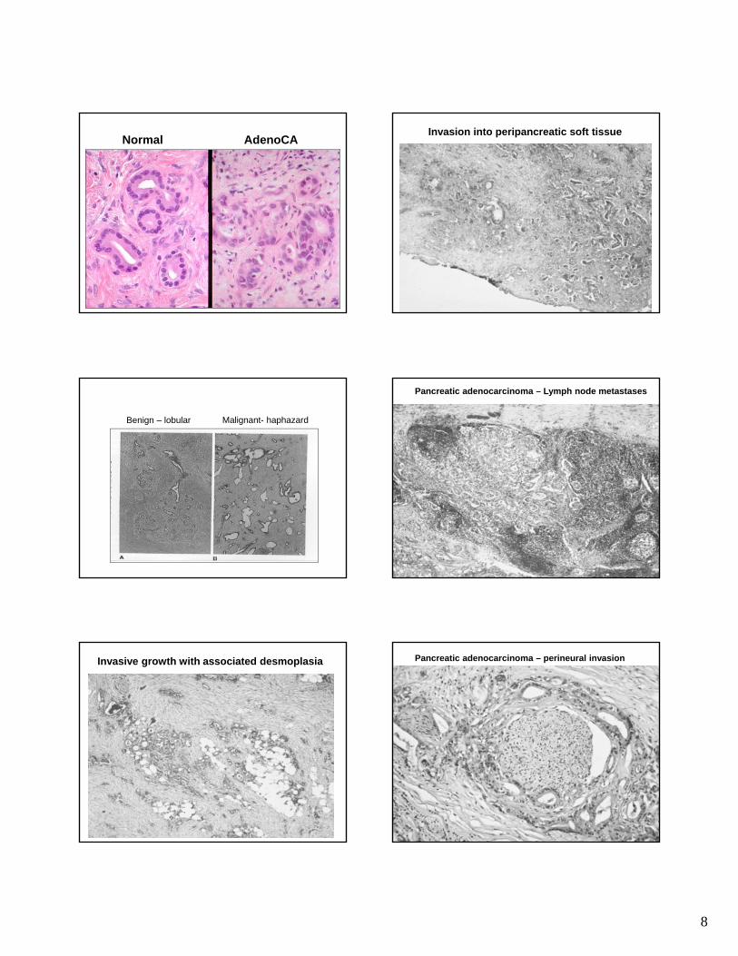
Mucinous cystic NeoplasmNOT connected with pancreatic ducts!
Mucinous cystadenocarcinoma
Mucinous cystadenoma
Mucinous cystic neoplasm
Mucinous epithelium; “ovarian type stroma”Clinical spectrum: benign to malignant
Mucinous Cystic Neoplasms MCNs
Mainly Females (mean age 45)Abdominal Pain or MassSample extensively to rule out invasive componentClassification: Mucinous cystadenoma (minimal atypia)
Borderline MCN (moderate atypia, papillary architecture)( yp , p p y )MCN with CIS (high grade cytology, cribriform architecture)Invasive CA (destructive stromal invasion, usual ductal CA)
Gross: large (mean 10cm); Body/Tail; Multilocular, unilocular rareNO communication with pancreatic duct.
Micro: Columnar mucin cells; intestinal or gastric foveolar type.Ovarian stroma (ER+, PR+, inhibin+)
DD: IPMN (Head/communicate with ducts/ no ovarian stroma)Pancreatic Pseudocyst (MCN lining can be denuded)
PANCREATIC CYSTIC LESIONS
MCNIPMN
12
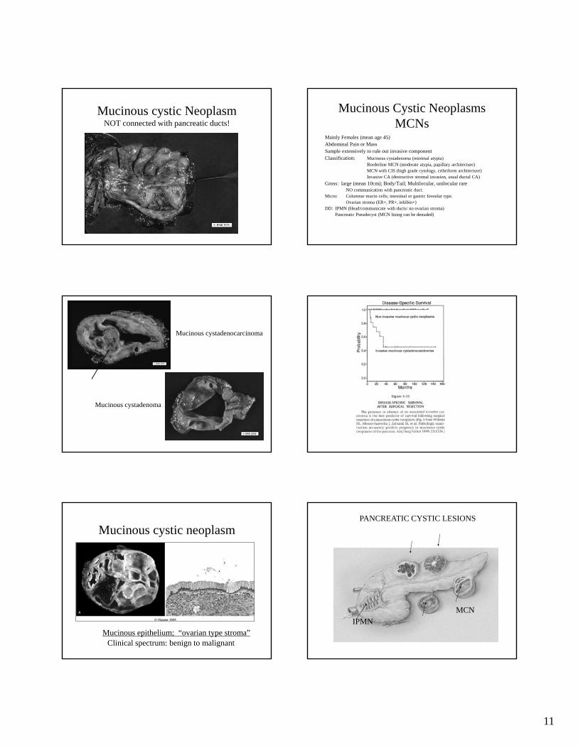
Intraductal Papillary Mucinous Neoplasm (IPMN)
IPMNs (Intraductal Papillary Mucinous Neoplasms)
Communicates with duct Mucin oozes out of ampullae
Intraductal papillary mucinous neoplasm (IPMN)
Associated with the pancreatic ductClinical spectrum: benign to malignant
Intraductal Papillary Mucinous Neoplasm (IPMN)
• IPMT (first named in 1995)• Radiologic or grossly visible lesion (>1cm)• Contiguous or multicentric with cytologic atypia• Head of pancreas; More common in male >60 y.• Invasive tumors associated with 30% of IPMNs (often colloid type –
more indolent clinical behavior than usual invasive ductal-NOS.)• Resection often with frozen sections, since most lesions are
contiguous.• Grade (Benign, Borderline, Intraductal CA); • DD: Mucinous cystic neoplasms, PanIN (resembles small IPMN)
PANCREATIC CYSTIC LESIONSSerous Cystadenoma
MCNIPMN

13
Pancreatic serous cystadenoma
BENIGN
Serous Cystadenoma• Aka microcystic adenoma, glycogen rich adenoma• F/M =7/1; mean age 66y• Association with von Hippel Lindau syndrome• Symptoms: none, local pain, obstruction if in head• Clinical behavior: benign • Gross: mean 11 cm; multiloculated mass, cysts filled with clear
fluid; spongy; often central scar.• Micro: small cystic spaces lined with cuboidal cells with clear
cytoplasm (glycogen rich); round nuclei; some cases papillary.
PANCREATIC CYSTIC LESIONSSerous Cystadenoma
MCNIPMN Pseudocyst
Pancreatic Pseudocyst
NOT NEOPLASTIC - RESULT OF ACUTE PANCREATITIS
Pseudocyst• Localized collections of pancreatic secretions that follow pancreatitis,
trauma, ductal calculi.• Symptoms: Painful, Hemorrhage, Infection, Perforation• Treatment: excise small pseudocysts in body/tail; drain cysts in head.
• Gross: 85% solitary, usually unilocular in/near pancreas; thick irregular wall, ragged inner surface.
• Micro: No epithelial lining, fluid has high amylase • Cyst arises from drainage of pancreatic secretions from damaged ducts
into interstitial tissue; wall consists of fibrous tissue/granulation tissue.
Pancreatic Endocrine Neoplasms
• 5% of pancreatic neoplasms• “Islet cell Tumors” – inaccurate; arise from
pluripotential ductal cells that differentiate along neuroendocrine lines.
• All have malignant potential except microadenomas (<5mm); No definite criteria to distinguish between benign and malignant (except for mets)

14
Pancreatic Endocrine Tumors
Pancreatic Endocrine NeoplasmsMicroscopic
Nests of uniform polygonal cells
Delicate vasculature
Salt and Pepper (stippled) chromatin.
Often (no necrosis, low mitotic activity)
Immunostains do not correlate with secretion. (Other than chromogranin and synaptophysin; specific stains: glucagon, insulin, PP, VIP, ACTH, somatostatin not really useful.)
Pancreatic Endocrine Neoplasms
Functional - recognizable syndrome; detect hormone in serum.• Insulinoma (most common); hypoglycemia; 10% malignant
• 10% assoc with MEN1
• Gastrinoma; duodenal ulcers; 75% malignantGastrinoma; duodenal ulcers; 75% malignant• 25% assoc with MEN1
Nonfunctional - no syndrome; normal serum hormone levels (except Pancreatic Polypeptide).
• Incidental; Obstructive Sx- head of pancreas; 50 – 90% malignant.
Pancreatic Endocrine Neoplasms
• Usually occur in body/tail• Hypervascular, circumscribed• Highlighted with Octreotide Scan (somatostatin receptors)• Usually slow growing, mets to LNs, liver, bone
(recommend resection of mets)
15
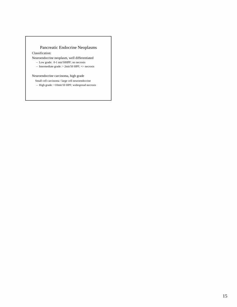
Pancreatic Endocrine NeoplasmsClassification:Neuroendocrine neoplasm, well differentiated
– Low grade: 0-1 mit/50HPF; no necrosis– Intermediate grade: > 2mit/50 HPF; +/- necrosis
Neuroendocrine carcinoma, high gradeSmall cell carcinoma / large cell neuroendocrine– High grade: >10mit/10 HPF; widespread necrosis

![Tumor Microenvironment Characterization in Gastric Cancer ... · tumor[Title]) OR gastric carcinoma[Title]) OR stomach cancer[Title]) OR stomach adenocarcinoma[Title]) OR stom-ach](https://img.pdfslide.net/doc/110x75/5f21882bfc26e208e73f9df9/tumor-microenvironment-characterization-in-gastric-cancer-tumortitle-or-gastric.jpg)


![Tumor-Specific Chromosome Mis-Segregation Controls Cancer … · supported by prediction of tumor progression with genetic clonal diversity in esophageal adenocarcinoma [3], and now](https://img.pdfslide.net/doc/110x75/5faa35bda88b342e6e09c934/tumor-specific-chromosome-mis-segregation-controls-cancer-supported-by-prediction.jpg)