Embed Size (px)
Citation preview
Korean J Hepatobiliary Pancreat Surg 2015;19:78-81http://dx.doi.org/10.14701/kjhbps.2015.19.2.78 Case Report
Gallstone ileus inducing obstructive jaundice at the afferent loop ofRoux-en-Y hepaticojejunostomy after bile duct cancer surgery:
a case report
Hyun Gu Lee, Shin Hwang, Yo-Han Joo, Yu-Jeong Cho, and Kyunghak Choi
Department of Surgery, Asan Medical Center, University of Ulsan College of Medicine, Seoul, Korea
The diagnosis of gallstone ileus is occasionally challenging due to the variability of its presentation. We herein present a very rare case of gallstone ileus inducing obstructive jaundice at the afferent loop of Roux-en-Y hepaticojejunostomy after 10 years of bile duct cancer surgery. We describe the case of a 74-year-old Korean woman with obstructive jaundice, treated conservatively. She showed severely impaired liver function test and obstructive jaundice. The com-puted tomography (CT) scan led to a diagnosis of very rare type of gallstones ileus at the afferent jejunal loop. Since the clinical manifestation was improved, we decided to observe her closely. On the next follow-up CT scan, the gall-stone disappeared with mild distension of the afferent bowel loop, implicating spontaneous passage of the gallstone. She recovered and returned to normal life after 10 days of initiation of clinical manifestations. We presume that the gallstone may enter the afferent jejunal loop through the hepaticojejunostomy and later increase in size. The presence of narrow tract of intestine may facilitate the incidence of gallstone ileus. It appears to be the first report on this rare type of gallstone ileus inducing obstructive jaundice. (Korean J Hepatobiliary Pancreat Surg 2015;19:78-81)
Key Words: Gallstone; Ileus; Roux-en-Y jejunostomy; Biliary obstruction; Bile duct cancer
Received: May 25, 2015; Revised: May 28, 2015; Accepted: May 30, 2015Corresponding author: Shin HwangDepartment of Surgery, Asan Medical Center, University of Ulsan College of Medicine, 88 Olympic-ro 43-gil, Songpa-gu, Seoul 138-736, KoreaTel: +82-2-3010-3930, Fax: +82-2-3010-6701, E-mail: [email protected]
Copyright Ⓒ 2015 by The Korean Association of Hepato-Biliary-Pancreatic SurgeryThis is an Open Access article distributed under the terms of the Creative Commons Attribution Non-Commercial License (http://creativecommons.org/
licenses/by-nc/4.0) which permits unrestricted non-commercial use, distribution, and reproduction in any medium, provided the original work is properly cited.Korean Journal of Hepato-Biliary-Pancreatic Surgery ∙ pISSN: 1738-6349ㆍeISSN: 2288-9213
INTRODUCTION
Gallstone ileus is a rare mechanical bowel obstruction
caused by the transition of a gallstone in the gastrointestinal
tract through a biliary-enteric fistula, or following endo-
scopic retrograde cholangiopancreatography (ERCP), which
occurs in 1 to 3% of all cases of mechanical ileus.1
Gallstones can cause bowel obstruction by intraluminal
impaction anywhere between the stomach and the rectum,
thus abdomen computed tomography (CT) is very informative
for diagnosis of gallstone ileus.2,3
Large gallstones inducing ileus are usually formed with-
in the gallbladder, thus it is very rare to present gallstone
ileus after cholecystectomy.4 In contrast, gallstones have
been occasionally formed at the proximal side of hep-
aticojejunostomy after resection of the common bile duct
and then passed through the lumen of hepaticojejunostomy.
In this type of intrahepatic duct stone formation, the stones
are usually muddy, not being solid stones due to the luminal
environment of the hepaticojejunostomy.
We herein present an extremely rare case of gallstone
ileus, in whom the gallstone stone was formed at the hep-
aticojejunostomy and entrapped at afferent loop of the
Roux-en-Y jejunojejunostomy, by which obstructive jaun-
dice happened and resolved after spontaneous passage.
CASE
A 74-year-old Korean woman was referred to our in-
stitution due to obstructive jaundice. She had undergone
curative resection of the extrahepatic bile duct cancer (2
cm-sized moderately differentiated adenocarcinoma of
T2N0M0) 10 years before. She had been followed up reg-
ularly until the end of postoperative 9 years without any
evidence of disease recurrence. However, 3 days before
referral, at 10 years after surgery, she complained of ab-
Hyun Gu Lee, et al. Afferent loop gallstone ileus 79
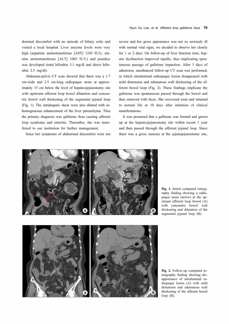
Fig. 1. Initial computed tomog-raphy finding showing a radio-paque stone (arrow) at the up-stream afferent loop bowel (A) with concentric bowel wall thickening and dilatation of the segmental jejunal loop (B).
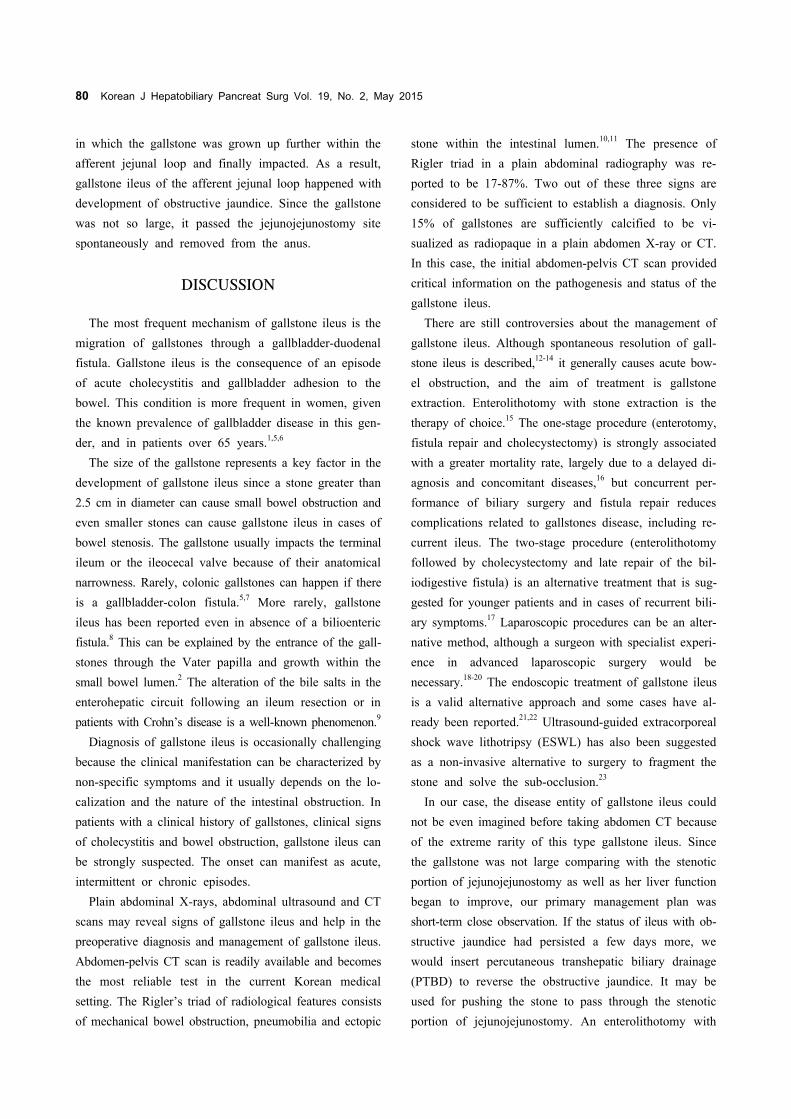
Fig. 2. Follow-up computed to-mography finding showing dis-appearance of intraluminal ra-diopaque lesion (A) with mild distension and edematous wall thickening of the afferent bowelloop (B).
dominal discomfort with no episode of biliary colic and
visited a local hospital. Liver enzyme levels were very
high (aspartate aminotransferase [AST] 1245 IU/L; ala-
nine aminotransferase [ALT] 1065 IU/L) and jaundice
was developed (total bilirubin 3.1 mg/dl and direct bilir-
ubin 2.5 mg/dl).
Abdomen-pelvis CT scan showed that there was a 1.7
cm-wide and 2.5 cm-long radiopaque stone at approx-
imately 15 cm below the level of hepaticojejunostomy site
with upstream afferent loop bowel dilatation and concen-
tric bowel wall thickening of the segmental jejunal loop
(Fig. 1). The intrahepatic ducts were also dilated with in-
homogeneous enhancement of the liver parenchyma. Thus
the primary diagnosis was gallstone ileus causing afferent
loop syndrome and enteritis. Thereafter, she was trans-
ferred to our institution for further management.
Since her symptoms of abdominal discomfort were not
severe and her gross appearance was not so seriously ill
with normal vital signs, we decided to observe her closely
for 1 or 2 days. On follow-up of liver function tests, hep-
atic dysfunction improved rapidly, thus implicating spon-
taneous passage of gallstone impaction. After 3 days of
admission, unenhanced follow-up CT scan was performed,
in which intraluminal radiopaque lesion disappeared with
mild distension and edematous wall thickening of the af-
ferent bowel loop (Fig. 2). These findings implicate the
gallstone was spontaneous passed through the bowel and
then removed with feces. She recovered soon and returned
to normal life at 10 days after initiation of clinical
manifestations.
It was presumed that a gallstone was formed and grown
up at the hepaticojejunostomy site within recent 1 year
and then passed through the afferent jejunal loop. Since
there was a gross stenosis at the jejunojejunostomy site,
80 Korean J Hepatobiliary Pancreat Surg Vol. 19, No. 2, May 2015
in which the gallstone was grown up further within the
afferent jejunal loop and finally impacted. As a result,
gallstone ileus of the afferent jejunal loop happened with
development of obstructive jaundice. Since the gallstone
was not so large, it passed the jejunojejunostomy site
spontaneously and removed from the anus.
DISCUSSION
The most frequent mechanism of gallstone ileus is the
migration of gallstones through a gallbladder-duodenal
fistula. Gallstone ileus is the consequence of an episode
of acute cholecystitis and gallbladder adhesion to the
bowel. This condition is more frequent in women, given
the known prevalence of gallbladder disease in this gen-
der, and in patients over 65 years.1,5,6
The size of the gallstone represents a key factor in the
development of gallstone ileus since a stone greater than
2.5 cm in diameter can cause small bowel obstruction and
even smaller stones can cause gallstone ileus in cases of
bowel stenosis. The gallstone usually impacts the terminal
ileum or the ileocecal valve because of their anatomical
narrowness. Rarely, colonic gallstones can happen if there
is a gallbladder-colon fistula.5,7 More rarely, gallstone
ileus has been reported even in absence of a bilioenteric
fistula.8 This can be explained by the entrance of the gall-
stones through the Vater papilla and growth within the
small bowel lumen.2 The alteration of the bile salts in the
enterohepatic circuit following an ileum resection or in
patients with Crohn’s disease is a well-known phenomenon.9
Diagnosis of gallstone ileus is occasionally challenging
because the clinical manifestation can be characterized by
non-specific symptoms and it usually depends on the lo-
calization and the nature of the intestinal obstruction. In
patients with a clinical history of gallstones, clinical signs
of cholecystitis and bowel obstruction, gallstone ileus can
be strongly suspected. The onset can manifest as acute,
intermittent or chronic episodes.
Plain abdominal X-rays, abdominal ultrasound and CT
scans may reveal signs of gallstone ileus and help in the
preoperative diagnosis and management of gallstone ileus.
Abdomen-pelvis CT scan is readily available and becomes
the most reliable test in the current Korean medical
setting. The Rigler’s triad of radiological features consists
of mechanical bowel obstruction, pneumobilia and ectopic
stone within the intestinal lumen.10,11 The presence of
Rigler triad in a plain abdominal radiography was re-
ported to be 17-87%. Two out of these three signs are
considered to be sufficient to establish a diagnosis. Only
15% of gallstones are sufficiently calcified to be vi-
sualized as radiopaque in a plain abdomen X-ray or CT.
In this case, the initial abdomen-pelvis CT scan provided
critical information on the pathogenesis and status of the
gallstone ileus.
There are still controversies about the management of
gallstone ileus. Although spontaneous resolution of gall-
stone ileus is described,12-14 it generally causes acute bow-
el obstruction, and the aim of treatment is gallstone
extraction. Enterolithotomy with stone extraction is the
therapy of choice.15 The one-stage procedure (enterotomy,
fistula repair and cholecystectomy) is strongly associated
with a greater mortality rate, largely due to a delayed di-
agnosis and concomitant diseases,16 but concurrent per-
formance of biliary surgery and fistula repair reduces
complications related to gallstones disease, including re-
current ileus. The two-stage procedure (enterolithotomy
followed by cholecystectomy and late repair of the bil-
iodigestive fistula) is an alternative treatment that is sug-
gested for younger patients and in cases of recurrent bili-
ary symptoms.17 Laparoscopic procedures can be an alter-
native method, although a surgeon with specialist experi-
ence in advanced laparoscopic surgery would be
necessary.18-20 The endoscopic treatment of gallstone ileus
is a valid alternative approach and some cases have al-
ready been reported.21,22 Ultrasound-guided extracorporeal
shock wave lithotripsy (ESWL) has also been suggested
as a non-invasive alternative to surgery to fragment the
stone and solve the sub-occlusion.23
In our case, the disease entity of gallstone ileus could
not be even imagined before taking abdomen CT because
of the extreme rarity of this type gallstone ileus. Since
the gallstone was not large comparing with the stenotic
portion of jejunojejunostomy as well as her liver function
began to improve, our primary management plan was
short-term close observation. If the status of ileus with ob-
structive jaundice had persisted a few days more, we
would insert percutaneous transhepatic biliary drainage
(PTBD) to reverse the obstructive jaundice. It may be
used for pushing the stone to pass through the stenotic
portion of jejunojejunostomy. An enterolithotomy with
Hyun Gu Lee, et al. Afferent loop gallstone ileus 81
stone extraction would be the last therapeutic for this
patient.
In conclusion, our case demonstrates that the gallstone
may enter the afferent jejunal loop through the hep-
aticojejunostomy and later increase in size. The presence
of a narrow tract of intestine can facilitate the incidence
of gallstone ileus. It appears to be the first report on this
type of gallstone ileus.
REFERENCES
1. Masannat Y, Masannat Y, Shatnawei A. Gallstone ileus: a review. Mt Sinai J Med 2006;73:1132-1134.
2. Pezzoli A, Maimone A, Fusetti N, Pizzo E. Gallstone ileus treat-ed with non-surgical conservative methods: a case report. J Med Case Rep 2015;9:15.
3. Michele D, Luciano G, Massimiliano F, Stefano R, Roberta D, Ernesto S, et al. Usefulness of CT-scan in the diagnosis and ther-apeutic approach of gallstone ileus: report of two surgically treat-ed cases. BMC Surg 2013;13 Suppl 2:S6.
4. Zens T, Liebl RS. Gallstone ileus 30 years status postcholecystectomy. WMJ 2010;109:332-334.
5. Reisner RM, Cohen JR. Gallstone ileus: a review of 1001 re-ported cases. Am Surg 1994;60:441-446.
6. Halabi WJ, Kang CY, Ketana N, Lafaro KJ, Nguyen VQ, Stamos MJ, et al. Surgery for gallstone ileus: a nationwide comparison of trends and outcomes. Ann Surg 2014;259:329-335.
7. Heaney RM. Colonic gallstone ileus: the rolling stones. BMJ Case Rep 2014 [Epub ahead of print].
8. Armitage G, Fowweather FS, Johnstone AS. Observations on bile-acid enteroliths with an account of a recent case. Br J Surg 1950;38:21-25.
9. Andersson H, Bosaeus I, Fasth S, Hellberg R, Hultén L. Cholelithiasis and urolithiasis in Crohn's disease. Scand J Gastroenterol 1987;22:253-256.
10. Rigler LG, Borman CN, Noble JF. Gallstone obstruction: patho-
genesis and roentgen manifestations. JAMA 1941;117:1753-1759.11. Roothans D, Anguille S. Rigler triad in gallstone ileus. CMAJ
2013;185:E690.12. Tandon A, Usha T, Bhargava SK, Bhatt S, Bhargava S, Prakash
M, et al. Resolution of gallstone ileus with spontaneous evacua-tion of gallstone: a case report. Indian J Surg 2013;75:228-231.
13. Roberts J, Lambrianides A. Spontaneous resolution of a gallstone ileus. J Surg Case Rep 2012;2012:3.
14. Miyasaka T, Yoshida H, Makino H, Watanabe M, Uchida E, Uchida E. Response of gallstone ileus to conservative therapy. J Nippon Med Sch 2014;81:388-391.
15. Mallipeddi MK, Pappas TN, Shapiro ML, Scarborough JE. Gallstone ileus: revisiting surgical outcomes using National Surgical Quality Improvement Program data. J Surg Res 2013; 184:84-88.
16. Warshaw AL, Bartlett MK. Choice of operation for gallstone in-testinal obstruction. Ann Surg 1966;164:1051-1055.
17. Zaliekas J, Munson JL. Complications of gallstones: the Mirizzi syndrome, gallstone ileus, gallstone pancreatitis, complications of "lost" gallstones. Surg Clin North Am 2008;88:1345-1368.
18. Ferraina P, Gancedo MC, Elli F, Nallar M, Ferraro A, Sarotto L, et al. Video-assisted laparoscopic enterolithotomy: new tech-nique in the surgical management of gallstone ileus. Surg Laparosc Endosc Percutan Tech 2003;13:83-87.
19. Bircan HY, Koc B, Ozcelik U, Kemik O, Demirag A. Laparoscopic treatment of gallstone ileus. Clin Med Insights Case Rep 2014;7: 75-77.
20. Zygomalas A, Karamanakos S, Kehagias I. Totally laparoscopic management of gallstone ileus--technical report and review of the literature. J Laparoendosc Adv Surg Tech A 2012;22:265-268.
21. Lübbers H, Mahlke R, Lankisch PG. Gallstone ileus: endoscopic removal of a gallstone obstructing the upper jejunum. J Intern Med 1999;246:593-597.
22. Muratori R, Cennamo V, Menna M, Cecinato P, Eusebi LH, Mazzella G, et al. Colonic gallstone ileus treated with radio-logically guided extracorporeal shock wave lithotripsy followed by endoscopic extraction. Endoscopy 2012;44 Suppl 2 UCTN: E88-E89.
23. Meyenberger C, Michel C, Metzger U, Koelz HR. Gallstone ileus treated by extracorporeal shockwave lithotripsy. Gastrointest Endosc 1996;43:508-511.
![Clinical and radiological diagnosis of gallstone ileus: a ... · order to cause obstruction at an anatomically wide part of the gastrointestinal tract [40–42]. This is estimated](https://img.pdfslide.net/doc/110x75/5d62e92788c993e9588b86bc/clinical-and-radiological-diagnosis-of-gallstone-ileus-a-order-to-cause.jpg)