Embed Size (px)
Citation preview
International Journal of Medical Imaging 2019; 7(1): 25-28
http://www.sciencepublishinggroup.com/j/ijmi
doi: 10.11648/j.ijmi.20190701.14
ISSN: 2330-8303 (Print); ISSN: 2330-832X (Online)
Gallstone Ileus: An Uncommon Cause of Acute Abdomen
Rachida Saouab1, Fatima Zahra Rhouni
2, Mohammed Mahi
2, Hassan Boumdien
2, Touriya Amil
2,
Jamal El Fenni2
1Department of Radiology, Military Hospital, Faculty of Medicine and Pharmacy, Cadi Ayyad University, Marrakech, Morocco 2Department of Radiology, Military Hospital, Faculty of Medicine and Pharmacy, Mohammed V University, Rabat, Morocco
Email address:
To cite this article: Rachida Saouab, Fatima Zahra Rhouni, Mohammed Mahi, Hassan Boumdien, Touriya Amil, Jamal El Fenni. Gallstone Ileus: An Uncommon
Cause of Acute Abdomen. International Journal of Medical Imaging. Vol. 7, No. 1, 2019, pp. 25-28. doi: 10.11648/j.ijmi.20190701.14
Received: March 5, 2019; Accepted: April 26, 2019; Published: May 17, 2019
Abstract: Gallstone ileus is a rare cause of acute abdomen, representing less than 1% of intestinal obstruction. It is due to
mechanical small bowel obstruction (SBO) by a migrated gallstone. we report this observation to illustrate the imaging
findings especially in CT scan. A 62-year-old woman was admitted to emergency for acute bowel obstruction. The CT scan
has confirmed jejunal bowel distension with fluid levels upstream endoluminal formation which was rounded and well limited,
measuring 5 cm of long axis. This training was slightly hyperdense with hypodense areas of fat density. Moreover, there was
aerobilia with multiple fistulas between the duodenum (D1) and gallbladder, which was empty. The triad of aerobilia, small
bowel obstruction and migrant gallstone was suggestive of gallstone ileus diagnosis. The patient was operated with good
evolution. Gallstone ileus is an uncommon presentation of gallstone disease. It is frequently observed in elderly patients with a
history of cholelithiasis or cholecystitis. Early diagnosis is crucial and would avoid morbidity and mortality related to this
disease. CT scan shows the classical triad of Rigler with pneumobilia, an ectopic stone and mechanical ileus. CT may improve
the diagnosis providing important information regarding the exact number, size, and location of ectopic stones and the site of
intestinal obstruction or direct visualization of a biliary–enteric fistula, to help clinicians in the therapeutic management of
patients.
Keywords: Gallstone, Ileus, Bowel Obstruction, Pneumobilia, Acute Abdomen, CT Scan
1. Introduction
Gallstone ileus is an uncommon cause of bowel
obstruction which mainly affects the elderly population. It is
a rare and serious complication of chronic cholecystitis and
occurs when a gallstone passes into the small bowel and
usually impacts at the ileocaecal valve. The associated
mortality is estimated to be up to 30% [1]. This high
mortality is attributed to comorbidities, particularly
cardiovascular, respiratory, and endocrine (diabetes and
obesity), or delayed diagnosis [2, 3]. Diagnostic imaging
plays an important role in the management of patients. We
report a case of a woman presented with jejunal obstruction
due to migrant gallstone in order to remember the CT scan
findings.
2. Case Report
A 62-year-old woman was admitted to emergency with
acute abdominal pain and vomiting. Abdominal history was
significant for gallstone. Physical examination revealed an
afebrile patient with diffuse abdominal tenderness more
marked at the right upper quadrant. The laboratory tests
showed leukocytosis at 13000/µl with slight increase in C-
reactive protein (CRP) and creatinine. Abdominal ultrasound
found aerobilia with distended small bowel loops. Additional
computed tomography scan (CT) was requested. It was made
without injection of contrast material because of kidney
failure. The CT scan has confirmed aerobilia and presence of
multiple fistulas between the duodenum (D1) and
gallbladder, which was empty, with the presence of stones in
the cystic duct (figure 1).
26 Rachida Saouab et al.: Gallstone Ileus: An Uncommon Cause of Acute Abdomen
Figure 1. Coronal reconstruction computed tomography image without IV contrast injection reveals pneumobilia with multiple fistulas between the duodenum
(D1) and gallbladder.
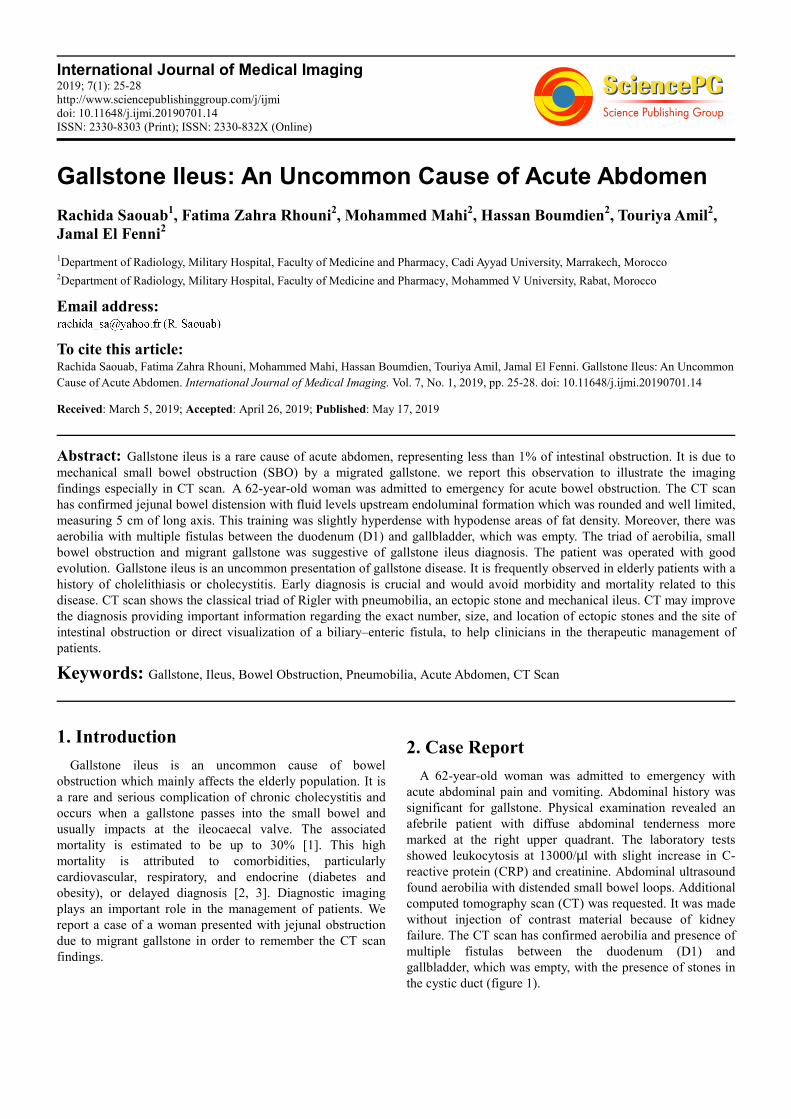
Moreover, there was small bowel distension with fluid levels upstream endoluminal formation which was rounded and well
limited, measuring 5 cm of long axis. This training was slightly hyperdense with hypodense areas of fat density suggesting a
migrant gallstone by the biliary- enteric fistula (figure 2).
Figure 2. Axial computed tomography image without IV contrast injection reveals jejunal distension with endoluminal gallstone, measuring 5 cm of long axis.
It was slightly hyperdense with hypodense areas of fat density.
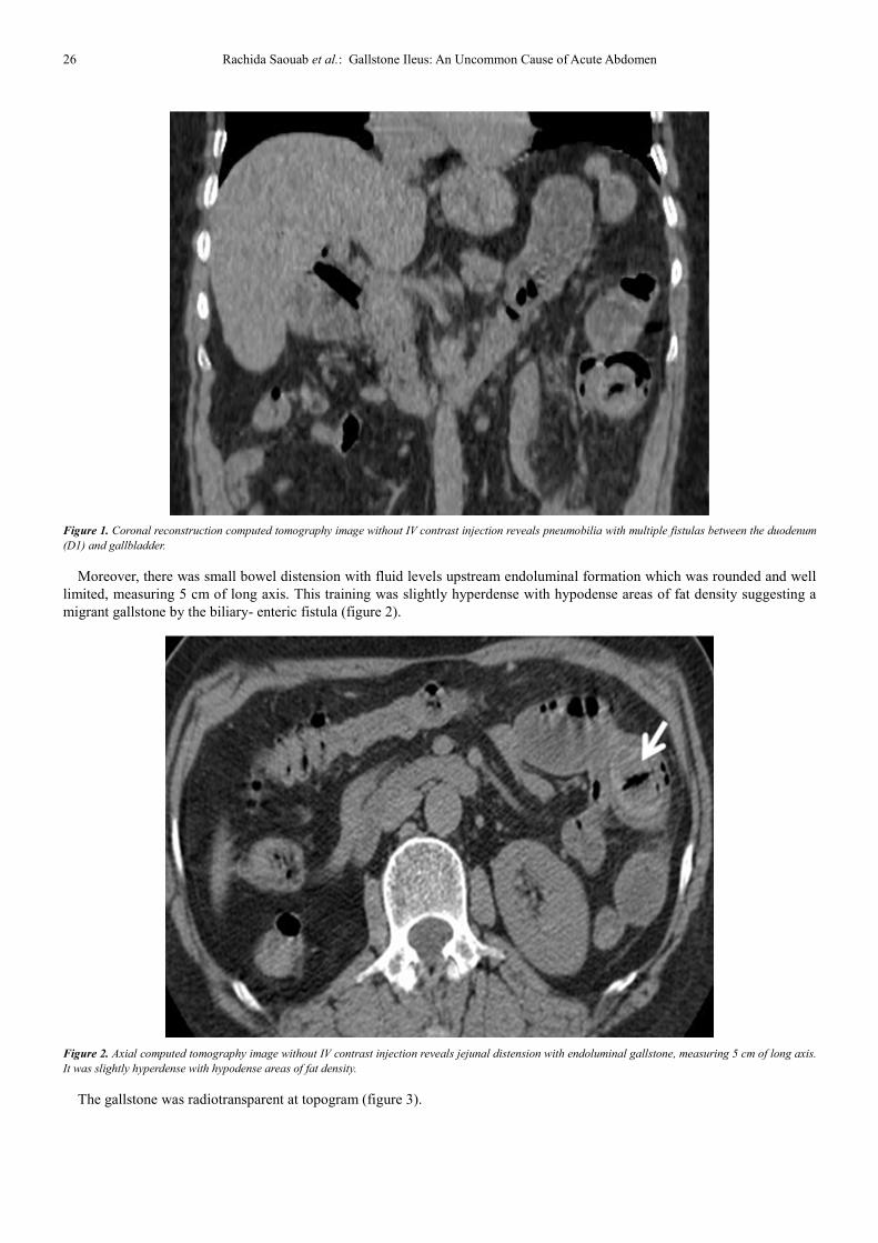
The gallstone was radiotransparent at topogram (figure 3).
International Journal of Medical Imaging 2019; 7(1): 25-28 27
Figure 3. CT scan topogram reveals jejunal distension at the left
hypochondrium without individualization of gallstone which is
radiotransparent.
Ileum and colon were collapsed. The triad of aerobilia,
small bowel obstruction and migrant gallstone was
suggestive of gallstone ileus diagnosis. The patient was
operated with good evolution.
3. Discussion
Gallstone ileus is an unusual complication of
cholelithiasis, occurring in less than 0.5 percent of patients. It
represents only 1 to 4% of small bowel obstruction caused by
an impaction of a gallstone within the lumen of the small
intestine via a cholecysto-enteric fistula. Large stones, greater
than 2.5 cm in diameter, are thought to predispose to fistula
formation by gradual erosion through the gallbladder fundus.
Sixty percent are cholecystoduodenal fistulas, but
cholecystocolonic and cholecystogastric fistulas can also
result in gallstone ileus [4, 5]. Common places for gallstones
include the ileum and ileocecal valve due to the anatomical
narrow lumen in 60% of cases, jejunum in up to 16%,
stomach in 15%, and colon (gallstone coleus) in 2–8% of
cases [6, 7].
The average age of patients with gallstone ileus is 70
years, with the youngest reported patient being 13 years of
age. Women are 3 to 16 times more likely to be affected [8].
Typically, patients have a long history of recurrent right
upper quadrant pain. The acute presentation of gallstone ileus
is that of a small bowel obstruction, with colicky abdominal
pain, abdominal distension and vomiting. Jaundice has been
found in only 15% of patients or less. Upper gastrointestinal
bleeding, secondary to duodenal erosion caused by the
offending gallstone, with hematemesis and melena, can be
seen in 15% and 7%, respectively [9, 10].
The diagnosis of gallstone ileus is difficult and usually
depends on the imaging findings. The classic Rigler’s triad
includes mechanical bowel obstruction, pneumobilia, and
presence of an ectopic gallstone within the bowel lumen [11].
Plain abdominal radiographic films usually show non-
specific findings, because only 10% of gallstones are
sufficiently calcified [12]. CT is the gold standard for
positive diagnosis. The overall sensitivity, specificity and
accuracy of CT in diagnosing gallstone ileus is around 93%,
100%; and 99%, respectively. It allows detection of the stone
whatever the density, its exact location and direct
visualization of the biliary–enteric fistula. Typically, stones
are large and measure several centimeters (2-3 cm). It is
important to look for free fluid, free gas, portal venous gas,
or mural gas, as signs of more advanced disease and poorer
prognosis [13-16].
Gallstone ileus usually requires emergency surgery to
relieve intestinal obstruction with the removal of the stone
(enterolithotomy) and repair of the choledochoenteric fistula,
accompanied by a cholecystectomy [17, 18].
Any delay in diagnosis and treatment may lead to serious
complications such as electrolyte imbalance, ischemic
lesions, ulcerations of the bowel, abscess formation, and,
occasionally, free perforation and peritonitis. The morbidity
and mortality rate of gallstone ileus remain very high, partly
because of misdiagnosis and delayed diagnosis and otherwise
because of the age-related co- morbidities of the afflicted
patients [19].
4. Conclusion
Gallstone ileus is a frequently misdiagnosed clinical entity.
Early diagnosis is crucial and allows earlier therapy and
would avoid morbidity and mortality related to this disease.
The use of radiological imaging is invaluable in the diagnosis
of gallstone ileus. In fact, CT scanning is a powerful and
gold-standard tool to diagnose the condition and to guide its
management. The combination of pneumobilia and
gastrointestinal obstruction should suggest the diagnosis of
gallstone ileus. In addition to the Rigler radiologic triad, CT
allows detection of the exact location of the ectopic stones,
the site of obstruction and direct visualization of the biliary–
enteric fistula.
References
[1] Liisa Chang, Minna Chang, Hanna M. Chang, Aina I. Chang, Fuju Chang. Clinical and radiological diagnosis of gallstone ileus: a mini review. Emerg Radiol (2018) 25:189–196
[2] Halabi WJ, Kang CY, Ketana N, Lafaro KJ, Nguyen VQ, Stamos MJ, Imagawa DK, Demirjian AN Surgery for gallstone ileus: a nationwide comparison of trends and outcomes. Ann Surg (2014) 259(2):329–335.
[3] Bass G, Gilani SN, Walsh TN Validating the 5Fs mnemonic for cholelithiasis: time to include family history. Postgrad Med J (2013) 89(1057):638–641.
[4] Farrell I, Turner P. A simple case of gallstone ileus? J Surg Case Rep 2015: rju148.
28 Rachida Saouab et al.: Gallstone Ileus: An Uncommon Cause of Acute Abdomen
[5] Ploneda-Valencia CF, Gallo-Morales M, Rinchon C, Navarro-Muñiz E, Bautista-López CA, de la Cerda-Trujillo LF et al Gallstone ileus: an overview of the literature. Rev Gastroenterol (2017) Mex 82(3):248–254.
[6] Dai XZ, Li GQ, Zhang F, Wang XH, Zhang CY. Gallstone ileus: case report and literature review. World J Gastroenterol (2013) 19(33):5586–5589.
[7] Howells L, Liasis L, Demosthenous M. Gallstone coleus: a rare relation of gallstone ileus. Int J Surg Res (2016) 2(4):28–31.
[8] Mallipeddi MK, Pappas TN, Shapiro ML, Scarborough JE. Gallstone ileus: revisiting surgical outcomes using National Surgical Quality Improvement Program data. J Surg Res (2013)184(1): 84–88.
[9] Martín-Pérez J, Delgado-Plasencia L, Bravo-Gutiérrez A, Burillo-Putze G, Martínez-Riera A, Alarcó-Hernández A, Medina-Arana V. Gallstone ileus as a cause of acute abdomen. Importance of early diagnosis for surgical treatment. Cir Esp 9 (2013) 1(8):485–489.
[10] Nuño-Guzmán CM, Marín-Contreras ME, Figueroa-Sánchez M, Corona JL. Gallstone ileus, clinical presentation, diagnostic and treatment approach. World J Gastrointest Surg (2016) 8(1):65–76.
[11] Rigler L. G., Borman C. N., Noble J. F.: Gallstone obstruction: pathogenesis and roentgen manifestations. JAMA, 1941, 117: 1753-1759.
[12] Chou J.-W., Hsu C.-H., Liao K.-F., et al.: Gallstone ileus: Report of two cases and review of the literature. World J Gastroenterol, 2007, 13: 1295-1298.
[13] Swift SE, Spencer JA. Gallstone ileus: CT findings. Clin Radiol (1998) 53(6):451–454.
[14] Liang X, Li W, Zhao B, Zhang L, Cheng Y (2015) Comparative analysis of MDCT and MRI in diagnosing chronic gallstone perforation and ileus. Eur J Radiol 84(10):1835–1842.
[15] Furukawa A, Yamasaki M, Furuichi K, Yokoyama K, Nagata T, Takahashi M, Murata K, Sakamoto T. Helical CT in the diagnosis of small bowel obstruction. Radiographics (2001) 21(2):341–355.
[16] Barakos JA, Ralls PW, Lapin SA, Johnson MB, Radin DR, Colletti PM, Boswell WD Jr, Halls JM Cholelithiasis: evaluation with CT. Radiology (1987) 162(2):415–418.
[17] Yu C. Y., Lin C. C., Shyu R. Y., et al.: Value of CT in the diagnosis and management of gallstone ileus. World J Gastroenterol, 2005, 11: 2142- 2147
[18] Ihara E., Ochiai T., Yamamoto K., et al.: A case of gallstone ileus with a spontaneous evacuation. Am J Gastro -enterol, 2002, 97: 1259-1260.
[19] Lobo D. N., Jobling J. C., Balfour T. W.: Gallstone ileus: diagnostic pitfalls and therapeutic successes. J Clin Gastroenterol, 2000, 30: 72-76.


![Clinical and radiological diagnosis of gallstone ileus: a ... · order to cause obstruction at an anatomically wide part of the gastrointestinal tract [40–42]. This is estimated](https://img.pdfslide.net/doc/110x75/5d62e92788c993e9588b86bc/clinical-and-radiological-diagnosis-of-gallstone-ileus-a-order-to-cause.jpg)