Embed Size (px)
Citation preview
PICTURES IN DIGESTIVE PATHOLOGY
Gallstone ileus presenting as obstructive gangrenous appendicitisJosé Cruz-Santiago1,2, Giuseppe Briceño-Sáenz1, Javier García-Álvarez1 and José Luis Beristain-Hernández2
1Department of General Surgery. Hospital Juárez de México. Ciudad de México, Mexico. 2Department of General Surgery. Hospital de Especialidades. Centro Médico Nacional “La Raza”. Ciudad de México, Mexico
1130-0108/2017/109/2/150-151Revista española de enfeRmedades digestivas© Copyright 2017. sepd y © ARÁN EDICIONES, S.L.
Rev esp enfeRm dig2017, Vol. 109, N.º 2, pp. 150-151
INTRODUCTION
We present the very unusual case of a 38-year-old woman with acute appendicitis and intestinal obstruction. During surgery, a 2.5 cm gallstone impacted at the base of the cecal appendix was found as the cause of a gangrenous appendicitis and obstruction; a choledochal-duodenal fis-tula was found during the same surgery with no gallstones remaining in the gallbladder or elsewhere.
The case was managed by appendectomy with retrieval of the gallstones, and no other procedure was performed for the gallbladder or the fistula, since no other gallstone was found on examination. Previously, she was found to have a round, radio-opaque image on the right iliac fossa; on imaging it was initially identified as an appendicolith, but after pathological examination it turned out to contain cholesterol and calcium bilirubinate.
Gallstone ileus as the cause of an obstructive gangre-nous appendicitis is a very unusual disease presentation that should be kept in mind when finding an unusual appendicolith presentation in or out of the appendix.
CASE REPORT
The patient was a 38-year-old female without any rel-evant medical history. She was admitted with a history
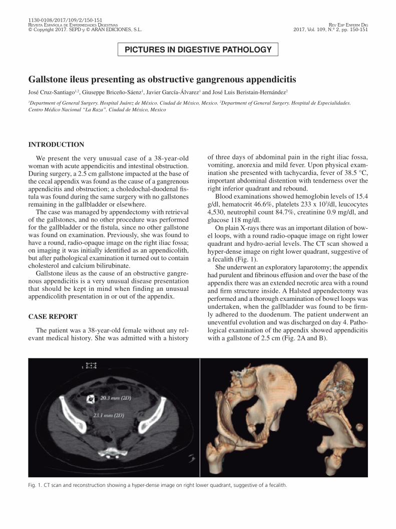
Fig. 1. CT scan and reconstruction showing a hyper-dense image on right lower quadrant, suggestive of a fecalith.
of three days of abdominal pain in the right iliac fossa, vomiting, anorexia and mild fever. Upon physical exam-ination she presented with tachycardia, fever of 38.5 °C, important abdominal distention with tenderness over the right inferior quadrant and rebound.
Blood examinations showed hemoglobin levels of 15.4 g/dl, hematocrit 46.6%, platelets 233 x 103/dl, leucocytes 4,530, neutrophil count 84.7%, creatinine 0.9 mg/dl, and glucose 118 mg/dl.
On plain X-rays there was an important dilation of bow-el loops, with a round radio-opaque image on right lower quadrant and hydro-aerial levels. The CT scan showed a hyper-dense image on right lower quadrant, suggestive of a fecalith (Fig. 1).
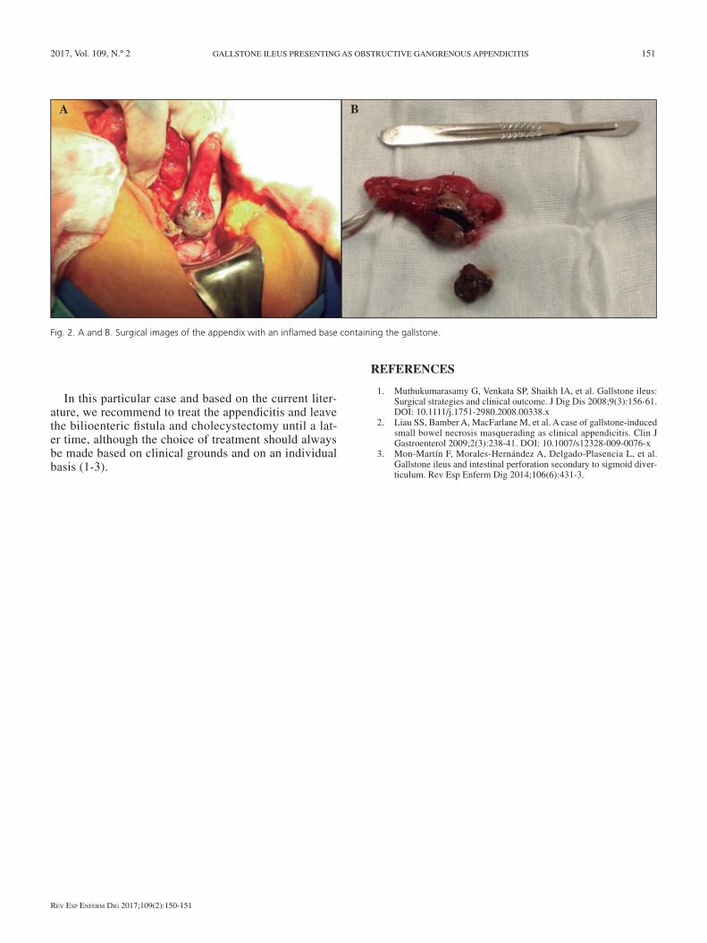
She underwent an exploratory laparotomy; the appendix had purulent and fibrinous effusion and over the base of the appendix there was an extended necrotic area with a round and firm structure inside. A Halsted appendectomy was performed and a thorough examination of bowel loops was undertaken, when the gallbladder was found to be firm-ly adhered to the duodenum. The patient underwent an uneventful evolution and was discharged on day 4. Patho-logical examination of the appendix showed appendicitis with a gallstone of 2.5 cm (Fig. 2A and B).
2017, Vol. 109, N.º 2 GALLSTONE ILEUS PRESENTING AS OBSTRUCTIVE GANGRENOUS APPENDICITIS 151
Rev esp enfeRm Dig 2017;109(2):150-151
In this particular case and based on the current liter-ature, we recommend to treat the appendicitis and leave the bilioenteric fistula and cholecystectomy until a lat-er time, although the choice of treatment should always be made based on clinical grounds and on an individual basis (1-3).
Fig. 2. A and B. Surgical images of the appendix with an inflamed base containing the gallstone.
A B
REFERENCES
1. Muthukumarasamy G, Venkata SP, Shaikh IA, et al. Gallstone ileus: Surgical strategies and clinical outcome. J Dig Dis 2008;9(3):156-61. DOI: 10.1111/j.1751-2980.2008.00338.x
2. Liau SS, Bamber A, MacFarlane M, et al. A case of gallstone-induced small bowel necrosis masquerading as clinical appendicitis. Clin J Gastroenterol 2009;2(3):238-41. DOI: 10.1007/s12328-009-0076-x
3. Mon-Martín F, Morales-Hernández A, Delgado-Plasencia L, et al. Gallstone ileus and intestinal perforation secondary to sigmoid diver-ticulum. Rev Esp Enferm Dig 2014;106(6):431-3.


![Clinical and radiological diagnosis of gallstone ileus: a ... · order to cause obstruction at an anatomically wide part of the gastrointestinal tract [40–42]. This is estimated](https://img.pdfslide.net/doc/110x75/5d62e92788c993e9588b86bc/clinical-and-radiological-diagnosis-of-gallstone-ileus-a-order-to-cause.jpg)