Embed Size (px)
Citation preview
Fitoterapia 81 (2010) 1033–1036
Contents lists available at ScienceDirect
Fitoterapia
j ourna l homepage: www.e lsev ie r.com/ locate / f i to te
Ganoderic acid Df, a new triterpenoid with aldose reductase inhibitoryactivity from the fruiting body of Ganoderma lucidum
Sri Fatmawati a,b, Kuniyoshi Shimizu a, Ryuichiro Kondo a,⁎a Department of Forest and Forest Products Sciences, Faculty of Agriculture, Kyushu University, Fukuoka, Japan, 812-8581b Department of Chemistry, Faculty of Mathematics and Natural Sciences, Sepuluh Nopember Institute of Technology, Kampus ITS-Sukolilo, Surabaya, Indonesia, 60111
a r t i c l e i n f o
⁎ Corresponding author. Tel./fax: +81 92 642 2811E-mail address: [email protected] (R. K
0367-326X/$ – see front matter © 2010 Elsevier B.V.doi:10.1016/j.fitote.2010.06.025
a b s t r a c t
Article history:Received 15 April 2010Accepted in revised form 21 June 2010Available online 1 August 2010
Ganoderic acid Df, a new lanostane-type triterpenoid, was isolated from the fruiting body ofGanoderma lucidum. Its structure was characterized as 7β, 11β-dihydroxy-3, 15, 23-trioxo-5α-lanosta-8-en-26-oic acid by 1D- and 2D-NMR spectra. This compound exhibited potent humanaldose reductase inhibitory activity, with an IC50 of 22.8 μM in vitro. A carboxyl group of thiscompound's side chain is essential for eliciting inhibitory activity because its methyl ester ismuch less active.
© 2010 Elsevier B.V. All rights reserved.
Keywords:Ganoderma lucidumAldose reductase inhibitorGanoderic acid1. Introduction
Ganoderma lucidum (Leyss;Fr) Karst. (Ganodermataceae)are well-known medicinal woody mushrooms called“Lingzhi” in Chinese, “Reishi” in Japanese, and “Yeongji” inKorean. For hundreds of years, this mushroom has been usedto prevent and treat various human diseases. G. lucidum hasbeen reported to produce many biologically active com-pounds such as sterol, polysaccharide, and triterpenoids.More than 100 triterpenoids have been isolated from G.lucidum and the genus Ganoderma [1]. Triterpenoids thathave been isolated from the fruiting body of G. lucidum aredivided into two groups: ganoderma acids with a carboxylgroup in the side chain and ganoderma alcohols with ahydroxyl group in the side chain [2]. The ganoderma acidsisolated from this mushroom show anti-androgenic, anti-5α-reductase, anti-inflammatory, anti-tumor and other biologi-cal activities [3–5].
Aldose reductase (alditol: NAD(P)+ 1-oxidoreductase, EC1.1.1.21) is the first enzyme in the polyol pathway. Thisenzyme catalyzes the reduction of glucose to sorbitol bycoupling with the oxidation of NADPH to NADP+. The
.ondo).
All rights reserved.
accumulation of sorbitol then leads to diabetic complications.For this reason, aldose reductase inhibitors have beenintroduced as a vehicle for the treatment of diabeticcomplications. In our continuing search for an aldosereductase inhibitory constituent, we have focused on thefruiting body of G. lucidum.
In our previous research, we found that the extract of G.lucidum had the strongest aldose reductase inhibitoryactivity among 17 edible and medicinal mushrooms, andsignificantly alleviated the galactitol accumulation in theeye lenses of galactosemic rats [6]. By using a chloroformextract of G. lucidum, we isolated a new compound, 7β,11β-dihydroxy-3,15,23-trioxo-5α-lanosta-8-en-26-oic acid,which we named ganoderic acid Df (1). This acid potentlyinhibited aldose reductase. In the present study, weevaluated the isolated compound (1) and its methyl ester (2)on aldose reductase inhibition.
2. Experimental
2.1. General experimental procedures
The compound was isolated with p-HPLC by usingWaters™ 600 Controller, Waters™ 486 Tunable absorbancedetector, and Waters 600 Multisolvent Delivery System.
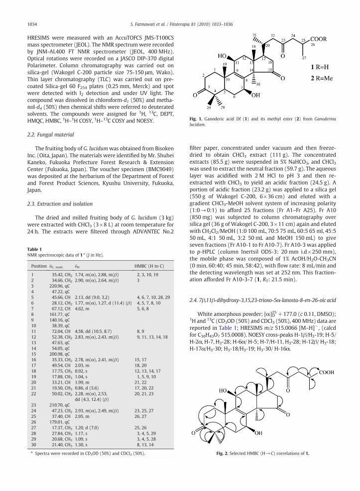
Fig. 1. Ganoderic acid Df (1) and its methyl ester (2) from Ganodermalucidum.
1034 S. Fatmawati et al. / Fitoterapia 81 (2010) 1033–1036
HRESIMS were measured with an AccuTOFCS JMS-T100CSmass spectrometer (JEOL). The NMR spectrumwere recordedby JNM-AL400 FT NMR spectrometer (JEOL, 400 MHz).Optical rotations were recorded on a JASCO DIP-370 digitalPolarimeter. Column chromatography was carried out onsilica-gel (Wakogel C-200 particle size 75-150 μm, Wako).Thin layer chromatography (TLC) was carried out on pre-coated Silica-gel 60 F254 plates (0.25 mm, Merck) and spotwere detected with I2 detection and under UV light. Thecompound was dissolved in chloroform-d1 (50%) and metha-nol-d4 (50%) then chemical shifts were referred to deuteratedsolvents. The compounds were assigned for 1H, 13C, DEPT,HMQC, HMBC, 1H–1H COSY, 1H–13C COSY and NOESY.
2.2. Fungal material
The fruiting body of G. lucidumwas obtained from BisokenInc. (Oita, Japan). The materials were identified by Mr. ShuheiKaneko, Fukuoka Prefecture Forest Research & ExtensionCenter (Fukuoka, Japan). The voucher specimen (BMC9049)was deposited at the herbarium of the Department of Forestand Forest Product Sciences, Kyushu University, Fukuoka,Japan.
2.3. Extraction and isolation
The dried and milled fruiting body of G. lucidum (3 kg)were extracted with CHCl3 (3×8 L) at room temperature for24 h. The extracts were filtered through ADVANTEC No.2
Table 1NMR spectroscopic data of 1 a (J in Hz).
Position δC, mult. δH HMBC (H to C)
1 35.42, CH2 1.74, m(α), 2.88, m(β) 2, 3, 10, 192 34.66, CH2 2.90, m(α), 2.64, m(β) 33 220.96, qC4 47.22, qC5 45.66, CH 2.13, dd (9.0, 3.2) 4, 6, 7, 10, 28, 296 28.12, CH2 1.77, m(α), 1.27, d (11.4) (β) 4, 5, 7, 8, 107 67.12, CH 4.62, m 5, 6, 88 161.77, qC9 140.16, qC10 38.39, qC11 72.04, CH 4.58, dd (10.5, 8.7) 8, 912 52.38, CH2 2.83, m(α), 2.43, m(β) 9, 11, 13, 14, 1813 47.63, qC14 54.05, qC15 200.98, qC16 35.33, CH2 2.78, m(α), 2.41, m(β) 15, 1717 49.54, CH 2.03, m 18, 2018 17.75, CH3 0.92, s 12, 13, 14, 1719 17.88, CH3 1.04, s 1, 5, 9, 1020 33.21, CH 1.99, m 21, 2221 19.50, CH3 0.86, d (5.6) 17, 20, 2222 50.02, CH2 2.28, m(α), 2.53,
dd (4.3, 12.4) (β)20, 21, 23
23 210.70, qC24 47.23, CH2 2.93, m(α), 2.49, m(β) 23, 25, 2725 37.40, CH 2.95, m 26, 2726 179.01, qC27 17.37, CH3 1.20, d (7.0) 25, 2628 27.84, CH3 1.17, s 3, 4, 5, 2929 20.68, CH3 1.09, s 3, 4, 5, 2830 21.40, CH3 1.30, s 8, 13, 14
a Spectra were recorded in CD3OD (50%) and CDCl3 (50%). Fig. 2. Selected HMBC (H→C) correlations of 1.
filter paper, concentrated under vacuum and then freeze-dried to obtain CHCl3 extract (111 g). The concentratedextracts (85.5 g) were suspended in 5% NaHCO3, and CHCl3was used to extract the neutral fraction (59.7 g). The aqueouslayer was acidified with 2 M HCl to pH 3 and then re-extracted with CHCl3 to yield an acidic fraction (24.5 g). Aportion of acidic fraction (23.2 g) was applied to a silica gel(550 g of Wakogel C-200, 6×36 cm) and eluted with agradient CHCl3-MeOH solvent system of increasing polarity(1:0→0:1) to afford 25 fractions (Fr A1–Fr A25). Fr A10(850 mg) was subjected to column chromatography oversilica gel (36 g of Wakogel C-200, 3×11 cm) again and elutedwith CH2Cl2/MeOH (1:0 100 mL, 70:5 75 mL, 60:5 65 ml, 45:550 mL, 4:1 50 mL, 3:2 50 mL and MeOH 150 mL) to giveseven fractions (Fr A10-1 to Fr A10-7). Fr A10-3 was appliedto p-HPLC (column Inertsil ODS-3: 20 mm i.d×250 mm),the mobile phase was composed of 1% AcOH/H2O-CH3CN(0 min, 60:40; 45 min, 58:42), with flow rate: 8 mL/min andthe detecting wavelength was set at 252 nm. This fraction-ation afforded Fr A10-3-7 (1, RT: 21.5 min).
2.4. 7β,11β-dihydroxy-3,15,23-trioxo-5α-lanosta-8-en-26-oic acid
White amorphous powder; [α]25D +177.0 (c 0.11, DMSO);1H and 13C (CD3OD (50%) and CDCl3 (50%), 400 MHz) data arereported in Table 1; HRESIMS m/z 515.0066 [M–H]−, (calcdfor C30H43O7 515.0008). NOESY cross-peaks H-1β/H3-19; H-5/H-2α, H-7, H3-28; H-6α/ H-5; H-7/H-11, H3-28; H-12β/ H3-18;H-17α/H3-30; H3-18/H3-19; H3-30/ H-16α.
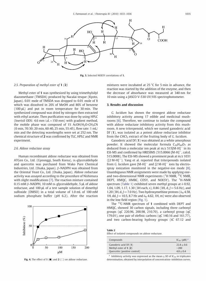
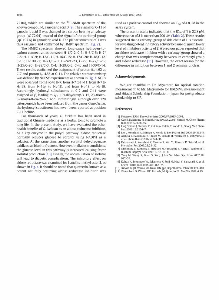
Fig. 3. Selected NOESY correlations of 1.
1035S. Fatmawati et al. / Fitoterapia 81 (2010) 1033–1036
2.5. Preparation of methyl ester of 1 (2)
Methyl ester of 1 was synthesized by using trimethylsilyldiazomethane (TMSDA) produced by Nacalai tesque (Kyoto,Japan). 0.01 mole of TMSDA was dropped to 0.01 mole of 1which was dissolved in 20% of MeOH and 80% of benzene(100 μL) and put in room temperature for 30 min. Thesynthesized compound was dried by nitrogen then extractedwith ethyl acetate. Then purification was done by using HPLC(Inertsil ODS: 4.6 mm i.d.×150 mm) with gradient method,the mobile phase was composed of 1% AcOH/H2O-CH3CN(0 min, 70:30; 20 min, 60:40, 25 min, 55:45), flow rate: 1 mL/min and the detecting wavelengths were set at 252 nm. Thechemical structure of 2was confirmed by TLC, HPLC and NMRexperiment.
2.6. Aldose reductase assay
Human recombinant aldose reductase was obtained fromATGen Co., Ltd. (Gyeonggi, South Korea), DL-glyceraldehydeand quercetin was purchased from Wako Pure ChemicalIndustries, Ltd. (Osaka, Japan). β-NADPH was obtained fromthe Oriental Yeast Co., Ltd. (Osaka, Japan). Aldose reductaseactivity was assayed according to the procedure of Nishimurawith slight modifications [7]. The reaction mixture contained0.15 mM β-NADPH, 10 mM DL-glyceraldehyde, 5 μL of aldosereductase, and 100 μL of a test sample solution of dimethylsulfoxide (DMSO) in a total volume of 1.0 mL of 100 mMsodium phosphate buffer (pH 6.2). After the reaction
Fig. 4. The effect of 1 (■) and 2 (□) on aldose reductase.
mixtures were incubated at 25 °C for 5 min in advance, thereaction was started by the addition of the enzyme, and thenthe decrease of absorbance was measured at 340 nm for10 min using a JASCO V-530 UV/VIS spectrophotometer.
3. Results and discussion
G. lucidum has shown the strongest aldose reductaseinhibitory activity among 17 edible and medicinal mush-rooms [6]. Therefore, we continue to isolate the compoundwith aldose reductase inhibitory activity from this mush-room. A new triterpenoid, which we named ganoderic acidDf (1), was isolated as a potent aldose reductase inhibitorfrom the CHCl3 extract of the fruiting body of G. lucidum.
Ganoderic acid Df (1) was obtained as a white amorphouspowder. It showed the molecular formula C30H44O7 asdeduced from a molecular ion peak at m/z 515[M-H]− in itsESI-MS and confirmed by HRESIMS (515.0066 [M-H]−, calcd.515.0008). The ESI-MS showed a prominent peak at m/z 1031([2 M-H]−). Yang et al. reported that triterpenoids isolatedfrom G. lucidum gave [M-H]− and [2 M-H]− ions by electro-spray ionization monitored in the negative-ion mode [8].Unambiguous NMR assignments weremade by applying one-and two-dimensional NMR experiments (1H NMR, 13C NMR,DEPT, HMQC, HMBC, COSY, and NOESY). The 1H-NMRspectrum (Table 1) exhibited seven methyl groups at δ 0.92,1.04, 1.09, 1.17, 1.30 (3H each, s), 0.86 (3H, d, J=5.6 Hz), and1.20 (3H, d, J=7.0 Hz). Two hydroxymethine protons (δH 4.58,1H, dd, J=10.5, 8.7 Hz and δH 4.62, 1H, m) were also observedin the low field region (Fig. 1).
The 13C-NMR spectrum of 1 combined with DEPT andHMQC, showed 30 carbon signals, including three carbonylgroups (qC 220.96, 200.98, 210.70), a carboxyl group (qC179.01), one pair of olefinic carbons (qC 140.16 and 161.77),and two carbon-bearing hydroxy groups (tC 67.12 and
Table 2Effect of isolated compounds on aldose reductase.
Compounds IC50 (μM) a
Ganoderic acid Df (1) 22.8±0.6Methyl ester of 1 (2) N200Quercetin (positive control) 4.8±0.4
a Inhibitory activity was expressed as the mean±SD of IC50 in triplicatesdetermination, obtained by interpolation of concentration–inhibition curves.
1036 S. Fatmawati et al. / Fitoterapia 81 (2010) 1033–1036
72.04), which are similar to the 13C-NMR spectrum of theknown compound, ganoderic acid D [9]. The signal for C-11 ofganoderic acid D was changed to a carbon bearing a hydroxygroup (tC 72.04) instead of the signal of the carbonyl group(qC 197.6) in ganoderic acid D. The planar structure of 1 wasthus assigned and confirmed by HMBC spectrum (Fig. 2).
The HMBC spectrum showed long-range hydrogen-to-carbon connectivities between H-1/C-2, C-3; H-6/C-5; H-7/C-8; H-11/C-9; H-12/C-11, H-16/C-15; H-17/C-20; H-18/C-12,C-13; H-19/C-1; H-21/C-20; H-24/C-23, C-25; H-27/C-25;H-25/C-26; H-28/C-3, C-4; H-29/C-3, C-4; and H-30/C-14.These results confirmed the assignment of proton δH 4.62 atC-7 and proton δH 4.58 at C-11. The relative stereochemistrywas defined by NOESY experiments as shown in Fig. 3. NOEswere observed fromH-5 to H-7; fromH-7 to H-11; fromH-7 toH3-28; from H-12β to H3-18; and from H3-18 to H3-19.Accordingly, hydroxyl substituents at C-7 and C-11 wereassigned as β, leading to 7β, 11β-dihydroxy-3, 15, 23-trioxo-5-lanosta-8-en-26-oic acid. Interestingly, although over 120triterpenoids have been isolated from the genus Ganoderma,the hydroxyl substituent has never been reported at positionC-11 before.
For thousands of years, G. lucidum has been used intraditional Chinese medicine as a herbal tonic to promote along life. In the present study, we have evaluated the otherhealth benefits of G. lucidum as an aldose reductase inhibitor.As a key enzyme in the polyol pathway, aldose reductasenormally reduces glucose to sorbitol using NADPH as acofactor. At the same time, another sorbitol dehydrogenaseoxidizes sorbitol to fructose. However, in diabetic conditions,the glucose level in this pathway is increased, causing fastersorbitol production [10]. Finally, the accumulation of sorbitolwill lead to diabetic complications. The inhibitory effect onaldose reductasewas examined for 1 and its methyl ester 2, asshown in Fig. 4. It should be noted that quercetin, known as apotent naturally occurring aldose reductase inhibitor, was
used as a positive control and showed an IC50 of 4.8 μM in theassay system.
The present results indicated that the IC50 of 1 is 22.8 μM,whereas that of 2 is more than 200 μM(Table 2). These resultssuggested that a carboxyl group of side chain of 1 is essentialfor revealing potent inhibitory activity because of much lowerlevel of inhibitory activity of 2. A previous paper reported thatan aldose reductase inhibitor with a carboxyl group showed acharge that was complementary between its carboxyl groupand aldose reductase [11]. However, the exact reason for thedifference in inhibition between 1 and 2 remains unclear.
Acknowledgements
We are thankful to Dr. Miyamoto for optical rotationmeasurement, to Mr. Matsumoto for HRESIMS measurementand Hitachi Scholarship Foundation - Japan, for postgraduatescholarship to S.F.
References
[1] Patterson RRM. Phytochemistry 2006;67:1985–2001.[2] Gao JJ, Nakamura N, Min BS, Hirakawa A, Zuo F, Hattori M. Chem Pharm
Bull 2004;52:688–95.[3] Liu J, Shiono J, Shimizu K, Kukita A, Kukita T, Kondo R. Bioorg Med Chem
Lett 2009;19:2154–7.[4] Liu J, Kurashiki K, Shimizu K, Kondo R. Biol Pharm Bull 2006;29:392–5.[5] Akihisa T, Nakamura Y, Tagata M, Tokuda H, Yasukawa K, Uchiyama E,
et al. Chem Biodiv 2007;4:224–31.[6] Fatmawati S, Kurashiki K, Takeno S, Kim Y, Shimizu K, Sato M, et al.
Phytother Res 2009;23:28–32.[7] Nishimura C, Yamaoka T, Mizutani M, Yamashita K, Akera T, Tanimoto T.
Biochim Biophys Acta 1991;1078:171–8.[8] Yang M, Wang X, Guan S, Xia J. J Am Soc Mass Spectrom 2007;18:
927–39.[9] Kohda H, Tokumoto W, Sakamoto K, Fujii M, Hirai Y, Yamasaki K, et al.
Chem Pharm Bull 1985;33:1367–74.[10] Kinoshita JH, Varma SD, Fukui HN. Jpn J Ophthalmol 1976;20:399–410.[11] El-Kabbani O, Wilson DK, Petrash JM, Quiocho FA. Mol Vis 1998;4:19.