Embed Size (px)
Citation preview
Note: This copy is for your personal non-commercial use only. To order presentation-ready copies for distribution to your colleagues or clients, contact us at www.rsna.org/rsnarights.
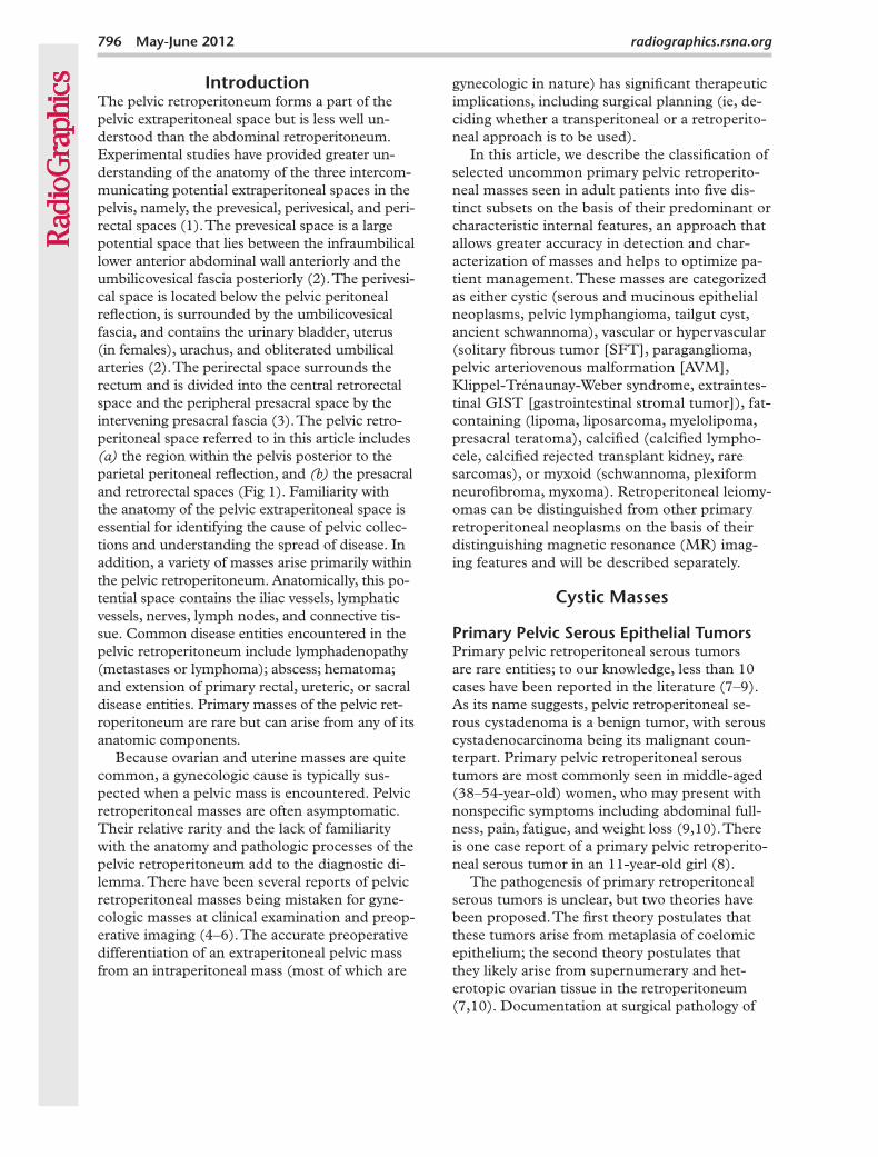
795GASTROINTESTINAL IMAGING
Alampady K. Shanbhogue, MD • Najla Fasih, FRCR • David B. Macdonald, MD • Adnan M. Sheikh, MD • Christine O. Menias, MD Srinivasa R. Prasad, MD
There is a broad spectrum of primary pelvic retroperitoneal masses in adults that demonstrate characteristic epidemiologic and histo-pathologic features and natural histories. These masses may be classi-fied into five distinct subgroups using a pattern-based approach that takes anatomic distribution and certain imaging characteristics into account, allowing greater accuracy in their detection and characteriza-tion and helping to optimize patient management. The five groups are cystic (serous and mucinous epithelial neoplasms, pelvic lymphangio-ma, tailgut cyst, ancient schwannoma), vascular or hypervascular (soli-tary fibrous tumor, paraganglioma, pelvic arteriovenous malformation, Klippel-Trénaunay-Weber syndrome, extraintestinal GIST [gastroin-testinal stromal tumor]), fat-containing (lipoma, liposarcoma, myelo-lipoma, presacral teratoma), calcified (calcified lymphocele, calcified rejected transplant kidney, rare sarcomas), and myxoid (schwannoma, plexiform neurofibroma, myxoma).Cross-sectional imaging modali-ties help differentiate the more common gynecologic neoplasms from more unusual masses. In particular, the tissue-specific multiplanar ca-pability of high-resolution magnetic resonance imaging permits better tumor localization and internal characterization, thereby serving as a road map for surgery.©RSNA, 2012 • radiographics.rsna.org
Uncommon Primary Pelvic Retroperitoneal Masses in Adults: A Pattern-based Imaging Approach1
Abbreviations: AVM = arteriovenous malformation, GIST = gastrointestinal stromal tumor, MIBG = metaiodobenzylguanidine, SFT = solitary fibrous tumor
RadioGraphics 2012; 32:795–817 • Published online 10.1148/rg.323115020 • Content Codes: 1From the Department of Radiology, University of Texas Health Science Center at San Antonio, 7703 Floyd Curl Dr, San Antonio, TX 78229 (A.K.S.); Department of Radiology, Ottawa Hospital, Ottawa, Ont, Canada (N.F., D.B.M., A.M.S.); Mallinckrodt Institute of Radiology, Washington University School of Medicine, St Louis, Mo (C.O.M.); and Department of Radiology, University of Texas M. D. Anderson Cancer Center, Houston, Tex (S.R.P.). Presented as an education exhibit at the 2009 RSNA Annual Meeting. Received February 7, 2011; revision requested July 21 and received November 18; accepted December 7. For this journal-based CME activity, the authors, editor, and reviewers have no relevant relationships to disclose. Address correspondence to A.K.S. (e-mail: [email protected]).
©RSNA, 2012
ONLINE-ONLY CME
See www.rsna .org/education /rg_cme.html
LEARNING OBJECTIVES
After completing this journal-based CME activity, participants
will be able to:
■ List the various uncommon primary pelvic retroperito-neal masses seen in adults.
■ Discuss the imag-ing features of these masses, with empha-sis on masses that are unique to the pelvis.
■ Describe a pat-tern-based approach that enables the radiologist to nar-row the differential diagnosis.
796 May-June 2012 radiographics.rsna.org
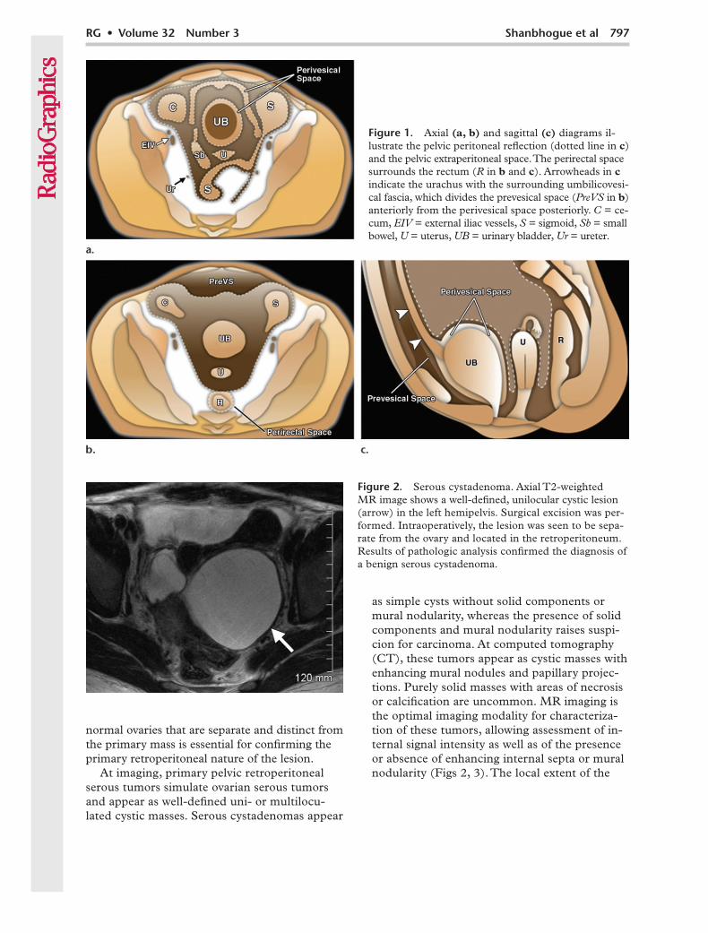
IntroductionThe pelvic retroperitoneum forms a part of the pelvic extraperitoneal space but is less well un-derstood than the abdominal retroperitoneum. Experimental studies have provided greater un-derstanding of the anatomy of the three intercom-municating potential extraperitoneal spaces in the pelvis, namely, the prevesical, perivesical, and peri-rectal spaces (1). The prevesical space is a large potential space that lies between the infraumbilical lower anterior abdominal wall anteriorly and the umbilicovesical fascia posteriorly (2). The perivesi-cal space is located below the pelvic peritoneal reflection, is surrounded by the umbilicovesical fascia, and contains the urinary bladder, uterus (in females), urachus, and obliterated umbilical arteries (2). The perirectal space surrounds the rectum and is divided into the central retrorectal space and the peripheral presacral space by the intervening presacral fascia (3). The pelvic retro-peritoneal space referred to in this article includes (a) the region within the pelvis posterior to the parietal peritoneal reflection, and (b) the presacral and retrorectal spaces (Fig 1). Familiarity with the anatomy of the pelvic extraperitoneal space is essential for identifying the cause of pelvic collec-tions and understanding the spread of disease. In addition, a variety of masses arise primarily within the pelvic retroperitoneum. Anatomically, this po-tential space contains the iliac vessels, lymphatic vessels, nerves, lymph nodes, and connective tis-sue. Common disease entities encountered in the pelvic retroperitoneum include lymphadenopathy (metastases or lymphoma); abscess; hematoma; and extension of primary rectal, ureteric, or sacral disease entities. Primary masses of the pelvic ret-roperitoneum are rare but can arise from any of its anatomic components.
Because ovarian and uterine masses are quite common, a gynecologic cause is typically sus-pected when a pelvic mass is encountered. Pelvic retroperitoneal masses are often asymptomatic. Their relative rarity and the lack of familiarity with the anatomy and pathologic processes of the pelvic retroperitoneum add to the diagnostic di-lemma. There have been several reports of pelvic retroperitoneal masses being mistaken for gyne-cologic masses at clinical examination and preop-erative imaging (4–6). The accurate preoperative differentiation of an extraperitoneal pelvic mass from an intraperitoneal mass (most of which are
gynecologic in nature) has significant therapeutic implications, including surgical planning (ie, de-ciding whether a transperitoneal or a retroperito-neal approach is to be used).
In this article, we describe the classification of selected uncommon primary pelvic retroperito-neal masses seen in adult patients into five dis-tinct subsets on the basis of their predominant or characteristic internal features, an approach that allows greater accuracy in detection and char-acterization of masses and helps to optimize pa-tient management. These masses are categorized as either cystic (serous and mucinous epithelial neoplasms, pelvic lymphangioma, tailgut cyst, ancient schwannoma), vascular or hypervascular (solitary fibrous tumor [SFT], paraganglioma, pelvic arteriovenous malformation [AVM], Klippel-Trénaunay-Weber syndrome, extraintes-tinal GIST [gastrointestinal stromal tumor]), fat-containing (lipoma, liposarcoma, myelolipoma, presacral teratoma), calcified (calcified lympho-cele, calcified rejected transplant kidney, rare sarcomas), or myxoid (schwannoma, plexiform neurofibroma, myxoma). Retroperitoneal leiomy-omas can be distinguished from other primary retroperitoneal neoplasms on the basis of their distinguishing magnetic resonance (MR) imag-ing features and will be described separately.
Cystic Masses
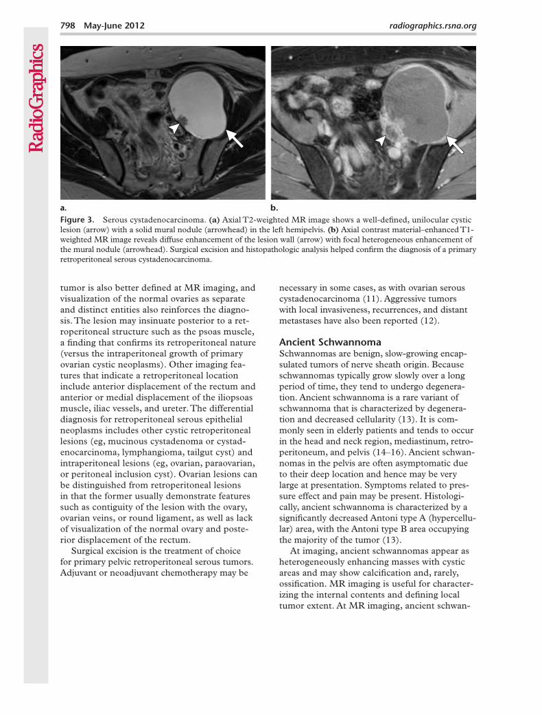
Primary Pelvic Serous Epithelial TumorsPrimary pelvic retroperitoneal serous tumors are rare entities; to our knowledge, less than 10 cases have been reported in the literature (7–9). As its name suggests, pelvic retroperitoneal se-rous cystadenoma is a benign tumor, with serous cystadenocarcinoma being its malignant coun-terpart. Primary pelvic retroperitoneal serous tumors are most commonly seen in middle-aged (38–54-year-old) women, who may present with nonspecific symptoms including abdominal full-ness, pain, fatigue, and weight loss (9,10). There is one case report of a primary pelvic retroperito-neal serous tumor in an 11-year-old girl (8).
The pathogenesis of primary retroperitoneal serous tumors is unclear, but two theories have been proposed. The first theory postulates that these tumors arise from metaplasia of coelomic epithelium; the second theory postulates that they likely arise from supernumerary and het-erotopic ovarian tissue in the retroperitoneum (7,10). Documentation at surgical pathology of
RG • Volume 32 Number 3 Shanbhogue et al 797
normal ovaries that are separate and distinct from the primary mass is essential for confirming the primary retroperitoneal nature of the lesion.
At imaging, primary pelvic retroperitoneal serous tumors simulate ovarian serous tumors and appear as well-defined uni- or multilocu-lated cystic masses. Serous cystadenomas appear
as simple cysts without solid components or mural nodularity, whereas the presence of solid components and mural nodularity raises suspi-cion for carcinoma. At computed tomography (CT), these tumors appear as cystic masses with enhancing mural nodules and papillary projec-tions. Purely solid masses with areas of necrosis or calcification are uncommon. MR imaging is the optimal imaging modality for characteriza-tion of these tumors, allowing assessment of in-ternal signal intensity as well as of the presence or absence of enhancing internal septa or mural nodularity (Figs 2, 3). The local extent of the
Figure 1. Axial (a, b) and sagittal (c) diagrams il-lustrate the pelvic peritoneal reflection (dotted line in c) and the pelvic extraperitoneal space. The perirectal space surrounds the rectum (R in b and c). Arrowheads in c indicate the urachus with the surrounding umbilicovesi-cal fascia, which divides the prevesical space (PreVS in b) anteriorly from the perivesical space posteriorly. C = ce-cum, EIV = external iliac vessels, S = sigmoid, Sb = small bowel, U = uterus, UB = urinary bladder, Ur = ureter.
Figure 2. Serous cystadenoma. Axial T2-weighted MR image shows a well-defined, unilocular cystic lesion (arrow) in the left hemipelvis. Surgical excision was per-formed. Intraoperatively, the lesion was seen to be sepa-rate from the ovary and located in the retroperitoneum. Results of pathologic analysis confirmed the diagnosis of a benign serous cystadenoma.
798 May-June 2012 radiographics.rsna.org
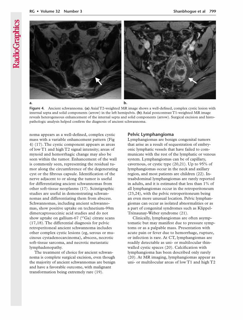
Figure 3. Serous cystadenocarcinoma. (a) Axial T2-weighted MR image shows a well-defined, unilocular cystic lesion (arrow) with a solid mural nodule (arrowhead) in the left hemipelvis. (b) Axial contrast material–enhanced T1-weighted MR image reveals diffuse enhancement of the lesion wall (arrow) with focal heterogeneous enhancement of the mural nodule (arrowhead). Surgical excision and histopathologic analysis helped confirm the diagnosis of a primary retroperitoneal serous cystadenocarcinoma.
tumor is also better defined at MR imaging, and visualization of the normal ovaries as separate and distinct entities also reinforces the diagno-sis. The lesion may insinuate posterior to a ret-roperitoneal structure such as the psoas muscle, a finding that confirms its retroperitoneal nature (versus the intraperitoneal growth of primary ovarian cystic neoplasms). Other imaging fea-tures that indicate a retroperitoneal location include anterior displacement of the rectum and anterior or medial displacement of the iliopsoas muscle, iliac vessels, and ureter. The differential diagnosis for retroperitoneal serous epithelial neoplasms includes other cystic retroperitoneal lesions (eg, mucinous cystadenoma or cystad-enocarcinoma, lymphangioma, tailgut cyst) and intraperitoneal lesions (eg, ovarian, paraovarian, or peritoneal inclusion cyst). Ovarian lesions can be distinguished from retroperitoneal lesions in that the former usually demonstrate features such as contiguity of the lesion with the ovary, ovarian veins, or round ligament, as well as lack of visualization of the normal ovary and poste-rior displacement of the rectum.
Surgical excision is the treatment of choice for primary pelvic retroperitoneal serous tumors. Adjuvant or neoadjuvant chemotherapy may be
necessary in some cases, as with ovarian serous cystadenocarcinoma (11). Aggressive tumors with local invasiveness, recurrences, and distant metastases have also been reported (12).
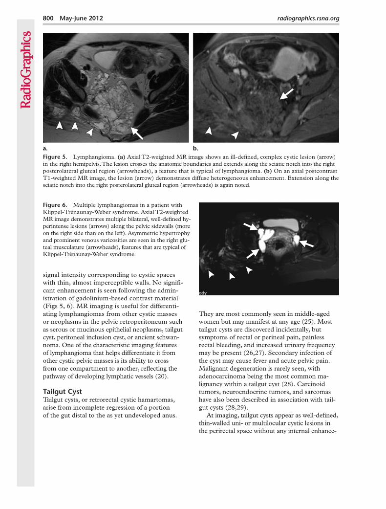
Ancient SchwannomaSchwannomas are benign, slow-growing encap-sulated tumors of nerve sheath origin. Because schwannomas typically grow slowly over a long period of time, they tend to undergo degenera-tion. Ancient schwannoma is a rare variant of schwannoma that is characterized by degenera-tion and decreased cellularity (13). It is com-monly seen in elderly patients and tends to occur in the head and neck region, mediastinum, retro-peritoneum, and pelvis (14–16). Ancient schwan-nomas in the pelvis are often asymptomatic due to their deep location and hence may be very large at presentation. Symptoms related to pres-sure effect and pain may be present. Histologi-cally, ancient schwannoma is characterized by a significantly decreased Antoni type A (hypercellu-lar) area, with the Antoni type B area occupying the majority of the tumor (13).
At imaging, ancient schwannomas appear as heterogeneously enhancing masses with cystic areas and may show calcification and, rarely, ossification. MR imaging is useful for character-izing the internal contents and defining local tumor extent. At MR imaging, ancient schwan-
RG • Volume 32 Number 3 Shanbhogue et al 799
noma appears as a well-defined, complex cystic mass with a variable enhancement pattern (Fig 4) (17). The cystic component appears as areas of low T1 and high T2 signal intensity; areas of myxoid and hemorrhagic change may also be seen within the tumor. Enhancement of the wall is commonly seen, representing the residual tu-mor along the circumference of the degenerating cyst or the fibrous capsule. Identification of the nerve adjacent to or along the tumor is useful for differentiating ancient schwannomas from other soft-tissue neoplasms (17). Scintigraphic studies are useful in demonstrating schwan-nomas and differentiating them from abscess. Schwannomas, including ancient schwanno-mas, show positive uptake on technetium-99m dimercaptosuccinic acid studies and do not show uptake on gallium-67 (67Ga) citrate scans (17,18). The differential diagnosis for pelvic retroperitoneal ancient schwannoma includes other complex cystic lesions (eg, serous or mu-cinous cystadenocarcinoma), abscess, necrotic soft-tissue sarcoma, and necrotic metastatic lymphadenopathy.
The treatment of choice for ancient schwan-noma is complete surgical excision, even though the majority of ancient schwannomas are benign and have a favorable outcome, with malignant transformation being extremely rare (19).
Pelvic LymphangiomaLymphangiomas are benign congenital tumors that arise as a result of sequestration of embry-onic lymphatic vessels that have failed to com-municate with the rest of the lymphatic or venous system. Lymphangiomas can be of capillary, cavernous, or cystic type (20,21). Up to 95% of lymphangiomas occur in the neck and axillary region, and most patients are children (22). In-traabdominal lymphangiomas are rarely reported in adults, and it is estimated that less than 1% of all lymphangiomas occur in the retroperitoneum (23,24), with the pelvic retroperitoneum being an even more unusual location. Pelvic lymphan-giomas can occur as isolated abnormalities or as a part of congenital syndromes such as Klippel-Trénaunay-Weber syndrome (21).
Clinically, lymphangiomas are often asymp-tomatic but may manifest due to pressure symp-toms or as a palpable mass. Presentation with acute pain or fever due to hemorrhage, rupture, or infection is rare. At CT, lymphangiomas are readily detectable as uni- or multilocular thin-walled cystic spaces (20). Calcification with lymphangioma has been described only rarely (20). At MR imaging, lymphangiomas appear as uni- or multilocular areas of low T1 and high T2
Figure 4. Ancient schwannoma. (a) Axial T2-weighted MR image shows a well-defined, complex cystic lesion with internal septa and solid components (arrow) in the left hemipelvis. (b) Axial postcontrast T1-weighted MR image reveals heterogeneous enhancement of the internal septa and solid components (arrow). Surgical excision and histo-pathologic analysis helped confirm the diagnosis of ancient schwannoma.
800 May-June 2012 radiographics.rsna.org
Figure 5. Lymphangioma. (a) Axial T2-weighted MR image shows an ill-defined, complex cystic lesion (arrow) in the right hemipelvis. The lesion crosses the anatomic boundaries and extends along the sciatic notch into the right posterolateral gluteal region (arrowheads), a feature that is typical of lymphangioma. (b) On an axial postcontrast T1-weighted MR image, the lesion (arrow) demonstrates diffuse heterogeneous enhancement. Extension along the sciatic notch into the right posterolateral gluteal region (arrowheads) is again noted.
signal intensity corresponding to cystic spaces with thin, almost imperceptible walls. No signifi-cant enhancement is seen following the admin-istration of gadolinium-based contrast material (Figs 5, 6). MR imaging is useful for differenti-ating lymphangiomas from other cystic masses or neoplasms in the pelvic retroperitoneum such as serous or mucinous epithelial neoplasms, tailgut cyst, peritoneal inclusion cyst, or ancient schwan-noma. One of the characteristic imaging features of lymphangioma that helps differentiate it from other cystic pelvic masses is its ability to cross from one compartment to another, reflecting the pathway of developing lymphatic vessels (20).
Tailgut CystTailgut cysts, or retrorectal cystic hamartomas, arise from incomplete regression of a portion of the gut distal to the as yet undeveloped anus.
They are most commonly seen in middle-aged women but may manifest at any age (25). Most tailgut cysts are discovered incidentally, but symptoms of rectal or perineal pain, painless rectal bleeding, and increased urinary frequency may be present (26,27). Secondary infection of the cyst may cause fever and acute pelvic pain. Malignant degeneration is rarely seen, with adenocarcinoma being the most common ma-lignancy within a tailgut cyst (28). Carcinoid tumors, neuroendocrine tumors, and sarcomas have also been described in association with tail-gut cysts (28,29).
At imaging, tailgut cysts appear as well-defined, thin-walled uni- or multilocular cystic lesions in the perirectal space without any internal enhance-
Figure 6. Multiple lymphangiomas in a patient with Klippel-Trénaunay-Weber syndrome. Axial T2-weighted MR image demonstrates multiple bilateral, well-defined hy-perintense lesions (arrows) along the pelvic sidewalls (more on the right side than on the left). Asymmetric hypertrophy and prominent venous varicosities are seen in the right glu-teal musculature (arrowheads), features that are typical of Klippel-Trénaunay-Weber syndrome.
RG • Volume 32 Number 3 Shanbhogue et al 801
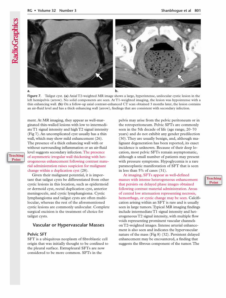
Figure 7. Tailgut cyst. (a) Axial T2-weighted MR image shows a large, hyperintense, unilocular cystic lesion in the left hemipelvis (arrow). No solid components are seen. At T1-weighted imaging, the lesion was hypointense with a thin enhancing wall. (b) On a follow-up axial contrast-enhanced CT scan obtained 3 months later, the lesion contains an air-fluid level and has a thick enhancing wall (arrow), findings that are consistent with secondary infection.
ment. At MR imaging, they appear as well-mar-ginated thin-walled lesions with low to intermedi-ate T1 signal intensity and high T2 signal intensity (Fig 7). An uncomplicated cyst usually has a thin wall, which may show mild enhancement (26). The presence of a thick enhancing wall with or without surrounding inflammation or an air-fluid level suggests secondary infection. The presence of asymmetric irregular wall thickening with het-erogeneous enhancement following contrast mate-rial administration raises suspicion for malignant change within a duplication cyst (28).
Given their malignant potential, it is impor-tant that tailgut cysts be differentiated from other cystic lesions in this location, such as epidermoid or dermoid cyst, rectal duplication cyst, anterior meningocele, and cystic lymphangioma. Cystic lymphangioma and tailgut cysts are often multi-locular, whereas the rest of the aforementioned cystic lesions are commonly unilocular. Complete surgical excision is the treatment of choice for tailgut cysts.
Vascular or Hypervascular Masses
Pelvic SFTSFT is a ubiquitous neoplasm of fibroblastic cell origin that was initially thought to be confined to the pleural surface. Extrapleural SFTs are now considered to be more common. SFTs in the
pelvis may arise from the pelvic peritoneum or in the retroperitoneum. Pelvic SFTs are commonly seen in the 5th decade of life (age range, 20–70 years) and do not exhibit any gender predilection (30). They are usually benign, and, although ma-lignant degeneration has been reported, its exact incidence is unknown. Because of their deep lo-cation, most pelvic SFTs remain asymptomatic, although a small number of patients may present with pressure symptoms. Hypoglycemia is a rare paraneoplastic manifestation of SFT that is seen in less than 5% of cases (31).
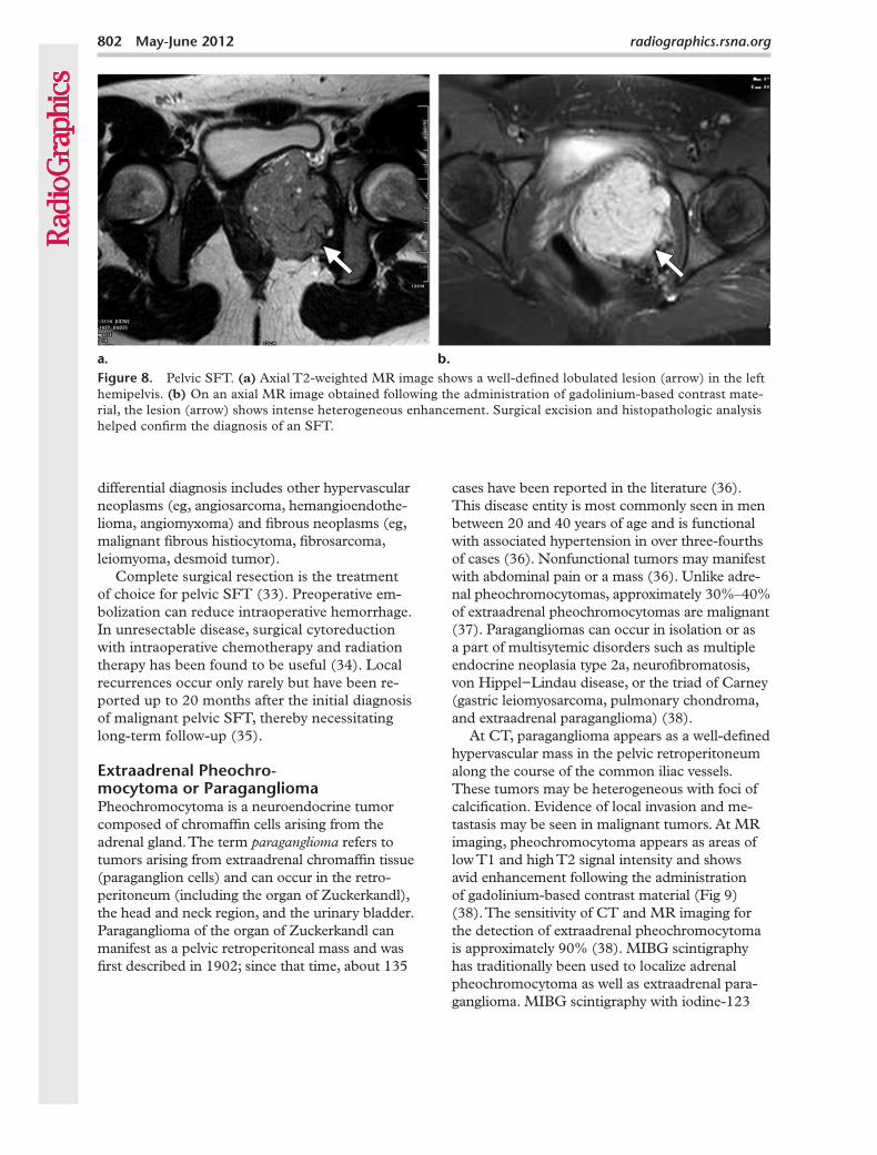
At imaging, SFTs appear as well-defined masses with intense heterogeneous enhancement that persists on delayed phase images obtained following contrast material administration. Areas of central low attenuation representing necrosis, hemorrhage, or cystic change may be seen. Calcifi-cation arising within an SFT is rare and is usually seen in large tumors. Typical MR imaging findings include intermediate T1 signal intensity and het-erogeneous T2 signal intensity, with multiple flow voids representing prominent vascular channels on T2-weighted images. Intense arterial enhance-ment is also seen and indicates the hypervascular nature of the mass (Fig 8) (32). Persistent delayed enhancement may be encountered, a finding that suggests the fibrous component of the tumor. The
802 May-June 2012 radiographics.rsna.org
Figure 8. Pelvic SFT. (a) Axial T2-weighted MR image shows a well-defined lobulated lesion (arrow) in the left hemipelvis. (b) On an axial MR image obtained following the administration of gadolinium-based contrast mate-rial, the lesion (arrow) shows intense heterogeneous enhancement. Surgical excision and histopathologic analysis helped confirm the diagnosis of an SFT.
differential diagnosis includes other hypervascular neoplasms (eg, angiosarcoma, hemangioendothe-lioma, angiomyxoma) and fibrous neoplasms (eg, malignant fibrous histiocytoma, fibrosarcoma, leiomyoma, desmoid tumor).
Complete surgical resection is the treatment of choice for pelvic SFT (33). Preoperative em-bolization can reduce intraoperative hemorrhage. In unresectable disease, surgical cytoreduction with intraoperative chemotherapy and radiation therapy has been found to be useful (34). Local recurrences occur only rarely but have been re-ported up to 20 months after the initial diagnosis of malignant pelvic SFT, thereby necessitating long-term follow-up (35).
Extraadrenal Pheochro- mocytoma or ParagangliomaPheochromocytoma is a neuroendocrine tumor composed of chromaffin cells arising from the adrenal gland. The term paraganglioma refers to tumors arising from extraadrenal chromaffin tissue (paraganglion cells) and can occur in the retro-peritoneum (including the organ of Zuckerkandl), the head and neck region, and the urinary bladder. Paraganglioma of the organ of Zuckerkandl can manifest as a pelvic retroperitoneal mass and was first described in 1902; since that time, about 135
cases have been reported in the literature (36). This disease entity is most commonly seen in men between 20 and 40 years of age and is functional with associated hypertension in over three-fourths of cases (36). Nonfunctional tumors may manifest with abdominal pain or a mass (36). Unlike adre-nal pheochromocytomas, approximately 30%–40% of extraadrenal pheochromocytomas are malignant (37). Paragangliomas can occur in isolation or as a part of multisytemic disorders such as multiple endocrine neoplasia type 2a, neurofibromatosis, von Hippel−Lindau disease, or the triad of Carney (gastric leiomyosarcoma, pulmonary chondroma, and extraadrenal paraganglioma) (38).
At CT, paraganglioma appears as a well-defined hypervascular mass in the pelvic retroperitoneum along the course of the common iliac vessels. These tumors may be heterogeneous with foci of calcification. Evidence of local invasion and me-tastasis may be seen in malignant tumors. At MR imaging, pheochromocytoma appears as areas of low T1 and high T2 signal intensity and shows avid enhancement following the administration of gadolinium-based contrast material (Fig 9) (38). The sensitivity of CT and MR imaging for the detection of extraadrenal pheochromocytoma is approximately 90% (38). MIBG scintigraphy has traditionally been used to localize adrenal pheochromocytoma as well as extraadrenal para-ganglioma. MIBG scintigraphy with iodine-123
RG • Volume 32 Number 3 Shanbhogue et al 803
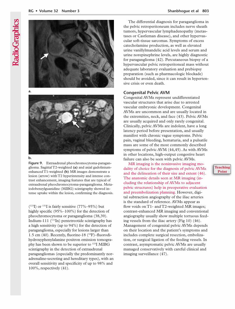
Figure 9. Extraadrenal pheochromocytoma-paragan-glioma. Sagittal T2-weighted (a) and axial gadolinium-enhanced T1-weighted (b) MR images demonstrate a lesion (arrow) with T2 hyperintensity and intense con-trast enhancement, imaging features that are typical of extraadrenal pheochromocytoma-paraganglioma. Meta-iodobenzylguanidine (MIBG) scintigraphy showed in-tense uptake within the lesion, confirming the diagnosis.
(123I) or 131I is fairly sensitive (77%–95%) but highly specific (95%–100%) for the detection of pheochromocytoma or paraganglioma (38,39). Indium-111 (111In) pentetreotide scintigraphy has a high sensitivity (up to 94%) for the detection of paraganglioma, especially for lesions larger than 1.5 cm (40). Recently, fluorine-18 (18F)–fluorodi-hydroxyphenylalanine positron emission tomogra-phy has been shown to be superior to 123I MIBG scintigraphy in the detection of extraadrenal paragangliomas (especially the predominantly nor-adrenaline-secreting and hereditary types), with an overall sensitivity and specificity of up to 98% and 100%, respectively (41).
The differential diagnosis for paraganglioma in the pelvic retroperitoneum includes nerve sheath tumors, hypervascular lymphadenopathy (metas-tases or Castleman disease), and other hypervas-cular soft-tissue sarcomas. Symptoms of excess catecholamine production, as well as elevated urine vanillylmandelic acid levels and serum and urine norepinephrine levels, are highly diagnostic for paraganglioma (42). Percutaneous biopsy of a hypervascular pelvic retroperitoneal mass without adequate laboratory evaluation and prebiopsy preparation (such as pharmacologic blockade) should be avoided, since it can result in hyperten-sive crisis or even death.
Congenital Pelvic AVMCongenital AVMs represent undifferentiated vascular structures that arise due to arrested vascular embryonic development. Congenital AVMs are uncommon and are usually located in the extremities, neck, and face (43). Pelvic AVMs are usually acquired and only rarely congenital. Clinically, pelvic AVMs are indolent, have a long latency period before presentation, and usually manifest with chronic vague symptoms. Pelvic pain, vaginal bleeding, hematuria, and a pulsatile mass are some of the most commonly described symptoms of pelvic AVM (44,45). As with AVMs in other locations, high-output congestive heart failure can also be seen with pelvic AVMs.
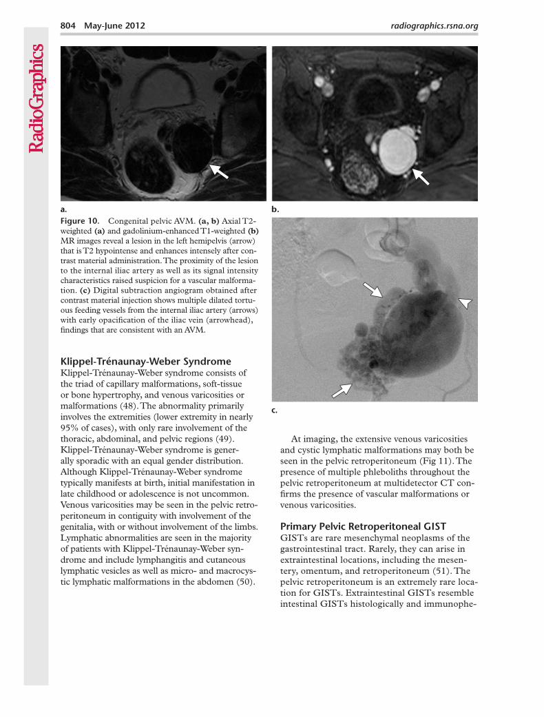
MR imaging is the noninvasive imaging mo-dality of choice for the diagnosis of pelvic AVMs and the delineation of their size and extent (46). The anatomic details seen at MR imaging (in-cluding the relationship of AVMs to adjacent pelvic structures) help in preoperative evaluation and preembolization planning. However, digi-tal subtraction angiography of the iliac arteries is the standard of reference. AVMs appear as flow voids on T1- and T2-weighted MR images; contrast-enhanced MR imaging and conventional angiography usually show multiple tortuous feed-ing vessels from the iliac artery (Fig 10) (46). Management of congenital pelvic AVMs depends on their location and the patient’s symptoms and includes complete surgical resection, emboliza-tion, or surgical ligation of the feeding vessels. In contrast, asymptomatic pelvic AVMs are usually managed conservatively with careful clinical and imaging surveillance (47).
804 May-June 2012 radiographics.rsna.org
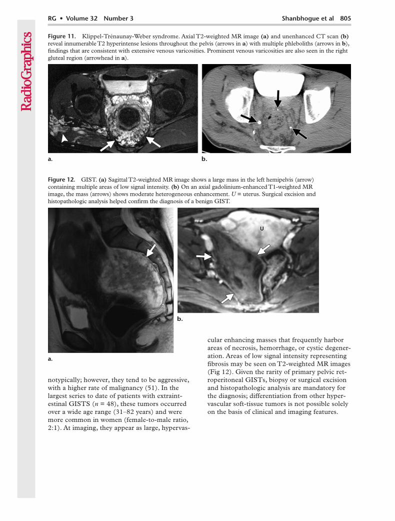
At imaging, the extensive venous varicosities and cystic lymphatic malformations may both be seen in the pelvic retroperitoneum (Fig 11). The presence of multiple phleboliths throughout the pelvic retroperitoneum at multidetector CT con-firms the presence of vascular malformations or venous varicosities.
Primary Pelvic Retroperitoneal GISTGISTs are rare mesenchymal neoplasms of the gastrointestinal tract. Rarely, they can arise in extraintestinal locations, including the mesen-tery, omentum, and retroperitoneum (51). The pelvic retroperitoneum is an extremely rare loca-tion for GISTs. Extraintestinal GISTs resemble intestinal GISTs histologically and immunophe-
Figure 10. Congenital pelvic AVM. (a, b) Axial T2-weighted (a) and gadolinium-enhanced T1-weighted (b) MR images reveal a lesion in the left hemipelvis (arrow) that is T2 hypointense and enhances intensely after con-trast material administration. The proximity of the lesion to the internal iliac artery as well as its signal intensity characteristics raised suspicion for a vascular malforma-tion. (c) Digital subtraction angiogram obtained after contrast material injection shows multiple dilated tortu-ous feeding vessels from the internal iliac artery (arrows) with early opacification of the iliac vein (arrowhead), findings that are consistent with an AVM.
Klippel-Trénaunay-Weber SyndromeKlippel-Trénaunay-Weber syndrome consists of the triad of capillary malformations, soft-tissue or bone hypertrophy, and venous varicosities or malformations (48). The abnormality primarily involves the extremities (lower extremity in nearly 95% of cases), with only rare involvement of the thoracic, abdominal, and pelvic regions (49). Klippel-Trénaunay-Weber syndrome is gener-ally sporadic with an equal gender distribution. Although Klippel-Trénaunay-Weber syndrome typically manifests at birth, initial manifestation in late childhood or adolescence is not uncommon. Venous varicosities may be seen in the pelvic retro-peritoneum in contiguity with involvement of the genitalia, with or without involvement of the limbs. Lymphatic abnormalities are seen in the majority of patients with Klippel-Trénaunay-Weber syn-drome and include lymphangitis and cutaneous lymphatic vesicles as well as micro- and macrocys-tic lymphatic malformations in the abdomen (50).
RG • Volume 32 Number 3 Shanbhogue et al 805
notypically; however, they tend to be aggressive, with a higher rate of malignancy (51). In the largest series to date of patients with extraint-estinal GISTS (n = 48), these tumors occurred over a wide age range (31–82 years) and were more common in women (female-to-male ratio, 2:1). At imaging, they appear as large, hypervas-
Figure 11. Klippel-Trénaunay-Weber syndrome. Axial T2-weighted MR image (a) and unenhanced CT scan (b) reveal innumerable T2 hyperintense lesions throughout the pelvis (arrows in a) with multiple phleboliths (arrows in b), findings that are consistent with extensive venous varicosities. Prominent venous varicosities are also seen in the right gluteal region (arrowhead in a).
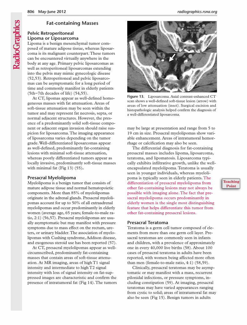
Figure 12. GIST. (a) Sagittal T2-weighted MR image shows a large mass in the left hemipelvis (arrow) containing multiple areas of low signal intensity. (b) On an axial gadolinium-enhanced T1-weighted MR image, the mass (arrows) shows moderate heterogeneous enhancement. U = uterus. Surgical excision and histopathologic analysis helped confirm the diagnosis of a benign GIST.
cular enhancing masses that frequently harbor areas of necrosis, hemorrhage, or cystic degener-ation. Areas of low signal intensity representing fibrosis may be seen on T2-weighted MR images (Fig 12). Given the rarity of primary pelvic ret-roperitoneal GISTs, biopsy or surgical excision and histopathologic analysis are mandatory for the diagnosis; differentiation from other hyper-vascular soft-tissue tumors is not possible solely on the basis of clinical and imaging features.
806 May-June 2012 radiographics.rsna.org
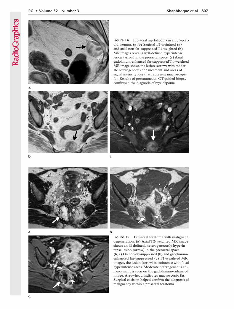
Figure 13. Liposarcoma. Axial contrast-enhanced CT scan shows a well-defined soft-tissue lesion (arrow) with areas of low attenuation (inset). Surgical excision and histopathologic analysis helped confirm the diagnosis of a well-differentiated liposarcoma.
Fat-containing Masses
Pelvic Retroperitoneal Lipoma or LiposarcomaLipoma is a benign mesenchymal tumor com-posed of mature adipose tissue, whereas liposar-coma is its malignant counterpart. These tumors can be encountered virtually anywhere in the body at any age. Primary pelvic liposarcomas as well as retroperitoneal liposarcomas extending into the pelvis may mimic gynecologic disease (52,53). Retroperitoneal and pelvic liposarco-mas can be asymptomatic for a long period of time and commonly manifest in elderly patients (5th−7th decades of life) (54,55).
At CT, lipomas appear as well-defined homo-geneous masses with fat attenuation. Areas of soft-tissue attenuation may be seen within the tumor and may represent fat necrosis, septa, or normal adjacent structures. However, the pres-ence of a predominantly solid soft-tissue compo-nent or adjacent organ invasion should raise sus-picion for liposarcoma. The imaging appearance of liposarcoma varies depending on the tumor grade. Well-differentiated liposarcomas appear as well-defined, predominantly fat-containing lesions with minimal soft-tissue attenuation, whereas poorly differentiated tumors appear as locally invasive, predominantly soft-tissue masses with minimal fat (Fig 13) (55).
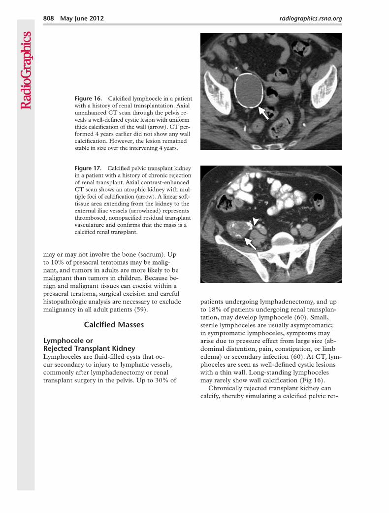
Presacral MyelolipomaMyelolipoma is a benign tumor that consists of mature adipose tissue and normal hematopoietic components. More than 85% of myelolipomas originate in the adrenal glands. Presacral myeloli-pomas account for up to 50% of all extraadrenal myelolipomas and occur predominantly in elderly women (average age, 65 years; female-to-male ra-tio, 2:1) (56,57). Presacral myelolipomas are usu-ally asymptomatic but may manifest with pressure symptoms due to mass effect on the rectum, ure-ters, or urinary bladder. The association of myelo-lipomas with Cushing syndrome, Addison disease, and exogenous steroid use has been reported (57).
At CT, presacral myelolipomas appear as well-circumscribed, predominantly fat-containing masses that contain areas of soft-tissue attenu-ation. At MR imaging, areas of high T1 signal intensity and intermediate to high T2 signal intensity with loss of signal intensity on fat-sup-pressed images are characteristic and confirm the presence of intratumoral fat (Fig 14). The tumors
may be large at presentation and range from 5 to 19 cm in size. Presacral myelolipomas show vari-able enhancement. Areas of intratumoral hemor-rhage or calcification may also be seen.
The differential diagnosis for fat-containing presacral masses includes lipoma, liposarcoma, teratoma, and lipomatosis. Liposarcoma typi-cally exhibits infiltrative growth, unlike the well-encapsulated myelolipoma. Teratoma is usually seen in younger individuals, whereas myeloli-poma is typically seen in elderly patients. The differentiation of presacral myelolipoma from other fat-containing lesions may not always be possible with imaging alone. The fact that pre-sacral myelolipoma occurs predominantly in elderly women is the single most distinguishing feature that helps differentiate this tumor from other fat-containing presacral lesions.
Presacral TeratomaTeratoma is a germ cell tumor composed of ele-ments from more than one germ cell layer. Pre-sacral teratomas are commonly seen in infants and children, with a prevalence of approximately one in every 40,000 live births (58). About 100 cases of presacral teratoma in adults have been reported, with women being affected more often than men (female-to-male ratio, 4:1) (58,59).
Clinically, presacral teratomas may be asymp-tomatic or may manifest with a mass, recurrent pilonidal infections, or pressure symptoms, in-cluding constipation (59). At imaging, presacral teratomas may have varied appearances ranging from cystic to solid; areas of intratumoral fat may also be seen (Fig 15). Benign tumors in adults
RG • Volume 32 Number 3 Shanbhogue et al 807
Figure 14. Presacral myelolipoma in an 85-year-old woman. (a, b) Sagittal T2-weighted (a) and axial non-fat-suppressed T1-weighted (b) MR images reveal a well-defined hyperintense lesion (arrow) in the presacral space. (c) Axial gadolinium-enhanced fat-suppressed T1-weighted MR image shows the lesion (arrow) with moder-ate heterogeneous enhancement and areas of signal intensity loss that represent macroscopic fat. Results of percutaneous CT-guided biopsy confirmed the diagnosis of myelolipoma.
Figure 15. Presacral teratoma with malignant degeneration. (a) Axial T2-weighted MR image shows an ill-defined, heterogeneously hyperin-tense lesion (arrow) in the presacral space. (b, c) On non-fat-suppressed (b) and gadolinium-enhanced fat-suppressed (c) T1-weighted MR images, the lesion (arrow) is isointense with focal hyperintense areas. Moderate heterogeneous en-hancement is seen on the gadolinium-enhanced image. Arrowhead indicates macroscopic fat. Surgical excision helped confirm the diagnosis of malignancy within a presacral teratoma.
808 May-June 2012 radiographics.rsna.org
Figure 17. Calcified pelvic transplant kidney in a patient with a history of chronic rejection of renal transplant. Axial contrast-enhanced CT scan shows an atrophic kidney with mul-tiple foci of calcification (arrow). A linear soft-tissue area extending from the kidney to the external iliac vessels (arrowhead) represents thrombosed, nonopacified residual transplant vasculature and confirms that the mass is a calcified renal transplant.
Figure 16. Calcified lymphocele in a patient with a history of renal transplantation. Axial unenhanced CT scan through the pelvis re-veals a well-defined cystic lesion with uniform thick calcification of the wall (arrow). CT per-formed 4 years earlier did not show any wall calcification. However, the lesion remained stable in size over the intervening 4 years.
may or may not involve the bone (sacrum). Up to 10% of presacral teratomas may be malig-nant, and tumors in adults are more likely to be malignant than tumors in children. Because be-nign and malignant tissues can coexist within a presacral teratoma, surgical excision and careful histopathologic analysis are necessary to exclude malignancy in all adult patients (59).
Calcified Masses
Lymphocele or Rejected Transplant KidneyLymphoceles are fluid-filled cysts that oc-cur secondary to injury to lymphatic vessels, commonly after lymphadenectomy or renal transplant surgery in the pelvis. Up to 30% of
patients undergoing lymphadenectomy, and up to 18% of patients undergoing renal transplan-tation, may develop lymphocele (60). Small, sterile lymphoceles are usually asymptomatic; in symptomatic lymphoceles, symptoms may arise due to pressure effect from large size (ab-dominal distention, pain, constipation, or limb edema) or secondary infection (60). At CT, lym-phoceles are seen as well-defined cystic lesions with a thin wall. Long-standing lymphoceles may rarely show wall calcification (Fig 16).
Chronically rejected transplant kidney can calcify, thereby simulating a calcified pelvic ret-
RG • Volume 32 Number 3 Shanbhogue et al 809
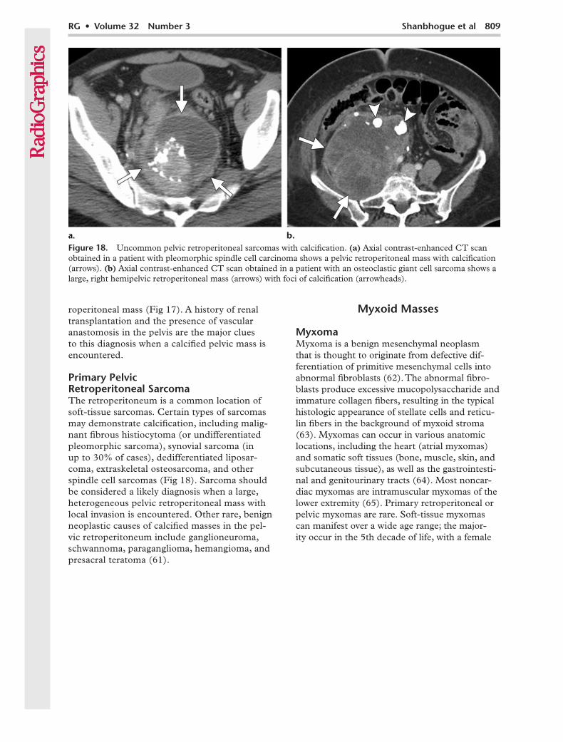
Figure 18. Uncommon pelvic retroperitoneal sarcomas with calcification. (a) Axial contrast-enhanced CT scan obtained in a patient with pleomorphic spindle cell carcinoma shows a pelvic retroperitoneal mass with calcification (arrows). (b) Axial contrast-enhanced CT scan obtained in a patient with an osteoclastic giant cell sarcoma shows a large, right hemipelvic retroperitoneal mass (arrows) with foci of calcification (arrowheads).
roperitoneal mass (Fig 17). A history of renal transplantation and the presence of vascular anastomosis in the pelvis are the major clues to this diagnosis when a calcified pelvic mass is encountered.
Primary Pelvic Retroperitoneal SarcomaThe retroperitoneum is a common location of soft-tissue sarcomas. Certain types of sarcomas may demonstrate calcification, including malig-nant fibrous histiocytoma (or undifferentiated pleomorphic sarcoma), synovial sarcoma (in up to 30% of cases), dedifferentiated liposar-coma, extraskeletal osteosarcoma, and other spindle cell sarcomas (Fig 18). Sarcoma should be considered a likely diagnosis when a large, heterogeneous pelvic retroperitoneal mass with local invasion is encountered. Other rare, benign neoplastic causes of calcified masses in the pel-vic retroperitoneum include ganglioneuroma, schwannoma, paraganglioma, hemangioma, and presacral teratoma (61).
Myxoid Masses
MyxomaMyxoma is a benign mesenchymal neoplasm that is thought to originate from defective dif-ferentiation of primitive mesenchymal cells into abnormal fibroblasts (62). The abnormal fibro-blasts produce excessive mucopolysaccharide and immature collagen fibers, resulting in the typical histologic appearance of stellate cells and reticu-lin fibers in the background of myxoid stroma (63). Myxomas can occur in various anatomic locations, including the heart (atrial myxomas) and somatic soft tissues (bone, muscle, skin, and subcutaneous tissue), as well as the gastrointesti-nal and genitourinary tracts (64). Most noncar-diac myxomas are intramuscular myxomas of the lower extremity (65). Primary retroperitoneal or pelvic myxomas are rare. Soft-tissue myxomas can manifest over a wide age range; the major-ity occur in the 5th decade of life, with a female
810 May-June 2012 radiographics.rsna.org
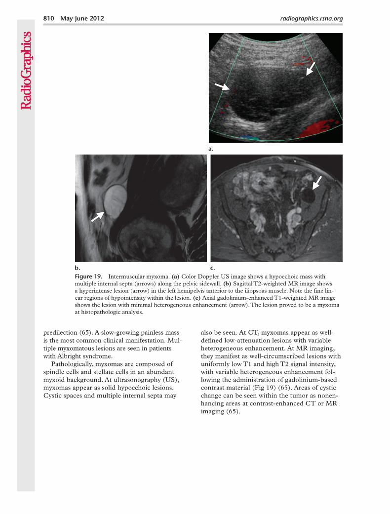
Figure 19. Intermuscular myxoma. (a) Color Doppler US image shows a hypoechoic mass with multiple internal septa (arrows) along the pelvic sidewall. (b) Sagittal T2-weighted MR image shows a hyperintense lesion (arrow) in the left hemipelvis anterior to the iliopsoas muscle. Note the fine lin-ear regions of hypointensity within the lesion. (c) Axial gadolinium-enhanced T1-weighted MR image shows the lesion with minimal heterogeneous enhancement (arrow). The lesion proved to be a myxoma at histopathologic analysis.
predilection (65). A slow-growing painless mass is the most common clinical manifestation. Mul-tiple myxomatous lesions are seen in patients with Albright syndrome.
Pathologically, myxomas are composed of spindle cells and stellate cells in an abundant myxoid background. At ultrasonography (US), myxomas appear as solid hypoechoic lesions. Cystic spaces and multiple internal septa may
also be seen. At CT, myxomas appear as well-defined low-attenuation lesions with variable heterogeneous enhancement. At MR imaging, they manifest as well-circumscribed lesions with uniformly low T1 and high T2 signal intensity, with variable heterogeneous enhancement fol-lowing the administration of gadolinium-based contrast material (Fig 19) (65). Areas of cystic change can be seen within the tumor as nonen-hancing areas at contrast-enhanced CT or MR imaging (65).
RG • Volume 32 Number 3 Shanbhogue et al 811
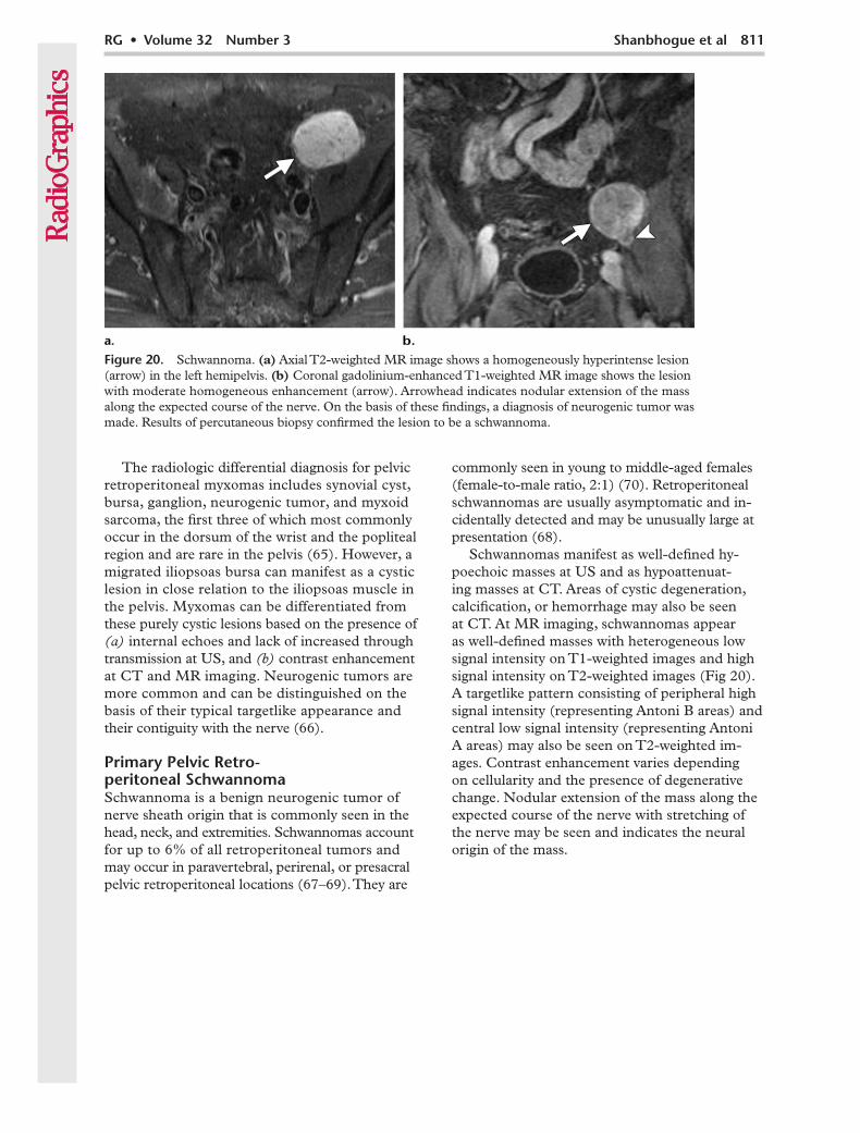
Figure 20. Schwannoma. (a) Axial T2-weighted MR image shows a homogeneously hyperintense lesion (arrow) in the left hemipelvis. (b) Coronal gadolinium-enhanced T1-weighted MR image shows the lesion with moderate homogeneous enhancement (arrow). Arrowhead indicates nodular extension of the mass along the expected course of the nerve. On the basis of these findings, a diagnosis of neurogenic tumor was made. Results of percutaneous biopsy confirmed the lesion to be a schwannoma.
The radiologic differential diagnosis for pelvic retroperitoneal myxomas includes synovial cyst, bursa, ganglion, neurogenic tumor, and myxoid sarcoma, the first three of which most commonly occur in the dorsum of the wrist and the popliteal region and are rare in the pelvis (65). However, a migrated iliopsoas bursa can manifest as a cystic lesion in close relation to the iliopsoas muscle in the pelvis. Myxomas can be differentiated from these purely cystic lesions based on the presence of (a) internal echoes and lack of increased through transmission at US, and (b) contrast enhancement at CT and MR imaging. Neurogenic tumors are more common and can be distinguished on the basis of their typical targetlike appearance and their contiguity with the nerve (66).
Primary Pelvic Retro- peritoneal SchwannomaSchwannoma is a benign neurogenic tumor of nerve sheath origin that is commonly seen in the head, neck, and extremities. Schwannomas account for up to 6% of all retroperitoneal tumors and may occur in paravertebral, perirenal, or presacral pelvic retroperitoneal locations (67–69). They are
commonly seen in young to middle-aged females (female-to-male ratio, 2:1) (70). Retroperitoneal schwannomas are usually asymptomatic and in-cidentally detected and may be unusually large at presentation (68).
Schwannomas manifest as well-defined hy-poechoic masses at US and as hypoattenuat-ing masses at CT. Areas of cystic degeneration, calcification, or hemorrhage may also be seen at CT. At MR imaging, schwannomas appear as well-defined masses with heterogeneous low signal intensity on T1-weighted images and high signal intensity on T2-weighted images (Fig 20). A targetlike pattern consisting of peripheral high signal intensity (representing Antoni B areas) and central low signal intensity (representing Antoni A areas) may also be seen on T2-weighted im-ages. Contrast enhancement varies depending on cellularity and the presence of degenerative change. Nodular extension of the mass along the expected course of the nerve with stretching of the nerve may be seen and indicates the neural origin of the mass.
812 May-June 2012 radiographics.rsna.org
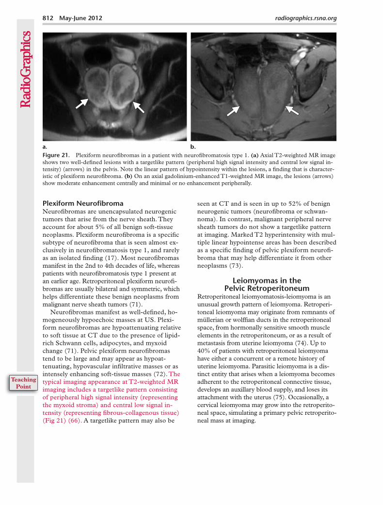
Figure 21. Plexiform neurofibromas in a patient with neurofibromatosis type 1. (a) Axial T2-weighted MR image shows two well-defined lesions with a targetlike pattern (peripheral high signal intensity and central low signal in-tensity) (arrows) in the pelvis. Note the linear pattern of hypointensity within the lesions, a finding that is character-istic of plexiform neurofibroma. (b) On an axial gadolinium-enhanced T1-weighted MR image, the lesions (arrows) show moderate enhancement centrally and minimal or no enhancement peripherally.
Plexiform NeurofibromaNeurofibromas are unencapsulated neurogenic tumors that arise from the nerve sheath. They account for about 5% of all benign soft-tissue neoplasms. Plexiform neurofibroma is a specific subtype of neurofibroma that is seen almost ex-clusively in neurofibromatosis type 1, and rarely as an isolated finding (17). Most neurofibromas manifest in the 2nd to 4th decades of life, whereas patients with neurofibromatosis type 1 present at an earlier age. Retroperitoneal plexiform neurofi-bromas are usually bilateral and symmetric, which helps differentiate these benign neoplasms from malignant nerve sheath tumors (71).
Neurofibromas manifest as well-defined, ho-mogeneously hypoechoic masses at US. Plexi-form neurofibromas are hypoattenuating relative to soft tissue at CT due to the presence of lipid-rich Schwann cells, adipocytes, and myxoid change (71). Pelvic plexiform neurofibromas tend to be large and may appear as hypoat-tenuating, hypovascular infiltrative masses or as intensely enhancing soft-tissue masses (72). The typical imaging appearance at T2-weighted MR imaging includes a targetlike pattern consisting of peripheral high signal intensity (representing the myxoid stroma) and central low signal in-tensity (representing fibrous-collagenous tissue) (Fig 21) (66). A targetlike pattern may also be
seen at CT and is seen in up to 52% of benign neurogenic tumors (neurofibroma or schwan-noma). In contrast, malignant peripheral nerve sheath tumors do not show a targetlike pattern at imaging. Marked T2 hyperintensity with mul-tiple linear hypointense areas has been described as a specific finding of pelvic plexiform neurofi-broma that may help differentiate it from other neoplasms (73).
Leiomyomas in the Pelvic Retroperitoneum
Retroperitoneal leiomyomatosis-leiomyoma is an unusual growth pattern of leiomyoma. Retroperi-toneal leiomyoma may originate from remnants of müllerian or wolffian ducts in the retroperitoneal space, from hormonally sensitive smooth muscle elements in the retroperitoneum, or as a result of metastasis from uterine leiomyoma (74). Up to 40% of patients with retroperitoneal leiomyoma have either a concurrent or a remote history of uterine leiomyoma. Parasitic leiomyoma is a dis-tinct entity that arises when a leiomyoma becomes adherent to the retroperitoneal connective tissue, develops an auxillary blood supply, and loses its attachment with the uterus (75). Occasionally, a cervical leiomyoma may grow into the retroperito-neal space, simulating a primary pelvic retroperito-neal mass at imaging.
RG • Volume 32 Number 3 Shanbhogue et al 813
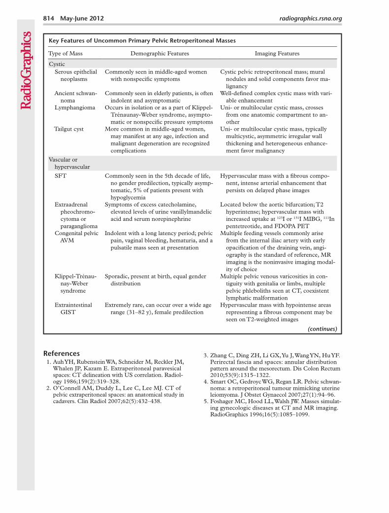
Figure 23. Parasitic leiomyoma. Sagittal T2-weighted MR image shows an oval hypointense lesion (arrows) in the pelvic retroperitoneal space. The lesion proved to be a parasitic leio-myoma at surgical excision.
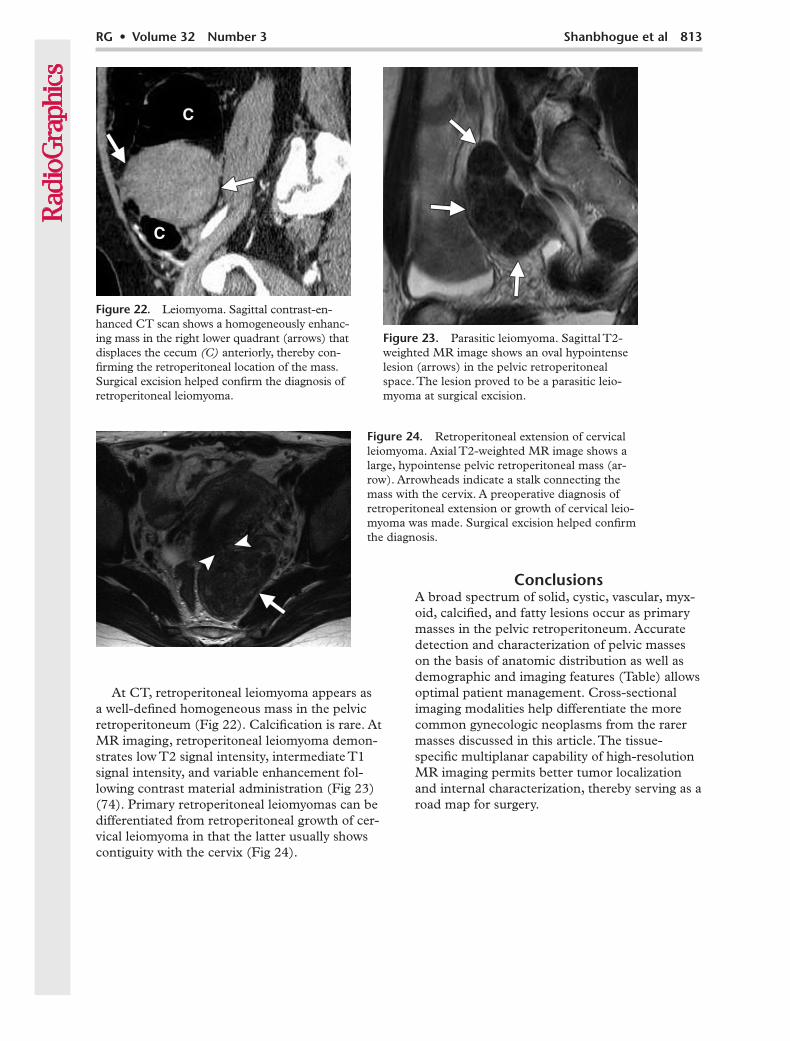
Figure 22. Leiomyoma. Sagittal contrast-en-hanced CT scan shows a homogeneously enhanc-ing mass in the right lower quadrant (arrows) that displaces the cecum (C) anteriorly, thereby con-firming the retroperitoneal location of the mass. Surgical excision helped confirm the diagnosis of retroperitoneal leiomyoma.
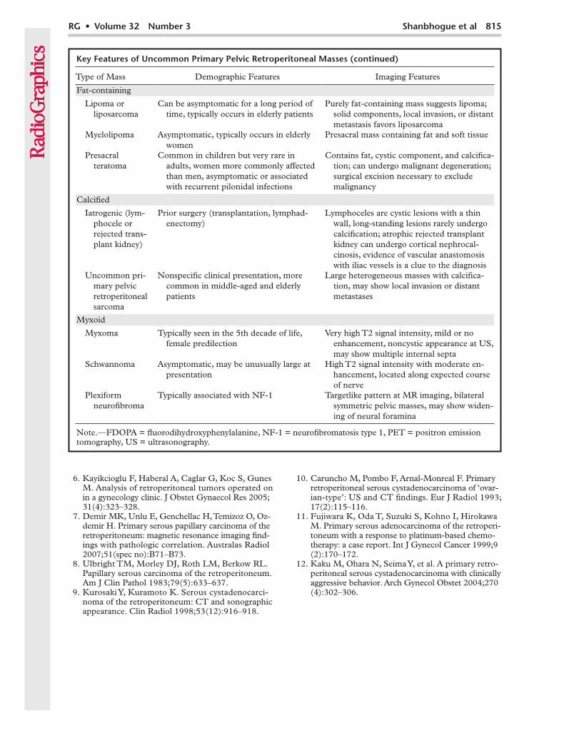
At CT, retroperitoneal leiomyoma appears as a well-defined homogeneous mass in the pelvic retroperitoneum (Fig 22). Calcification is rare. At MR imaging, retroperitoneal leiomyoma demon-strates low T2 signal intensity, intermediate T1 signal intensity, and variable enhancement fol-lowing contrast material administration (Fig 23) (74). Primary retroperitoneal leiomyomas can be differentiated from retroperitoneal growth of cer-vical leiomyoma in that the latter usually shows contiguity with the cervix (Fig 24).
ConclusionsA broad spectrum of solid, cystic, vascular, myx-oid, calcified, and fatty lesions occur as primary masses in the pelvic retroperitoneum. Accurate detection and characterization of pelvic masses on the basis of anatomic distribution as well as demographic and imaging features (Table) allows optimal patient management. Cross-sectional imaging modalities help differentiate the more common gynecologic neoplasms from the rarer masses discussed in this article. The tissue-specific multiplanar capability of high-resolution MR imaging permits better tumor localization and internal characterization, thereby serving as a road map for surgery.
Figure 24. Retroperitoneal extension of cervical leiomyoma. Axial T2-weighted MR image shows a large, hypointense pelvic retroperitoneal mass (ar-row). Arrowheads indicate a stalk connecting the mass with the cervix. A preoperative diagnosis of retroperitoneal extension or growth of cervical leio-myoma was made. Surgical excision helped confirm the diagnosis.
814 May-June 2012 radiographics.rsna.org
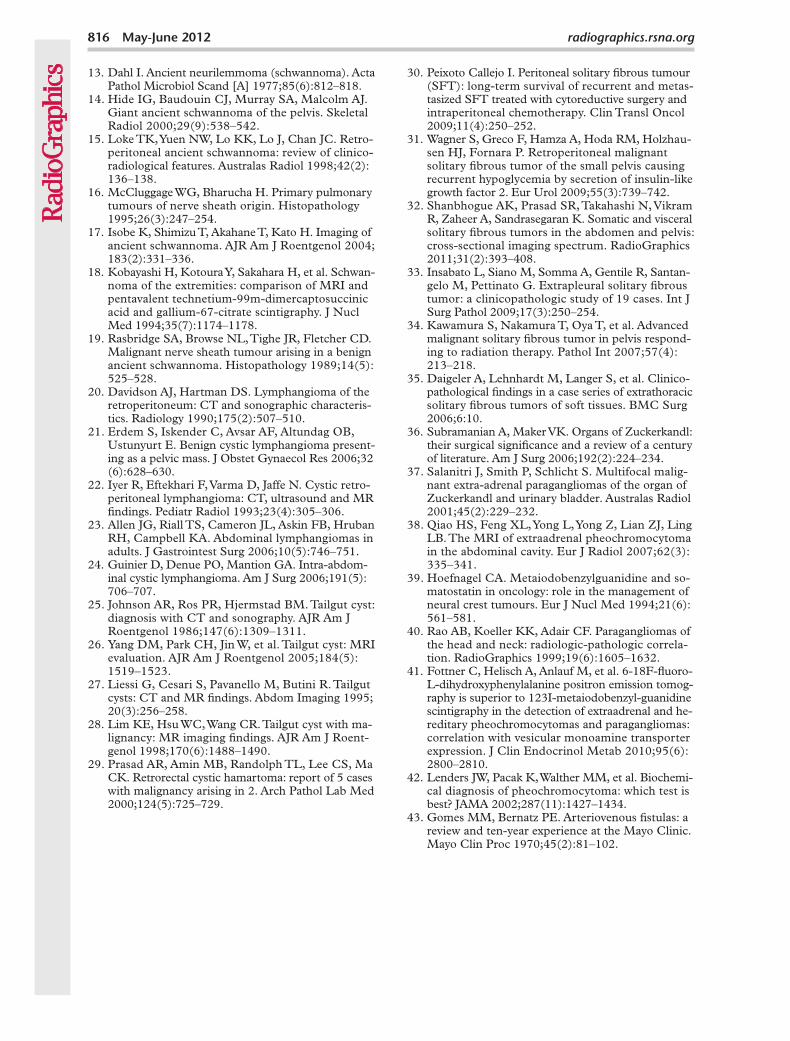
Key Features of Uncommon Primary Pelvic Retroperitoneal Masses
Type of Mass Demographic Features Imaging Features
Cystic Serous epithelial
neoplasmsCommonly seen in middle-aged women
with nonspecific symptomsCystic pelvic retroperitoneal mass; mural
nodules and solid components favor ma-lignancy
Ancient schwan- noma
Commonly seen in elderly patients, is often indolent and asymptomatic
Well-defined complex cystic mass with vari-able enhancement
Lymphangioma Occurs in isolation or as a part of Klippel-Trénaunay-Weber syndrome, asympto-matic or nonspecific pressure symptoms
Uni- or multilocular cystic mass, crosses from one anatomic compartment to an-other
Tailgut cyst More common in middle-aged women, may manifest at any age, infection and malignant degeneration are recognized complications
Uni- or multilocular cystic mass, typically multicystic, asymmetric irregular wall thickening and heterogeneous enhance-ment favor malignancy
Vascular or hypervascular
SFT Commonly seen in the 5th decade of life, no gender predilection, typically asymp-tomatic, 5% of patients present with hypoglycemia
Hypervascular mass with a fibrous compo-nent, intense arterial enhancement that persists on delayed phase images
Extraadrenal pheochromo- cytoma or paraganglioma
Symptoms of excess catecholamine, elevated levels of urine vanillylmandelic acid and serum norepinephrine
Located below the aortic bifurcation; T2 hyperintense; hypervascular mass with increased uptake at 123I or 131I MIBG, 111In pentetreotide, and FDOPA PET
Congenital pelvic AVM
Indolent with a long latency period; pelvic pain, vaginal bleeding, hematuria, and a pulsatile mass seen at presentation
Multiple feeding vessels commonly arise from the internal iliac artery with early opacification of the draining vein, angi-ography is the standard of reference, MR imaging is the noninvasive imaging modal-ity of choice
Klippel-Trénau- nay-Weber syndrome
Sporadic, present at birth, equal gender distribution
Multiple pelvic venous varicosities in con-tiguity with genitalia or limbs, multiple pelvic phleboliths seen at CT, coexistent lymphatic malformation
Extraintestinal GIST
Extremely rare, can occur over a wide age range (31–82 y), female predilection
Hypervascular mass with hypointense areas representing a fibrous component may be seen on T2-weighted images
(continues)
References 1. Auh YH, Rubenstein WA, Schneider M, Reckler JM,
Whalen JP, Kazam E. Extraperitoneal paravesical spaces: CT delineation with US correlation. Radiol-ogy 1986;159(2):319–328.
2. O’Connell AM, Duddy L, Lee C, Lee MJ. CT of pelvic extraperitoneal spaces: an anatomical study in cadavers. Clin Radiol 2007;62(5):432–438.
3. Zhang C, Ding ZH, Li GX, Yu J, Wang YN, Hu YF. Perirectal fascia and spaces: annular distribution pattern around the mesorectum. Dis Colon Rectum 2010;53(9):1315–1322.
4. Smart OC, Gedroyc WG, Regan LR. Pelvic schwan-noma: a retroperitoneal tumour mimicking uterine leiomyoma. J Obstet Gynaecol 2007;27(1):94–96.
5. Foshager MC, Hood LL, Walsh JW. Masses simulat-ing gynecologic diseases at CT and MR imaging. RadioGraphics 1996;16(5):1085–1099.
RG • Volume 32 Number 3 Shanbhogue et al 815
Key Features of Uncommon Primary Pelvic Retroperitoneal Masses (continued)
Type of Mass Demographic Features Imaging Features
Fat-containing
Lipoma or liposarcoma
Can be asymptomatic for a long period of time, typically occurs in elderly patients
Purely fat-containing mass suggests lipoma; solid components, local invasion, or distant metastasis favors liposarcoma
Myelolipoma Asymptomatic, typically occurs in elderly women
Presacral mass containing fat and soft tissue
Presacral teratoma
Common in children but very rare in adults, women more commonly affected than men, asymptomatic or associated with recurrent pilonidal infections
Contains fat, cystic component, and calcifica-tion; can undergo malignant degeneration; surgical excision necessary to exclude malignancy
Calcified
Iatrogenic (lym- phocele or rejected trans- plant kidney)
Prior surgery (transplantation, lymphad-enectomy)
Lymphoceles are cystic lesions with a thin wall, long-standing lesions rarely undergo calcification; atrophic rejected transplant kidney can undergo cortical nephrocal-cinosis, evidence of vascular anastomosis with iliac vessels is a clue to the diagnosis
Uncommon pri- mary pelvic retroperitoneal sarcoma
Nonspecific clinical presentation, more common in middle-aged and elderly patients
Large heterogeneous masses with calcifica-tion, may show local invasion or distant metastases
Myxoid
Myxoma Typically seen in the 5th decade of life, female predilection
Very high T2 signal intensity, mild or no enhancement, noncystic appearance at US, may show multiple internal septa
Schwannoma Asymptomatic, may be unusually large at presentation
High T2 signal intensity with moderate en-hancement, located along expected course of nerve
Plexiform neurofibroma
Typically associated with NF-1 Targetlike pattern at MR imaging, bilateral symmetric pelvic masses, may show widen-ing of neural foramina
Note.—FDOPA = fluorodihydroxyphenylalanine, NF-1 = neurofibromatosis type 1, PET = positron emission tomography, US = ultrasonography.
6. Kayikcioglu F, Haberal A, Caglar G, Koc S, Gunes M. Analysis of retroperitoneal tumors operated on in a gynecology clinic. J Obstet Gynaecol Res 2005; 31(4):323–328.
7. Demir MK, Unlu E, Genchellac H, Temizoz O, Oz-demir H. Primary serous papillary carcinoma of the retroperitoneum: magnetic resonance imaging find-ings with pathologic correlation. Australas Radiol 2007;51(spec no):B71–B73.
8. Ulbright TM, Morley DJ, Roth LM, Berkow RL. Papillary serous carcinoma of the retroperitoneum. Am J Clin Pathol 1983;79(5):633–637.
9. Kurosaki Y, Kuramoto K. Serous cystadenocarci-noma of the retroperitoneum: CT and sonographic appearance. Clin Radiol 1998;53(12):916–918.
10. Caruncho M, Pombo F, Arnal-Monreal F. Primary retroperitoneal serous cystadenocarcinoma of ‘ovar-ian-type’: US and CT findings. Eur J Radiol 1993; 17(2):115–116.
11. Fujiwara K, Oda T, Suzuki S, Kohno I, Hirokawa M. Primary serous adenocarcinoma of the retroperi-toneum with a response to platinum-based chemo-therapy: a case report. Int J Gynecol Cancer 1999;9 (2):170–172.
12. Kaku M, Ohara N, Seima Y, et al. A primary retro-peritoneal serous cystadenocarcinoma with clinically aggressive behavior. Arch Gynecol Obstet 2004;270 (4):302–306.
816 May-June 2012 radiographics.rsna.org
13. Dahl I. Ancient neurilemmoma (schwannoma). Acta Pathol Microbiol Scand [A] 1977;85(6):812–818.
14. Hide IG, Baudouin CJ, Murray SA, Malcolm AJ. Giant ancient schwannoma of the pelvis. Skeletal Radiol 2000;29(9):538–542.
15. Loke TK, Yuen NW, Lo KK, Lo J, Chan JC. Retro-peritoneal ancient schwannoma: review of clinico-radiological features. Australas Radiol 1998;42(2): 136–138.
16. McCluggage WG, Bharucha H. Primary pulmonary tumours of nerve sheath origin. Histopathology 1995;26(3):247–254.
17. Isobe K, Shimizu T, Akahane T, Kato H. Imaging of ancient schwannoma. AJR Am J Roentgenol 2004; 183(2):331–336.
18. Kobayashi H, Kotoura Y, Sakahara H, et al. Schwan-noma of the extremities: comparison of MRI and pentavalent technetium-99m-dimercaptosuccinic acid and gallium-67-citrate scintigraphy. J Nucl Med 1994;35(7):1174–1178.
19. Rasbridge SA, Browse NL, Tighe JR, Fletcher CD. Malignant nerve sheath tumour arising in a benign ancient schwannoma. Histopathology 1989;14(5): 525–528.
20. Davidson AJ, Hartman DS. Lymphangioma of the retroperitoneum: CT and sonographic characteris-tics. Radiology 1990;175(2):507–510.
21. Erdem S, Iskender C, Avsar AF, Altundag OB, Ustunyurt E. Benign cystic lymphangioma present-ing as a pelvic mass. J Obstet Gynaecol Res 2006;32 (6):628–630.
22. Iyer R, Eftekhari F, Varma D, Jaffe N. Cystic retro-peritoneal lymphangioma: CT, ultrasound and MR findings. Pediatr Radiol 1993;23(4):305–306.
23. Allen JG, Riall TS, Cameron JL, Askin FB, Hruban RH, Campbell KA. Abdominal lymphangiomas in adults. J Gastrointest Surg 2006;10(5):746–751.
24. Guinier D, Denue PO, Mantion GA. Intra-abdom-inal cystic lymphangioma. Am J Surg 2006;191(5): 706–707.
25. Johnson AR, Ros PR, Hjermstad BM. Tailgut cyst: diagnosis with CT and sonography. AJR Am J Roentgenol 1986;147(6):1309–1311.
26. Yang DM, Park CH, Jin W, et al. Tailgut cyst: MRI evaluation. AJR Am J Roentgenol 2005;184(5): 1519–1523.
27. Liessi G, Cesari S, Pavanello M, Butini R. Tailgut cysts: CT and MR findings. Abdom Imaging 1995; 20(3):256–258.
28. Lim KE, Hsu WC, Wang CR. Tailgut cyst with ma-lignancy: MR imaging findings. AJR Am J Roent-genol 1998;170(6):1488–1490.
29. Prasad AR, Amin MB, Randolph TL, Lee CS, Ma CK. Retrorectal cystic hamartoma: report of 5 cases with malignancy arising in 2. Arch Pathol Lab Med 2000;124(5):725–729.
30. Peixoto Callejo I. Peritoneal solitary fibrous tumour (SFT): long-term survival of recurrent and metas-tasized SFT treated with cytoreductive surgery and intraperitoneal chemotherapy. Clin Transl Oncol 2009;11(4):250–252.
31. Wagner S, Greco F, Hamza A, Hoda RM, Holzhau-sen HJ, Fornara P. Retroperitoneal malignant solitary fibrous tumor of the small pelvis causing recurrent hypoglycemia by secretion of insulin-like growth factor 2. Eur Urol 2009;55(3):739–742.
32. Shanbhogue AK, Prasad SR, Takahashi N, Vikram R, Zaheer A, Sandrasegaran K. Somatic and visceral solitary fibrous tumors in the abdomen and pelvis: cross-sectional imaging spectrum. RadioGraphics 2011;31(2):393–408.
33. Insabato L, Siano M, Somma A, Gentile R, Santan-gelo M, Pettinato G. Extrapleural solitary fibrous tumor: a clinicopathologic study of 19 cases. Int J Surg Pathol 2009;17(3):250–254.
34. Kawamura S, Nakamura T, Oya T, et al. Advanced malignant solitary fibrous tumor in pelvis respond-ing to radiation therapy. Pathol Int 2007;57(4): 213–218.
35. Daigeler A, Lehnhardt M, Langer S, et al. Clinico-pathological findings in a case series of extrathoracic solitary fibrous tumors of soft tissues. BMC Surg 2006;6:10.
36. Subramanian A, Maker VK. Organs of Zuckerkandl: their surgical significance and a review of a century of literature. Am J Surg 2006;192(2):224–234.
37. Salanitri J, Smith P, Schlicht S. Multifocal malig-nant extra-adrenal paragangliomas of the organ of Zuckerkandl and urinary bladder. Australas Radiol 2001;45(2):229–232.
38. Qiao HS, Feng XL, Yong L, Yong Z, Lian ZJ, Ling LB. The MRI of extraadrenal pheochromocytoma in the abdominal cavity. Eur J Radiol 2007;62(3): 335–341.
39. Hoefnagel CA. Metaiodobenzylguanidine and so-matostatin in oncology: role in the management of neural crest tumours. Eur J Nucl Med 1994;21(6): 561–581.
40. Rao AB, Koeller KK, Adair CF. Paragangliomas of the head and neck: radiologic-pathologic correla-tion. RadioGraphics 1999;19(6):1605–1632.
41. Fottner C, Helisch A, Anlauf M, et al. 6-18F-fluoro-L-dihydroxyphenylalanine positron emission tomog-raphy is superior to 123I-metaiodobenzyl-guanidine scintigraphy in the detection of extraadrenal and he-reditary pheochromocytomas and paragangliomas: correlation with vesicular monoamine transporter expression. J Clin Endocrinol Metab 2010;95(6): 2800–2810.
42. Lenders JW, Pacak K, Walther MM, et al. Biochemi-cal diagnosis of pheochromocytoma: which test is best? JAMA 2002;287(11):1427–1434.
43. Gomes MM, Bernatz PE. Arteriovenous fistulas: a review and ten-year experience at the Mayo Clinic. Mayo Clin Proc 1970;45(2):81–102.
RG • Volume 32 Number 3 Shanbhogue et al 817
44. Calligaro KD, Sedlacek TV, Savarese RP, Carneval P, DeLaurentis DA. Congenital pelvic arteriovenous malformations: long-term follow-up in two cases and a review of the literature. J Vasc Surg 1992;16 (1):100–108.
45. Smith MR. Pulsatile pelvic masses: options for evaluation and management of pelvic arteriovenous malformations. Am J Obstet Gynecol 1995;172(6): 1857–1862; discussion 1862–1863.
46. Wei CJ, Tseng HS, Wu MH, Tiu CM, Yang MJ, Chang CY. Congenital pelvic arteriovenous malfor-mation: two cases and MR findings. Eur J Obstet Gynecol Reprod Biol 2003;108(2):226–228.
47. Game X, Berlizot P, Hassan T, et al. Congenital pelvic arteriovenous malformation in male patients: a rare cause of urological symptoms and role of em-bolization. Eur Urol 2002;42(4):407–412.
48. Jacob AG, Driscoll DJ, Shaughnessy WJ, Stanson AW, Clay RP, Gloviczki P. Klippel-Trénaunay syn-drome: spectrum and management. Mayo Clin Proc 1998;73(1):28–36.
49. Berry SA, Peterson C, Mize W, et al. Klippel-Tre-naunay syndrome. Am J Med Genet 1998;79(4): 319–326.
50. James CA, Allison JW, Waner M. Klippel-Trénaunay syndrome. RadioGraphics 1999;19(4):1093–1096.
51. Reith JD, Goldblum JR, Lyles RH, Weiss SW. Extra-gastrointestinal (soft tissue) stromal tumors: an anal-ysis of 48 cases with emphasis on histologic predictors of outcome. Mod Pathol 2000;13(5):577–585.
52. Hsiao JC, Yang YC, Lin WC, Lin YN, Wang KG. Primary retroperitoneal liposarcoma mimicking ovarian cancer: a case report. Zhonghua Yi Xue Za Zhi (Taipei) 1998;61(5):295–300.
53. Kuppuvelumani P, Rachagan SP, Syed N, Kumar G, Cheah PL. Rare case of huge retroperitoneal liposarcoma presenting as a gynaecological prob-lem. Eur J Obstet Gynecol Reprod Biol 1993;48(3): 220–222.
54. Susini T, Taddei G, Massi D, Massi G. Giant pelvic retroperitoneal liposarcoma. Obstet Gynecol 2000; 95(6 pt 2):1002–1004.
55. Szklaruk J, Tamm EP, Choi H, Varavithya V. MR imaging of common and uncommon large pelvic masses. RadioGraphics 2003;23(2):403–424.
56. Kammen BF, Elder DE, Fraker DL, Siegelman ES. Extraadrenal myelolipoma: MR imaging findings. AJR Am J Roentgenol 1998;171(3):721–723.
57. Singla AK, Kechejian G, Lopez MJ. Giant presacral myelolipoma. Am Surg 2003;69(4):334–338.
58. Bryant P, Leditschke JF, Hewett P. Hereditary presacral teratoma. Aust N Z J Surg 1996;66(6): 418–420.
59. Bull J Jr, Yeh KA, McDonnell D, Caudell P, Davis J. Mature presacral teratoma in an adult male: a case report. Am Surg 1999;65(6):586–591.
60. Kim JK, Jeong YY, Kim YH, Kim YC, Kang HK, Choi HS. Postoperative pelvic lymphocele: treat-ment with simple percutaneous catheter drainage. Radiology 1999;212(2):390–394.
61. Secil M, Mungan U, Yorukoglu K, Dicle O. Retro-peritoneal extraskeletal osteosarcoma. Radiology 2005;237(3):880–883.
62. Ireland DC, Soule EH, Ivins JC. Myxoma of so-matic soft tissues: a report of 58 patients, 3 with multiple tumors and fibrous dysplasia of bone. Mayo Clin Proc 1973;48(6):401–410.
63. Stout AP. Myxoma, the tumor of primitive mesen-chyme. Ann Surg 1948;127(4):706–719.
64. Allen PW. Myxoma is not a single entity: a review of the concept of myxoma. Ann Diagn Pathol 2000; 4(2):99–123.
65. Murphey MD, McRae GA, Fanburg-Smith JC, Temple HT, Levine AM, Aboulafia AJ. Imaging of soft-tissue myxoma with emphasis on CT and MR and comparison of radiologic and pathologic find-ings. Radiology 2002;225(1):215–224.
66. Murphey MD, Smith WS, Smith SE, Kransdorf MJ, Temple HT. Imaging of musculoskeletal neurogenic tumors: radiologic-pathologic correlation. Radio-Graphics 1999;19(5):1253–1280.
67. Lane RH, Stephens DH, Reiman HM. Primary retroperitoneal neoplasms: CT findings in 90 cases with clinical and pathologic correlation. AJR Am J Roentgenol 1989;152(1):83–89.
68. Kim SH, Choi BI, Han MC, Kim YI. Retroperito-neal neurilemoma: CT and MR findings. AJR Am J Roentgenol 1992;159(5):1023–1026.
69. Kinoshita T, Naganuma H, Ishii K, Itoh H. CT fea-tures of retroperitoneal neurilemmoma. Eur J Radiol 1998;27(1):67–71.
70. Rha SE, Byun JY, Jung SE, Chun HJ, Lee HG, Lee JM. Neurogenic tumors in the abdomen: tumor types and imaging characteristics. RadioGraphics 2003;23(1):29–43.
71. Bass JC, Korobkin M, Francis IR, Ellis JH, Cohan RH. Retroperitoneal plexiform neurofibromas: CT findings. AJR Am J Roentgenol 1994;163(3): 617–620.
72. Fortman BJ, Kuszyk BS, Urban BA, Fishman EK. Neurofibromatosis type 1: a diagnostic mimicker at CT. RadioGraphics 2001;21(3):601–612.
73. Ros PR, Eshaghi N. Plexiform neurofibroma of the pelvis: CT and MRI findings. Magn Reson Imaging 1991;9(3):463–465.
74. Fasih N, Prasad Shanbhogue AK, Macdonald DB, et al. Leiomyomas beyond the uterus: unusual loca-tions, rare manifestations. RadioGraphics 2008;28 (7):1931–1948.
75. Rader JS, Binette SP, Brandt TD, Sreekanth S, Chhablani A. Ileal hemorrhage caused by a parasitic uterine leiomyoma. Obstet Gynecol 1990;76(3 pt 2):531–534.
This journal-based CME activity has been approved for AMA PRA Category 1 CreditTM. See www.rsna.org/education/rg_cme.html.
Teaching Points May-June Issue 2012
Uncommon Primary Pelvic Retroperitoneal Masses in Adults: A Pattern-based Imaging ApproachAlampady K. Shanbhogue, MD • Najla Fasih, FRCR • David B. Macdonald, MD • Adnan M. Sheikh, MD • Christine O. Menias, MD • Srinivasa R. Prasad, MD
RadioGraphics 2012; 32:795–817 • Published online 10.1148/rg.323115020 • Content Codes:
Page 801The presence of asymmetric irregular wall thickening with heterogeneous enhancement following contrast material administration raises suspicion for malignant change within a duplication cyst.
Page 801At imaging, SFTs appear as well-defined masses with intense heterogeneous enhancement that persists on delayed phase images obtained following contrast material administration. Areas of central low attenuation representing necrosis, hemorrhage, or cystic change may be seen.
Page 803MR imaging is the noninvasive imaging modality of choice for the diagnosis of pelvic AVMs and the delin-eation of their size and extent (46). The anatomic details seen at MR imaging (including the relationship of AVMs to adjacent pelvic structures) help in preoperative evaluation and preembolization planning.
Page 806The differentiation of presacral myelolipoma from other fat-containing lesions may not always be pos-sible with imaging alone. The fact that presacral myelolipoma occurs predominantly in elderly women is the single most distinguishing feature that helps differentiate this tumor from other fat-containing presacral lesions.
Page 812 (Figure on page 812)The typical imaging appearance at T2-weighted MR imaging includes a targetlike pattern consisting of peripheral high signal intensity (representing the myxoid stroma) and central low signal intensity (representing fibrous-collagenous tissue) (Fig 21) (66).