Embed Size (px)
Citation preview

Gastrointestinal Neoplasms
Harvard-MIT Division of Health Sciences and Technology HST.121: Gastroenterology, Fall 2005 Instructors: Dr. Jonathan Glickman

Neoplastic Diseases of the Stomach
• Mucosal polyps – Hyperplastic (regenerative) polyps – Cystic fundic gland polyp – Inflammatory fibroid polyp – Polyposis syndromes – Adenomas
• Gastric adenocarcinoma – Variants: Adenosquamous; lymphoepithelial; hepatoid; parietal
cell • Neuroendocrine tumors • Stromal tumors • Lymphomas

Overview of the Lecture• Epithelial tumors
– Epithelial polyps – Colorectal ACA – Gastric ACA – Esophageal ACA – Esophageal SCC
• Neuroendocrine tumors • Lymphomas • Gastrointestinal stromal tumors

The Art of Terminology!• A tumor is a mass lesion without reference to tissue
composition or malignant potential
• GI tumors typically present as protrusions of mucosal tissue into the lumen (polyps); Polyps may have a broad base (sessile polyps) or be attached to the wall by a stalk (pedunculated polyps)
• Over the years, the term tumor has almost become synonymous with a neoplastic growth

The Art of Terminology
• A neoplasm is the new (new onset) growth (overgrowth) of a specific cell or tissue type, which may or may not form a tumor
• Based on their natural history, neoplasms may be benign, malignant, or locally aggressive
• If the new growth consists of benign indigenous cell or tissue elements, it may be called a hamartoma or a hyperplasia

The Art of Terminology• In the GI tract, dysplasia implies the presence of pre-malignant
epithelial abnormalities (this is not necessarily true for other organs)
• Dysplasia has a cytological spectrum from mild to severe (or from low-grade to high-grade)
• Carcinoma indicates the presence of severe dysplasia, which may be confined by the basement membrane (carcinoma-in-situ) or invade through the basement membrane (invasive carcinoma)

Epithelial Polyps
• Inflammatory polyps
– Inflammatory (pseudo)polyps – Sporadic juvenile polyps
• Hamartomatous polyps
– Juvenile polyposis syndrome – Peutz-Jeghers syndrome
• Hyperplastic polyps • Adenomas

Juvenile Polyps and Polyposis
• Juvenile polyps consist of abnormal epithelial glands nested in an inflammatory background
• Sporadic polyps (also called retention polyps) are typically found in the rectosigmoid of children presenting with blood in stools
• Juvenile polyposis syndrome may be sporadic or familial (AD) and is associated with an increased risk of ACA and extraintestinal manifestations

Hamartomatous Polyps• Polyps consist of indigenous epithelial elements with an arborizing
muscular framework and little to no inflammation
• Peutz-Jeghers syndrome is an AD disease with gastrointestinal hamartomatous polyps and mucocutaneous pigmented macules
• Molecular defect: STK11/LKB1 gene (serine-threonine kinase)
• PJS patients have an increased risk of gastro-intestinal neoplasms and neoplasms of many other organs including ovaries, testes, cervix, breast, thyroid, biliary tree, and urogenital tract

Hyperplastic Polyps
• The most common type of colorectal polyp
• Hyperplastic polyps are typically small and sessile protrusions of “hypermature” colonic epithelium with little inflammation and no muscular component
• Large hyperplastic polyps are much less common, but may be associated with an increased risk of dysplasia and ACA
• “Serrated neoplasia pathway”- methylation silencing of tumor suppressor genes

Adenomas
• Adenomas are benign but dysplastic epithelial neoplasms of the GI tract
• Adenomas are common lesions, occuring in 25-50% of individuals over the age of 60
• Most adenomas (~90%) are colonic
• Most colonic adenomas (~75%) are in the rectosigmoid
• Most adenomas (~75%) are single

Classification of Adenomas
• Adenomas are divided into three types based on their glandular architecture
–Tubular (most common)
–Villous (least common)
–Tubulovillous
• The above three types are histological variants of the same neoplastic process

Familial Adenomatous Polyposis
• AD disease characterized by progressive development of hundreds of adenomatous polyps (primarily colonic)
• Incidence of 1 in 10,000 live births
• Inherited in 80% of cases
• Associated with 100% risk of ACA
• Associated with mutations of APC gene (5q21)

APC Genotype-FAP Phenotype Associations
• Mutations in exons 3, 4, and distal 15 are associated with Attenuated FAP (also known as Flat Adenoma Syndrome)
• Mutations in codons 1309/1328 of exon 15 are associated with an early aggressive FAP
• Mutations in distal portion of exon 15 are weakly associated with Gardner’s Syndrome (FAP + desmoids + osteomas + other)
• Mutations between exons 9 and 15 are associated with CHRPE

The APC Gene• APC is a basolateral membrane protein that functions as a
tumor suppressor protein presumably through interactions with β-catenin (a cytoskeletal protein that can exert a suppressive effect on cellular proliferation through the Wnt signaling pathway)
• Numerous mutations of the APC gene have been described in FAP; Somatic APC mutations are critical in sporadic colorectal carcinogenesis

APC Gene in Colorectal CarcinogenesisNormal Epithelium
APC gene (5q loss or mutation)
Methylation Abnormalities
k-Ras gene (12p mutation)
DCC/SMAD (18q loss)
p53 gene (17p loss)
Other mutations
Proliferative Epithelium
Early Adenoma
Intermediate Adenoma
Late Adenoma
Invasive Carcinoma
Metastases

Genomic Instability in CRC
• Chromosomal instability (majority of CRCs): Allelic losses, translocations, and other gross chromosomal abnormalities in key regulatory proteins
• Microsatellite instability (minority of CRCs): Increased intragenic mutations due to instability of short tandemly repeated DNA sequences (microsatellites)

Microsatellite Instability (MSI)• Nucleotide mismatches that “normally” occur when DNA
polymerase inserts the wrong base in the newly synthesized DNA are typically repaired by mismatch repair enzymes
• Defects in the process of mismatch repair lead to MSI (instability in >40% of loci)
• Mutations in DNA mismatch repair (MMR) genes (primarily MSH2 & MLH1) are found in sporadic CRCs with MSI and in families with HNPCC

MLH1
PMS1
Cancer Research 61:7369;2001
MSH2 MSH3
MSH2 MSH3
MLH1 PMS1
MSH2 MSH3
DNA mismatch
conformation change
mismatch binding
recruit partners
Modified from Fishel et al

Herediatry Non-Polyposis Colorectal Cancer1. Three relatives with colorectal cancer (CRC), one a first-
Original International Collaborative Group criteria degree relative of the other two (the Amsterdam Criteria) 2. CRC involving at least 2 generations
3. >1 CRC diagnosed before the age of 50y • In very small families: Modified Amsterdam Criteria 1. Two CRC's in first-degree relatives
2. CRC involving at least 2 generations 3. >1 CRC diagnosed before the age of 50y
• In families with 2 first-degree relatives affected by CRC, the presence of a third relative with an unusually early onset of CRC or endometrial cancer
NCI Workshop (Bethesda Guidelines) • Cancer in families that fulfill Amsterdam criteria • Two HNPCC-related cancers • CRC or endometrial cancer before the age of 45 • CRC and a first-degree relative with CRC and/or HNPCC-
related cancer and/or colorectal adenoma; one of the cancers before the age of 45 and adenoma before the age of 40
• Right-sided CRC with "undifferentiated" histology before the age of 45
• Signet-ring-cell-type CRC before the age of 45 • Adenomas before the age of 40

Colorectal Adenocarcinoma (CRC)
• In 1999, CRC was the third most common carcinoma and the third leading cause of cancer deaths in the US
• Greater than 130,000 new cases per year • Rare before the age of 40 • M:F ratio of 1 (but ~2 for rectal cancers) • Risk factors: ? environmental, ? diet • Five-year survival ~65% in 1994

Pathology of CRC• Most CRCs are in the rectosigmoid
• Left-sided tumors tend to produce “napkin-ring” lesions and present with obstruction
• Right-sided tumors tend to be large and centrally necrotic polypoid masses
• Most tumors are gland-forming and well- to moderately-differentiated; ~10% are mucinous
• Survival generally related to depth of invasion, nodal status, and metastases
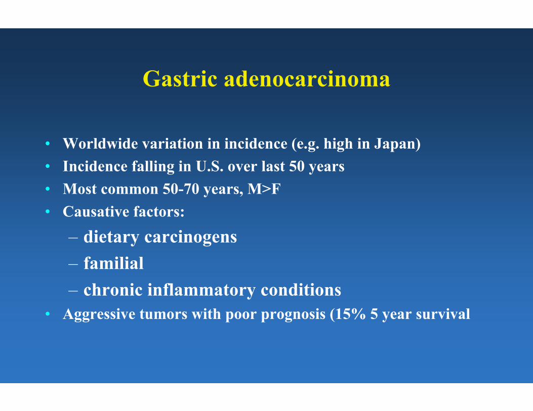
Gastric adenocarcinoma
• Worldwide variation in incidence (e.g. high in Japan) • Incidence falling in U.S. over last 50 years • Most common 50-70 years, M>F • Causative factors:
– dietary carcinogens – familial – chronic inflammatory conditions
• Aggressive tumors with poor prognosis (15% 5 year survival

Genetic Progressionin Gastric Neoplasia
•C-met/HGF amplification/overexpression
•9p21 deletion/p16 inactivation•19q12 amplification/Cyclin E
overexpression •18q deletion
•5q21 deletion/APC inactivation •16q22 deletion/E-cadherin loss •17p13 deletion/p53 mutation •chromosomal deletions (1p, 1q, •MLH1 methylation 7q, 13q)
Normal Metaplasia Dysplasia AdenoCa

Gastric carcinoma- pathology
• Location: antrum (70%)>lesser curvature, cardia (25%)>diffuse (5%)
• Gross configuration: polypoid, ulcerating, or infiltrating • Intestinal type: gland formation, associated with intestinal
metaplasia, dysplasia • Diffuse type: signet ring cells, arises directly from surface
foveolar cells, not associated with environmental factors

Esophageal carcinomas
• Two major types
–Squamous cell carcinoma –Adenocarcinoma
• Squamous cell carcinoma more common worldwide • Incidence of adenocarcinomas rising in U.S., Western Europe,
now accounts for 50% of esophageal malignancies in those regions

Esophageal adenocarcinoma
• Peak age 60-70 years, M>>F • Symptoms: dysphagia, weight loss • Arises in setting of Barrett’s esophagus (columnar metaplasia
with goblet cells) in distal esophagus • Proceeds through dysplasia-carcinoma sequence • Microscopically similar to adenocarcinomas elsewhere in GI
tract • Aggressive tumors; key to survival is early detection

Esophageal squamous cell carcinoma
• Incidence highest in Africa, Iran, China • Peak age: 55-65 years, M>F • Causative factors
– Alcohol, tobacco – Corrosive esophagitis – Achalasia – ?HPV
• Symptoms: dysphagia, weight loss • Aggressive tumors (10% 5 year survival)
Neuroendocrine (carcinoid) tumors
• Arise from neuroendocrine cells of gastrointestinal mucosa and its derivatives (e.g. lung, pancreas)
• Variable clinical behavior but often slow-growing • Appendix most common site (35%) followed by ileum (20%) • Pathology: uniform cells with round nuclei, “salt and pepper”
chromatin • Extra-appendiceal carcinoids frequently invade wall,
metastasize

Carcinoid syndrome
• Only develops in patients with liver metastases • Tumors elaborate serotonin, plus histamine, others • Flushing, diarrhea, bronchoconstriction, valvular changes in
right heart • Treatment: removal or ablation of metastasis or
antagonism/suppression of circulating serotonin

GI tract lymphomas
• Nearly all non-Hodgkin’s lymphomas (NHL) • GI tract involved in 70% of patients with NHL • Stomach most common site, followed by intestine and colon • Nearly all B cell type, except for enteropathy associated T cell
lymphoma (a/w celiac diseae) • MALT lymphoma: gastric lymphomas develop in setting of H.
pylori infection (potentially treatable by H, pylori eradication)

Gastrointestinal stromal tumors (GISTs)
• Spindle cell neoplasms arising from interstitial cells of Cajal (pacemaker cells)
• Most associated with activating mutations in c-kit tyrosine kinase; sensitive to treatment with inhibitor (Gleevec)
• Variable aggressiveness • Prognostic factors: size, location, histologic grade • Distinguish leiomyomas (true neoplasms of smooth muscle)

Other tumors
• Adenocarcinoma of small intestine, appendix • Anal squamous cell carcinoma • Mesotheliomas od peritoneum • Melanoma (rectum, anus, esophagus) • Lipoma (colon, stomach) • Kaposi’s sarcoma