Embed Size (px)
Citation preview
Scientific American Surgery DOI 10.2310/7800.2076
06/15
© 2015 Decker Intellectual Properties Inc
gastrointestinal tract and abdomen
U p p e r G a s t r o i n t e s t i n a l e n d o s c o p y
Jeffrey Marks, MD, FACS, and Hahn Soe-Lin, MD*
since the beginning of the 1970s, flexible endoscopy of the gastrointestinal tract has been the dominant modality for the diagnosis of gastrointestinal disease. over the same period, developments in technology and methodology have made possible the use of endoscopy to treat a host of conditions that once were considered to be manageable only by means of surgical intervention. the concept of performing endo-scopic surgery has become a reality with the advancement of endoscopic therapies to control hemorrhage and perform mucosal ablations, resections, and tissue approximation. in addition, investigation into intra-abdominal interventions has begun with the progression of natural orifice translumi-nal endoscopic surgery (notes). the tools created for both intraluminal and transluminal procedures will allow the endoscopist to continue to supplant surgical therapies for numerous gastrointestinal diseases. the integration of flex-ible endoscopic techniques into the armamentarium of the gastrointestinal surgeon permits a more multidimensional approach to the treatment of digestive system disorders. the modern surgeon must continue to stay abreast of these endoscopic advances to provide appropriate patient care.
Endoscope History
the flexible endoscope was initially developed in 1957 as an imaging device dependent on the delivery of light and transmission of the image along multiple bundles of chemi-cally treated glass fibers. the image undergoes a series of internal reflections within each fiber, which are coated with low optical density glass to prevent escape of light as it is transmitted up the bundle. the endoscopist views the image through an eyepiece at the top of the instrument handle.1 a teaching head could be fixed onto this eyepiece, allowing a second person to view the endoscopic image. With the advancement of early video camera systems, a video camera could be affixed to the scope eyepiece to transmit an image to a video monitor. eventually, the video system was imbed-ded into the scope, allowing the progression from fiber- optic scopes to the videoendoscopes we use today. this has been the key component in the advancements in our ability to perform more involved therapies, educate physicians and endoscopic assistants, and obtain static and dynamic recorded data images for improved clinical management.
Endoscope Structure
although the basic structure of the endoscope is unchanged, there are now a wide variety of lengths and diameters of scopes, with an assortment of channel numbers and sizes and adjunct imaging modalities.2 By knowing the
differences among available scopes, the skilled endoscopist is better able to match his or her tools to the desired proce-dure. the knobs for controlling scope tip manipulation are located on the right side of the headpiece and provide quadridirectional movement. the inner control knob is for up/down and the outer knob for left/right. the two buttons on the front of the scope provide lens cleaning, air insuffla-tion, and suction. newer models now include additional buttons for changing optical modalities, discussed subse-quently in this review.
the suction channel also functions as the biopsy channel so that any endoscopic tools placed into the biopsy channel will limit the ability to suction fluids through the endoscope. a small button on the front of the handpiece above the suction button allows for freezing of the image and digital recording by pressing the image capture button on the back of the handpiece.
Advancements in Imaging Technology
there have been many recent advances in endoscopic imaging techniques with the goal of improving immediate detection of dysplasia and other mucosal abnormalities. the ability to perform an “optical biopsy” at the time of the endoscopic procedure can, in theory, enhance directed tissue sampling or even resection compared with unaided tissue biopsy techniques. it should be noted that these tech-niques can be used for both upper and lower endoscopic procedures.
chromoendoscopy
this technique is based on the staining of mucosal sur-faces to differentiate different cellular activities and subtle mucosal abnormalities. agents commonly used as topical mucosal dyes include lugol (potassium iodide) solution, methylene blue, indigo carmine, congo red, and acetic acid. diseases, including Barrett esophagus and squamous cell carcinoma of the esophagus, have been detected with the use of these agents. normal keratinized squamous epitheli-um stains a deep brown with lugol solution as a result of a reaction between the potassium iodide and glycogen, but inflammation, dysplasia, and carcinoma do not stain because of a relative lack of glycogen. Methylene blue is actively absorbed by epithelium, whereas indigo carmine accumulates in pits and grooves to act as a countercontrast for the superficial esophageal mucosa. Methylene blue has largely fallen from favor given failure to demonstrate improved diagnostic yields over traditional white light endoscopy. acetic acid and indigo carmine have shown some evidence of increased diagnostic yields when com-bined with more modern high-resolution white light endos-copy techniques. However, they remain limited from routine use secondary to the cumbersome nature of the staining process.
* the authors and editors gratefully acknowledge the contri-butions of the previous author, Jeffrey l. ponsky, Md, Facs, to the development and writing of this review.
Scientific American Surgery
06/15
gastro upper gastrointestinal endoscopy — 2
spectroscopy/autofluorescence imaging
spectroscopy is an additional modality that relies on light-based excitation of tissues at the subcellular level. indi-vidual organelles produce different wavelength patterns in response to a pulse of lower frequency light, and these characteristic patterns generate a fluorescence spectrum that is sufficiently unique to distinguish between neoplastic and nonneoplastic tissue. an optical probe is passed through the working channel of the endoscope and is required to contact tissue directly with a spatial resolution of 0.5 mm in diam-eter. the use of white light reduces the cost of the system, and the speed of the pulsatile light probe acquiring data facilitates surveillance of a 10 cm Barrett segment in approx-imately 2 minutes.5 although this modality has demonstrated increased detection of areas of neoplasia not identified by white light endoscopy, the gain in diagnostic yield when compared with standard random quadrantic biopsies was calculated at only 2%.6 a newer endoscopic application of one such spectroscopic technique known as raman spectroscopy induces excitational inelastic vibrations at the molecular level, which can subsequently be analyzed near real time to differentiate tissues based on their molecular signature. promising early data from ex vivo studies com-paring data collected from this probing and histopathologic samples have demonstrated sensitivities and specificities of 86% and 88%, respectively, in the detection of high-grade dysplasia and esophageal adenocarcinoma.7 challenges to clinical translation have included weak signal in in vivo tissues and slow speed of spectral measurements. More recently, contact probes have been developed that can fit through the biopsy channel of a standard endoscope and provide 1-second data collection times and demonstrate the promise that this modality may one day significantly improve detection of early high-grade dysplasia.8 as with nBi, the clinical advantage may ultimately be a reduction in the number of biopsies required to identify dysplasia, although this advantage has yet to be confirmed in large prospective trials.
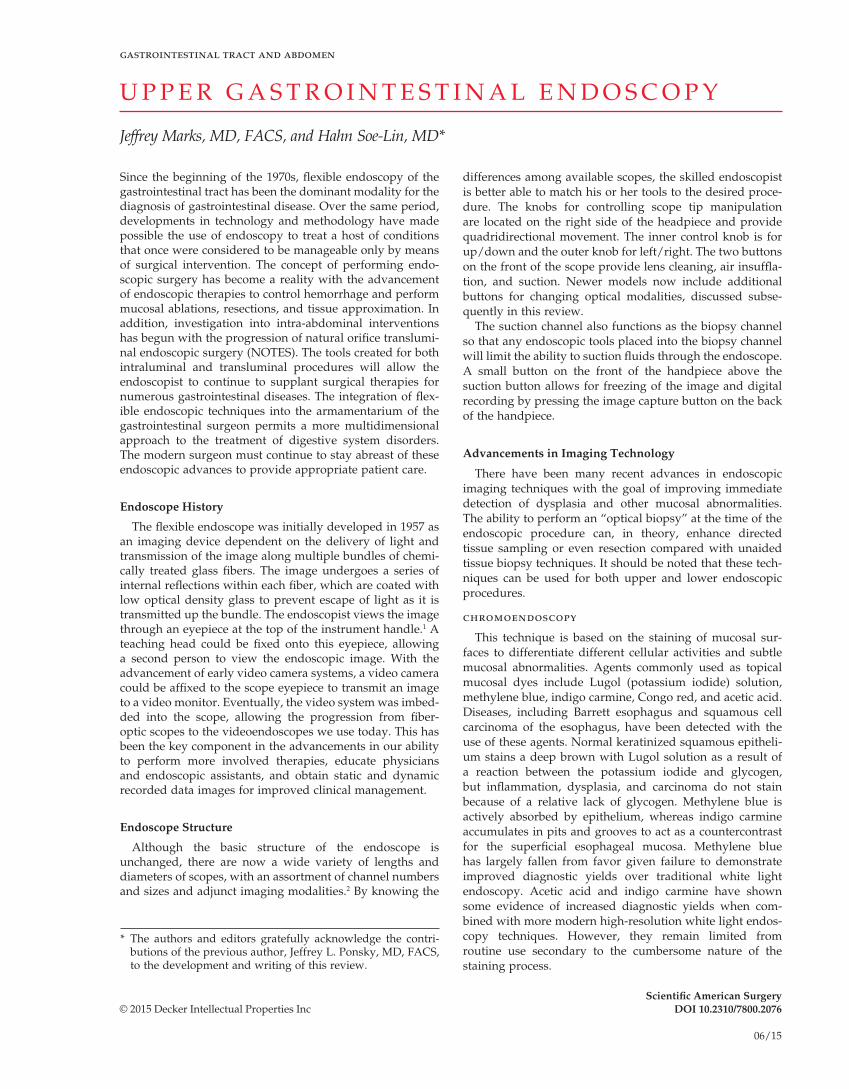
narrow-band imaging
Most endoscopes now have the ability to switch the bandwidth of projected light just by pressing a button on the scope handle. narrow-band imaging (nBi) is a digital chromoendoscopy technique that takes advantage of the different absorption spectra of hemoglobin compared with adjacent epithelial structures. this allows visual enhance-ment of underlying vasculature and better differentiation of squamous from nonsquamous epithelium to help identify Barrett esophagus [see Figure 1 and Figure 2] without the use of cumbersome chemical dyes. the clinical advantage of nBi is that it can direct the endoscopist toward suspicious areas worthy of biopsy and thus obtain comparable diagnos-tic yields with fewer biopsies compared with standard four-quadrant surveillance biopsy protocols.3 the use of white light as well as nBi has also enabled endoscopists to provide an immediate assessment of small colonic and gastric lesions without histopathologic evaluation.4 Whereas nBi is consid-ered a preprocessing technique where a specific wavelength component of white light is selected to shine directly on tissue, postprocessing chromoendoscopy uses white light excitation and then digitally reprocesses the ensuing image to enhance selective wavelengths, allowing tissues that enhance best at different wavelengths to be rapidly evaluated using a single white light source.
optical coherence tomography
this technique uses reflection of near-infrared light to produce real-time two-dimensional cross-sectional images of the gastrointestinal tract, with a spatial resolution of between 1 and 15 µm. a small probe similar to an endoscope ultrasound probe that does not require tissue contact is passed through the scope. optical coherence tomography produces a high-resolution image of the layers of the gastro-intestinal tract and for this reason has particular promise for endoscopic staging of esophageal squamous cell carcinomas. although not yet in widespread use, early investigations suggest promising improvements in staging accuracy over traditional endoscopic ultrasonography.4
Figure 1 The squamocolumnar junction seen with routine white light.
Figure 2 The squamocolumnar junction as seen with narrow-band imaging.
Scientific American Surgery
06/15
gastro upper gastrointestinal endoscopy — 3
Basic Upper Endoscopy
indications
Upper endoscopy, or esophagogastroduodenoscopy (eGd), is indicated when a patient has abnormal findings on tradi-tional gastrointestinal x-ray series or one of the “alarm symptoms,” such as weight loss, early satiety, hematemesis, dysphagia, odynophagia, epigastric pain that does not respond to medical therapy, persistent heartburn, suspected foreign body, or iron deficiency anemia. it is also indicated for surveillance of patients at high risk for malignancy and for sampling of gastrointestinal tissue or fluid. Unsedated transnasal endoscopy with slim endoscopes can also be performed in patients with suspected esophageal or gastric pathology. Biopsies can be performed, but no therapeutic interventions can be provided because of the limitation of the biopsy channel.
contraindications
contraindications to eGd include underlying patient comorbidity and inability to tolerate conscious sedation. recent surgery or anastomosis is not a contraindication to an eGd, and the risks and benefits of any procedure must be weighed before proceeding.
assessment and monitoring
Following the initial patient evaluation, including ameri-can society of anesthesiologists classification and Malam-pati scoring to assess the risk of airway compromise, the patient is placed in a left-side-down decubitus position. one prepares for the examination by ensuring the patient’s hemodynamic stability, having the patient fast for 6 to 8 hours beforehand, and then proceeding with the delivery of medications to provide conscious sedation, which gener-ally involves applying a topical anesthetic to the posterior pharynx and administering a narcotic and a benzodiazepine intravenously. Monitoring of arterial blood pressure and oxygen saturation throughout the procedure is now standard practice.
technique of upper endoscopy
With the patient in the left lateral decubitus position, a topical anesthetic may be applied to the posterior pharynx and an intravenous sedative administered. the forward-viewing panendoscope—a small-caliber instrument that is long enough to permit examination of the foregut from the mouth to the third portion of the duodenum—is employed. the endoscope is held in the left hand regardless of the individual physician’s hand dominance. the internal upward and downward deflection knob is controlled by the left thumb, whereas the air, water, and suction knobs are controlled by the left index and middle fingers. the smaller left/right knob then is usually manipulated by the right hand.
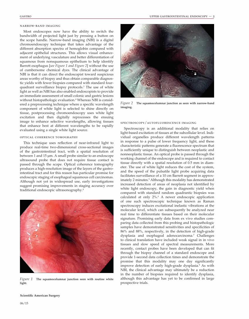
the endoscope should be introduced under direct visual-ization rather than using two-handed techniques, which increase the risk for both the patient and the physician. the instrument is advanced slowly until the epiglottis and vocal cords are visualized [see Figure 3]; it is then angled posteri-orly to the esophageal introitus and gently advanced as the patient is asked to swallow. insufflation of air is begun to
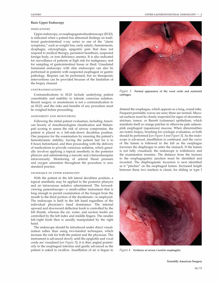
distend the esophagus, which appears as a long, round tube. Frequent peristaltic waves are seen; these are normal. Muco-sal surfaces must be closely inspected for signs of ulceration, stricture, tumor, or Barrett (columnar) epithelium, which manifests itself as orange patches in otherwise pale salmon-pink esophageal (squamous) mucosa. When abnormalities are noted, biopsy, brushing for cytologic evaluation, or both should be performed [see Figure 4 and Figure 5]. as the endo-scope is advanced, insufflation is continued, and the curve of the lumen is followed to the left as the esophagus traverses the diaphragm to enter the stomach. if the lumen is not fully visualized, the endoscope is withdrawn and the examination resumes. the distance from the incisors to the esophagogastric junction must be identified and recorded. the diaphragmatic incursion is next identified as it “pinches” on the esophageal lumen. increased length between these two markers is classic for sliding or type i
Figure 3 Normal appearance of the vocal cords and arytenoid cartilages.
Figure 4 Evidence of severe Candida esophagitis.
Scientific American Surgery
06/15
gastro upper gastrointestinal endoscopy — 4
hiatal hernia, as is the presence of gastric folds above this pinched area. the pinching may be better visualized when the patient sniffs.
When the stomach is entered, the tip of the endoscope is elevated so as to center it within the gastric lumen. it should be noted that with the patient lying in the left lateral decubitus position, the stomach is also on its side, with the greater curvature at 6 o’clock, the lesser curvature at 12 o’clock, the posterior wall at 3 o’clock, and the anterior wall at 9 o’clock. air should be insufflated to distend the stomach fully and permit careful inspection of all mucosal surfaces [see Figure 6].
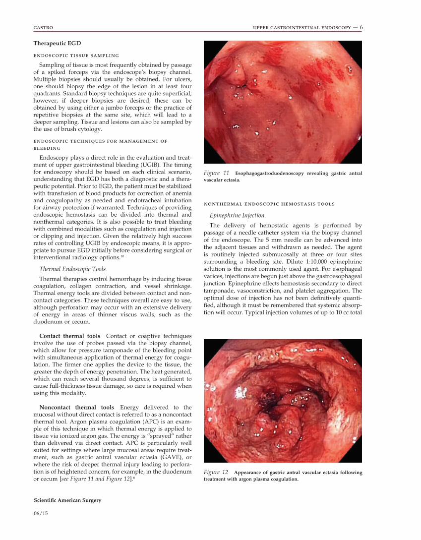
after the stomach has been viewed, the instrument is advanced under direct vision through the pylorus and into the duodenal bulb. insufflation of air should continue as the scope is pressed against the pylorus to facilitate passage of
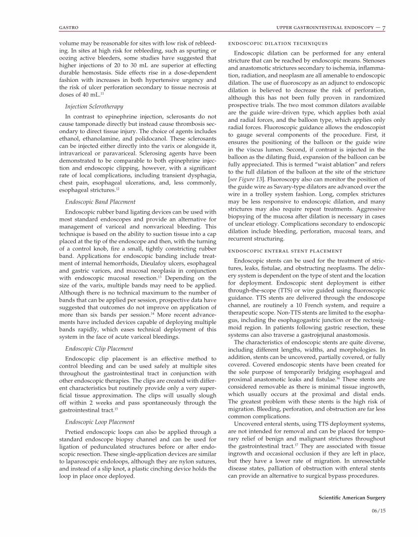
the instrument [see Figure 7]. the scope tends to pop into the duodenal bulb rather than slide smoothly; it should be pulled back slightly to allow one to observe the mucosal surfaces of the bulb before moving ahead. Unlike the rest of the small bowel, the duodenal bulb has no semicircular folds [see Figure 8]. the tip of the scope must be rotated slightly to permit examination of the walls of the bulb. it is advisable to pull the instrument back into the stomach while observing the walls of the bulb and the pyloric channel for lesions; several such withdrawals may be required for full assessment of this area.
once the duodenal bulb has been examined, the endo-scope is advanced just past the bulb to the point where the first duodenal folds are observed. Here the duodenum turns sharply to the rear and downward as it becomes retroperi-toneal. advancement of the scope into the second portion of the duodenum is one of the few endoscopic maneuvers that cannot be accomplished under direct vision [see Figure 9]. Because of the sharp angle of the turn, one will experience a moment of so-called “red-out” as the tip of the endoscope touches the mucosa during the turn. to ensure that the turn is accomplished safely, the instrument is advanced as far through the bulb as is possible under direct vision. the control handle of the scope is then rotated approximately 90° to the right as the tip of the scope is turned to the right and angled first upward and then downward. as the second portion of the duodenum appears, the scope is rotated back to its neutral position. When done correctly, the turn is actu-ally quite easy. it should never be forced: if the instrument does not proceed easily into the descending duodenum, the scope should be pulled back and the attempt repeated. pushing against resistance may result in perforation.
entering the descending duodenum causes the scope to form a large loop in the stomach. therefore, once the second portion of the duodenum is successfully entered, the shaft of the instrument is pulled back. paradoxically, as this move-ment straightens the gastric loop, it also advances the tip of the instrument deeper into the duodenum. Further advance-ment of the instrument under direct vision often permits
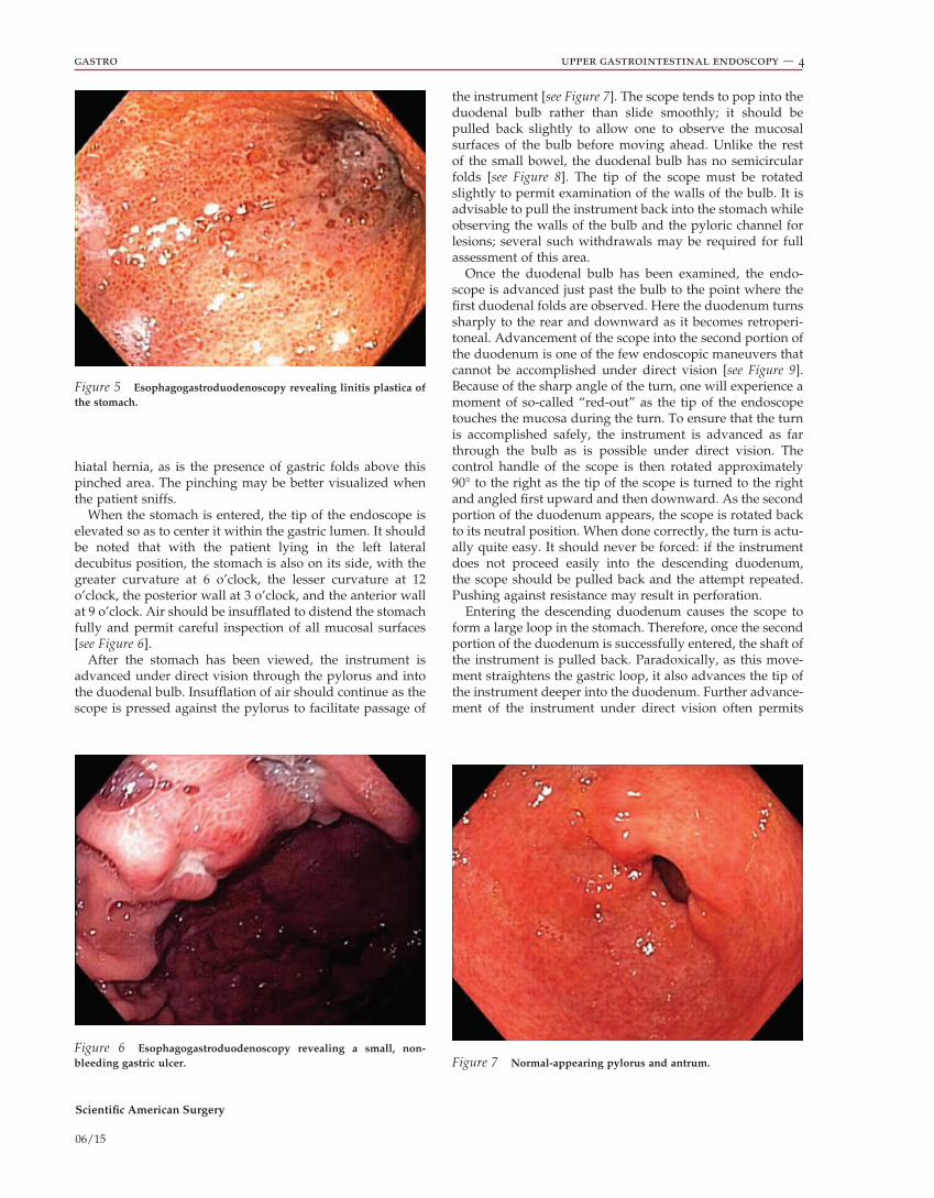
Figure 5 Esophagogastroduodenoscopy revealing linitis plastica of the stomach.
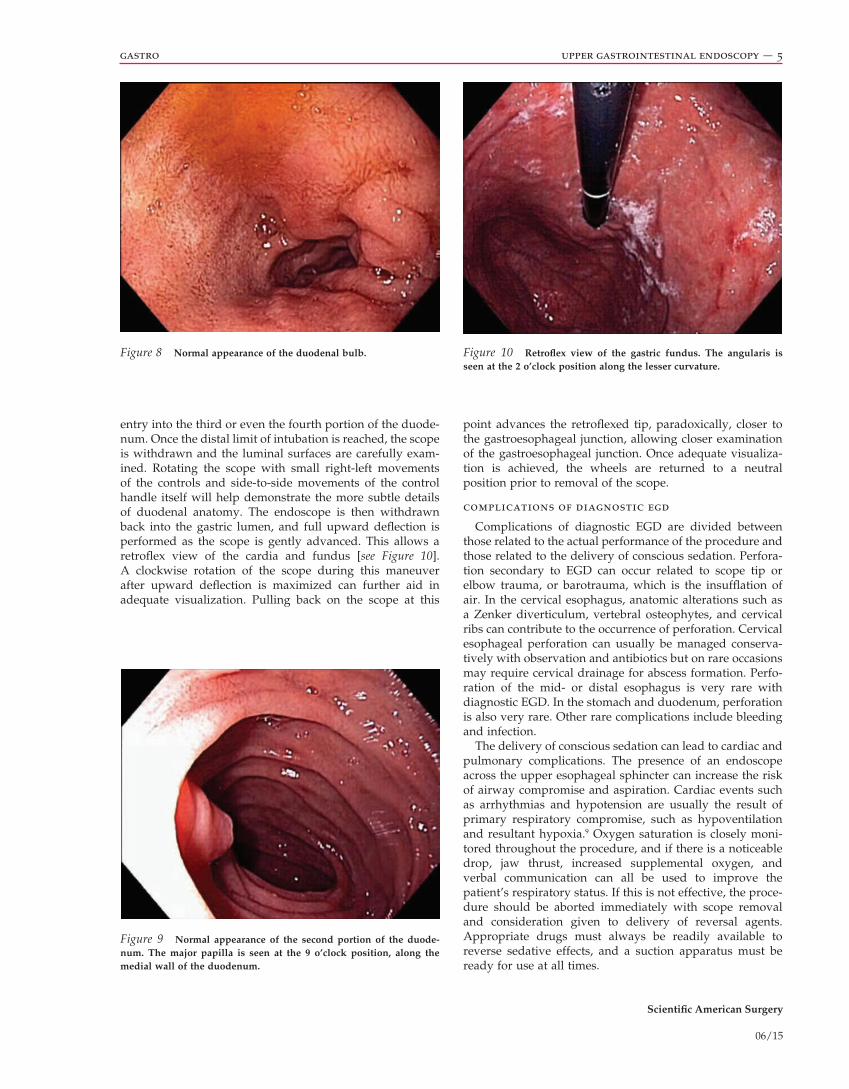
Figure 6 Esophagogastroduodenoscopy revealing a small, non-bleeding gastric ulcer. Figure 7 Normal-appearing pylorus and antrum.
Scientific American Surgery
06/15
gastro upper gastrointestinal endoscopy — 5
entry into the third or even the fourth portion of the duode-num. once the distal limit of intubation is reached, the scope is withdrawn and the luminal surfaces are carefully exam-ined. rotating the scope with small right-left movements of the controls and side-to-side movements of the control handle itself will help demonstrate the more subtle details of duodenal anatomy. the endoscope is then withdrawn back into the gastric lumen, and full upward deflection is performed as the scope is gently advanced. this allows a retroflex view of the cardia and fundus [see Figure 10]. a clockwise rotation of the scope during this maneuver after upward deflection is maximized can further aid in adequate visualization. pulling back on the scope at this
point advances the retroflexed tip, paradoxically, closer to the gastroesophageal junction, allowing closer examination of the gastroesophageal junction. once adequate visualiza-tion is achieved, the wheels are returned to a neutral position prior to removal of the scope.
complications of diagnostic egd
complications of diagnostic eGd are divided between those related to the actual performance of the procedure and those related to the delivery of conscious sedation. perfora-tion secondary to eGd can occur related to scope tip or elbow trauma, or barotrauma, which is the insufflation of air. in the cervical esophagus, anatomic alterations such as a Zenker diverticulum, vertebral osteophytes, and cervical ribs can contribute to the occurrence of perforation. cervical esophageal perforation can usually be managed conserva-tively with observation and antibiotics but on rare occasions may require cervical drainage for abscess formation. perfo-ration of the mid- or distal esophagus is very rare with diagnostic eGd. in the stomach and duodenum, perforation is also very rare. other rare complications include bleeding and infection.
the delivery of conscious sedation can lead to cardiac and pulmonary complications. the presence of an endoscope across the upper esophageal sphincter can increase the risk of airway compromise and aspiration. cardiac events such as arrhythmias and hypotension are usually the result of primary respiratory compromise, such as hypoventilation and resultant hypoxia.9 oxygen saturation is closely moni-tored throughout the procedure, and if there is a noticeable drop, jaw thrust, increased supplemental oxygen, and verbal communication can all be used to improve the patient’s respiratory status. if this is not effective, the proce-dure should be aborted immediately with scope removal and consideration given to delivery of reversal agents. appropriate drugs must always be readily available to reverse sedative effects, and a suction apparatus must be ready for use at all times.
Figure 8 Normal appearance of the duodenal bulb.
Figure 9 Normal appearance of the second portion of the duode-num. The major papilla is seen at the 9 o’clock position, along the medial wall of the duodenum.
Figure 10 Retroflex view of the gastric fundus. The angularis is seen at the 2 o’clock position along the lesser curvature.
Scientific American Surgery
06/15
gastro upper gastrointestinal endoscopy — 6
nonthermal endoscopic hemostasis tools
Epinephrine Injection
the delivery of hemostatic agents is performed by passage of a needle catheter system via the biopsy channel of the endoscope. the 5 mm needle can be advanced into the adjacent tissues and withdrawn as needed. the agent is routinely injected submucosally at three or four sites surrounding a bleeding site. dilute 1:10,000 epinephrine solution is the most commonly used agent. For esophageal varices, injections are begun just above the gastroesophageal junction. epinephrine effects hemostasis secondary to direct tamponade, vasoconstriction, and platelet aggregation. the optimal dose of injection has not been definitively quanti-fied, although it must be remembered that systemic absorp-tion will occur. typical injection volumes of up to 10 cc total
Therapeutic EGD
endoscopic tissue sampling
sampling of tissue is most frequently obtained by passage of a spiked forceps via the endoscope’s biopsy channel. Multiple biopsies should usually be obtained. For ulcers, one should biopsy the edge of the lesion in at least four quadrants. standard biopsy techniques are quite superficial; however, if deeper biopsies are desired, these can be obtained by using either a jumbo forceps or the practice of repetitive biopsies at the same site, which will lead to a deeper sampling. tissue and lesions can also be sampled by the use of brush cytology.
endoscopic techniques for management of bleeding
endoscopy plays a direct role in the evaluation and treat-ment of upper gastrointestinal bleeding (UGiB). the timing for endoscopy should be based on each clinical scenario, understanding that eGd has both a diagnostic and a thera-peutic potential. prior to eGd, the patient must be stabilized with transfusion of blood products for correction of anemia and coagulopathy as needed and endotracheal intubation for airway protection if warranted. techniques of providing endoscopic hemostasis can be divided into thermal and nonthermal categories. it is also possible to treat bleeding with combined modalities such as coagulation and injection or clipping and injection. Given the relatively high success rates of controlling UGiB by endoscopic means, it is appro-priate to pursue eGd initially before considering surgical or interventional radiology options.10
Thermal Endoscopic Tools
thermal therapies control hemorrhage by inducing tissue coagulation, collagen contraction, and vessel shrinkage. thermal energy tools are divided between contact and non-contact categories. these techniques overall are easy to use, although perforation may occur with an extensive delivery of energy in areas of thinner viscus walls, such as the duodenum or cecum.
Contact thermal tools contact or coaptive techniques involve the use of probes passed via the biopsy channel, which allow for pressure tamponade of the bleeding point with simultaneous application of thermal energy for coagu-lation. the firmer one applies the device to the tissue, the greater the depth of energy penetration. the heat generated, which can reach several thousand degrees, is sufficient to cause full-thickness tissue damage, so care is required when using this modality.
Noncontact thermal tools energy delivered to the mucosal without direct contact is referred to as a noncontact thermal tool. argon plasma coagulation (apc) is an exam-ple of this technique in which thermal energy is applied to tissue via ionized argon gas. the energy is “sprayed” rather than delivered via direct contact. apc is particularly well suited for settings where large mucosal areas require treat-ment, such as gastric antral vascular ectasia (GaVe), or where the risk of deeper thermal injury leading to perfora-tion is of heightened concern, for example, in the duodenum or cecum [see Figure 11 and Figure 12].6
Figure 11 Esophagogastroduodenoscopy revealing gastric antral vascular ectasia.
Figure 12 Appearance of gastric antral vascular ectasia following treatment with argon plasma coagulation.
Scientific American Surgery
06/15
gastro upper gastrointestinal endoscopy — 7
endoscopic dilation techniques
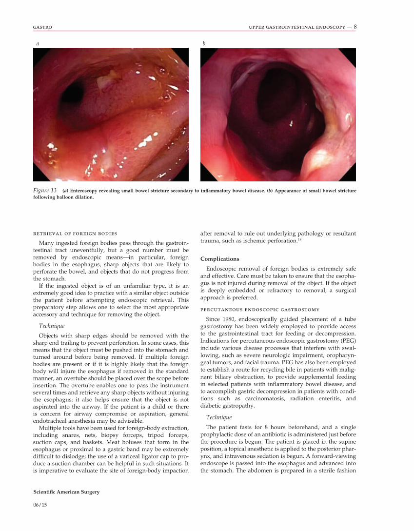
endoscopic dilation can be performed for any enteral stricture that can be reached by endoscopic means. stenoses and anastomotic strictures secondary to ischemia, inflamma-tion, radiation, and neoplasm are all amenable to endoscopi c dilation. the use of fluoroscopy as an adjunct to endoscopic dilation is believed to decrease the risk of perforation, although this has not been fully proven in randomized prospective trials. the two most common dilators available are the guide wire–driven type, which applies both axial and radial forces, and the balloon type, which applies only radial forces. Fluoroscopic guidance allows the endoscopist to gauge several components of the procedure. First, it ensures the positioning of the balloon or the guide wire in the viscus lumen. second, if contrast is injected in the balloon as the dilating fluid, expansion of the balloon can be fully appreciated. this is termed “waist ablation” and refers to the full dilation of the balloon at the site of the stricture [see Figure 13]. Fluoroscopy also can monitor the position of the guide wire as savary-type dilators are advanced over the wire in a trolley system fashion. long, complex strictures may be less responsive to endoscopic dilation, and many strictures may also require repeat treatments. aggressive biopsying of the mucosa after dilation is necessary in cases of unclear etiology. complications secondary to endoscopic dilation include bleeding, perforation, mucosal tears, and recurrent structuring.
endoscopic enteral stent placement
endoscopic stents can be used for the treatment of stric-tures, leaks, fistulae, and obstructing neoplasms. the deliv-ery system is dependent on the type of stent and the location for deployment. endoscopic stent deployment is either through-the-scope (tts) or wire guided using fluoroscopic guidance. tts stents are delivered through the endoscope channel, are routinely a 10 French system, and require a therapeutic scope. non-tts stents are limited to the esopha-gus, including the esophagogastric junction or the rectosig-moid region. in patients following gastric resection, these systems can also traverse a gastrojejunal anastomosis.
the characteristics of endoscopic stents are quite diverse, including different lengths, widths, and morphologies. in addition, stents can be uncovered, partially covered, or fully covered. covered endoscopic stents have been created for the sole purpose of temporarily bridging esophageal and proximal anastomotic leaks and fistulae.16 these stents are considered removable as there is minimal tissue ingrowth, which usually occurs at the proximal and distal ends. the greatest problem with these stents is the high risk of migration. Bleeding, perforation, and obstruction are far less common complications.
Uncovered enteral stents, using tts deployment systems, are not intended for removal and can be placed for tempo-rary relief of benign and malignant strictures throughout the gastrointestinal tract.17 they are associated with tissue ingrowth and occasional occlusion if they are left in place, but they have a lower rate of migration. in unresectable disease states, palliation of obstruction with enteral stents can provide an alternative to surgical bypass procedures.
volume may be reasonable for sites with low risk of rebleed-ing. in sites at high risk for rebleeding, such as spurting or oozing active bleeders, some studies have suggested that higher injections of 20 to 30 ml are superior at effecting durable hemostasis. side effects rise in a dose-dependent fashion with increases in both hypertensive urgency and the risk of ulcer perforation secondary to tissue necrosis at doses of 40 ml.11
Injection Sclerotherapy
in contrast to epinephrine injection, sclerosants do not cause tamponade directly but instead cause thrombosis sec-ondary to direct tissue injury. the choice of agents includes ethanol, ethanolamine, and polidocanol. these sclerosants can be injected either directly into the varix or alongside it, intravariceal or paravariceal. sclerosing agents have been demonstrated to be comparable to both epinephrine injec-tion and endoscopic clipping, however, with a significant rate of local complications, including transient dysphagia, chest pain, esophageal ulcerations, and, less commonly, esophageal strictures.12
Endoscopic Band Placement
endoscopic rubber band ligating devices can be used with most standard endoscopes and provide an alternative for management of variceal and nonvariceal bleeding. this technique is based on the ability to suction tissue into a cap placed at the tip of the endoscope and then, with the turning of a control knob, fire a small, tightly constricting rubber band. applications for endoscopic banding include treat-ment of internal hemorrhoids, dieulafoy ulcers, esophageal and gastric varices, and mucosal neoplasia in conjunction with endoscopic mucosal resection.13 depending on the size of the varix, multiple bands may need to be applied. although there is no technical maximum to the number of bands that can be applied per session, prospective data have suggested that outcomes do not improve on application of more than six bands per session.14 More recent advance-ments have included devices capable of deploying multiple bands rapidly, which eases technical deployment of this system in the face of acute variceal bleedings.
Endoscopic Clip Placement
endoscopic clip placement is an effective method to control bleeding and can be used safely at multiple sites throughout the gastrointestinal tract in conjunction with other endoscopic therapies. the clips are created with differ-ent characteristics but routinely provide only a very super-ficial tissue approximation. the clips will usually slough off within 2 weeks and pass spontaneously through the gastrointestinal tract.15
Endoscopic Loop Placement
pretied endoscopic loops can also be applied through a standard endoscope biopsy channel and can be used for ligation of pedunculated structures before or after endo-scopic resection. these single-application devices are similar to laparoscopic endoloops, although they are nylon sutures, and instead of a slip knot, a plastic cinching device holds the loop in place once deployed.
Scientific American Surgery
06/15
gastro upper gastrointestinal endoscopy — 8
retrieval of foreign bodies
Many ingested foreign bodies pass through the gastroin-testinal tract uneventfully, but a good number must be removed by endoscopic means—in particular, foreign bodies in the esophagus, sharp objects that are likely to perforate the bowel, and objects that do not progress from the stomach.
if the ingested object is of an unfamiliar type, it is an extremely good idea to practice with a similar object outside the patient before attempting endoscopic retrieval. this preparatory step allows one to select the most appropriate accessory and technique for removing the object.
Technique
objects with sharp edges should be removed with the sharp end trailing to prevent perforation. in some cases, this means that the object must be pushed into the stomach and turned around before being removed. if multiple foreign bodies are present or if it is highly likely that the foreign body will injure the esophagus if removed in the standard manner, an overtube should be placed over the scope before insertion. the overtube enables one to pass the instrument several times and retrieve any sharp objects without injuring the esophagus; it also helps ensure that the object is not aspirated into the airway. if the patient is a child or there is concern for airway compromise or aspiration, general endotracheal anesthesia may be advisable.
Multiple tools have been used for foreign-body extraction, including snares, nets, biopsy forceps, tripod forceps, suction caps, and baskets. Meat boluses that form in the esophagus or proximal to a gastric band may be extremely difficult to dislodge; the use of a variceal ligator cap to pro-duce a suction chamber can be helpful in such situations. it is imperative to evaluate the site of foreign-body impaction
ba
Figure 13 (a) Enteroscopy revealing small bowel stricture secondary to inflammatory bowel disease. (b) Appearance of small bowel stricture following balloon dilation.
after removal to rule out underlying pathology or resultant trauma, such as ischemic perforation.18
Complications
endoscopic removal of foreign bodies is extremely safe and effective. care must be taken to ensure that the esopha-gus is not injured during removal of the object. if the object is deeply embedded or refractory to removal, a surgical approach is preferred.
percutaneous endoscopic gastrostomy
since 1980, endoscopically guided placement of a tube gastrostomy has been widely employed to provide access to the gastrointestinal tract for feeding or decompression. indications for percutaneous endoscopic gastrostomy (peG) include various disease processes that interfere with swal-lowing, such as severe neurologic impairment, oropharyn-geal tumors, and facial trauma. peG has also been employed to establish a route for recycling bile in patients with malig-nant biliary obstruction, to provide supplemental feeding in selected patients with inflammatory bowel disease, and to accomplish gastric decompression in patients with condi-tions such as carcinomatosis, radiation enteritis, and diabetic gastropathy.
Technique
the patient fasts for 8 hours beforehand, and a single prophylactic dose of an antibiotic is administered just before the procedure is begun. the patient is placed in the supine position, a topical anesthetic is applied to the posterior phar-ynx, and intravenous sedation is begun. a forward-viewing endoscope is passed into the esophagus and advanced into the stomach. the abdomen is prepared in a sterile fashion
Scientific American Surgery
06/15
gastro upper gastrointestinal endoscopy — 9
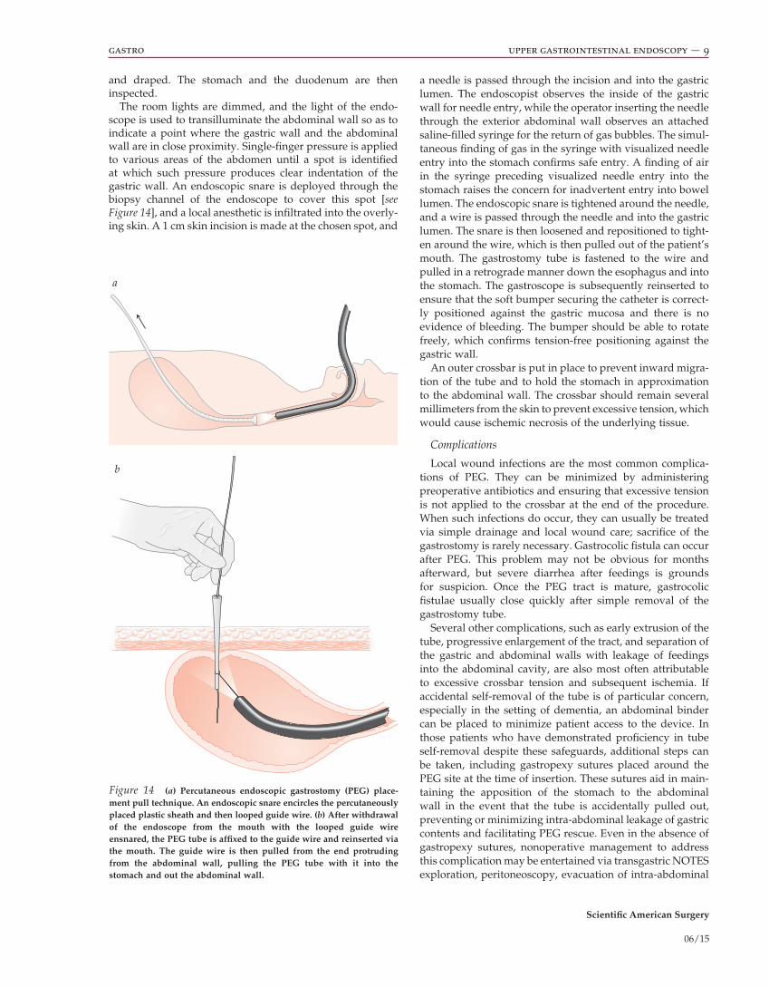
a needle is passed through the incision and into the gastric lumen. the endoscopist observes the inside of the gastric wall for needle entry, while the operator inserting the needle through the exterior abdominal wall observes an attached saline-filled syringe for the return of gas bubbles. the simul-taneous finding of gas in the syringe with visualized needle entry into the stomach confirms safe entry. a finding of air in the syringe preceding visualized needle entry into the stomach raises the concern for inadvertent entry into bowel lumen. the endoscopic snare is tightened around the needle, and a wire is passed through the needle and into the gastric lumen. the snare is then loosened and repositioned to tight-en around the wire, which is then pulled out of the patient’s mouth. the gastrostomy tube is fastened to the wire and pulled in a retrograde manner down the esophagus and into the stomach. the gastroscope is subsequently reinserted to ensure that the soft bumper securing the catheter is correct-ly positioned against the gastric mucosa and there is no evidence of bleeding. the bumper should be able to rotate freely, which confirms tension-free positioning against the gastric wall.
an outer crossbar is put in place to prevent inward migra-tion of the tube and to hold the stomach in approximation to the abdominal wall. the crossbar should remain several millimeters from the skin to prevent excessive tension, which would cause ischemic necrosis of the underlying tissue.
Complications
local wound infections are the most common complica-tions of peG. they can be minimized by administering preoperative antibiotics and ensuring that excessive tension is not applied to the crossbar at the end of the procedure. When such infections do occur, they can usually be treated via simple drainage and local wound care; sacrifice of the gastrostomy is rarely necessary. Gastrocolic fistula can occur after peG. this problem may not be obvious for months afterward, but severe diarrhea after feedings is grounds for suspicion. once the peG tract is mature, gastrocolic fistulae usually close quickly after simple removal of the gastrostomy tube.
several other complications, such as early extrusion of the tube, progressive enlargement of the tract, and separation of the gastric and abdominal walls with leakage of feedings into the abdominal cavity, are also most often attributable to excessive crossbar tension and subsequent ischemia. if accidental self-removal of the tube is of particular concern, especially in the setting of dementia, an abdominal binder can be placed to minimize patient access to the device. in those patients who have demonstrated proficiency in tube self-removal despite these safeguards, additional steps can be taken, including gastropexy sutures placed around the peG site at the time of insertion. these sutures aid in main-taining the apposition of the stomach to the abdominal wall in the event that the tube is accidentally pulled out, preventing or minimizing intra-abdominal leakage of gastric contents and facilitating peG rescue. even in the absence of gastropexy sutures, nonoperative management to address this complication may be entertained via transgastric notes exploration, peritoneoscopy, evacuation of intra-abdominal
and draped. the stomach and the duodenum are then inspected.
the room lights are dimmed, and the light of the endo-scope is used to transilluminate the abdominal wall so as to indicate a point where the gastric wall and the abdominal wall are in close proximity. single-finger pressure is applied to various areas of the abdomen until a spot is identified at which such pressure produces clear indentation of the gastric wall. an endoscopic snare is deployed through the biopsy channel of the endoscope to cover this spot [see Figure 14], and a local anesthetic is infiltrated into the overly-ing skin. a 1 cm skin incision is made at the chosen spot, and
Figure 14 (a) Percutaneous endoscopic gastrostomy (PEG) place-ment pull technique. An endoscopic snare encircles the percutaneously placed plastic sheath and then looped guide wire. (b) After withdrawal of the endoscope from the mouth with the looped guide wire ensnared, the PEG tube is affixed to the guide wire and reinserted via the mouth. The guide wire is then pulled from the end protruding from the abdominal wall, pulling the PEG tube with it into the stomach and out the abdominal wall.
a
b
Scientific American Surgery
06/15
gastro upper gastrointestinal endoscopy — 10
fluid, and reestablishment of the peG tube via the original gastrostomy tract.19 Failure of this less invasive option would mandate traditional operative intervention for washout and repair.
PEG with Jejunostomy Tube Extension
in patients who fail to tolerate gastric feedings as a result of severe gastroesophageal reflux or gastroparesis, transpy-loric feeding can be provided via a jejunostomy tube passed through an existing peG. peG–J tube placement is achieved by passing a jejunal feeding tube through the peG lumen (a 24 French peG tube accommodates up to a 12.5 French J tube; a standard 20 French peG tube accommodates an 8.5 French J tube). endoscopically, the jejunal tube is guided into the duodenum under direct vision with either a biopsy forceps or an endoscopic clip.
direct percutaneous endoscopic jejunostomy tube
Feedings beyond the ligament of treitz using direct percutaneous endoscopic jejunostomy (peJ) are associated with a lower incidence of gastroesophageally induced aspi-ration compared with simple postpyloric feeding but are associated with increased procedural risks, including bleed-ing, inadvertent viscus injury, and leakage.20 performance of direct peJ requires both endoscopic and fluoroscopic guidance. abdominal wall depression with a hemostat is performed at this site to try to identify a loop of small bowe l adjacent to the abdominal wall. safe tract techniques are then used to access the identified bowel, and a “pull” peJ is performed with either a 16 French or a 20 French tube.
endoscopic retrograde cholangiopancreatography
endoscopic retrograde cholangiopancreatography (ercp) is an advanced procedure that is technically more challeng-ing than standard upper gastrointestinal endoscopy; however, it can be mastered by most endoscopists who are willing to dedicate sufficient time to learning the method. ercp yields a radiologic image of the pancreatic and biliary trees and, in many cases, provides access for therapy. indi-cations for ercp include suspected benign or malignant maladies of the common bile duct (cBd), the ampulla of Vater, or the pancreas. cholelithiasis is not an indication for ercp unless choledocholithiasis is suspected. diagnostic ercp is rarely performed because of the high risks of this procedure. Magnetic resonance cholangiopancreatography (Mrcp) provides safer radiographic imaging of the pancre-aticobiliary trees.21 the therapeutic potential of ercp is quite extensive and has supplanted surgical intervention for many diseases of the pancreas and biliary systems.
Technique
as with standard upper gastrointestinal endoscopy, the patient fasts for 6 to 8 hours beforehand. intravenous seda-tion is administered, and prophylactic antibiotics are given when biliary obstruction is suspected. the patient is initially placed in the left lateral decubitus position but is later rotated to the prone position after the scope is in place in the second portion of the duodenum. a side-viewing endoscope is employed because it allows the best visualization of the
ampulla of Vater. the instrument is passed into the esopha-gus and maneuvered through the stomach, across the pylorus, and into the duodenum.
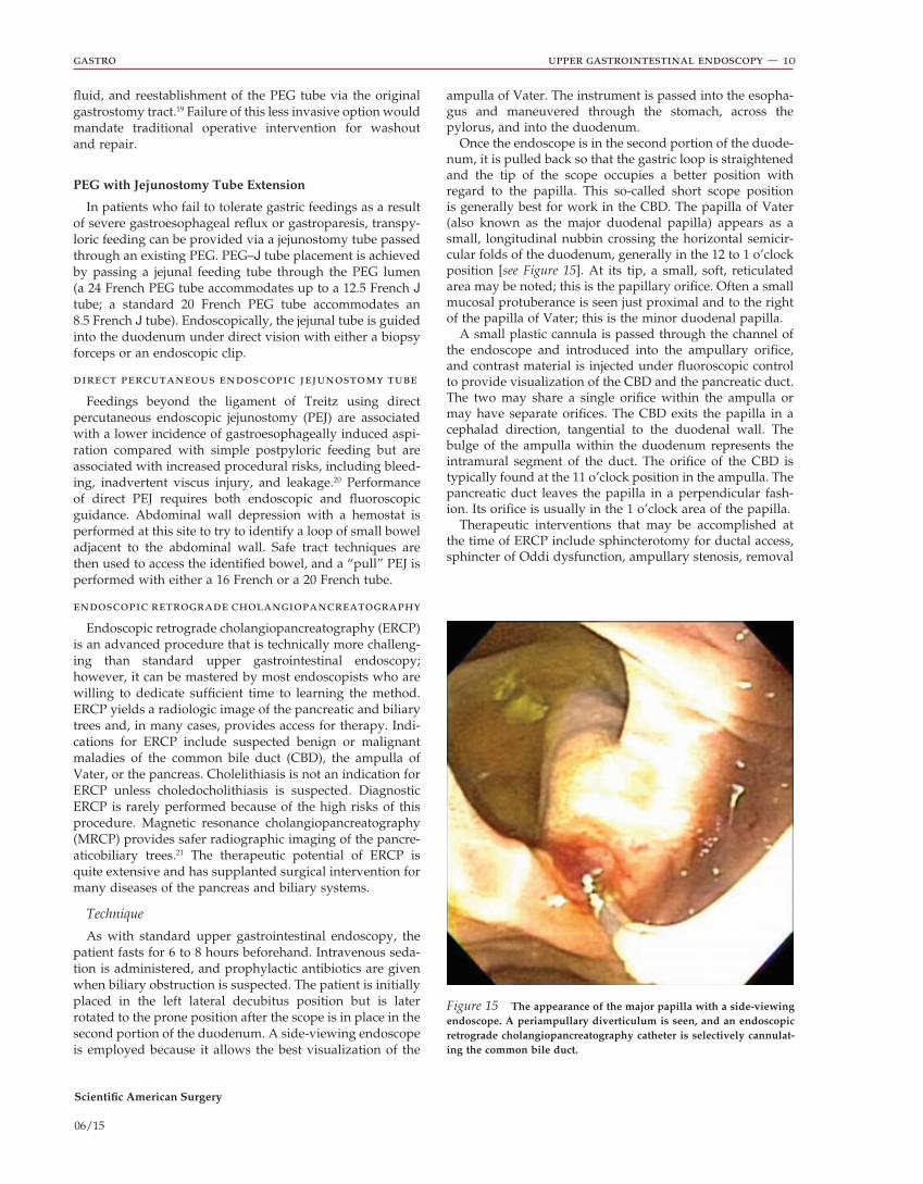
once the endoscope is in the second portion of the duode-num, it is pulled back so that the gastric loop is straightened and the tip of the scope occupies a better position with regard to the papilla. this so-called short scope position is generally best for work in the cBd. the papilla of Vater (also known as the major duodenal papilla) appears as a small, longitudinal nubbin crossing the horizontal semicir-cular folds of the duodenum, generally in the 12 to 1 o’clock position [see Figure 15]. at its tip, a small, soft, reticulated area may be noted; this is the papillary orifice. often a small mucosal protuberance is seen just proximal and to the right of the papilla of Vater; this is the minor duodenal papilla.
a small plastic cannula is passed through the channel of the endoscope and introduced into the ampullary orifice, and contrast material is injected under fluoroscopic control to provide visualization of the cBd and the pancreatic duct. the two may share a single orifice within the ampulla or may have separate orifices. the cBd exits the papilla in a cephalad direction, tangential to the duodenal wall. the bulge of the ampulla within the duodenum represents the intramural segment of the duct. the orifice of the cBd is typically found at the 11 o’clock position in the ampulla. the pancreatic duct leaves the papilla in a perpendicular fash-ion. its orifice is usually in the 1 o’clock area of the papilla.
therapeutic interventions that may be accomplished at the time of ercp include sphincterotomy for ductal access, sphincter of oddi dysfunction, ampullary stenosis, removal
Figure 15 The appearance of the major papilla with a side-viewing endoscope. A periampullary diverticulum is seen, and an endoscopic retrograde cholangiopancreatography catheter is selectively cannulat-ing the common bile duct.
Scientific American Surgery
06/15
gastro upper gastrointestinal endoscopy — 11
Bleeding may also occur with endoscopic sphincterotomy at the time of the procedure or in a delayed fashion up to 1 to 2 weeks later. it is usually controllable at the time of the procedure with balloon tamponade, injection sclerotherapy,
of cBd stones, dilation of benign and malignant biliary strictures, and insertion of stents to maintain ductal patency [see Figure 16]. pancreatic duct interventions include remov-al of stones, bridging of ductal disruptions, and drainage of pseudocysts.
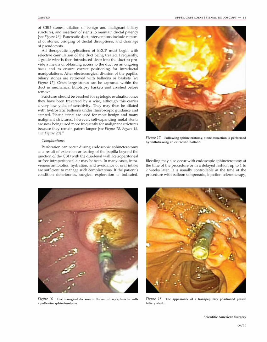
all therapeutic applications of ercp must begin with selective cannulation of the duct being treated. Frequently, a guide wire is then introduced deep into the duct to pro-vide a means of obtaining access to the duct on an ongoing basis and to ensure correct positioning for intraductal manipulations. after electrosurgical division of the papilla, biliary stones are retrieved with balloons or baskets [see Figure 17]. often large stones can be captured within the duct in mechanical lithotripsy baskets and crushed before removal.
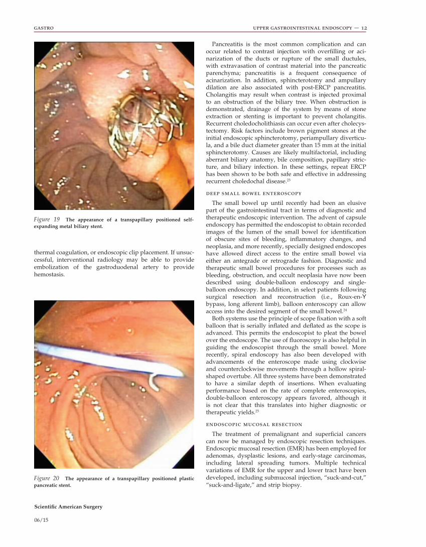
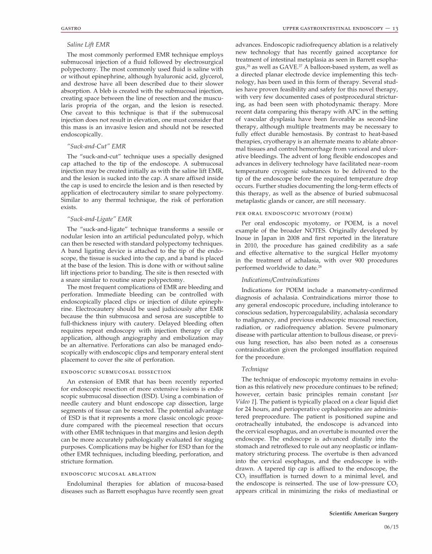
strictures should be brushed for cytologic evaluation once they have been traversed by a wire, although this carries a very low yield of sensitivity. they may then be dilated with hydrostatic balloons under fluoroscopic guidance and stented. plastic stents are used for most benign and many malignant strictures; however, self-expanding metal stents are now being used more frequently for malignant strictures because they remain patent longer [see Figure 18, Figure 19, and Figure 20].22
Complications
perforation can occur during endoscopic sphincterotomy as a result of extension or tearing of the papilla beyond the junction of the cBd with the duodenal wall. retroperitoneal or free intraperitoneal air may be seen. in many cases, intra-venous antibiotics, hydration, and avoidance of oral intake are sufficient to manage such complications. if the patient’s condition deteriorates, surgical exploration is indicated.
Figure 16 Electrosurgical division of the ampullary sphincter with a pull-wire sphincterotome.
Figure 17 Following sphincterotomy, stone extraction is performed by withdrawing an extraction balloon.
Figure 18 The appearance of a transpapillary positioned plastic biliary stent.
Scientific American Surgery
06/15
gastro upper gastrointestinal endoscopy — 12
pancreatitis is the most common complication and can occur related to contrast injection with overfilling or aci-narization of the ducts or rupture of the small ductules, with extravasation of contrast material into the pancreatic parenchyma; pancreatitis is a frequent consequence of acinarization. in addition, sphincterotomy and ampullary dilation are also associated with post-ercp pancreatitis. cholangitis may result when contrast is injected proximal to an obstruction of the biliary tree. When obstruction is demonstrated, drainage of the system by means of stone extraction or stenting is important to prevent cholangitis. recurrent choledocholithiasis can occur even after cholecys-tectomy. risk factors include brown pigment stones at the initial endoscopic sphincterotomy, periampullary diverticu-la, and a bile duct diameter greater than 15 mm at the initial sphincterotomy. causes are likely multifactorial, including aberrant biliary anatomy, bile composition, papillary stric-ture, and biliary infection. in these settings, repeat ercp has been shown to be both safe and effective in addressing recurrent choledochal disease.23
deep small bowel enteroscopy
the small bowel up until recently had been an elusive part of the gastrointestinal tract in terms of diagnostic and therapeutic endoscopic intervention. the advent of capsule endoscopy has permitted the endoscopist to obtain recorded images of the lumen of the small bowel for identification of obscure sites of bleeding, inflammatory changes, and neoplasia, and more recently, specially designed endoscopes have allowed direct access to the entire small bowel via either an antegrade or retrograde fashion. diagnostic and therapeutic small bowel procedures for processes such as bleeding, obstruction, and occult neoplasia have now been described using double-balloon endoscopy and single- balloon endoscopy. in addition, in select patients following surgical resection and reconstruction (i.e., roux-en-Y bypass, long afferent limb), balloon enteroscopy can allow access into the desired segment of the small bowel.24
Both systems use the principle of scope fixation with a soft balloon that is serially inflated and deflated as the scope is advanced. this permits the endoscopist to pleat the bowel over the endoscope. the use of fluoroscopy is also helpful in guiding the endoscopist through the small bowel. More recently, spiral endoscopy has also been developed with advancements of the enteroscope made using clockwise and counterclockwise movements through a hollow spiral-shaped overtube. all three systems have been demonstrated to have a similar depth of insertions. When evaluating performance based on the rate of complete enteroscopies, double-balloon enteroscopy appears favored, although it is not clear that this translates into higher diagnostic or therapeutic yields.25
endoscopic mucosal resection
the treatment of premalignant and superficial cancers can now be managed by endoscopic resection techniques. endoscopic mucosal resection (eMr) has been employed for adenomas, dysplastic lesions, and early-stage carcinomas, including lateral spreading tumors. Multiple technical variations of eMr for the upper and lower tract have been developed, including submucosal injection, “suck-and-cut,” “suck-and-ligate,” and strip biopsy.
thermal coagulation, or endoscopic clip placement. if unsuc-cessful, interventional radiology may be able to provide embolization of the gastroduodenal artery to provide hemostasis.
Figure 20 The appearance of a transpapillary positioned plastic pancreatic stent.
Figure 19 The appearance of a transpapillary positioned self- expanding metal biliary stent.
Scientific American Surgery
06/15
gastro upper gastrointestinal endoscopy — 13
Saline Lift EMR
the most commonly performed eMr technique employs submucosal injection of a fluid followed by electrosurgical polypectomy. the most commonly used fluid is saline with or without epinephrine, although hyaluronic acid, glycerol, and dextrose have all been described due to their slower absorption. a bleb is created with the submucosal injection, creating space between the line of resection and the muscu-laris propria of the organ, and the lesion is resected. one caveat to this technique is that if the submucosal injection does not result in elevation, one must consider that this mass is an invasive lesion and should not be resected endoscopically.
“Suck-and-Cut” EMR
the “suck-and-cut” technique uses a specially designed cap attached to the tip of the endoscope. a submucosal injection may be created initially as with the saline lift eMr, and the lesion is sucked into the cap. a snare affixed inside the cap is used to encircle the lesion and is then resected by application of electrocautery similar to snare polypectomy. similar to any thermal technique, the risk of perforation exists.
“Suck-and-Ligate” EMR
the “suck-and-ligate” technique transforms a sessile or nodular lesion into an artificial pedunculated polyp, which can then be resected with standard polypectomy techniques. a band ligating device is attached to the tip of the endo-scope, the tissue is sucked into the cap, and a band is placed at the base of the lesion. this is done with or without saline lift injections prior to banding. the site is then resected with a snare similar to routine snare polypectomy.
the most frequent complications of eMr are bleeding and perforation. immediate bleeding can be controlled with endoscopically placed clips or injection of dilute epineph-rine. electrocautery should be used judiciously after eMr because the thin submucosa and serosa are susceptible to full-thickness injury with cautery. delayed bleeding often requires repeat endoscopy with injection therapy or clip application, although angiography and embolization may be an alternative. perforations can also be managed endo-scopically with endoscopic clips and temporary enteral stent placement to cover the site of perforation.
endoscopic submucosal dissection
an extension of eMr that has been recently reported for endoscopic resection of more extensive lesions is endo-scopic submucosal dissection (esd). Using a combination of needle cautery and blunt endoscope cap dissection, large segments of tissue can be resected. the potential advantage of esd is that it represents a more classic oncologic proce-dure compared with the piecemeal resection that occurs with other eMr techniques in that margins and lesion depth can be more accurately pathologically evaluated for staging purposes. complications may be higher for esd than for the other eMr techniques, including bleeding, perforation, and stricture formation.
endoscopic mucosal ablation
endoluminal therapies for ablation of mucosa-based diseases such as Barrett esophagus have recently seen great
advances. endoscopic radiofrequency ablation is a relatively new technology that has recently gained acceptance for treatment of intestinal metaplasia as seen in Barrett esopha-gus,26 as well as GaVe.27 a balloon-based system, as well as a directed planar electrode device implementing this tech-nology, has been used in this form of therapy. several stud-ies have proven feasibility and safety for this novel therapy, with very few documented cases of postprocedural strictur-ing, as had been seen with photodynamic therapy. More recent data comparing this therapy with apc in the setting of vascular dysplasia have been favorable as second-line therapy, although multiple treatments may be necessary to fully effect durable hemostasis. By contrast to heat-based therapies, cryotherapy is an alternate means to ablate abnor-mal tissues and control hemorrhage from variceal and ulcer-ative bleedings. the advent of long flexible endoscopes and advances in delivery technology have facilitated near–room temperature cryogenic substances to be delivered to the tip of the endoscope before the required temperature drop occurs. Further studies documenting the long-term effects of this therapy, as well as the absence of buried submucosal metaplastic glands or cancer, are still necessary.
per oral endoscopic myotomy (poem)
per oral endoscopic myotomy, or poeM, is a novel example of the broader notes. originally developed by inoue in Japan in 2008 and first reported in the literature in 2010, the procedure has gained credibility as a safe and effective alternative to the surgical Heller myotomy in the treatment of achalasia, with over 900 procedures performed worldwide to date.28
Indications/Contraindications
indications for poeM include a manometry-confirmed diagnosis of achalasia. contraindications mirror those to any general endoscopic procedure, including intolerance to conscious sedation, hypercoagulability, achalasia secondary to malignancy, and previous endoscopic mucosal resection, radiation, or radiofrequency ablation. severe pulmonary disease with particular attention to bullous disease, or previ-ous lung resection, has also been noted as a consensus contraindication given the prolonged insufflation required for the procedure.
Technique
the technique of endoscopic myotomy remains in evolu-tion as this relatively new procedure continues to be refined; however, certain basic principles remain constant [see Video 1]. the patient is typically placed on a clear liquid diet for 24 hours, and perioperative cephalosporins are adminis-tered preprocedure. the patient is positioned supine and orotracheally intubated, the endoscope is advanced into the cervical esophagus, and an overtube is mounted over the endoscope. the endoscope is advanced distally into the stomach and retroflexed to rule out any neoplastic or inflam-matory stricturing process. the overtube is then advanced into the cervical esophagus, and the endoscope is with-drawn. a tapered tip cap is affixed to the endoscope, the co2 insufflation is turned down to a minimal level, and the endoscope is reinserted. the use of low-pressure co2 appears critical in minimizing the risks of mediastinal or
Scientific American Surgery
06/15
gastro upper gastrointestinal endoscopy — 14
subcutaneous emphysema and pneumothorax, which were reported in earlier series using air insufflation.29
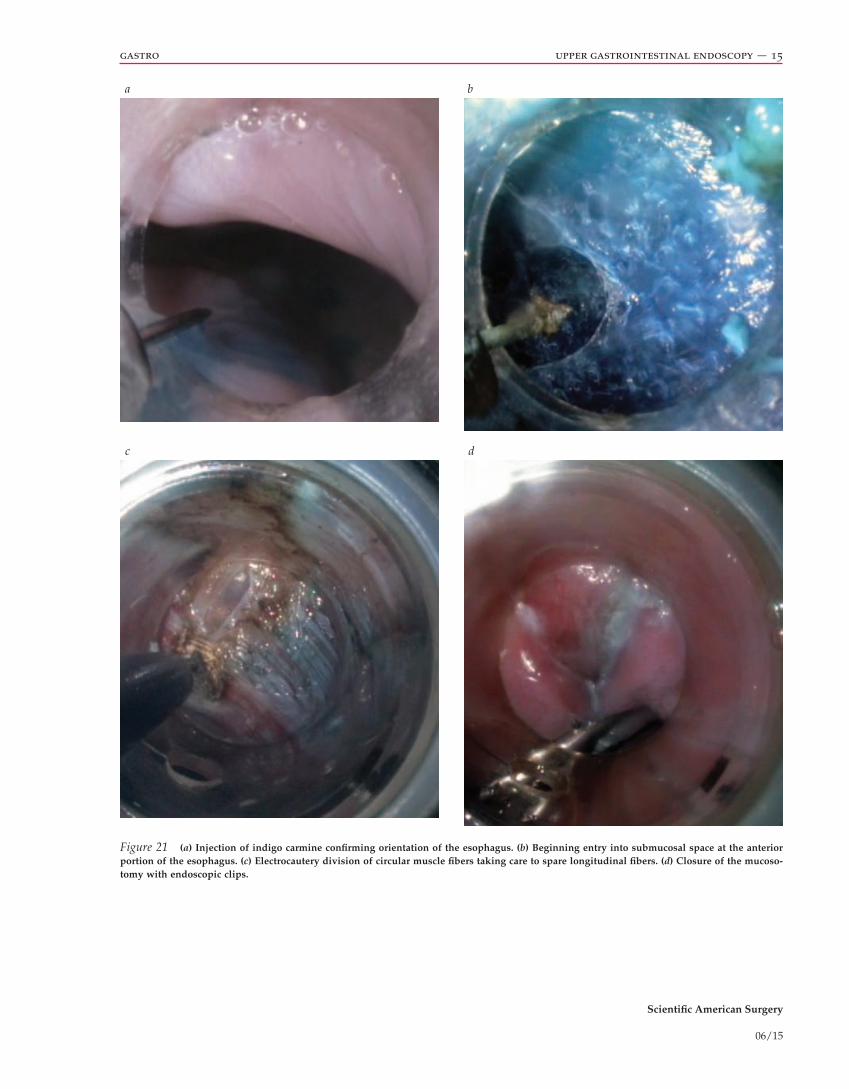
Submucosal tunnel creation at this point, we instill indigo carmine solution and use gravity to confirm the posterior segment of the esophagus [see Figure 21a]. Using a triangle-tip knife, a 2 cm longitudinal mucosectomy is created, typically starting 14 cm from the gastroesophageal junction. the anterior portion of the esophagus is then injected with 10 ml of an indigo carmine solution with epinephrine, and the submucosal space is entered [see Figure 21b]. the endoscope tapered tip cap is then exchanged for an oblique tipped cap. Using electrocautery on the triangle-tip knife, the submucosal space is then dissected distally, and this dissection plane is carried out onto the stomach, typically 4 cm past the gastroesophageal junction and onto the gastric cardia. indigo carmine is periodically injected during this time to delineate the submucosal space. the endoscope is then returned to the native lumen and retroflexed to confirm adequate distal dissection onto the gastric wall.
Esophageal and cardiomyotomy at the inferior aspect of this submucosal plane, the myotomy is started and car-ried across the lower esophageal sphincter onto the gastric wall, dividing the circular fibers, but with particular atten-tion to preserve the longitudinal fibers as much as possible [see Figure 21c]. the endoscope is then returned to the native gastric lumen, and smooth passage of the endoscope across the gastroesophageal junction, in conjunction with observed patulousness of the lower esophageal sphincter, and blanched gastric mucosa on retroflexion confirm adequate myotomy.
Mucosectomy closure the endoscope is then returned to the submucosal tunnel, and a bacitracin solution is injected into the tunnel. the mucosotomy is then closed using endoclips [see Figure 21d], the overtube is carefully withdrawn, and the mucosal tissues are again evaluated to confirm no evidence of injury and adequate hemostasis.
Perioperative complications complications associated with poeM procedures are relatively infrequent but include full-thickness mucosal injuries to the esophagus and stom-ach, which can be repaired at the time of the procedure with endoscopic techniques. pleural effusions and delayed hem-orrhage have also been described, although they appear to be either relatively rare or self-resolving with conservative management.
endoscopic ultrasonography
the 1980s saw the introduction of endoscopic ultrasonog-raphy (eUs). extracavitary ultrasonographic methods have been hampered by the presence of air within the gas-trointestinal tract, which precludes high-resolution imaging. consequently, they had been relegated to gross estimates of disease and detection of displacement of other tissues or fluid accumulation proximal to stenoses, such as ductal dilation in patients with cBd stones.
three advances have proven invaluable in allowing eUs to carve out a niche in the field of gastrointestinal diagnosis. First is the improvement in endoscopes that allows trans-ducer and receiver channels to traverse a tortuous path. second is the development of multiple-frequency options in conjunction with circumferential visualization. Higher frequencies provide higher resolutions, allowing useful differentiation of the various layers of the intestinal tract.
Video 1 Peroral endoscopic myotomy.
Scientific American Surgery
06/15
gastro upper gastrointestinal endoscopy — 15
a b
c d
Figure 21 (a) Injection of indigo carmine confirming orientation of the esophagus. (b) Beginning entry into submucosal space at the anterior portion of the esophagus. (c) Electrocautery division of circular muscle fibers taking care to spare longitudinal fibers. (d) Closure of the mucoso-tomy with endoscopic clips.
Scientific American Surgery
06/15
gastro upper gastrointestinal endoscopy — 16
the benefits that can be gained from combining imaging techniques in appropriate circumstances.
Future of Endoscopy
as endoscopic tools become increasingly sophisticated, so too will applications aimed at addressing increasingly complex surgical problems.33 poeM is perhaps the most prominent example of an idea over a decade in development now resulting in a viable intraluminal alternative to a laparoscopic procedure. the lessons learned in developing submucosal tunneling and endoscopic resection are now finding broader applicability as surgical endoscopists attempt resection of full-thickness lesions. already case series are reporting removal of gastrointestinal stromal tumors, leiomyomas,34,35 colonic adenomas,36 and even segments of Barrett esophagus.37 recently, the first case of nonexposed endoscopic wall inversion surgery (neWs) described the inversion of a gastric lesion laparoscopically into the lumen of the stomach to facilitate an endoluminal full-thickness resection and sentinel node basin dissection, removing the sample perorally.38 earlier on in the develop-ment process are feasibility studies investigating transanal total mesorectal excision for low rectal cancers, which has already been attempted in a small series of patients. together these reports provide a hopeful future for the expansion of endoscopic technique in oncologic resections.
as the array of interventions available to the surgical endoscopist continues to evolve and expand, the need for training a future generation of surgeons facile with these techniques has become increasingly pressing. the era of work-hour restrictions and the expansion of tools and tech-niques across other swaths of general surgery have placed further demands on the limited time of surgical trainees. recently, a surgical endoscopy curriculum was implemented by the american Board of surgery to codify and standardize a set of fundamental endoscopic skills so that surgical grad-uates will have a platform on which to further develop and innovate in this exciting and still nascent field of minimally invasive surgery.
Financial Disclosures: Jeffrey Marks, MD, FACS, is a consultant for US Endosur-gery, Apollo Surgery, GI Supply, and Olympus. Hahn Soe-Lin, MD, has no relevant financial relationships to disclose. This review was previously authored by Jeffrey L. Ponsky, MD, FACS, with disclosure made at the time of initial publication. This review has been reviewed, updated, and rereleased by the authors listed.
References
1. ponsky Jl. endoluminal surgery: past, present and future. surg endosc 2006;20 suppl 2:s500–2.
2. pearl Jp, Marks JM. new technology in endoscopy. in: soper nJ, editor. Mastery of endoscopic and laparoscopic surgery. 2nd ed. philadelphia: lippincott Williams & Wilkins; 2009. p. 17–23.
3. sharma p, Hawes rH, Bansal a, et al. standard endoscopy with random biopsies versus narrow band imaging targeted biopsies in Barrett’s oesophagus: a prospective, international, randomised controlled trial. Gut 2013;62: 15–21.
4. Hatta W, Uno K, Koike t, et al. a prospective comparative study of optical coherence tomography and eUs for tumor
third is the evolution of treatment protocols keyed to the accurate staging of tumors—information that is sometimes unobtainable from other imaging techniques.30
this technology has now been firmly established as an accurate way to identify carcinoma. More recent develop-ments are allowing eUs to expand from the field of diagno-sis into the realm of intervention. examples of eUs-guided procedures include fine-needle aspiration, lymph node sampling, and drainage of pancreatic pseudocysts.31
eUs devices come in both linear and radial transducers.32 radial transducers have the advantage of providing circum-ferential visualization that parallels the standard modes of perceiving the gastrointestinal tract. linear images allow eUs-directed biopsies and have the potential to provide color and pulsed doppler imaging. probes can be mounted on the top of an oblique-viewing fiber-optic scope or come in an over-the-wire format for use in the pancreaticobiliary tree. a series of frequencies are available, with higher fre-quencies providing greater resolution but less tissue depth penetration. lower-frequency probes allow deeper tissue assessment and a broader view, but at the price of reduced resolution. nevertheless, any form of eUs will provide better resolution than transcutaneous ultrasonography, allowing markedly improved two-point discrimination and hence more accurate tissue diagnosis.
the benefits of accurate staging of gastrointestinal tumors paved the way for eUs development. tissue sampling techniques are further benefited by this technology. the sensitivity of eUs makes it one of the best modalities for the evaluation and detection of pancreatic tumors. its sensitivi-ty, which is in excess of 95%, contrasts favorably with those of other modalities, including ultrasonography (75%), com-puted tomography (ct) (80%), and angiography (89%). the accuracy of t staging by eUs in esophageal cancer (80 to 90%) is greater than that of staging determined by ct (50 to 60%). this finding has led to the development of several staging schemes that are based solely on eUs findings. eUs has established a role in the identification of early pancreati-tis; the detection of cBd stones and mediastinal masses; and the assessment of anastomotic strictures, thickened gastric folds, and the integrity of the anal sphincter. it has also proved a useful adjunct in the determination of whether a tumor is amenable to eMr techniques or is better served by adjuvant therapies or surgical interventions.
the sensitivity of eUs is rooted in its ability to delineate the various layers of the alimentary canal. experienced endoscopists can easily evaluate the submucosa and differ-entiate intramural from extrinsic masses. characteristic pat-terns are readily learned and rapidly recognized, obviating tissue diagnoses in straightforward cases. criteria have also been established to aid in the differentiation of benign and malignant lesions. With the continued use of this technique, additional algorithms will be established in conjunction with more innovative interventional adjuncts. However, two lim-itations have caused many practitioners to remain skeptical: cost and training issues. other imaging modalities, such as ct and magnetic resonance imaging, have also made tre-mendous strides recently. although these various modalities are often considered competitors—a view arising from the perceived need for a single imaging modality—the issue of which is superior to the others pales in comparison with
Scientific American Surgery
06/15
gastro upper gastrointestinal endoscopy — 17
staging of superficial esophageal squamous cell carcinoma. Gastrointest endosc 2012;76:548–55.
5. lovat lB, Johnson K, Mackenzie Gd, et al. elastic scattering spectroscopy accurately detects high grade dysplasia and cancer in Barrett’s oesophagus. Gut 2006;55:1078–83.
6. Boerwinkel dF, Holz Ja, Kara Ma, et al. effects of autofluorescence imaging on detection and treatment of early neoplasia in patients with Barrett’s esophagus. clin Gastroenterol Hepatol 2014;12:774–81.
7. almond lM, Hutchings J, lloyd G, et al. endoscopic raman spectroscopy enables objective diagnosis of dysplasia in Barrett’s esophagus. Gastrointest endosc 2014;79:37–45.
8. Bergholt Ms, Zheng W, Ho Ky, et al. Fiberoptic confocal raman spectroscopy for real-time in vivo diagnosis of dysplasia in Barrett’s esophagus. Gastroenterology 2014; 146:27–32.
9. Qadeer Ma, rocio lopez a, dumot Ja, et al. risk factors for hypoxemia during ambulatory gastrointestinal endos-copy in asa i-ii patients. dig dis sci 2009;54:1035–40.
10. tang sJ, lee sy, Hynan ls, et al. endoscopic hemostasis in nonvariceal upper gastrointestinal bleeding: comparison of physician practice in the east and the west. dig dis sci 2009;54:2418–26.
11. liou tc, lin sc, Wang Hy, chang WH. optimal injection volume of epinephrine for endoscopic treatment of peptic ulcer bleeding. World J Gastroenterol 2006;12:3108–13.
12. Jaspersen d, schwacha H, sauer B, et al. complications of endoscopic sclerotherapy of esophageal varices. leber Magen darm 1995;25:171–4.
13. Zepeda-Gómez s, Marcon ne. endoscopic band ligation for nonvariceal bleeding: a review. can J Gastroenterol 2008; 22:748–52.
14. ramirez Fc, colon VJ, landan d, et al. the effects of the number of rubber bands placed at each endoscopic session upon variceal outcomes: a prospective, randomized study. am J Gastroenterol 2007;102:1372–6.
15. yuan y, Wang c, Hunt rH. endoscopic clipping for acute nonvariceal upper-Gi bleeding: a meta-analysis and critical appraisal of randomized controlled trials. Gastrointest endosc 2008;68:339–51.
16. Babor r, talbot M, tyndal a. treatment of upper gastroin-testinal leaks with a removable, covered, self-expanding metallic stent. surg laparosc endosc percutan tech 2009; 19:e1–4.
17. Huang Q, dai dK, Qian XJ, et al. treatment of gastric outlet and duodenal obstructions with uncovered expandable metal stents. World J Gastroenterol 2007;13:5376–9.
18. prasad Ga, reddy JG, Boyd-enders Ft, et al. predictors of recurrent esophageal food impaction: a case-control study. J clin Gastroenterol 2008;42:771–5.
19. Marks J, ponsky J, pearl J, et al. peG “rescue”: a practical notes technique. surg endosc 2007;21:816–9.
20. delegge MH. small bowel endoscopic enteral access. Gastrointest endosc clin n am 2007;17:663–86.
21. scaffidi MG, luigiano c, consolo p, et al. Magnetic resonance cholangiopancreatography versus endoscopic retrograde cholangiopancreatography in the diagnosis of common bile duct stones: a prospective comparative study. Minerva Med 2009;100:341–8.
22. perdue dG, Freeman Ml, disario Ja, et al. plastic versus self-expanding metallic stents for malignant hilar biliary
obstruction: a prospective multicenter observational cohort study. J clin Gastroenterol 2008;42:1040–6.
23. sugiyama M, suzuki y, abe n, et al. endoscopic retreat-ment of recurrent choledocholithiasis alter sphincterotomy. Gut 2004;53:1856–9.
24. pohl J, May a, aschmoneit i, et al. double-balloon endoscopy for retrograde cholangiography in patients with choledochojejunostomy and roux-en-y reconstruction. Z Gastroenterol 2009;47:215–9.
25. Jeon sr, Kim Jo. deep enteroscopy: which technique will survive? clin endosc 2013;46:480–5.
26. Fleischer de, overholt BF, sharma VK, et al. endoscopic ablation of Barrett’s esophagus: a multicenter study with 2.5-year follow-up. Gastrointest endosc 2008;68:867–76.
27. McGorisk t, Krishnan K, Keffer l, Komanduri s. radiofre-quency ablation for refractory gastric antral vascular ectasia (with video). Gastrointest endosc 2013;78:584–8.
28. stavropoulos sn, Modayil rJ, Friedel d, savides t. the international per oral endoscopic Myotomy survey (ipoeMs): a snapshot of the global poeM experience. surg endosc 2013;27:3322–38.
29. ren Z, Zhong y, Zhou p, et al. perioperative management and treatment for complications during and after peroral endoscopic myotomy (poeM) for esophageal achalasia (ea) (data from 119 cases). surg endosc 2012;26:3267–72.
30. Hunt Gc, Faigel do. assessment of eUs for diagnosing, staging, and determining resectability of pancreatic cancer: a review. Gastrointest endosc 2002;55:232–7.
31. park dH, lee ss, Moon sH, et al. endoscopic ultrasound-guided versus conventional transmural drainage for pancreatic pseudocysts: a prospective randomized trial. endoscopy 2009;41:842–8.
32. siemsen M, svendsen lB, Knigge U, et al. a prospective randomized comparison of curved array and radial echoen-doscopy in patients with esophageal cancer. Gastrointest endosc 2003;58:671–6.
33. pearl Jp, Marks JM, ponsky Jl. Hybrid surgery: combined laparoscopy and natural orifice surgery. Gastrointest endosc clin n am 2008;18:325–32.
34. lu J, lu X, Jiao t, Zheng M. endoscopic management of upper gastrointestinal submucosal tumors arising from muscularis propria. J clin Gastroenterol 2014;48:667–73.
35. Huang ly, cui J, Wu cr, et al. endoscopic full-thickness resection and laparoscopic surgery for treatment of gastric stromal tumors. World J Gastroenterol 2014;20:8253–9.
36. Valli pV, Kaufmann M, Vrugt B, Bauerfeind p. endoscopic resection of a diverticulum-arisen colonic adenoma using a full thickness resection device. Gastroenterology 2014 aug 2. [epub ahead of print]
37. chennat J, Konda VJ, ross as, et al. complete Barrett’s eradication endoscopic mucosal resection: an effective treat-ment modality for high-grade dysplasia and intramucosal carcinoma—an american single-center experience. am J Gastroenterol 2009;104:2684–92.
38. Goto o, takeuchi H, Kawakubo H, et al. First case of non-exposed endoscopic wall-inversion surgery with sentinel node basin dissection for early gastric cancer. Gastric cancer 2014 aug 3. [epub ahead of print]
Acknowledgment
Figure 14 tom Moore