Embed Size (px)
Citation preview
Research Article
GATA transcription factors, SOX17 and TFAP2C, drive thehuman germ-cell specification programYoji Kojima1,2,3 , Chika Yamashiro1,2 , Yusuke Murase1,2, Yukihiro Yabuta1,2, Ikuhiro Okamoto1,2, Chizuru Iwatani4,Hideaki Tsuchiya4, Masataka Nakaya1,4, Tomoyuki Tsukiyama1,4 , Tomonori Nakamura1,2,7 , Takuya Yamamoto1,3,5,6,Mitinori Saitou1,2,3
The in vitro reconstitution of human germ-cell developmentprovides a robust framework for clarifying key underlyingmechanisms. Here, we explored transcription factors (TFs) thatengender the germ-cell fate in their pluripotent precursors.Unexpectedly, SOX17, TFAP2C, and BLIMP1, which act under theBMP signaling and are indispensable for human primordial germ-cell-like cell (hPGCLC) specification, failed to induce hPGCLCs.In contrast, GATA3 or GATA2, immediate BMP effectors, combinedwith SOX17 and TFAP2C, generated hPGCLCs. GATA3/GATA2knockouts dose-dependently impaired BMP-induced hPGCLCspecification, whereas GATA3/GATA2 expression remainedunaffected in SOX17, TFAP2C, or BLIMP1 knockouts. In cyn-omolgus monkeys, a key model for human development,GATA3, SOX17, and TFAP2C were co-expressed exclusively inearly PGCs. Crucially, the TF-induced hPGCLCs acquired ahallmark of bona fide hPGCs to undergo epigenetic reprog-ramming and mature into oogonia/gonocytes in xenogeneicreconstituted ovaries. By uncovering a TF circuitry drivingthe germ line program, our study provides a paradigm for TF-based human gametogenesis.
DOI 10.26508/lsa.202000974 | Received 27 November 2020 | Revised 7 January2021 | Accepted 5 February 2021 | Published online 19 February 2021
Introduction
Germ cells are the carriers of genetic as well as epigenetic infor-mation into new individuals, and thus serve as an enduring linkbetween generations. Accordingly, they bear a capacity to replicategenetic information with high fidelity (1, 2, 3). On the other hand,they also create genetic and epigenetic diversity through meioticrecombination and epigenetic reprogramming/programming, re-spectively, providing a driving force for evolution (4, 5). Anomalies in
such processes often lead to diseased states, including infertilityand genetic/epigenetic disorders of offspring. Therefore, in-vestigations into the mechanism of germ-cell development notonly promote our understanding of fundamental principles ofheredity and evolution but also provide salient information re-garding the etiology of critical diseases.
Despite such importance, investigations into human germ-celldevelopment have been limited because of the difficulties inaccessing relevant experimental materials and ethical restrictions.Notably, recent advancements in the in vitro reconstitution ofhuman germ-cell development using human pluripotent stem cells(hPSCs), including embryonic stem cells (hESCs) and inducedpluripotent stem cells (hiPSCs), have created novel opportunitiesfor such studies, permitting investigations into the mechanisms ofhuman germ-cell development as an emerging frontier in repro-ductive biology/medicine (6, 7). Accordingly, hPSCs are induced into cellsbearing properties similar to human primordial germ cells (hPGCs) (8, 9),the founding population of the human germ-cell lineage that eventuallygives rise to either spermatozoa or oocytes. The induced hPGC-like cells(hPGCLCs) are further differentiated into oogonia/early oocyte-like cellswith appropriate epigenetic reprogramming in a reconstituted ovaryculture (10, 11), or into pro-spermatogonia-like cells in a reconstitutedtestis culture (12). Although further reconstitution of human germ-celldevelopment remains a key challenge, these advances recapitulate aperiod ofmore than 10wk of human germ-cell development, leading to anumber of key findings with regard to the mechanism of human germ-cell development ingeneral, andgerm-cell specification inparticular (8, 9,13, 14, 15, 16, 17).
In the case of germ-cell specification, humans as well as non-human primates such as cynomolgus monkeys (Macaca fas-cicularis), use transcriptional and signaling programs evolutionarilydistinct from those in mice, which have long been a paradigmfor mammalian development (8, 9, 13, 18, 19). Specifically, inhumans, WNT signaling induces EOMES, which, together with bone
1Institute for the Advanced Study of Human Biology (ASHBi), Kyoto University, Yoshida-Konoe-cho, Kyoto, Japan 2Department of Anatomy and Cell Biology, GraduateSchool of Medicine, Kyoto University, Yoshida-Konoe-cho, Kyoto, Japan 3Center for iPS Cell Research and Application (CiRA), Kyoto University, Shogoin-Kawahara-cho,Kyoto, Japan 4Research Center for Animal Life Science, Shiga University of Medical Science, Seta-Tsukinowa-cho, Otsu, Japan 5AMED-CREST, AMED, Tokyo, Japan6Medical-Risk Avoidance Based on iPS Cells Team, RIKEN Center for Advanced Intelligence Project (AIP), Kyoto, Japan 7The Hakubi Center for Advanced Research, KyotoUniversity, Yoshida-Konoe-cho, Kyoto, Japan
Correspondence: [email protected]; [email protected]
© 2021 Kojima et al. https://doi.org/10.26508/lsa.202000974 vol 4 | no 5 | e202000974 1 of 24
on 15 January, 2022life-science-alliance.org Downloaded from http://doi.org/10.26508/lsa.202000974Published Online: 19 February, 2021 | Supp Info:
morphogenetic protein 4 (BMP4) signaling, induces SOX17 as one ofthe most upstream transcription factors (TFs) for hPGC(LC)specification (13). SOX17 is essential for the expression of keydownstream genes, including BLIMP1, and for activating othergerm-cell specification programs (8, 13). TFAP2C also serves as a keyupstream TF that functions in parallel and in an interdependentfashion with SOX17 and is critical for the repression of somaticprograms (13, 15). Such programs for germ-cell specification appearto be relatively well conserved in cynomolgus monkeys (18, 19). Incontrast, in mice, Sox17 has no role in germ-cell specification (20),and BMP4 signaling activates endogenous WNT signaling that inturn induces T (T has no role in humans (13)), which up-regulatesBlimp1 and Prdm14, two of the most upstream TFs for germ-cellspecification (21, 22, 23). Blimp1, Prdm14, and Tfap2c are essentialand sufficient for the global control of downstream programs,including by reactivating pluripotency programs, repressing so-matic programs, and initiating epigenetic reprogramming (22, 23, 24,25). These findings demonstrate that the TFs and TF hierarchiesinvolved in conferring the germ-cell fate in humans are distinctfrom those in mice, highlighting the importance of further pro-moting human germ-cell biology.
In regard to the mechanism of human germ-cell specification, afundamental question remains to be answered: That is, which TFs orTF combinations are sufficient to give rise to the germ-cell fate intheir precursors? The answer to this question could help establish afoundation for TF-based human gametogenesis. In mice, three TFs(Blimp1, Prdm14, and Tfac2c), and to a lesser extent, two TFs (Blimp1and Tfap2c; Prdm14 and Tfap2c) or a single TF (Prdm14), are suf-ficient to confer the germ-cell fate to their precursors, and such TF-induced mouse PGCLCs (mPGCLCs) contribute to spermatogenesis(25). We therefore set out to define the TFs that replace the BMP4signaling and are sufficient to establish the identity of hPGCs ontheir precursors. Unexpectedly, we found that three TFs that areessential for hPGCLC specification—that is, SOX17, TFAP2C, andBLIMP1—are nonetheless not sufficient, and in contrast, the GATAfamily of TFs, combined with SOX17 and TFAP2C, drives the hPGCLCprogram.
Results
SOX17, TFAP2C, and BLIMP1 are not sufficient to generate hPGCLCs
For hPGCLC induction, hiPSCs are first induced into incipientmesoderm-like cells (iMeLCs) by stimulating with activin A and aWNT signal activator (CHIR99021) for 2 d, and iMeLCs are then in-duced into hPGCLCs by stimulating with bone morphogeneticprotein 4, together with proliferation/survival factors, includingstem cell factor (SCF), EGF, and leukemia inhibitory factor (LIF),under a floating aggregate condition (9, 13, 26). hPGCLCs that ex-press key genes such as SOX17, TFAP2C, BLIMP1, and NANOS3 areinduced as early as day 2 of induction (d2 hPGCLCs), show a pro-gressive maturation, and persist at least until around d10 (9, 13, 26).
We set out to identify TFs that are sufficient to confer the germ-cell fate on iMeLCs in the absence of BMP signaling. At the outset,we evaluated whether SOX17, TFAP2C, or BLIMP1, three TFs essential
for hPGCLC specification (8, 9, 13), would be sufficient to induce thegerm-cell fate when expressed either singly or in one of variouscombinations. For this purpose, hiPSCs bearing the BLIMP1-2A-tdTomato (BT) and TFAP2C-2A-EGFP (AG) alleles (585B1 BTAG (XY))(9) were transfected with piggyBac-based vectors expressing (i) thereverse tetracycline trans-activator (rtTA) under a constitutivelyactive promoter and (ii) the genes of interest (SOX17, TFAP2C, SOX17/TFAP2C, SOX17/BLIMP1, TFAP2C/BLIMP1, or SOX17/TFAP2C/BLIMP1)under the control of tetracycline regulatory elements with tran-scription termination by the rabbit β-globin poly A sequence(rBGpA), so that the genes of interest exhibited timed expression ina doxycycline (Dox)-dependent manner and could be distinguishedfrom the endogenous ones by the presence of rBGpA (Fig 1A). Foreach transfectant, we selected two clones that exhibited transgeneexpression levels in hiPSCs comparable with the correspondingendogenous gene expression levels in hPGCLCs (Figs 1B and S1A).The expression of the transgenes in a clone expressing SOX17/TFAP2C/BLIMP1 was confirmed with Western blotting (Fig S1B). Allthe hiPSC clones selected exhibited undifferentiated morphology.
We first examined the effects of the transgene expressionin hiPSCs cultured with Dox for 24 h. Quantitative Real Time-PCR(qRT-PCR) for the endogenous key loci (SOX17, TFAP2C, BLIMP1,and NANOS3) showed that no clones up-regulated SOX17, whereasthe SOX17, SOX17/TFAP2C, and SOX17/TFAP2C/BLIMP1 clones up-regulated TFAP2C mildly and BLIMP1 to an extent comparable withthat in d2 hPGCLCs (Fig 1B). The TFAP2C/BLIMP1, SOX17/TFAP2C, andSOX17/TFAP2C/BLIMP1 clones up-regulated endogenousNANOS3 toa level similar to that in d2 hPGCLCs (Fig 1B). Because the TFAP2Cclones had no impact on all these genes (TFAP2C appeared torepress endogenous TFAP2C) (Fig 1B), we excluded them from thesubsequent analyses.
We next analyzed whether the expression of these genes iniMeLCs might induce the germ-cell fate (Fig 1C). The iMeLCs inducedby activin A and CHIR99021 from all the clones bore a morphologyindistinguishable from that of the parental clone (Fig 1D). Upon d4of induction by BMP4 or BMP4 and Dox, iMeLC aggregates from allthe clones exhibited a distinct cluster of BT-positive (BT+) and AG-positive (AG+) cells, as revealed by observation under a fluores-cence dissection microscope or FACS (Fig 1E). We noted that theSOX17/TFAP2C clones stimulated by BMP4 and Dox, althoughforming small aggregates, differentiated into BT+AG+ cells at a veryhigh efficiency (~90%), whereas the other clones formed BT+AG+
cells with an efficiency of ~20–30% (Fig 1E and F). This might bebecause SOX17 and TFAP2C expression could be a rate-limitingevent for hPGCLC specification, and the Dox-induced expressionof SOX17 and TFAP2C would create a state highly competent forBMP-induced hPGCLC specification. In addition, the iMeLC aggre-gates of the SOX17, SOX17/TFAP2C, and TFAP2C/BLIMP1 clones be-came smaller when stimulated with BMP4 and Dox, which mighthave been due to a subtle but significant difference in the ex-pression levels of SOX17, TFAP2C, or BLIMP1 (e.g., Blimp1/BLIMP1 isknown to induce cell-cycle arrest in various contexts (27, 28)).
In contrast, with Dox stimulation alone, no iMeLC aggregatesshowed BT+AG+ cells (Fig 1E and F). Upon stimulation with Dox, theSOX17 clones induced weak BT+ cells, the SOX17/BLIMP1 clonesshowed no BTAG positivity, the TFAP2C/BLIMP1 clones generatedsmall aggregates with weak BTAG positivity (less than the threshold
Induction of human germ-cell fate Kojima et al. https://doi.org/10.26508/lsa.202000974 vol 4 | no 5 | e202000974 2 of 24
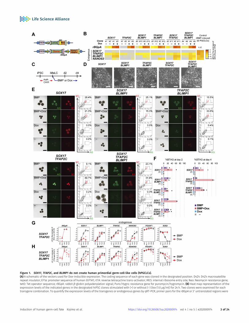
Figure 1. SOX17, TFAP2C, and BLIMP1 do not create human primordial germ-cell-like cells (hPGCLCs).(A) A schematic of the vectors used for Dox-inducible expression. The coding sequence of each gene was cloned in the designated position. D4Z4: D4Z4 macrosatelliterepeat insulator; EF1α: promoter sequence of human EEF1A1; rtTA: reverse tetracycline trans-activator; IRES: internal ribosome entry site; Neo: Neomycin resistance gene;tetO: Tet operator sequence; rBGpA: rabbit β-globin polyadenylation signal; Puro/Hygro: resistance gene for puromycin/hygromycin. (B) Heat map representation of theexpression levels of the indicated genes in the designated hiPSC clones stimulated with (+) or without (−) Dox (1.0 μg/ml) for 24 h. Two clones were examined for eachtransgene combination. To quantify the expression levels of the transgenes or endogenous genes by qRT-PCR, primer pairs for the rBGpA or 39 untranslated regions were
Induction of human germ-cell fate Kojima et al. https://doi.org/10.26508/lsa.202000974 vol 4 | no 5 | e202000974 3 of 24
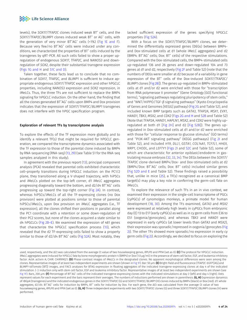
levels), the SOX17/TFAP2C clones induced weak BT+ cells, and theSOX17/TFAP2C/BLIMP1 clones induced weak BT+ or AG+ cells, withthe generation of very minor BT+AG+ cells (<1%) (Fig 1E and F).Because very few/no BT+AG+ cells were induced under any con-ditions, we characterized the properties of BT+ cells induced by thetransgenes by qRT-PCR. All such cells failed to show a proper up-regulation of endogenous SOX17, TFAP2C, and NANOS3 and down-regulation of SOX2, despite their substantial transgene expression(Figs 1G and H and S1C and D).
Taken together, these facts lead us to conclude that no com-bination of SOX17, TFAP2C, and BLIMP1 is sufficient to induce ap-propriate endogenous SOX17/TFAP2C expression and other hPGCLCproperties, including NANOS3 expression and SOX2 repression, iniMeLCs. Thus, the three TFs are not sufficient to replace the BMP4signaling for hPGCLC induction. On the other hand, the finding thatall the clones generated BT+AG+ cells upon BMP4 and Dox provisionindicates that the expression of SOX17/TFAP2C/BLIMP1 transgenesdoes not interfere with the hPGC specification program.
Exploration of relevant TFs by transcriptome analysis
To explore the effects of the TF expression more globally and toidentify a relevant TF(s) that might be required for hPGCLC gen-eration, we compared the transcriptome dynamics associated withthe TF expression to those of the parental clone induced by BMP4by RNA-sequencing (RNA-seq) (13, 29) (see Table S1 for the RNA-seqsamples analyzed in this study).
In agreement with the previous report (13), principal componentanalysis (PCA) revealed that parental cells exhibited characteristiccell–property transitions during hPGCLC induction: on the PC1/2plane, they transitioned along a V-shaped trajectory, with hiPSCsand iMeLCs plotted on the top-left corner, d1 iMeLC aggregatesprogressing diagonally toward the bottom, and d2/d4 BT+AG+ cellsprogressing up toward the top-right corner (Fig 2A). In contrast,whereas hiPSCs/iMeLCs of all the TF-expressing clones (no Doxprovision) were plotted at positions similar to those of parentalhiPSCs/iMeLCs, upon Dox provision on iMeLC aggregates (i.e., TFexpression), all the clones shifted their positions in parallel alongthe PC1 coordinate with a retention or some down-regulation oftheir PC2 scores, but none of the clones acquired a state similar tod4 hPGCLCs (Fig 2B–E). We examined the expression of 481 genesthat characterize the hPGCLC specification process (13), whichrevealed that the d2 TF-expressing cells failed to show a propertysimilar to those of d2/d4 BT+AG+ cells (Fig S2A). Most notably, they
lacked sufficient expression of the genes specifying hPGCLCproperties (Fig S2A).
With a focus on the SOX17/TFAP2C/BLIMP1 clones, we deter-mined the differentially expressed genes (DEGs) between BMP4-and Dox-stimulated cells at d1 (whole iMeLC aggregates) and d2(BMP4: BT+AG+ cells; Dox: BT+ cells) of the respective stimulations.Compared with the Dox-stimulated cells, the BMP4-stimulated cellsup-regulated 136 and 26 genes and down-regulated 104 and 73genes at d1 and d2, respectively (Fig 2F and Table S2) (note that thenumbers of DEGs were smaller at d2 because of a variability in geneexpression of the BT+ cells of the Dox-induced SOX17/TFAP2C/BLIMP1 clones [Fig 2B]). The genes up-regulated in BMP4-stimulatedcells at d1 and/or d2 were enriched with those for “transcriptionfrom RNA polymerase II promoter” (Gene Ontology [GO] functionalterms), “signaling pathways regulating pluripotency of stem cells,”and “WNT/HIPPO/TGF-β signaling pathways” (Kyoto Encyclopediaof Genes and Genomes [KEGG] pathway) (Fig 2G and Table S2), andincluded known BMP targets such as GATA3, TFAP2A, MSX1, EVX1,HAND1, TBX3,MSX2, and CDX2 (Figs 2G and H and S2B and Table S2)(Note that TFAP2A, HAND1, HAPLN1,MSX2, and CDX2were highly up-regulated at both d1 [Fig 2H] and d2 [Fig S2B]). The genes up-regulated in Dox-stimulated cells at d1 and/or d2 were enrichedwith those for “cellular response to glucose stimulus” (GO terms)and “PI3K-AKT signaling pathway” (KEGG pathways) (Fig 2I andTable S2), and included H19, DLL1, GSTA1, COL14A1, TCF7L1, HHEX,NRP1, CHODL, and LEFTY1 (Figs 2I and S2C and Table S2), some ofwhich are characteristic for anterior epiblast/endoderm in gas-trulatingmouse embryos (32, 33, 34). The DEGs between the SOX17/TFAP2C clone-derived BMP4/Dox- and Dox-stimulated cells at d2(BMP4/Dox: BT+AG+ cells; Dox: BT+ cells) included similar genes(Fig S2D and E and Table S2). These findings raised a possibilitythat, unlike in mice (25), a TF(s) recognized as a canonical BMPtarget(s) may play a key role in conferring the germ-cell fate oniMeLCs.
To explore the relevance of such TFs in an in vivo context, weexamined their expression in the single-cell transcriptome of PGCs(cyPGCs) of cynomolgus monkeys, a primate model for humandevelopment (18, 30). Among the TFs examined, GATA3 and MSX2were expressed at relatively high levels in cyPGCs from embryonicday (E) 13 to E17 (early cyPGCs) as well as in cy germ cells from E36 toE51 (oogonia/gonocytes), and whereas TBX3 and HAND1 wereexpressed in early cyPGCs (at lower levels than GATA3 and MSX2),their expression was sporadic/repressed in oogonia/gonocytes (Fig2J). The other TFs showed more sporadic/no expression in early cygerm cells (Fig 2J). Accordingly, we decided to focus on exploring the
used, respectively, and the ΔCt was calculated from the average Ct value of two housekeeping genes, RPLP0 and PPIA (set as 0). (C) The protocol for hPGCLC induction.iMeLC aggregates were induced for hPGCLC fate by bonemorphogenetic protein 4 (BMP4) or Dox (1.0 μg/ml) in the presence of stem cell factor, EGF, and leukemia inhibitoryfactor. ActA: activin A; CHIR: CHIR99021. (D) Phase-contrast images of iMeLCs in the designated clones. No apparent morphological differences were seen among theclones. Representative images of at least two independent experiments are shown (shown in Fig 1F). Bar: 50 μm. (E) Bright-field and fluorescence (TFAP2C-EGFP [AG] andBLIMP1-tdTomato [BT]) images, and FACS analyses for BTAG expression in floating aggregates of the indicated transgene-expressing clones at day 4 of the indicatedstimulation. (−): induction only with stem cell factor, EGF and leukemia inhibitory factor. Representative images of at least two independent experiments are shown (seeFig 1F). Bars, 200 μm. (F) Percentage of BT+AG+ cells of the indicated transgene-expressing clones with the indicated stimulations at day 2 (left) and day 4 (right). Dotsrepresent values for each experiment and the bars represent their averages. The numbers of inductions performed are shown in parenthesis. (G, H) Expression dynamicsof rBGpA (transgenes) and the indicated endogenous genes in the SOX17/TFAP2C (G) and SOX17/TFAP2C/BLIMP1 (H) clones induced by BMP4 (black) or Dox (red). d1: wholeaggregates; d2/d4: BT+AG+ cells for induction by BMP4, BT+ cells for induction by Dox. For each gene, the ΔCt was calculated from the average Ct value of twohousekeeping genes, RPLP0 and PPIA (set as 0). (G, H) Three independent experiments with two SOX17/TFAP2C clones (G) and three SOX17/TFAP2C/BLIMP1 clones (H) wereperformed.
Induction of human germ-cell fate Kojima et al. https://doi.org/10.26508/lsa.202000974 vol 4 | no 5 | e202000974 4 of 24
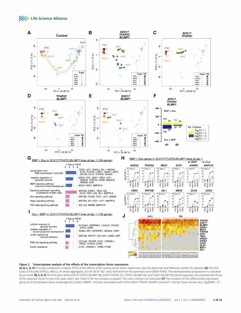
Figure 2. Transcriptome analysis of the effects of the transcription factor expression.(A, B, C, D, E) Principal component analysis (PCA) of the effects of the transcription factor expression (see the Materials and Methods section for details). (A) The PCAplots of the cells (hiPSCs, iMeLCs, d1 whole aggregates, d2/d4/d6 BT+AG+ cells) derived from the parental clone (585B1 BTAG). The developmental progression is indicatedby an arrow. (B, C, D, E) The PCA plots of the SOX17/TFAP2C/BLIMP1 (B), SOX17/TFAP2C (C), TFAP2C/BLIMP1 (D), and SOX17/BLIMP1 (E) clones (squares) are overlaid with thoseof the parental clone (circles with pale color). See Table S1 for the samples analyzed. The color coding is as indicated. (F) The numbers of the differentially expressedgenes at d1/d2 between bonemorphogenetic protein (BMP)– and Dox-stimulated cells of the SOX17/TFAP2C/BLIMP1 clones (P < 0.01 by Tukey–Kramer test, log2[RPM + 1] >
Induction of human germ-cell fate Kojima et al. https://doi.org/10.26508/lsa.202000974 vol 4 | no 5 | e202000974 5 of 24
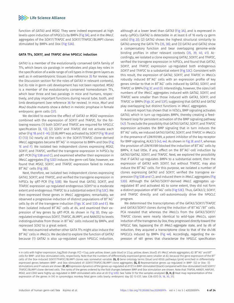
function of GATA3 and MSX2. They were indeed expressed at highlevels upon induction of hPGCLCs by BMP4 (Fig 3A), and in the iMeLCaggregates of the SOX17/TFAP2C and SOX17/TFAP2C/BLIMP1 clonesstimulated by BMP4 and Dox (Fig S3A).
GATA TFs, SOX17, and TFAP2C drive hPGCLC induction
GATA3 is a member of the evolutionarily conserved GATA family ofTFs, which bears six paralogs in vertebrates and plays key roles inthe specification of a wide range of cell types in three germ layers aswell as in extraembryonic tissues (see reference 35 for review; seethe Discussion section for the roles of GATA3 in relevant contexts),but its role in germ-cell development has not been reported. MSX2is a member of the evolutionarily conserved homeodomain TFs,which bear three and two paralogs in mice and humans, respec-tively, and play important functions during neural tube, tooth, andlimb development (see reference 36 for review). In mice, Msx1 andMsx2 double mutants show a defect in meiotic prophase in femaleembryonic germ cells (37).
We decided to examine the effect of GATA3 or MSX2 expressioncombined with the expression of SOX17 and TFAP2C, for the fol-lowing reasons: (1) both SOX17 and TFAP2C are required for hPGCLCspecification (8, 13); (2) SOX17 and TFAP2C did not activate eachother (Fig 1B and E–H); (3) BLIMP1 was activated by SOX17 (Fig 1B andE) (13); (4) nearly all the cells in the SOX17/TFAP2C clone-derivediMeLC aggregates became BT+AG+ in response to BMP4 and Dox (Fig1E and F). We isolated two independent clones expressing MSX2,SOX17, and TFAP2C, verified the transgene expression in hiPSCs byqRT-PCR (Fig S3B and C), and examined whether their expression iniMeLC aggregates (Fig S3D) induces the germ-cell fate; however, wefound that MSX2, SOX17, and TFAP2C expression failed to induceBT+AG+ cells (Fig 3B).
Next, therefore, we isolated two independent clones expressingGATA3, SOX17, and TFAP2C, and verified the transgene expression inhiPSCs by qRT-PCR (Fig S3B). We found that GATA3, SOX17, andTFAP2C expression up-regulated endogenous SOX17 to a moderateextent and endogenous TFAP2C to a substantial extent (Fig S3C). Wethen expressed these genes in iMeLC aggregates; remarkably, weobserved a progressive induction of distinct populations of BT+AG+
cells by d4 of the transgene induction (Figs 3C and S3D and E). Wethen isolated induced BT+AG+ cells at d4, and examined their ex-pression of key genes by qRT-PCR. As shown in Fig 3D, they up-regulated endogenous SOX17, TFAP2C, BLIMP1, andNANOS3 to levelsindistinguishable from those in BT+AG+ cells induced by BMP4, andrepressed SOX2 to a great extent.
We next examined whether other GATA TFs might also induce theBT+AG+ cells in iMeLCs. We decided to explore the function of GATA2because (1) GATA2 is also up-regulated upon hPGCLC induction,
although at a lower level than GATA3 (Fig 3A), and is expressed inearly cyPGCs (GATA2 is detectable in at least 6 of 16 early cy germcells) (Fig 2J); (2) GATA2 shows the highest structural similarity toGATA3 among the GATA TFs (35, 38); and (3) GATA3 and GATA2 showa compensatory function and bear overlapping genome-widebinding profiles in other relevant contexts (35, 39, 40, 41). Ac-cordingly, we isolated a clone expressing GATA2, SOX17, and TFAP2C,verified the transgene expression in hiPSCs, and found that GATA2,SOX17, and TFAP2C expression up-regulated both endogenousSOX17 and TFAP2C to a substantial extent (Fig S3C). Consistent withthis result, the expression of GATA2, SOX17, and TFAP2C in iMeLCsrobustly induced BT+AG+ cells with an expression profile of keygenes similar to that in BT+AG+ cells induced by GATA3, SOX17, andTFAP2C or BMP4 (Fig 3C and D). Interestingly, however, the sizes/cellnumbers of the iMeLC aggregates induced with GATA2, SOX17, andTFAP2C were smaller than those induced with GATA3, SOX17, andTFAP2C or BMP4 (Figs 3C and S3F), suggesting that GATA3 and GATA2play overlapping but distinct functions in iMeLC aggregates.
A recent report has shown that in hESCs, BMP signaling activatesGATA3, which in turn up-regulates BMP4, thereby creating a feed-forward loop for persistent activation of the BMP signaling pathway(42). To exclude the possibility that GATA3/GATA2, SOX17, and TFAP2Cexpression activates the BMP signaling that in turn induces theBT+AG+ cells, we induced GATA3/GATA2, SOX17, and TFAP2C in iMeLCsin the presence of LDN193189, a potent inhibitor of the key receptorfor BMP signaling, ALK2/3 (43, 44). As shown in Fig 3E and F, whereasthe provision of LDN193189 blocked the induction of BT+AG+ cells byBMP4, it had little, if any, effect on the BT+AG+-cell induction byGATA3/GATA2, SOX17, and TFAP2C. Next, we explored the possibilitythat if GATA3 up-regulates BMP4 to a substantial extent, then theexpression of GATA3 with SOX17, but without TFAP2C, may alsoinduce the BT+AG+ cells. For this purpose, we isolated a number ofclones expressing GATA3 and SOX17, verified the transgene ex-pression (Fig S3B and C), and induced them in iMeLC aggregates (FigS3D). Although the GATA3/SOX17-derived iMeLC aggregates up-regulated BT and activated AG to some extent, they did not forma distinct population of BT+AG+ cells (Fig S3E). Thus, GATA3/2, SOX17,and TFAP2C directly and cell-autonomously drive the hPGCLCprogram.
We determined the transcriptomes of the GATA3/SOX17/TFAP2Cand GATA3/SOX17 clones during the induction of BT+AG+/BT+ cells.PCA revealed that whereas the iMeLCs from the GATA3/SOX17/TFAP2C clones were nearly identical to wild-type iMeLCs, uponinduction of the transgenes by Dox, they progressed directly toward thehPGCLC fate, bypassing the d1 iMeLC-aggregate state, and by d6 ofinduction, they acquired a transcriptome close to that of the d4/d6hPGCLCs induced by BMP4 (Fig 4A). Accordingly, regarding the ex-pression of 481 genes that characterize the hPGCLC specification
4 in cells with higher expression, log2[fold change: FC] >1 [up, pale yellow; down, pale blue] or 2 [up, yellow; down, blue]). d1: iMeLC whole aggregates; d2: BT+AG+ and BT+
cells for BMP- and Dox-stimulated cells, respectively. Note that the numbers of differentially expressed genes were smaller at d2, because the gene expression of the BT+
cells of the Dox-induced SOX17/TFAP2C/BLIMP1 clones was somewhat variable. (G, I) Gene ontology terms (blue) and KEGG pathways (pink) enriched in differentiallyexpressed genes between BMP- and Dox-stimulated d1 SOX17/TFAP2C/BLIMP1 clone aggregates. (G, I) Representative genes up-regulated in BMP- (G) or Dox- (I)stimulations and P-values are shown. (F, H) Expression dynamics of the genes up-regulated at d1 (F) in BMP-stimulated (black) compared with Dox-stimulated (red) SOX17/TFAP2C/BLIMP1 clone-derived cells. The ranks of the genes ordered by the fold changes between BMP and Dox stimulation are shown. Note that TFAP2A, HAND1, HAPLN1,MSX2, and CDX2 were highly up-regulated in BMP-stimulated cells also at d2 (Fig S2B). See Table S1 for the samples analyzed. (E, H, J) Heat map representation of theexpression of the genes in (H) in cynomolgus monkey fetal germ cells (early: embryonic day (E) 13-E17; late: E36-E51) (9, 18, 30, 31).
Induction of human germ-cell fate Kojima et al. https://doi.org/10.26508/lsa.202000974 vol 4 | no 5 | e202000974 6 of 24
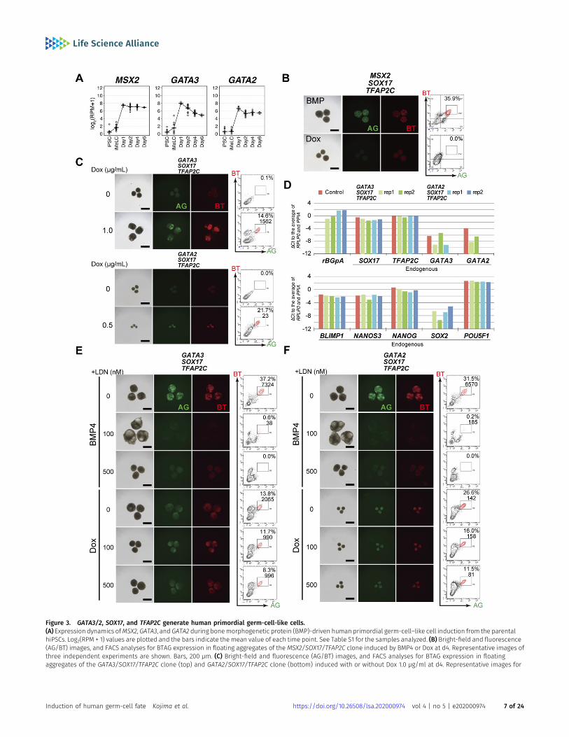
Figure 3. GATA3/2, SOX17, and TFAP2C generate human primordial germ-cell-like cells.(A) Expression dynamics ofMSX2, GATA3, and GATA2 during bonemorphogenetic protein (BMP)-driven human primordial germ-cell–like cell induction from the parentalhiPSCs. Log2(RPM + 1) values are plotted and the bars indicate the mean value of each time point. See Table S1 for the samples analyzed. (B) Bright-field and fluorescence(AG/BT) images, and FACS analyses for BTAG expression in floating aggregates of the MSX2/SOX17/TFAP2C clone induced by BMP4 or Dox at d4. Representative images ofthree independent experiments are shown. Bars, 200 μm. (C) Bright-field and fluorescence (AG/BT) images, and FACS analyses for BTAG expression in floatingaggregates of the GATA3/SOX17/TFAP2C clone (top) and GATA2/SOX17/TFAP2C clone (bottom) induced with or without Dox 1.0 μg/ml at d4. Representative images for
Induction of human germ-cell fate Kojima et al. https://doi.org/10.26508/lsa.202000974 vol 4 | no 5 | e202000974 7 of 24
process (13) (Fig S2A), d6 BT+AG+ cells from the GATA3/SOX17/TFAP2Cclone exhibited high similarity/correlation to d2/d4/d6 wild-typeBT+AG+ cells induced by BMP4 (Fig 4B). In contrast, although d2/d4BT+ cells induced from the GATA3/SOX17 clone appeared to take asimilar pathway until d2, they failed to progress further by d4 (Fig S3G).
The numbers of DEGs between BT+AG+ cells from the GATA3/SOX17/TFAP2C clone induced by Dox and from the parental cells induced byBMP4 were the largest at d2 (527), and decreased thereafter (d4: 265; d6:53) (Fig 4C and Table S2). The genes up-regulated in BMP4-induced cellsat day 2 (334 genes) were enriched with those for “negative regulation oftranscription from RNA pol II promoter,” “embryonic forelimb morpho-genesis” (GO terms), and “TGF-β signaling pathway” (KEGG pathway), andincluded key BMP targets, such as ID1, ID3, CDX2, TBX3,MSX1,MSX2,HAND1,and TFAP2A (Fig 4D and E), suggesting that these BMP effectors aredispensable for hPGCLC specification. In contrast, the genes up-regulatedinGATA3/SOX17/TFAP2C-induced cells at day 2 (193 genes) were enrichedwith those for “negative regulation of BMP signaling pathway,” “anterior/posterior axis specification” (GO terms), and “PI3K-Akt signaling pathway”(KEGG pathway), and included FGF2, FGF12, FGF19, FGFR2,MAP2K1,MAP2K6,IRS2, and SOS2 (Fig 4F and G). The genes up-regulated with high fold-changes included epiblast/ectoderm genes such as SOX2, ZIC3, andSALL3, which were repressed in slower kinetics by the transgene ex-pression (Fig 4G).
We addressed whether GATA3 and GATA2 expression were af-fected by other TFs relevant for hPGCLC specification. As shown inFig 4H, in any of the knockout clones for EOMES, SOX17, TFAP2C, andBLIMP1 induced for the germ-cell fate by BMP4 (13), GATA3 andGATA2 up-regulation was un-affected, indicating that their ex-pression is independent from these TF pathways. We conclude thatamong the BMP effectors, the GATA TFs are the key that, togetherwith SOX17 and TFAP2C, is sufficient to drive the transcriptionalprogram for hPGCLC specification.
Critical requirements of the GATA TF paralogs for hPGCLCspecification
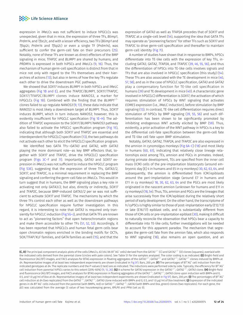
We next explored whether GATA TFs are essential for hPGCLC in-duction. Using CRISPR/Cas9 technology (45), we targeted GATA3 orGATA2 loci in parental 585B1 BTAG hiPSCs, and isolated four andthree clones bearing frameshift mutations in both alleles of GATA3or GATA2, respectively (GATA3 or GATA2 homozygous knockout−/−
clones) (Fig S4A). The lack of GATA3 or GATA2 expression in theseclones was verified by Western blot analyses following the dif-ferentiation of these clones into TE-like cells (46) (Fig S4B).
We induced these clones into iMeLCs (Fig S4C), and then intohPGCLCs by BMP4. Unexpectedly, all theGATA3−/−orGATA2−/− cloneswereinduced into BT+AG+ cells in a manner similar to the parental clone (Figs5A and S4D and E). We isolated total RNAs from iMeLCs and d2/d4/d6BT+AG+ cells induced from all the clones, analyzed the expression of keygenes by qRT-PCR, and found that the GATA3−/− or GATA2−/− clones
expressed relevant genes for hPGCLC specification in an apparentlynormal fashion (Fig 5B). We performed an RNA-seq analysis, whichrevealed that GATA3−/− and GATA2−/− cells differentiated into BT+AG+ cellsin amanner equivalent to theparental clone (Fig 5C andD), andexhibitedsmall numbers of DEGs comparedwith the parental counterparts (Fig S4Fand G). However, we noted that the BT+AG+-cell induction efficiencies atd4 of the GATA3−/− clones (~14.5%) were significantly lower than those ofthe control (~31.4%) or of the GATA2−/− clones (~25.7%) (Fig 5F), raising thepossibility that GATA3 and GATA2 have a compensatory function, withGATA3 playing the more dominant role, during hPGCLC induction.
To investigate this possibility, we knocked out the GATA2 alleles in theGATA3−/− clone, and obtained one line with the GATA3−/−; GATA2+/− ge-notype and one linewith the GATA3−/−;GATA2−/− genotype (Fig S4A). Uponthe differentiation of these clones into TE-like cells (46), the GATA3−/−;GATA2+/− cells formed an epithelial-sheet structure with a typical TE-likecobblestone morphology, but the GATA3−/−; GATA2−/− cells failed to showsuch differentiation and exhibited a mesenchyme-like appearance (FigS4H). Consistently, we confirmed that theGATA3−/−;GATA2−/− cells, but notthe GATA3−/−; GATA2+/− cells, lost the expression of GATA2 proteins (FigS4B).We then induced these cells into iMeLCs (Fig S4I) and successively tohPGCLCs, which revealed that theGATA3−/−;GATA2+/− cells still formed theBT+AG+ cells, but at a further reduced efficiency (~9.5%), whereas theGATA3−/−;GATA2−/− cells barely differentiated into such a state (~1.6%) (Fig5E and F).
To exclude the possibility that the differentiation failure of theGATA3−/−; GATA2−/− clone was due to a clonal effect, we performed arescue experiment. We introduced the Dox-inducible GATA3 ex-pression system into the GATA3−/−; GATA2−/− clone and isolated aline that showed an appropriate GATA3 expression in hiPSCs. Weinduced this line into iMeLCs (Fig S4I), and stimulated the iMeLCaggregates with BMP4 and Dox. Although we found that a contin-uous stimulation of the iMeLC aggregates with BMP4 and Dox led tomajor cell death for an unknown reason, the timed stimulation ofDox (~32 h) resulted in the induction of BT+AG+ cells in a Dox-dosedependent manner (Fig 5G–I), and the induced BT+AG+ cellsexpressed key genes for hPGCLCs in an appropriate fashion (Fig 5J).Thus, we conclude that the GATA TF paralogs, GATA3 and GATA2,show a dose-dependent requirement for hPGCLC specification.Considering that GATA3 was expressed at a higher level than GATA2during hPGCLC induction (Fig 3A) and upon cyPGC specification (Fig2I) and that GATA3 knockouts, but not GATA2 knockouts, exhibited asignificant decrease in hPGCLC induction efficiency (Fig 5F), wepropose that GATA3 plays a major role in hPGCLC induction.
GATA3 expression in post-implantation primate embryos
To explore the spatial relationship of GATA3, SOX17, and TFAP2Cexpression in a developmental context, we examined their expres-sion during PGC specification in the early post-implantation embryosof cynomolgus monkeys. By immunofluorescence analysis, we
10 (GATA3/SOX17/TFAP2C) and six (GATA2/SOX17/TFAP2C) experiments are shown. Bars, 200 μm. (D) Expression of rBGpA (transgenes) and the indicated endogenousgenes in BMP-induced parental clone-derived and Dox-induced GATA3/SOX17/TFAP2C clone- and GATA2/SOX17/TFAP2C clone-derived d4 BT+AG+ cells. Two replicates fromindependent experiments were analyzed. For each gene, the ΔCt was calculated from the average Ct value of two housekeeping genes, RPLP0 and PPIA (set as 0). (E, F)Bright-field and fluorescence (AG/BT) images, and FACS analyses for BTAG expression at d4 in floating aggregates of the GATA3/SOX17/TFAP2C (E) and GATA2/SOX17/TFAP2C (F) clones induced by BMP4 or Dox with 0, 100, 500 nM of LDN193189. Representative images of at least two independent experiments are shown. Bars, 200 μm.
Induction of human germ-cell fate Kojima et al. https://doi.org/10.26508/lsa.202000974 vol 4 | no 5 | e202000974 8 of 24
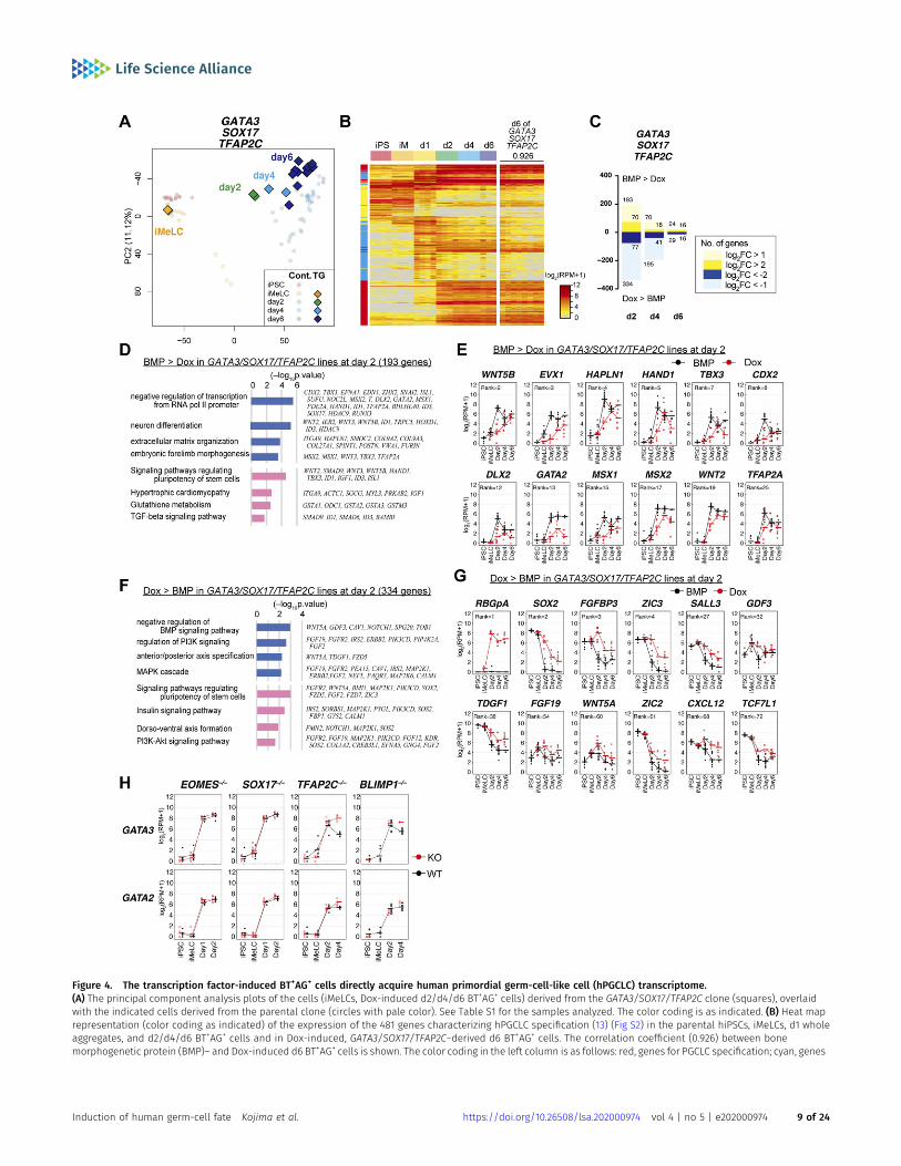
Figure 4. The transcription factor-induced BT+AG+ cells directly acquire human primordial germ-cell-like cell (hPGCLC) transcriptome.(A) The principal component analysis plots of the cells (iMeLCs, Dox-induced d2/d4/d6 BT+AG+ cells) derived from the GATA3/SOX17/TFAP2C clone (squares), overlaidwith the indicated cells derived from the parental clone (circles with pale color). See Table S1 for the samples analyzed. The color coding is as indicated. (B) Heat maprepresentation (color coding as indicated) of the expression of the 481 genes characterizing hPGCLC specification (13) (Fig S2) in the parental hiPSCs, iMeLCs, d1 wholeaggregates, and d2/d4/d6 BT+AG+ cells and in Dox-induced, GATA3/SOX17/TFAP2C–derived d6 BT+AG+ cells. The correlation coefficient (0.926) between bonemorphogenetic protein (BMP)– and Dox-induced d6 BT+AG+ cells is shown. The color coding in the left column is as follows: red, genes for PGCLC specification; cyan, genes
Induction of human germ-cell fate Kojima et al. https://doi.org/10.26508/lsa.202000974 vol 4 | no 5 | e202000974 9 of 24
detected SOX17+ and TFAP2C+ cyPGCs in the dorsal amnion at E12,in the posterior amnion at E13, and near the primitive streakregion between the epiblast and hypoblast at E15 (Fig 6A–D).During this period, SOX17 was also expressed in the hypoblast,and TFAP2C was also expressed diffusely in the amnion andcytotrophoblast/syncytiotrophoblast at E12 but became negativein the amnion thereafter (Fig 6A–C). We found that GATA3 wasexpressed strongly in the cytotrophoblast/syncytiotrophoblastand also in the amnion and the hypoblast, and importantly, inthe SOX17+/TFAP2C+ cyPGCs (Fig 6A–D). Along with the embryonicdevelopment, we noted a gradual decrease in the ratio of GATA3+
cyPGCs (Fig 6D). Notably, the epiblast did not express any of theseTFs.
In addition, we re-analyzed GATA3/GATA2 expression in thesingle-cell transcriptome of cy post-implantation embryos (E13–E20)(30). In addition to early cyPGCs (Fig 6A–D), GATA3 was expressed inthe TE, extra-embryonic mesenchyme, visceral endoderm/yolk-sacendoderm, and gastrulating cells, and weakly/sporadically in theepiblast (Fig 6E). GATA2 was expressed strongly in the TE, but wasweak/sporadic in the other cell types, including early cyPGCs (Figs 2Jand 6E). These findings delineate the spatial relationship of theexpression of GATA3, SOX17, and TFAP2C during primate development,demonstrating that cyPGCs, but not other relevant cell types, co-express these TFs.
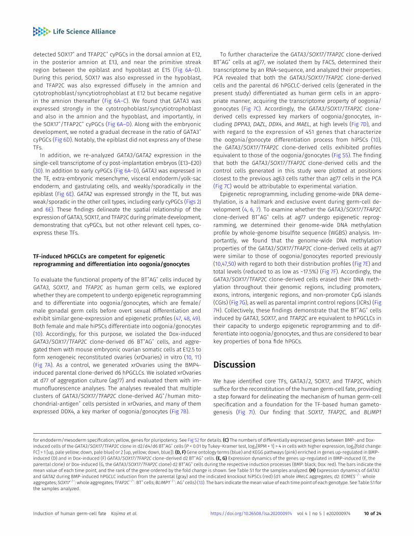
TF-induced hPGCLCs are competent for epigeneticreprogramming and differentiation into oogonia/gonocytes
To evaluate the functional property of the BT+AG+ cells induced byGATA3, SOX17, and TFAP2C as human germ cells, we exploredwhether they are competent to undergo epigenetic reprogrammingand to differentiate into oogonia/gonocytes, which are female/male gonadal germ cells before overt sexual differentiation andexhibit similar gene-expression and epigenetic profiles (47, 48, 49).Both female and male hiPSCs differentiate into oogonia/gonocytes(10). Accordingly, for this purpose, we isolated the Dox-inducedGATA3/SOX17/TFAP2C clone-derived d6 BT+AG+ cells, and aggre-gated them with mouse embryonic ovarian somatic cells at E12.5 toform xenogeneic reconstituted ovaries (xrOvaries) in vitro (10, 11)(Fig 7A). As a control, we generated xrOvaries using the BMP4-induced parental clone-derived d6 hPGCLCs. We isolated xrOvariesat d77 of aggregation culture (ag77) and evaluated them with im-munofluorescence analyses. The analyses revealed that multipleclusters of GATA3/SOX17/TFAP2C clone-derived AG+/human mito-chondrial-antigen+ cells persisted in xrOvaries, and many of themexpressed DDX4, a key marker of oogonia/gonocytes (Fig 7B).
To further characterize the GATA3/SOX17/TFAP2C clone-derivedBT+AG+ cells at ag77, we isolated them by FACS, determined theirtranscriptome by an RNA-sequence, and analyzed their properties.PCA revealed that both the GATA3/SOX17/TFAP2C clone-derivedcells and the parental d6 hPGCLC-derived cells (generated in thepresent study) differentiated as human germ cells in an appro-priate manner, acquiring the transcriptome property of oogonia/gonocytes (Fig 7C). Accordingly, the GATA3/SOX17/TFAP2C clone-derived cells expressed key markers of oogonia/gonocytes, in-cluding DPPA3, DAZL, DDX4, and MAEL, at high levels (Fig 7D), andwith regard to the expression of 451 genes that characterizethe oogonia/gonocyte differentiation process from hiPSCs (10),the GATA3/SOX17/TFAP2C clone-derived cells exhibited profilesequivalent to those of the oogonia/gonocytes (Fig S5). The findingthat both the GATA3/SOX17/TFAP2C clone-derived cells and thecontrol cells generated in this study were plotted at positionsclosest to the previous ag63 cells rather than ag77 cells in the PCA(Fig 7C) would be attributable to experimental variation.
Epigenetic reprogramming, including genome-wide DNA deme-thylation, is a hallmark and exclusive event during germ-cell de-velopment (4, 6, 7). To examine whether the GATA3/SOX17/TFAP2Cclone-derived BT+AG+ cells at ag77 undergo epigenetic reprog-ramming, we determined their genome-wide DNA methylationprofile by whole-genome bisulfite sequence (WGBS) analysis. Im-portantly, we found that the genome-wide DNA methylationproperties of the GATA3/SOX17/TFAP2C clone-derived cells at ag77were similar to those of oogonia/gonocytes reported previously(10,47,50) with regard to both their distribution profiles (Fig 7E) andtotal levels (reduced to as low as ~17.5%) (Fig 7F). Accordingly, theGATA3/SOX17/TFAP2C clone-derived cells erased their DNA meth-ylation throughout their genomic regions, including promoters,exons, introns, intergenic regions, and non-promoter CpG islands(CGIs) (Fig 7G), as well as parental imprint control regions (ICRs) (Fig7H). Collectively, these findings demonstrate that the BT+AG+ cellsinduced by GATA3, SOX17, and TFAP2C are equivalent to hPGCLCs intheir capacity to undergo epigenetic reprogramming and to dif-ferentiate into oogonia/gonocytes, and thus are considered to bearkey properties of bona fide hPGCs.
Discussion
We have identified core TFs, GATA3/2, SOX17, and TFAP2C, whichsuffice for the reconstitution of the human germ-cell fate, providinga step forward for delineating the mechanism of human germ-cellspecification and a foundation for the TF-based human gameto-genesis (Fig 7I). Our finding that SOX17, TFAP2C, and BLIMP1
for endoderm/mesoderm specification; yellow, genes for pluripotency. See Fig S2 for details. (C) The numbers of differentially expressed genes between BMP- and Dox-induced cells of the GATA3/SOX17/TFAP2C clone in d2/d4/d6 BT+AG+ cells (P < 0.01 by Tukey–Kramer test, log2[RPM + 1] > 4 in cells with higher expression, log2[fold change:FC] > 1 [up, pale yellow; down, pale blue] or 2 [up, yellow; down, blue]). (D, F) Gene ontology terms (blue) and KEGG pathways (pink) enriched in genes up-regulated in BMP-induced (D) and in Dox-induced (F) GATA3/SOX17/TFAP2C clone-derived d2 BT+AG+ cells. (E, G) Expression dynamics of the genes up-regulated in BMP-induced (E, theparental clone) or Dox-induced (G, the GATA3/SOX17/TFAP2C clone) d2 BT+AG+ cells during the respective induction processes (BMP: black; Dox: red). The bars indicate themean value of each time point, and the rank of the gene ordered by the fold change is shown. See Table S1 for the samples analyzed. (H) Expression dynamics of GATA3and GATA2 during BMP-induced hPGCLC induction from the parental (gray) and the indicated knockout hiPSCs (red) (d1: whole iMeLC aggregates; d2: EOMES−/−: wholeaggregates; SOX17−/−: whole aggregates; TFAP2C−/−: BT+ cells; BLIMP1−/−: AG+ cells) (13). The bars indicate themean value of each time point of each genotype. See Table S1 forthe samples analyzed.
Induction of human germ-cell fate Kojima et al. https://doi.org/10.26508/lsa.202000974 vol 4 | no 5 | e202000974 10 of 24
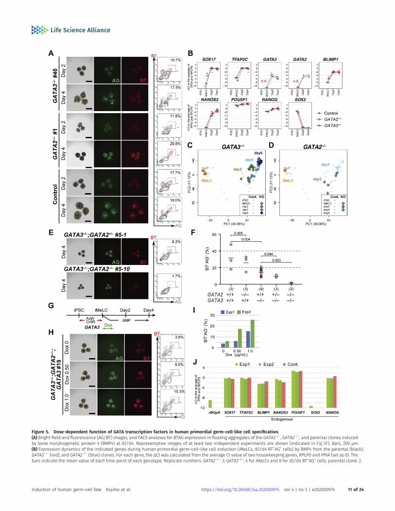
Figure 5. Dose-dependent function of GATA transcription factors in human primordial germ-cell-like cell specification.(A) Bright-field and fluorescence (AG/BT) images, and FACS analyses for BTAG expression in floating aggregates of the GATA3−/−, GATA2−/−, and parental clones inducedby bone morphogenetic protein 4 (BMP4) at d2/d4. Representative images of at least two independent experiments are shown (indicated in Fig 5F). Bars, 200 μm.(B) Expression dynamics of the indicated genes during human primordial germ-cell–like cell induction (iMeLCs, d2/d4 BT+AG+ cells) by BMP4 from the parental (black),GATA3−/− (red), and GATA2−/− (blue) clones. For each gene, the ΔCt was calculated from the average Ct value of two housekeeping genes, RPLP0 and PPIA (set as 0). Thebars indicate the mean value of each time point of each genotype. Replicate numbers: GATA2−/−: 3; GATA3−/−: 4 for iMeLCs and 8 for d2/d4 BT+AG+ cells; parental clone: 2.
Induction of human germ-cell fate Kojima et al. https://doi.org/10.26508/lsa.202000974 vol 4 | no 5 | e202000974 11 of 24
expression in iMeLCs was not sufficient to induce hPGCLCs wasunexpected, given that in mice, the expression of three TFs, Blimp1,Prdm14, and Tfac2c, and with a lesser efficiency, two TFs (Blimp1 andTfap2c; Prdm14 and Tfap2c) or even a single TF (Prdm14), wassufficient to confer the germ-cell fate on their precursors (25).Notably, none of these TFs are known as direct effectors of the BMPsignaling in mice; TFAP2C and BLIMP1 are shared by humans; andPRDM14 is expressed in both hiPSCs and iMeLCs (9, 16). Thus, themechanism of human germ-cell specification is distinct from that inmice not only with regard to the TFs themselves and their hier-archies of actions (13), but also in terms of how the key TFs regulateeach other to drive the downstream PGC pathways.
We showed that SOX17 induces BLIMP1 in both hiPSCs and iMeLCaggregates (Fig 1B and E), and the TFAP2C/BLIMP1, SOX17/TFAP2C,SOX17/TFAP2C/BLIMP1 clones induce NANOS3, a marker forhPGCLCs (Fig 1B). Combined with the finding that the BLIMP1−/−
clones failed to up-regulate NANOS3 (9, 13), these data indicate thatNANOS3 is most likely a downstream target of BLIMP1. Thus, SOX17induces BLIMP1, which in turn induces NANOS3; however, this isevidently insufficient for hPGCLC specification (Fig 1E–H). The ad-dition of TFAP2C expression to the SOX17/BLIMP1/NANOS3 pathwayalso failed to activate the hPGCLC specification program (Fig 1E),indicating that although both SOX17 and TFAP2C are essential andinterdependent for hPGC(LC) specification (13), the two pathways donot activate each other to elicit the hPGC(LC) specification program.
We identified two GATA TFs—GATA3 and GATA2, with GATA3playing the more dominant role—as key BMP effectors that, to-gether with SOX17 and TFAP2C, drive the hPGC(LC) specificationprogram (Figs 3C–F and 7I). Importantly, GATA3 and SOX17 ex-pression in iMeLCs was not sufficient to induce the hPGCLC program(Fig S3E), suggesting that the expression of three TFs, GATA3/2,SOX17, and TFAP2C, is a minimal requirement in replacing the BMPsignaling and conferring the germ-cell fate on iMeLCs. This would inturn suggest that in humans, the BMP signaling plays a key role inactivating not only GATA3/2, but also, directly or indirectly, SOX17and TFAP2C, because BMP-induced GATA3/2 per se was not suffi-cient to activate SOX17 and TFAP2C. The mechanisms by which thethree TFs control each other as well as the downstream pathwaysfor hPGCLC specification require further investigation. In thisregard, it is interesting to note that GATA3 is required only tran-siently for hPGCLC induction (Fig 5G–J), and that GATA TFs are knownto act as “pioneering factors” that open heterochromatin regionsand make them accessible to other TFs (51, 52, 53). Accordingly, ithas been reported that hPGCLCs and human fetal germ cells bearopen chromatin regions enriched in the binding motifs for OCT4,SOX/TFAP/KLF families, and GATA families as well (15), and that the
expression of GATA3 as well as TFAP2A precedes that of SOX17 andTFAP2C at a single-cell level (54), supporting the idea that GATA TFsmay operate as “pioneering factors” for other TFs such as SOX17 andTFAP2C to drive germ-cell specification and thereafter to maintaingerm-cell identity (Fig 7I).
A number of studies have shown that in response to BMP4, hPSCsdifferentiate into TE-like cells with the expression of key TFs, in-cluding GATA3, GATA2, TFAP2A, and TFAP2C (39, 46, 55, 56), and thusthe differentiation of hPSCs into TE-like cells involves signals andTFs that are also involved in hPGCLC specification (this study) (54).These TFs are also associated with the TE development in mice (40,57, 58), and as in the case of hPGCLC specification, GATA3 and GATA2play a compensatory function for TE-like cell specification inhumans (39) and TE development in mice (40). A characteristic geneinvolved in hPGC(LC) differentiation is SOX17, the activation of whichrequires stimulation of hPSCs by WNT signaling that activatesEOMES expression (i.e., iMeLC induction), before stimulation by BMPsignaling (13). In contrast, TE-like cell differentiation requires directstimulation of hPSCs by BMP signaling (39, 55, 56) and such dif-ferentiation has been shown to be significantly promoted byinhibiting endogenous WNT activity elicited by BMP (59). Thus,evidently, a prior activation of the WNT pathway in hPSCs is a key tothe differential cell-fate specification between the germ-cell fateand TE-like cell fate upon BMP stimulation.
Notably, GATA3, GATA2, TFAP2A, and TFAP2C are also expressed inthe amnion in cynomolgus monkeys (Fig 6A–C) (18) and most likelyin humans (60, 61), indicating that relatively close lineage rela-tionships exist among TEs, amnion and PGCs in primates. Indeed,during primate development, TEs are specified from the inner cellmass (ICM) cells of the pre-implantation blastocysts (around em-bryonic day [E] 4 in humans and E5 in cy monkeys) 30, 62, 63, 64, andsubsequently, the amnion is differentiated from ICM/epiblastsaround the peri-implantation stage (around E7 in humans andE11 in cy monkeys) 18, 30, 62, 63, 64 and the PGCs are most likelyoriginated in the nascent amnion (unknown for humans and E11 incymonkeys) (18, 54). Thus, TEs, amnion and PGCs are the lineages thatarise successively from the ICM/epiblast during the relatively shortperiod of early development. On the other hand, the transcriptome ofh/cyPSCs is highly similar to those of post-implantation early (E12/13)or late (E16/17) epiblast cells, and is substantially different fromthose of ICM cells or pre-implantation epiblast (30), making it difficultto naturally reconcile the observation that hPSCs bear a capacity todifferentiate into TE-like cells; further investigations will be neededto account for this apparent paradox. The mechanism that segre-gates the germ-cell fate from the amnion fate, which also respondsto WNT signaling (18), also remains an open question, and an
(C, D) The principal component analysis plots of the cells (iMeLCs, d2/d4/d6 BT+AG+ cells) derived from the GATA3−/− (C) and GATA2−/− (D) clones (squares), overlaid withthe indicated cells derived from the parental clone (circles with pale colors). See Table S1 for the samples analyzed. The color coding is as indicated. (E) Bright-field andfluorescence (AG/BT) images, and FACS analyses for BTAG expression in floating aggregates of the GATA3−/−; GATA2+/− and GATA3−/−; GATA2−/− clones induced by BMP4 atd4. Representative images of at least two independent experiments are shown (indicated in Fig 5F). Bars, 200 μm. (F) The percentages of BT+AG+ cell induction from theindicated genotypes at d4. The replicate numbers and the P-values (t test) are as indicated. The inductions were performed side by side. Typically, the efficiency for BT+AG+
cell induction from parental hiPSCs varies to this extent (20%~60%) (9, 13, 26). (G) A scheme for GATA3 expression in the GATA3−/−; GATA2−/−; GATA3 clone. (H) Bright-fieldand fluorescence (AG/BT) images, and FACS analyses for BTAG expression in floating aggregates of the GATA3−/−; GATA2−/−; GATA3 clone upon induction with BMP4 and 0,0.5, and 1.0 μg/ml of Dox at d4. Representative images of at least two independent experiments are shown (indicated in Fig 5F). Bars, 200 μm. (I) The percentages of BT+AG+
cell induction at d4 (two replicates) from the GATA3−/−; GATA2−/−; GATA3 clone induced with BMP4 and 0, 0.5, and 1.0 μg/ml of Dox treatment. (J) Expression of the indicatedgenes in d4 BT+AG+ cells induced from the parental (with BMP4, red) or GATA3−/−; GATA2−/−; GATA3 (with BMP4 and Dox, green) clones (two replicates). For each gene, theΔCt was calculated from the average Ct value of two housekeeping genes, RPLP0 and PPIA (set as 0).
Induction of human germ-cell fate Kojima et al. https://doi.org/10.26508/lsa.202000974 vol 4 | no 5 | e202000974 12 of 24
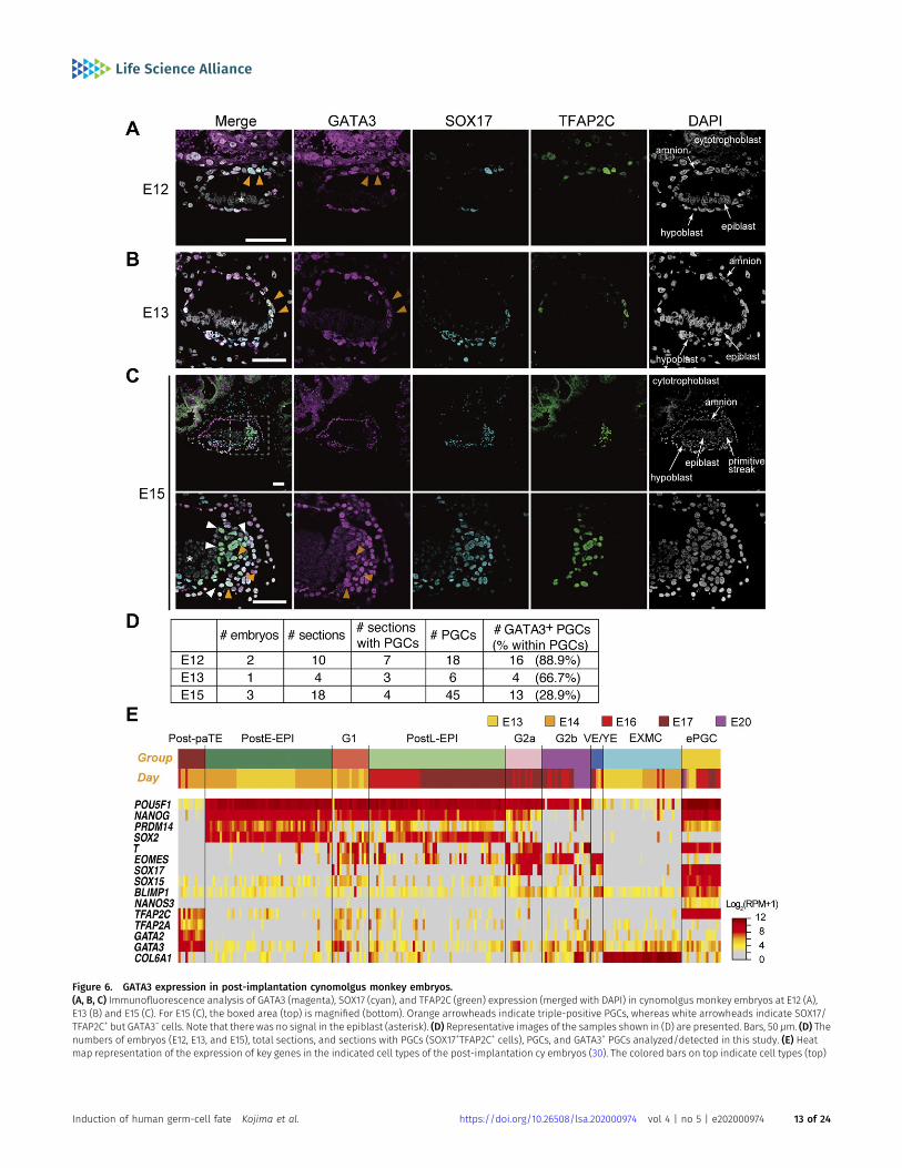
Figure 6. GATA3 expression in post-implantation cynomolgus monkey embryos.(A, B, C) Immunofluorescence analysis of GATA3 (magenta), SOX17 (cyan), and TFAP2C (green) expression (merged with DAPI) in cynomolgus monkey embryos at E12 (A),E13 (B) and E15 (C). For E15 (C), the boxed area (top) is magnified (bottom). Orange arrowheads indicate triple-positive PGCs, whereas white arrowheads indicate SOX17/TFAP2C+ but GATA3− cells. Note that there was no signal in the epiblast (asterisk). (D) Representative images of the samples shown in (D) are presented. Bars, 50 μm. (D) Thenumbers of embryos (E12, E13, and E15), total sections, and sections with PGCs (SOX17+TFAP2C+ cells), PGCs, and GATA3+ PGCs analyzed/detected in this study. (E) Heatmap representation of the expression of key genes in the indicated cell types of the post-implantation cy embryos (30). The colored bars on top indicate cell types (top)
Induction of human germ-cell fate Kojima et al. https://doi.org/10.26508/lsa.202000974 vol 4 | no 5 | e202000974 13 of 24
understanding of this mechanism may lead to a more efficient in-duction of the germ-cell fate from hPSCs.
Crucially, we showed that the TF-induced BT+AG+ cells, whencultured in xrOvaries, underwent epigenetic reprogramming anddifferentiated into oogonia/gonocytes (Fig 7), demonstrating thatthe TF-induced BT+AG+ cells bear one of the key functions of bonafide hPGCs. UnlikemPGCLC specification, which is directly coupled withepigenetic reprogramming (65, 66, 67, 68), hPGCLC specification itselfdoes not appear to be sufficient to elicit the epigenetic reprogram-ming: further signaling/environmental cues, including those providedby xrOvaries, are necessary to activate such key processes (10, 50).Upon mPGCLC specification, Blimp1, Prdm14, and Tfap2c repress theexpression of genes such asDnmt3a/b and Uhrf1, and create a cellularstate with little, if any, de novo and maintenance DNA methyl-transferase (DNMT) activities (50, 65, 66, 67, 68), and this leads to areplication-coupled passive genome-wide DNA demethylation uponmPGCLC proliferation (67, 68). In contrast, themechanism of epigeneticreprogramming, including genome-wide DNA demethylation, inhumans is unclear, and may involve a divergence from that in mice.The identification of the TFs sufficient to create the hPGCLC state (thisstudy), coupled with the development of a method for hPGCLC ex-pansion (50), will be instrumental in clarifying the mechanism ofepigenetic reprogramming in human germ cells.
The mechanisms of germ-cell specification in metazoans areclassified largely into two modes, “epigenesis” and “preformation”(69, 70). The former, as in mammals, involves a strategy to inducethe germ-cell fate into pluripotent precursors by signaling mole-cules and is evolutionarily ancestral, whereas the latter, as in fliesand frogs, involves “preformed” germ plasm in oocytes for germ-cell specification and has been acquired in diverse metazoanlineages as a result of convergent evolution (69, 70). Notably, in the“epigenesis” mode, BMP has been identified as an evolutionarilyconserved key signal in species as diverse as gryllus (71), axolotl(72), and mammals, including mice (73, 74), rabbits (75), pigs (76),monkeys (18, 19), and humans (8, 9). On the other hand, there hasbeen a lack of knowledge as to the mechanism of action, includingvia direct effectors, of the BMP signaling for PGC specification inthese species. In future investigations, it would be useful to in-vestigate whether GATA TFs—which are widely evolutionarily con-served—play a similar role in diverse species, including mice.
Materials and Methods
Animal care and use
All animal experiments were performed under the ethical guide-lines of Kyoto University and Shiga University of Medical Science.Pregnant ICR female mice were purchased from Japan SLC. Ex-perimental procedures using cynomolgus monkeys were approved
by the Animal Care and Use Committee of the Shiga University ofMedical Science.
Human iPSC culture
All the experiments on the induction of hPGCLCs from hiPSCs and ge-nome editing were approved by the Institutional Review Board of KyotoUniversity andwereperformedaccording to the guidelines of theMinistryof Education, Culture, Sports, Science, and Technology (MEXT) of Japan.
The 585B1 BTAG hiPSCs (46, XY) (9) weremaintained in StemFit AK03Nmedium (Ajinomoto) on cell culture plates coated with iMatrix-511(Nippi) (77). The medium was changed every other day. For the pas-sageor the inductionofdifferentiation, the cellswere treatedwith a 1 to 1mixture of TrypLE Select (Life Technologies) and 0.5 mM EDTA/PBS todissociate into single cells, and 10 μMof a ROCK inhibitor (Y-27632; WakoPure Chemical Industries) was added for 24 h after plating.
Generation of TF-expression lines
The vectors for the Doxycycline-induced expression were constructedbased on the Gateway System (Thermo Fisher Scientific) as describedpreviously (13). The full-length cDNA sequences of SOX17, TFAP2C, BLIMP1,MSX2, GATA3, and GATA2 were PCR amplified from d2 hPGCLCs derivedfrom the 585B1 BTAG hiPSC line. Nucleotide sequences for the epitopetags with linkers, 3×FLAG-G4S, V5-G4S, and 2×TY1-G4S, were added to the5-prime ends of SOX17, TFAP2C, and BLIMP1/MSX2/GATA3/GATA2, re-spectively. Primers used for the construction are shown in Table S3. ThePCR products were cloned between the BamHI and XhoI sites of thepENTR1a vector and were subsequently recombined into the destinationvector with LR clonase. In the destination vector, the transgenes wereclonedunder the TetOpromoter repeat regionand followedby the rabbitβ-globin poly A (rBGpA) sequence. In the region downstream of rβGpA,thepuromycin-resistant genedrivenby theEF1α promoterwas cloned forSOX17 and TFAP2C, neomycin for rtTA, and hygromycin for the rest of thegenes.
Transfection was performed with the electroporator NEPA21 typeII (Nepagene). Half a million hiPSCs were transfected with 500 ng ofthe piggybac transposase expression vector and 1 μg of eachtransgene expression vector, except for TFAP2C, which was added at1.5 μg, then resuspended in 100 μl of OptiMEM (Thermo FisherScientific). Selection antibiotics (200 μg/ml geneticin [G418], 10 μg/ml puromycin, and 800 μg/ml hygromycin [all from Thermo FisherScientific]) were added 2 d after the transfection and maintaineduntil the surviving colonies were picked up at 12–14 d. The inductionof the transgenes with 1.0 μg/ml doxycycline (Takara-Clontech) forthe selected hiPSC clones was assessed at 24 h of culture.
Generation of knockout lines
pX335-U6-Chimeric BB-CBh-hSpCas9n (D10A) was a gift of FengZhang (plasmid #42335; Addgene) (45), and the eGFP sequence wasreplaced with the mCherry sequence bearing a silent mutation
and embryonic days (bottom), respectively. The color coding is as indicated. Post-paTE, PostE-EPI, post-implantation early epiblast; G1, gastrulating cells, group 1; postL-EPI, post-implantation late epiblast; G2a/G2b, gastrulating cells, group 2a/2b; VE/YE, visceral endoderm/yolk sac endoderm; EXMC, extraembryonic mesenchyme; ePGC,early PGC.
Induction of human germ-cell fate Kojima et al. https://doi.org/10.26508/lsa.202000974 vol 4 | no 5 | e202000974 14 of 24
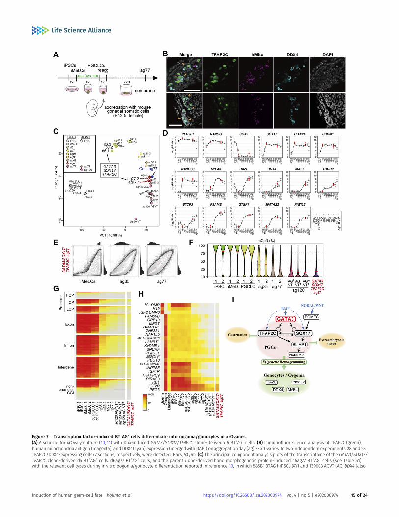
Figure 7. Transcription factor-induced BT+AG+ cells differentiate into oogonia/gonocytes in xrOvaries.(A) A scheme for xrOvary culture (10, 11) with Dox-induced GATA3/SOX17/TFAP2C clone-derived d6 BT+AG+ cells. (B) Immunofluorescence analysis of TFAP2C (green),humanmitochondria antigen (magenta), and DDX4 (cyan) expression (merged with DAPI) on aggregation day (ag) 77 xrOvaries. In two independent experiments, 28 and 23TFAP2C/DDX4-expressing cells/7 sections, respectively, were detected. Bars, 50 μm. (C) The principal component analysis plots of the transcriptome of the GATA3/SOX17/TFAP2C clone-derived d6 BT+AG+ cells, d6ag77 BT+AG+ cells, and the parent clone-derived bone morphogenetic protein-induced d6ag77 BT+AG+ cells (see Table S1)with the relevant cell types during in vitro oogonia/gonocyte differentiation reported in reference 10, in which 585B1 BTAG hiPSCs (XY) and 1390G3 AGVT (AG; DDX4 [also
Induction of human germ-cell fate Kojima et al. https://doi.org/10.26508/lsa.202000974 vol 4 | no 5 | e202000974 15 of 24
(G432A) for convenience in clone selection. For one recombinationsite, a pair of gRNA sequences nicking each strand of genomicDNA were designed using a CRISPR design website (crispr.mit.edu/:now renovated). Two oligo DNAs bearing the gRNA sequence andcompatible ends for the BbsI-digested overhang sequence werephosphorylated, annealed and ligated into the BbsI site of thepX335 vector. 2 μg each of the CRISPR vectors in 100 μl of OptiMEMwere transfected into 5 × 10⁵ 585B1 BTAG hiPSCs using NEPA21 type II(Nepagene). The cells were cultured in AK03N with 10 μM Y27632 for24 h, thenmaintained in AK03N alone for the next 24 h, and the cellswith high mCherry expression (≈top 0.5%) were plated onto 96-wellplates precoated with iMatrix at a single cell per well with theAutomatic Cell Deposition Unit of the FACS Aria III (BD Biosciences).The cells were then cultured in AK03Nwith 10 μMY27632 for 72 h andsubsequently cultured in AK03N alone. 10–14 d after plating, pro-liferating colonies were collected: half of the cells were frozen inStem CellBanker (Zenoaq) and the remaining half were pelletedand lysed for genotyping.
Genotype
To extract the genomic DNA, the cells were lysed in 40 μl of KOD PlusNeo buffer (TOYOBO) supplemented with 0.5% NP40 and 0.8 mg/mlProteinase K (TakaraBio) at 55°C for 3 h, followed by a proteinaseinactivation step at 95°C for 10 min. PCR amplification at the CRISPRrecombination site was performed from 1 μl of the cell lysate withKOD Plus Neo (TOYOBO) and the primers listed in Table S3. Tosequence each allele separately, the amplicon was A-tailed withTaq polymerase (Greiner), cloned into pGEM-T easy (Promega), andtransformed into the DH5α Escherichia coli strain, followed byplating onto LB plates with a blue-white selection. White colonieswere picked for direct PCR with KOD FX Neo (TOYOBO) using theprimers M13-RV and M13-M4. The amplified fragments were se-quenced by Eurofins Genomics with the M13-FW primer andsearched for insertions and/or deletions.
Western blot analysis
Cell pellets were lysed in Bolt Sample Buffer Reducing Agent andthe protein concentrations were measured by protein quantifica-tion assay (Macharey-Nagel). Bolt 10% Bis-Tris Plus Gels were usedfor SDS–PAGE with 1 μg of samples per lane and subsequentlytransferred to Polyvinylidene difluoride (PVDF) membrane (0.22 μmpore) with an iBlot2 Dry Blotting System. All the reagents anddevices were from Thermo Fisher Scientific if not specified and allthe experiments were performed following the manufacturer’sinstructions. The transferred membrane was washed in PBST (PBSwith 0.1% Tween20), blocked with the blocking solution (5% skim
milk in PBST) for 20 min at room temperature with continuousrocking and incubated overnight at 4°C with primary antibodiesdiluted at 1:1,000 in blocking solution. After washing three times ×5 min with PBST, the membrane was incubated with the HRP-conjugated secondary antibodies diluted at 1:1,000 in blockingsolution for 1–3 h at room temperature. After washing three times ×5 min with PBST, the chemiluminescent reaction was induced withAmersham ECL Western Blotting Detection Reagent (GE HealthcareLife Sciences) and the signal was detected with Fusion Solo S(Vilber). All the antibodies used are listed in the Reagents and Toolstable.
hPGCLC induction
The induction of hPGCLCs via iMeLCs was performed as describedpreviously (9, 13, 26). For the induction of iMeLCs, hiPSCs were platedat a density of 5 × 10⁴ cells/cm2 onto a fibronectin (FC010; Millipore)-coated plate. Either 24-well, 12-well or 6-well plates were usedaccording to the number of cells required. The cells were culturedin GK15 medium (GMEM with 15% KSR, 0.1 mM NEAA, 2 mM L-glu-tamine, 1 mM sodium pyruvate, penicillin-streptomycin, and 0.1 mM2-mercaptoethanol) supplemented with 50 ng/ml activin A (R&DSystems), 3 μM CHIR99021 (TOCRIS), and 10 μM of Y-27632 (WakoPure Chemical Industries) for 44–48 h. Then the cells were disso-ciated into single cells with TrypLE Select and aggregated in a low-cell-binding V-bottom 96-well plate (Greiner) at 5,000 cells per wellin 100 μl of GK15 medium supplemented with 200 ng/ml BMP4 (R&DSystems), 100 ng/ml SCF (R&D Systems), 50 ng/ml EGF (R&D Sys-tems), 1,000 U/ml LIF (Millipore), and 10 μM of Y-27632 to be inducedinto hPGCLCs. For the transgene-mediated induction, BMP wasreplaced with doxycycline (Dox) at 1.0 μg/ml except in the case ofGATA2 overexpression, for which 0.5 μg/ml Dox was used. Themedium was not changed until the analysis up to the sixth day ofinduction except in the case of the GATA3 rescue experiment. Forthe GATA3 rescue experiment, 100 μl of medium containing bothDox and BMP4 was applied first, and then, after 32 h, 90 μl of themedium was aspirated and replaced with the same amount ofmedium containing BMP4, SCF, EGF and LIF, and the culture wascontinued for the remaining days. The images of the aggregateswere taken under an M205C stereo microscope (Leica Micro-systems) equipped with a DP72 CCD camera and DP2-BSW software(Olympus).
FACS
For analysis of the cellular contents of the aggregates, the ag-gregates were collected on the designated days of induction,washed once in PBS, and dissociated with 0.25% Trypsin–EDTA for
known as VASA]-tdTomato [VT]) hiPSCs (XX) were used as starting materials. Numbers following “ag” indicate the culture days in xrOvaries. For the AGVT cells, ag77 and120 AG+VT− (AG), AG+VT+ (AGVT) or AG−VT+ (VT) were used for analysis. (D) Expression dynamics of the key genes in GATA3/SOX17/TFAP2C clone-derived d6 BT+AG+ cells, andd6ag77 BT+AG+ cells (n = 2, red circles) (see Table S1), overlaid with those in the relevant cell types during the in vitro oogonia/gonocyte differentiation reported inreference 10. (E) Scatter-plot comparisons, combined with histogram representations (top and right of scatter plots), of the genome-wide 5 mC levels (genome-wide2-kb windows) between the indicated cell types. (F) Violin-plot representation of the genome-wide 5 mC levels determined by whole-genome bisulfite sequence analysisin the cell types indicated. The mean levels are indicated by red bars. (G, H) Heat map representation showing the 5 mC levels in the indicated genomic elements on theautosomes (G) and in the differentially methylated regions of the indicated imprinted genes (H) in the indicated cells. HCP/ICP/LCP, high/intermediate/low-CpGpromoters. The color coding is as indicated. (I) A model of the transcription factor circuitry driving human primordial germ-cell like cell specification.
Induction of human germ-cell fate Kojima et al. https://doi.org/10.26508/lsa.202000974 vol 4 | no 5 | e202000974 16 of 24
Reagents and tools table
Reagent/resource Reference or source Identifier or catalog number
Experimental models
BTAG (BLIMP1-tdTomato and TFAP2C-eGFP knockinreporters in the 585B1 hiPSCs) Sasaki et al (2015). N/A
BTAG; SOX17 OE#1 This study N/A
BTAG; SOX17 OE#2 This study N/A
BTAG; TFAP2C OE#1 This study N/A
BTAG; TFAP2C OE#3 This study N/A
BTAG; SOX17+BLIMP1 OE#9 This study N/A
BTAG; SOX17+BLIMP1 OE#15 This study N/A
BTAG; TFAP2C+BLIMP1 OE#2 This study N/A
BTAG; TFAP2C+BLIMP1 OE#15 This study N/A
BTAG; SOX17+TFAP2C OE#2 This study N/A
BTAG; SOX17+TFAP2C OE#11 This study N/A
BTAG; SOX17+TFAP2C OE#28 This study N/A
BTAG; SOX17+TFAP2C+BLIMP1 OE#4 This study N/A
BTAG; SOX17+TFAP2C+BLIMP1 OE#9 This study N/A
BTAG; SOX17+TFAP2C+BLIMP1 OE#22 This study N/A
BTAG; SOX17+TFAP2C+BLIMP1 OE#28 This study N/A
BTAG; SOX17+TFAP2C+BLIMP1 OE#31 This study N/A
BTAG; SOX17+TFAP2C+MSX2 OE#7 This study N/A
BTAG; SOX17+TFAP2C+MSX2 OE#9 This study N/A
BTAG; GATA3+SOX17+TFAP2C OE#1 This study N/A
BTAG; GATA3+SOX17+TFAP2C OE#5 This study N/A
BTAG; GATA2+SOX17+TFAP2C OE#29 This study N/A
BTAG; GATA2−/− #1 This study N/A
BTAG; GATA2−/− #6 This study N/A
BTAG; GATA2−/− #12 This study N/A
BTAG; GATA3−/− #17 This study N/A
BTAG; GATA3−/− #18 This study N/A
BTAG; GATA3−/− #30 This study N/A
BTAG; GATA3−/− #40 This study N/A
BTAG; HAND1−/− #6 This study N/A
BTAG; GATA3−/−; GATA2+/− #5-1 This study N/A
BTAG; GATA3−/−; GATA2−/− #5-10 This study N/A
BTAG; GATA3−/−; GATA2−/−; GATA3 OE #19 This study N/A
Recombinant DNA
pX335-U6-Chimeric BB-CBh-hSpCas9n (D10A) Addgene Cat. no. 42335
Antibodies
Goat anti-SOX17 R&D Systems AF1924; RRID: AB_355060
Mouse anti-TFAP2C Santa Cruz sc-12762; RRID: AB_667770
Mouse anti-BLIMP1 R&D Systems MAB36081; RRID: AB_10718104
Mouse anti-GATA3 BIOCARE ACR405A; RRID: AB_10895444
(Continued on following page)
Induction of human germ-cell fate Kojima et al. https://doi.org/10.26508/lsa.202000974 vol 4 | no 5 | e202000974 17 of 24
Continued
Reagent/resource Reference or source Identifier or catalog number
Rabbit anti-GATA3 Cell Signaling CST5852S; RRID:AB_10835690
Rabbit anti-GATA2 Novus NBP82581; RRID:AB_11026191
Rabbit anti-GATA2 Santa Cruz sc9008; RRID:AB_2294456
Mouse anti-human mitochondria Merck Millipore MAB1273; RRID:AB_94052
Goat anti-DDX4 R&D Systems AF2030; RRID:AB_2277369
Mouse IgG – HRP conjugated Sigma-Aldrich A5906; RRID: AB_258264
Mouse anti-α Tubulin Sigma-Aldrich T9026; RRID: AB_477593
Mouse IgG – HRP conjugated Sigma-Aldrich A5906; RRID: AB_258264
Rabbit IgG – HRP conjugated Sigma-Aldrich A6154; RRID: AB_258284
Goat IgG – HRP conjugated Sigma-Aldrich A5420; RRID: AB_258242
SSEA1 (CD15) microbeads for human and mouse Miltenyi Biotec 130-094+530
CD31 microbeads for mouse Miltenyi Biotec 130-097-418
Oligonucleotides and sequence-based reagents
qRT-PCR primers This study Table S3
Chemicals, enzymes and other reagents
StemFit AK03N Ajinomoto N/A
iMatrix-511 Nippi
Puromycin Thermo Fisher Scientific A1113803
G418, Geneticin Thermo Fisher Scientific #10131035
Hygromycin B Thermo Fisher Scientific #10131035
Doxycycline Takara-Clontech Z1311N
Fibronectin Millipore FC010
GMEM Thermo Fisher Scientific #11710035
Knockout serum replacement Thermo Fisher Scientific A3181502
Activin A Peprotech 120-14E
CHIR99021 TOCRIS #4423
Y27632 FujiFilm 030-24021
BMP4 R&D Systems 314-BP
SCF R&D Systems 255-SC
EGF R&D Systems 236-EG
LIF Millipore LIF1010
LDN193189 StemGent 04-0074
Glutamax Thermo Fisher Scientific 35050-061
HEPES Thermo Fisher Scientific 15630-106
α-Minimum Essential Medium Thermo Fisher Scientific 32571-036
L-ascorbic acid Sigma-Aldrich A4403
Software
FACSDiva Software BD Biosciences N/A
DAVID (v6.8; GO analysis) https://david.ncifcrf.gov/ N/A
FV10-ASW Olympus N/A
R (v3.6.0; PCA, DEG, and graphs) https://www.R-project.org N/A
Bowtie2 v2.2.7 http://bowtie-bio.sourceforge.net/bowtie2/index.shtml N/A
TopHat v2.1.0 https://ccb.jhu.edu/software/tophat/index.shtml N/A
(Continued on following page)
Induction of human germ-cell fate Kojima et al. https://doi.org/10.26508/lsa.202000974 vol 4 | no 5 | e202000974 18 of 24
10–15 min at 37°C with gentle pipetting every 5 min. Trypsin wasneutralized with a 5× volume of 10% FBS in DMEM, and theresuspended cells were processed with FACS Aria III system (BDBiosciences) and analyzed with FACS Diva software.
The method for selecting CRISPR-mediated knockout clones isdescribed in the section “Generation of knockout lines.”
cDNA amplification, qRT-PCR and RNA-seq analysis
Total RNA was extracted from the frozen cell pellets using RNeasykits (QIAGEN) or NucleoSpin RNA kits (Macherey-Nagel) followingthe manufacturers’ instructions. The amount of RNA was measuredwith Qubit 2.0 (Thermo Fisher Scientific) and the cDNAs weresynthesized through amplification of their 39 ends starting from 1 ngof total RNA as described previously (29). The RNA sample wasmixed with ERCC (External RNA Controls Consortium; Thermo FisherScientific) spike RNA and then reverse transcribed with V1-(dT)24primer using SuperScript III for 5 min at 50°C. SuperScript III wasimmediately inactivated at 70°C for 10 min, and the excess primerwas digested with Exonuclease I (TakaraBio) for 30 min at 37°Cfollowed by heat inactivation for 25 min at 80°C. Then the poly A tailwas added to the cDNA with Terminal Deoxynucleotidyl Transferase
(TakaraBio) for 15 min at 37°C and heat inactivated for 10 min at70°C. Subsequently, PCR amplification was done using ExTaq HSpolymerase (TakaraBio); the first cycle was run with V3-(dT)24primer alone, followed by 14 cycles using both V1-(dT)24 and V3-(dT24). The PCR product was then purified and the primer dimerswere removed by adding a 0.6× volume of AMPure XP (Agencourt),washed with 80% ethanol two times, and eluted with 50 μl 5 mMTris–HCl (pH 8.0) on a magnetic stand. In some cases, an AxyPrepMAG PCR Clean Up Kit (Corning) was used in place of AMPure XP; thetwo provided comparable results.
qRT-PCR was performed with PowerSYBR Green PCR Master Mix(Thermo Fisher Scientific) on a CFX384 Real-Time PCR DetectionSystem (Bio-Rad) using the primers listed in Table S3. The quality ofthe amplified cDNA was assessed according to the Ct values byqPCR of the ERCC spike RNA and the housekeeping genes (PPIA andRPLP0).
The cDNA library was prepared as described previously (78). 5 ngaliquots of quality-controlled cDNA samples were further amplifiedby PCR using ExTaq HS (TakaraBio) with the N-V3 (dT)24 and V1 (dT)24primers for four cycles, purifiedwith three rounds of binding, washingand eluting stepswith AMPureXP, and then fragmentedwith a CovarisE220 sonicator. The fragmented products were end-polished with T4
Continued
Reagent/resource Reference or source Identifier or catalog number
HTSeq v0.9.1 https://htseq.readthedocs.io/en/master/overview.html N/A
ImageJ/Fiji Fiji.sc N/A
Trim_galore v0.6.3 https://www.bioinformatics.babraham.ac.uk/projects/trim_galore/ N/A
cutadapt v118 http://cutadapt.readthedocs.io/en/stable/guide.html N/A
Bismark v0.22.1 https://www.bioinformatics.babraham.ac.uk/projects/bismark/ N/A
SAMtools v1.9 http://samtools.source-forge.net N/A
Other
pGEM-T Easy Kit Promega A3600
Gateway LR Clonase Enzyme Mix Thermo Fisher Scientific #11791043
v-bottom 96-well plate Greiner #651970
RNeasy Micro Kit QIAGEN #74004
NucleoSpin RNA XS Macherey-Nagel #740902
Qubit RNA HS assay kit Thermo Fisher Scientific Q32855
PowerSYBR Green PCR Master Mix Thermo Fisher Scientific #4367659
Qubit dsDNA HS assay kit Thermo Fisher Scientific Q32851
Protein Quantification Assay Macherey-Nagel #740967
ECL Western Blotting Detection Reagent GE Healthcare Life Sciences RPN2106
EZ DNA Methylation-Gold Kit Zymogen D5005
DP72 Olympus N/A
FV1000-IX81 confocal microscope system Olympus N/A
CFX384 Touch Real-Time PCR detection system Bio-Rad Laboratories N/A
NextSeq500/550 Illumina N/A
Hiseq2500 Illumina N/A
Induction of human germ-cell fate Kojima et al. https://doi.org/10.26508/lsa.202000974 vol 4 | no 5 | e202000974 19 of 24
DNA polymerase (NEB) and T4 polynucleotide kinase (NEB) for 30minat 20°C. The products were then purified again with a 0.7× volume ofAMPureXP, followed by addition of a 0.9× volume of AMPureXP to thesupernatant, and a final washing and elution. To attach adaptorsequences, the cDNA solution was treated first with Rd2SP-V1(dT)20primer using ExTaqHS, followed by addition of Rd1SP-adaptor with T4DNA ligase (NEB), and purified with a 0.8× volume of AMPureXP. Theadapter attached cDNA was then PCR amplified using Nextera XTIndex 1 (N7XX) and Index 2 (S5XX) Primers (Illumina) with ExTaqHSfor 10 cycles and purified by two washings with a 0.9× volume ofAMPureXP.
The quality and quantity of the resultant library DNAs wereevaluated by the LabChip GX (Perkin Elmer), the Qubit dsDNA HSassay kit, and the Taqman-qPCR assay using Thunderbird ProbeqPCR mix (TOYOBO) and a TaqMan probe (Ac04364396; AppliedBiosystems). The sequence data were acquired using NextSeq 500(Illumina). Conversion of the sequence read data into expressionlevels was performed as described previously (29, 78). The readswere first processed with cutadapt-1.3 (79) to trim the V1 and V3adaptor sequences and poly-A sequences. The trimmed readslonger than 30 bp were then mapped onto the GRCh38.p2 genomeusing Tophat v2.1.0/Bowtie2 v2.2.7, with the “-no-coverage-search”option (80). The expression levels (reads permillion-mapped reads:RPM) were calculated from these mapped reads using the HTSeqv0.9.1 with default settings, and the GRCh38.p2 reference geneannotations were modified, where necessary, so that the transcripttermination sites were extended up to 10 kb downstream.
Data analysis of the RNA-seq
All statistical analyses were performed on R (ver 3.6.0) with stringr,gplots, and prcomp packages. The expression data were firstconverted into log2(RPM + 1) values, the genes with maximumlog2(RPM + 1) < 4 (equivalent to 10–20 copies per cell) in all thesamples were excluded, and the distribution of the expressionlevels was assessed with boxplots to confirm the quality of thesamples. For hPGCLC induction, 180 samples were added to the 230samples used in the former study with 12,909 genes (13), and forxenogeneic reconstituted ovary series, three iMeLC samples andthree ag77 samples were newly collected and analyzed with 30samples collected in the previous study (10) with 12,737 genes.
All the heat maps were drawn with the heatmap.2() function, thecorrelation coefficient between BMP-induced and overexpressedsamples was calculated with the cor() function with “Pearsoncorrelation,” and the PCA was performed with the prcomp() func-tion. DEGs among SOX17/TFAP2C/BLIMP1 samples were defined asP < 0.01 by one-way ANOVA among all time points and conditions ofSOX17/TFAP2C/BLIMP1 series, P < 0.01 in Tukey–Kramer post hoc testfor multiple comparisons, log2(RPM + 1) > 4 in the higher expressiongroup, and more than onefold change of the mean expressionvalues. GO analysis was performed on the DAVID website (https://david.ncifcrf.gov). The GO terms were extracted from the BiologicalProcess (GOTERM_BP_DIRECT) and the pathway terms fromKEGG_PATHWAY (Kyoto Encyclopedia of Genes and Genomes)(http://www.genome.jp/kegg/) (81).
Single-cell transcriptome data of cynomolgus monkey embryoswas retrieved from the Gene Expression Omnibus database under
accession numbers GSE67259, GSE74767 and GSE76267 (30, 31). Thefollowing cells were extracted: post-implantation parietal tro-phectoderm (designated Post-paTE), post-implantation early epi-blast (PostE-EPI), post-implantation late epiblast (PostL-EPI),gastrulating cells group 1 (G1), gastrulating cells group 2a (G2a),gastrulating cells group 2b (G2b), visceral endoderm or yolk sacendoderm (VE/YE), extraembryonic mesenchyme (EXMC), earlyPGCs (ePGC), and late PGCs (lPGC). The heat maps were drawn withthe heatmap.2() function on R (ver 3.6.0). Note that SOX17 and GATA2were named LOC101925698 and LOC101865311, respectively, in thesedatasets.
Immunofluorescence of iMeLC aggregates and cynomolgusmonkey embryos
Cynomolgus monkey embryos were isolated as described previously(18, 30). The samples were formalin-fixed, paraffin-embedded, andstored at 4°C. The sectioned samples were first deparaffinized andhydrated followed by antigen retrieval with HistoVT One (Nacalai) at90°C for 35 min. After washing in PBS, the samples were per-meabilized and blocked with blocking solution (5% donkey serum,0.2% Tween 20, in PBS) for 2 h at ambient temperature. The sampleswere then treated with primary antibodies in the blocking solutionovernight at 4°C, washed three times with PBS, incubated withsecondary antibodies at room temperature for 1 h, washed threetimes with PBS, mounted in VECTASHIELD Mounting Medium withDAPI (Vector Laboratories), and imaged under an Olympus FV1000confocal microscope.
For the immunofluorescence analysis of reconstituted xenoge-neic ovaries, the harvested reaggregates were fixed with 4%paraformaldehyde at 4°C for 2 h, followed by two-step cry-oprotection with 10% and 30% sucrose dissolved in PBS at 4°C for1 h and overnight, respectively, and finally, freezing in OptimalCutting Temperature (OCT) compound (Sakura Finetek). The sec-tioned samples were washed with PBS to remove the OCT com-pound, and then permeabilized and blocked using the protocoldescribed above.
Xenogeneic reconstituted ovary (xrOvaries) culture
d6 hPGCLCs were further differentiated by aggregation with mousefemale gonadal somatic cells at embryonic day (E) 12.5, which wetermed xrOvaries as described previously (10, 11). Pregnant ICRfemales were euthanized by cervical dislocation and the E12.5embryos were dissected in DMEM containing 10% FBS (Hyclone),2 mM GlutaMax, 10mMHEPES, and 100 U/ml penicillin/streptomycin.Fetal ovaries were dissected out with tungsten needles and dis-sociated into single cells with 0.25% Trypsin, the endogenousmouse PGCs were removed by MACS with anti-CD31 and anti-SSEA1antibodies (Miltenyi), and the remaining fetal ovarian somatic cellswere aliquoted and frozen until use. Thawed somatic cells (75,000cells/well) were mixed with d6 hPGCLCs (5,000 cells/well) andcultured in a Lipidure-coated U-bottom 96-well plate with GK15medium containing 10 μM Y27632 (Tocris) to form floatingcellular aggregates. After 2 d under this condition, the xrOvarieswere transferred onto Transwell-COL membrane inserts (Corning)with a mouth pipette and maintained as an air-liquid interface
Induction of human germ-cell fate Kojima et al. https://doi.org/10.26508/lsa.202000974 vol 4 | no 5 | e202000974 20 of 24
culture in α-MEM with 10% FBS, 55 μM 2-mercaptoethanol, 150μM l-ascorbic acid (Sigma-Aldrich), and 100 U/ml penicillin/streptomycin. The xrOvaries were cultured at 37°C, 5%CO2 in airand half of the medium was replaced every 3 d until harvest. Allthe reagents were from Thermo Fisher Scientific unless otherwisespecified.
WGBS
Genomic DNA was prepared from 5,000 cells, then resuspended inlysis buffer containing 0.1% SDS and 1 μg/μl Proteinase K in DNaseFreeWater (GIBCO) and incubated at 37°C for 60min; the ProteinaseK was then heat inactivated at 98°C for 15 min. This lysate wasspiked with 150 pg of unmethylated λ phage DNA (Promega), basedon the estimation that the amount of genomic DNA was 6 pg per celland the phage DNA accounted for 0.5% of the input genomic DNA.Bisulfite conversion and library construction were performed by thepost-bisulfite adaptor tagging (PBAT) method (82). The detailedprotocol of the PBATmethod has been published and is freely availableat the website of the International Human Epigenome Consortium(http://www.crest-ihec.jp/english/epigenome/index.html). All the re-agents used were the same as described in the protocol, except thatPhusion Hot Start II DNA Polymerase (Thermo Fisher Scientific) andan AxyPrep MAG PCR Clean-Up Kit (Corning) were substituted forthe Phusion Hot Start High-Fidelity DNA polymerase and Agen-court AMPure XP, respectively. Deep sequencing was performedon an Illumina Hiseq 2500 system to generate 101-nucleotide single-end sequence reads, and cluster generation and sequencing wereimplemented in single-read mode using a TruSeq SR Cluster Kit v3-cBot-HS and TruSeq SBS Kit v3-HS (Illumina) following themanufacturer's instructions.
WGBS data processing
The WGBS data were first processed with Trim_Galore v0.6.3 (https://www.bioinformatics.babraham.ac.uk/projects/trim_galore/)/cutadaptv1.18 (http://cutadapt.readthedocs.io/en/stable/guide.html) with the“--clip_R1 4,” “--trim1” and “-a AGATCGGAAGAGC” options to trim awaylow-quality bases (four bases from the 59 ends, one base from the39ends and bases with quality score <20) and the adapter sequences.The qualified reads were then mapped onto the human genome(GRCh38.p2) using Bismark v0.22.1 with the “--pbat” option, and thecytosine/methyl-cytosine count at every CpG site on the genome wasdetermined by the bismark_methylation_extractor program includedin the package.
For the genome-wide analysis, all the CpG sites with read depth≥4 were used for the following analyses. The percent methylationsof individual CpG sites were plotted in the Violin plot using thevioplot package. For the scatterplots, the average percent meth-ylations of the CpG sites in 2-kb non-overlapping bins that carriedfour or more CpG sites were used. The scatterplots were overlaidwith contour plots to enhance the visibility of the plot density, and ahistogram in each scatterplot shows the frequency of CpG meth-ylation at 5% intervals within the samples.
To compare the ratio of DNA methylation in particular regions,the genomic DNA sequence was grouped into the following regions.Promoters were defined as the region between 900-bp upstream
and 400-bp downstream of the transcription start sites, and thepromoters with high CpG (HCP), intermediate CpG (ICP), and lowCpG (LCP) were classified according to the previous report (83).Data for the CpG islands (84) and human imprint loci (85) weredownloaded from public repositories and converted to GRCh38format with LiftOver. No statistical analysis was performed onWGBS data.
Data and Code Availability
All the sequencing data have been deposited in the Gene Ex-pression Omnibus database under accession number GSE154691(RNA-seq: GSE154688; WGBS: GSE154690) and the R script is avail-able on request.
Supplementary Information
Supplementary Information is available at https://doi.org/10.26508/lsa.202000974.
Acknowledgements
We thank the members of our laboratory for their helpful input on this study.We are grateful to Y Nagai, N Konishi, E Tsutsumi, and M Kawasaki of theSaitou Laboratory, and J Asahira and M Kabata of the Yamamoto Laboratoryfor their technical assistance. This work was supported in part by Grants-in-Aid for Scientific Research from Japan Society for the Promotion of Sci-ence (JSPS) (15K08267 and 20K06654) to Y Kojima and by a Grant-in-Aid forSpecially Promoted Research from JSPS (17H06098), an Exploratory Researchfor Advanced Technology Grant from the Japan Science and TechnologyAgency (JST-ERATO) (JPMJER1104), a Grant from Human Science FrontierProgram (RGP0057/2018), and grants from the Pythias Fund and OpenPhilanthropy Project to M Saitou.
Author Contributions
Y Kojima: conceptualization, data curation, formal analysis, su-pervision, funding acquisition, validation, investigation, visualiza-tion, methodology, project administration, and writing—originaldraft, review, and editing.C Yamashiro: resources, investigation, and methodology.Y Murase: resources, investigation, and methodology.Y Yabuta: resources, formal analysis, investigation, andmethodology.I Okamoto: resources, investigation, and methodology.C Iwatani: resources, investigation, and methodology.H Tsuchiya: resources, investigation, and methodology.M Nakaya: resources, investigation, and methodology.T Tsukiyama: resources, investigation, and methodology.T Nakamura: resources, investigation, and methodology.T Yamamoto: resources, investigation, and methodology.M Saitou: conceptualization, formal analysis, supervision, fundingacquisition, investigation, methodology, project administration,and writing—original draft, review, and editing.
Induction of human germ-cell fate Kojima et al. https://doi.org/10.26508/lsa.202000974 vol 4 | no 5 | e202000974 21 of 24
Conflict of Interest Statement
The authors declare that they have no conflict of interest.
References
1. Lynch M (2010) Evolution of the mutation rate. Trends Genet 26: 345–352.doi:10.1016/j.tig.2010.05.003
2. Segurel L, Wyman MJ, Przeworski M (2014) Determinants of mutation ratevariation in the human germline. Annu Rev Genomics Hum Genet 15:47–70. doi:10.1146/annurev-genom-031714-125740
3. Milholland B, Dong X, Zhang L, Hao X, Suh Y, Vijg J (2017) Differencesbetween germline and somatic mutation rates in humans and mice. NatCommun 8: 15183. doi:10.1038/ncomms15183
4. Lee HJ, Hore TA, Reik W (2014) Reprogramming the methylome: Erasingmemory and creating diversity. Cell Stem Cell 14: 710–719. doi:10.1016/j.stem.2014.05.008
5. Baudat F, Imai Y, de Massy B (2013) Meiotic recombination in mammals:Localization and regulation. Nat Rev Genet 14: 794–806. doi:10.1038/nrg3573
6. Saitou M, Miyauchi H (2016) Gametogenesis from pluripotent stem cells.Cell Stem Cell 18: 721–735. doi:10.1016/j.stem.2016.05.001
7. Tang WW, Kobayashi T, Irie N, Dietmann S, Surani MA (2016) Specificationand epigenetic programming of the human germ line. Nat Rev Genet 17:585–600. doi:10.1038/nrg.2016.88
8. Irie N, Weinberger L, Tang WW, Kobayashi T, Viukov S, Manor YS,Dietmann S, Hanna JH, Surani MA (2015) SOX17 is a critical specifier ofhuman primordial germ cell fate. Cell 160: 253–268. doi:10.1016/j.cell.2014.12.013
9. Sasaki K, Yokobayashi S, Nakamura T, Okamoto I, Yabuta Y, Kurimoto K,Ohta H, Moritoki Y, Iwatani C, Tsuchiya H, et al (2015) Robust in vitroinduction of human germ cell fate from pluripotent stem cells. Cell StemCell 17: 178–194. doi:10.1016/j.stem.2015.06.014
10. Yamashiro C, Sasaki K, Yabuta Y, Kojima Y, Nakamura T, Okamoto I,Yokobayashi S, Murase Y, Ishikura Y, Shirane K, et al (2018) Generation ofhuman oogonia from induced pluripotent stem cells in vitro. Science 36:356–360. doi:10.1126/science.aat1674
11. Yamashiro C, Sasaki K, Yokobayashi S, Kojima Y, Saitou M (2020)Generation of human oogonia from induced pluripotent stemcells in culture. Nat Protoc 15: 1560–1583. doi:10.1038/s41596-020-0297-5
12. Hwang YS, Suzuki S, Seita Y, Ito J, Sakata Y, Aso H, Sato K, Hermann BP,Sasaki K (2020) Reconstitution of prospermatogonial specification invitro from human induced pluripotent stem cells. Nat Commun 11: 5656.doi:10.1038/s41467-020-19350-3
13. Kojima Y, Sasaki K, Yokobayashi S, Sakai Y, Nakamura T, Yabuta Y, NakakiF, Nagaoka S, Woltjen K, Hotta A, et al (2017) Evolutionarily distinctivetranscriptional and signaling programs drive human germ cell lineagespecification from pluripotent stem cells. Cell Stem Cell 21: 517–532.e5.doi:10.1016/j.stem.2017.09.005
14. Chen D, Liu W, Lukianchikov A, Hancock GV, Zimmerman J, Lowe MG, KimR, Galic Z, Irie N, Surani MA, et al (2017) Germline competency of humanembryonic stem cells depends on eomesodermin. Biol Reprod 97:850–861. doi:10.1093/biolre/iox138
15. Chen D, Liu W, Zimmerman J, Pastor WA, Kim R, Hosohama L, Ho J,Aslanyan M, Gell JJ, Jacobsen SE, et al (2018) The TFAP2C-regulated OCT4naive enhancer is involved in human germline formation. Cell Rep 25:3591–3602.e5. doi:10.1016/j.celrep.2018.12.011
16. Sybirna A, Tang WWC, Pierson Smela M, Dietmann S, Gruhn WH, Brosh R,Surani MA (2020) A critical role of PRDM14 in human primordial germ cell
fate revealed by inducible degrons. Nat Commun 11: 1282. doi:10.1038/s41467-020-15042-0
17. Pierson Smela M, Sybirna A, Wong FCK, Surani MA (2019) Testing the roleof SOX15 in human primordial germ cell fate. Wellcome Open Res 4: 122.doi:10.12688/wellcomeopenres.15381.2
18. Sasaki K, Nakamura T, Okamoto I, Yabuta Y, Iwatani C, Tsuchiya H, Seita Y,Nakamura S, Shiraki N, Takakuwa T, et al (2016) The germ cell fate ofcynomolgus monkeys is specified in the nascent amnion. Dev Cell 39:169–185. doi:10.1016/j.devcel.2016.09.007
19. Sakai Y, Nakamura T, Okamoto I, Gyobu-Motani S, Ohta H, Yabuta Y,Tsukiyama T, Iwatani C, Tsuchiya H, Ema M, et al (2020) Induction of thegerm cell fate from pluripotent stem cells in cynomolgus monkeys. BiolReprod 102: 620–638. doi:10.1093/biolre/ioz205
20. Kanai-Azuma M, Kanai Y, Gad JM, Tajima Y, Taya C, Kurohmaru M, Sanai Y,Yonekawa H, Yazaki K, Tam PP, et al (2002) Depletion of definitive gutendoderm in Sox17-null mutant mice. Development 129: 2367–2379.
21. Aramaki S, Hayashi K, Kurimoto K, Ohta H, Yabuta Y, Iwanari H, MochizukiY, Hamakubo T, Kato Y, Shirahige K, et al (2013) A mesodermal factor, T,specifies mouse germ cell fate by directly activating germlinedeterminants. Dev Cell 27: 516–529. doi:10.1016/j.devcel.2013.11.001
22. Ohinata Y, Payer B, O’Carroll D, Ancelin K, Ono Y, Sano M, Barton SC,Obukhanych T, Nussenzweig M, Tarakhovsky A, et al (2005) Blimp1 is acritical determinant of the germ cell lineage in mice. Nature 436:207–213. doi:10.1038/nature03813
23. Yamaji M, Seki Y, Kurimoto K, Yabuta Y, Yuasa M, Shigeta M, Yamanaka K,Ohinata Y, SaitouM (2008) Critical function of Prdm14 for the establishment ofthe germ cell lineage in mice. Nat Genet 40: 1016–1022. doi:10.1038/ng.186
24. Weber S, Eckert D, Nettersheim D, Gillis AJ, Schafer S, Kuckenberg P,Ehlermann J, Werling U, Biermann K, Looijenga LH, et al (2010) Criticalfunction of AP-2 gamma/TCFAP2C in mouse embryonic germ cellmaintenance. Biol Reprod 82: 214–223. doi:10.1095/biolreprod.109.078717
25. Nakaki F, Hayashi K, Ohta H, Kurimoto K, Yabuta Y, Saitou M (2013)Induction of mouse germ-cell fate by transcription factors in vitro.Nature 501: 222–226. doi:10.1038/nature12417
26. Yokobayashi S, Okita K, NakagawaM, Nakamura T, Yabuta Y, Yamamoto T,Saitou M (2017) Clonal variation of human induced pluripotent stemcells for induction into the germ cell fate. Biol Reprod 96: 1154–1166.doi:10.1093/biolre/iox038
27. Martins G, Calame K (2008) Regulation and functions of Blimp-1 in T andB lymphocytes. Annu Rev Immunol 26: 133–169. doi:10.1146/annurev.immunol.26.021607.090241
28. Bikoff EK, Morgan MA, Robertson EJ (2009) An expanding job descriptionfor Blimp-1/PRDM1. Curr Opin Genet Dev 19: 379–385. doi:10.1016/j.gde.2009.05.005
29. Nakamura T, Yabuta Y, Okamoto I, Aramaki S, Yokobayashi S, Kurimoto K,Sekiguchi K, Nakagawa M, Yamamoto T, Saitou M (2015) SC3-seq: Amethod for highly parallel and quantitative measurement of single-cellgene expression. Nucleic Acids Res 43: e60. doi:10.1093/nar/gkv134
30. Nakamura T, Okamoto I, Sasaki K, Yabuta Y, Iwatani C, Tsuchiya H, Seita Y,Nakamura S, Yamamoto T, Saitou M (2016) A developmental coordinateof pluripotency among mice, monkeys and humans. Nature 537: 57–62.doi:10.1038/nature19096
31. Nakamura T, Yabuta Y, Okamoto I, Sasaki K, Iwatani C, Tsuchiya H, SaitouM (2017) Single-cell transcriptome of early embryos and culturedembryonic stem cells of cynomolgus monkeys. Sci Data 4: 170067.doi:10.1038/sdata.2017.67
32. Thomas PQ, Brown A, Beddington RS (1998) Hex: A homeobox generevealing peri-implantation asymmetry in themouse embryo and an earlytransient marker of endothelial cell precursors. Development 125: 85–94.
33. Brennan J, Lu CC, Norris DP, Rodriguez TA, Beddington RS, Robertson EJ(2001) Nodal signalling in the epiblast patterns the early mouse embryo.Nature 411: 965–969. doi:10.1038/35082103
Induction of human germ-cell fate Kojima et al. https://doi.org/10.26508/lsa.202000974 vol 4 | no 5 | e202000974 22 of 24
34. Merrill BJ, Pasolli HA, Polak L, Rendl M, Garcıa-Garcıa MJ, Anderson KV,Fuchs E (2004) Tcf3: A transcriptional regulator of axis induction in theearly embryo. Development 131: 263–274. doi:10.1242/dev.00935
35. Tremblay M, Sanchez-Ferras O, Bouchard M (2018) GATA transcriptionfactors in development and disease. Development 145: dev164384.doi:10.1242/dev.164384
36. Ramos C, Robert B (2005) msh/Msx gene family in neural development.Trends Genet 21: 624–632. doi:10.1016/j.tig.2005.09.001
37. Le Bouffant R, Souquet B, Duval N, Duquenne C, Herve R, Frydman N,Robert B, Habert R, Livera G (2011) Msx1 and Msx2 promote meiosisinitiation. Development 138: 5393–5402. doi:10.1242/dev.068452
38. Lowry JA, Atchley WR (2000) Molecular evolution of the GATA family oftranscription factors: Conservation within the DNA-binding domain. JMol Evol 50: 103–115. doi:10.1007/s002399910012
39. Krendl C, Shaposhnikov D, Rishko V, Ori C, Ziegenhain C, Sass S, SimonL, Müller NS, Straub T, Brooks KE, et al (2017) GATA2/3-TFAP2A/Ctranscription factor network couples human pluripotent stem celldifferentiation to trophectoderm with repression of pluripotency.Proc Natl Acad Sci U S A 114: E9579–E9588. doi:10.1073/pnas.1708341114
40. Home P, Kumar RP, Ganguly A, Saha B, Milano-Foster J, Bhattacharya B,Ray S, Gunewardena S, Paul A, Camper SA, et al (2017) Geneticredundancy of GATA factors in the extraembryonic trophoblast lineageensures the progression of preimplantation and postimplantationmammalian development. Development 144: 876–888. doi:10.1242/dev.145318
41. Craven SE, Lim KC, Ye W, Engel JD, de Sauvage F, Rosenthal A (2004) Gata2specifies serotonergic neurons downstream of sonic hedgehog.Development 131: 1165–1173. doi:10.1242/dev.01024
42. Gunne-Braden A, Sullivan A, Gharibi B, Sheriff RSM, Maity A, Wang YF,Edwards A, Jiang M, Howell M, Goldstone R, et al (2020) GATA3mediates afast, irreversible commitment to BMP4-driven differentiation in humanembryonic stem cells. Cell Stem Cell 26: 693–706.e9. doi:10.1016/j.stem.2020.03.005
43. Cuny GD, Yu PB, Laha JK, Xing X, Liu JF, Lai CS, Deng DY, Sachidanandan C,Bloch KD, Peterson RT (2008) Structure-activity relationship study ofbone morphogenetic protein (BMP) signaling inhibitors. Bioorg MedChem Lett 18: 4388–4392. doi:10.1016/j.bmcl.2008.06.052
44. Yu PB, Deng DY, Lai CS, Hong CC, Cuny GD, Bouxsein ML, Hong DW,McManus PM, Katagiri T, Sachidanandan C, et al (2008) BMP type Ireceptor inhibition reduces heterotopic [corrected] ossification. NatMed 14: 1363–1369. doi:10.1038/nm.1888
45. Cong L, Ran FA, Cox D, Lin S, Barretto R, Habib N, Hsu PD, Wu X, Jiang W,Marraffini LA, et al (2013) Multiplex genome engineering using CRISPR/Cas systems. Science 339: 819–823. doi:10.1126/science.1231143
46. Li Y, Moretto-Zita M, Soncin F, Wakeland A, Wolfe L, Leon-Garcia S,Pandian R, Pizzo D, Cui L, Nazor K, et al (2013) BMP4-directed trophoblastdifferentiation of human embryonic stem cells is mediated through aDeltaNp63+ cytotrophoblast stem cell state. Development 140:3965–3976. doi:10.1242/dev.092155
47. Tang WW, Dietmann S, Irie N, Leitch HG, Floros VI, Bradshaw CR, HackettJA, Chinnery PF, Surani MA, et al (2015) A unique gene regulatory networkresets the human germline epigenome for development. Cell 161:1453–1467. doi:10.1016/j.cell.2015.04.053
48. Guo F, Yan L, Guo H, Li L, Hu B, Zhao Y, Yong J, Hu Y, Wang X, Wei Y, et al(2015) The transcriptome and DNA methylome landscapes of humanprimordial germ cells. Cell 161: 1437–1452. doi:10.1016/j.cell.2015.05.015
49. Li L, Dong J, Yan L, Yong J, Liu X, Hu Y, Fan X, Wu X, Guo H, Wang X, et al(2017) Single-cell RNA-seq analysis maps development of humangermline cells and gonadal niche interactions. Cell Stem Cell 20:858–873.e4. doi:10.1016/j.stem.2017.03.007
50. Murase Y, Yabuta Y, Ohta H, Yamashiro C, Nakamura T, Yamamoto T,Saitou M (2020) Long-term expansion with germline potential of human
primordial germ cell-like cells in vitro. EMBO J 39: e104929. doi:10.15252/embj.2020104929
51. Cirillo LA, Lin FR, Cuesta I, Friedman D, Jarnik M, Zaret KS (2002) Openingof compacted chromatin by early developmental transcription factorsHNF3 (FoxA) and GATA-4. Mol Cell 9: 279–289. doi:10.1016/s1097-2765(02)00459-8
52. Eeckhoute J, Keeton EK, Lupien M, Krum SA, Carroll JS, Brown M (2007)Positive cross-regulatory loop ties GATA-3 to estrogen receptor alphaexpression in breast cancer. Cancer Res 67: 6477–6483. doi:10.1158/0008-5472.can-07-0746
53. Kong SL, Li G, Loh SL, Sung WK, Liu ET (2011) Cellular reprogramming bythe conjoint action of ERalpha, FOXA1, and GATA3 to a ligand-induciblegrowth state. Mol Syst Biol 7: 526. doi:10.1038/msb.2011.59
54. Chen D, Sun N, Hou L, Kim R, Faith J, Aslanyan M, Tao Y, Zheng Y, Fu J, LiuW, et al (2019) Human primordial germ cells are specified from lineage-primed progenitors. Cell Rep 29: 4568–4582.e5. doi:10.1016/j.celrep.2019.11.083
55 Xu RH, Chen X, Li DS, Li R, Addicks GC, Glennon C, Zwaka TP, Thomson JA(2002) BMP4 initiates human embryonic stem cell differentiation totrophoblast. Nat Biotechnol 20: 1261–1264. doi:10.1038/nbt761
56 Drukker M, Tang C, Ardehali R, Rinkevich Y, Seita J, Lee AS, Mosley AR,Weissman IL, Soen Y (2012) Isolation of primitive endoderm,mesoderm, vascular endothelial and trophoblast progenitors fromhuman pluripotent stem cells. Nat Biotechnol 30: 531–542. doi:10.1038/nbt.2239
57 Auman HJ, Nottoli T, Lakiza O, Winger Q, Donaldson S, Williams T (2002)Transcription factor AP-2gamma is essential in the extra-embryoniclineages for early postimplantation development. Development 129:2733–2747.
58 Ralston A, Cox BJ, Nishioka N, Sasaki H, Chea E, Rugg-Gunn P, Guo G,Robson P, Draper JS, Rossant J (2010) Gata3 regulates trophoblastdevelopment downstream of Tead4 and in parallel to Cdx2.Development 137: 395–403. doi:10.1242/dev.038828
59. Kurek D, Neagu A, Tastemel M, Tüysüz N, Lehmann J, van de Werken HJG,Philipsen S, van der Linden R, Maas A, van IJcken WFJ, et al (2015)Endogenous WNT signals mediate BMP-induced and spontaneousdifferentiation of epiblast stem cells and human embryonic stem cells.Stem Cel Rep 4: 114–128. doi:10.1016/j.stemcr.2014.11.007
60. Shao Y, Taniguchi K, Gurdziel K, Townshend RF, Xue X, Yong KMA, Sang J,Spence JR, Gumucio DL, Fu J (2017) Self-organized amniogenesis byhuman pluripotent stem cells in a biomimetic implantation-like niche.Nat Mater 16: 419–425. doi:10.1038/nmat4829
61. Zheng Y, Xue X, Shao Y, Wang S, Esfahani SN, Li Z, Muncie JM, Lakins JN,Weaver VM, Gumucio DL, et al (2019) Controlled modelling of humanepiblast and amnion development using stem cells. Nature 573: 421–425.doi:10.1038/s41586-019-1535-2
62. O’Rahilly R, Müller F (1987) Developmental Stages in Human EmbryosIncluding a Revision of Streeter’s “Horizons” and a Survey of theCarnegie Collection. Washington, DC: Carnegie Institution.
63. Luckett WP (1975) The development of primordial and definitiveamniotic cavities in early Rhesus monkey and human embryos. Am JAnat 144: 149–167. doi:10.1002/aja.1001440204
64. Heuser, Streeter (1941) Development of the macaque embryoContributions to Embryology. pp: 15–55. Washington DC: CarnegieInstitution.
65. Hayashi K, Ohta H, Kurimoto K, Aramaki S, Saitou M (2011) Reconstitutionof the mouse germ cell specification pathway in culture by pluripotentstem cells. Cell 146: 519–532. doi:10.1016/j.cell.2011.06.052
66. Kurimoto K, Yabuta Y, Hayashi K, Ohta H, Kiyonari H, Mitani T, Moritoki Y,Kohri K, Kimura H, Yamamoto T, et al (2015) Quantitative dynamics ofchromatin remodeling during germ cell specification from mouseembryonic stem cells. Cell Stem Cell 16: 517–532. doi:10.1016/j.stem.2015.03.002
Induction of human germ-cell fate Kojima et al. https://doi.org/10.26508/lsa.202000974 vol 4 | no 5 | e202000974 23 of 24
67. Shirane K, Kurimoto K, Yabuta Y, Yamaji M, Satoh J, Ito S, Watanabe A,Hayashi K, Saitou M, Sasaki H (2016) Global landscape and regulatoryprinciples of DNA methylation reprogramming for germ cellspecification by mouse pluripotent stem cells. Dev Cell 39: 87–103.doi:10.1016/j.devcel.2016.08.008
68. Ohta H, Kurimoto K, Okamoto I, Nakamura T, Yabuta Y, Miyauchi H,Yamamoto T, Okuno Y, Hagiwara M, Shirane K, et al (2017) In vitroexpansion of mouse primordial germ cell-like cells recapitulates anepigenetic blank slate. EMBO J 36: 1888–1907. doi:10.15252/embj.201695862
69. Extavour CG, Akam M (2003) Mechanisms of germ cell specificationacross the metazoans: Epigenesis and preformation. Development 130:5869–5884. doi:10.1242/dev.00804
70. Johnson AD, Richardson E, Bachvarova RF, Crother BI (2011) Evolution ofthe germ line-soma relationship in vertebrate embryos. Reproduction141: 291–300. doi:10.1530/rep-10-0474
71. Donoughe S, Nakamura T, Ewen-Campen B, Green DA 2nd, Henderson L,Extavour CG (2014) BMP signaling is required for the generation ofprimordial germ cells in an insect. Proc Natl Acad Sci U S A 111: 4133–4138.doi:10.1073/pnas.1400525111
72. Chatfield J, O’Reilly MA, Bachvarova RF, Ferjentsik Z, Redwood C,Walmsley M, Patient R, Loose M, Johnson AD, et al (2014) Stochasticspecification of primordial germ cells from mesoderm precursors inaxolotl embryos. Development 141: 2429–2440. doi:10.1242/dev.105346
73. Lawson KA, Dunn NR, Roelen BA, Zeinstra LM, Davis AM, Wright CV,Korving JP, Hogan BL (1999) Bmp4 is required for the generation ofprimordial germ cells in the mouse embryo. Genes Dev 13: 424–436.doi:10.1101/gad.13.4.424
74. Ohinata Y, Ohta H, Shigeta M, Yamanaka K, Wakayama T, Saitou M (2009)A signaling principle for the specification of the germ cell lineage inmice. Cell 137: 571–584. doi:10.1016/j.cell.2009.03.014
75. Hopf C, Viebahn C, Puschel B (2011) BMP signals and the transcriptionalrepressor BLIMP1 during germline segregation in the mammalianembryo. Dev Genes Evol 221: 209–223. doi:10.1007/s00427-011-0373-5
76. Kobayashi T, Zhang H, Tang WWC, Irie N, Withey S, Klisch D, Sybirna A,Dietmann S, Contreras DA, Webb R, et al (2017) Principles of early humandevelopment and germ cell program from conserved model systems.Nature 546: 416–420. doi:10.1038/nature22812
77. Nakagawa M, Taniguchi Y, Senda S, Takizawa N, Ichisaka T, Asano K,Morizane A, Doi D, Takahashi J, Nishizawa M, et al (2014) A novel efficient
feeder-free culture system for the derivation of human inducedpluripotent stem cells. Sci Rep 4: 3594. doi:10.1038/srep03594
78. Ishikura Y, Yabuta Y, Ohta H, Hayashi K, Nakamura T, Okamoto I,Yamamoto T, Kurimoto K, Shirane K, Sasaki H, et al (2016) In Vitroderivation and propagation of spermatogonial stem cell activity frommouse pluripotent stem cells. Cell Rep 17: 2789–2804. doi:10.1016/j.celrep.2016.11.026
79. Martin M (2011) Cutadapt removes adapter sequences from high-throughput sequencing reads. EMBnet J 17: 10. doi:10.14806/ej.17.1.200
80. Kim D, Pertea G, Trapnell C, Pimentel H, Kelley R, Salzberg SL (2013)TopHat2: Accurate alignment of transcriptomes in the presence ofinsertions, deletions and gene fusions. Genome Biol 14: R36. doi:10.1186/gb-2013-14-4-r36
81. Huang daW, Sherman BT, Lempicki RA (2009) Bioinformatics enrichmenttools: Paths toward the comprehensive functional analysis of large genelists. Nucleic Acids Res 37: 1–13. doi:10.1093/nar/gkn923
82. Miura F, Enomoto Y, Dairiki R, Ito T (2012) Amplification-free whole-genome bisulfite sequencing by post-bisulfite adaptor tagging. NucleicAcids Res 40: e136. doi:10.1093/nar/gks454
83. Borgel J, Guibert S, Li Y, Chiba H, Schübeler D, Sasaki H, Forne T,Weber M (2010) Targets and dynamics of promoter DNAmethylation during early mouse development. Nat Genet 42:1093–1100. doi:10.1038/ng.708
84. Illingworth RS, Gruenewald-Schneider U, Webb S, Kerr AR, JamesKD, Turner DJ, Smith C, Harrison DJ, Andrews R, Bird AP (2010)Orphan CpG islands identify numerous conserved promoters inthe mammalian genome. PLoS Genet 6: e1001134. doi:10.1371/journal.pgen.1001134
85. Court F, Tayama C, Romanelli V, Martin-Trujillo A, Iglesias-Platas I,Okamura K, Sugahara N, Simón C, Moore H, Harness JV, et al (2014)Genome-wide parent-of-origin DNA methylation analysis reveals theintricacies of human imprinting and suggests a germline methylation-independent mechanism of establishment. Genome Res 24: 554–569.doi:10.1101/gr.164913.113
License: This article is available under a CreativeCommons License (Attribution 4.0 International, asdescribed at https://creativecommons.org/licenses/by/4.0/).
Induction of human germ-cell fate Kojima et al. https://doi.org/10.26508/lsa.202000974 vol 4 | no 5 | e202000974 24 of 24