Embed Size (px)
Citation preview

GE Port J Gastroenterol. 2016;23(1):54---55
www.elsevier.pt/ge
IMAGES IN GASTROENTEROLOGY AND HEPATOLOGY
Gelatinous Ascites: A Characteristic Finding of a RareEntity
Ascite Gelatinosa: Um Achado Característico de uma Entidade Rara
Susana Marques ∗, Joana Carmo, Miguel Bispo, Cristina Chagas
Gastroenterology Department, Centro Hospitalar Lisboa Ocidental, Lisbon, Portugal
Received 16 June 2015; accepted 3 July 2015Available online 1 September 2015
KEYWORDSAscites;Pseudomyxoma Peritonei
PALAVRAS-CHAVEAscite;Pseudomixoma Peritoneal
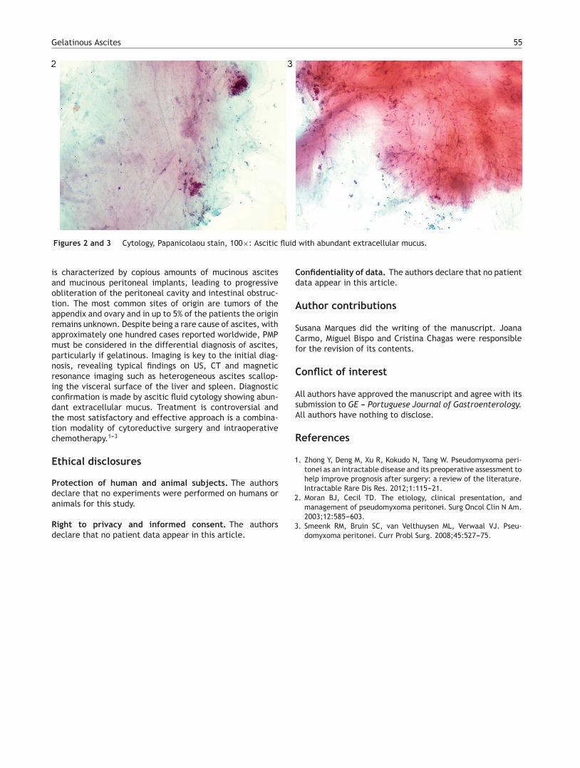
A 70-year-old man with significant past medical historyof hypertension, atrial fibrillation and cerebral vascular dis-ease was admitted for anorexia, weight loss (10 kg) andincreased abdominal girth for the past 3 months. On exam-ination, a voluminous ascites was noted, with no signs ofchronic liver disease. Laboratory studies showed an iso-lated elevation of serum inflammatory markers (C-reactiveprotein and erythrocyte sedimentation rate). Abdominalultrasound (US) and computed tomography (CT) were per-formed and revealed CT low-attenuation heterogeneousascites that scalloped the margins of the liver and spleen(Fig. 1). Diagnostic paracentesis revealed thick gelati-nous ascitic fluid, rich in mucin on cytology examination
∗ Corresponding author.E-mail address: [email protected] (S. Marques).
(Figs. 2 and 3). Ascitic fluid cell count was not possible dueto high viscosity and cultures were negative.
The radiologic and cytotogic findings were crucial tosupport the diagnosis of pseudomyxoma peritonei (PMP).Despite being proposed for surgery, there was a rapid clin-ical deterioration and the patient died 11 weeks after thediagnosis.
PMP is a rare and intractable clinical syndrome, withan estimated incidence of 1---2 per million per year. It
Figure 1 Abdominal CT axial view: low-attenuation hetero-geneous ascites scalloping the margins of the liver and spleen.
http://dx.doi.org/10.1016/j.jpge.2015.07.0012341-4545/© 2015 Sociedade Portuguesa de Gastrenterologia. Published by Elsevier España, S.L.U. This is an open access article under theCC BY-NC-ND license (http://creativecommons.org/licenses/by-nc-nd/4.0/).
Gelatinous Ascites 55
Figures 2 and 3 Cytology, Papanicolaou stain, 100×: Ascitic fluid with abundant extracellular mucus.
is characterized by copious amounts of mucinous ascitesand mucinous peritoneal implants, leading to progressiveobliteration of the peritoneal cavity and intestinal obstruc-tion. The most common sites of origin are tumors of theappendix and ovary and in up to 5% of the patients the originremains unknown. Despite being a rare cause of ascites, withapproximately one hundred cases reported worldwide, PMPmust be considered in the differential diagnosis of ascites,particularly if gelatinous. Imaging is key to the initial diag-nosis, revealing typical findings on US, CT and magneticresonance imaging such as heterogeneous ascites scallop-ing the visceral surface of the liver and spleen. Diagnosticconfirmation is made by ascitic fluid cytology showing abun-dant extracellular mucus. Treatment is controversial andthe most satisfactory and effective approach is a combina-tion modality of cytoreductive surgery and intraoperativechemotherapy.1---3
Ethical disclosures
Protection of human and animal subjects. The authorsdeclare that no experiments were performed on humans oranimals for this study.
Right to privacy and informed consent. The authorsdeclare that no patient data appear in this article.
Confidentiality of data. The authors declare that no patientdata appear in this article.
Author contributions
Susana Marques did the writing of the manuscript. JoanaCarmo, Miguel Bispo and Cristina Chagas were responsiblefor the revision of its contents.
Conflict of interest
All authors have approved the manuscript and agree with itssubmission to GE --- Portuguese Journal of Gastroenterology.All authors have nothing to disclose.
References
1. Zhong Y, Deng M, Xu R, Kokudo N, Tang W. Pseudomyxoma peri-tonei as an intractable disease and its preoperative assessment tohelp improve prognosis after surgery: a review of the literature.Intractable Rare Dis Res. 2012;1:115---21.
2. Moran BJ, Cecil TD. The etiology, clinical presentation, andmanagement of pseudomyxoma peritonei. Surg Oncol Clin N Am.2003;12:585---603.
3. Smeenk RM, Bruin SC, van Velthuysen ML, Verwaal VJ. Pseu-domyxoma peritonei. Curr Probl Surg. 2008;45:527---75.