Embed Size (px)
Citation preview

Clinical Science (2016) 130, 1711–1725 doi: 10.1042/CS20160004
Gender, aging and longevity in humans: anupdate of an intriguing/neglected scenario pavingthe way to a gender-specific medicineRita Ostan*1, Daniela Monti†1, Paola Gueresi‡, Mauro Bussolotto§, Claudio Franceschi‖2 andGiovannella Baggio§2
*Interdepartmental Centre “L. Galvani” (CIG) and Department of Experimental, Diagnostic and Specialty Medicine (DIMES), University of Bologna, ViaSan Giacomo 12, 40126 Bologna, Italy†Department of Clinical and Experimental Biomedical Sciences, University of Florence, Viale Morgagni 50, 50134 Florence, Italy‡Department of Statistical Sciences “Paolo Fortunati”, University of Bologna, Via Belle Arti 41, 40126 Bologna§Internal Medicine Unit, Department of Molecular Medicine, University of Padua, Italy‖IRCCS, Institute of Neurological Sciences of Bologna, 40139 Bologna, Italy
AbstractData showing a remarkable gender difference in life expectancy and mortality, including survival to extreme age, arereviewed starting from clinical and demographic data and stressing the importance of a comprehensive historicalperspective and a gene–environment/lifestyle interaction. Gender difference regarding prevalence and incidence ofthe most important age-related diseases, such as cardiovascular and neurodegenerative diseases, cancer, Type 2diabetes, disability, autoimmunity and infections, are reviewed and updated with particular attention to the role ofthe immune system and immunosenescence. On the whole, gender differences appear to be pervasive and stillpoorly considered and investigated despite their biomedical relevance. The basic biological mechanismsresponsible for gender differences in aging and longevity are quite complex and still poorly understood. The presentreview focuses on centenarians and their offspring as a model of healthy aging and summarizes availableknowledge on three basic biological phenomena, i.e. age-related X chromosome inactivation skewing, gutmicrobiome changes and maternally inherited mitochondrial DNA genetic variants. In conclusion, an appropriategender-specific medicine approach is urgently needed and should be systematically pursued in studies on healthyaging, longevity and age-related diseases, in a globalized world characterized by great gender differences whichhave a high impact on health and diseases.
Key words: aging, centenarians, gender, gender-specific medicine, gut microbiome, longevity, mitochondrial DNA, X chromosome inactivation.
INTRODUCTION
Lifespan and longevity are complex and multifactorial traits res-ulting from an intriguing combination of ‘Nature’ and ‘nurture’,the unique reciprocal interaction between environmental, genetic,epigenetic and stochastic factors, each contributing to the overallphenotype [1,2].
Women live longer than men and this difference in life expect-ancy is a worldwide phenomenon indicating that human longevityseems strongly influenced by gender defined as the combination
Abbreviations: AD, Alzheimer’s disease; AIRE, autoimmune regulator; APOE, apolipoprotein E; CRC, colorectal cancer; CRP, C-reactive protein; CVD, cardiovascular disease; DR, dietaryrestriction; GH, growth hormone; GM, gut microbiota; GST, Glutathione S-transferase; HF, heart failure; IF, intermittent fasting; IFN-γ , interferon γ ; IGF-1, insulin-like growth factor 1;IHD, ischaemic heart disease; IIS, insulin and IGF-1 signalling; IL, interleukin; LDL, low-density lipoprotein; mTOR, mammalian target of rapamycin; NK, natural killer; OXPHOS, oxidativephosphorylation; PD, Parkinson’s disease; SEM, stochastic epigenetic mutation; T2D, Type 2 diabetes; TNF-α, tumour necrosis factor α; TOR, target of rapamycin; XCI, X chromosomeinactivation.1 These authors are equal first authors.2 These authors are equal senior authors.
Correspondence: Professor Giovannella Baggio (email [email protected]).
between biological sexual characteristics (anatomy, reproductivefunctions, sex hormones, expression of genes on the X or Y chro-mosome) and factors related to behaviour, social role, lifestyleand life experiences [3–6].
Following a historical perspective, in Europe in the 19th Cen-tury, life expectancy was less than 40 years and longevity of thetwo genders was generally very similar. The high female mor-tality due to pregnancy and childbirth corresponded to a highermale mortality from causes related to work, accidental injuryor violence. Moreover, infectious and communicable diseases
1711c© 2016 The Author(s). This is an open access article published by Portland Press Limited on behalf of the Biochemical Society and distributed under theCreative Commons Attribution Licence 4.0 (CC BY).

R. Ostan and others
Table 1 Number of deaths, percentage of total deaths by sex regarding the 14 leading causes of death in U.S.A. in 2013Data refer to all races and all ages. Adapted from [14].
Male Female
Rank Cause of death Number Total deaths (%) Number Total deaths (%) Higher mortality for
1 Heart disease 321,347 24.6 289,758 22.4 ♂
2 Malignant neoplasms 307,559 23.5 277,322 21.5 ♂
3 Chronic lower respiratory diseases 70,317 5.4 78,888 6.1 ♀
4 Accidents (unintentional injuries) 81,916 6.3 48,641 3.8 ♂
5 Cerebrovascular diseases 63,691 4.1 75,287 5.8 ♀
6 Alzheimer’s disease 25,836 2.0 58,931 4.6 ♀
7 Diabetes mellitus 39,841 3.1 35,737 2.8 ♂
8 Influenza and pneumonia 26,804 2.1 30,175 2.3 ♀
9 Kidney diseases 23,493 1.8 23,619 1.8 =10 Suicide 32,055 2.5 9,094 0.7 ♂
11 Septicaemia 17,994 1.4 20,162 1.6 ♀
12 Chronic liver disease and cirrhosis 23,709 1.8 12,718 1.0 ♂
13 Essential hypertension-related diseases 12,963 1.0 17,807 1.4 ♀
14 Parkinson’s disease 15,088 1.2 10,108 0.8 ♂
15 All other causes 253,421 19.4 302,707 23.5 ♂
affected and killed men and women almost equally [7]. Through-out the 20th Century, mortality became concentrated in the olderages, non-communicable diseases became the prevailing causesof death, and a female survival advantage emerged and grew.This divergence in life expectancy can partly be explained by thedeclining rates in maternal mortality; however, a major contribu-tion is due to differences in behaviour and biology between malesand females [8].
Using historical data from 1763 birth cohorts from 1800 to1935 in 13 developed countries, Beltran-Sanchez et al. [9] showedthat gender asymmetry emerged in cohorts born after 1880, thatexcess adult male mortality is rooted in a specific age group (50–70) and that heart disease is the main condition associated withincreased excess male mortality in birth cohorts of 1900–1935.The authors have suggested that excess male mortality, foundeven after accounting for smoking-attributable deaths, may beexplained by underlying traits of vulnerability to CVD (cardi-ovascular disease) that emerged with the reduction of infectionsand changes in diet and other lifestyle factors [9].
The maximum difference in life expectancy between malesand females was found between the 1970s and the 1990s. Thesubsequent reduction of the gender gap can be attributed partlyto the narrowing of differences in risk behaviours between malesand females, along with the decline in mortality rates from CVDamong men [10]. In the EU-28 countries, the difference in lifeexpectancy between males and females was 5.5 years in 2013(http://ec.europa.eu/eurostat/statistics-explained/index.php/Mortality_and_life_expectancy_statistics); however, the gendergap varied largely across EU member states.
The survival advantage of women is counterbalanced by aworse quality of life in advanced age due to the increase in disab-ility and degenerative diseases [12]. Therefore men and womenhave a diverse chance to attain longevity and, at the same time,the aging process is qualitatively different between genders.
The impact of gender difference in aging has been extensivelyassessed, but the study of the interaction between a series of fun-damental aspects such as hormonal, immunological and meta-bolic pathways as well as genetic background remains largelyunknown.
Accordingly, the present review aims to (i) give an accurateanalysis of mortality causes and age-related diseases pattern inmen and women; (ii) describe the most important mechanismsunderpinning the gender difference in longevity and aging (sexhormones, immunity, genetic factors, nutrition and stress); (iii)attempt to explain the difference in longevity between males andfemales, in human models of extreme longevity such as centen-arians and long-lived families, suggesting the importance of anintegrated investigation of nuclear, mitochondrial DNA geneticsand gut microbiome; and (iv) stress the urgent need for a gender-specific medicine, taking into account the profound differencesin pathophysiological pathways, in clinical characteristics and inpharmacological response between men and women. In conclu-sion, the scientific world is obliged to revise all outcomes in allfields of medicine on the basis of gender differences.
GENDER AND AGE-RELATED DISEASES
The epidemiology of age-related diseases is substantially differ-ent between genders and changes dramatically in women aftermenopause [13]. Table 1 reports mortality data by sex regardingthe 14 leading causes of death in U.S.A. in 2013 and refers toall races and ages [14]. Women died at higher rates than menof chronic lower respiratory diseases, cerebrovascular diseases,AD (Alzheimer’s disease), influenza and pneumonia, septicaemiaand hypertension-related diseases [14]. Even in the EU, a sig-nificant gender gap exists in mortality rates in all countries.
1712 c© 2016 The Author(s). This is an open access article published by Portland Press Limited on behalf of the Biochemical Society and distributed under theCreative Commons Attribution Licence 4.0 (CC BY).
Gender, aging and longevity in humans
In particular, death rates for IHD (ischaemic heart disease) andstroke are higher for men than for women [10].
There are important inequalities in healthy life years betweenmen and women. In EU countries, life expectancy at age 50reached 29.8 years for men and 34.6 years for women in 2010, butthe average duration of life free from activity limitation remainedpractically the same in women (68.6 years) and men (67.9 years)[10,15,16], meaning that the almost 5 years of advantage in lifeexpectancy of women are years of diseases and disability.
Cardiovascular disease (CVD)Differences between women and men in the epidemiology, patho-physiology and symptoms of CVD are well-described. Thisgender gap should been taken into account because it stronglyimpinges on the effects of specific drugs and outcomes. Bothfactors linked to sex (gene expression from the sex chromosomes,sex hormones, metabolism of drugs by sex-specific cytochromeexpression) and gender (sociocultural processes, behaviours, ex-posure to specific environment, nutrition, lifestyle and attitudestowards treatments and prevention) play a fundamental role indetermining CVD risk [17].
Death rates for IHD are 70 % higher for men than for womenon average in all EU countries [10]. In addition, women showeda delayed onset of IHD (7–10 years on average) in several west-ern EU states, even though, due to harmful lifestyle modifica-tions, the prevalence of IHD is increasing in young women [17].Moreover, IHD in women may show different symptoms and painlocalization, and may need diverse diagnostic procedures anddrugs [18].
HF (heart failure) is one of the major health threats of Westernsocieties and affects up to 10 % of the elderly, in absolute numbersmore women than men [19]. However, women survive better thanmen and HF in women frequently occurs at older age and withless ischaemic aetiology than in men [20]. Recent data from theFramingham Heart Study showed that, in the latter half of the20th Century, incidence of HF has declined by about one-thirdin women, but not in men, even though, after adjusting for age,survival after HF onset was improved in both genders [21].
The difference in the epidemiology of hypertension betweenmen and women deeply changes with age. In particular, hyper-tension has a low prevalence in young and adult age when it ismore predominant among men. By contrast, hypertension is morecommon in women than in men in the elderly population [17].Indeed, falling oestrogen production during and after menopausehas been associated with hypertension in women [22].
During aging, there is a sex-specific ‘cardiac remodelling’.In particular, women develop more frequently concentric cardiachypertrophy with smaller internal cavity and relatively largerwall thickness, preserving a better ejection fraction and myocar-dial contractility than men. On the other hand, men show morefrequently eccentric hypertrophy leading to an increased strokevolume and dilatation [17].
Women are particularly susceptible to the deleterious impactof T2D (Type 2 diabetes) and hypertension on cardiovascularhealth. These conditions were associated with higher risk of HFin women with respect to men (T2D increases 3.4-fold and 2-fold the risk of HF in women and men respectively; hyperten-
sion increases 5-fold and 2-fold the risk of HF in women andmen respectively) [21]. In addition, T2D worsens the coronaryartery disease outcome more in women than in men [23]. Finally,some pregnancy-associated conditions, such as pre-eclampsiaand other hypertensive disorders, further contribute to increasedrisk for future chronic hypertension, CVD, cerebrovascular dis-eases and death in women [24].
Type 2 diabetes (T2D)Recent data show that the difference in the global estimates ofT2D between men and women in terms of cases (male, 197.7million; female, 184.1 million), prevalence (male, 8.7 %; female,8.1 %) and age-specific prevalence [25] is small. Even if T2Dprevalence is similar in men and women, it is slightly higher inmen under 60 years of age and in women at older ages. Indeed,the longer survival of women is one of the factors leading to ahigher prevalence of diabetes for women than for men at advancedage [26]. However, a stronger connection between diabetes andcoronary heart disease has been demonstrated in women. In par-ticular, the relative risk for mortality due to coronary heart diseasein diabetic patients is 50 % higher in women than in men, sug-gesting that T2D may induce a more unfavourable cardiovascularrisk profile among women. Diabetic women have significantlyhigher levels of blood pressure and lipids than men with diabetes[27]. Moreover, findings from different studies conducted in theU.K. and the U.S.A. showed that the greater coronary risk as-sociated with T2D observed in women may reflect a treatmentbias that favours men. In particular, in these countries, diabeticmen with cardiovascular problems are more frequently treatedwith hypoglycaemic drugs, aspirin, statins or anti-hypertensivedrugs than women with similar pathological conditions[28–30].
Moreover, women over 65 years of age have a higher fre-quency of insulin resistance, dyslipidaemias, central adiposityand hypertension (named the metabolic syndrome) which in turnis a greater risk factor for CVD in women [31,32]. In partic-ular, central adiposity tends to be more pronounced in post-menopausal women than in men playing a determinant role inthe increase in CVD risk. It has been demonstrated that visceraladipose tissue contributes to insulin resistance secreting a varietyof inflammatory mediators [such as IL (interleukin)-6, TNF-α(tumour necrosis factor α), leptin and resistin]. Moreover, lipidprofile [HDL- (high-density lipoprotein) and LDL (low-densitylipoprotein)-cholesterol as well as triacylglycerols] dramaticallyworsens after the menopause favouring atherogenesis [32].
CancerCancer mortality rates are higher for men than for women inindustrialized countries. In some EU countries (i.e. Lithuania,Spain, Latvia, Estonia, the Slovak Republic, Portugal and Croa-tia), the mortality rates for neoplasms in men are dramaticallyincreased. This gender difference can be explained partly bythe greater prevalence of risk factors among men as well asby reduced availability/use of screening programmes for can-cers affecting men, leading to lower survival rates after diagnosis[10]. For instance, lung cancer accounts for the greatest numberof cancer deaths among men in almost all EU states. In 2011,
1713c© 2016 The Author(s). This is an open access article published by Portland Press Limited on behalf of the Biochemical Society and distributed under theCreative Commons Attribution Licence 4.0 (CC BY).
R. Ostan and others
death rates from lung cancer among men were the highest inall EU countries where smoking habits among men remain veryfrequent (Hungary, Poland and Croatia). However, lung cancermortality in American women has increased from 1950 to 1995by 500 % [18]. Evidence has suggested that the developmentof lung cancer is different in women in comparison with men.Non-smoking women have a 2.5-fold higher risk than men todevelop lung cancer at a younger age, but they respond betterto treatment. Women who smoke have a higher susceptibility tocigarette-smoking damage probably related to the polymorphismof Glutathione S-transferase (GST) Mu 1, which plays a role indetoxifying environmental carcinogens [33]. However, womenwith lung cancer survive longer than men, regardless of therapyand stage.
CRC (colorectal cancer) is the second leading cause of cancerdeath in both genders; in women it occurs 5 years later than inmen. For this reason, the population screening for CRC shouldbe extended beyond 70 years of age. Moreover, CRC in wo-men is more often located in the right colon, the histology ismucinous, occult blood in stool may be negative until the laststages and it is frequently diagnosed in an urgent/emergencysituation. Nevertheless, the survival is better in female patientswith respect to male patients [34]. CRC in women more fre-quently expresses microsatellite instability showing a lower sens-itivity to fluoropyrimidines, cornerstone drugs for the treatmentof colorectal carcinoma [35].
Currently, prostate cancer has become the most common can-cer among men after skin cancer in the majority of EU countries,particularly among men aged 65 years and over. However, deathrates for prostate cancer are lower than for lung cancer. Theprimary risk factors are obesity, lack of exercise, age and familyhistory [10].
Breast cancer is the second most common form of cancerin women after skin cancer in all EU countries. It can occur inboth men and women, but it is very rare in men (10 %). The in-cidence rates of breast cancer have increased in the last decade,but the death rates have diminished or remained stable, indicat-ing an improving of survival rates due to earlier diagnosis andbetter treatment [36]. Numerous risk factors for breast cancer inwomen have been identified, including age, personal history ofcertain benign breast diseases or breast cancer, early menstru-ation or late menopause, never having been pregnant or havinga first pregnancy after age 30, use of oral contraceptives, familyhistory of breast cancer, presence of certain genetic mutations(BRCA1 and BRCA2), history of radiation therapy to the chest,long-term use of combined hormone therapy, use of DES (di-ethylstilbestrol), increased breast density, alcohol use and obesityafter menopause. Risk factors for men for breast cancer includeobesity, Klinefelter’s syndrome and an excess of breast tissue(http://www.cancer.gov/research/progress/snapshots/breast).
Neurodegenerative diseasesWomen are more affected than men by dementia (definition com-prising different conditions including AD and vascular dementia)showing a more frequent and rapid decline of cognitive functionwith aging. Prevalence rates among populations vary consid-erably because of methodological reasons (diagnostic criteria,
sampling strategies and statistical analysis) [38]. Among peopleaged 90 years and over, the gender gap rises to 30 % of prevalencefor men and 47 % for women [10].
The biological basis of gender impact on AD and neurodegen-eration are still unclear. Indeed, the development and functioningof the central nervous system is strongly influenced by gender.The main risk factor for AD is age, and the fact that the majorityof AD patients are females has been attributed to longer life ex-pectancy. However, women are reported to have higher rates ofAD than men, even after adjusting for survival [39,40]. The neg-ative effect of the APOE (apolipoprotein E) ε4 allele, one of themost established genetic risk factors for AD, may explain, at leastin part, this gender gap. Different studies have observed that fe-male APOE ε4 carriers show a higher risk of AD compared withmales [41,42]. A recent paper demonstrated that female APOEε4 carriers presented widespread brain hypometabolism and cor-tical thinning compared with female non-carriers, whereas maleAPOE ε4 carriers showed only a small cluster of hypometa-bolism and regions of cortical thickening compared with malenon-carriers, suggesting that the impact of APOE ε4 on brainmetabolism and structure is strongly dependent on gender [43].
AD can be caused by defects in mitochondrial oxidativephosphorylation. Given that the mitochondrial genome (mtDNA)codes for polypeptides that are essential components of the res-piratory chain, a number of studies have investigated the associ-ation between mtDNA-inherited variants and AD. In particular,research conducted on AD patients and controls from Italy hasidentified the sub-haplogroup H5 as a risk factor for AD for fe-males in particular and independently of the APOE genotype [44].
It is also worth noting that sex hormones have a critical rolein neurodegeneration processes. Oestrogen has been shown tobe protective towards AD reducing amyloid β-peptide aggreg-ation and improving neural functions [45–47]. During aging,the decrease in gonadal hormones production is gradual in men(testosterone), whereas in women, the fall of oestrogen is quickafter menopause when the incidence of AD suddenly increases[39,48].
A neuroprotective effect of oestrogen on the risk of PD (Par-kinson’s disease) onset and disease progression has also beenreported. Both the prevalence and the incidence of the PD ishigher in men than in women [49,50]. In women, the risk of PDis related to the fertile lifespan considering that a later age atmenopause is associated with a later age at onset of PD [51,52],whereas a premature menopause increases the risk of PD [53].These data suggest a relationship between the duration of endo-genous oestrogen exposure and the susceptibility to develop PDin women.
DisabilityIt is important to underline that women pay for their survivaladvantage with a worse quality of life in their old age due to anincreased prevalence of a variety of disabling non-lethal patho-logical conditions [15].
Diseases influencing the ADL (Activities of Daily Living)and IADL (Instrumental Activities of Daily Living) scales inwomen are the consequences of CVD, osteoarthritis, osteoporosisand cognitive decline. Women are more medicalized in terms of
1714 c© 2016 The Author(s). This is an open access article published by Portland Press Limited on behalf of the Biochemical Society and distributed under theCreative Commons Attribution Licence 4.0 (CC BY).

Gender, aging and longevity in humans
frequency of medical visits, days of hospitalization and numberof drugs routinely administrated [13,54]. A recent Italian studyon a cohort of hospitalized elderly patients (REPOSI) describes agender dimorphism in the demographic and morbidity profiles aswell as in the overall medication pattern of hospitalized elderlypeople [55,56]. In all EU countries, women reported a poorer self-perceived health, more long-standing illnesses and/or more healthproblems than men [10]. A possible explanation for this gender-associated health–survival paradox may be found in a higherfemale sensibility to physical discomfort that led the womento seek medical attention more frequently. Actually, the higherprevalence and severity of arthritis and musculoskeletal diseaseamong older women widely contributes to their worse healthand functional status. In particular, women are more frequentlyaffected by severe forms of osteoarthritis affecting the hand, footand knee, and the incidence of this condition highly increases atthe time of menopause, suggesting a role for oestrogens in thepathogenesis of osteoarthritis. Moreover, gender disparities mayalso be caused by differences in bone strength, posture, ligamentlaxity, pregnancy and neuromuscular strength [57,58].
Stress and spousal bereavementOwing to their longer life expectancy and the tendency to marryolder men, women are more likely to become widows. Conjugalloss in advanced age is a stressful life experience able to drastic-ally alter the social environment of the surviving spouse. There-fore widowhood is often associated with a feeling of loneliness,depression, loss of physical and cognitive functions, and poor nu-tritional status [59–62]. However, spousal bereavement may nothave the same implications for women and men. For example,widows maintain higher levels of social contacts with family,friends and neighbours [63] than widowers and this behaviouraldifference may alleviate some of the undesirable effects of wid-owhood. Thus, even if widows are more numerous than widowers,it has been shown that widowhood has a more negative impacton health status and mortality in men than women [62,64–66].
GENDER AND THE IMMUNE SYSTEM
Much research has been carried out into the role of sex hor-mones in determining lifespan [67] and one hypothesis is thatsex hormones appear to influence the immune system. This candetermine a sexual dimorphism in the immune response in hu-mans [68]. For instance, females produce more vigorous cellularand humoral immune reactions and are more resistant to cer-tain infections. In contrast, men are more susceptible to manyillness caused by viruses, bacteria, parasites and fungi. It iswell known that oestrogens, androgens and progesterone affectcells of the innate and adaptive immune system differently dur-ing the reproductive phase of life [69]. Oestrogens inhibit NK(natural killer) cell cytotoxicity, reduce neutrophil chemotaxisand consequently inflammation [70,71]. Moreover, macrophagestreated in vitro with oestradiol display a reduced production ofpro-inflammatory cytokines, i.e. IL-1β, IL-6 and TNF-α [72].Oestrogens and androgens are responsible for a reduced im-
mature number of T-lymphocytes and thymus involution afterpuberty [73] and can also influence the adaptive immunity inan opposing way. Androgens polarize naıve CD4+ T-cells to-wards the Th1 subset and activate CD8+ T-cells; conversely,oestrogens stimulate Th2 responses and activate antibody pro-duction [74]. Testosterone increases IL-10 production, and menwith androgen deficiencies have higher levels of IL-1β, IL-2 andTNF-α, higher antibody titres and higher CD4+/CD8+ T-cell ra-tios [75]. Oestradiol reduces the apoptosis of immature B-cellsand also increases somatic hypermutation and isotype-switch re-combination leading to high-affinity Ig-producing cells. Theseeffects might contribute to an improved humoral response in wo-men, but also favour the appearance of autoreactive clones andthe susceptibility to autoimmune diseases [76]. Moreover, oestro-gens down-regulate autoimmune regulator gene (AIRE) expres-sion in mTECs (medullary thymic epithelial cells), that plays animportant role in protection against autoimmunity, triggering thenegative selection of self-reactive T-cells [77]. In addition AIREinduces Treg (regulatory T-cell) development; consequently oes-trogens contribute to increased susceptibility to autoimmunity[77]. Several studies showed that females are 2–10-fold moresusceptible than males to a series of disabling autoimmune dis-eases such as rheumatoid arthritis, multiple sclerosis, systemicrheumatoid arthritis, systemic lupus erythaematosus, myastheniagravis, Sjogren’s syndrome and Hashimoto’s thyroiditis [78–80].The better immune response of females is also evident after vac-cinations; women reveal higher levels of immunoglobulins andseroconversion and lower rates of disease [75]. In short, sex hor-mones have different effects on immune responses, with oestro-gens exerting an immune-improving action, and progesterone andtestosterone having an immune-suppressive effect.
The sudden loss of ovarian oestrogen and progesterone pro-duction that characterizes menopause, induces pathophysiolo-gical changes in different organs and systems [81]. Menopausereflects the inevitable final hallmark of a woman’s fertile lifespanand of the above-described beneficial effects of oestrogens on im-mune responses. Menopause affects various women’s health as-pects, including bone density, breast cellular composition, cardi-ovascular health, mood/cognitive function and sexual wellbeing.Moreover, old women lose their immunological privilege towardsinfection [69] because the rapid reduction of oestrogen levels res-ults in an increased susceptibility and mortality towards a seriesof infectious diseases (hepatitis, and meningococcal and pneumo-coccal infections) [69,79]. It is noteworthy that women will soonspend half of their life in post-menopause, if the current trend ofincreasing human life expectancy should persist. Various stud-ies have reported an association between late-onset menopauseand reduction in all causes of morbidity and mortality [82]. Bothfecundity at an older age and a high age at menopause have beenassociated with longevity [83]. Several studies have suggestedthat ovarian sex steroid loss favours immunosenescence by con-tributing to the remodelling of the immune system. Immunosen-escence is a multifaceted phenomenon that increases morbidityand mortality due to infections and age-related pathologies, andis characterized by changes in innate and adaptive immune re-sponses to foreign antigens [84,85]. In Figure 1, the main aspectsof immunosenescence [86–92] are shown and it is indicated that
1715c© 2016 The Author(s). This is an open access article published by Portland Press Limited on behalf of the Biochemical Society and distributed under theCreative Commons Attribution Licence 4.0 (CC BY).

R. Ostan and others
Figure 1 The progression to immunosenescence characterized by age-related changes in immune cells and inflammatorymediators is faster in men than in women [86–93]TLR, Toll-like receptor.
age-related changes in immune cells and inflammatory mediat-ors, i.e. the progression to immunosenescence, are faster in menthan in women [93].
Functional aspects of age/gender-specific differences of theimmune system and its interplay with changing sex steroidhormone levels have not been investigated extensively. Post-menopausal women exhibit a reduced number of total lympho-cytes, mainly B- and CD4+ T-lymphocytes [94] and an altered ex-pression of inflammatory mediators such as an increased plasmalevel of IL-1β, IL-6, IL-10 and TNF-α [95–98]. After a transi-ent rise in post-menopausal women, IFN-ɣ (interferon γ ) levelsgradually decrease with age. Yet the production of IL-10 in-creases during the post-menopausal period [95]. Moreover, inin vitro stimulation studies, IFN-ɣ and IL-17 secretion is dimin-ished in aged men in comparison with women [99]. In contrast,the anti-inflammatory cytokine IL-10 increases in aged womenbut not in men. Centenarians, mainly females, present markersof inflammation [e.g. increased plasma levels of IL-6 and CRP(C-reactive protein) and hypercoagulable state], but do not suffermost of the detrimental effects of inflammaging. Accordingly,centenarians seem to be equipped with gene variants that allowthem to optimize the balance between pro- and anti-inflammatorymolecules, thus minimizing the effects of the lifelong exposureto environmental insults and stressors [100].
GENDER AND NUTRIENT-SENSINGPATHWAYS
DR (dietary restriction) without malnutrition, intended as a re-duced intake of all dietary constituents except vitamins and min-erals, is a well-known intervention to improve most aspects of
health during aging and to extend lifespan in model organismsfrom invertebrates and rodents to primates, including humans[101]. However, in humans, this practice remains difficult, if notimpossible, to sustain because it envisages unrealistic levels ofself-deprivation, can impair reproductive function and libido, res-istance to infection and wound healing, and can increase the riskof osteoporosis and fractures, anaemia and cardiac arrhythmias[101]. Therefore interest in interventions able to recapture thebeneficial effects of DR has grown. Among the mechanisms me-diating the effects of DR, particular attention has to be paidto nutrient-sensing pathways, such as IIS [insulin and IGF-1(insulin-like growth factor 1) signalling] by their transcriptionfactor FOXO (forkhead box O) or via mTOR (mammalian targetof rapamycin), which are considered key modulators of lifespanand the aging process [102,103]. These highly conserved path-ways are designated to couple nutritional status to energeticallyexpensive processes, such as growth, reproduction and metabol-ism [104]. Several studies on experimental animal models havetried to disentangle the effect of IIS/TOR (target of rapamycin)signalling network on biological processes. Specifically, inter-ventions aimed at the down-regulation of this pathway affect theexpression of hundreds of genes involved in immunity and stressresponses, activate anti-aging responses and are able to extendlifespan mimicking the action of DR.
On the whole, data on animal models have shown that geneticmutations inhibiting IIS and TOR nutrient sensing signalling havea stronger effects on lifespan extension in females [101]. For ex-ample, Drosophila mutants with impaired insulin-like signallinghave a significant life extension in females [105,106] and het-erozygous IGF-1R (IGF-1 receptor)-knockout female mice arelong-lived and show a higher oxidative stress resistance thanwild-type mice, whereas the difference is not significant in males
1716 c© 2016 The Author(s). This is an open access article published by Portland Press Limited on behalf of the Biochemical Society and distributed under theCreative Commons Attribution Licence 4.0 (CC BY).

Gender, aging and longevity in humans
[107]. The deletion of S6K1 (ribosomal S6 protein kinase 1), acomponent of the nutrient-responsive mTOR signalling pathway,leads to a significantly increased lifespan and to an improvementin a number of age-sensitive biomarkers of aging (fewer memoryand more naıve T-cells, lower plasma leptin levels and fat mass) infemales only [108]. Similarly, rapamycin, an inhibitor of mTORkinase, increases the lifespan of genetically heterogeneous UM-HET3 mice more in females than in males at each dose evaluated[109].
In humans, IIS and mTOR signalling have been investigatedfor their role in the development of diseases, such as diabetes andcancer, and for their impact on longevity [110]. Large cohort stud-ies have shown a significant interaction with gender. For example,genetic variation in IIS pathway components [GHRHR (growthhormone-releasing hormone receptor), GH (growth hormone),IGF-1, insulin, IRS1 (insulin receptor substrate 1)] have a higherinfluence on body size and are more beneficial for old age sur-vival in women with respect to men [111]. Some human studieshave investigated the role of sex hormones in regulating the so-matotropic axis (GH and IGF-1) underlining gender differencesin the impact of suppression of the nutrient-sensing pathwayson aging and longevity. For instance, oestradiol reduces hepaticsensitivity to GH, whereas testosterone plays an opposite roleenhancing the growth-promoting effects of the somatotropic axis[13,112,113] and increasing the risk of some age-related patho-logies such as prostate cancer and cardiomyocyte hypertrophy[114,115].
To date, there is a lack of data on the effect of DR on hu-man longevity and whether this practice has a different impactaccording to the gender is largely unknown. However, increasinginterest has been paid to trials on the effects of IF (intermit-tent fasting) or adjusted rhythm of feeding on women’s health. Astudy in overweight or obese pre-menopausal women has demon-strated that IF (two non-consecutive days per week over a 6-monthperiod) is an effective intervention to reduce weight, fat massand waist circumference as well as to improve insulin sensitivityand other biomarkers such as total and LDL-cholesterol, triacyl-glycerols, CRP and arterial blood pressure [116]. Therefore IFmay be considered as an alternative and more feasible practicethan DR to reduce disease risk [116]. Interestingly, a ‘breakfastdiet’ (980 kcal breakfast, 640 kcal lunch and 190 kcal dinner;1 kcal = 4.184 kJ) on lean women with polycystic ovary syn-drome improves glucose metabolism, decreases free testosteroneand increases the ovulation rate with respect to an isocaloric ‘din-ner diet’ (190 kcal breakfast, 640 kcal lunch and 980 kcal dinner)[117].
However, to date, few studies have assessed the differencesbetween men and women in response to nutritional interven-tions. Several papers have described the effects of a 4-weekfully controlled isoenergetic Mediterranean diet on a group of 38men and 32 pre-menopausal women (24–53 years). The resultshave shown an improvement in lipid profile, cardiovascular riskand inflammation markers which was significant in both genders[118,119]. Such a short-term consumption of Mediterranean dietsignificantly ameliorates insulin homoeostasis [120], leads to afavourable redistribution of LDL subclasses [121] and reducesadiponectin levels [122] only in men. The greater improvements
in dietary intakes obtained in men with respect to women canexplain, at least in part, these gender-related responses [123], butit is worth considering that gender differences in the remodelling,distribution and secretory activity of adipose tissue as well as thelevels and ratio of androgenic and oestrogenic steroids may playa fundamental role in metabolism homoeostasis. These data un-derline the importance of considering gender in further studiesevaluating the effects of dietary intervention on diseases, agingand longevity taking into account that men and women can showvery different responses and require personalized treatments.
HUMAN POPULATION MODELS TO STUDYGENDER EFFECT ON AGING AND LONGEVITY
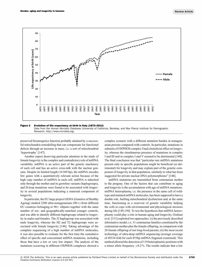
The particular combination of genetic, environmental, historical,anthropological, socio-economic and cultural factors as well asgeographical origin could contribute to the longer female life ex-pectancy worldwide. To increase our knowledge on these aspects,the model of centenarians could represent a useful approach.These extraordinary individuals (mostly women) are character-ized by a peculiar and heterogeneous phenotype embodying thebest example of longevity and successful aging. Most of themhave survived, escaped or delayed the onset of major age-relateddiseases [124–126]. Centenarians are the outcome of a numberof biological processes that exert their effects lifelong, from birth(and even before) until the extreme limits of human life. Froma demographic point of view, the high number of centenariansin our societies is the integrated result of complex interactionsbetween humans and their environment(s) which underwent con-sistent changes since the beginning of the 20th century and whichare continuing today. Therefore the study of centenarians repres-ents a sort of ‘historical probing’ that allows the tracing of theabove-mentioned complex basis of the longevity today. A histor-ical perspective of demographic data on gender and longevity inItaly is shown in Box 1.
The model of centenarians has some disadvantages due totheir rarity, lack of an age-matched control group and pheno-typical frailty related to their extreme age. Literature suggeststhat longevity ‘runs in families’ through different generations[136] and, indeed, centenarian offspring appear to be healthier[137,138] and to have a more favourable biological signature[139] with respect to age-matched controls, thus representing asuitable model to identify early biological factors/markers cor-related to healthy aging and higher ‘risk’ of longevity. Thus fe-male offspring of centenarian parents could represent a peculiarsubgroup of women characterized by a survival advantage not ac-companied by the worst quality of life typical of elderly women.
Within this scenario, where plenty of data have described thehormonal, immunological and metabolic gender differences, thestudy of long-lived families has allowed us to address peculiaraspects of the genetics of aging in women, following the ‘threegenetics conceptualization’ we have proposed recently [140]. Wehave suggested that an integrated investigation of nuclear genet-ics, mitochondrial DNA genetics and gut microbiome is essentialto grasp the genetic contribution to aging and longevity in humansconsidered as meta-organisms.
1717c© 2016 The Author(s). This is an open access article published by Portland Press Limited on behalf of the Biochemical Society and distributed under theCreative Commons Attribution Licence 4.0 (CC BY).

R. Ostan and others
Box 1 Demographic data on gender and longevity: a historicalperspective in Italy
Low-mortality countries, as well as Italy, in the recent decades haveseen a process of reduction in mortality at all ages of life that hasallowed pronounced gains in life expectancy [127]. Currently, theaverage life expectancy at birth in Italy is among the highest in theworld, having reached 80 years for men and 85 years for women.The improvement in living conditions, education, nutrition and life-styles, and progress in the prevention, diagnosis and treatment ofdiseases, have been crucial in reducing the risk of death even inadvanced ages of life. Indeed, mortality rates at older ages showeda linear downward trend between 1950 and 2012 [128].
The decline in old-age mortality is thought to be the main causeof the dramatic increase in centenarians [129], whose numberdoubles approximately every 7 years. According to data of the ItalianNational Institute of Statistics (http://demo.istat.it/), the numberof centenarians has reached 19095 (i.e. over 31 per 100 thou-sand residents) on 1 January 2015. At the same date, accordingto the ISTAT register, 878 residents on Italian territory were semi-supercentenarians (persons aged 105 or over), whereas 17 weresupercentenarians (persons aged 110 or over).
Increased levels of survival are linked to the long process of epi-demiologic transition that saw a radical transformation of mortalityin its gender, age and cause profiles [131,132].As shown in Figure 2, life expectancy in Italy was very similar formales and females until the early 20th Century. Afterwards, theevolution of life expectancy, while being dramatically marked bya sharp drop during the two World Wars, showed a differentiationbetween males and females that reached a maximum of 6.75 yearsin 1979 and 1980.
The recent decrease in the gender gap is mainly due to the reduc-tion of excess male mortality between the ages of 45 and 75 years.On the other hand, the disadvantage of men compared with womenat older ages is emphasized. This phenomenon might be related toa generation effect: whereas in the younger generations with morehealthy lifestyles the gap is reducing, the cohorts born in the early20th Century and the mid-1930s are ‘carriers’ of an excess mor-tality [133]. The current centenarians emerge from these cohortsand show geographical differences in the female/male ratio (F/M),which is higher in the North-West and North-East areas (around7:1 and 6:1 respectively), intermediate in the Centre (around 5:1)and lower in the South and Islands (around 4:1), according to themost recent ISTAT and census data. It is worth mentioning that ina mountainous zone of Sardinia (Nuoro province), an exceptionallyhigh number of male centenarians was identified, together with anunusually low F/M ratio [134].
The pattern of distribution of extremely long-lived individuals iscertainly affected by environmental factors which shaped the geo-graphy of life expectancy in Italy through a different impact on themain causes of death in the elderly [135]. However, a role of ge-netic factors is suggested by the finding of a correlation betweencentenarians’ gender ratio across the national territory and the firstprincipal component obtained by studying the polymorphic variationat 95 different loci [5].
X chromosome inactivation (XCI) skewing inhuman aging and longevityIn cells from females, one of the two X chromosomes is epi-genetically and randomly inactivated in early embryonic life.Young women are a mosaic of two cell populations in whicheither the maternal or the paternal X chromosome is inactivated,and the ratio is close to 50 % for each chromosome. A generalconcordance was seen in the XCI (X chromosome inactivation)pattern between haemopoietic tissue (blood and/or spleen) andseveral other tissues (e.g. brain, skin, heart, lung, muscle, kid-ney and gastrointestinal). According to the ‘Heterogametic SexHypothesis’, having two copies of the X chromosome may beadvantageous for females because of possible selection with age
of the better X chromosome while inactivating the deleteriousone [141]. In addition, previous data reveal that a small portion(∼17 %) of the genes on the inactivated X chromosome are par-tially active providing a further survival advantages for females[141]. During aging, a marked deviation from the equivalent ratio(50:50) between maternal and paternal X chromosome inactiva-tion occurs (skewing of XCI) in blood cells and the concordanceof XCI among tissues may weaken with age. In particular, com-paring haemopoietic tissues and brain in the oldest women, thegreatest difference between inactivation values of the two tissueswere found [142]. The XCI patterns in brain are of particularclinical relevance, because the X chromosome is relatively en-riched for genes involved in neuronal functioning [143]. Someauthors suggested that age-associated XCI skewing could be in-volved in the pathogenesis of several diseases such as autoim-munity and cancer [144]. Our proposed experimental model oflongevity/healthy aging consisting of female centenarians, theirfemale offspring, female offspring born from non-long-lived par-ents (age-matched controls) and young women has allowed us toextend to centenarians the study of XCI skewing and to demon-strate that this process was significantly less severe and frequentin centenarian offspring compared with their age-matched con-trols [145]. These results highlight a possible detrimental linkbetween the rate of XCI skewing and healthy aging/longevity,fitting the hypothesis that the balanced female mosaic is a win-ning strategy, sustaining a co-operative adaptive mechanism withpossible biological advantages, whereas a skewed situation infavour of one of the two X chromosomes would represent an un-favourable condition to attain health and longevity. Conversely,the absence of a similar mosaic strategy in men might contributeto their shorter lifespan [1].
A recent paper has described the correlation between SEMs(stochastic epigenetic mutations) (i.e. rare or stochastic epimuta-tions not shared among individuals) and XCI skewing duringaging demonstrating that the number of SEMs was low in child-hood and increased exponentially with age [146]. Moreover,a multivariate analysis has indicated a significant correlationbetween SEMs and degree of XCI skewing after adjustment forage, indicating for the first time that XCI skewing may not be adirect consequence of aging, but is mediated by the number ofSEMs. The data from this study support the hypothesis that anincreased number of SEMs might influence haemopoietic stemcells viability or might create conditions able to induce clonalstochastic loss of a specific type of haemopoietic cells [146].
mtDNA and gender in human aging and longevityMitochondria are considered to be important determinants of cellaging because they are involved in several fundamental processessuch as cellular energy/ATP production, the urea cycle, heat pro-duction, apoptosis, inflammasome activation and cell senescence.Mitochondria are also the main producers of ROS (reactive oxy-gen species), the most important by-products of OXPHOS (ox-idative phosphorylation), which, besides their physiological rolein cell signalling, have been suggested to play a role in the agingprocess as well as in age-related diseases. Data from primaryculture of fibroblasts from long-living individuals, including fe-male centenarians, indicate that longevity is characterized by a
1718 c© 2016 The Author(s). This is an open access article published by Portland Press Limited on behalf of the Biochemical Society and distributed under theCreative Commons Attribution Licence 4.0 (CC BY).
Gender, aging and longevity in humans
Figure 2 Evolution of life expectancy at birth in Italy (1872–2012)Data from the Human Mortality Database (University of California, Berkeley, and Max Planck Institute for DemographicResearch, http://www.mortality.org).
preserved bioenergetics function probably attained by a success-ful mitochondria remodelling that can compensate for functionaldefects through an increase in mass, i.e. a sort of mitochondrial‘hypertrophy’ [147].
Another aspect deserving particular attention in the study offemale longevity is the complex and contradictory role of mtDNAvariability. mtDNA is an active part of the genetic machineryof each cell and has an active cross-talk with the nuclear gen-ome. Despite its limited length (16 569 bp), the mtDNA encodesfew genes with a quantitatively relevant action because of thehigh copy number of mtDNA in each cell. mtDNA is inheritedonly through the mother and its germline variants (haplogroups),and D-loop mutations were found to be associated with longev-ity in several populations indicating a maternal component oflongevity.
In particular, the EU large project GEHA (Genetics of HealthyAgeing) studied 2200 ultra-nonagenarians (90+) from differentEU countries belonging to 90+ sibpairs together with the samenumber of sex- and geographically-matched younger controls,and was able to identify different haplogroups related to longev-ity in males and females. The J2 haplogroup was associated withmale longevity, whereas the H2 and T2 haplogroups were as-sociated with female longevity [148]. Taking advantage of thecomplete sequencing of a high number of mtDNA molecules,it was also possible to evaluate for the first time the cumulativeeffect of specific and concomitant mtDNA mutations, includingthose that have a low or very low impact. The analysis of themutations occurring in different OXPHOS complexes showed a
complex scenario with a different mutation burden in nonagen-arian persons compared with controls. In particular, mutations insubunits of OXPHOS complex I had a beneficial effect on longev-ity, whereas the simultaneous presence of mutations in complexI and III and in complex I and V seemed to be detrimental [148].The final conclusion was that “particular rare mtDNA mutationspresent only in specific populations might be beneficial (or det-rimental) for longevity and may explain part of the genetic com-ponent of longevity in that population, similarly to what has beensuggested for private nuclear DNA polymorphisms” [148].
mtDNA mutations are transmitted from centenarian motherto the progeny. One of the factors that can contribute to agingand longevity is the accumulation with age of mtDNA mutations.mtDNA heteroplasmy, i.e. the presence in the same cell of wild-type and mutated mtDNA molecules, has been supposed to have adouble role, fuelling mitochondrial dysfunction and, at the sametime, functioning as a reservoir of genetic variability helpingthe cells to cope with environmental and physiological stressorsduring life [149,150]. To test the hypothesis that mtDNA hetero-plasmy could play a role in human aging and longevity, Giulianiet al. [151] exploited two approaches: (i) the previously describedinformative model, i.e. 31 centenarian families constituted by thecentenarian mother plus the female offspring, in comparison with28 female offspring of not long-lived parents; (ii) the most recenttechnology of ultra-deep mtDNA sequencing (average coverageof 49334-fold for each 853bp mtDNA fragment examined). Thismethod allowed the detection of 119 heteroplasmic positions witha minor allele frequency �0.2 %. The results indicate that a low
1719c© 2016 The Author(s). This is an open access article published by Portland Press Limited on behalf of the Biochemical Society and distributed under theCreative Commons Attribution Licence 4.0 (CC BY).

R. Ostan and others
level of heteroplasmies are transmitted and maintained withinfamilies until extreme age. However, a non-heteroplasmic vari-ant associated with longevity and healthy aging was identified buta particular and unique heteroplasmy profile for each family wasdrawn. Therefore mtDNA heteroplasmy appears to be a familialtrait transmitted by the mothers which can contribute to healthyaging and longevity [151].
On the other hand, a number of studies have investigated theassociation between mtDNA inherited variants and multifactorialdiseases, such as diabetes [152], ischaemic disease [153] andneurodegenerative diseases such as PD [154] and AD [44]. Asdescribed previously, a high-resolution analysis (sequencing ofdisplacement loop and restriction analysis of specific markers inthe coding region of mtDNA) found that sub-haplogroup H5 is arisk factor for AD in particular for females and independently ofthe APOE genotype partially explaining the higher prevalence ofAD in women [44].
Gut microbiota and gender in human aging andlongevityHumans have to be considered as metaorganisms due to symbi-otic relationship with the numerous microbial communities (‘mi-crobiota’) present in various anatomical locations of the humanbody. Several hundreds of individual bacterial species colonizemouth, upper airways, skin, vagina and intestinal tract constitut-ing a complex and dynamic ecosystem which cross-talk with theenvironment as well as the rest of the body, including liver andbrain among others. At present, the microbiota associated withthe intestinal tract [GM (gut microbiota)] is the most studied. TheGM are essential for the synthesis of some fundamental nutrientsand energy production from food and are able to strongly mod-ulate innate and specific immunity. The gastrointestinal tract ofnewborns becomes colonized immediately after birth with micro-organisms, mainly from the mother. The composition of vaginaltract microbiota of the mother, the mode of delivery (natural orCaesarean) and breast or formula feeding have a deep impacton the GM of human offspring since the very beginning of life.Strong evidence has suggested that the early composition of themicrobiota of newborns plays an important role for the postnataldevelopment and functionality of the immune system [155].
Data regarding the association between genders and spe-cific GM communities are still unreliable even if some reportsfound that some specific taxa (Bacteroides, Ruminococcus, Eu-bacterium and Blautia) are more abundant in men, whereas Tre-ponema is prevalent in women [78,156]. Probably, these differ-ences in GM composition are due to lifestyle and dietary factorsas well as cultural gender-related habits rather than sex hormoneeffects [78]. Alterations of the GM have been observed in numer-ous diseases such as obesity, T2D, inflammatory bowel diseaseand CRC. In particular, specific signatures of GM patterns areassociated with autoimmunity affecting prevalently women andcontributing to the increase in their morbidity [78]. Thus thereis an urgent need to consider the role of gender background inthe GM ecology and its relationship with autoimmunity diseaseonset and therapy effects. This consideration is reinforced bythe fact that the importance of GM in human aging is dramat-ically emerging. This endogenous ecosystem, together with the
external antigenic load, is coming out as a crucial driving forceof the homoeostasis of the immune system, and lifelong GMchanges, from newborns to centenarians, can represent an im-portant source of inflammatory stimuli. Our group has shownthat female centenarians have a different composition of the GMin comparison with sex-matched younger persons, which is as-sociated with an increase in inflammaging (high plasma levels ofpro-inflammatory cytokines such as IL-6 and IL-8). In general,with aging, a decrease in the biodiversity of the composition ofthe GM is observed, with a trend towards an increase in potentiallypathogenic bacteria (pathobionts) with respect to the beneficialones (symbionts producing butyrate and other short-chain fattyacids) [157]. However, data on the remodelling of the GM and itsassociation with inflammaging are still lacking in men, underlin-ing again the importance of conducting gender-specific studiesto fill this gap.
AN AGENDA FOR THE FUTURE: AMANDATORY NEED FOR A GENDER-SPECIFICMEDICINE
The aging process starts ‘in utero’ and early events exert potenteffects later in life both in adult age and in old age. This lifelongperspective of aging and age-related diseases let emerge the im-portance of going beyond sex and to consider ‘gender’. Indeed,men and women differ not only biologically (biology, physiologyand genetics), but also regarding lifestyle and habits (smoking,nutrition, physical activity, type of work and education, amongothers) as well as regarding the capability of coping with stress(spousal bereavement, serving as care-givers to family members).These biological and non-biological factors interact continuouslylifelong, playing an overwhelming role in modulating healthand/or the propensity to diseases and disabilities later in life.
From basic to clinical sciences, there is a mandatory needfor studies where gender is appropriately and fully considered.The enormous progress of medicine in the last 50 years hasbeen reached by scientific investigations and publications wheregender has been rather neglected: ‘put gender on the agenda’ hasbeen repeatedly stated by top journals such as Nature since 2010[158,159].
Gender medicine can be considered quite a new but man-datory dimension of medicine that has to go much deeper inunderstanding the differences between men and women regard-ing all pathophysiological pathways, clinical characteristics andpharmacological responsiveness, as well as the importance oflifestyle and cultural aspects [18].
Within this scenario, it is even better to speak about a ‘gender-specific medicine’ and not only an indefinite and/or separated‘gender medicine’ since the gender perspective is broader, shouldbe more pervasive and penetrate all specialties of medicine.Gender medicine is not a separate exercise, or a separate branchof medicine. Therefore gender should be the focus for the clinicalapproach and this task requires a deep cultural change of mindas well as a reorganization of clinical services in all countriesand health systems. Gender medicine is even more necessary inneglected countries such as in Africa where the conditions of
1720 c© 2016 The Author(s). This is an open access article published by Portland Press Limited on behalf of the Biochemical Society and distributed under theCreative Commons Attribution Licence 4.0 (CC BY).

Gender, aging and longevity in humans
women are worse and gender differences are stronger and havea higher impact on the health status. At the same time, it is cur-rently no longer possible to conduct medical as well as biologicalsciences and education programmes without taking into consid-erations gender differences in the medical schools as well as inall educational programmes.
The knowledge of the biology of gender differences in humansare still in their infancy and there is an urgent need for specific-ally targeted large studies across countries, to take into accountthe above-mentioned cultural and anthropological differences ina globalized world where migration of persons from countriescharacterized by different genetic, cultural and anthropologicaltraits and habits is a hot topic.
In conclusion, the development of a gender-specific medicineis of the utmost importance in order to complete our understand-ing of the main mechanisms of aging as well as the differencesin prevention, care, treatment, evolution and outcomes of non-communicable diseases in both genders.
ACKNOWLEDGEMENTS
Thanks are due to Francesco Scalone for his suggestions aboutItalian demographic data.
FUNDING
This study was supported by the European Union’s Seventh Frame-work Programme [grant numbers 266486 (“NU-AGE: New diet-ary strategies addressing the specific needs of the elderly pop-ulation for healthy aging in Europe”), 602757 (“HUMAN: Healthand the understanding of Metabolism, Aging and Nutrition”) and259679 (“IDEAL: Integrated research on DEvelopmental determin-ants of Aging and Longevity”) (to C.F.)], by the Italian Ministry ofUniversity and Research “Progetto CL.A.N. Pros.it” [grant numberCTN01_00230_413096 (to C.F.)] and by the University of Florence(to D.M.), and by ’Progetto di Ricerca Sanitaria Finalizzata’ VenetoRegion, Italy.
REFERENCES1 Ostan, R., Monti, D. and Franceschi, C. (2015) Gender and
longevity. Ital. J. Gender-Specific Med. 1, 10–142 Cevenini, E., Bellavista, E., Tieri, P., Castellani, G., Lescai, F.,
Francesconi, M., Mishto, M., Santoro, A., Valensin, S., Salvioli,S. et al. (2010) Systems biology and longevity: an emergingapproach to identify innovative anti-aging targets andStrategies. Curr. Pharm. Des. 16, 802–813 CrossRef PubMed
3 Regitz-Zagrosek, V. (2012) Sex and gender differences inhealth. EMBO Rep. 13, 596–603 CrossRef PubMed
4 Franceschi, C., Motta, L., Valensin, S., Rapisarda, R., Franzone,A., Berardelli, M., Motta, M., Monti, D., Bonafe, M., Ferrucci, L.et al. (2000) Do men and women follow different trajectories toreach extreme longevity? Italian Multicenter Study onCentenarians (IMUSCE). Aging (Milano) 12, 77–84 PubMed
5 Passarino, G., Calignano, C., Vallone, A., Franceschi, C., Jeune,B., Robine, J.M., Yashin, A.I., Cavalli Sforza, L.L. and DeBenedictis, G. (2002) Male/female ratio in centenarians: apossible role played by population genetic structure. Exp.Gerontol. 37, 1283–1289 CrossRef PubMed
6 Barford, A. and Dorling, D. (2006) Life expectancy: women nowon top everywhere. BMJ 332, 808 CrossRef PubMed
7 Livi Bacci, M. (2015) La differenza di genere nella longevita: siattenua il vantaggio delle donne. In Longevita, Vecchiaia,Salute. (Salvini, S., ed.), pp. 34–38, Neodemos, Firenze
8 Wisser, O. and Vaupel, J.W. (2014) The sex differential inmortality: a historical comparison of the adult-age pattern of theratio and the difference, MPIDR Working Paper WP 2014-005.,Max Planck Institute for Demographic Research, Rostock
9 Beltran-Sanchez, H., Finch, C.E. and Crimmins, E.M. (2015)Twentieth century surge of excess adult male mortality. Proc.Natl. Acad. Sci. U.S.A. 112, 8993–8998 CrossRef PubMed
10 OECD (2014) Life expectancy and healthy life expectancy atbirth., OECD Publishing, Paris 16–17, Health at a Glance:Europe 2014
11 Reference deleted12 Van Oyen, H., Nusselder, W., Jagger, C., Kolip, P., Cambois, E.,
Robine, J.M. (2013) Gender differences in healthy life yearswithin the EU: an exploration of the “health-survival” paradox.Int. J. Public Health 58, 143–155 CrossRef PubMed
13 Austad, S.N. and Bartke, A. (2015) Sex differences in longevityand in responses to anti-aging interventions: a mini-review.Gerontology 62, 40–46 CrossRef PubMed
14 Heron, M. (2016) Deaths: leading causes for 2013. Natl. Vital.Stat. Rep. 65, 1–95
15 Jagger, C., Gillies, C., Moscone, F., Cambois, E., Van Oyen, H.,Nusselder, W. and Robine, J.-M. (2008) Inequalities in healthylife years in the 25 countries of the European Union in 2005: across-national meta-regression analysis. Lancet 372,2124–2131 CrossRef PubMed
16 Fouweather, T., Gillies, C., Wohland, P., Van Oyen, H., Nusselder,W., Robine, J.M., Cambois, E. and Jagger, C. (2015)Comparison of socio-economic indicators explaininginequalities in Healthy Life Years at age 50 in Europe: 2005and 2010. Eur. J. Public Health 25, 978–983 CrossRef PubMed
17 Regitz-Zagrosek, V., Oertelt-Prigione, S., Prescott, E., Franconi,F., Gerdts, E., Foryst-Ludwig, A., Maas, A.H., Kautzky-Willer, A.,Knappe-Wegner, D., Kintscher, U. et al. (2016) Gender incardiovascular diseases: impact on clinical manifestations,management, and outcomes. Eur. Heart. J. 37, 24–34CrossRef PubMed
18 Baggio, G., Corsini, A., Floreani, A., Giannini, S. and Zagonel, V.(2013) Gender medicine: a task for the third millennium. Clin.Chem. Lab. Med. 51, 713–727 CrossRef PubMed
19 Ambrosy, A.P., Fonarow, G.C., Butler, J., Chioncel, O., Greene,S.J., Vaduganathan, M., Nodari, S., Lam, C.S., Sato, N., Shah,A.N. and Gheorghiade, M. (2014) The global health andeconomic burden of hospitalizations for heart failure: lessonslearned from hospitalized heart failure registries. J. Am. Coll.Cardiol. 63, 1123–1133 CrossRef PubMed
20 Levy, D., Kenchaiah, S., Larson, M.G., Benjamin, E.J., Kupka,M.J., Ho, K.K., Murabito, J.M. and Vasan, R.S. (2002)Long-term trends in the incidence of and survival with heartfailure. N. Engl. J. Med. 347, 1397–1402 CrossRef PubMed
21 Kenchaiah, S. and Vasan, R.S. (2015) Heart failure in women:insights from the Framingham Heart Study. Cardiovasc. DrugsTher. 29, 377–390 CrossRef PubMed
22 Barton, M. and Meyer, M.R. (2009) Postmenopausalhypertension: mechanisms and therapy. Hypertension 54,11–18 CrossRef PubMed
23 Barrett-Connor, E.L., Cohn, B.A., Wingard, D.L. and Edelstein,SL. (1991) Why is diabetes mellitus a stronger risk factor forfatal ischemic heart disease in women than in men? TheRancho Bernardo Study. JAMA 265, 627–631CrossRef PubMed
24 Leslie, M.S. and Briggs, L.A. (2016) Preeclampsia and the riskof future vascular disease and mortality: a review. J. MidwiferyWomens Health 61, 315–324 CrossRef PubMed
25 Guariguata, L., Shaw, J.E., Whiting, D.W. and Linnenkamp, U.(2014) Determinants of gender differences in the prevalence ofdiabetes. Diabetes Res. Clin. Pract. 106, e14–e16CrossRef PubMed
1721c© 2016 The Author(s). This is an open access article published by Portland Press Limited on behalf of the Biochemical Society and distributed under theCreative Commons Attribution Licence 4.0 (CC BY).

R. Ostan and others
26 Wild, S., Roglic, G., Green, A., Sicree, R. and King, H. (2004)Global prevalence of diabetes: estimates for the year 2000 andprojections for 2030. Diabetes Care 27, 1047e53 CrossRef
27 Huxley, R., Barzi, F. and Woodward, M. (2006) Excess risk offatal coronary heart disease associated with diabetes in menand women: meta-analysis of 37 prospective cohort studies.BMJ 332, 73–78 CrossRef PubMed
28 Cull, C.A., Neil, H.A. and Holman, R.R. (2004) Changing aspirinuse in patients with Type 2 diabetes in the UKPDS. Diabet Med.21, 1368–1371 CrossRef PubMed
29 Wexler, D.J., Grant, R.W., Meigs, J.B., Nathan, D.M. andCagliero, E. (2005) Sex disparities in treatment of cardiac riskfactors in patients with type 2 diabetes. Diabetes Care 28,514–520 CrossRef PubMed
30 Persell, S.D. and Baker, D.W. (2004) Aspirin use among adultswith diabetes: recent trends and emerging sex disparities.Arch. Intern. Med. 164, 2492–2499 CrossRef PubMed
31 Maggi, S., Noale, M., Gallina, P., Bianchi, D., Marzar,i, C.,Limongi, F. and Crepaldi, G. (2006) Metabolic syndrome,diabetes, and cardiovascular disease in an elderly Caucasiancohort: the Italian Longitudinal Study on Aging. J. Gerontol. ABiol. Sci. Med. Sci. 61, 505–510 CrossRef PubMed
32 Mottillo, S., Filion, K.B., Genest, J., Joseph, L., Pilote, L.,Poirier, P., Rinfret, S., Schiffrin, E.L. and Eisenberg, M.J. (2010)The metabolic syndrome and cardiovascular risk a systematicreview and meta-analysis. J. Am. Coll. Cardiol. 56, 1113–1132CrossRef PubMed
33 Donington, J.S. and Colson, Y.L. (2011) Sex and genderdifferences in non-small cell lung cancer. Semin. Thorac.Cardiovasc. Surg. 23, 137–145 CrossRef PubMed
34 Nelson, R.L., Dollear, T., Freels, S. and Persky, V. (2007) Therelation of age, race, and gender to the subside location ofcolorectal carcinoma. Cancer 80, 103–107
35 Strimpakos, A.S., Syrigos, K.N. and Saif, M.V. (2009)Pharmacogenetics and biomarkers in colorectal cancer.Pharmacogenomics J. 9, 147–160 CrossRef PubMed
36 Ferlay, J., Steliarova-Foucher, E., Lortet-Tieulent, J., Rosso, S.,Coebergh, J.W., Comber, H., Forman, D. and Bray, F. (2013)Cancer incidence and mortality patterns in Europe: estimatesfor 40 countries in 2012. Eur. J. Cancer 49, 1374–1403CrossRef PubMed
37 Reference deleted PubMed38 Alzheimer Europe (2013) Dementia in Europe Yearbook 2013.,
Alzheimer Europe, Luxembourg PubMed39 Gabelli, C., Codemo, A. (2015) Gender differences in cognitive
decline and Alzheimer’s disease. Ital. J. Gender-Specific Med.1, 21–28
40 Bachman, D.L., Wolf, P.A., Linn, R., Knoefel, J.E., Cobb, J.,Belanger, A., D’Agostino, R.B. and White, L.R. (1992)Prevalence of dementia and probable senile dementia of theAlzheimer type in the Framingham Study. Neurology 42,115–159 CrossRef PubMed
41 Bretsky, P.M., Buckwalter, J.G., Seeman, T.E., Miller, C.A.,Poirier, J., Schellenberg, G.D., Finch, C.E. and Henderson, V.W.(1999) Evidence for an interaction between apolipoprotein Egenotype, gender, and Alzheimer disease. Alzheimer Dis.Assoc. Disord. 13, 216–221 CrossRef PubMed
42 Farrer, L.A., Cupples, L.A., Hainesm, J.L., Hyman, B., Kukull,W.A., Mayeux, R., Myers, R.H., Pericak-Vance, M.A., Risch, N.and van Duijn, C.M. (1997) Effects of age, sex, and ethnicity onthe association between apolipoprotein E genotype andAlzheimer disease: a meta-analysis. JAMA 278, 1349–1356CrossRef PubMed
43 Sampedro, F., Vilaplana, E., de Leon, M.J., Alcolea, D.,Pegueroles, J., Montal, V., Carmona-Iragui, M., Sala, I.,Sanchez-Saudinos, M.B., Anton-Aguirre, S. et al. (2015)Alzheimer’s Disease Neuroimaging Initiative: APOE-by-sexinteractions on brain structure and metabolism in healthyelderly controls. Oncotarget 6, 26663–26674CrossRef PubMed
44 Santoro, A., Balbi, V., Balducci, E., Pirazzini, C., Rosini, F.,Tavano, F., Achilli, A., Siviero, P., Minicuci, N., Bellavista, E.et al. (2010) Evidence for sub-haplogroup H5 of mitochondrialDNA as a risk factor for late onset Alzheimer’s disease. PLoSOne 5, e12037 CrossRef PubMed
45 Jaffe, A.B., Toran-Allerand, C.D., Greengard, P. and Gandy, S.E.(1994) Estrogen regulates metabolism of Alzheimer amyloid β
precursor protein. J. Biol. Chem. 269, 13065–13068PubMed
46 Wang, Q., Santizo, R., Baughman, V.L., Pelligrino, D.A. andIadecola, C. (1999) Estrogen provides neuroprotection intransient forebrain ischemia through perfusion-independentmechanisms in rats. Stroke 30, 630–637 CrossRef PubMed
47 Aenlle, K.K., Kumar, A., Cui, L., Jackson, T.C. and Foster, T.C.(2009) Estrogen effects on cognition and hippocampaltranscription in middle-aged mice. Neurobiol. Aging 30,932–945 CrossRef PubMed
48 Rocca, W.A., Grossardt, B.R. and Shuster, L.T. (2010)Oophorectomy, menopause, estrogen, and cognitive aging: thetiming hypothesis. Neurodegener. Dis. 7, 163–166CrossRef PubMed
49 Litim, N., Morissette, M. and Di Paolo, T. (2015) Neuroactivegonadal drugs for neuroprotection in male and female modelsof Parkinson’s disease. Neurosci. Biobehav. Rev. CrossRef
50 Pringsheim, T., Jette, N., Frolkis, A. and Steeves, T.D.L. (2014)The prevalence of Parkinson’s disease: a systematicreview and meta-analysis. Mov. Disord. 29, 1583–1590CrossRef PubMed
51 Nitkowska, M., Czyzyk, M. and Friedman, A. (2014)Reproductive life characteristics in females affected withParkinson’s disease and in healthy control subjects: acomparative study on Polish population. Neurol. Neurochir. Pol.48, 322–327 PubMed
52 Ragonese, P., D’Amelio, M., Salemi, G., Aridon, P., Gammino,M., Epifanio, A., Morgante, L. and Savettieri, G. (2004) Risk ofParkinson disease in women: effect of reproductivecharacteristics. Neurology 62, 2010–2014 CrossRef PubMed
53 Rocca, W.A., Bower, J.H., Maraganore, D.M., Ahlskog, J.E.,Grossardt, B.R., de Andrade, M. and Melton, L.J. (2008)Increased risk of parkinsonism in women who underwentoophorectomy before menopause. Neurology 70, 200–209CrossRef PubMed
54 Christensen, K., Doblhammer, G., Rau, R. and Vaupel, J.W.(2009) Ageing populations: the challenges ahead. Lancet 374,1196–1208 CrossRef PubMed
55 Corrao, S., Santalucia, P., Argano, C., Djade, C.D., Barone, E.,Tettamanti, M., Pasina, L., Franchi, C., Kamal Eldin, T.,Marengoni, A. et al. (2014) Gender-differences in diseasedistribution and outcome in hospitalized elderly: data from theREPOSI study. Eur. J. Intern. Med. 25, 617–623CrossRef PubMed
56 Santalucia, P., Franchi, C., Djade, C.D., Tettamanti, M., Pasina,L., Corrao, S., Salerno, F., Marengoni, A., Marcucci, M., Nobili,A. and Mannucci, P.M. (2015) Gender difference in drug use inhospitalized elderly patients. Eur. J. Intern. Med. 26, 483–490CrossRef PubMed
57 Johnson, V.L. and Hunter, D.J. (2014) The epidemiology ofosteoarthritis. Best Pract. Res. Clin. Rheumatol. 28, 5–15CrossRef PubMed
58 Srikanth, V.K., Fryer, J.L., Zhai, G., Winzenberg, T.M., Hosmer,D. and Jones, G. (2005) A meta-analysis of sex differencesprevalence, incidence and severity of osteoarthritis.Osteoarthritis Cartilage 13, 769–781 CrossRef PubMed
59 Sundstrom, A., Westerlund, O., Mousavi-Nasab, H., Adolfsson,R. and Nilsson, L.G. (2014) The relationship between maritaland parental status and the risk of dementia. Int. Psychogeriatr.26, 749–757 CrossRef PubMed
60 Heuberger, R. and Wong, H. (2014) The association betweendepression and widowhood and nutritional status in olderadults. Geriatr. Nurs. 35, 428–433 CrossRef PubMed
1722 c© 2016 The Author(s). This is an open access article published by Portland Press Limited on behalf of the Biochemical Society and distributed under theCreative Commons Attribution Licence 4.0 (CC BY).
Gender, aging and longevity in humans
61 Cole, M.G. and Dendukuri, N. (2003) Risk factors fordepression among elderly community subjects: a systematicreview and meta-analysis. Am. J. Psychiatry 160, 1147–1156CrossRef PubMed
62 Dahlberg, L., Andersson, L., McKee, K.J. and Lennartsson, C.(2015) Predictors of loneliness among older women and men inSweden: a national longitudinal study. Aging Ment. Health 19,409–417 CrossRef PubMed
63 Dykstra, P.A. and Fokkema, T. (2007) Social and emotionalloneliness among divorced and married men and women:comparing the deficit and cognitive perspectives. Basic Appl.Social Psychol. 29, 112 CrossRef
64 Lennartsson, C. and Lundberg, O. (2007) ‘What’s maritalstatus got to do with it?’ Gender inequalities in economicresources, health and functional abilities among older adults.In Health Inequalities and Welfare Resources: Continuity andChange in Sweden. (Fritzell, J. and Lundberg, O., eds),pp. 179–198, The Policy Press, Bristol
65 Moon, J.R., Kondo, N., Glymour, M.M. and Subramanian, S.V.(2011) Widowhood and mortality: a meta-analysis. PLoS One 6,e23465 CrossRef PubMed
66 Clouston, S.A., Lawlor, A. and Verdery, A.M. (2014) The role ofpartnership status on late-life physical function. Can. J. Aging33, 413–425 CrossRef PubMed
67 Vina, J., Borras, C., Gambini, J., Sastre, J. and Pallardo, F.V.(2005) Why females live longer than males: control of longevityby sex hormones. Sci. Aging Knowledge Environ. 23, pe17
68 Bouman, A., Heineman, M.J. and Faas, M.M. (2005) Sexhormones and the immune response in humans. Hum. Reprod.Update 11, 411–423 CrossRef PubMed
69 Giefing-Kroll, C., Berger, P., Lepperdinger, G. andGrubeck-Loebenstein, B. (2015) How sex and age affectimmune responses, susceptibility to infections, and responseto vaccination. Aging Cell 14, 309–321 CrossRef PubMed
70 Hao, S., Zhao, J., Zhou, J., Zhao, S., Hu, Y. and Hou, Y. (2007)Modulation of 17β -estradiol on the number and cytotoxicity ofNK cells in vivo related to MCM and activating receptors. Int.Immunopharmacol. 7, 1765–1775 CrossRef PubMed
71 Ashcroft, G.S., Greenwell-Wild, T., Horan, M.A., Wahl, S.M. andFerguson, M.W. (1999) Topical estrogen accelerates cutaneouswound healing in aged humans associated with an alteredinflammatory response. Am. J. Pathol. 155, 1137–1146CrossRef PubMed
72 Kramer, P.R., Kramer, S.F. and Guan, G. (2004) 17β -Estradiolregulates cytokine release through modulation of CD16expression in monocytes and monocyte-derived macrophages.Arthritis Rheum. 50, 1967–1975 CrossRef PubMed
73 Olsen, N.J. and Kovacs, W.J. (2011) Evidence that androgensmodulate human thymic T cell output. J. Investig. Med. 59,32–35 CrossRef PubMed
74 Gonzalez, D.A., Diaz, B.B., Rodriguez Perez Mdel, C.,Hernandez, A.G., Chico, B.N. and de Leon, A.C. (2010) Sexhormones and autoimmunity. Immunol. Lett. 133, 6–13CrossRef PubMed
75 Klein, S.L., Jedlicka, A. and Pekosz, A. (2010) The Xs and Y ofimmune responses to viral vaccines. Lancet Infect. Dis. 10,338–349 CrossRef PubMed
76 Sakiani, S., Olsen, N.J. and Kovacs, W.J. (2013) Gonadalsteroids and humoral immunity. Nat. Rev. Endocrinol. 9, 56–62CrossRef PubMed
77 Bakhru, P. and Su, M.A. (2016) Estrogen turns down “the AIRE”.J. Clin. Invest. 126, 1239–1241 CrossRef PubMed
78 Gomez, A., Luckey, D. and Taneja, V. (2015) The gut microbiomein autoimmunity: sex matters. Clin. Immunol. 159, 154–162CrossRef PubMed
79 Fish, E.N. (2008) The X-files in immunity: sex-based differencespredispose immune responses. Nat. Rev. Immunol. 8,737–744 CrossRef PubMed
80 Zandman-Goddard, G., Peeva, E. and Shoenfeld, Y. (2007)Gender and autoimmunity. Autoimmun. Rev. 6, 366–372CrossRef PubMed
81 Vrachnis, N., Zygouris, D., Iliodromiti, Z., Daniilidis, A.,Valsamakis, G. and Kalantaridou, S. (2014) Probing the impactof sex steroids and menopause-related sex steroid deprivationon modulation of immune senescence. Maturitas 78, 174–178CrossRef PubMed
82 Ossewaarde, M.E., Bots, M.L., Verbeek, A.L., Peeters, P.H., vander Graaf, Y., Grobbee, D.E. and van der Schouw, Y.T. (2005)Age at menopause, cause-specific mortality and total lifeexpectancy. Epidemiology 4, 556–562 CrossRef
83 Gagnon, A., Smith, K.R., Tremblay, M., Vezina, H., Pare, P.P. andDesjardins, B. (2009) Is there a trade-off between fertility andlongevity? A comparative study of women from three largehistorical databases accounting for mortality selection Am. J.Hum. Biol. 4, 533–540 CrossRef
84 Weinberger, B. and Grubeck-Loebenstein, B. (2012) Vaccinesfor the elderly. Clin. Microbiol. Infect. 18 Suppl. 5, 100–108CrossRef PubMed
85 Scholz, J.L., Diaz, A., Riley, R.L., Cancro, M.P. and Frasca, D.(2013) A comparative review of aging and B cell function inmice and humans. Curr. Opin. Immunol. 25, 504–510CrossRef PubMed
86 Solana, R., Tarazona, R., Gayoso, I., Lesur, O., Dupuis, G. andFulop, T. (2012) Innate immunosenescence: effect of aging oncells and receptors of the innate immune system in humans.Semin. Immunol. 24, 331–341 CrossRef PubMed
87 Shaw, A.C., Panda, A., Joshi, S.R., Qian, F., Allore, H.G. andMontgomery, R.R. (2011) Dysregulation of human Toll-likereceptor function in aging. Ageing Res. Rev. 10, 346–353CrossRef PubMed
88 Salminen, A., Ojala, J., Kaarniranta, K. and Kauppinen, A.(2012) Mitochondrial dysfunction and oxidative stress activateinflammasomes: impact on the aging process and age-relateddiseases. Cell. Mol. Life Sci. 69, 2999–3013 CrossRef PubMed
89 Sansoni, P., Vescovini, R., Fagnoni, F., Biasini, C., Zann,i, F.,Zanlari, L., Telera, A., Lucchini, G., Passeri, G., Monti, D.,Franceschi, C. and Passeri, M. (2008) The immune system inextreme longevity. Exp. Gerontol. 43, 61–65 CrossRef PubMed
90 Franceschi, C., Bonafe, M., Valensin, S., Olivieri, F., De Luca,M., Ottaviani, E. and De Benedictis, G. (2000) Inflamm-aging:an evolutionary perspective on immunosenescence. Ann. N.Y.Acad. Sci. 908, 244–254 CrossRef PubMed
91 Cevenini, E., Monti, D. and Franceschi, C. (2013)Inflamm-ageing. Curr. Opin. Clin. Nutr. Metab. Care 16, 14–20CrossRef PubMed
92 Franceschi, C. and Campisi, J. (2014) Chronic inflammation(inflammaging) and its potential contribution to age-associateddiseases. J. Gerontol. A Biol. Sci. Med. Sci. 69 (Suppl. 1),S4–S9 CrossRef PubMed
93 Hirokawa, K., Utsuyama, M., Hayashi, Y., Kitagawa, M.,Makinodan, T. and Fulop, T. (2013) Slower immune systemaging in women versus men in the Japanese population. ImmunAgeing 15, 10–19
94 Giglio, T., Imro, M.A., Filaci, G., Scudeletti, M., Puppo, F., DeCecco, L., Indiveri, F. and Costantini, S. (1994) Immune cellcirculating subsets are affected by gonadal function. Life Sci54, 1305–1312 CrossRef PubMed
95 Deguchi, K., Kamada, M., Irahara, M., Maegawa, M.,Yamamoto, S., Ohmoto, Y., Murata, K., Yasui, T., Yamano, S.and Aono, T. (2001) Postmenopausal changes in production oftype 1 and type 2 cytokines and the effects of hormonereplacement therapy. Menopause 8, 266–273CrossRef PubMed
96 Kamada, M., Irahara, M., Maegawa, M., Ohmoto, Y., Takeji, T.,Yasui, T. and Aono, T. (2001) Postmenopausal changes inserum cytokine levels and hormone replacement therapy. Am.J. Obstet. Gynecol. 184, 309–314 CrossRef PubMed
97 Vural, P., Akgul, C. and Canbaz, M. (2006) Effects of hormonereplacement therapy on plasma pro-inflammatory andanti-inflammatory cytokines and some bone turnover markers inpostmenopausal women. Pharmacol. Res. 54, 298–302CrossRef PubMed
1723c© 2016 The Author(s). This is an open access article published by Portland Press Limited on behalf of the Biochemical Society and distributed under theCreative Commons Attribution Licence 4.0 (CC BY).

R. Ostan and others
98 Yasui, T., Maegawa, M., Tomita, J., Miyatani, Y., Yamada, M.,Uemura, H., Matsuzaki, T., Kuwahara, A., Kamada, M.,Tsuchiya, N. et al. (2007) Changes in serum cytokineconcentrations during the menopausal transition. Maturitas 56,396–403 CrossRef PubMed
99 Goetzl, E.J., Huang, M.C., Kon, J., Patel, K., Schwartz, J.B.,Fast, K., Ferrucci, L., Madara, K., Taub, D.D. and Longo, D.L.(2010) Gender specificity of altered human immune cytokineprofiles in aging. FASEB J. 24, 3580–3589 CrossRef PubMed
100 Ostan, R., Bucci, L., Capri, M., Salvioli, S., Scurti, M., Pini, E.,Monti, D. and Franceschi, C. (2008) Immunosenescence andimmunogenetics of human longevity. Neuroimmunomodulation15, 224–240 CrossRef PubMed
101 Fontana, L. and Partridge, L. (2015) Promoting health andlongevity through diet: from model organisms to humans. Cell161, 106–118 CrossRef PubMed
102 Fontana, L., Partridge, L. and Longo, V.D. (2010) Extendinghealthy lifespan: from yeast to humans. Science 328, 321–326CrossRef PubMed
103 Longo, V.D., Antebi, A., Bartke, A., Barzilai, N., Brown-Borg,H.M., Caruso, C., Curiel, T.J., de Cabo, R., Franceschi, C.,Gems, D. et al. (2015) Interventions to slow aging in humans:are we ready? Aging Cell 14, 497–510 CrossRef PubMed
104 Regan, J.C. and Partridge, L. (2013) Gender and longevity: whydo men die earlier than women? Comparative and experimentalevidence. Best Pract. Res. Clin. Endocrinol. Metab. 27,467–479 CrossRef PubMed
105 Clancy, D.J., Gems, D., Harshman, L.G., Oldham, S., Stocker,H., Hafen, E., Leevers, S.J. and Partridge, L. (2001) Extensionof life-span by loss of CHICO, a Drosophila insulin receptorsubstrate protein. Science 292, 104–106 CrossRef PubMed
106 Tatar, M., Kopelman, A., Epstein, D., Tu, M.P., Yin, C.M. andGarofalo, R.S. (2001) A mutant Drosophila insulin receptorhomolog that extends life-span and impairs neuroendocrinefunction. Science 292, 107–110 CrossRef PubMed
107 Holzenberger, M., Dupont, J., Ducos, B., Leneuve, P., Geloen,A., Even, P.C., Cervera, P. and Le Bouc, Y. (2003) IGF-1 receptorregulates lifespan and resistance to oxidative stress in mice.Nature 421, 182–187 CrossRef PubMed
108 Selman, C., Tullet, J.M., Wieser, D., Irvine, E., Lingard, S.J.,Choudhury, A.I., Claret, M., Al-Qassab, H., Carmignac, D.,Ramadani, F. et al. (2009) Ribosomal protein S6 kinase 1signaling regulates mammalian life span. Science 326,140–144 CrossRef PubMed
109 Miller, R.A., Harrison, D.E., Astle, C.M., Fernandez, E., Flurkey,K., Han, M., Javors, M.A., Li, X., Nadon, N.L., Nelson, J.F. et al.(2014) Rapamycin-mediated lifespan increase in mice is doseand sex dependent and metabolically distinct from dietaryrestriction. Aging Cell 13, 468–477 CrossRef PubMed
110 Passtoors, W.M., Beekman, M., Deelen, J., van der Breggen,R., Maier, A.B., Guigas, B., Derhovanessian, E., van Heemst,D., de Craen, A.J., Gunn, D.A. et al. (2013) Gene expressionanalysis of mTOR pathway: association with human longevity.Aging Cell 12, 24–31 CrossRef PubMed
111 van Heemst, D., Beekman, M., Mooijaart, S.P., Heijmans, B.T.,Brandt, B.W., Zwaan, B.J., Slagboom, P.E. and Westendorp, R.G.(2005) Reduced insulin/IGF-1 signalling and human longevity.Aging Cell 4, 79–85 CrossRef PubMed
112 Leung, K.C., Johannsson, G., Leong, G.M. and Ho, K.K. (2004)Estrogen regulation of growth hormone action. Endocr. Rev. 25,693–721 CrossRef PubMed
113 Bondanelli, M., Ambrosio, M.R., Margutti, A., Franceschetti, P.,Zatelli, M.C. and degli Uberti, E.C. (2003) Activation of thesomatotropic axis by testosterone in adult men: evidence for arole of hypothalamic growth hormone-releasing hormone.Neuroendocrinology 77, 380–387 CrossRef PubMed
114 Xu, Y., Chen, S.Y., Ross, K.N. and Balk, S.P. (2006) Androgensinduce prostate cancer cell proliferation through mammaliantarget of rapamycin activation and post-transcriptionalincreases in cyclin D proteins. Cancer Res. 66, 7783–7792CrossRef PubMed
115 Altamirano, F., Oyarce, C., Silva, P., Toyos, M., Wilson, C.,Lavandero, S., Uhlen, P. and Estrada, M. (2009) Testosteroneinduces cardiomyocyte hypertrophy through mammalian targetof rapamycin complex 1 pathway. J. Endocrinol. 202, 299–307CrossRef PubMed
116 Harvie, M.N., Pegington, M., Mattson, M.P., Frystyk, J., Dillon,B., Evans, G., Cuzick, J., Jebb, S.A., Martin, B., Cutler, R.G.et al. (2011) The effects of intermittent or continuous energyrestriction on weight loss and metabolic disease risk markers:a randomized trial in young overweight women. Int. J. Obes.(Lond.) 35, 714–727 CrossRef PubMed
117 Jakubowicz, D., Barnea, M., Wainstein, J. and Froy, O. (2013)Effects of caloric intake timing on insulin resistance andhyperandrogenism in lean women with polycystic ovarysyndrome. Clin. Sci. (Lond.) 125, 423–432 CrossRef PubMed
118 Bedard, A., Lamarche, B., Corneau, L., Dodin, S. and Lemieux,S. (2015) Sex differences in the impact of the Mediterraneandiet on systemic inflammation. Nutr. J. 14, 46CrossRef PubMed
119 Bedard, A., Riverin, M., Dodin, S., Corneau, L. and Lemieux, S.(2012) Sex differences in the impact of the Mediterranean dieton cardiovascular risk profile. Br. J. Nutr. 108, 1428–1434CrossRef PubMed
120 Bedard, A., Corneau, L., Lamarche, B., Dodin, S. and Lemieux,S. (2014) Sex-related differences in the effects of theMediterranean diet on glucose and insulin homeostasis. J.Nutr. Metab. 2014, 424130 PubMed
121 Bedard, A., Corneau, L., Lamarche, B., Dodin, S. and Lemieux,S. (2015) Sex differences in the impact of the Mediterraneandiet on LDL particle size distribution and oxidation. Nutrients 7,3705–3723 CrossRef PubMed
122 Bedard, A., Tchernof, A., Lamarche, B., Corneau, L., Dodin, S.and Lemieux, S. (2014) Effects of the traditional Mediterraneandiet on adiponectin and leptin concentrations in men andpremenopausal women: do sex differences exist? Eur. J. Clin.Nutr. 68, 561–566 CrossRef
123 Leblanc, V., Hudon, A.M., Royer, M.M., Corneau, L., Dodin, S.,Begin, C. and Lemieux, S. (2015) Differences between menand women in dietary intakes and metabolic profile in responseto a 12-week nutritional intervention promoting theMediterranean diet. J. Nutr. Sci. 4, e13 CrossRef PubMed
124 Franceschi, C., Motta, L., Motta, M., Malaguarnera, M., Capri,M., Vasto, S., Candore, G. and Caruso, C. (2008) The extremelongevity: the state of the art in Italy. Exp. Gerontol. 43, 45–52CrossRef PubMed
125 Salvioli, S., Capri, M., Bucci, L., Lanni, C., Racchi, M., Uberti,D., Memo, M., Mari, D., Govoni, S. and Franceschi, C. (2009)Why do centenarians escape or postpone cancer? The role ofIGF-1, inflammation and p53. Cancer Immunol. Immunother.58, 1909–1917 CrossRef PubMed
126 Paolisso, G., Gambardella, A., Ammendola, S., D’Amore, A.,Balbi, V., Varricchio, M. and D’Onofrio, F. (1996) Glucosetolerance and insulin action in healthy centenarians. Am. J.Physiol. 270, E890–E894 PubMed
127 Barbi, E. (2009) Piu longevi e meno diversi: recente evoluzionedella sopravvivenza. In Rapporto Sulla Popolazione: Salute eSopravvivenza. (Ongaro, F. and Salvini, S., eds), pp. 11–17, IlMulino, Bologna
128 Livi Bacci, M. (2015) Vivere a lungo, ma quanto a lungo?(Salvini, S., ed.), pp. 14–19, Neodemos, Firenze, In Longevita,Vecchiaia, Salute
129 Robine, J.M. and Caselli, G. (2005) An unprecedented increasein the number of centenarians. Genus 61, 57–82
130 Reference deleted131 Prati, S., Frova, L. (2011) Sopravvivenza e salute: l’evoluzione
della sopravvivenza. In Rapporto Sulla Popolazione. L’Italia a150 Anni dall’Unita. (Salvini, S. and De Rose, A., eds),pp. 79–88, Il Mulino, Bologna
132 Caselli, G. and Egidi, V. (1991) A new insight into morbidity andmortality transition in Italy. Genus 47, 1–29 PubMed
1724 c© 2016 The Author(s). This is an open access article published by Portland Press Limited on behalf of the Biochemical Society and distributed under theCreative Commons Attribution Licence 4.0 (CC BY).

Gender, aging and longevity in humans
133 di Fraia, G. (2011) Il recupero dello svantaggio maschile nellasopravvivenza. In Rapporto Sulla Popolazione. L’Italia a 150Anni dall’Unita. (Salvini, S. and De Rose, A., eds), pp. 88–90, IlMulino, Bologna
134 Poulain, M., Pes, G.M., Grasland, C., Carru, C., Ferrucci, L.,Baggio, G., Franceschi, C. and Deiana, L. (2004) Identificationof a geographic area characterized by extreme longevity in theSardinia island: the AKEA study. Exp. Gerontol. 39, 1423–1429CrossRef PubMed
135 Lipsi, R.M. and Caselli, G. (2002), Evoluzione della Geografiadella Mortalita in Italia: Tavole Provinciali e Probabilita di Morteper Causa. Anni: 1971–1973, 1981–1983, 1991–1993,Dipartimento di Scienze Demografiche, Universita degli StudiLa Sapienza, Roma
136 Caselli, G., Pozzi, L., Vaupel, J.W., Deiana, L., Pes, G., Carru,C., Franceschi, C. and Baggio, G. (2006) Family clustering inSardinian longevity: a genealogical approach. Exp. Gerontol.41, 727–736 CrossRef PubMed
137 Gueresi, P., Miglio, R., Monti, D., Mari, D., Sansoni, P., Caruso,C., Bonafede, E., Bucci, L., Cevenini, E., Ostan, R. et al. (2013)Does the longevity of one or both parents influence the healthstatus of their offspring? Exp. Gerontol 48, 395–400
138 Bucci, L., Ostan, R., Cevenini, E., Pini, E., Scurti, M., Vitale, G.,Mari, D., Caruso, C., Sansoni, P., Fanelli, F. et al. (2016)Centenarians’ offspring as a model of healthy aging: areappraisal of the data on Italian subjects and a comprehensiveoverview. Aging (Albany NY) 8, 510–520 CrossRef PubMed
139 Horvath, S., Pirazzini, C., Bacalini, M.G., Gentilini, D., Di Blasio,A.M., Delledonne, M., Mari, D., Arosio, B., Monti, D., Passarino,G. et al. (2015) Decreased epigenetic age of PBMCs fromItalian semi-supercentenarians and their offspring. Aging(Albany NY) 7, 1159–1170 CrossRef PubMed
140 Garagnani, P., Pirazzini, C., Giuliani, C., Candela, M., Brigidi, P.,Sevini, F., Luiselli, D., Bacalini, M.G., Salvioli, S., Capri, M.et al. (2014) The three genetics (nuclear DNA, mitochondrialDNA, and gut microbiome) of longevity in humans consideredas metaorganisms. Biomed. Res. Int. 2014, 560340CrossRef PubMed
141 Austad, S.N. (2006) Why women live longer than men: sexdifferences in longevity. Gend. Med. 3, 79–92CrossRef PubMed
142 Bittel, D.C., Theodoro, M.F., Kibiryeva, N., Fischer, W.,Talebizadeh, Z. and Butler, M.G. (2008) Comparison ofX-chromosome inactivation patterns in multiple tissues fromhuman females. J. Med. Genet. 45, 309–313CrossRef PubMed
143 Deng, X., Berletch, J.B., Nguyen, D.K. and Disteche, C.M.(2014) X chromosome regulation: diverse patterns indevelopment, tissues and disease. Nat. Rev. Genet. 15,367–378 CrossRef PubMed
144 Ozcelik, T. (2008) X chromosome inactivation and femalepredisposition to autoimmunity. Clin. Rev. Allergy Immunol. 34,348–351 CrossRef PubMed
145 Gentilini, D., Castaldi, D., Mari, D., Monti, D., Franceschi, C., DiBlasio, A.M. and Vitale, G. (2012) Age-dependent skewing of Xchromosome inactivation appears delayed in centenarians’offspring: is there a role for allelic imbalance in healthy agingand longevity? Aging Cell 11, 277–283 CrossRef PubMed
146 Gentilini, D., Garagnani, P., Pisoni, S., Bacalini, M.G., Calzari, L.,Mari, D., Vitale, G., Franceschi, C. and Di Blasio, A.M. (2015)Stochastic epigenetic mutations (DNA methylation) increaseexponentially in human aging and correlate with X chromosomeinactivation skewing in females. Aging (Albany NY) 7, 568–578CrossRef PubMed
147 Sgarbi, G., Matarrese, P., Pinti, M., Lanzarini, C., Ascione, B.,Gibellini, L., Dika, E., Patrizi, A., Tommasino, C., Capri, M. et al.(2014) Mitochondria hyperfusion and elevated autophagicactivity are key mechanisms for cellular bioenergeticpreservation in centenarians. Aging (Albany NY) 6, 296–310CrossRef PubMed
148 Raule, N., Sevini, F., Li, S., Barbieri, A., Tallaro, F., Lomartire, L.,Vianello, D., Montesanto, A., Moilanen, J.S., Bezrukov, V. et al.(2014) The co-occurrence of mtDNA mutations on differentoxidative phosphorylation subunits, not detected by haplogroupanalysis, affects human longevity and is population specific.Aging Cell 13, 401–407 CrossRef PubMed
149 Rose, G., Passarino, G., Scornaienchi, V., Romeo, G., Dato, S.,Bellizzi, D., Mari, V., Feraco, E., Maletta, R., Bruni, A. et al.(2007) The mitochondrial DNA control region shows geneticallycorrelated levels of heteroplasmy in leukocytes ofcentenarians and their offspring. BMC Genomics 8, 293CrossRef PubMed
150 Rose, G., Romeo, G., Dato, S., Crocco, P., Bruni, A.C.,Hervonen, A., Majamaa, K., Sevini, F., Franceschi, C. andPassarino, G. (2010) Somatic point mutations in mtDNA controlregion are influenced by genetic background and associatedwith healthy aging: a GEHA study. PLoS One 5, e13395CrossRef PubMed
151 Giuliani, C., Barbieri, C., Li, M., Bucci, L., Monti, D., Passarino,G., Luiselli, D., Franceschi, C., Stoneking, M. and Garagnani, P.(2014) Transmission from centenarians to theiroffspring of mtDNA heteroplasmy revealed by ultra-deepsequencing. Aging (Albany NY) 6, 454–467CrossRef PubMed
152 Achilli, A., Olivieri, A., Pala, M., Hooshiar Kashani, B., Carossa,V., Perego, U.A., Gandini, F., Santoro, A., Battaglia, V., Grugni, V.et al. (2011) Mitochondrial DNA backgrounds might modulatediabetes complications rather than T2DM as a whole. PLoSOne 6, e21029 CrossRef PubMed
153 Chinnery, P.F., Elliot, H.R., Syed, A. and Rothwell, P.M. (2010)Mitochondrial DNA haplogroups and risk of transient ischaemicattack and ischaemic stroke: a genetic association study.Lancet. Neurol. 9, 498–503 CrossRef PubMed
154 Gaweda-Walerych, K., Maruszak, A., Safranow, K., Bialecka, M.,Klodowska-Duda, G., Czyzewski, K., Slawek, J., Rudzinska, M.,Styczynska, M., Opala, G. et al. (2008) Mitochondrial DNAhaplogroups and subhaplogroups are associated withParkinson’s disease risk in a Polish PD cohort. J. Neural.Transm. 115, 1521–1526 CrossRef
155 Lee, Y.K. and Mazmanian, S.K. (2010) Has the microbiotaplayed a critical role in the evolution of the adaptiveimmune system? Science 330, 1768–1773CrossRef PubMed
156 Mueller, S., Saunier, K., Hanisch, C., Norin, E., Alm, L.,Midtvedt, T., Cresci, A., Silvi, S., Orpianesi, C., Verdenelli, M.C.et al. (2006) Differences in fecal microbiota in differentEuropean study populations in relation to age,gender, and country: a cross-sectional study. Appl.Environ. Microbiol. 72, 1027–1033CrossRef PubMed
157 Biagi, E., Nylund, L., Candela, M., Ostan, R., Bucci, L., Pini, E.,Nikkıla, J., Monti, D., Satokari, R., Franceschi, C. et al. (2010)Through ageing, and beyond: gut microbiota and inflammatorystatus in seniors and centenarians. PLoS One 5, e10667CrossRef PubMed
158 Anon (2010) Putting gender on the agenda. Nature 465, 665159 Schiebinger, L. (2014) Scientific research must take gender
into account. Nature 507, 9 CrossRef PubMed
Received 14 January 2016/21 June 2016; accepted 27 June 2016
Version of Record published 23 August 2016, doi: 10.1042/CS20160004
1725c© 2016 The Author(s). This is an open access article published by Portland Press Limited on behalf of the Biochemical Society and distributed under theCreative Commons Attribution Licence 4.0 (CC BY).