Embed Size (px)
Citation preview
Altex 27, 1/10 17
Markers of Murine Embryonic and Neural Stem Cells, Neurons and Astrocytes: Reference Points for Developmental Neurotoxicity Testing*Philipp B. Kuegler1,2, Bastian Zimmer1, Tanja Waldmann1, Birte Baudis1, Sten Ilmjärv3,4, Jürgen Hescheler5, Phil Gaughwin6, Patrik Brundin7, William Mundy8, Anna K. Bal-Price9, André Schrattenholz10, Karl-Heinz Krause11, Christoph van Thriel12, Mahendra S. Rao13, Suzanne Kadereit1 and Marcel Leist1
1Doerenkamp-Zbinden Chair for in vitro toxicology and Biomedicine, University of Konstanz, Germany; 2Konstanz Research School Chemical Biology, University of Konstanz, Germany; 3Quretec, tartu, estonia; 4Department of Physiology, University of tartu, estonia; 5Institute of Neurophysiology, University of Cologne, Germany; 6Stem Cell and Developmental Biology, Genome Institute of Singapore; 7Department of experimental Medical Science, Wallenberg Neuroscience Center, Sweden; 8Neu-rotoxicologyDivision,NationalHealthandEnvironmentalEffectsResearchLaboratory,OfficeofResearchand Development, USePA, NC, USA; 9european Centre for the Validation of Alternative Methods, Institute of Health and Consumer Protection, JRC, Ispra, Italy; 10ProteoSys, Mainz, Germany; 11Department of Genetic and laboratory Medicine, Geneva University Hospitals, Switzerland; 12leibniz Research Centre for Working environment and Human Factors, technical University of Dortmund, Germany; 13life technologies, Frederick, MD, USA
1 Introduction
embryonic stem cell (eSC)-based novel test systems are amongst the most dynamic areas of in vitro toxicology and biomedicine, and their development is funded e.g. by a large scale eU project (eSNAtS http://www.esnats.eu/). they may become future alternatives to animal testing and a key element of modern risk
assessment approaches (Pellizer et al., 2005). At the start of such aparadigmshiftintoxicologyitisessentialtodefinethenewtestsystems and their performance to the maximum possible degree. Thereforethisreviewundertakesafirstattempttodefinemarkersfor meSC and derived cell types as a starting point for an intense scientificdiscussionandfurtherimprovementsinthisarea.
* a report of t4 – the transatlantic think tank for toxicology, reviewed by T. Hartung and A. Goldberg (Baltimore, MD, USA) This manuscript has been reviewed by the National Health and Environmental Effects Research Laboratory, U.S. Environmental Protection Agency, and approved for publication. Approval does not signify that the contents reflect the views of the Agency, nor does mention of trade names or commercial products constitute endorsement or recommendation for use.
SummaryDevelopmental neurotoxicity (DNT) is a serious concern for environmental chemicals, as well as for food and drug constituents. Animal-based DNT models have relatively low sensitivity, and they are burdened by high work-load, cost and animal ethics. Murine embryonic stem cells (mESC) recapitulate several critical processes involved in the development of the nervous system if they are induced to differentiate into neural cells. They therefore represent an alternative toxicological model to predict human hazard. In this review, we discuss how mESC can be used for DNT assays. We have compiled a list of mRNA markers that define undifferentiated mESC (n = 42), neural stem cells (n = 73), astrocytes (n = 25) and the pattern of different neuronal and non-neuronal cell types generated (n = 57). We propose that transcriptional profiling can be used as a sensitive endpoint in toxicity assays to distinguish neural differentiation states during normal and disturbed development. Importantly, we believe that it can be scaled up to relatively high throughput whilst still providing rich information on disturbances affecting small cell subpopulations. Moreover, this approach can provide insight into underlying mechanisms and pathways of toxicity. We broadly discuss the methodological basis of marker lists and DNT assay design. The discussion is put in the context of a new generation of alternative assays (embryonic stem cell based DNT testing = ESDNT V2.0), that may later include human induced pluripotent stem cells, and that are not designed for 1:1 replacement of animal experiments, but are rather intended to improve human risk assessment by using independent scientific principles.
Keywords: stem cell, development, neurotoxicity, gene ontology, astrocyte, systems biology

Kuegler et al.
Altex 27, 1/1018
of methylmercury intoxication, autopsy studies revealed that this compound targeted the fetal neural system (e.g. eto et al., 1992),andthereby,Minamatadiseasecontributedsignificantlyto the identificationofdevelopmentalneurotoxicity (DNT)asan important endpoint in toxicology.
At the same time, the problem of developmental ecotoxicol-ogy (e.g. reduced reproductive success of birds due to pesticides in their food chain) was introduced by Rachel Carson in her book “silent spring”. the above mentioned examples provide insights intothespecificproblemsofDT.AnotherexampleforspecificDNTissuesisthalidomidethathasadefined“windowofsensi-tivity”. It did not cause problems when taken by pregnant women earlier than about 20 days after conception or later than about 35 days after conception. However, within this window it caused different effects, such as facial paralysis, when taken rather early, malformations of arms and legs in the middle and e.g. deformi-ties of the intestine when only taken late during the window of sensitivity. Notably, although thalidomide acted as a sedative in rats and mice (just as in humans), it had no teratogenic effects in these rodent species most frequently used for toxicity testing.
In Minamata, Chisso Corporation was found responsible for having caused the disease by introducing mercury waste into the Minamatabay.However,itwasmuchmoredifficultinthecaseof the victims of the congenital disease (who had never eaten contaminatedfish, but hadbeen exposed inutero) toprove acausal relationship between their disease and the methylmer-cury contamination. the situation was similar with other envi-ronmental contaminants, where a cause-effect relationship was disputed until R. Carson’s book became one of the key triggers for a wave of public concern that resulted in the ban of dichlor-diphenyltrichlorethane (DDt). these examples illustrate the specificproblemsofthedisciplineofdevelopmentaltoxicology,i.e.thedifficultytoprovideevidenceforcause-effectrelation-ships, and to identify suitable test systems. this fundamental weakness is also evident from less dramatic and more prevalent human poisonings that have reached the pandemic scale. the most prominent example of such an omnipresent contaminant is lead. It causes human developmental neurotoxicity, associated with a reduction of intelligence estimated to have resulted in an economic cost of > 100 billion $/year for each birth cohort born between 1960 and 1990 (Grandjean and landrigan, 2006). the average lead blood levels in children fell by 90% after the even-tual ban of lead additives to gasoline (Grandjean and landrigan, 2006). However, those exposed earlier may keep suffering from lead neurotoxicity due to its long biological half-life in addition to the DNt effects (Cory-Slechta, 1990). In the case of the de-velopmental toxicity of lead, the overwhelming epidemiological evidencefinallyhelpedtoconvinceregulatorstoreduceaccept-able thresholds, and the availability of trustworthy human ref-erence data helped to optimise a suitable experimental system to improve the toxicity evaluation. there are still many other wide-spread contaminants with effects below the threshold of a pandemic, but with the potential to affect a large population.
For most of these hazardous compounds evidence from hu-man epidemiology is not available. therefore, standardised test systems, mainly rodent-based bioassays, are used to de-rive points of departure (POD) for human health risk assess-
Murine embryonic stem cells (meSC) are pluripotent cells able to differentiate into all cell types in the mouse, including functional germ cells. Under appropriate conditions, meSC can be kept as in vitrocultureswithanindefinitecapacityforself-renewal (evans and Kaufman, 1981; Martin, 1981). the deriva-tion, use and properties of murine and human embryonic stem cells (eSC) have been reviewed earlier (leist et al., 2008a), also with the perspective of generating induced pluripotent stem cells (iPSC) by reprogramming of somatic cells from various species, including humans (Baker, 2010; Nagy and Nagy, 2010; lee and Studer, 2010). Pluripotent cells are suitable for molecu-lar biological manipulations, such as homologous recombina-tions with exogenous DNA to alter sequences of their genome. these properties have been used successfully for the generation ofknock-outandknock-inmicefrommodifiedmESC(Capec-chi, Martin and Smithies, Nobel Prize 2007). Such mice stand as in vivo proof that every stage and every cell of the nervous system can develop from meSC under appropriate conditions, and that the produced cells display different phenotypes accord-ing to the genotype of the meSC used initially for generation of the mice. It has also been demonstrated, that meSC can dif-ferentiate in vitro to different neuronal or glial subtypes (Wobus and Boheler, 2005). In theory, this offers the possibility to study all steps – in detail, in real time and at the resolution of indi-vidual cells – that lead from the multipotent meSC to the for-mation of neuroectoderm tissue, and further to the generation of neural stem cells (NSC), neuroblasts and various intermediate and mature types of neural cells (Bain et al., 1995; Fraichard et al., 1995; Strübing et al., 1995; Ying and Smith, 2003; Conti et al., 2005). the in vitro differentiation of meSC or human eSC (heSC), as well as of murine or human iPSC or neural precur-sorcells(NPC)toneuronsorotherdefinedcelltypesisofhighinterest to the understanding of developmental biology, but also its disturbances. thus, such test systems appear useful for the examinationofteratogenicityandthewidefieldofreproductivetoxicity (Rt). Moreover, introduction of neural endpoints rel-evant for developmental neurotoxicity (DNt) at different stages of development and development of more predictive and more sensitivemodelsystemsmaysignificantlyimprovethistestingstrategy (Breier et al., 2009; Moors et al., 2009; Coecke et al., 2007; lein et al., 2007).
2 Towards new test systems for developmental neurotoxicity
2.1 Lessons from the history of developmental neurotoxicity (DNT) testingthe area of developmental toxicology (Dt) came into public focus 50 years ago. At that time, the drug thalidomide caused severe birth defects, while the metal-organic contaminant meth-ylmercury caused Minamata disease (Harada, 1995). the latter also includes a congenital form, which is triggered by exposure of the unborn fetus to the toxicant. It has been shown that the mercuryconcentration inumbilicalcordbloodcanbesignifi-cantly higher than in the maternal blood (Sakamoto et al., 2004). Decades later, but still in consequence of this miniepidemic
Kuegler et al.
Altex 27, 1/10 19
substances should all be evaluated for their reproductive toxicity, experiments involving millions of animals would be performed to satisfy the legal requirements (Hartung and Rovida, 2009). However, these tests of individual chemicals constitute only the tipoftheiceberg.Practically,andscientifically,weshouldalsoconsider mixtures of compounds that humans and the environ-ment are exposed to. Already a dozen compounds can form thou-sands of different mixtures, which would be impossible to test by classical toxicological approaches based on animal experiments. even though some of the most relevant chemicals will be tested for their effects on reproduction, these tests will most likely leave open the safety questions concerning low dose effects on DNt. As indicated above, testing for Dt in the low-dose range and basinglegaldecisionsonthesedatahasprovenverydifficult,ifnot impossible, in most cases. this is even more an issue for the subarea of DNt. Within the ReACH testing requirements, DNt isonlyaddressedinexceptionalcasestriggeredbypositivefind-ings from other studies. Dedicated studies are otherwise not re-quired. thus, the concern remains that subtle, and predominantly functional, DNt effects triggered by chemicals might remain undiscovered. A comprehensive safety assessment will therefore require alternative approaches. technical (limited test capaci-ties),ethical(reductionofanimaltesting)andscientificreasonscall for new strategies in toxicology testing (leist et al., 2008b; Hartung, 2009a; Stingl et al., 2009; Bottini et al., 2007). One such strategy was suggested by the National Research Council (NRC, 2007). this milestone publication has been described in many reviews (Collins et al., 2008; leist et al., 2008c; Hartung and leist, 2008; Hartung, 2009b), and the strategy is now often summarised under the heading “tox21c” (toxicology for the 21st century).Twochangesareparticularlyimportant:first,noveltestsystems would be based on cell cultures (human, where possible) andsimplemodelorganisms(e.g.wormsandflies)insteadofro-dents and other higher vertebrates; second, the essential primary endpoints should cover disturbances of cellular (e.g. signalling, metabolic, homeostatic, proliferation, differentiation) pathways, and the overall resulting toxicological effect on humans would be predicted by systems biology-based approaches from these mechanistic data. the vision is that the new test systems would allow a much higher throughput of compounds and would work better in the low-dose range relevant for human exposure. the use of a systems-based approach (e.g. omics data, quantitative models linking cellular processes to adverse effects) is expected to be more predictive of human toxicity (see above issue of ro-dent testing of thalidomide). Added value may come from the possibility to use and to compare cells of different species, in-cluding humans.
For this vision to become reality, the new methods must be trusted and accepted globally (Bottini and Hartung, 2009; Bottini etal.,2007).Forinstance,technical/scientificbarriersarelinkedto the problem of validation (Hartung, 2007), as detailed for the areas of food safety and cosmetics safety (Hartung, 2008b; Hartung and Koëter, 2008; Vogel, 2009). New technologies and ideas can be imported and developed with specialists of other disciplines (e.g. Mitterhauser and toegel, 2008; Schrattenholz and Klemm, 2007), and teaching of alternative approaches may be achieved in different ways (Jukes, 2008; Jukes, 2009; leist,
ment in regulatory toxicology. In the 1960s, it became evident that developmental exposure to chemicals and drugs can alter behavioural function in young and adult animals (e.g. Werboff and Dembicki, 1962). As an indirect measure of neurotoxicity, behavioural readouts have been used and validated since the 1960s. these behavioural alterations are considered as an ob-servable expression of effects on nervous system function (Re-iter, 1978). therefore, guidelines and test batteries have been developed (Moser and MacPhail, 1990, 1992) and validated for use in behavioural toxicology. In the 1980s, the U.S. en-vironmentalProtectionAgency(U.S.EPA)developedthefirstDNt guidelines and initiated the standardisation of this testing strategy by the Organisation of economic Co-operation and De-velopment (OeCD). the development of the pertinent OeCD testguideline426,whichwasfinallyacceptedin2007(Makrisetal.,2009),wasguidedbytwoideas:first,themethodsneedto yield reproducible results within and across laboratories, and second, they must be sensitive to the effects of a range of neu-rotoxic agents (Middaugh et al., 2003). A recent review (Makris et al., 2009) revealed that just over 100 compounds have been tested in studies using the OeCD 426 draft guideline. Most of these compounds were pesticides (66%) and only 8 industrial chemicalswereincluded.Anotherreviewidentifiedabout174compounds for which neurobehavioural risk assessment had been performed, in many cases also on the offspring of the ex-posed animals (F1 generation). Only 1% of these compounds were industrial chemicals (Middaugh et al., 2003). the avail-able data for this relatively new area of toxicology of industrial chemicals is therefore rather limited. Some of the studies indi-cate that compounds exist for which DNt testing is the most sensitive of all toxicity endpoints in a broad safety evaluation battery. therefore inclusion of DNt testing in compound safety evaluation programmes such as ReACH is likely to add impor-tant information for regulatory decisions (Makris et al., 2009; Middaugh et al., 2003). At present the available data is insuf-ficienttopredicthowrepresentativethesefindingsare.
In summary, the historical development of DNt testing strate-gies was strongly based on the statistical concepts of reliability and sensitivity, and biological modes of action played a relatively minor role. In addition to the relatively low numbers of animal studies, few human reference data are available. thus, the predic-tive value of traditional DNt testing for human health is hard to estimate. establishment of alternative and additional approaches remainsahugescientificchallengerequiringnewstrategies.
2.2 The road to a mechanism-based developmental toxicologythe number of chemicals with potential for environmental ex-posure is large. the new european law entitled ReACH trig-gered an administrative procedure aiming at registration, evalu-ation and authorisation of all chemicals produced in the eU at > 1 t/year and not tested under the chemical safety law of 1982. It is expected that at least 30,000 chemicals will be registered, amongst these several thousand that are produced or used at > 100 t/year (Rovida and Hartung, 2009). A considerable percentage of these chemicals is found in the environment or at work places, where human exposure could potentially trigger Dt. As these
Kuegler et al.
Altex 27, 1/1020
ing, standardisation of protocols and exploratory activities, and a large variety of different approaches should be promoted and exploredforasufficientlylongtimebeforearationalselectionprocess can be initiated with the goal of identifying a smaller set of assays that may be used for regulatory decisions. therefore only some general considerations are highlighted here:
3.1.1 SpeciesFor human predictivity, heSC may appear more promising than rodent systems. However, for comparison with already exist-ing murine and rat in vivo databases, meSC may be more suit-able. In general, meSC presently represent a system with higher throughput and robustness: neurons are generated much faster and with higher yield than in the human system. As many more laboratories have worked with meSC compared to heSC, there is more experience in using the murine cultures. they are easier to handle, and the tools to genetically modify these cells are more advanced, while heSC show considerable variability in vivo and in vitro (Parsons et al., 2009; Wu et al., 2007; Osafune et al., 2008; Abeyta et al., 2004). It is also evident that heSC
2006; Hartung et al., 2009). However, much research in the 3R field addresses technical problems within already establishedconcepts (e.g. Rothen-Rutishauser, 2008; Heindl, 2008; Wan-ner, 2008; li, 2008a,b; Hagelschuer et al., 2009; Bahramsoltani et al., 2009; Manzer et al., 2009; Hartung and Hoffmann, 2009; Sauer et al., 2009). the next generation of methods (see chapter below on eSDNt V2.0) should set its own standards instead of aiming at a 1:1 substitution of existing animal protocols with their own set of problems (Hartung, 2008a; Pelkonen et al., 2009; Vedani et al., 2009; Sauer, 2009).
3 Markers for DNT testing
3.1 Challenges for an in vitro DNT test systemA number of questions arise when one considers develop-ing meSC, iPSC or heSC as potential test systems for DNt. these involve species, source, genotype, developmental status, throughput and endpoints of the model system. At the present stage, all different options and their combinations require test-
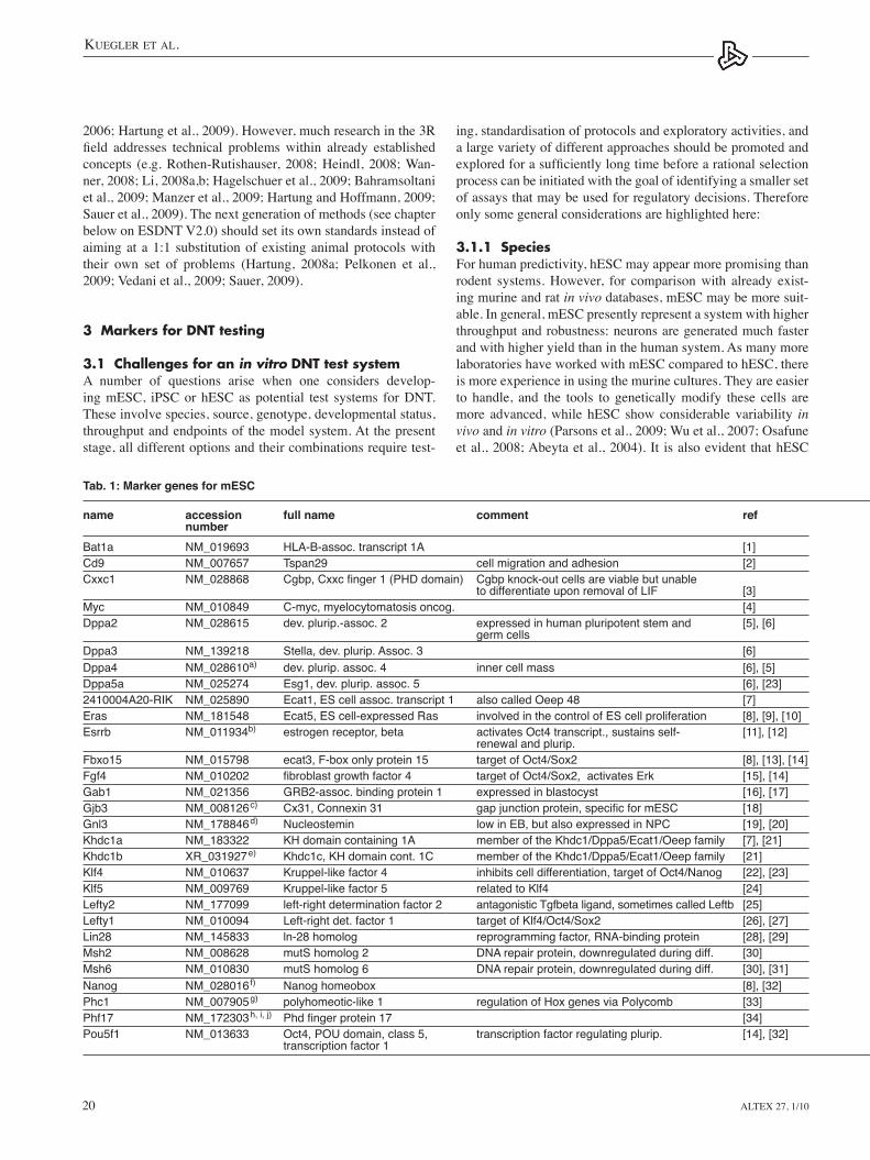
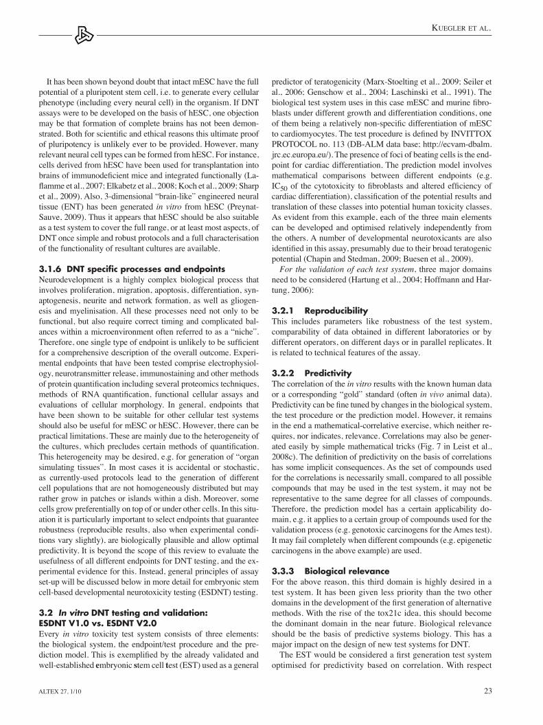
Tab. 1: Marker genes for mESC
name accession full name comment ref number Bat1a NM_019693 HLA-B-assoc. transcript 1A [1]Cd9 NM_007657 Tspan29 cell migration and adhesion [2]Cxxc1 NM_028868 Cgbp, Cxxc finger 1 (PHD domain) Cgbp knock-out cells are viable but unable to differentiate upon removal of LIF [3]Myc NM_010849 C-myc, myelocytomatosis oncog. [4]Dppa2 NM_028615 dev. plurip.-assoc. 2 expressed in human pluripotent stem and [5], [6] germ cells Dppa3 NM_139218 Stella, dev. plurip. Assoc. 3 [6]Dppa4 NM_028610a) dev. plurip. assoc. 4 inner cell mass [6], [5]Dppa5a NM_025274 Esg1, dev. plurip. assoc. 5 [6], [23]2410004A20-RIK NM_025890 Ecat1, ES cell assoc. transcript 1 also called Oeep 48 [7]Eras NM_181548 Ecat5, ES cell-expressed Ras involved in the control of ES cell proliferation [8], [9], [10]Esrrb NM_011934b) estrogen receptor, beta activates Oct4 transcript., sustains self- [11], [12] renewal and plurip. Fbxo15 NM_015798 ecat3, F-box only protein 15 target of Oct4/Sox2 [8], [13], [14]Fgf4 NM_010202 fibroblast growth factor 4 target of Oct4/Sox2, activates Erk [15], [14]Gab1 NM_021356 GRB2-assoc. binding protein 1 expressed in blastocyst [16], [17]Gjb3 NM_008126c) Cx31, Connexin 31 gap junction protein, specific for mESC [18]Gnl3 NM_178846d) Nucleostemin low in EB, but also expressed in NPC [19], [20]Khdc1a NM_183322 KH domain containing 1A member of the Khdc1/Dppa5/Ecat1/Oeep family [7], [21]Khdc1b XR_031927e) Khdc1c, KH domain cont. 1C member of the Khdc1/Dppa5/Ecat1/Oeep family [21]Klf4 NM_010637 Kruppel-like factor 4 inhibits cell differentiation, target of Oct4/Nanog [22], [23]Klf5 NM_009769 Kruppel-like factor 5 related to Klf4 [24]Lefty2 NM_177099 left-right determination factor 2 antagonistic Tgfbeta ligand, sometimes called Leftb [25]Lefty1 NM_010094 Left-right det. factor 1 target of Klf4/Oct4/Sox2 [26], [27]Lin28 NM_145833 ln-28 homolog reprogramming factor, RNA-binding protein [28], [29]Msh2 NM_008628 mutS homolog 2 DNA repair protein, downregulated during diff. [30]Msh6 NM_010830 mutS homolog 6 DNA repair protein, downregulated during diff. [30], [31]Nanog NM_028016f) Nanog homeobox [8], [32]Phc1 NM_007905g) polyhomeotic-like 1 regulation of Hox genes via Polycomb [33]Phf17 NM_172303h, i, j) Phd finger protein 17 [34] Pou5f1 NM_013633 Oct4, POU domain, class 5, transcription factor regulating plurip. [14], [32] transcription factor 1

Kuegler et al.
Altex 27, 1/10 21
behave differently from meSC concerning the pathways that control stemness. It has been suggested that they correspond to epiblast stem cells rather than to inner cell mass-derived cells, as do meSC, and they may not be able to form chimeras and an organism (li and Ding, 2009). Continuing basic research on ro-bust and more rapid heSC protocols is still needed to eventually provide a model system that avoids the species differences and the necessity for an interspecies extrapolation.
3.1.2 Type of cells used as starting materialDifferent cell types have been used to study aspects of DNt. eSC are derived from the inner cell mass of blastocysts (Mar-tin, 1981; evans and Kaufman, 1981; reviewed in leist et al., 2008a), and, using eSC-based models, all developmental steps are accessible for examination (Winkler et al., 2009). the down-side of this approach is that the cells need to be directed through all differentiation steps, preferably in a synchronised way, even under circumstances when only information on the last step is of interest. to avoid this problem, various other cell types have been used to study particular stages of DNt. For instance pri-
mary neurons or certain neuroblastoma, phaeochromocytoma or teratoma cells can differentiate to a partially neuronal pheno-type (e.g. axonal elongation and maturation), and this forms the basis for many test systems, which are of more limited scope but often of high reproducibility and throughput (Radio and Mundy, 2008; Radio et al., 2008, 2009; Hogberg et al., 2009, 2010). An intermediate solution would be the use of neural stem cells or neuroblast-like cells, which may be developed from eSC and that do not necessitate the initial differentiation steps required for eSC but still have the potential to develop into a number of different, morphologically and functionally mature neuronal and glial cell types (Buzanska et al., 2009; Breier et al., 2008; Wang et al., 2007). the advantages and disadvantages of such systems illustrate an important issue of DNt testing. the down-side is that such NSC-based systems cannot model the initial phaseofneuroectodermspecificationandformation.Thus,theeffect of compounds on this developmental period, associated with an important coordinated wave of gene transcription, can-not be tested. the upside of the use of NSC is that other phases, e.g. the step from NSC or neuroblasts, can be examined with
name accession full name comment ref number Rest NM_011263 RE1-silencing transcription factor maintains self-renewal and plurip., [35-39] (also NSC), discussed Sox2 NM_011443 SRY-box containing gene 2 transcription factor regulating plurip., (also NSC) [14], [32]Stat3 NM_213660k, l) signal transducer and activator involved in LIF signaling [40], [23] of transcription 3 Stip1 NM_016737 stress-ind. phosphoprot. role in plurip. signaling [41]Tcfcp2l1 NM_023755 transcription factor CP2-like 1 [2], [23]Tdgf1 NM_011562 Cripto, teratocarcinoma-derived target of nanog, Oct4, SMAD [8], [42] growth factor 1 Tdh NM_021480 L-threonine dehydrogenase [43]Tead4 NM_011567 TEA domain family member 4, expressed from 2 cell stage on to blastocyst [44] TEF-1-related factor 1 Tert NM_009354 telomerase (RT) reverse transcriptase [45]Tex19.1 NM_028602 Nuclear protein also germ line [46]Timp1 NM_011593m) tissue inhibitor of metallo- [2], [47] proteinase 1 Utf1 NM_009482 undifferentiated embryonic cell target of Oct4/Sox2 [8], [14], transcription factor 1 [48-50]Zfp42 NM_009556 Rex1, zinc finger protein 42 [8]Zic3 NM_009575 zinc finger protein of the required for maintenance of plurip. in ES cells [51], [52] cerebellum 3 and neural crest development
Additional accession numbers: a) NM_001018002, b) NM_001159500, c) NM_001160012, d) NM_153547, e) NM_001033904, f) NM_001080945, g) NM_001042623, h) NM_001130184, i) NM_001130185, j) NM_001130186, k) NM_213659, l) NM_213660, m) NM_0010443841. Sharov et al., 2003; 2: Abranches et al., 2009; 3. Carlone et al., 2005; 4. Lewitzky and Yamanaka, 2007; 5. Maldonado-Saldivia et al., 2007; 6. Bortvin et al., 2003; 7. Imamura et al., 2006; 8. Mitsui et al., 2003; 9. Takahashi et al., 2003; 10. Sorrentino et al., 2007; 11. Zhang et al., 2008; 12. Feng et al., 2009; 13. Tokuzawa et al., 2003; 14. Okumura-Nakanishi et al., 2005; 15. Kunath et al., 2007; 16. Schaeper et al., 2007; 17. Xie et al., 2005; 18. Worsdorfer et al., 2008; 19. Tsai and McKay, 2002; 20. Beekman et al., 2006; 21. Pierre et al., 2007; 22. Li et al., 2005; 23. Wei et al., 2005; 24. Ema et al., 2008; 25. Hamada et al., 2001; 26. Farthing et al., 2008; 27. Nakatake et al., 2006; 28. Hagan et al., 2009; 29. Hanna et al., 2009; 30. Roos et al., 2007; 31. Mason et al., 2009; 32. Chambers and Tomlinson, 2009; 33. Isono et al., 2005; 34. Tzouanacou et al., 2003; 35. Singh et al., 2008; 36. Canzonetta et al., 2008; 37. Johnson et al., 2008; 38. Buckley et al., 2009; 39. Jørgensen et al., 2009; 40. Kues et al., 2005; 41. Longshaw et al., 2009; 42. Liu et al., 2005; 43. Wang et al., 2009; 44. Nishioka et al., 2009; 45. Armstrong et al., 2005; 46. Kuntz et al., 2008; 47. Singla and McDonald, 2007; 48. van den Boom et al., 2007; 49. Nishimoto et al., 2005; 50. Okuda et al., 1998; 51. Lim et al., 2007; 52. Nakata et al., 1998
Kuegler et al.
Altex 27, 1/1022
nation of the role of certain genes in diseases and pathologies. especially the availability of meSC with reporter constructs has been broadly applied to high-throughput screens, e.g. for com-pounds affecting DNt (Suter and Krause, 2008; Suter et al., 2009; Conti et al., 2005). Similar reporter constructs have been introduced and used in heSC or iPSC, but there is still ample room for further development and improvement.
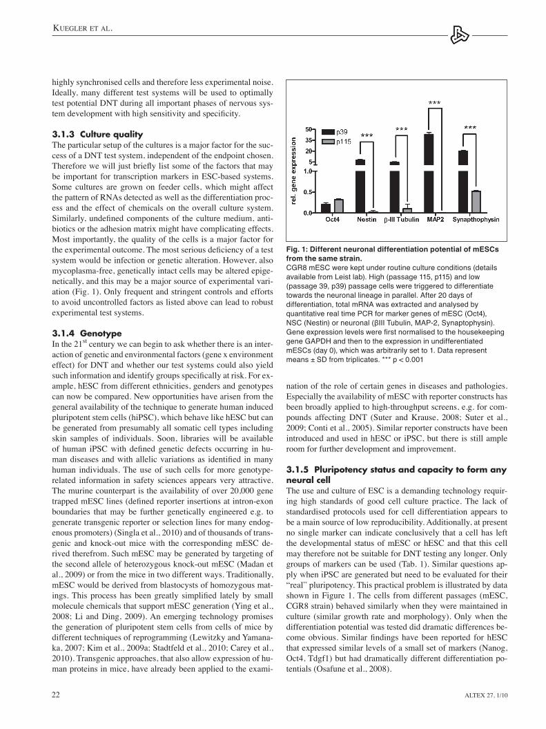
3.1.5 Pluripotency status and capacity to form any neural cell the use and culture of eSC is a demanding technology requir-ing high standards of good cell culture practice. the lack of standardised protocols used for cell differentiation appears to be a main source of low reproducibility. Additionally, at present no single marker can indicate conclusively that a cell has left the developmental status of meSC or heSC and that this cell may therefore not be suitable for DNt testing any longer. Only groups of markers can be used (tab. 1). Similar questions ap-ply when iPSC are generated but need to be evaluated for their “real” pluripotency. this practical problem is illustrated by data shown in Figure 1. the cells from different passages (meSC, CGR8 strain) behaved similarly when they were maintained in culture (similar growth rate and morphology). Only when the differentiation potential was tested did dramatic differences be-comeobvious.Similarfindings havebeen reported for hESCthat expressed similar levels of a small set of markers (Nanog, Oct4, tdgf1) but had dramatically different differentiation po-tentials (Osafune et al., 2008).
highly synchronised cells and therefore less experimental noise. Ideally, many different test systems will be used to optimally test potential DNt during all important phases of nervous sys-temdevelopmentwithhighsensitivityandspecificity.
3.1.3 Culture qualitythe particular setup of the cultures is a major factor for the suc-cess of a DNt test system, independent of the endpoint chosen. Thereforewewilljustbrieflylistsomeofthefactorsthatmaybe important for transcription markers in eSC-based systems. Some cultures are grown on feeder cells, which might affect the pattern of RNAs detected as well as the differentiation proc-ess and the effect of chemicals on the overall culture system. Similarly,undefinedcomponentsof theculturemedium,anti-biotics or the adhesion matrix might have complicating effects. Most importantly, the quality of the cells is a major factor for theexperimentaloutcome.Themostseriousdeficiencyofatestsystem would be infection or genetic alteration. However, also mycoplasma-free, genetically intact cells may be altered epige-netically, and this may be a major source of experimental vari-ation (Fig. 1). Only frequent and stringent controls and efforts to avoid uncontrolled factors as listed above can lead to robust experimental test systems.
3.1.4 Genotype In the 21st century we can begin to ask whether there is an inter-action of genetic and environmental factors (gene x environment effect) for DNt and whether our test systems could also yield suchinformationandidentifygroupsspecificallyatrisk.Forex-ample, heSC from different ethnicities, genders and genotypes can now be compared. New opportunities have arisen from the general availability of the technique to generate human induced pluripotent stem cells (hiPSC), which behave like heSC but can be generated from presumably all somatic cell types including skin samples of individuals. Soon, libraries will be available ofhuman iPSCwithdefinedgenetic defects occurring inhu-mandiseasesandwithallelicvariationsas identified inmanyhuman individuals. the use of such cells for more genotype-related information in safety sciences appears very attractive. the murine counterpart is the availability of over 20,000 gene trappedmESClines(definedreporterinsertionsatintron-exonboundaries that may be further genetically engineered e.g. to generate transgenic reporter or selection lines for many endog-enous promoters) (Singla et al., 2010) and of thousands of trans-genic and knock-out mice with the corresponding meSC de-rived therefrom. Such meSC may be generated by targeting of the second allele of heterozygous knock-out meSC (Madan et al., 2009) or from the mice in two different ways. traditionally, meSC would be derived from blastocysts of homozygous mat-ings.Thisprocesshasbeengreatly simplified latelyby smallmolecule chemicals that support meSC generation (Ying et al., 2008; li and Ding, 2009). An emerging technology promises the generation of pluripotent stem cells from cells of mice by different techniques of reprogramming (lewitzky and Yamana-ka, 2007; Kim et al., 2009a; Stadtfeld et al., 2010; Carey et al., 2010). transgenic approaches, that also allow expression of hu-man proteins in mice, have already been applied to the exami-
Fig. 1: Different neuronal differentiation potential of mESCs from the same strain. CGR8 mESC were kept under routine culture conditions (details available from Leist lab). High (passage 115, p115) and low (passage 39, p39) passage cells were triggered to differentiate towards the neuronal lineage in parallel. After 20 days of differentiation, total mRNA was extracted and analysed by quantitative real time PCR for marker genes of mESC (Oct4), NSC (Nestin) or neuronal (βIII Tubulin, MAP-2, Synaptophysin). Gene expression levels were first normalised to the housekeeping gene GAPDH and then to the expression in undifferentiated mESCs (day 0), which was arbitrarily set to 1. Data represent means ± SD from triplicates. *** p < 0.001
Kuegler et al.
Altex 27, 1/10 23
predictor of teratogenicity (Marx-Stoelting et al., 2009; Seiler et al., 2006; Genschow et al., 2004; laschinski et al., 1991). the biologicaltestsystemusesinthiscasemESCandmurinefibro-blasts under different growth and differentiation conditions, one ofthembeingarelativelynon-specificdifferentiationofmESCtocardiomyocytes.ThetestprocedureisdefinedbyINVITTOXPROtOCOl no. 113 (DB-AlM data base; http://ecvam-dbalm.jrc.ec.europa.eu/). the presence of foci of beating cells is the end-point for cardiac differentiation. the prediction model involves mathematical comparisons between different endpoints (e.g. IC50of thecytotoxicity tofibroblastsandalteredefficiencyofcardiacdifferentiation),classificationofthepotentialresultsandtranslation of these classes into potential human toxicity classes. As evident from this example, each of the three main elements can be developed and optimised relatively independently from the others. A number of developmental neurotoxicants are also identifiedinthisassay,presumablyduetotheirbroadteratogenicpotential (Chapin and Stedman, 2009; Buesen et al., 2009).
For the validation of each test system, three major domains need to be considered (Hartung et al., 2004; Hoffmann and Har-tung, 2006):
3.2.1 Reproducibilitythis includes parameters like robustness of the test system, comparability of data obtained in different laboratories or by different operators, on different days or in parallel replicates. It is related to technical features of the assay.
3.2.2 Predictivitythe correlation of the in vitro results with the known human data or a corresponding “gold” standard (often in vivo animal data). Predictivitycanbefinetunedbychangesinthebiologicalsystem,the test procedure or the prediction model. However, it remains in the end a mathematical-correlative exercise, which neither re-quires, nor indicates, relevance. Correlations may also be gener-ated easily by simple mathematical tricks (Fig. 7 in leist et al., 2008c).Thedefinitionofpredictivityonthebasisofcorrelationshas some implicit consequences. As the set of compounds used for the correlations is necessarily small, compared to all possible compounds that may be used in the test system, it may not be representative to the same degree for all classes of compounds. therefore, the prediction model has a certain applicability do-main, e.g. it applies to a certain group of compounds used for the validation process (e.g. genotoxic carcinogens for the Ames test). It may fail completely when different compounds (e.g. epigenetic carcinogens in the above example) are used.
3.3.3 Biological relevance For the above reason, this third domain is highly desired in a test system. It has been given less priority than the two other domainsinthedevelopmentofthefirstgenerationofalternativemethods. With the rise of the tox21c idea, this should become the dominant domain in the near future. Biological relevance should be the basis of predictive systems biology. this has a major impact on the design of new test systems for DNt.TheESTwouldbeconsideredafirstgenerationtestsystem
optimised for predictivity based on correlation. With respect
It has been shown beyond doubt that intact meSC have the full potential of a pluripotent stem cell, i.e. to generate every cellular phenotype (including every neural cell) in the organism. If DNt assays were to be developed on the basis of heSC, one objection may be that formation of complete brains has not been demon-strated.Bothforscientificandethicalreasonsthisultimateproofof pluripotency is unlikely ever to be provided. However, many relevant neural cell types can be formed from heSC. For instance, cells derived from heSC have been used for transplantation into brainsofimmunodeficientmiceandintegratedfunctionally(La-flammeetal.,2007;Elkabetzetal.,2008;Kochetal.,2009;Sharpet al., 2009). Also, 3-dimensional “brain-like” engineered neural tissue (eNt) has been generated in vitro from heSC (Preynat-Sauve, 2009). thus it appears that heSC should be also suitable as a test system to cover the full range, or at least most aspects, of DNt once simple and robust protocols and a full characterisation of the functionality of resultant cultures are available.
3.1.6 DNT specific processes and endpointsNeurodevelopment is a highly complex biological process that involves proliferation, migration, apoptosis, differentiation, syn-aptogenesis, neurite and network formation, as well as gliogen-esis and myelinisation. All these processes need not only to be functional, but also require correct timing and complicated bal-ances within a microenvironment often referred to as a “niche”. Therefore,onesingletypeofendpointisunlikelytobesufficientfor a comprehensive description of the overall outcome. experi-mental endpoints that have been tested comprise electrophysiol-ogy, neurotransmitter release, immunostaining and other methods ofproteinquantificationincludingseveralproteomicstechniques,methods of RNA quantification, functional cellular assays andevaluations of cellular morphology. In general, endpoints that have been shown to be suitable for other cellular test systems should also be useful for meSC or heSC. However, there can be practical limitations. these are mainly due to the heterogeneity of thecultures,whichprecludescertainmethodsofquantification.this heterogeneity may be desired, e.g. for generation of “organ simulating tissues”. In most cases it is accidental or stochastic, as currently-used protocols lead to the generation of different cell populations that are not homogeneously distributed but may rather grow in patches or islands within a dish. Moreover, some cells grow preferentially on top of or under other cells. In this situ-ation it is particularly important to select endpoints that guarantee robustness (reproducible results, also when experimental condi-tions vary slightly), are biologically plausible and allow optimal predictivity. It is beyond the scope of this review to evaluate the usefulness of all different endpoints for DNt testing, and the ex-perimental evidence for this. Instead, general principles of assay set-up will be discussed below in more detail for embryonic stem cell-based developmental neurotoxicity testing (eSDNt) testing.
3.2 In vitro DNT testing and validation: ESDNT V1.0 vs. ESDNT V2.0every in vitro toxicity test system consists of three elements: the biological system, the endpoint/test procedure and the pre-dictionmodel.Thisisexemplifiedbythealreadyvalidatedandwell-established embryonic stem cell test (eSt) used as a general
Kuegler et al.
Altex 27, 1/1024
the eSt may be adapted in different ways for DNt testing. However, in all cases a fundamental difference between cardio-teratogenicity and neuroteratogenity needs to be considered: the heart consists of a limited number of cell types in a relatively homogeneous tissue arrangement, and most developmental ef-fects on the heart have some form of histological or morpho-logical correlate. the nervous system consists of many different cell populations, and DNt, as well as many CNS diseases, can have predominantly behavioural and functional consequences (e.g. on regulation of mood, intelligence, attention, concentra-tion, motor activity) without obvious morphological correlates. this needs to be taken into account when test systems are be-
toneurally-active teratogens(DNTfield) itmaybecalledaneSDNt V1.0 (embryonic stem cell based developmental neu-rotoxicity test, version 1.0). It operates predominantly as a black box system, similar to reproductive toxicology studies in animals. Understanding of the mechanisms is not required to derive the results and the regulatory consequences in both positiveornegativecases.Moreover,anditisdifficulttoob-tain information from this system on why positive compounds are positive and why negative compounds are negative. How-ever, as this information is not required for regulatory testing ofchemicals,agoodcorrelationwassufficientforsuccessfulvalidation.
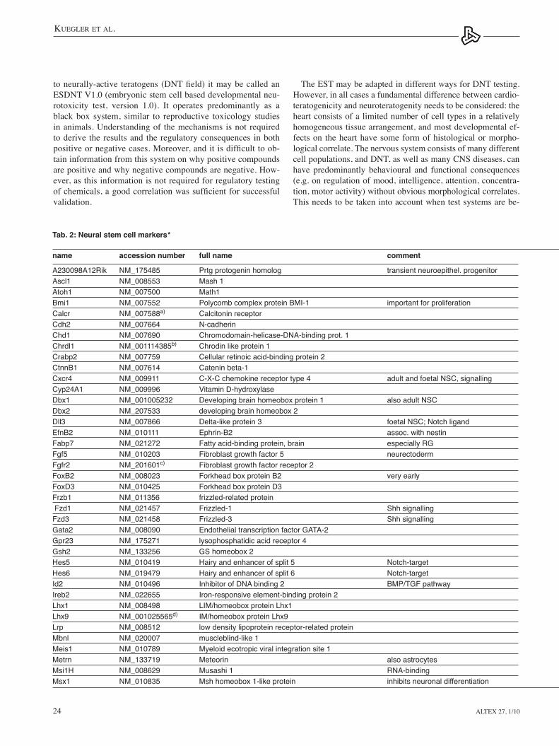
Tab. 2: Neural stem cell markers* name accession number full name comment
A230098A12Rik NM_175485 Prtg protogenin homolog transient neuroepithel. progenitorAscl1 NM_008553 Mash 1 Atoh1 NM_007500 Math1 Bmi1 NM_007552 Polycomb complex protein BMI-1 important for proliferationCalcr NM_007588a) Calcitonin receptor Cdh2 NM_007664 N-cadherin Chd1 NM_007690 Chromodomain-helicase-DNA-binding prot. 1 Chrdl1 NM_001114385b) Chrodin like protein 1 Crabp2 NM_007759 Cellular retinoic acid-binding protein 2 CtnnB1 NM_007614 Catenin beta-1 Cxcr4 NM_009911 C-X-C chemokine receptor type 4 adult and foetal NSC, signallingCyp24A1 NM_009996 Vitamin D-hydroxylase Dbx1 NM_001005232 Developing brain homeobox protein 1 also adult NSC Dbx2 NM_207533 developing brain homeobox 2 Dll3 NM_007866 Delta-like protein 3 foetal NSC; Notch ligandEfnB2 NM_010111 Ephrin-B2 assoc. with nestinFabp7 NM_021272 Fatty acid-binding protein, brain especially RGFgf5 NM_010203 Fibroblast growth factor 5 neurectodermFgfr2 NM_201601c) Fibroblast growth factor receptor 2 FoxB2 NM_008023 Forkhead box protein B2 very earlyFoxD3 NM_010425 Forkhead box protein D3 Frzb1 NM_011356 frizzled-related protein Fzd1 NM_021457 Frizzled-1 Shh signallingFzd3 NM_021458 Frizzled-3 Shh signallingGata2 NM_008090 Endothelial transcription factor GATA-2 Gpr23 NM_175271 lysophosphatidic acid receptor 4 Gsh2 NM_133256 GS homeobox 2 Hes5 NM_010419 Hairy and enhancer of split 5 Notch-targetHes6 NM_019479 Hairy and enhancer of split 6 Notch-targetId2 NM_010496 Inhibitor of DNA binding 2 BMP/TGF pathwayIreb2 NM_022655 Iron-responsive element-binding protein 2 Lhx1 NM_008498 LIM/homeobox protein Lhx1 Lhx9 NM_001025565d) IM/homeobox protein Lhx9 Lrp NM_008512 low density lipoprotein receptor-related protein Mbnl NM_020007 muscleblind-like 1 Meis1 NM_010789 Myeloid ecotropic viral integration site 1 Metrn NM_133719 Meteorin also astrocytesMsi1H NM_008629 Musashi 1 RNA-bindingMsx1 NM_010835 Msh homeobox 1-like protein inhibits neuronal differentiation
Kuegler et al.
Altex 27, 1/10 25
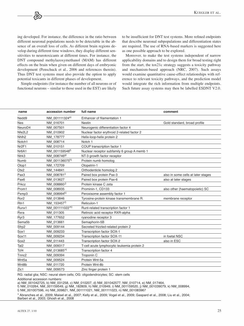
name accession number full name comment
Nedd9 NM_001111324e) Enhancer of filamentation 1 Nes NM_016701 Nestin Gold standard, broad profileNeuroD4 NM_007501 Neurogenic differentiation factor 4 Nfe2L2 NM_010902 Nuclear factor erythroid 2-related factor 2 Nhlh2 NM_178777 Helix-loop-helix protein 2 Notch1 NM_008714 Notch 1 Nr2F1 NM_010151 COUP transcription factor 1 Nr6A1 NM_001159548f) Nuclear receptor subfamily 6 group A memb 1 Ntrk3 NM_008746g) NT-3 growth factor receptor Numb NM_001136075h) Protein numb homolog Otop1 NM_172709 Otopetrin-1 Otx2 NM_144841 Orthodenticle homolog 2 Pax3 NM_008781i) Paired box protein Pax-3 also in some cells at later stagesPax6 NM_013627 Paired box protein Pax-6 also at later stagesPrkcz NM_008860 j) Protein kinase C zeta Prom1 NM_008935 Prominin-1, CD133 also other (haematopoietic) SCPxmp3 NM_008994k) Peroxisome assembly factor 1 Ror2 NM_013846 Tyrosine-protein kinase transmembrane R. membrane receptorRtn1 NM_153457l) Reticulon-1 Runx1 NM_001111022m) Runt-related transcription factor 1 Rxra NM_011305 Retinoic acid receptor RXR-alpha Ryr3 NM_177652 ryanodine receptor 3 Sema5b NM_013661 Semaphorin-5B Sfrp2 NM_009144 Secreted frizzled-related protein 2 Sox1 NM_009233 Transcription factor SOX-1 Sox11 NM_009234 Transcription factor SOX-11 in foetal NSCSox2 NM_011443 Transcription factor SOX-2 also in ESCTal2 NM_009317 T-cell acute lymphocytic leukemia protein 2 Tcf4 NM_013685n) Transcription factor 4 Tnnc2 NM_009394 Troponin C Wnt5a NM_009524 Protein Wnt-5a Wnt8b NM_011720 Protein Wnt-8b Zic1 NM_009573 Zinc finger protein 1 RG: radial glia; NSC: neural stem cells; OG: oligodendrocytes; SC: stem cellsAdditional accession numbers: a) NM_001042725, b) NM_031258, c) NM_010207, d) NM_001042577, NM_010714, e) NM_017464, f) NM_010264, NM_001159549, g) NM_182809, h) NM_010949, i) NM_001159520, j) NM_001039079, k) NM_008994, l) NM_001007596, m) NM_009821, NM_001111021, NM_001111023, n) NM_001083967* Abranches et al., 2009; Maisel et al., 2007; Kelly et al., 2009; Vogel et al., 2009; Gaspard et al., 2008; Liu et al., 2004; Barberi et al., 2003; Ghosh et al., 2008
tobeinsufficientforDNTtestsystems.Morerefinedendpointsthat describe neuronal subpopulations and differentiation states are required. the use of RNA-based markers is suggested here as one possible approach to be explored.
Moreover, to make the test systems independent of narrow applicability domains and to design them for broad testing right from the start, the tox21c strategy suggests a toxicity pathway and mechanism-based approach (NRC, 2007). Such assays would examine quantitative cause-effect relationships with ref-erence to relevant toxicity pathways, and the prediction model would integrate the rich information from multiple endpoints. Such future assay systems may then be labelled eSDNt V2.0.
ing developed. For instance, the difference in the ratio between different neuronal populations needs to be detectable in the ab-sence of an overall loss of cells. As different brain regions de-velop during different time windows, they display different sen-sitivities to neurotoxicants at different times. For instance, the DNt compound methylazoxymethanol (MAM) has different effects on the brain when given on different days of embryonic development (Penschuck et al., 2006 and references therein). thus DNt test systems must also provide the option to apply potential toxicants in different phases of development.
Simple endpoints (for instance the number of all neurons or of functional neurons – similar to those used in the eSt) are likely
Kuegler et al.
Altex 27, 1/1026
from a non-homogeneous mixed cell population and can give information on its relative composition. the transcriptional profiling approach has particular advantages for quantitativestudies in inhomogeneous populations of adherent cells or for complexmixturesofcells,ifappropriatecellspecificmarkersand reference genes are available (see below – point (3)). the big disadvantage of the technology is that co-localisation stud-iesarenotreadilypossible,andthereforethespecificcellsub-population that undergoes changes in response to the toxin can bedifficulttoidentify.
the use of transcription based endpoints (e.g. Northern blot, gene microarrays and PCR) also requires some technical con-siderations, asbriefly summarised inTab.3.Microarrayplat-forms may indicate relative expression differences with varying sensitivities and accuracies for different genes. Without detailed background data, information on a single gene may not be reli-able.Asanalternative,setsofinterestingcell-orstate-specificgenescanbeselectedfordetailedquantificationofrelativegeneexpression changes by quantitative real-time PCR methods. If profiling isperformedbyPCRonaselectedsetofgenes, thetechnology is available in most laboratories at reasonable cost andthroughput,andoptimisedprimersforamplificationcanbederived from online databases (RtPrimerDB, http://medgen.ugent.be/rtprimerdb/).
As this review focuses on the compilation of gene lists that should be useful as background description of cellular states in DNt assays, three major technical issues of gene selection and classificationwillbediscussed:
4.1.1 Gene annotationFirst, the literature, including also relatively recent publications, isfilledwithstronglyvaryingabbreviationsforonegivengene.this is due to the discovery and cloning process, which often occurred in parallel in different places, initial discovery in dif-ferent species, protein and antigen names that differ from the
Here an initial basis is provided for the characterisation of the cells used in such assays.
4 The definition of stem cell genes
4.1 Transcription-based markersForthedefinitionofcelltypesandoftransitionsfromonecelltype to another, different sets of markers may be applied. these rangefromdefinitionofthegenome(primarysequenceandepi-geneticstatus)todefinitionoftheproteome(proteinbasedoran-tigen-based), and include the metabolome, functional character-istics (e.g. electrophysiological responses) and characterisation of the transcriptome (mRNAs and miRNAs). these approaches havedifferentsensitivities,dynamicranges,specificities,sam-ple requirements, technical requirements and throughput.
the most frequently used approaches are antigen based meth-ods and transcriptional profiling.The former have been dealtwithelsewhere,andextensivestudiesinthestemcellfieldhavebeen performed e.g. by BD Biosciences (www.bdbiosciences.com).Briefly,theyareparticularlyusefulforsinglecellcharac-terisation and for sorting cells, only limited by antibody avail-ability (works best for surface antigens). Quantitative evalua-tionsbythisapproachusuallyinvolveflowcytometricanalysisand work particularly well in non-adherent cultures or with cells that can be detached by enzymatic treatment without affecting the epitope. Use on adherent cells requires advanced imaging technologies and is often harder to quantify and to control. On a semi-quantitative or qualitative level, antigen staining offers an easy option to characterise mixed cell populations and to deter-mine co-localisation of different markers within a given cell.
RNA-based measurements have been suggested to be par-ticularly useful to characterise the differentiation of eSC (Noaksson et al., 2005) and to detect neurotoxicity and DNt (Hogberg et al., 2009; Bal-Price et al., 2009; Stummann et al., 2009).Transcriptionalprofilinghasbeenusedinmanyfields,for instance to indicate cellular activation states (Henn et al., 2009; lund et al., 2006; Falsig et al., 2006). the method is fre-quently used successfully for quantitative studies in homogene-ous populations of cells. More or less every gene transcript can be examined (few exceptions due to highly repetitive or highly GC-rich sequences). the expression pattern can be interpreted as a “signature” of the status of the tested cell population. the “signature” can be examined in terms of known cell specificmarkers,geneontology(GO)classificationsystemsandknowngene interaction networks. For instance, different types and differentiation stages of neurons and glial cells differ in their RNAprofiles,andtheseprofilesdifferfromthatofESC(Tab.1)orneuralstemcells(NSC–Tab.2).Therefore,definitionofreferenceprofilesfordifferentculturestatesshouldpermitthedetection of subtle effects of developmental neurotoxicants and give information on the affected pathways. Deviations from the “default transcription signature” may permit the detection of subtle effects of developmental neurotoxicants, and give infor-mation as to the pathways affected. they may also occur as a consequence of cell cycle progression or cellular activation state. Such signatures and their alterations can also be obtained
Tab. 3: Issues concerning identification and selection of transcription-based markers
Definition of applicability domainSelection of criteria for appropriate markers (assay dependent)Method for identification/qualification of markersSelection of negative (exclusion) and positive markersAssembly of set of markers (no single marker is adequate)(Semi-)Quantitative relationship of markers (ratios; thresholds; yes/no)Definition of differentiation statusComposition of culture over time Selection of control population(s) for cell type specific endpointsBiological validation of endpoint-markers with (positive and negative) controlsTiming of chemical exposure (duration and differentiation status)Use of reference databases for cross-validation of dataStatistical and standardisation issues within and between experimentsKnown species differences

Kuegler et al.
Altex 27, 1/10 27
GOdoesnotexist,asGOsdonotdefinecelltypes,butratherrelated functions of genes. therefore meSC genes as endpoints ofDNTtestsneedtobedefinedandagreeduponasinTable1.
4.1.3 Standardisation and statistical issuesTheycanstronglyinfluencetheidentificationofmarkergenes.this applies in different ways to individual studies as well as to meta-analyses. In the former, normalisation, standardi-sation and cut-off procedures are mostly hidden in materials and methods in a way that makes them hard to control or to reproduce by peers. Alterations of expression levels are often calculated relative to housekeeping genes, but the stability and variance of these reference points is only very rarely indicat-ed. However, these data and procedures have a large impact on specificity and sensitivity of the overall analysis. House-keeping genes may be selected based on various criteria. Most importantly, the gene needs to be expressed in equal amounts relative to the total amount of cellular mRNA. In many cell types,thisconditionisfulfilledforGapdh, 18S ribosomal RNA (18S rRNA), and β2 microglobulin (b2m). Other markers that are also used frequently comprise Hprt, 28S ribosomal RNA (28S rRNA), Actb or Acta1. More rarely found options are Ribosomal protein L32 (RPl32) or Phosphoglycerate kinase 1 (PGK1). However, these housekeeping mRNAs do not al-ways behave according to the criteria set above (e.g. Der et al., 1998). this problem is particularly pronounced in differentia-tionexperiments,asdescribedhere.Inthiscase,thefinalcelltype in the dish can be very different (overall phenotype, size, cell cycle status, metabolic activity, etc.) from the starting cell, and therefore express housekeeping genes at different levels. Similar problems may occur upon exposure to toxicants. An-other type of problem lies in the heterogeneity of cells in DNt test systems. the cultures may contain different subpopulations that express house-keeping genes at different levels. Upon dif-ferentiation, the relative amounts of these subpopulations may change dramatically, leading to enormous practical challenges concerning the standardisation of gene expression levels. to circumvent this, samples are often referenced to a group of housekeeping genes instead of a single gene only. In other cas-es,itmaybeusefultoemploysubpopulation-specificreferencepoints, such as B3 tubulin or Fox-3 (NeuN) for neurons, and e.g. Doublecortin or Neurogenin to refer shifts in patterning markersmore specifically to neuroblast-like cells within theoverall population. Concerning meta-analysis (e.g. Assou et al., 2007; International Stem Cell Initiative, 2007; Bhattacharya et al., 2005; Bhattacharya et al., 2009), additional problems need to be considered. the statistical criteria and quality of the stud-ies included in the meta-analysis might vary strongly, and the initial conditions and rules set within these analyses might be hard to trace. therefore, it is dangerous to rely blindly on the summary of the outcome. this applies also to the table compi-lations presented here. If they are put to experimental scrutiny and trigger a constructive discussion and an improved second version, then a major goal of this review will already have been reached. Possibly subsets will have to be selected, according to the specific culture conditions and cell lines used and thequestions asked.
gene name, and changes of names upon consolidation of the fully sequenced mouse and human genomes. We have chosen to include thecurrently-usedofficialgenesymbol thatcanberetrieved from PubMed (http://www.ncbi.nlm.nih.gov/pubmed) in addition to various other names in common use. In addition, thetranscriptaccessionnumber(asanunambiguousidentifier)is listed. Notably, these accession numbers do not refer to the genesas such,butdefinecDNAs.Theymaye.g.characteriseparticular transcripts of genes with multiple splicing variants. thus, one gene can have more than one accession number. this is highly important for expression analysis and corresponding database searches, as a given gene can form different transcripts in different cell types or at different differentiation stages. therefore, problematic situations might arise where analysis of gene regulation by different methods (different PCR primers, different hybridisation oligos, etc.) yields different results. In such situations, different transcripts might have been analysed. to cover this situation, accession numbers for different splice and annotation variants of the same gene are also included in the tables. the NCBI RefSeq database provides annotated individu-al transcripts and protein sequences (derived from its predeces-sor, Genbank) with accession numbers that are distinguished by a two-letter prefix (http://www.ncbi.nlm.nih.gov/RefSeq/key.html). Curated transcripts for mRNA, noncoding RNA and proteinsequencesaredistinguishedbytheprefixesNM_,NR_,andNP_,respectively.OtherprefixesindicateoriginalGenbankannotations (two-letter prefixes without a following under-score) or Refseq sequences that are undergoing annotation or curation(TypicallyXM_,XR_,XP_)amongothers.Ensembl,an alternative informative annotation and curation effort by the european Bioinformatics Institute (eBI) also curates sequences and splice variants derived thereof (www.ensembl.org). typi-cally, it is helpful to design gene expression strategies against the curated sequences, although it is important to be aware of (and design around) the potential for underlying variation in that transcript. the collective variation in gene expression can be viewed with the aid of online genome browsers as provided by the University of Santa Cruz (http://genome.ucsc.edu/) or the eBI (http://www.ensembl.org/index.html).
4.1.2 GO categoriesWhenlarge-scaletranscriptionalprofilingisperformed,identi-fiedgenesareoftenassociatedwithgeneontologies(GO).TheGene Ontology Project is an initiative to classify genes and gene products according to known molecular functions with a defined and finite vocabulary (http://www.geneontology.org).GOclassificationsassociategeneproductswithappropriatecat-egories in the three principal areas “cellular component”, “bio-logical process”, and “molecular function”. they are organised by a hierarchical relationship between these groups. When the transcriptionalprofileofacellpopulationchanges,thealteredtranscripts often cluster to certain GOs, and these GOs can give useful information on the types of changes that are occurring (structural, signalling, differentiation). thus, it may be useful topickthegenesofahypothetical“mESCGO”todefinethestarting population of DNt experiments and the changes of genes characteristic for this population. Unfortunately such a
Kuegler et al.
Altex 27, 1/1028
2007). In addition, much of the variation may be due to real spe-cies differences. In fact, the biology of meSC and heSC shows distinct differences with regard to signals required to maintain pluripotency (Wei et al., 2005; eckfeldt et al., 2005; Wang et al., 2009). In this situation, it is tempting to conclude quickly that heSC are more relevant for human physiology. However, strong evidence indicates that meSC may resemble cells of the human inner cell mass of the blastocyst more closely than heSC (li and Ding, 2009). Moreover, it is not known whether some differences of eSC in culture have any effects on readouts for DNt. this can only be determined experimentally, and should be done so.Theoverallapproachofdifferentialtranscriptionprofilingto
identify eSC markers has some conceptual shortcomings: First, the factor of differential expression that is used as cut-off is often relatively low (e.g. 2-3 fold). this means that it would be very hard to identify an eSC contamination of around 30% within an otherwise fully differentiated cell population. this low cut-offalsoreducesthelevelofspecificitysuchstudiescanachieve.Second, the “differentiated population” used for comparison was frequently obtained from embryoid bodies (eBs), i.e. 3-di-mensional spheroids formed from eSCs when they are left to differentiate “wildly” (in a non-guided way, only triggered by withdrawal of pluripotency factors). this population contains cells from all three germ layers, and may not be relevant for the identification of differentially-expressed genes between ESCand differentiating neurons. thirdly, this approach is bound to identify many “false positives”, as two populations with differ-ent proliferation characteristics are being compared. thus, genes involved in DNA synthesis, chromatin structuring and cell cy-cle regulation would be selected as putative stem cell genes. A variant of this approach was taken by the International Stem Cell Initiative(ISCI)todefinehESCmarkers.Genesweregroupedaccording to the similarity of their behaviour to that of Nanog when over 50 heSC lines were differentiated to eBs. the top 6 group comprises Nanog, Tdgf1, Gabrb3, Dnmt3b, Gdf3, Pou5f1/Oct4 and the top 20 group additionally contains Fgf4, Gal, Leftb, Ifitm1, Nodal, Tert, Utf1, Foxd3, Ebaf, Lin28, Grb7, Podxl, Cd9 and Brix (the International Stem Cell Initiative, 2007).
A third approach to identify stem cell genes is based on the concept that genes qualify for inclusion when they are required for the function and maintenance of eSC. these genes would be biologicallydefinedas“stemnessgenes”.Thisdefinitionwouldalso form the basis for the opening of a GO category under the fieldof“biologicalfunction”.Themostprominentexamplesforsuch genes are Pou5f1/Oct4 and Nanog (Mitsui et al., 2003) or the Klf (Krüppel-like factor) genes. However, Oct4 is also found in germ stem cells or cardiac differentiation (Stefanovic and Puceat, 2007), Nanog plays a role in neuronal differentiation (Molero et al., 2009) and Klf-4 is also an oncogen (Rowland et al., 2005). the Wnt, FGF and BMP/tGF-ß pathways – and as-sociated genes – are clearly involved in the maintenance of stem-ness, but they also play a role in dozens of other processes. the same type of ambiguity is found when one examines the genes that can be used for reprogramming. In addition to Oct4, Na-nog and Klf-4 above, for instance Sox2, Lin28 and Myc are used. Sox2 and Myc play roles not only in reprogramming but also in stemcellmaintenance.However,theyarenotspecificforESCs,
4.2 What are stem cell genes?Atfirstsight,itmayseemeasytoextractstemcellgenesfromthe existing literature. Dozens of papers have dealt with such genes and large numbers of microarray studies have been per-formed to identify such genes, but also doubt has been voiced onwhetherstemcellsarereallyadefinedentityatall,orwheth-er they rather represent one of many possible transient states (efroni et al., 2009; Zipori, 2004). the expression pattern as-sociated with such states may vary between different stem cell lines. Such effects may be linked to higher dynamics of the genome than commonly expected. For instance, non-protein coding line elements, which make up a large proportion of the human genome, have been shown to be active as transposons in eSC and, even more commonly, in NSC. Such activity might affect the activity of classical genes directly, e.g. by insertion, or indirectly,bymodificationofpervasivetranscription(Coufaletal., 2009; Muotri et al., 2007; Garcia-Perez et al., 2007; Muotri and Gage, 2006; Muotri et al., 2005).
For assay development we have to take a closer look at the important distinction between “stemness genes” and “stem cell markergenes”.Forthespecificpurposeofthisreview,thefocusof the discussion will be on eSC (and NSC) markers.
Unfortunately, the term “stem cell marker gene” is less clear uponcloserinspectionthanitappears.Aneasydefinitionwouldfor instance be “a gene that is only expressed in meSC, and in no other cell type”. Unfortunately, no such gene exists. the re-versedefinitionmayalsobeapplied.“Negativestemcellmark-ers” are genes that are by no means expressed in meSC. this isadefinitionthatisusefulinpracticeforqualitycontrolandfordefiningtheonsetofdifferentiation,butitisnotsufficientfor definingmESC (Assou et al., 2007;Bhattarachyan et al.,2009).Thedefinitionof negativemarkers is alsonotwithoutambiguity, as meSC cultures may often be contaminated with more differentiated cells. Upon transcriptome analysis it may then appear that apparently pure stem cells “express” certain genes usually not associated with meSC, such as B3 tubulin, Keratins-8 and -18 or Alpha cardiac actin (Ginnis et al., 2004; Bhattacharya et al., 2005). to establish an eSC database free of contaminations, cells may be sorted prior to analysis or selected on the basis of the activity of a sharply-regulated stemness gene like Utf-1 (tan et al., 2007).ApragmaticapproachtotheidentificationofESCgenesisto
characterise transcriptome changes when eSC differentiate and todefinethosegenesthataredifferentiallyhighlyexpressedineSC as stem cell genes. this approach has been taken many times in many variations (reviewed in Bhattacharya et al., 2009 and efroni et al., 2009). the result was that these approaches consistentlyidentifiedasmallgroupof“usualsuspectgenes”foreSC, such as Lefty2, Oct3/4, Nanog, Utf-1 and Tdgf1. However, astonishingly large differences were observed between the stud-ies. It was surprising that some studies found that meSC genes overlap with heSC genes only to a low degree, i.e. between 15 and 35% (Bhattacharya et al., 2005; Ivanova et al., 2002; Ram-alho-Santos et al., 2002). this may indicate some intrinsic weak-nesses of these studies (see e.g. paragraph on standardisation and statistics issue). An alternative explanation may be that the deri-vatisation of the lines affects their later phenotype (Navara et al.,

Kuegler et al.
Altex 27, 1/10 29
larasforthemarkersidentifiedbyexpressionlevel.Stemnessgenes are not included when their expression is relatively low compared to neural tissue expression.(e) Only mRNAs coding for proteins have been considered for this analysis. Information on micro RNAs (miRNA) is still rela-tively limited. However, it appears that expression of miRNA can be relatively cell type-specific. Thus, miRNAswith rela-tively high expression levels in meSC compared to other cell typesmay be identified (Lakshmipathy et al., 2007a,b). Simi-larly,miRNAsimportantfordefinedstepsinneuronaldevelop-menthavebeenidentified(Yooetal.,2009).Futureprofilesmaytherefore also include miRNAs. A further step may be a more detailed analyses of the promoters themselves and their epige-netic state by chip-on ChIP experiments (microarray analysis of chromatinimmunoprecipitates),bisulfitesequencing(identifica-tion of methylcytosine as altered base in the DNA structure) or one of the many related new technologies. For instance, it has been suggested that the eSC genome may be characterised and definedbyrelativelyopenchromatinarchitecture(Zipori,2004;eckfeldt et al., 2005). this has been corroborated on the mo-lecular level by genome-wide mapping of the chromatin state of eSC and other cells and indeed has functional consequences (Mikkelsen et al., 2007). the resultant pervasive transcription is particularly prominent in eSC, and a major difference between eSC state and more lineage committed differentiation stages may be the extent of this genome wide transcriptional activity (efroni et al., 2009), that involves many non-protein coding RNAs (Ber-retta and Morillon, 2009; Dinger et al., 2009; Jacquier, 2009; Mikkelsen et al., 2007). to transform this knowledge into robust testendpointsanddefiningmarkers,theidentificationofESC-specificnon-codingRNAwouldappearuseful.Indeed,recentlyover a thousand conserved large intervening non-coding RNAs (lincRNAs)havebeenidentified(Guttmanetal.,2009).About100 were regulated by Oct4 and Nanog and functionally impli-cated in a stemness network, and at least one was only expressed in eSC. thus, lincRNAs are candidates for future lists of differ-entiationandcellactivation-defininglistsofmarkers.(f)Last,butdefinitelynotleast,negativemarkersshouldbeusedin transcription-based cell characterisations. the meSC table contains only positive markers, as naturally all genes listed in table 2 (or other tables presented here) represent the correspond-ing negative markers. typical markers for endodermal differen-tiation (e.g. intestine, glands, liver) would be VegfR2, Sox17, Ttr, ApoA1, Lim1, Cytokeratin19, FoxA2, Alphafetoprotein or Gata-4 (also mesendoderm and cardiac mesoderm); for mesoderm (e.g. muscles, bones, heart, blood): Hand1, Brachyury, Smooth mus-cle actin, Cd31, Cd34, Cd325 or Eomes (also trophoblast), and e.g. Ncam1 or certain keratins (Krt 18) indicate ectoderm. Other useful and sensitive markers for initial differentiation away from eSC may be Fibronectin-1, Naalad2, Profilin-1 and Slc40a1.
4.4 Neurodevelopmental biology and definition of neural stem cell markersDifferentiation of meSC towards neurons triggers coordinated wavesofgenetranscriptionthatcanbeidentifiedbyunbiasedcluster analyses (Abranches et al., 2009; Schulz et al., 2009). Accordingly, the cells move from the multipotent stem cell state
as e.g. Sox2 is highly expressed (and functional) in NSCs, and Myc is upregulated in many tumours and rapidly dividing cells.
In conclusion, simple rules for the selection of eSC marker genes cannot be applied. More advanced algorithms based on multiple markers are required as described below.
4.3 Definition of mESC markersBasedon theabove,markerswerefiltered from the literatureaccording to the following criteria: (a) the gene needs to be expressed in meSC (differences be-tween meSC and heSC need to be taken into account).(b) the gene needs to be expressed in meSC considerably high-er than in most other cell types. Frequently, eSC were com-pared to embryoid bodies (eBs). In other approaches meSC were compared to mNSC and other stem cell types (haemat-opoietic) to identify unique marker genes (Ivanova et al., 2002; Ramalho-Santos et al., 2002). An interesting approach in that direction was also taken by groups at the NIH (Bhattacharya et al., 2004; Bhattacharya et al., 2005; Bhattacharya et al., 2009; Ginis et al., 2004), when eSC were compared to RNA pools from normal differentiated tissue. this approach was taken one step further in a large meta-analysis, in which heSC expression profileswerecomparedtodataretrievedfromdatabasesonover100 tissue analyses (Assou et al., 2007). For the compilation of table 1, especially co-expression at similar levels in NSC was usedasanexclusioncriterion.Notably,asmESCaredefinedbya group of genes, the criterion of absence of expression in other cells needs not be applied stringently, providing that it refers to differentcelltypesfordifferentmarkergenes.Ifasufficientlylarge group of meSC marker genes is selected, it is likely that expression in other cells is cancelled out (averaged), while each of the genes should be expressed in meSC.(c) the marker gene should not be expressed in neural stem cells and neuroectodermal cells and thus be different from the oneslistedinTable2.Thisconditionisaspecificlimitationofcondition 2 and applies particularly for meSC markers used in DNt experiments. For instance, Galanin is a frequently-identi-fiedmESCgene,butalsoplaysaroleinNSCandcertainmatureneurons. Genes with such behaviour may not be downregulated upon meSC differentiation towards the neuronal lineage and are therefore useless as meSC markers for this particular purpose. A vast amount of gene expression data is available to identify relevant genes. Here, both individual papers (e.g. Abranches et al., 2009) and databaseswere used for identification and ex-clusion of candidates. For instance, the EU fifth frameworkresearch programme (FP5)-consortium FunGeneS provides extensive transcriptome profiling information on the differ-entiation of meSC to neurons, coupled to web-based analysis software (FunGeneS consortium http://www.fungenes.org/) (Schulz et al., 2009). Similar approaches are taken for instance by the StemBase of the Ontario Genomics Innovation Center (StemBase http://www.stembase.ca/?path=/) (Perez-Iratxeta et al., 2005; Porter et al., 2007).(d) Genes with a known functional role for the maintenance of meSC (e.g. loss of stemness upon their knockdown or knockout (Misui et al., 2003)) are included as markers if they do not have multiple roles also in other cell types. the reasoning is simi-

Kuegler et al.
Altex 27, 1/1030
larly,ESC-derivedNSC-like cells can acquire region-specificphenotypes depending on the differentiation protocol (Bouhon etal.,2006;Gaspardetal.,2009).Therefore,notallcellsfulfill-ing basic criteria for NSC can still be differentiated to all CNS cell types. Differences also exist between eSC-derived NSC, and brain-derived NSC (both can only be obtained by extensive in vitro culturing, potentially leading to artefacts), for instance in the readiness to generate astroglial cells, or between spinal cord NSC and cortical NSC in the expression of many pattern-ing marks and genes with broadly varying biological function (Kelly et al., 2009). thus, it is not a straightforward and unam-biguousapproachtodefineNSCmarkersbycharacterisingonegiven NSC population that can be maintained in culture. DefinedprotocolsformESCneuronaldifferentiationtypical-
lyinvolveinitialneuralspecificationandexpansionunderNSCgrowth conditions, followed by withdrawal of growth factors
over an early neuroectoderm state to a state in which they can form rosettes that still have the potential to develop to central and peripheral neurons. this state is closely linked to the production of neural precursor cells or NSC. Such NSCs (human or murine) may be enriched and clonally expanded under appropriate cul-ture conditions (Ying and Smith, 2003; Conti et al., 2005; Bar-beri et al., 2003; Koch et al., 2009; elkabetz et al., 2008; Okabe et al., 1996). NSC markers may be derived from gene expres-sionprofilingofclonally-expandedNSC-likecells.Thishasforinstance been done for human rosette-type cells vs. heSC (elk-abetz et al., 2008), but multiple comparisons against different populations (including more mature neurons) would be required todefinethegenuineNSCgenes.NSCs,whilesharingcommonproperties of undifferentiated progenitors, may exhibit distinct regionalcapacitiesforneuraldifferentiationtospecificlineagesor neurotransmitter phenotypes (Klein and Fishell, 2004). Simi-
Tab. 4: Markers for fine mapping of DNT effects in developing neural cells
category name accession number full name commentrostral-caudal FoxG1 NM_008241a) forkhead box G1 (Bf1) very rostral Emx1 NM_010131 empty spiracles homolog 1 very rostral Emx2 NM_010132 empty spiracles homolog 2 forebrain Dlx1 NM_010053 distal-less homeobox 1 forebrain Nkx2.1 NM_009385 Titf1, NK2 homeobox 1 forebrain Gsx2 NM_133256 GS homeobox 2 forebrain En1 NM_010133 engrailed 1 midbrain Otx1 NM_011023 orthodenticle homolog 1 dorsal fore- and midbrain Otx2 NM_144841 orthodenticle homolog 2 dorsal fore- and midbrain Atoh1 NM_007500 Math1, atonal homolog 1 hindbrain Irx3 NM_008393 Iroquois related homeobox 1 caudal HoxA2 NM_010451 Homeobox A2 rostral hindbrain HoxB1 NM_008266 Homeobox B1 rostral hindbrain HoxB4 NM_010459 Homeobox B4 caudal hindbrain HoxB6 NM_008269 Homeobox B6 spinal corddorsal-ventral Shh NM_009170 sonic hedgehog ventral Gli3 NM_008130 GLI-Kruppel family member dorsal forebrain Olig2 NM_016967 OC transcription factor 2 hindbrain Isl1 NM_021459 ISL1 transcription factor forebrain Nkx2.2 NM_010919b) NK2 transcription factor ventral Neurog2 NM_009718 neurogenin 2 dorsal forebrain Nr2f1 NM_010151 nuclear receptor subfamily 2 ventral forebrain Ascl1 NM_008553 Mash1, achaete-scute complex homolog 1 ventral forebrain Msx1 NM_010835 homeobox msh-like 1 ventral midbrain Dll1 NM_007865 delta-like 1 ventral midbrainneuronal subtypes GAD2 NM_008078 glutamic acid decarboxylase 2 GABAergic neurons Gat3 NM_144512 solute carrier family 6a13 GABAergic neurons Calb1 NM_009788 calbindin 1 GABA subtypes Calb2 NM_007586 calbindin 2, calretinin GABA subtypes TH NM_009377 tyrosine hydroxylase dopaminergic neurons VGlut2 NM_080853 solute carrier family 17a6 glutamatergic neurons Adra2b NM_009633 adrenergic receptor adrenergic neurons Tph1 NM_009414 tryptophan hydroxylase 1 serotonergic neurons Chat NM_009891 choline acetyltransferase motor neurons Mnx1 NM_019944 HB9, motor neu. And panc. homeobox 1 motor neurons

Kuegler et al.
Altex 27, 1/10 31
category name accession number full name commentNCC Sox10 XM_128139 SRY-box containing gene 10 transcription factorneuroblast Dcx NM_010025c) Doublecortin cytoplasmic protein NeuroD4 NM_007501 neurogenic differentiation 4 transcriptional activator Tubb3 NM_023279 tubulin beta 3 cytoskeleton protein Elavl4 NM_010488d) emb. lethal abnormal vision-like 4 RNA binding protein Epha7 NM_010141e) Eph receptor A7 growing axonsnon ectodermal germ layers Sox17 NM_011441 SRY-box containing gene 17 mesoderm Acta2 NM_007392f) SMA, actin, alpha 2, smooth muscle mesoderm T NM_009309 Brachyury mesoderm Gata4 NM_008092 GATA binding protein 4 endoderm Afp NM_007423 alpha fetoprotein endodermECM components Col4a1 NM_009931 collagen type IV alpha 1 early marker in diff. Ncan NM_007789g) Neurocan early marker in diff. Tnc NM_011607 tenascin C late marker in diff. Col1a1 NM_007742 collagen type I, alpha 1 late marker in diff.neuronal marker Syp NM_009305 Synaptophysin synaptic vesicle assoc. Grin1 NM_008169 NMDA1 ionotropic glutamate R. Nrg1 NM_178591 neuregulin 1 schiz. assoc. Nrxn 1 NM_020252 neurexin I autism, schiz. assoc. Stx1a NM_016801 syntaxin 1A synapse assoc. Snca NM_009221h) Synuclein alpha parkinson assoc. Mapt NM_010838i) microtubule-associated protein tau alzheimer assoc.
OC: oligodendrocyte, panc: pancreas, neu: neuron, NCC: neural crest cell, emb: embryonic, R: receptor, schiz: schizophrenia, assoc: associated Additional accession numbers: a) NM_001160112, b) NM_001077632, c) NM_001110222, NM_001110224, NM_001110223, d) NM_001038698, e) NM_001122889, f) NM_183274, g) XM_913832, h) NM_001042451, i) NM_001038609
regional patterning is thought to be achieved by cell-extrinsic, contrasting gradients of morphogens and growth factors, includ-ing Bone Morphogenetic Proteins (BMPs), Sonic Hedgehog (Shh), Retinoic acid, Fibroblast Growth Factors (FGFs), etc. these chemical gradients establish a positional axis that confers region-specificpatternsofgeneexpressionanddirectslineage-specificdifferentiation.Thisinformationhasbeenusedtocom-pile the list of markers for in vitro differentiation (tab. 2). the translation of knowledge from developmental gene expression to in vitro gene expression is not without caveats. For instance, the gradients formed in vivo are complex and not stable over time. For instance, NSC formation in the neural tube structure begins rostrally, and zones of NSC formation and patterning are moving in a rostro-caudal (from head to tail) way along the neu-ral tube (Wilson and Maden, 2005). In vivo neurulation is also a desynchronised process. Homogenates used for transcriptional
and neuronal differentiation. the differentiation process is usu-ally not 100% synchronised, and cellular differentiation stages form a continuum. therefore, the wave of NSC gene expression may overlap with the antecedent meSC gene expression and with the following wave of NSC-derived neuronal/glial gene expression.Consequently, it isdifficult tostrictlydefineNSCpatterns of gene expression solely within the context of an in vitro differentiation system. For this reason, changes in gene ex-pression are interpreted with reference to those observed during neuralspecificationandlineageprogressionin vivo (Rubenstein and Puelles, 1994; Rubenstein et al., 1998).
A basic characteristic of the nervous system is the high diver-sity of different cell types, which is necessary for appropriate function.Neuronaldifferentiationproceedsinaregion-specificmanner, depending on the position of neuroepithelial progeni-tors along the rostrocaudal or dorsoventral axes (Fig. 2). this

Kuegler et al.
Altex 27, 1/1032
Similar processes occur also in in vitro culture, which ex-plains that e.g. the density of cells has a major impact on the end result of the differentiation. Chemicals can act in this phase on the cells or their signalling molecules, and the exposure may result in a shift of the balance between neuronal subtypes (Gaspard et al., 2008) or between glia and neurons (Fritsche et al., 2005; Steinhart et al., 2007). Such early events have been speculated to have a late impact e.g. on development of neuro-degenerative disease (landrigan et al., 2005), and it has been demonstrated experimentally that e.g. exposure to polychlorin-
profilingwill contain bothNSCs and differentiating neurons.therefore, NSC and their progeny can be hard to disentangle at the level of transcription without reference to cellular, spatial distinctionsingeneexpressionprofiles(e.g.asdeterminedbyhigh resolution in situ hybridisation).
Ideally, meSC-derived NSC gene expression should broadly recapitulate developmental patterns of gene expression ob-served during neuroepithelium specification, orwithin prolif-erative progenitor zones at later stages. Such information has alsobeenusedhere(amongstothers)forfilteringofsuggestedmarker genes (Fig. 2). For our compilation, we used the strong expression and/or significant role of a gene in differentiatedneurons and/or glia as exclusion for its definition as anNSCmarker, even though the gene may be expressed in NSCs. this importantfilteringstepisadmittedlybiased,andnewinforma-tion will require adaptations.
A different approach would be to look at functional impor-tance. While some of the most frequently used meSC markers also have a functional role in stemness, many of the typical NSC and radial glia markers are e.g. cytoskeletal elements (Nestin, Gfap, Vimentin) without known function in NSC maintenance. Others, with known and important functions, such as the tran-scription factors Sox2 or Zic1, or signal transduction molecules like Jak2, Hes5 and Fgfr3 also have roles in other cells. thus, at present, it appears necessary to combine all different approach-estoselectcandidateNSCgenesandtofurtherfilterthosebymanual cherry-picking based on literature studies. Such at-tempts have been made repeatedly, and the table (tab. 2) pre-sented here is strongly based on the publications of Abranches et al., 2009; Maisel et al., 2007; Kelly et al., 2009; Vogel et al., 2009; Gaspard et al., 2008; liu et al., 2004; Barberi et al., 2003; Ghosh et al., 2008.
4.5 Definition of differentiation markers for different neuronal stagesAfter the generation of NSC, neuronal differentiation proceeds. the currently accepted model of neural developmental pro-poses that extracellular signalling molecules act on NSC and their progeny and determine what type of neurons or glia they will become. this would be accompanied by migration of the neuroblasts and by generation of new signalling gradients due to factors secreted from neural cells themselves. Cells at a dif-ferentiation stage after the NSC stage that are not yet mature neurons are frequently referred to as neuroblasts. However, the definitionofthesecellshassomecaveats.Duringorgandevel-opment, the neuroblast is essentially a post-mitotic neuron that is distinguished from a maturing or mature neuron by its spe-cialisation for migration rather than for functional integration. Postnatally, neuroblasts are a distinct population of cells, which are capable of proliferation and are neuronally committed.
therefore, we use the expression NSC in this review to signify proliferative cells with self-renewal capacity, the ability to form neuronal and glial cells, and a dependence on eGF and bFGF for optimal proliferation (meSC depend on lIF). the neuronally committed progeny of NSC includes maturing neurons at differ-ent stages, which we denote as “neuroblasts” when referring to early stages and as neurons when referring to late stages.
Fig. 2: Basic concepts of neurodevelopmentVery early during embryonic development (about day 7.5) the neural plate forms as an area of early neuroectodermal tissue, whereas flanking regions form ectoderm (ED). A. Within the next 24 h this plate invaginates and closes to form the neural tube, which is the precursor stage of the central nervous system. Cells at the lateral margins of the neural tube form the neural crest cells (NC) that migrate to various locations and form parts of the peripheral nervous system among other cell types. B. The neural tube (light gray) is flanked by non-neural tissue (dark grey) and extends from the head region towards the prospective tail region. At this stage, clear patterns of neurons along different axes are established, which lead to different neuronal subpopulations in the adult. The major axes are from back (dorsal = d) to belly-side (ventral = v) and from head (rostral = r) to tail-side (caudal = c). C. lateral view of a day 10.5 embryo (E10.5): the caudal end (c) represents the spinal cord (SC), the rostral end (r) develops into the brain, where forebrain (FB), midbrain (MB) and hindbrain (HB) can be distinguished. The dorso-ventral axes (d-v) remains present both in the spinal cord (motor neurons in the ventral part) and in the brain (e.g. the dorsal forebrain differentiates to cortical structures (Ctx). D. Embryonic stem cells (ESC) can differentiate to neural stem cells (NSC) with characteristics resembling those of proliferating cells found in the early development of the nervous system. The mESC-derived NSC-like cells, like their in vivo counterparts, retain the capacity to acquire region-specific identities and differentiate into neurons and/or glia via intermediate lineage-restricted progenitor cell stages in vitro.

Kuegler et al.
Altex 27, 1/10 33
4.6 Astrocyte markersA discussion of all markers relevant for the different develop-mental phases is beyond the scope of this review. As one ex-ample for the complexity, we chose a relatively simple neural population: astrocytes. Although these cells make up more than half of the brain mass, they have been relatively neglected as potential targets of toxicity or DNt. It is generally assumed that astrocytes are identified by the intermediary filament proteinGFAP. However, recent research has shown that antibodies to this protein also label radial glia (NSC-related cells) (Seri et al., 2001; Ganat et al., 2006; Götz and Steindler, 2003; Buffo et al., 2008), and that about 50% of astrocytes in the brain may not ex-presssignificantamountsofGFAP(Lovattetal.,2007;Cahoyet al., 2008). Moreover, knockout of Gfap has no major effects on astrocyte development or brain function (Pekny et al., 1995; Gomi et al., 1995). thus, a broader panel of astrocyte mark-ers, as compiled here (tab. 5), is urgently needed, similar to the markers for meSC and mNSC presented in tables 1 and 2.
ated biphenyls (PCBs) in utero can affect the outcome of stroke in later life without major effects on brain development (Dzien-nis et al., 2008). thus, we have to assume that DNt does not necessarily affect the number of neurons or other major cell types,butthespecificpatterningofthenervoussystemandre-lationships between neuronal populations. For such endpoints, transcriptionalprofilingatdifferentphasesofdevelopmentap-pears to be a useful approach to detect deviations from the nor-malpattern.AnexampleofmarkerstodefinesuchpatterningisdisplayedinTable4.Afuturerefinementmaybetheselec-tion of differentially spliced genes that form highly cell-type or development-specifictranscripts.ForinstancethewellknownneuronalmarkerNeuNaffectsneuronal-specificsplicing(Kimetal.,2009b),and themitochondrial fusion-fission-regulatinggene Drp1expressesaspecificsplicevariantonlyinbrain(Uoet al., 2009). New microarray platforms that allow reliable de-tection of exon splicing may enable detailed analysis of post-mitotic neuronal differentiation.
Tab. 5: Marker genes for astrocytes
name accession number full name comment referenceActA2 NM_007392 Alpha-actin-2 also in smooth muscle [1]Aldh1L1 NM_027406 Aldehyde dehydrogenase family 1 member L1 also in GFAP-neg AC [2]AldoC NM_009657 Fructose-bisphosphate aldolase C [3]ApoE NM_009696 Apolipoprotein E also synthesised by MG [4]Aqp4 NM_009700 Aquaporin-4 also RG, endfeet at vessels [5], [6]Bysl NM_016859 Bystin in reactive AC [7]Car2 NM_009801 Carbonic anhydrase 2 also in OC Cbs NM_178224a) Cystathionine beta-synthase [3]Csad NM_144942 Cysteine sulfinic acid decarboxylase taurine biosynthesis Gfap NM_001131020b) Glial fibrillary acidic protein labels subset of AC [8], [9]GjA1 NM_010288 Connexin 43 AC specific in the brain [6], [10]GjB6 NM_001010937c) Connexin 30 [10]Glul NM_008131 Glutamine Synthetase also in GFAP-neg AC [11], [12]Kcnj10 NM_001039484 ATP-sensitive inward rectifier potassium channel 10, Kir4.1 absent in immature AC [3]MaoB NM_172778 Monoamine oxidase type B mitochondrial NFIA NM_010905d) Nuclear factor 1 A-type also in OC, NSC [13]NFIB NM_001113209e) Nuclear factor 1 B-type also in OC, NSC [13]NFIX NM_001081982f) Nuclear factor 1 X-type also in OC [13]Pla2g7 NM_013737 PAF acetylhydrolase useful RNA marker [2]PygB NM_153781 Glycogen phosphorylase, brain form specific for AC in brain [14]S100b NM_009115 S100beta also in early AC, NSC [15]Slc1A2 NM_001077515g) GLT-1, excitatory amino acid transporter 2 early AC, also NSC, RG [16]Slc1A3 NM_148938 Glast-1, excitatory amino acid transporter 1 mature AC [16]SparcL1 NM_010097 SC1, SPARC-like protein 1 [17]Vim NM_011701 Vimentin also RG, NSC, early AC [18], [19]
AC: astrocytes; RG: radial glia; NSC: neural stem cells; OG: oligodendrocytes; MG: microglia
Additional accession numbers: a) NM_144855, b) NM_010277, c) NM_008128, d) NM_001122952, NM_001122953, e) NM_008687, NM_001113210, f) NM_010906, NM_001081981, g) NM_011393, NM_001077514
1: Lecain et al., 1991; 2: Cahoy et al., 2008; 3: Hatada et al., 2008; 4: Gee et al., 2005; 5: Nakahama et al., 1999; 6: Fatemi et al., 2008; 7: Sheng et al., 2004; 8: Rodnight et al., 1997; 9: Ghandour et al.,, 1979; 10: Lovatt et al., 2007; 11: Steffek et al., 2008; 12: Wu et al., 2005; 13: Wilczynska et al., 2009; 14: Pfeiffer et al., 1992; 15: Burette et al., 1998; 16: Chaudhry et al., 1995; 17: McKinnon et al., 1996; 18: Dahl et al., 1981; 19: Zamora et al., 1988

Kuegler et al.
Altex 27, 1/1034
5 Conclusions
the transcription-based markers discussed in this review represent an effort to characterise subtle disturbances in the waves of gene inductions leading from meSC to differentiated neuralcells.Withmoreexperience,andforspecificapplica-tions, possibly small subgroups of markers can be selected to obtain relevant information. A step further would be the use of genetically-modifiedmESCwith reporter constructs.In this case, easily quantifiable enzymes, like luciferase orsecreted alkaline phosphatase (Suter et al., 2009; Volbracht et al., 2009), driven by cell- and stage-specific promoterswould be used as endpoints of gene induction, also in very complex cell mixtures. As with all transcription-based assays, the endpoints suggested here do not necessarily correlate with protein or function, and this issue will require further charac-terisationandvalidation(SchrattenholzandSoskić,2008).Inthe end, the proof of the pudding is in the eating. In extreme cases, certain well-established markers cannot be detected at all on the RNA-level for technical reasons (e.g. highly re-petitivesequences)orbecausetheyareantigenicallydefined.these comprise the early astrocyte precursor marker A2B5, the meSC marker SSeA-1 or the NSC marker polysialylated-NCAM (PSA-NCAM). Suchmarkers are linked to specificglycosylations or keratin sulfates, and in extreme cases gly-cosylation itself (GalNAc-epitopes on multiple proteins) can be an excellent marker for meSC (Nash et al., 2007). Such antigenic markers are ideally combined with RNA markers and, in the future, RNA marker sophistication will increase by the inclusion of miRNAs, lincRNAs or other non-coding RNAs,andoftissuespecificsplicevariants.Altogether, thisapproachseemstobepowerfulenoughtodefineexactlythedifferentiation capacity of eSC, also to germ layers other than neuroectoderm, and it should therefore be a suitable substitute for old-fashioned assays testing teratoma formation in vivo to establish pluripotency of a cell population. this review fo-cussed mainly on meSC differentiation, but the underlying principles also apply to heSC. For the human counterparts, some excellent compilations of stem cell markers exist (Assou et al., 2007; International Stem Cell Initiative, 2007; Bhatta-charya et al., 2005, 2009), and it needs to be noted in this con-text that clear species differences may exist (Ginis et al., 2004; Sato et al., 2003). For instance, the above mentioned marker SSeA-1 does not work for heSC, while those are character-ised by SSeA-3/4, which do not work for meSC. Genes like threonine dehydrogenase (tdh) (Wang et al., 2009), FoxD3 or the genes coding for the receptor of the meSC growth fac-tor lIF (which is dispensable for heSC) are regulated in a species-specificmanner.With thesewell known differencestaken into account, meSC still remain a very robust system for studying neural development and are possibly able to provide human DNt relevant information on compounds more sensi-tively than the currently used animal models.
Mostnotably,theunderlyingprincipleforthedefinitionofas-trocyte markers differs from that of stem cell markers. All of the stem cell markers are expected to be expressed in all stem cells. In contrast to this, not all “astrocyte markers” are expressed in allastrocytes.Theyratherdefinesubpopulationsofastrocytesand different developmental stages of such subpopulations. Only this comprehensive picture based on multiple markers will yield meaningful information on the fate of the diverse group of astrocytesasawholeandoneffectsofchemicalandotherinflu-ences on their development.
4.7 Toxicity pathwaysIn this review we have focussed on markers useful for the des-cription of subtle phenotypic effects caused by toxicants – in-dependent of their mode of action. An interesting additional aspect of transcription-based endpoints may be the possibility toidentifycellulartoxicitypathwaysandfingerprintsinvolvedintheeffectofthechemicals.Especiallyfingerprintingisal-ready established for other organ toxicities, in particular hepa-totoxicity (Ruepp et al., 2005; Steiner et al., 2004; Blomme et al., 2009). these two different approaches may be applied independently or be combined. An example may best demon-strate the underlying principle: For instance, a chemical may be identifiedasapotentialdevelopmentalneurotoxicantbasedonshifts in the patterning markers presented in table 5. It may e.g. increase dorsal markers and decrease ventral markers relative to house keeping genes. On closer (mechanistic) examination, one may notice, that in particular sonic hedgehog (Shh) target genes were down-regulated upon exposure to the chemical. the mechanism of toxicity may thus involve inhibition of Shh signalling.Alternatively,chemicalsmaybescreenedspecifi-cally for disturbances of key signalling pathways by reporter assays or transcriptome analysis coupled with systems biology approaches.OnecompoundmaybeidentifiedtoblocktheShhsignalling pathway, and upon subsequent examination of DNt effects, it would lead to a dorsalisation of the developing neu-rons. Cyclopamine is a substance that behaves as described above.Other toxicity pathways,whichmay be identified ina similar manner, involve e.g. retinoic acid synthesis, notch processing or Wnt, tGF-beta or Ah-receptor signalling. the above examples show the independence of mechanistic and phenotypic approaches and the huge potential of using and combining both. In the context of eSC-based neurodevelop-mental test systems, it is important to note that the phenotypic approachnecessarilyrequiresacomplexanddifficultexperi-mental test system (differentiating eSC). In contrast to this, the mechanistic approach may also be applied to (and work much better in) much simpler systems involving the respec-tive pathways. Differentiating eSC are in fact, due to their complexity, not very suitable as a mechanistic screen system. this review has predominantly focussed on the markers that may be useful for DNt/teratogenicity screening approaches in the nearer future.

Kuegler et al.
Altex 27, 1/10 35
Bottini, A. A., Amcoff, P. and Hartung, t. (2007). Food for thought ... on globalisation of alternative methods. ALTEX 24, 255-269.
Bouhon, I. A., Joannides, A., Kato, H. et al. (2006). embryonic stem cell-derived neural progenitors display temporal restric-tion to neural patterning. Stem Cells 24, 1908-1913.
Breier, J. M., Radio, N. M., Mundy, W. R. et al. (2008). De-velopment of a high-throughput screening assay for chemical effects on proliferation and viability of immortalized human neural progenitor cells. Toxicol. Sci. 105, 119-133.
Breier, J. M., Gassmann, K., Kayser, R. et al. (2009). Neural progenitor cells as models for high-throughput screens of de-velopmental neurotoxicity: State of the science. Neurotoxicol. Teratol., (epub ahead of print).
Buckley, N. J., Johnson, R., Sun, Y. M. and Stanton, l. W. (2009). Is ReSt a regulator of pluripotency? Nature 457, e5-6; discussion e7.
Buesen, R., Genschow, e., Slawik, B. et al. (2009). embryonic stem cell test remastered: comparison between the validated eSt and the new molecular FACS-eSt for assessing devel-opmental toxicity in vitro. Toxicol. Sci. 108, 389-400.
Buffo, A., Rite, I., tripathi, P. et al. (2008). Origin and progeny of reactive gliosis: a source of multipotent cells in the injured brain. Proc. Natl. Acad. Sci. USA 105, 3581-3586.
Burette, A., Jalenques, I. and Romand, R. (1998). Developmen-tal distribution of astrocytic proteins in the rat cochlear nu-cleus. Brain Res. Dev. Brain Res. 107, 179-189.
Buzanska, l., Sypecka, J., Nerini-Molteni, S. et al. (2009). A human stem cell-based model for identifying adverse effects of organic and inorganic chemicals on the developing nervous system. Stem Cells 27, 2591-2601.
Cahoy, J. D., emery, B., Kaushal, A. et al. (2008). A transcrip-tome database for astrocytes, neurons, and oligodendrocytes: a new resource for understanding brain development and function. J. Neurosci. 28, 264-278.
Canzonetta, C., Mulligan, C., Deutsch, S. et al. (2008). DYR-K1A-dosage imbalance perturbs NRSF/ReSt levels, dereg-ulating pluripotency and embryonic stem cell fate in Down syndrome. Am. J. Hum. Genet. 83, 388-400.
Capecchi, M. R., Martin, J. e. and Smithies, O. (2007). No-bel Price 2007. http://nobelprize.org/nobel_prizes/medicine/laureates/2007/
Carey, B. W., Markoulaki, S., Beard, C. et al. (2010). Single-gene transgenic mouse strains for reprogramming adult so-matic cells. Nat. Methods 7, 56-59.
Carlone, D. l., lee, J. H., Young, S. R. et al. (2005). Reduced genomic cytosine methylation and defective cellular differen-tiation in embryonic stem cells lacking CpG binding protein. Mol. Cell Biol. 25, 4881-4891.
Chambers, I. and tomlinson, S. R. (2009). the transcriptional foundation of pluripotency. Development 136, 2311-2322.
Chapin, R. e. and Stedman, D. B. (2009). endless possibilities: stem cells and the vision for toxicology testing in the 21st cen-tury. Toxicol. Sci. 112, 17-22.
Chaudhry, F. A., lehre, K. P., van lookeren Campagne, M. et al. (1995). Glutamate transporters in glial plasma membranes:
ReferencesAbeyta, M. J., Clark, A. t., Rodriguez, R. t. et al. (2004). Unique
gene expression signatures of independently-derived human embryonic stem cell lines. Hum. Mol. Genet. 13, 601-608.
Abranches, e., Silva, M., Pradier, l. et al. (2009). Neural dif-ferentiation of embryonic stem cells in vitro: a road map to neurogenesis in the embryo. PLoS One 4, e6286.
Armstrong, l., Saretzki, G., Peters, H. et al. (2005). Overex-pression of telomerase confers growth advantage, stress re-sistance, and enhanced differentiation of eSCs toward the hematopoietic lineage. Stem Cells 23, 516-529.
Assou, S., le Carrour, t., tondeur, S. et al. (2007). A meta-anal-ysis of human embryonic stem cells transcriptome integrated into a web-based expression atlas. Stem Cells 25, 961-973.
Bahramsoltani, M., Plendl, J., Janczyk, P. et al. (2009). Quanti-tation of angiogenesis and antiangiogenesis in vivo, ex vivo and in vitro – an overview. ALTEX 26, 95-107.
Bain, G., Kitchens, D., Yao, M. et al. (1995). embryonic stem cells express neuronal properties in vitro. Dev. Biol. 168, 342-357.
Baker, M. (2010). iPS cells: potent stuff. Nat. Methods 7, 17-19.Bal-Price, A. K., Hogberg, H. t., Buzanska, l. et al. (2009).
In vitro developmental neurotoxicity (DNt) testing: relevant models and endpoints. Neurotoxicology Dec 5, (epub ahead of print).
Barberi, t., Klivenyi, P., Calingasan, N. Y. et al. (2003). Neural subtypespecificationoffertilizationandnucleartransferem-bryonic stem cells and application in parkinsonian mice. Nat. Biotechnol. 21, 1200-1207.
Beekman, C., Nichane, M., De Clercq, S. et al. (2006). evolu-tionarily conserved role of nucleostemin: controlling prolif-eration of stem/progenitor cells during early vertebrate devel-opment. Mol. Cell Biol. 26, 9291-9301.
Berretta, J. and Morillon, A. (2009). Pervasive transcription constitutes a new level of eukaryotic genome regulation. EMBO Rep. 10, 973-982.
Bhattacharya, B., Puri, S. and Puri, R. K. (2009). A review of geneexpressionprofilingofhumanembryonicstemcelllinesand their differentiated progeny. Curr. Stem Cell Res. Ther. 4, 98-106.
Bhattacharya, B., Cai, J., luo, Y. et al. (2005). Comparison of the gene expression profile of undifferentiated human em-bryonic stem cell lines and differentiating embryoid bodies. BMC Dev. Biol. 5, 22.
Bhattacharya, B., Miura, t., Brandenberger, R. et al. (2004). Gene expression in human embryonic stem cell lines: unique molecular signature. Blood 103, 2956-2964.
Blomme, e. A., Yang, Y. and Waring, J. F. (2009). Use of toxi-cogenomics to understand mechanisms of drug-induced hepa-totoxicity during drug discovery and development. Toxicol. Lett. 186, 22-31.
Bortvin, A., eggan, K., Skaletsky, H. et al. (2003). Incomplete reactivation of Oct4-related genes in mouse embryos cloned from somatic nuclei. Development 130, 1673-1680.
Bottini, A. A. and Hartung, t. (2009). Food for thought ... on the economics of animal testing. ALTEX 26, 3-16.
Kuegler et al.
Altex 27, 1/1036
cations for their innate immune function. J. Neurochem. 96, 893-907.
Farthing, C. R., Ficz, G., Ng, R. K. et al. (2008). Global map-ping of DNA methylation in mouse promoters reveals epige-netic reprogramming of pluripotency genes. PLoS Genet. 4, e1000116.
Fatemi, S. H., Folsom, t. D., Reutiman, t. J. et al. (2008). ex-pression of astrocytic markers aquaporin 4 and connexin 43 is altered in brains of subjects with autism. Synapse 62, 501-507.
Feng, B., Jiang, J., Kraus, P. et al. (2009). Reprogramming of fibroblasts into induced pluripotent stem cells with orphannuclear receptor esrrb. Nat. Cell Biol. 11, 197-203.
Fraichard, A., Chassande, O., Bilbaut, G. et al. (1995). In vitro differentiation of embryonic stem cells into glial cells and functional neurons. J. Cell Sci. 108, 3181-3188.
Fritsche, e., Cline, J. e., Nguyen, N. H. et al. (2005). Polychlo-rinated biphenyls disturb differentiation of normal human neural progenitor cells: clue for involvement of thyroid hor-mone receptors. Environ. Health Perspect. 113, 871-876.
Ganat, Y. M., Silbereis, J., Cave, C. et al. (2006). early postna-tal astroglial cells produce multilineage precursors and neural stem cells in vivo. J. Neurosci. 26, 8609-8621.
Garcia-Perez, J. l., Marchetto, M. C., Muotri, A. R. et al. (2007). lINe-1 retrotransposition in human embryonic stem cells. Hum. Mol. Genet. 16, 1569-1577.
Gaspard, N., Bouschet, t., Herpoel, A. et al. (2009). Generation of cortical neurons from mouse embryonic stem cells. Nat. Protoc. 4, 1454-1463.
Gaspard, N., Bouschet, t., Hourez, R. et al. (2008). An intrinsic mechanism of corticogenesis from embryonic stem cells. Na-ture 455, 351-357.
Gee, J. R. and Keller, J. N. (2005). Astrocytes: regulation of brain homeostasis via apolipoprotein e. Int. J. Biochem. Cell Biol. 37, 1145-1150.
Genschow, e., Spielmann, H., Scholz, G. et al. (2004). Valida-tion of the embryonic stem cell test in the international eC-VAM validation study on three in vitro embryotoxicity tests. ATLA 32, 209-244.
Ghandour, M. S., langley, O. K., Vincendon, G. et al. (1979). Double labeling immunohistochemical technique provides evidenceofthespecificityofglialcellmarkers.J. Histochem. Cytochem. 27, 1634-1637.
Ghosh, S., Marquardt, t., thaler, J. P. et al. (2008). Instructive role of aPKCzeta subcellular localization in the assembly of adherens junctions in neural progenitors. Proc. Natl. Acad. Sci. USA 105, 335-340.
Ginis, I., luo, Y., Miura, t. et al. (2004). Differences between human and mouse embryonic stem cells. Dev. Biol. 269, 360-380.
Gomi, H., Yokoyama, t., Fujimoto, K. et al. (1995). Mice de-voidoftheglialfibrillaryacidicproteindevelopnormallyandare susceptible to scrapie prions. Neuron 14, 29-41.
Götz, M. and Steindler, D. (2003). to be glial or not – how glial are the precursors of neurons in development and adulthood? Glia 43, 1-3.
highly differentiated localizations revealed by quantitative ul-trastructural immunocytochemistry. Neuron 15, 711-720.
Coecke, S., Goldberg, A. M., Allen, S. et al. (2007). Workgroup report: incorporating in vitro alternative methods for develop-mental neurotoxicity into international hazard and risk assess-ment strategies. Environ. Health Perspect. 115, 924-931.
Collins, F. S., Gray, G. M. and Bucher, J. R. (2008). toxicol-ogy – transforming environmental health protection. Science 319, 906-907.
Conti, l., Pollard, S. M., Gorba, t. et al. (2005). Niche-inde-pendent symmetrical self-renewal of a mammalian tissue stem cell. PLoS Biol. 3, e283.
Cory-Slechta, D. A. (1990). lead exposure during advanced age: alterations in kinetics and biochemical effects. Toxicol. Appl. Pharmacol. 104, 67-78.
Coufal, N. G., Garcia-Perez, J. l., Peng, G. e. et al. (2009). l1 retrotransposition in human neural progenitor cells. Nature 460, 1127-1131.
Dahl, D., Rueger, D. C., Bignami, A. et al. (1981). Vimentin, the 57000molecularweightproteinoffibroblastfilaments,isthemajor cytoskeletal component in immature glia. eur. J. Cell Biol. 24, 191-196.
Der,S.D.,Zhou,A.,Williams,B.R.etal.(1998).Identificationof genes differentially regulated by interferon alpha, beta, or gamma using oligonucleotide arrays. Proc. Natl. Acad. Sci. USA 95, 15623-15628.
Dewhurst, D. (2006). Computer-based alternatives in higher education – past, present and future. ALTEX 23, 197-201.
Dinger, M. e., Amaral, P. P., Mercer, t. R. et al. (2009). Per-vasive transcription of the eukaryotic genome: functional indices and conceptual implications. Brief Funct. Genomic Proteomic 8, 407-423.
Dziennis, S., Yang, D., Cheng, J. et al. (2008). Developmental exposuretopolychlorinatedbiphenylsinfluencesstrokeout-come in adult rats. Environ. Health Perspect. 116, 474-480.
eckfeldt, C. e., Mendenhall, e. M. and Verfaillie, C. M. (2005). the molecular repertoire of the ‘almighty’ stem cell. Nat. Rev. Mol. Cell Biol. 6, 726-737.
Efroni,S.,Melcer,S.,Nissim-Rafinia,M.et al. (2009).Stemcells do play with dice: a statistical physics view of transcrip-tion. Cell Cycle 8, 43-48.
elkabetz, Y., Panagiotakos, G., Al Shamy, G. et al. (2008). Hu-man eS cell-derived neural rosettes reveal a functionally dis-tinct early neural stem cell stage. Genes Dev. 22, 152-165.
ema, M., Mori, D., Niwa, H. et al. (2008). Kruppel-like factor 5 is essential for blastocyst development and the normal self-renewal of mouse eSCs. Cell Stem Cell 3, 555-567.
eto, K., Oyanagi, S., Itai, Y. et al. (1992). A fetal type of Mi-namata disease. An autopsy case report with special reference to the nervous system. Mol. Chem. Neuropathol. 16, 171-186.
evans, M. J. and Kaufman, M. H. (1981). establishment in cul-ture of pluripotential cells from mouse embryos. Nature 292, 154-156.
Falsig, J., Pörzgen, P., Lund, S. et al. (2009).The inflamma-tory transcriptome of reactive murine astrocytes and impli-

Kuegler et al.
Altex 27, 1/10 37
Hoffmann, S. and Hartung, t. (2006). Designing validation studiesmoreefficientlyaccordingtothemodularapproach:retrospective analysis of the ePISKIN test for skin corrosion. ATLA 34, 177-191.
Hogberg, H. t., Kinsner-Ovaskainen, A., Hartung, t. et al. (2009). Gene expression as a sensitive endpoint to evaluate cell differentiation and maturation of the developing central nervous system in primary cultures of rat cerebellar granule cells (CGCs) exposed to pesticides. Toxicol. Appl. Pharma-col. 235, 268-286.
Hogberg, H. t., Kinsner-Ovaskainen, A., Coecke, S. et al. (2010). mRNA expression is a relevant tool to identify devel-opmental neurotoxicants using an in vitro approach. Toxicol. Sci. 113, 95-115.
Imamura, M., Miura, K., Iwabuchi, K. et al. (2006). transcrip-tional repression and DNA hypermethylation of a small set of eS cell marker genes in male germline stem cells. BMC Dev. Biol. 6, 34.
International StemCell Initiative,Adewumi,O.,Aflatoonian,B., Ahrlund-Richter, l. et al. (2007). Characterization of hu-man embryonic stem cell lines by the International Stem Cell Initiative. Nat. Biotechnol. 25, 803-816.
Isono, K., Fujimura, Y., Shinga, J. et al. (2005). Mammalian polyhomeotic homologues Phc2 and Phc1 act in synergy to mediate polycomb repression of Hox genes. Mol. Cell Biol. 25, 6694-6706.
Ivanova, N. B., Dimos, J. t., Schaniel, C. et al. (2002). A stem cell molecular signature. Science 298, 601-604.
Jacquier, A. (2009). the complex eukaryotic transcriptome: un-expected pervasive transcription and novel small RNAs. Nat. Rev. Genet. 10, 833-844.
Johnson, R., teh, C. H., Kunarso, G. et al. (2008). ReSt regu-lates distinct transcriptional networks in embryonic and neu-ral stem cells. PLoS Biol. 6, e256.
Jørgensen, H. F., Chen, Z. F., Merkenschlager, M. et al. (2009). Is ReSt required for eSC pluripotency? Nature 457, e4-5; discussion e7.
Jukes, N. (2009). Alternatives across latin America: catalysing change in the curriculum. ALTEX 26, 55-57.
Jukes, N. (2008). Russia: update on animal experiments and al-ternatives in education. ALTEX 25, 56-62.
Kelly, t. K., Karsten, S. l., Geschwind, D. H. et al. (2009). Cell lineage and regional identity of cultured spinal cord neural stem cells and comparison to brain-derived neural stem cells. PLoS One 4, e4213.
Kim, J. B., Zaehres, H., Araúzo-Bravo, M. J. et al. (2009a). Generation of induced pluripotent stem cells from neural stem cells. Nat. Protoc. 4, 1464-1470.
Kim, K. K., Adelstein, R. S. and Kawamoto, S. (2009b). Iden-tificationofneuronalnuclei(NeuN)asFox-3,anewmemberof the Fox-1 gene family of splicing factors. J. Biol. Chem. 284, 31052-31061.
Klein, C. and Fishell, G. (2004). Neural stem cells: progeni-tors or panacea? Dev. Neurosci.26, 82-92. Review. PubMed PMID: 15711052.
Koch, P., Opitz, t., Steinbeck, J. A. et al. (2009). A rosette-type,
Guttman, M., Amit, I., Garber, M. et al. (2009). Chromatin sig-nature reveals over a thousand highly conserved large non-coding RNAs in mammals. Nature 458, 223-227.
Grandjean, P. and landrigan, P. J. (2006). Developmental neu-rotoxicity of industrial chemicals. Lancet 368, 2167-2178.
Hagan, J. P., Piskounova, e. and Gregory, R. I. (2009). lin28 recruits the tUtase Zcchc11 to inhibit let-7 maturation in mouse embryonic stem cells. Nat. Struct. Mol. Biol. 16, 1021-1025.
Hagelschuer, I., Hauff, P., Strube, A. et al. (2009). luminescent imaging technology as an opportunity to reduce and refineanimal experiments: light at the end of the tunnel? ALTEX 26, 177-185.
Hamada, H., Meno, C., Saijoh, Y. et al. (2001). Role of asym-metric signals in left-right patterning in the mouse. Am. J. Med. Genet. 101, 324-327.
Hanna, J., Saha, K., Pando, B. et al. (2009). Direct cell repro-gramming is a stochastic process amenable to acceleration. Nature 462, 595-601.
Harada, M. (1995). Minamata disease: methylmercury poison-ing in Japan caused by environmental pollution. Crit. Rev. Toxicol. 25, 1-24.
Hartung, t. (2009a). Food for thought ... on evidence-based toxicology. ALTEX 26, 75-82.
Hartung,T.(2009b).Toxicologyforthetwenty-firstcentury.Na-ture 460, 208-212.
Hartung, t. (2008a). Food for thought … on animal tests. AL-TEX 25, 3-9.
Hartung, t. (2008b). Food for thought ... on alternative methods for cosmetics safety testing. ALTEX 25, 147-162.
Hartung, t. (2007). Food for thought … on validation. ALTEX 24, 67-72.
Hartung, t. and Hoffmann, S. (2009). Food for thought ... on in silico methods in toxicology. ALTEX 26, 155-166.
Hartung, t. and Rovida, C. (2009). Chemical regulators have overreached. Nature 460, 1080-1081.
Hartung, t., and Koëter, H. (2008). Food for thought ... on food safety testing. ALTEX 25, 259-264.
Hartung, t. and leist, M. (2008). Food for thought … on the evolution of toxicology and phasing out of animal testing. ALTEX 25, 91-96.
Hartung, t., Blaauboer, B. and leist, M. (2009). Food for thought ... on education in toxicology. ALTEX 26, 255-263.
Hartung, t., Bremer, S., Casati, S. et al. (2004). A modular ap-proach to the eCVAM principles on test validity. ATLA 32, 467-472.
Hatada, I., Namihira, M., Morita, S. et al. (2008). Astrocyte-specificgenesaregenerallydemethylatedinneuralprecursorcells prior to astrocytic differentiation. PLoS One 3, e3189.
Heindl,C.,Hess,A.andBrune,K.(2008).Refinementandre-duction in animal experimentation: options for new imaging techniques. ALTEX 25, 121-125.
Henn, A., lund, S., Hedtjärn, M. et al. (2009). the suitability of BV2 cells as alternative model system for primary microglia culturesor foranimalexperimentsexaminingbrain inflam-mation. ALTEX 26, 83-94.

Kuegler et al.
Altex 27, 1/1038
li, A. P. (2008a). Human hepatocytes as an effective alterna-tive experimental system for the evaluation of human drug properties: general concepts and assay procedures. ALTEX 25, 33-42.
li, A. P. (2008b). In vitro evaluation of human xenobiotic toxic-ity:scientificconceptsandthenovelintegrateddiscretemul-tiple cell co-culture (IdMOC) technology. ALTEX 25, 43-49.
li, W. and Ding, S. (2009). Small molecules that modulate embryonic stem cell fate and somatic cell reprogramming. Trends Pharmacol. Sci., (epub ahead of print).
li, Y., McClintick, J., Zhong, l. et al. (2005). Murine embry-onic stem cell differentiation is promoted by SOCS-3 and inhibitedby thezincfinger transcription factorKlf4.Blood 105, 635-637.
lim, l. S., loh, Y. H., Zhang, W. et al. (2007). Zic3 is required for maintenance of pluripotency in embryonic stem cells. Mol. Biol. Cell 18, 1348-1358.
liu, H., Harris, t. M., Kim, H. H. et al. (2005). Cardiac myocyte differentiation: the Nkx2.5 and Cripto target genes in P19 clone 6 cells. Funct. Integr. Genomics 5, 218-239.
liu, Y., Helms, A. W. and Johnson, J. e. (2004). Distinct ac-tivities of Msx1 and Msx3 in dorsal neural tube development. Development 131, 1017-1028.
longshaw, V. M., Baxter, M., Prewitz, M. et al. (2009). Knock-down of the co-chaperone Hop promotes extranuclear accu-mulation of Stat3 in mouse embryonic stem cells. Eur. J. Cell Biol. 88, 153-166.
lovatt, D., Sonnewald, U., Waagepetersen, H. S. et al. (2007). the transcriptome and metabolic gene signature of protoplas-mic astrocytes in the adult murine cortex. J. Neurosci. 27, 12255-12266.
lund, S., Christensen, K. V., Hedtjärn, M. et al. (2006). the dy-namicsoftheLPStriggeredinflammatoryresponseofmurinemicroglia under different culture and in vivo conditions. J. Neuroimmunol. 180, 71-87.
Madan, B., Madan, V., Weber, O. et al. (2009). the pluripoten-cy-associated gene Dppa4 is dispensable for embryonic stem cell identity and germ cell development but essential for em-bryogenesis. Mol. Cell Biol. 29, 3186-3203.
Maisel, M., Herr, A., Milosevic, J. et al. (2007). transcription profilingofadultandfetalhumanneuroprogenitorsidentifiesdivergent paths to maintain the neuroprogenitor cell state. Stem Cells 25, 1231-1240.
Makris, S. l., Raffaele, K., Allen, S. et al. (2009). A retrospec-tive performance assessment of the developmental neurotox-icity study in support of OeCD test guideline 426. Environ. Health Perspect. 117, 17-25.
Maldonado-Saldivia, J., van den Bergen, J., Krouskos, M. et al. (2007). Dppa2 and Dppa4 are closely linked SAP motif genes restricted to pluripotent cells and the germ line. Stem Cells 25, 19-28.
Manzer, A. K., lombardi-Borgia, S., Schäfer-Korting, M. et al. (2009). SV40-transformed human corneal keratocytes: optimisation of serum-free culture conditions. ALTEX 26, 33-39.
Martin, G. R. (1981). Isolation of a pluripotent cell line from
self-renewing human eS cell-derived neural stem cell with potential for in vitro instruction and synaptic integration. Proc. Natl. Acad. Sci. USA 106, 3225-3230.
Kues, W. A., Carnwath, J. W. and Niemann, H. (2005). From fibroblastsandstemcells:implicationsforcelltherapiesandsomatic cloning. Reprod. Fertil. Dev. 17, 125-134.
Kunath, t., Saba-el-leil, M. K., Almousailleakh, M. et al. (2007). FGF stimulation of the erk1/2 signalling cascade triggers transition of pluripotent embryonic stem cells from self-renewal to lineage commitment. Development 134, 2895-2902.
Kuntz, S., Kieffer, e., Bianchetti, l. et al. (2008). tex19, a mammalian-specific protein with a restricted expression in pluripotent stem cells and germ line. Stem Cells 26, 734-744.
Laflamme,M.A.,Chen,K.Y.,Naumova,A.V.etal. (2007).Cardiomyocytes derived from human embryonic stem cells in pro-survival factors enhance function of infarcted rat hearts. Nat. Biotechnol. 25, 1015-1024.
lakshmipathy, U., love, B., Goff, l. A. et al. (2007a). Micro-RNA expression pattern of undifferentiated and differenti-ated human embryonic stem cells. Stem Cells Dev. 16, 1003-1016.
lakshmipathy, U., love, B., Adams, C. et al. (2007b). Micro RNAprofiling:aneasyandrapidmethodtoscreenandchar-acterize stem cell populations. Methods Mol. Biol. 407, 97-114.
landrigan, P. J., Sonawane, B., Butler, R. N. et al. (2005). early environmental origins of neurodegenerative disease in later life. Environ. Health Perspect. 113, 1230-1233.
laschinski, G., Vogel, R. and Spielmann, H. (1991). Cytotoxici-ty test using blastocyst-derived euploid embryonal stem cells: a new approach to in vitro teratogenesis screening. Reprod. Toxicol. 5, 57-64.
lecain, e., Alliot, F., laine, M. C. et al. (1991). Alpha isoform of smooth muscle actin is expressed in astrocytes in vitro and in vivo. J. Neurosci. Res. 28, 601-606.
lee, G. and Studer, l. (2010). Induced pluripotent stem cell technology for the study of human disease. Nat. Methods 7, 25-27.
lein, P., locke, P. and Goldberg, A. (2007). Meeting report: al-ternatives for developmental neurotoxicity testing. Environ. Health Perspect. 115, 764-768.
leist, M. (2006). What can a chair on alternatives to animal experimentation effectuate? ALTEX 23, 211-213.
leist, M., Bremer, S., Brundin, P. et al. (2008a). the biological and ethical basis of the use of human embryonic stem cells for in vitro test systems or cell therapy. ALTEX 25, 163-190.
leist, M., Kadereit, S. and Schildknecht, S. (2008b). Food for thought ... on the real value of 3R approaches. ALTEX 25, 17-24.
leist, M., Hartung, t. and Nicotera, P. (2008c). the dawning of a new age of toxicology. ALTEX 25, 103-114.
lewitzky, M. and Yamanaka, S. (2007) Reprogramming somat-iccellstowardspluripotencybydefinedfactors.Curr. Opin. Biotechnol. 18, 467-473.

Kuegler et al.
Altex 27, 1/10 39
Nakata, K., Nagai, t., Aruga, J. et al. (1998). xenopus Zic fam-ily and its role in neural and neural crest development. Mech. Dev. 75, 43-51.
Nakatake, Y., Fukui, N., Iwamatsu, Y. et al. (2006). Klf4 cooper-ates with Oct3/4 and Sox2 to activate the lefty1 core promot-er in embryonic stem cells. Mol. Cell Biol. 26, 7772-7782.
Nash, R., Neves, l., Faast, R. et al. (2007). the lectin Dolichos biflorusagglutininrecognizesglycanepitopesonthesurfaceof murine embryonic stem cells: a new tool for characteriz-ing pluripotent cells and early differentiation. Stem Cells 25, 974-982.
Navara, C. S., Mich-Basso, J. D., Redinger, C. J. et al. (2007). Pedigreed primate embryonic stem cells express homogene-ousfamilialgeneprofiles.Stem Cells 25, 2695-2704.
Noaksson, K., Zoric, N., Zeng, x. et al. (2005). Monitoring dif-ferentiation of human embryonic stem cells using real-time PCR. Stem Cells 23, 1460-1467.
Nishimoto, M., Miyagi, S., Yamagishi, t. et al. (2005). Oct-3/4 maintains the proliferative embryonic stem cell state via spe-cificbindingtoavariantoctamersequenceintheregulatoryregion of the UtF1 locus. Mol. Cell Biol. 25, 5084-5094.
Nishioka, N., Inoue, K., Adachi, K. et al. (2009). the Hippo signaling pathway components lats and Yap pattern tead4 activity to distinguish mouse trophectoderm from inner cell mass. Dev. Cell 16, 398-410.
NRC – National Research Council US (2007). Committee on toxicity testing and assessment of environmental agents, Na-tional Research Council: toxicity testing in the 21st century: a vision and a strategy. The National Academies Press 196. http://www.nap.edu/catalog.php?record_id=11970
Osafune, K., Caron, l., Borowiak, M. et al. (2008). Marked dif-ferences in differentiation propensity among human embry-onic stem cell lines. Nat. Biotechnol. 26, 313-315.
Okabe, S., Forsberg-Nilsson, K., Spiro, A. C. et al. (1996). De-velopment of neuronal precursor cells and functional postmi-totic neurons from embryonic stem cells in vitro. Mech. Dev. 59, 89-102.
Okuda, A., Fukushima, A., Nishimoto, M. et al. (1998). UtF1, a novel transcriptional coactivator expressed in pluripotent em-bryonic stem cells and extra-embryonic cells. EMBO J. 17, 2019-2032.
Okumura-Nakanishi, S., Saito, M., Niwa, H. et al. (2005). Oct-3/4 and Sox2 regulate Oct-3/4 gene in embryonic stem cells. J. Biol. Chem. 280, 5307-5317.
Parsons, x. H., teng, Y. D., Snyder, e. Y. (2009). Important pre-cautionswhenderivingpatient-specificneuralelementsfrompluripotent cells. Cytotherapy 11, 815-824.
Pekny, M., levéen, P., Pekna, M. et al. (1995). Mice lacking glial fibrillary acidic protein display astrocytes devoid ofintermediatefilamentsbutdevelopandreproducenormally.EMBO J. 14, 1590-1598.
Pelkonen, O., tolonen, A., Rousu, t. et al. (2009). Comparison ofmetabolicstabilityandmetaboliteidentificationof55EC-VAM/ICCVAM validation compounds between human and rat liver homogenates and microsomes – a preliminary analy-sis. ALTEX 26, 214-222.
early mouse embryos cultured in medium conditioned by teratocarcinoma stem cells. Proc. Natl. Acad. Sci USA 78, 7634-7638.
Marx-Stoelting, P., Adriaens, e., Ahr, H. J. et al. (2009). A review of the implementation of the embryonic stem cell test (eSt). the report and recommendations of an eCVAM/ ReProtect Workshop. ATLA 37, 313-328.
Mason, M. J., Fan, G., Plath, K. et al. (2009). Signed weighted gene co-expression network analysis of transcriptional regu-lation in murine embryonic stem cells. BMC Genomics 10, 327.
McKinnon, P. J. and Margolskee, R. F. (1996). SC1: a marker for astrocytes in the adult rodent brain is upregulated during reactive astrocytosis. Brain Res. 709, 27-36.
Mertens, C. and Rülicke, t. (2007). Welfare assessment and phenotypic characterization of transgenic mice. ALTEX 24 Spec. Issue, 46-48.
Middaugh, l. D., Dow-edwards, D., li, A. A. et al. (2003). Neurobehavioral assessment: a survey of use and value in safety assessment studies. Toxicol. Sci. 76, 250-261.
Mikkelsen, t. S., Ku, M., Jaffe, D. B. et al. (2007). Genome-wide maps of chromatin state in pluripotent and lineage-com-mitted cells. Nature 448, 553-560.
Mitsui, K., tokuzawa, Y., Itoh, H. et al. (2003). the homeo-protein Nanog is required for maintenance of pluripotency in mouse epiblast and eS cells. Cell 113, 631-642.
Mitterhauser, M. and toegel, S. (2008). An in vitro model for the comparative evaluation of bone seeking pharmaceuticals. ALTEX 25, 51-55.
Molero, A. e., Gokhan, S., Gonzalez, S. et al. (2009). Impairment of developmental stem cell-mediated striatal neurogenesis andpluripotencygenesinaknock-inmodelofHuntington̓sdisease. Proc. Natl. Acad. Sci. USA 106, 21900-21905.
Moors, M., Rockel, t. D., Abel, J. et al. (2009). Human neu-rospheres as three-dimensional cellular systems for develop-mental neurotoxicity testing. Environ. Health Perspect. 117, 1131-1138.
Moser, V. C. and MacPhail, R. C. (1990). Comparative sensitiv-ity of neurobehavioral tests for chemical screening. Neuro-toxicology 11, 335-344.
Moser, V. C. and MacPhail, R. C. (1992). International valida-tion of a neurobehavioral screening battery: the IPCS/WHO collaborative study. Toxicol. Lett. 64-65 Spec. No, 217-223.
Muotri, A. R., Chu, V. t., Marchetto, M. C. et al. (2005). So-matic mosaicism in neuronal precursor cells mediated by l1 retrotransposition. Nature 435, 903-910.
Muotri, A. R. and Gage, F. H. (2006). Generation of neuronal variability and complexity. Nature 441, 1087-1093.
Muotri, A. R., Marchetto, M. C., Coufal, N. G. et al. (2007). the necessary junk: new functions for transposable elements. Hum. Mol. Genet. 16 Spec. No. 2, R159-167.
Nagy, A. and Nagy, K. (2010). the mysteries of induced pluripo-tency: where will they lead. Nat. Methods 7, 22-24.
Nakahama, K., Nagano, M., Fujioka, A. et al. (1999). effect of tPA on aquaporin 4 mRNA expression in cultured rat astro-cytes. Glia 25, 240-246.

Kuegler et al.
Altex 27, 1/1040
numbers and costs for in vivo tests to accomplish ReACH legislation requirements for chemicals – a report by the trans-atlantic think tank for toxicology (t4). ALTEX 26, 187-208.
Rowland, B. D., Bernards, R. and Peeper, D. S. (2005). the KlF4 tumour suppressor is a transcriptional repressor of p53 that acts as a context-dependent oncogene. Nature Cell Biol-ogy 7, 1074-1082.
Rubenstein, J. l. and Puelles, l. (1994). Homeobox gene ex-pression during development of the vertebrate brain. Curr. Top. Dev. Biol. 29, 1-63.
Rubenstein, J. l., Shimamura, K., Martinez, S. et al. (1998). Re-gionalization of the prosencephalic neural plate. Annu. Rev. Neurosci. 21, 445-477.
Ruepp, S., Boess, F., Suter, l. et al. (2005). Assessment of hepa-totoxicliabilitiesbytranscriptprofiling.Toxicol. Appl. Phar-macol. 207, 161-170.
Sakamoto, M., Kubota, M., liu, x. J. et al. (2004). Maternal and fetal mercury and n-3 polyunsaturated fatty acids as a risk and benefit offish consumption to fetus.Environ. Sci. Technol. 38, 3860-3863.
Sato, N., Sanjuan, I. M., Heke, M. et al. (2003). Molecular sig-nature of human embryonic stem cells and its comparison with the mouse. Dev. Biol. 260, 404-413.
Seiler, A., Buesen, R., Hayess, K. et al. (2006). Current status of the embryonic stem cell test: the use of recent advances in the fieldof stemcell technology andgene expression analysis.ALTEX 23 Suppl., 393-399.
Sauer, U. G. (2009). Animal and non-animal experiments in na-notechnology – the results of a critical literature survey. AL-TEX 26, 109-128.
Sauer, U. G., Wächter, t., Grune, B. et al. (2009). Go3R – se-mantic Internet search engine for alternative methods to ani-mal testing. ALTEX 26, 17-31.
Schaeper, U., Vogel, R., Chmielowiec, J. et al. (2007). Distinct requirements for Gab1 in Met and eGF receptor signaling in vivo. Proc. Natl. Acad. Sci. USA 104, 15376-15381.
Schrattenholz,A. and Soskić, V. (2008).What does systemsbiology mean for drug development? Curr. Med. Chem. 15, 1520-1528.
Schrattenholz, A. and Klemm, M. (2007). Neuronal cell culture from human embryonic stem cells as in vitro model for neu-roprotection. ALTEX 24, 9-15.
Schulz, H., Kolde, R., Adler, P. et al. (2009). the FunGeneS database: a genomics resource for mouse embryonic stem cell differentiation. PLoS One 4, e6804.
Seri, B., García-Verdugo, J. M., Mcewen, B. S. et al. (2001). Astrocytes give rise to new neurons in the adult mammalian hippocampus. J. Neurosci. 21, 7153-7160.
Sharov, A. A., Piao, Y., Matoba, R. et al. (2003). transcriptome analysis of mouse stem cells and early embryos. PLoS Biol. 1, e74.
Sharp, J., Frame, J., Siegenthaler, M. et al. (2009). Human em-bryonic Stem Cell-Derived Oligodendrocyte Progenitor Cell transplants Improve Recovery after Cervical Spinal Cord In-jury. Stem Cells, 152-163.
Sheng, J., Yang, S., xu, l. et al. (2004). Bystin as a novel marker
Pellizzer, C., Bremer, S. and Hartung, t. (2005). Developmental toxicity testing from animal towards embryonic stem cells. ALTEX 22, 47-57.
Penschuck, S., Flagstad, P., Didriksen, M. et al. (2006). Decrease in parvalbumin-expressing neurons in the hippocampus and increased phencyclidine-induced locomotor activity in the rat methylazoxymethanol (MAM) model of schizophrenia. Eur. J. Neurosci. 23, 279-284.
Perez-Iratxeta, C., Palidwor, G., Porter, C. J. et al. (2005). Study of stem cell function using microarray experiments. FEBS Letters 579, 1795-1801.
Pfeiffer, B., Meyermann, R. and Hamprecht, B. (1992). Immu-nohistochemical co-localization of glycogen phosphorylase withtheastroglialmarkersglialfibrillaryacidicproteinandS-100 protein in rat brain sections. Histochemistry 97, 405-412.
Pierre, A., Gautier, M., Callebaut, I. et al. (2007). Atypical structure and phylogenomic evolution of the new eutherian oocyte- and embryo-expressed KHDC1/DPPA5/eCAt1/OOeP gene family. Genomics 90, 583-594.
Porter, C. J., Palidwor, G. A., Sandie, R. et al. (2007). Stem-Base: a resource for the analysis of stem cell gene expression data. Methods Mol. Biol. 407, 137-148.
Preynat-Seauve, O., Suter, D. M., tirefort, D. et al. (2009). De-velopment of human nervous tissue upon differentiation of embryonic stem cells in three-dimensional culture. Stem Cells 27, 509-520.
Radio, N. M. and Mundy, W. R. (2008). Developmental neuro-toxicity testing in vitro: models for assessing chemical effects on neurite outgrowth. Neurotoxicology 29, 361-376.
Radio, N. M., Breier, J. M., Shafer, t. J. et al. (2008). Assess-ment of chemical effects on neurite outgrowth in PC12 cells using high content screening. Toxicol. Sci. 105, 106-118.
Radio, N. M., Freudenrich, t. M., Robinette, B. l. et al. (2010). Comparison of PC12 and cerebellar granule cell cultures for evaluating neurite outgrowth using high content analysis. Neurotoxicol. Teratol. 32, 25-35.
Ramalho-Santos, M., Yoon, S., Matsuzaki, Y. et al. (2002). “Stemness”:transcriptionalprofilingofembryonicandadultstem cells. Science 298, 597-600.
Reiter, l. (1978). An introduction to neurobehavioral toxicol-ogy. Environ. Health Perspect. 26, 5-7.
Rodnight, R., Goncalves, C. A., Wofchuk, S. t. et al. (1997). Control of the phosphorylation of the astrocyte marker glial fibrillaryacidicprotein(GFAP)intheimmaturerathippoc-ampus by glutamate and calcium ions: possible key factor in astrocytic plasticity. Braz. J. Med. Biol. Res. 30, 325-338.
Roos, W. P., Christmann, M., Fraser, S. t. et al. (2007). Mouse embryonic stem cells are hypersensitive to apoptosis triggered by the DNA damage O(6)-methylguanine due to high e2F1 regulated mismatch repair. Cell Death Differ. 14, 1422-1432.
Rothen-Rutishauser, B., Mueller, l., Blank, F. et al. (2008). A newly developed in vitro model of the human epithelial air-way barrier to study the toxic potential of nanoparticles. AL-TEX 25,191-196.
Rovida, C. and Hartung, t. (2009). Re-evaluation of animal

Kuegler et al.
Altex 27, 1/10 41
eRas in promoting tumour-like properties in mouse embry-onic stem cells. Nature 423, 541-545.
tan, S. M., Wang, S. t., Hentze, H. et al. (2007). A UtF1-based selection system for stable homogeneously pluripotent human embryonic stem cell cultures. Nucleic Acids Res. 35, e118.
tokuzawa, Y., Kaiho, e., Maruyama, M. et al. (2003). Fbx15 is a novel target of Oct3/4 but is dispensable for embryonic stem cell self-renewal and mouse development. Mol. Cell Biol. 23, 2699-2708.
tsai, R. Y. and McKay, R. D. (2002). A nucleolar mechanism controlling cell proliferation in stem cells and cancer cells. Genes Dev. 16, 2991-3003.
Tzouanacou,E.,Tweedie,S.,andWilson,V.(2003).Identifica-tionofJade1,ageneencodingaPHDzincfingerprotein,ina gene trap mutagenesis screen for genes involved in antero-posterior axis development. Mol. Cell Biol. 23, 8553-8562.
Uo, t., Dworzak, J., Kinoshita, C. et al. (2009). Drp1 levels con-stitutively regulate mitochondrial dynamics and cell survival in cortical neurons. Exp. Neurol. 218, 274-285.
van den Boom, V., Kooistra, S. M., Boesjes, M. et al. (2007). UtF1 is a chromatin-associated protein involved in eS cell differentiation. J. Cell Biol. 178, 913-924.
Vedani,A.,Smiesko,M.,Spreafico,M.etal. (2009).Virtual-toxlab – in silico prediction of the toxic (endocrine-disrupt-ing) potential of drugs, chemicals and natural products. two years and 2,000 compounds of experience: a progress report. ALTEX 26, 167-176.
Vogel, R. (2009). Alternatives to the use of animals in safety testing as required by the eU-Cosmetics Directive 2009. AL-TEX 26, 223-226.
Vogel, t., Ahrens, S., Büttner, N. and Krieglstein, K. (2009). Transforminggrowthfactorβpromotesneuronalcellfateofmouse cortical and hippocampal progenitors in vitro and in vivo:identificationofNedd9asanessentialsignalingcom-ponent. Cereb. Cortex, (epub ahead of print).
Volbracht, C., Penzkofer, S., Mansson, D. et al. (2009). Meas-urement of cellular beta-site of APP cleaving enzyme 1 activ-ity and its modulation in neuronal assay systems. Anal. Bio-chem. 387, 208-220.
Wang, J., Alexander, P., Wu, l. et al. (2009). Dependence of mouse embryonic stem cells on threonine catabolism. Science 325, 435-439.
Wang, l., Zhang, Z. G., Gregg, S. R. et al. (2007). the Sonic hedgehog pathway mediates carbamylated erythropoietin-enhanced proliferation and differentiation of adult neural pro-genitor cells. J. Biol. Chem. 282, 32462-32470.
Wanner, R. and Schreiner, M. (2008). An in vitro assay to screen for the sensitizing potential of xenobiotics. ALTEX 25, 115-120.
Wei, C. l., Miura, t., Robson, P. et al. (2005). transcriptome profiling of human and murine ESCs identifies divergentpaths required to maintain the stem cell state. Stem Cells 23, 166-185.
Werboff, J. and Dembicki, e. l. (1962). toxic effects of tran-quilizers administered to gravid rats. J. Neuropsychiatr. 4, 87-91.
for reactive astrocytes in the adult rat brain following injury. Eur. J. Neurosci. 20, 873-884.
Singh, S. K., Kagalwala, M. N., Parker-thornburg, J. et al. (2008). ReSt maintains self-renewal and pluripotency of embryonic stem cells. Nature 453, 223-227.
Singla, D. K. and McDonald, D. e. (2007). Factors released from embryonic stem cells inhibit apoptosis of H9c2 cells. Am. J. Physiol. Heart Circ. Physiol. 293, H1590-1595.
Singla, V., Hunkapiller, J., Santos, N. et al. (2010). Floxin, a resource for genetically engineering mouse eSCs. Nat. Meth-ods 7, 50-52.
Sorrentino, e., Nazzicone, V., Farini, D. et al. (2007). Compara-tive transcript profiles of cell cycle-related genes inmouseprimordial germ cells, embryonic stem cells and embryonic germ cells. Gene Expr. Patterns 7, 714-721.
Stadtfeld, M., Maherali, N., Borkent, M. et al. (2010). A repro-grammable mouse strain from gene-targeted embryonic stem cells. Nat. Methods 7, 53-55.
Stefanovic, S. and Pucéat, M. (2007). Oct-3/4: not just a gate-keeper of pluripotency for embryonic stem cell, a cell fate instructor through a gene dosage effect. Cell Cycle 6, 8-10.
Steffek, A. e., McCullumsmith, R. e., Haroutunian, V. et al. (2008).Cortical expression of glial fibrillary acidic proteinand glutamine synthetase is decreased in schizophrenia. Schizophr. Res. 103, 71-82.
Steiner, G., Suter, l., Boess, F. et al. (2004). Discriminating differentclassesoftoxicantsbytranscriptprofiling.Environ. Health Perspect. 112, 1236-1248.
Steinhart, R., Kazimirsky, G., Okhrimenko, H. et al. (2007). PKCepsilon induces astrocytic differentiation of multipoten-tial neural precursor cells. Glia 55, 224-232.
Stingl, l., Völkel, M., lindl, t. (2009). 20 Years of hyperten-sion research using geneticallymodified animals: no clini-cally promising approaches in sight. ALTEX 26, 41-51.
Strübing, C., Ahnert-Hilger, G., Shan, J. et al. (1995). Differen-tiation of pluripotent embryonic stem cells into the neuronal lineage in vitro gives rise to mature inhibitory and excitatory neurons. Mech. Dev. 53, 275-287.
Stummann, t. C., Hareng, l., Bremer, S. (2009). Hazard as-sessment of methylmercury toxicity to neuronal induction in embryogenesis using human embryonic stem cells. Toxicol-ogy 257, 117-126.
Suter, D. M. and Krause, K. H. (2008). Neural commitment of embryonic stem cells: molecules, pathways and potential for cell therapy. J. Pathol. 215, 355-368.
Suter, D. M., Preynat-Seauve, O., tirefort, D. et al. (2009). Phenazopyridine induces and synchronizes neuronal differ-entiation of embryonic stem cells. J. Cell Mol. Med., (epub ahead of print) PubMed PMID: 19175469.
Suter, D. M., Dubois-Dauphin, M., Krause, K. H. (2006). Ge-netic engineering of embryonic stem cells. Swiss Med. Wkly. 136, 413-415.
Suter, D. M., Cartier, l., Bettiol, e. et al. (2005). Rapid gen-eration of stable transgenic embryonic stem cell lines using modular lentivectors. Stem Cells 24, 615-623.
takahashi, K., Mitsui, K. and Yamanaka, S. (2003). Role of

Kuegler et al.
Altex 27, 1/1042
Ying, Q. l., Wray, J., Nichols, J. et al. (2008). the ground state of embryonic stem cell self-renewal. Nature 453, 519-523.
Yoo, A. S., Staahl, B. t., Chen, l. et al. (2009). MicroRNA-mediated switching of chromatin-remodelling complexes in neural development. Nature 460, 642-646.
Zamora,A.J.andMutin,M.(1988).Vimentinandglialfibril-laryacidicproteinfilamentsinradialgliaoftheadulturodelespinal cord. Neuroscience 27, 279-288.
Zhang, x., Zhang, J., Wang, t. et al. (2008). esrrb activates Oct4 transcription and sustains self-renewal and pluripotency in embryonic stem cells. J. Biol. Chem. 283, 35825-35833.
Zipori, D. (2004). the nature of stem cells: state rather than entity. Nat. Rev. Genet. 5, 873-878.
Acknowledgementsthis work was supported by the Doerenkamp-Zbinden Founda-tion, the FP7 eSNAtS project, the DFG international research training school IRtG1331, and the Konstanz Research School Chemical Biology of the German excellence Initiative. We are grateful to many who gave useful advice and help on many oc-casions and especially to Brigitte Schanze for excellent secre-tarial support.
Correspondence toProf. Dr. Marcel leistDoerenkamp-Zbinden Chair for in vitro toxicology and BiomedicineUniversity of Konstanz,Department of Biology, Box 65778457 KonstanzGermanye-mail: [email protected]
Wilczynska, K. M., Singh, S. K., Adams, B. et al. (2009). Nu-clear factor I isoforms regulate gene expression during the differentiation of human neural progenitors to astrocytes. Stem Cells 27, 1173-1181.
Wilson, l. and Maden, M. (2005). the mechanisms of dors-oventral patterning in the vertebrate neural tube. Dev. Biol. 282, 1-13.
Winkler, J., Sotiriadou, I., Chen, S. et al. (2009). the potential of embryonic stem cells combined with -omics technologies as model systems for toxicology. Curr. Med. Chem. 16, 4814-4827.
Wobus, A. M. and Boheler, K. R. (2005). embryonic stem cells: prospects for developmental biology and cell therapy. Physi-ol. Rev. 85, 635-678.
Worsdorfer, P., Maxeiner, S., Markopoulos, C. et al. (2008). Connexin expression and functional analysis of gap junction-al communication in mouse embryonic stem cells. Stem Cells 26, 431-439.
Wu, H., xu, J., Pang, Z. P. et al. (2007). Integrative genomic and functional analyses reveal neuronal subtype differentiation bias in human embryonic stem cell lines. Proc. Natl. Acad. Sci. USA 104, 13821-13826.
Wu, Y., Zhang, A. Q. and Yew, D. t. (2005). Age related chang-es of various markers of astrocytes in senescence-accelerated mice hippocampus. Neurochem. Int. 46, 565-574.
xie, Y., Wang, Y., Sun, t. et al. (2005). Six post-implantation lethal knockouts of genes for lipophilic MAPK pathway pro-teins are expressed in preimplantation mouse embryos and trophoblast stem cells. Mol. Reprod. Dev. 71, 1-11.
Ying,Q. L. and Smith,A.G. (2003).Defined conditions forneural commitment and differentiation. Methods Enzymol. 365, 327-341.


![Review Article RegulationofAck ...Kos1 is a 47kDa protein that was cloned from differentiating murine embryonic stem cells [27]. The Kos1 gene is located in chromosome 11 in mice](https://img.pdfslide.net/doc/110x75/6117fcb82a1de378a81f3010/review-article-regulationofack-kos1-is-a-47kda-protein-that-was-cloned-from.jpg)