Embed Size (px)
Citation preview
Gene Function
Chapter 12
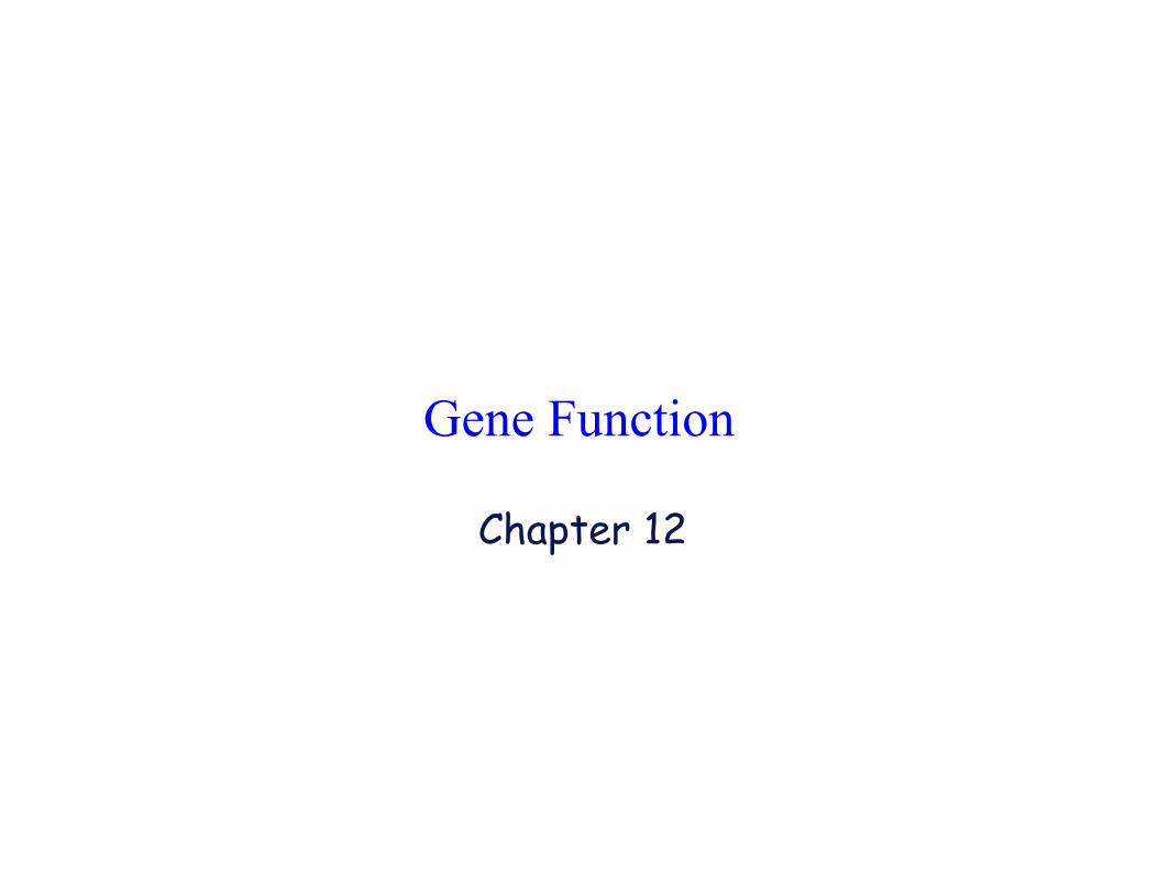
The Central Dogma of Biology
transcription
translation
GATC
GAUC
20 amino acids
Gene Control of Enzyme Structure
• Genes encode proteins, including enzymes.
• Genes work in sets to accomplish biochemicalpathways.
• Genes often work in cooperation with othergenes.
• These discoveries are the foundation of modernmolecular genetics.
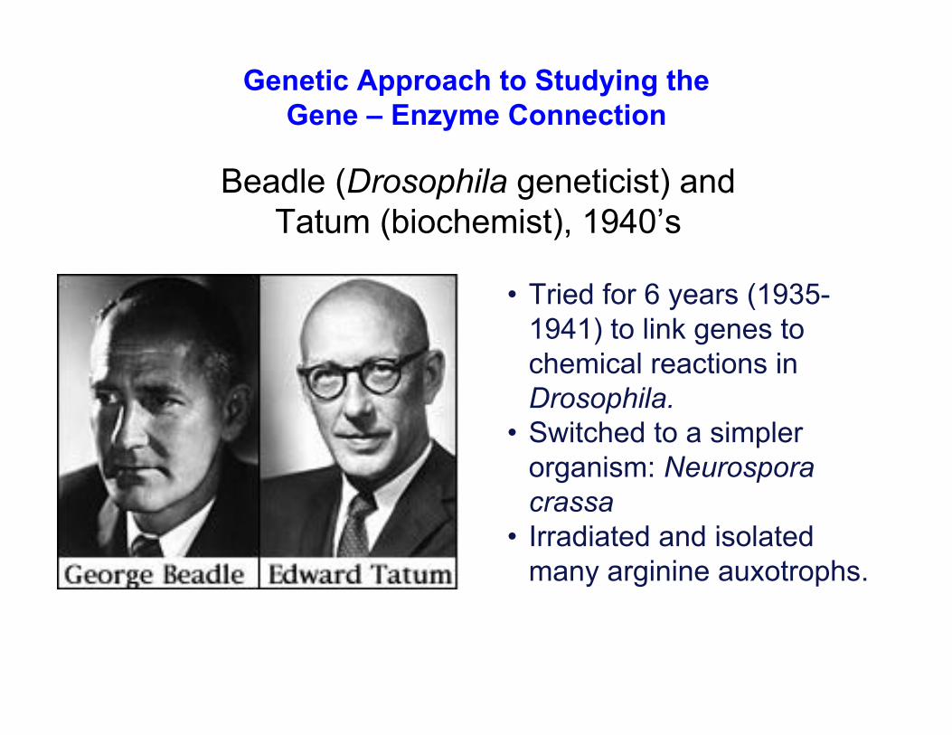
Genetic Approach to Studying the
Gene – Enzyme Connection
Beadle (Drosophila geneticist) and
Tatum (biochemist), 1940’s
• Tried for 6 years (1935-
1941) to link genes to
chemical reactions in
Drosophila.
• Switched to a simpler
organism: Neurospora
crassa
• Irradiated and isolated
many arginine auxotrophs.
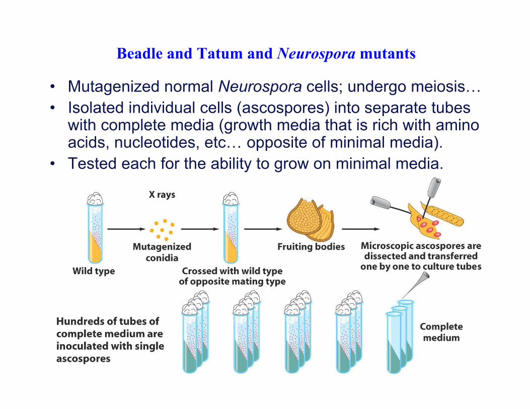
Beadle and Tatum and Neurospora mutants
• Mutagenized normal Neurospora cells; undergo meiosis…
• Isolated individual cells (ascospores) into separate tubeswith complete media (growth media that is rich with aminoacids, nucleotides, etc… opposite of minimal media).
• Tested each for the ability to grow on minimal media.
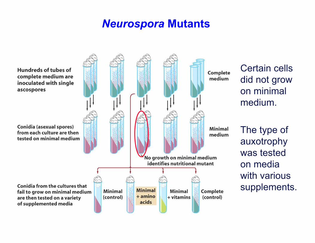
Neurospora Mutants
Certain cells
did not grow
on minimal
medium.
The type of
auxotrophy
was tested
on media
with various
supplements.
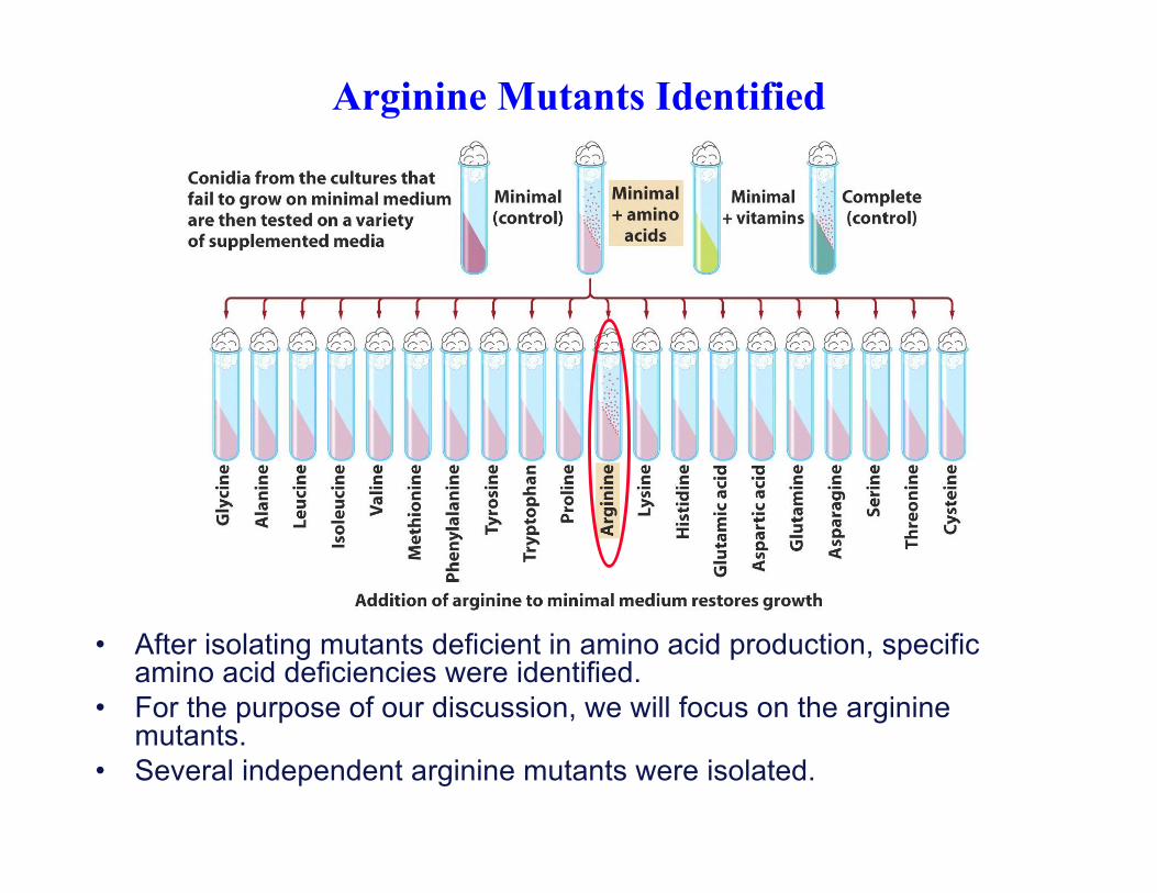
Arginine Mutants Identified
• After isolating mutants deficient in amino acid production, specificamino acid deficiencies were identified.
• For the purpose of our discussion, we will focus on the argininemutants.
• Several independent arginine mutants were isolated.
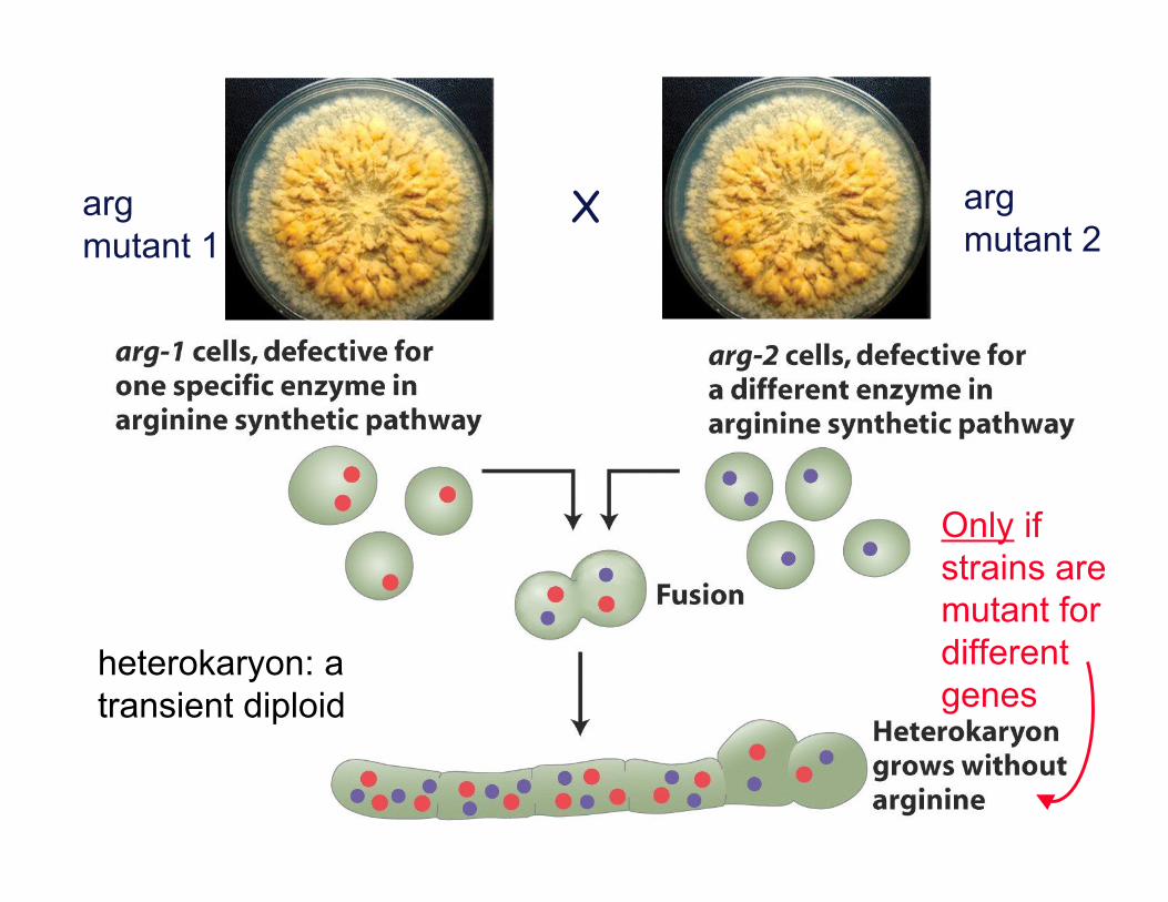
arg
mutant 1
heterokaryon: a
transient diploid
Only if
strains are
mutant for
different
genes
X arg
mutant 2
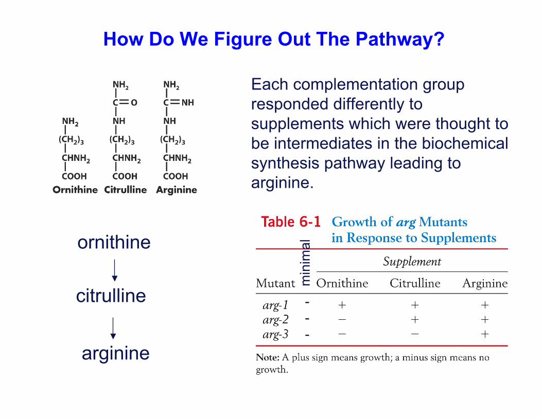
How Do We Figure Out The Pathway?
Each complementation group
responded differently to
supplements which were thought to
be intermediates in the biochemical
synthesis pathway leading to
arginine.
arginine
citrulline
ornithine
min
ima
l-
-
-
Mutant citrulline ornithine arginine
arg-1 + + +
arg-2 + - +
arg-3 - - +
precursor
arg-1
enz. X
argornithine citrulline
Next, figure out at which step in the pathway each
complementation group (gene) acts…
arg-2
enz. Y
arg-3
enz. Z
minimal
-
-
-
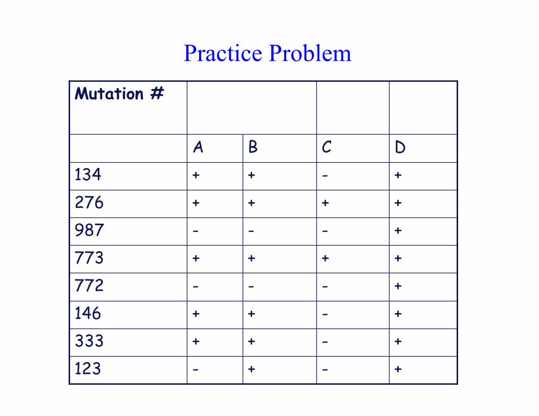
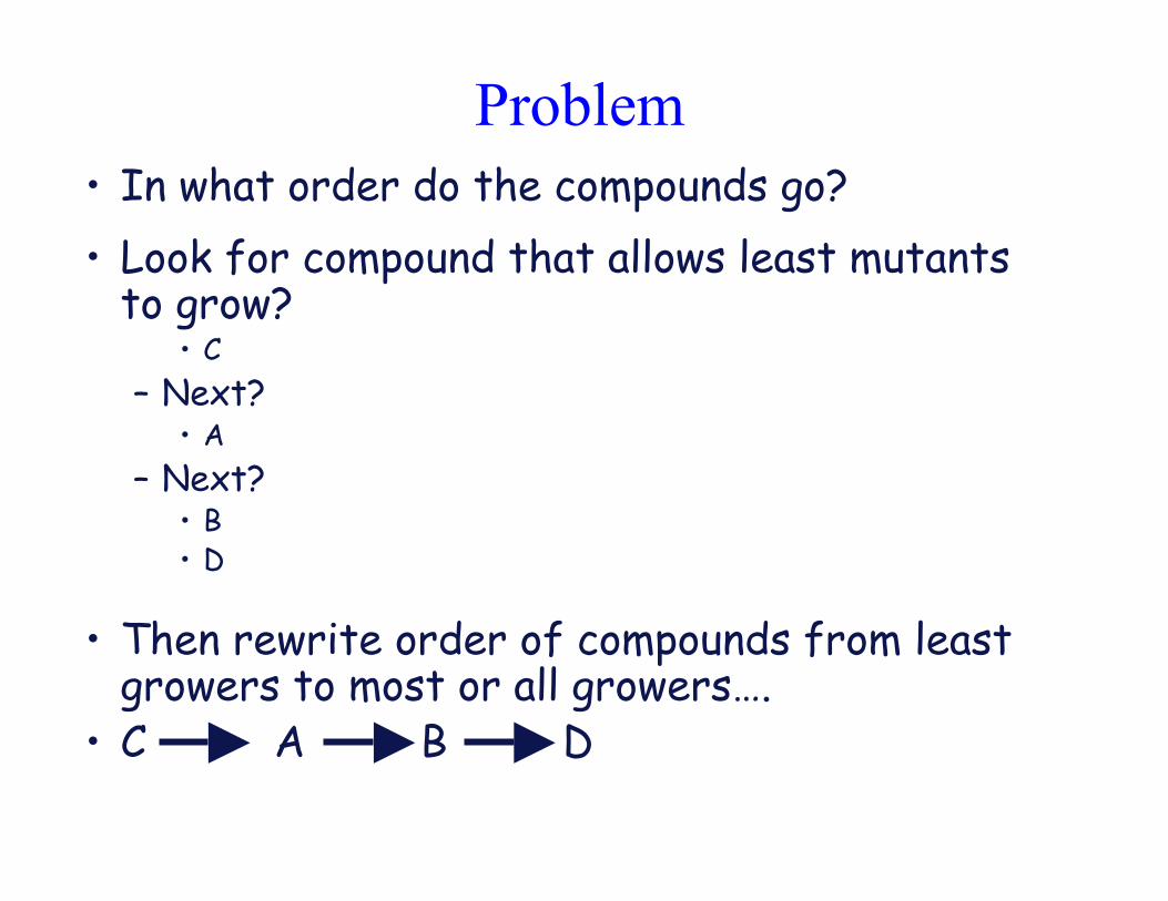
Practice Problem
+-+-123
+-++333
+-++146
+---772
++++773
+---987
++++276
+-++134
DCBA
Mutation #
Problem
• In what order do the compounds go?
• Look for compound that allows least mutantsto grow?
• C
– Next?• A
– Next?• B• D
• Then rewrite order of compounds from leastgrowers to most or all growers….
• C A B D
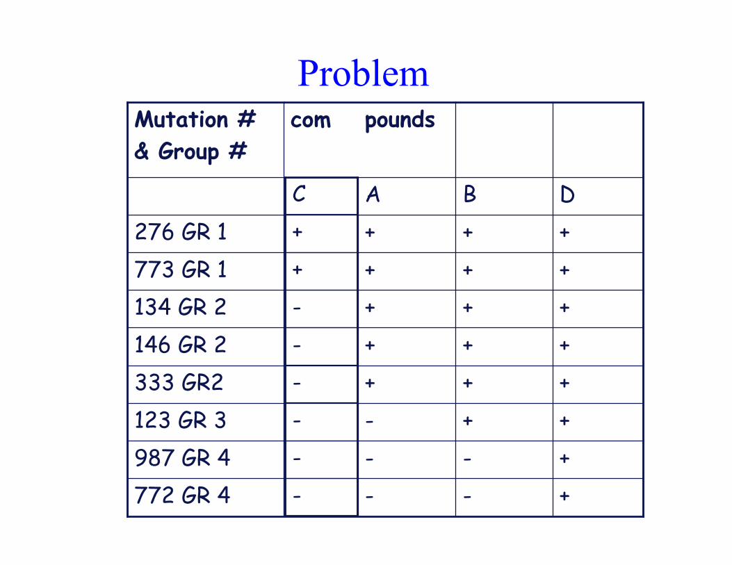
Problem
++-123 GR 3
+++333 GR2
+++146 GR 2
+++276 GR 1
+++773 GR 1
+--772 GR 4
+--987 GR 4
+++134 GR 2
DBA
poundscomMutation #
& Group #
-
-
-
-
-
-
+
+
C
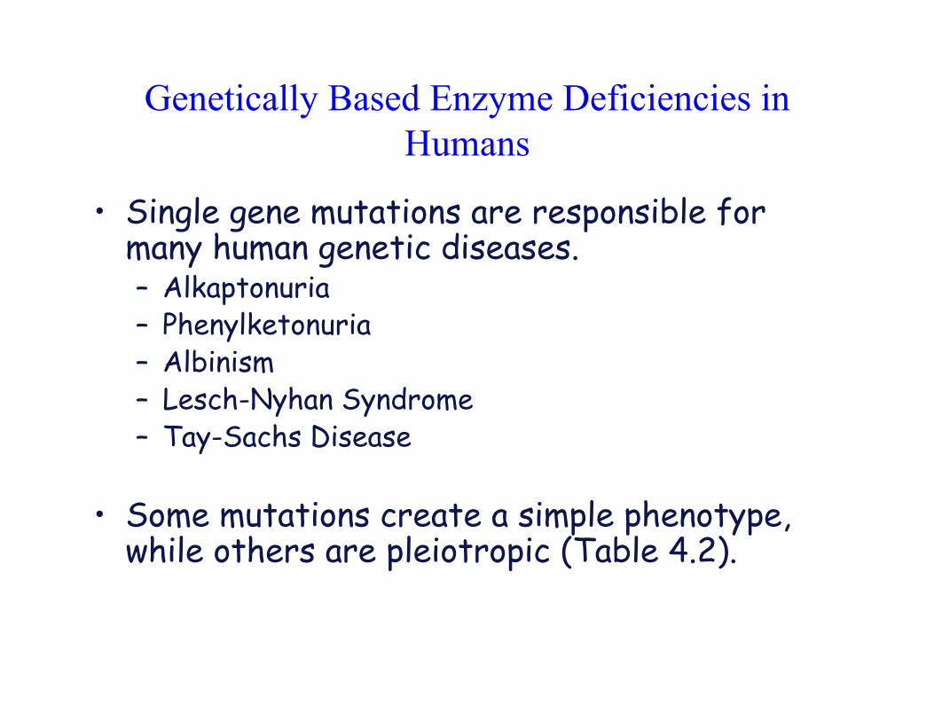
Genetically Based Enzyme Deficiencies in
Humans
• Single gene mutations are responsible formany human genetic diseases.– Alkaptonuria– Phenylketonuria– Albinism– Lesch-Nyhan Syndrome– Tay-Sachs Disease
• Some mutations create a simple phenotype,while others are pleiotropic (Table 4.2).
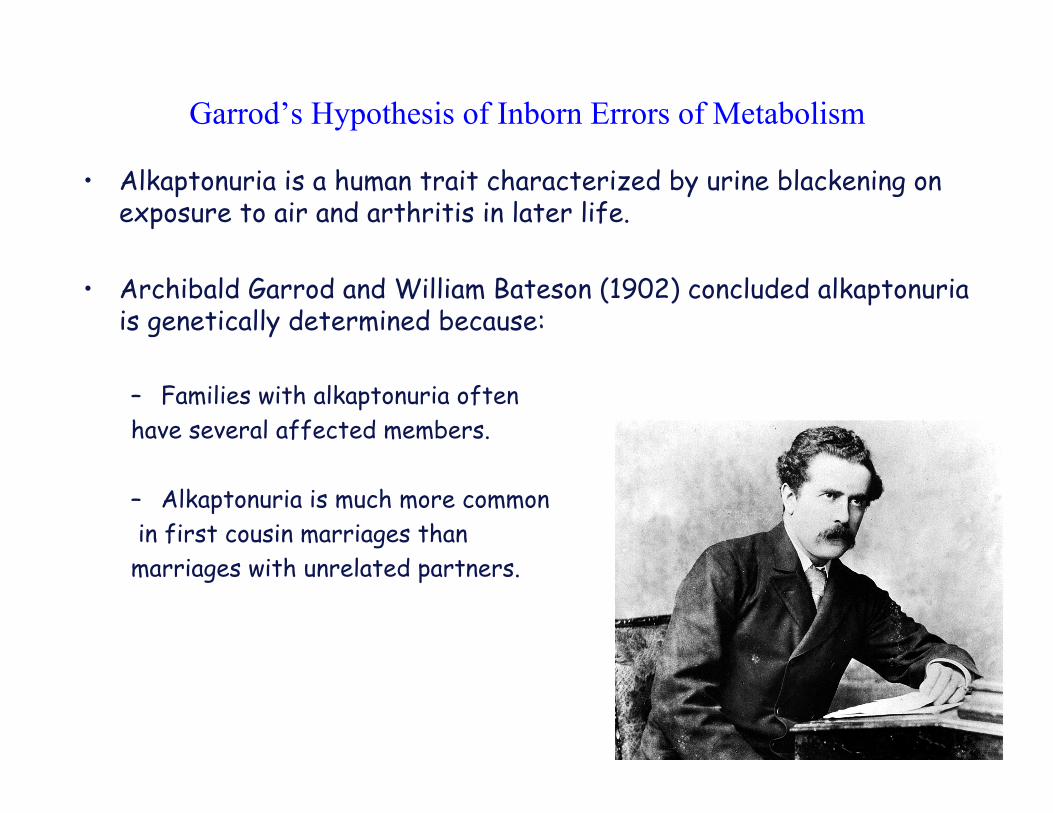
Garrod’s Hypothesis of Inborn Errors of Metabolism
• Alkaptonuria is a human trait characterized by urine blackening onexposure to air and arthritis in later life.
• Archibald Garrod and William Bateson (1902) concluded alkaptonuriais genetically determined because:
– Families with alkaptonuria often
have several affected members.
– Alkaptonuria is much more common
in first cousin marriages than
marriages with unrelated partners.
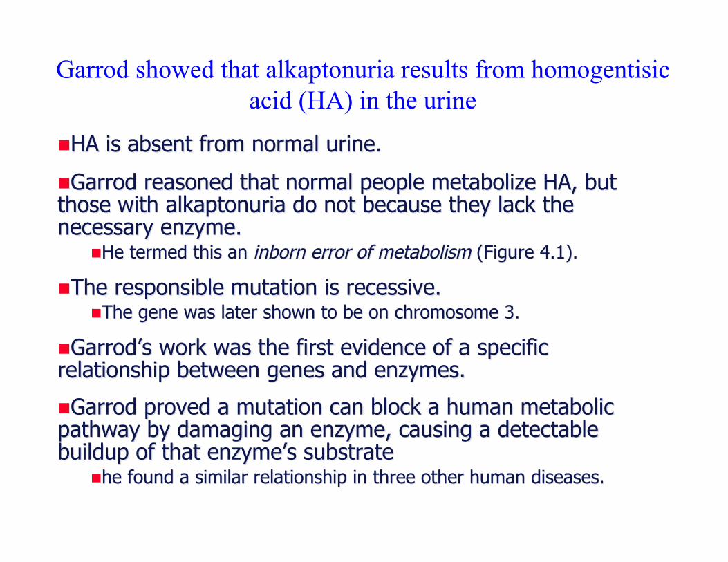
Garrod showed that alkaptonuria results from homogentisic
acid (HA) in the urine
!!HA is absent from normal urine.HA is absent from normal urine.
!!Garrod Garrod reasoned that normal people metabolize HA, butreasoned that normal people metabolize HA, butthose with those with alkaptonuria alkaptonuria do not because they lack thedo not because they lack thenecessary enzyme.necessary enzyme.
!!He termed this an He termed this an inborn error of metabolisminborn error of metabolism (Figure 4.1). (Figure 4.1).
!!The responsible mutation is recessive.The responsible mutation is recessive.!!The gene was later shown to be on chromosome 3.The gene was later shown to be on chromosome 3.
!!GarrodGarrod’’s s work was the first evidence of a specificwork was the first evidence of a specificrelationship between genes and enzymes.relationship between genes and enzymes.
!!Garrod Garrod proved a mutation can block a human metabolicproved a mutation can block a human metabolicpathway by damaging an enzyme, causing a detectablepathway by damaging an enzyme, causing a detectablebuildup of that enzymebuildup of that enzyme’’s substrates substrate
!!he found a similar relationship in three other human diseases.he found a similar relationship in three other human diseases.
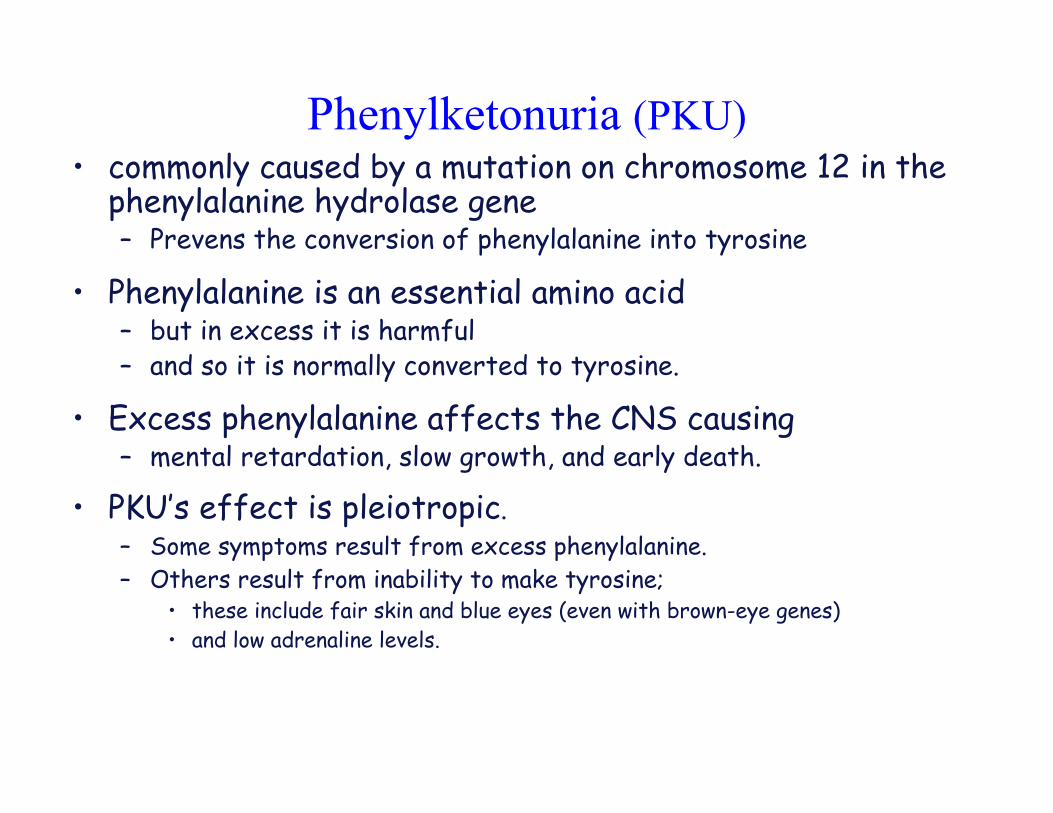
Phenylketonuria (PKU)• commonly caused by a mutation on chromosome 12 in the
phenylalanine hydrolase gene– Prevens the conversion of phenylalanine into tyrosine
• Phenylalanine is an essential amino acid– but in excess it is harmful– and so it is normally converted to tyrosine.
• Excess phenylalanine affects the CNS causing– mental retardation, slow growth, and early death.
• PKU’s effect is pleiotropic.– Some symptoms result from excess phenylalanine.
– Others result from inability to make tyrosine;• these include fair skin and blue eyes (even with brown-eye genes)
• and low adrenaline levels.
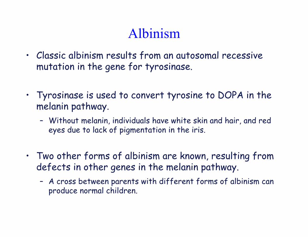
Albinism
• Classic albinism results from an autosomal recessivemutation in the gene for tyrosinase.
• Tyrosinase is used to convert tyrosine to DOPA in themelanin pathway.
– Without melanin, individuals have white skin and hair, and redeyes due to lack of pigmentation in the iris.
• Two other forms of albinism are known, resulting fromdefects in other genes in the melanin pathway.
– A cross between parents with different forms of albinism canproduce normal children.
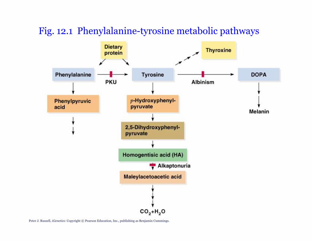
Peter J. Russell, iGenetics: Copyright © Pearson Education, Inc., publishing as Benjamin Cummings.
Fig. 12.1 Phenylalanine-tyrosine metabolic pathways
Lesch-Nyhan Syndrome
• results from a recessive mutation on the X chromosome
– in the gene for hypoxanthine-guanine phosphoribosyl transferase(HGPRT).
1. The fatal disease is found in males.
2. Heterozygous (carrier) females may show symptoms when lyonization(inactivation) of the normal X chromosome leaves the X chromosome withthe defective HGPRT gene in control of cells.
• HGPRT is an enzyme essential to purine utilization. In Lesch-Nyhansyndrome this pathway is highly impaired.
– results in the accumulation of purines
• which are eventually!converted to uric acid.
– individuals with HGPRT have high levels of uric acid in their bodies.
• The defect in a single enzyme, HGPRT, has very pleiotropiceffects
– giving rise to uremia, kidney failure, mental deficiency, and (so farinexplicably) self-mutilation.
Tay-Sachs Disease• Tay-Sachs is one of a group of diseases called lysosomal-storage
diseases.
– Generally caused by recessive mutations
– result from mutations in genes encoding lysosomal enzymes.
• Tay-Sachs disease (infantile amaurotic idiocy) results from arecessive mutation in the gene hexA
– which encodes the enzyme N-acetylhexosaminidase A.
– The HexA enzyme cleaves a terminal N-acetylgalactosamine group from abrain ganglioside
• Infants homozygous recessive for this gene will have nonfunctionalHexA enzyme.– Unprocessed ganglioside accumulates in brain cells, and causes various
clinical symptoms:• Infants have enhanced reaction to sharp sounds.• A cherry-colored spot surrounded by a white halo may be visible on the retina.• Rapid neurological degeneration begins about age 1
– brain loses control of normal functions due to accumulation of unprocessedganglioside.
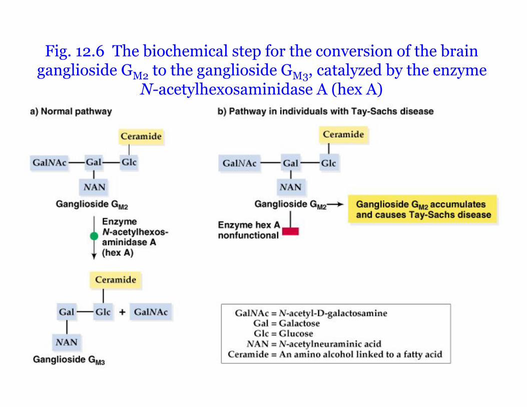
Peter J. Russell, iGenetics: Copyright © Pearson Education, Inc., publishing as Benjamin Cummings.
Fig. 12.6 The biochemical step for the conversion of the brainganglioside GM2 to the ganglioside GM3, catalyzed by the enzyme
N-acetylhexosaminidase A (hex A)
Gene Control of Protein Structure
• Genes also make proteins that are notenzymes.– Such proteins are called Structural Proteins.
• These proteins are often abundant, makingthem easier to isolate, purify, and analyze.– such as hemoglobin
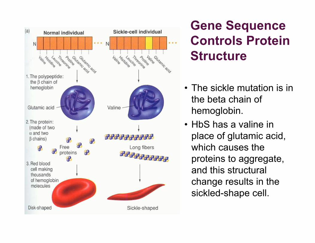
• The sickle mutation is in
the beta chain of
hemoglobin.
• HbS has a valine in
place of glutamic acid,
which causes the
proteins to aggregate,
and this structural
change results in the
sickled-shape cell.
Gene Sequence
Controls Protein
Structure
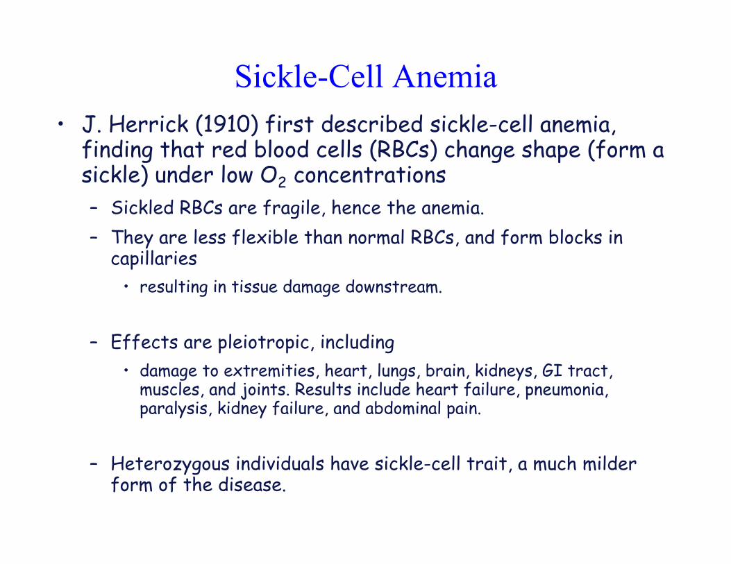
Sickle-Cell Anemia
• J. Herrick (1910) first described sickle-cell anemia,finding that red blood cells (RBCs) change shape (form asickle) under low O2 concentrations
– Sickled RBCs are fragile, hence the anemia.
– They are less flexible than normal RBCs, and form blocks incapillaries
• resulting in tissue damage downstream.
– Effects are pleiotropic, including
• damage to extremities, heart, lungs, brain, kidneys, GI tract,muscles, and joints. Results include heart failure, pneumonia,paralysis, kidney failure, and abdominal pain.
– Heterozygous individuals have sickle-cell trait, a much milderform of the disease.
Sickle-Cell Anemia
• The genetics and gene products involved in sickle-cell anemiaand trait are as follows:
– Wild-type ! chain allele is !A, which is codominant with mutant !S
allele.
– Hemoglobin of !A/!A individuals has normal ! subunits, while
hemoglobin of those with the genotype !S/!S has ! subunits that sickle at
low O2 tension.
– Hemoglobin of !A/!S individuals is 1⁄2 normal, and 1⁄
2 sickling form.
These heterozygotes may experience sickle-cell symptoms after a sharp
drop in the oxygen content of their environment.
• http://www.pbs.org/wgbh/evolution/library/01/2/l_012_02.html

Homework Problems
• Chapter 12
• # 12, 18, 34
• DON’T forget to take the online QUIZ!!
• DON’T forget to submit the online iActivity
•“Pathways”