Embed Size (px)
Citation preview
REVIEW Open Access
Gene specific therapies – the nexttherapeutic milestone in neurologyDavid Brenner1,2* , Albert C. Ludolph1,3 and Jochen H. Weishaupt1,2
Abstract
Gene selective approaches that either correct a disease mutation or a pathogenic mechanism will fundamentallychange the treatment of neurological disorders. Basically, gene specific therapies are designed to manipulate RNAexpression or reconstitute gene expression and function depending on the disease mechanism. Considerablemethodological advances in the last years have made successful clinical translation of gene selective approachespossible, based on RNA interference or viral gene reconstitution in spinal muscular atrophy (SMA), Duchennemuscular dystrophy (DMD), and familial amyloid polyneuropathy (FAP). In this review, we provide an overview ofthe existing and coming gene specific therapies in neurology and discuss benefits, risks and challenges.
BackgroundAfter the ground-breaking therapeutic advances in neu-rovascular and chronic inflammatory CNS diseases inthe previous decades neurology reaches the next thera-peutic milestone. Gene-specific approaches that eithercorrect a genetic defect directly or compensate a diseasemechanism will dramatically change the treatment ofboth genetic and sporadic neurological diseases. The ul-timate gene specific therapy would be the direct, preciseand permanent correction of the DNA defect. Advance-ment of genome editing tools and vector platformsmight make this dream come true one day. Meanwhile,indirect gene-specific strategies have been continuouslydeveloped and are now available to effectively compen-sate the effects of mutations or pathomechanisms. Fun-damentally, three different approaches are used inpractice today:
(1) RNA interference by antisense oligonucleotides(ASOs) or small interfering RNAs (siRNAs) tomanipulate stability, splicing and/or translation ofthe target mRNA.
(2) Splice modification by small molecules leading toin- or exclusion of a selected exon.
(3) Gene (transfer) therapy - viral delivery of anexogenous DNA encoding a healthy gene copy or aregulatory RNA (miRNA or shRNA) to influenceexpression of the target mRNA.
Gene mutations cause a partial or complete loss offunction (LoF), a gain of toxic function (GoF) or a com-bination of both. If the prevailing disease mechanism isa LoF the therapeutic aim is to restore the expression ofthe affected protein product (“gene reconstitution or re-placement”). In the case of a GoF the goal is to reducethe expression of the mutant protein product (“gene si-lencing”). Technically, gene delivery therapies and ASOsboth can be designed to down- or upregulate expressionof the target gene/mRNA. So far, gene-specific therapieshave been approved for three neurological indications:Spinal muscular atrophy (SMA), Duchenne musculardystrophy (DMD), and familial amyloid polyneuropathy(FAP). There is a constantly growing number of preclin-ical and clinical trials testing gene-specific approaches inother neurological diseases. This review provides a com-prehensive overview of the principles of and the latestdevelopments in gene specific therapies and illustratesthe risks and challenges coming with them.
© The Author(s). 2020 Open Access This article is licensed under a Creative Commons Attribution 4.0 International License,which permits use, sharing, adaptation, distribution and reproduction in any medium or format, as long as you giveappropriate credit to the original author(s) and the source, provide a link to the Creative Commons licence, and indicate ifchanges were made. The images or other third party material in this article are included in the article's Creative Commonslicence, unless indicated otherwise in a credit line to the material. If material is not included in the article's Creative Commonslicence and your intended use is not permitted by statutory regulation or exceeds the permitted use, you will need to obtainpermission directly from the copyright holder. To view a copy of this licence, visit http://creativecommons.org/licenses/by/4.0/.
* Correspondence: [email protected] of Neurology, University of Ulm, Ulm, Germany2Division of Neurodegenerative Diseases, Neurology Department, UniversityMedicine Mannheim, Mannheim, GermanyFull list of author information is available at the end of the article
Neurological Researchand Practice
Brenner et al. Neurological Research and Practice (2020) 2:25 https://doi.org/10.1186/s42466-020-00075-z
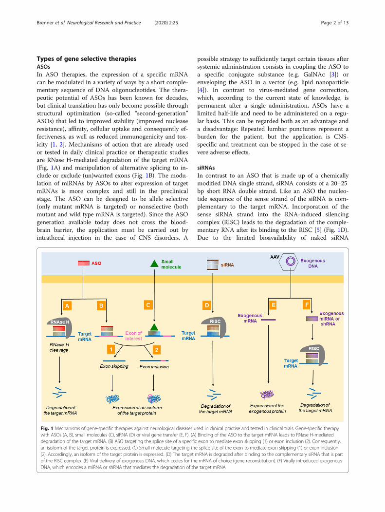
Types of gene selective therapiesASOsIn ASO therapies, the expression of a specific mRNAcan be modulated in a variety of ways by a short comple-mentary sequence of DNA oligonucleotides. The thera-peutic potential of ASOs has been known for decades,but clinical translation has only become possible throughstructural optimization (so-called “second-generation”ASOs) that led to improved stability (improved nucleaseresistance), affinity, cellular uptake and consequently ef-fectiveness, as well as reduced immunogenicity and tox-icity [1, 2]. Mechanisms of action that are already usedor tested in daily clinical practice or therapeutic studiesare RNase H-mediated degradation of the target mRNA(Fig. 1A) and manipulation of alternative splicing to in-clude or exclude (un)wanted exons (Fig. 1B). The modu-lation of miRNAs by ASOs to alter expression of targetmRNAs is more complex and still in the preclinicalstage. The ASO can be designed to be allele selective(only mutant mRNA is targeted) or nonselective (bothmutant and wild type mRNA is targeted). Since the ASOgeneration available today does not cross the blood-brain barrier, the application must be carried out byintrathecal injection in the case of CNS disorders. A
possible strategy to sufficiently target certain tissues aftersystemic administration consists in coupling the ASO toa specific conjugate substance (e.g. GalNAc [3]) orenveloping the ASO in a vector (e.g. lipid nanoparticle[4]). In contrast to virus-mediated gene correction,which, according to the current state of knowledge, ispermanent after a single administration, ASOs have alimited half-life and need to be administered on a regu-lar basis. This can be regarded both as an advantage anda disadvantage: Repeated lumbar punctures represent aburden for the patient, but the application is CNS-specific and treatment can be stopped in the case of se-vere adverse effects.
siRNAsIn contrast to an ASO that is made up of a chemicallymodified DNA single strand, siRNA consists of a 20–25bp short RNA double strand. Like an ASO the nucleo-tide sequence of the sense strand of the siRNA is com-plementary to the target mRNA. Incorporation of thesense siRNA strand into the RNA-induced silencingcomplex (RISC) leads to the degradation of the comple-mentary RNA after its binding to the RISC [5] (Fig. 1D).Due to the limited bioavailability of naked siRNA
Fig. 1 Mechanisms of gene-specific therapies against neurological diseases used in clinical practise and tested in clinical trials. Gene-specific therapywith ASOs (A, B), small molecules (C), siRNA (D) or viral gene transfer (E, F). (A) Binding of the ASO to the target mRNA leads to RNase H-mediateddegradation of the target mRNA. (B) ASO targeting the splice site of a specific exon to mediate exon skipping (1) or exon inclusion (2). Consequently,an isoform of the target protein is expressed. (C) Small molecule targeting the splice site of the exon to mediate exon skipping (1) or exon inclusion(2). Accordingly, an isoform of the target protein is expressed. (D) The target mRNA is degraded after binding to the complementary siRNA that is partof the RISC complex. (E) Viral delivery of exogenous DNA, which codes for the mRNA of choice (gene reconstitution). (F) Virally introduced exogenousDNA, which encodes a miRNA or shRNA that mediates the degradation of the target mRNA
Brenner et al. Neurological Research and Practice (2020) 2:25 Page 2 of 13
following systemic administration this approach requiresa vector. In the case of the first instance of a clinicallyapplicable siRNA therapy, Patisiran, a lipid nanoparticleserves as vector [6].
Small moleculesRNA splicing with in- or exclusion of (un)wanted exonscan also be manipulated by small molecules, which tar-get splice sites (Fig. 1C) and exhibit a comparably goodbiodistribution and tissue penetrance [7]. Risdiplam,which leads to selective inclusion of exon 7 in the SMN2mRNA to treat SMA, is the first approved drug based onthis approach (see below).
Vector-based gene therapyConstant optimization of delivery, transgene expressionefficacy and safety of AAVs in the recent years now fa-cilitate successful clinical translation of gene transfertherapies [8]. In virus-mediated in vivo gene therapy aviral vector, usually a non-replicating natural or engi-neered AAV capsid, is used to deliver a fully functionalcopy of a gene including a promoter and polyadenylationsignal into the nucleus of the patient’s cells without inte-gration in the patient’s genomic DNA [9]. The trans-ferred gene either encodes a functional protein tocompensate a LoF (“gene replacement”) (Fig. 1E) or aregulatory RNA (miRNA or shRNA) to downregulatethe expression of the target mRNA by RNA interferencein order to counteract a GoF (“gene silencing”) (Fig. 1F).The pharmacokinetics and tropism depend on the prop-erties of the natural or engineered AAV subtype. Thechoice of the promoter decides if the exogenous gene isconstitutively or conditionally expressed and in whichtissue it is expressed. Since tissue barriers limit the bio-availability of the vector, in some instances it needs tobe injected into the CSF or directly into the target tissue(muscle or CNS by stereotaxis). The great advantage ofgene delivery therapies over ASO, siRNA or small mol-ecule approaches is the durable expression and efficacy.A potential severe disadvantage is that treatment cannotbe terminated and unwanted effects may persist.
In vivo genome editingWith CRISPR/Cas9 (Clustered Regularly InterspacedShort Palindromic Repeats), TALEN (Transcriptionactivator-like effector nuclease) and ZFN (Zinc-fingernuclease) various gene editing methods are used in pre-clinical models of neurological diseases. These ap-proaches are potentially suitable for the direct, preciseand permanent correction of the DNA defect whatmight once become the ideal therapy. While TALENand ZFN recognize and bind the target genomic DNAsequence directly through protein/DNA interaction,CRISPR-Cas nucleases are guided by RNA (RNA/DNA
base pairing). The nucleases are designed to recognizeand cut the target DNA sequences and cause double-strand breaks that are repaired by the cell’s DNA repairsystems through non-homologous end joining orhomology-directed repair, given the presence of a donorDNA template. Either the nuclease, its mRNA or itsgene must be delivered to the target cells by a vectorsystem that can be a plasmid, virus or nanoparticle. Ineither case, many technical hurdles remain to be over-come until these therapies are safe and efficient enoughto treat patients [10]. Nevertheless, the pipelines of sev-eral pharma consortia already mention preclinical testingof genome editing strategies for various indications in-cluding Huntington’s disease and familial amyloidpolyneuropathy.
Present and future gene specific therapiesA myriad of gene specific therapies based on ASOs,siRNA, small molecules and gene transfer is currentlybeing tested in a preclinical and clinical trials by aca-demia and pharma industry. In the following, we high-light the most relevant developments in variousneurological diseases while focusing on therapies havingalready reached the clinical trial phase (see also Table 1).
Motor neuron disordersSpinal muscular atrophy (SMA) is caused by homozy-gous LoF mutations in the SMN1 gene that is ubiquitouslyexpressed in the body. The age of onset and the severity ofthe phenotype (SMA type 1–4) depend on the copy num-ber of the homologous SMN2 gene. The mature SMN2mRNA differs from the SMA1 mRNA by exclusion ofexon 7 following alterative splicing, which results in in-stability and reduced amounts of the protein product.Two gene specific therapies have been developed that me-diate the inclusion of exon 7 into the mature SMN2mRNA: the ASO Nusinersen (Fig. 1B) and the small mol-ecule Risdiplam (Fig. 1C). Nusinersen (NCT02193074[11], NCT02292537 [12]) is administered intrathecallyevery 4months after a 1-year dosing phase. Since its ap-proval it has revolutionized the treatment of infantile- andlater-onset SMA [13]. Meanwhile there is robust evidencethat adult patients with SMA also benefit from a therapywith Nusinersen [14]. Contrary to Nusinersen, Risdiplamis a small molecule and is given orally. It could in principlereach all tissues of the body. In a phase 3 study it hasshown a similar clinical effectiveness as Nusinersen forSMA type 1 (i.e. infantile SMA) and is also effective in theother SMA types (NCT02908685, NCT02913482,NCT03032172, NCT03779334). Its approval for all formsof SMA is expected soon. With Branaplam another smallmolecule inducing exon 7 inclusion is being tested in aphase 1/2 trial (NCT02268552). The third gene specifictherapy approach against SMA aims at restoring SMN1
Brenner et al. Neurological Research and Practice (2020) 2:25 Page 3 of 13
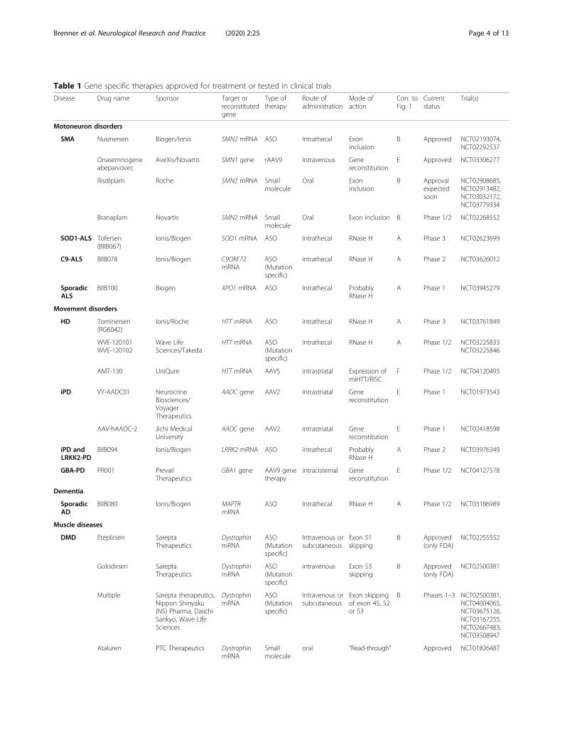
Table 1 Gene specific therapies approved for treatment or tested in clinical trials
Disease Drug name Sponsor Target orreconstitutedgene
Type oftherapy
Route ofadministration
Mode ofaction
Corr. toFig. 1
Currentstatus
Trial(s)
Motoneuron disorders
SMA Nusinersen Biogen/Ionis SMN2 mRNA ASO Intrathecal Exoninclusion
B Approved NCT02193074,NCT02292537
Onasemnogeneabeparvovec
AveXis/Novartis SMN1 gene rAAV9 Intravenous Genereconstitution
E Approved NCT03306277
Risdiplam Roche SMN2 mRNA Smallmolecule
Oral Exoninclusion
B Approvalexpectedsoon
NCT02908685,NCT02913482,NCT03032172,NCT03779334
Branaplam Novartis SMN2 mRNA Smallmolecule
Oral Exon inclusion B Phase 1/2 NCT02268552
SOD1-ALS Tofersen(BIIB067)
Ionis/Biogen SOD1 mRNA ASO Intrathecal RNase H A Phase 3 NCT02623699
C9-ALS BIIB078 Ionis/Biogen C9ORF72mRNA
ASO(Mutationspecific)
intrathecal RNase H A Phase 2 NCT03626012
SporadicALS
BIIB100 Biogen XPO1 mRNA ASO Intrathecal ProbablyRNase H
A Phase 1 NCT03945279
Movement disorders
HD Tominersen(RG6042)
Ionis/Roche HTT mRNA ASO intrathecal RNase H A Phase 3 NCT03761849
WVE-120101WVE-120102
Wave LifeSciences/Takeda
HTT mRNA ASO(Mutationspecific)
intrathecal RNase H A Phase 1/2 NCT03225833NCT03225846
AMT-130 UniQure HTT mRNA AAV5 intrastriatal Expression ofmiHTT/RISC
F Phase 1/2 NCT04120493
iPD VY-AADC01 NeurocrineBiosciences/VoyagerTherapeutics
AADC gene AAV2 intrastriatal Genereconstitution
E Phase 1 NCT01973543
AAV-hAADC-2 Jichi MedicalUniversity
AADC gene AAV2 intrastriatal Genereconstitution
E Phase 1 NCT02418598
iPD andLRKK2-PD
BIIB094 Ionis/Biogen LRRK2 mRNA ASO intrathecal ProbablyRNase H
A Phase 2 NCT03976349
GBA-PD PR001 PrevailTherapeutics
GBA1 gene AAV9 genetherapy
intracisternal Genereconstitution
E Phase 1/2 NCT04127578
Dementia
SporadicAD
BIIB080 Ionis/Biogen MAPTRmRNA
ASO Intrathecal RNase H A Phase 1/2 NCT03186989
Muscle diseases
DMD Eteplirsen SareptaTherapeutics
DystrophinmRNA
ASO(Mutationspecific)
Intravenous orsubcutaneous
Exon 51skipping
B Approved(only FDA)
NCT02255552
Golodirsen SareptaTherapeutics
DystrophinmRNA
ASO(Mutationspecific)
intravenous Exon 53skipping
B Approved(only FDA)
NCT02500381
Multiple Sarepta therapeutics,Nippon Shinyaku(NS) Pharma, DaiichiSankyo, Wave LifeSciences
DystrophinmRNA
ASO(Mutationspecific)
Intravenous orsubcutaneous
Exon skippingof exon 45, 52or 53
B Phases 1–3 NCT02500381,NCT04004065,NCT03675126,NCT03167255,NCT02667483,NCT03508947
Ataluren PTC Therapeutics DystrophinmRNA
Smallmolecule
oral “Read-through” Approved NCT01826487
Brenner et al. Neurological Research and Practice (2020) 2:25 Page 4 of 13
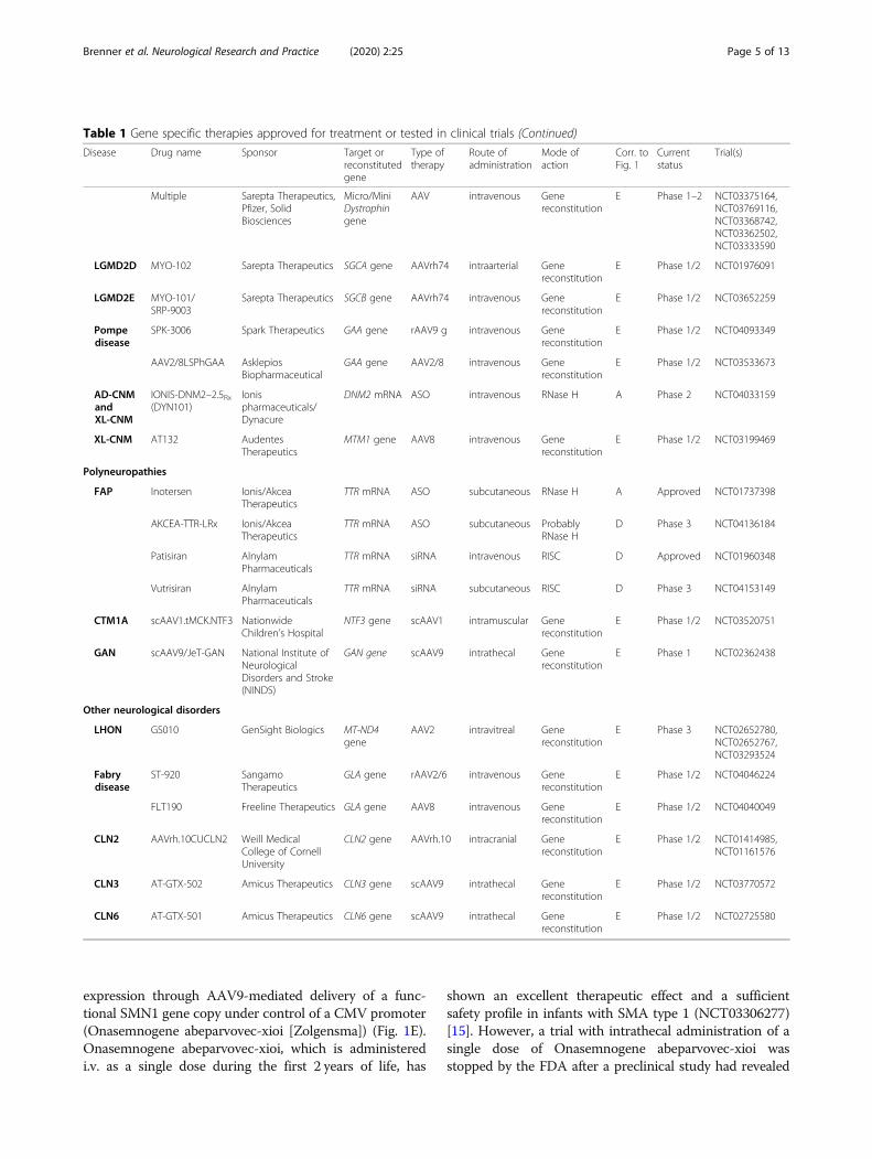
expression through AAV9-mediated delivery of a func-tional SMN1 gene copy under control of a CMV promoter(Onasemnogene abeparvovec-xioi [Zolgensma]) (Fig. 1E).Onasemnogene abeparvovec-xioi, which is administeredi.v. as a single dose during the first 2 years of life, has
shown an excellent therapeutic effect and a sufficientsafety profile in infants with SMA type 1 (NCT03306277)[15]. However, a trial with intrathecal administration of asingle dose of Onasemnogene abeparvovec-xioi wasstopped by the FDA after a preclinical study had revealed
Table 1 Gene specific therapies approved for treatment or tested in clinical trials (Continued)
Disease Drug name Sponsor Target orreconstitutedgene
Type oftherapy
Route ofadministration
Mode ofaction
Corr. toFig. 1
Currentstatus
Trial(s)
Multiple Sarepta Therapeutics,Pfizer, SolidBiosciences
Micro/MiniDystrophingene
AAV intravenous Genereconstitution
E Phase 1–2 NCT03375164,NCT03769116,NCT03368742,NCT03362502,NCT03333590
LGMD2D MYO-102 Sarepta Therapeutics SGCA gene AAVrh74 intraarterial Genereconstitution
E Phase 1/2 NCT01976091
LGMD2E MYO-101/SRP-9003
Sarepta Therapeutics SGCB gene AAVrh74 intravenous Genereconstitution
E Phase 1/2 NCT03652259
Pompedisease
SPK-3006 Spark Therapeutics GAA gene rAAV9 g intravenous Genereconstitution
E Phase 1/2 NCT04093349
AAV2/8LSPhGAA AsklepiosBiopharmaceutical
GAA gene AAV2/8 intravenous Genereconstitution
E Phase 1/2 NCT03533673
AD-CNMandXL-CNM
IONIS-DNM2–2.5Rx(DYN101)
Ionispharmaceuticals/Dynacure
DNM2 mRNA ASO intravenous RNase H A Phase 2 NCT04033159
XL-CNM AT132 AudentesTherapeutics
MTM1 gene AAV8 intravenous Genereconstitution
E Phase 1/2 NCT03199469
Polyneuropathies
FAP Inotersen Ionis/AkceaTherapeutics
TTR mRNA ASO subcutaneous RNase H A Approved NCT01737398
AKCEA-TTR-LRx Ionis/AkceaTherapeutics
TTR mRNA ASO subcutaneous ProbablyRNase H
D Phase 3 NCT04136184
Patisiran AlnylamPharmaceuticals
TTR mRNA siRNA intravenous RISC D Approved NCT01960348
Vutrisiran AlnylamPharmaceuticals
TTR mRNA siRNA subcutaneous RISC D Phase 3 NCT04153149
CTM1A scAAV1.tMCK.NTF3 NationwideChildren’s Hospital
NTF3 gene scAAV1 intramuscular Genereconstitution
E Phase 1/2 NCT03520751
GAN scAAV9/JeT-GAN National Institute ofNeurologicalDisorders and Stroke(NINDS)
GAN gene scAAV9 intrathecal Genereconstitution
E Phase 1 NCT02362438
Other neurological disorders
LHON GS010 GenSight Biologics MT-ND4gene
AAV2 intravitreal Genereconstitution
E Phase 3 NCT02652780,NCT02652767,NCT03293524
Fabrydisease
ST-920 SangamoTherapeutics
GLA gene rAAV2/6 intravenous Genereconstitution
E Phase 1/2 NCT04046224
FLT190 Freeline Therapeutics GLA gene AAV8 intravenous Genereconstitution
E Phase 1/2 NCT04040049
CLN2 AAVrh.10CUCLN2 Weill MedicalCollege of CornellUniversity
CLN2 gene AAVrh.10 intracranial Genereconstitution
E Phase 1/2 NCT01414985,NCT01161576
CLN3 AT-GTX-502 Amicus Therapeutics CLN3 gene scAAV9 intrathecal Genereconstitution
E Phase 1/2 NCT03770572
CLN6 AT-GTX-501 Amicus Therapeutics CLN6 gene scAAV9 intrathecal Genereconstitution
E Phase 1/2 NCT02725580
Brenner et al. Neurological Research and Practice (2020) 2:25 Page 5 of 13
dorsal root ganglia (DRG) mononuclear cell inflammationin non-human primates. The drug has meanwhile beenapproved for i.v. administration by the FDA and EMA.Nucleocytoplasmic translocation of TDP-43 with forma-
tion of cytosolic pTDP-43 inclusions that might cause anuclear LoF and cytoplasmic GoF toxicity is the prevailingneuropathology in sporadic ALS. The propagation ofTDP-43 pathology in the CNS correlates with the spread-ing of the clinical symptoms [16]. Corroborating the sig-nificance of TDP-43 in ALS, mutations in its gene(TARDBP) cause genetic forms of ALS [17, 18]. Conse-quently, the TDP-43 pathology appears a reasonedpharmacological target. However, the reduction of TDP-43 expression itself is not a suitable therapeutic approach,since TDP-43 is physiologically essential. Mice withhomozygous Tardbp deletion are not viable and thosewith heterozygous KO develop an ALS-like phenotype[19]. Therefore, approaches must aim at reducing thepTDP-43 inclusions or compensate for the loss of physio-logical function of TDP-43. Following this thought, anASO approach has been developed to downregulate theXPO1 mRNA (BIIB100), which codes for the proteinExportin 1. Exportin 1mediates the nuclear export ofmany proteins containing nuclear export signals includingTDP-43 [20] (Fig. 1A). Thus, XPO1 inhibition is supposedto reduce nucleocytoplasmic translocation and cytosolicaggregation of TDP-43 [21]. The recruitment of sporadicALS patients for a respective phase 1/2 study has juststarted at the end of 2019 (NCT03945279).Intermediate-length polyglutamine expansions in the
ATXN2 gene are associated with an increased risk ofALS. TDP-43 mutant mice with Atxn2-KO or treatedwith ASOs downregulating Atxn2 show a reduction ofphosphorylated TDP-43 inclusions and a dramaticallyincreased lifespan [22] (Fig. 1A). Consequently, an ASOdownregulating human ATXN2 is being developed to-wards a clinical trial in sporadic ALS patients.An intronic hexanucleotide repeat expansion in
C9Orf72 and missense mutations in SOD1 are the mostfrequent causes of genetic ALS (C9-ALS and SOD1-ALS) in Europe [23]. It is supposed that their pathogen-icity is predominantly based on a GoF toxicity, althougha LoF aspect is also discussed for C9Orf72 mutations.After most promising results in mutant disease models,ASOs downregulating the expression of SOD1 (Tofer-sen/BIIB067/IONIS-SOD1Rx; allele unselective) andC9Orf72 (BIIB078/IONIS-C9Rx; allele selective) throughRNase H mediated mRNA degradation are now beingtested in clinical trials (Fig. 1A). The trial testing IONIS-SOD1Rx is presently in phase 3 (NCT02623699); an in-terim analysis of phase 2 has raised hope for a positivestudy outcome [24]. The IONIS-C9Rx trial is currently inphase 2 (NCT03626012), there are no interim resultsavailable yet. Further, another consortium is developing
an allele-selective silencing ASO towards clinical transla-tion for C9-ALS and -FTD. AAV mediated gene deliveryof DNA coding miRNA or shRNA that downregulateSOD1 mRNA showed excellent efficacy in mutanthSOD1 rodent or primate models [25] (Fig. 1F). Conse-quently, an AAV9-SOD1-shRNA candidate is being de-veloped towards clinical translation by anotherconsortium.
Movement disordersThe current drug therapy of idiopathic Parkinson’sdisease (iPD) aims at restoring CNS dopamine levels.Two sponsors are currently testing MR-guided intrapu-taminal AAV2-delivered AADC gene transfer in iPD pa-tient in phase 1/2 trials (NCT02418598, NCT01973543).The AADC gene encodes the aromatic L-amino acid de-carboxylase responsible for converting Levodopa toDopamine. An interim analysis of NCT01973543 hasshown an increase in enzyme expression and dosedependent clinical improvements [26]. While thisapproach might prolong the time of symptom control itis probably – like current dopaminergic drugs – incap-able of influencing disease progression. Alpha-synuclein(a-syn) aggregations are the predominant neuropathol-ogy of iPD; its propagation in the CNS correlates withthe spreading of the clinical symptoms [27]. The signifi-cance of a-syn in iPS is further supported by the findingsthat multiplications, mutations, and single nucleotidepolymorphisms in the SNCA gene, encoding the alpha-synuclein protein, either cause or increase the risk foriPS. Cole and colleagues developed an ASO that down-regulates the SCNA mRNA after intraventricular appli-cation. It efficiently reduced seeded a-syn inclusion loadin wildtype mice and rats treated with exogenous pre-formed a-syn fibrils [28] (Fig. 1A). This therapy has notyet reached the clinical trial phase. Another approachtargeting the a-syn pathology consists in the downregu-lation of LRRK2. Missense mutations in LRRK2 that leadto a GoF are a frequent cause of genetic PD [29, 30] andcertain polymorphisms in LRRK2 locus modulate therisk for iPD [31]. In addition, increased LRRK2 proteinactivity in dopaminergic neurons in post-mortem tissueof iPD patients seems to drive the α-syn pathology [32].Thus, downregulation of LRRK2 appears an interestingtherapy strategy to alleviate α-syn pathology. Indeed,ASOs reducing expression of the LRRK2 mRNA dimin-ished fibril-induced seeding of a-syn inclusions in wild-type mice [28] (Fig. 1A). Consequently, a pharmaconsortium is testing an ASO targeting the humanLRRK2 mRNA (BIIB094/ION859) in a phase 2 trial inPD patients with or without LRRK2 GoF mutations(NCT03976349). In case the strategies reducing a-synpathology are clinically effective they might also be
Brenner et al. Neurological Research and Practice (2020) 2:25 Page 6 of 13

tested in other synucleinopathies, such as multiple sys-tem atrophy (MSA) and Lewy body dementia (LBD).Mutations in the GBA1 gene, which presumably lead to
a LOF, are the most frequent genetic contributor to PDpathogenesis (GBA-PD) [32]. GBA1 encodes the enzymebeta-glucocerebrosidase, which is required for the disposaland recycling of glycolipids. Accumulation of glycolipidsleads to lysosomal dysfunction that in turn exacerbateslysosomal accumulation of a-syn. Recently, a phase 1/2trial has been launched in patients with a pathogenicGBA1 mutation (PD-GBA) testing a gene reconstitutiontherapy where a GBA1 gene copy is delivered by an AAV9(NCT04127578) (Fig. 1E). The drug is administered intra-cisternally as a single dose. A phase 1/2 trial testing thesame therapeutic in patients with neuropathic Gaucherdisease, that is caused by biallelic LoF mutations in thesame gene, is expected to start soon. Further, the samesponsor announces the development of an approach com-bining GBA1 gene transfer and SCNA knockdown fortreatment of synucleinopathies in general.A CAG repeat expansion in the HTT gene that en-
codes the Huntingtin protein causes Huntington’s dis-ease (HD). After most promising results in preclinicalrodent studies [33], several gene-specific therapeuticstrategies are being tested in clinical trials in HD pa-tients. In NCT03761849, currently in phase 3, an alleleunselective ASO that downregulates pan-HTT mRNA(Tominersen/RG6042/IONIS-HTTRx) is administeredintrathecally (Fig. 1A). An interim analysis has demon-strated successful target engagement in that the HTTprotein was reduced by 40% on average in the CSF ofHD patients [34]. In NCT03225833 and NCT03225846,currently in phase 1/2, allele specific ASOs (targeting aprevalent SNP) selectively downregulate the mutantHTT mRNA after intrathecal adminstration (Fig. 1A).Third, in NCT04120493, currently in phase 1/2, anAAV5 vector is used to deliver a gene encoding amiRNA that blocks the HTT mRNA (miHTT). This vec-tor is administered by intrastriatal injection (Fig. 1F).
DementiasTau and beta-amyloid aggregates are the neuropatho-logical hallmarks of sporadic Alzheimer’s disease (AD).Mutations in the amyloid precursor protein gene APPcause genetic AD through a GoF mechanism. However,beta-amyloid immunotherapy has been largely disap-pointing in clinical trials so far, questioning beta-amyloid as the ideal drug target, at least in progressedAD cases. According to autopsy and Tau-PET imagingstudies spreading of Tau pathology seems to be highlycorrelated with disease progression [35, 36], which raiseshopes for a successful clinical translation of Tau targetedtherapies. Genetic deletion or ASO-mediated downregu-lation of the MAPT mRNA (encoding Tau protein)
alleviated neuropathology and clinical symptoms in gen-etic AD mouse models. Consequently, an ASO designedto downregulate the MAPT mRNA is being tested in ADpatients in a phase 1/2 clinical trial (NCT03186989)(Fig. 1A). If this strategy proves successful it could bealso tested in other tauopathies, such as the atypical Par-kinson syndromes supranuclear palsy (PSP) and cortico-basal syndrome (CBS).Sporadic frontotemporal dementia (FTD) is neuro-
pathologically associated with TDP-43, Tau or FUS pro-tein pathology that each is considered the cause of theclinical symptoms. Therapies aiming at alleviating theseproteinopathies are currently being tested in other indi-cations (BIIB100 for TDP-43 pathology in ALS; BIIB080for Tau pathology in AD, see respective paragraphsabove). They might also be tested for the treatment ofFTD patients, if the according ALS or AD studies arepositive. Another prerequisite would be the availabilityof PET imaging and respective tracers that allow to reli-ably identify the underlying proteinopathy. The threemost frequently mutated genes in genetic FTD areC9Orf72, GRN, and MAPT (with an autosomal dominantinheritance in all three cases). As described before, ASOsdownregulating C9Orf72 or MAPT (both predominantlycausing a GoF effect when mutated) are currently beingtested in C9-ALS and sporadic AD patients and may betested in C9-FTD cohorts next (see above). Mutations inGRN cause FTD by a LOF and aggravate TDP-43 inclu-sion pathology. An AAV9-based gene reconstitutiontherapy for GRN-FTD is currently developed towards aphase 1/2 trial (Fig. 1E).
PolyneuropathiesHereditary amyloidosis is caused by GoF mutations inthe ATTR gene leading to an abnormal, aggregation-prone TTR protein that is deposited in amyloid aggre-gates. The amyloidosis causes cardiomyopathy and/orPNP, also called familial amyloid PNP (FAP). Twogene specific therapies based on RNA interference, Ino-tersen and Patisiran, have been developed and approvedfor the treatment of PNP caused by hATTR amyloidosis.Inotersen is an ASO that is administered s.c. once perweek, while Patisiran is a first-in-class siRNA therapeuticthat is given i.v. every 3 weeks. They lead to the degrad-ation of the mutant and the wildtype hATTR mRNAthrough RNAse H (Inoteresen) (Fig. 1A) and RISC (Pati-siran) (Fig. 1D), respectively. Longer lasting compoundsbased on the same therapeutic principles (AKCEA-TTR-LRx and Vutrisiran) are currently being tested by thesame sponsors in phase 3 trials (NCT04136184 andNCT04153149).Giant axonal neuropathy (GAN) is a very rare, auto-
somal recessive childhood onset disease owing to LoFmutations in the GAN gene. It encodes the protein
Brenner et al. Neurological Research and Practice (2020) 2:25 Page 7 of 13
Gigaxonin. The mutations cause a progressive accumula-tion of neuronal intermediate filaments in axons. After asuccessful preclinical study [37] a phase 1 trial is recruit-ing patients to test the intrathecal administration of anAAV9 vector to deliver a functional copy of the GANgene (NCT02362438) (Fig. 1E).Duplications of the PMP22 gene cause the most
prevalent subtype of CMT, CMT1A. Zhao et al. havedeveloped an ASO to downregulate PMP22 mRNA.After s.c. administration the ASO results in restorationof myelination and improvement of electroneurographicparameters in a mouse model based on overexpressionof he human PMP22 gene [38] (Fig. 1A). A clinical trialhas not yet been announced. Further, the upregulationof neurotrophin-3 (NT-3) has been shown to lead to aremyelination in CMT1A mouse models and patients[39]. A gene transfer using an AAV1 vector has showngood efficacy in PMP22 mutant mice [40]. A clinicaltrial in CMT1A patients is underway (NCT03520751)(Fig. 1E). Preclinical studies testing gene reconstitutiontherapies have also been successful in mouse models ofother CMT types [41].
Muscle diseasesMuscle dystrophy is a X-linked genetic myopathycaused by mutations in the dystrophin gene DMD. De-pending on the residual function of the protein product,mutations either lead to the more severe phenotype ofDuchenne muscular dystrophy (DMD) (complete LoF)or the milder form of Becker muscular dystrophy (partialLoF). Two ASO therapeutics (Eteplirsen and Golodirsen)have been approved for the treatment of DMD in 2016and 2010, respectively, by the FDA, while the Europeanauthority EMA rejected their approval for reasons dis-cussed below [42]. Eteplirsen and Golodirsen, given s.c.or i.v. weekly, cause a skipping of exon 51 or 53, respect-ively, of the Dystrophin pre-mRNA. This strategy resultsin a shortened instead of an otherwise unfunctional pro-tein (Fig. 1B). Both ASOs are restricted to DMD patientswith mutations in exon 51 or 53, respectively. However,severe shortcomings of the clinical studies that led totheir approval and serious SAEs have raised significantdoubts about their efficacy and safety [43, 44]. The FDAhas instructed the responsible company to provide morerobust evidence for the clinical effectiveness of bothASO therapeutics in post marketing studies by 2021(Eteplirsen) and 2023 (Golodirsen), respectively. Mul-tiple other ASOs that lead to skipping of various DMDexons are being tested in clinical trials [45]. Beyond theaforementioned ASO therapeutics, the small moleculeAtaluren has been approved for treatment of patientswith nonsense DMD mutations, which produce a prema-ture stop codon, after showing some clinical benefit [46].Ataluren causes the ribosomal readthrough of mRNAs
with a premature stop codon and consequent translationof the complete protein. Further, multiple AAV-basedi.v. gene transfer therapies have been developed and arebeing tested in phase 1/2 trials in DMD patients (seeTable 1) (Fig. 1E). Since the Dystrophin gene exceeds thepackaging capacity of AAVs it has been shortened to theessential domains in these cases, called micro- or mini-dystrophin-genes.Limb-girdle muscular dystrophy (LGMD) is a slowly
progressive, symmetric, proximal myopathy with onset inchildhood or adolescence. It is caused by mono- or bialle-lic LoF mutations in various genes encoding sarcoglycans,which tie the intracellular cytoskeleton to the extracellularmatrix in muscle tissue. According to the mode of inherit-ance it is classified into LGMD1 (autosomal dominant)and LGMD2 (autosomal recessive). After successful pre-clinical studies in mice [47, 48] AAV gene reconstitutiontherapies that supply healthy copies of the mutated geneshave been developed and are being tested in phase 1/2 tri-als for the autosomal recessive LGMD2 forms D and E(NCT01976091 and NCT03652259 (Fig. 1E).Pompe disease is a progressive myopathy and auto-
somal recessively inherited disorder caused by biallelicLoF mutations in the GAA gene. GAA encodes the lyso-somal acidic alpha-glucosidase. Respective mutationslead to lysosomal accumulation of glycogen. After prom-ising results from preclinical mouse studies [49], AAV-mediated expression of GAA in hepatocytes by a singlei.v. infusion of the viral vector is now tested in Pompedisease patients in phase 1/2 trials (NCT04093349 andNCT03533673) (Fig. 1E). Perspectively, ASOs reducingglycogen synthesis, which are currently being developedfor the treatment of Lafora disease, might be a generaltherapeutic option for glycogenoses.Centronuclear Myopathy (CNM) is a group of con-
genital myopathies characterized by abnormal localizationof the nucleus in the center of muscles cells. Mutations inseveral genes have been made responsible for CNM. Themost severe from is X-linked CNM (XL-CNM; syn. Myo-tubular myopathy) is caused by LoF mutations in theMTM1 gene, while autosomal dominant CNM (AD-CNM) is mostly caused by GoF mutations in the DNM2gene. Mtm1-KO causes an overexpression of DNM2 andsystemic administration of an ASO downregulating Dnm2mRNA prevented and reverted myotubular myopathy inMtm1-KO mice [50]. Consequently, a consortium is test-ing its ASO candidate IONIS-DNM2–2.5Rx (DYN101)that is administered i.v. in patients with centronuclear my-opathies caused by mutations in either DNM2 or MTM1(NCT04033159). The study is currently in phase 2. Fur-ther, Audentes Therapeutics is testing an AAV8-deliveredreplacement of the MTM1 gene (AT132) by single dosei.v. administration in patients with XL-CNM in a phase 1/2 study (NCT03199469). Therapeutic efficacy has already
Brenner et al. Neurological Research and Practice (2020) 2:25 Page 8 of 13
been shown in Mtm1-KO mice and XLMTM dogs before.An interim analysis has yielded promising results [51].
Other neurological indicationsLeber hereditary optic neuropathy (LHON) is a mater-nally inherited mitochondrial disease characterized by thedegeneration of retinal ganglion cells and their axons lead-ing to vision loss. It is caused by LoF mutations in thegenes ND4, ND1 and ND6 encoding the mitochondrialNADH dehydrogenase proteins. AAV transfer of a healthyND4 gene copy after intraocular administration preventedretinal ganglion cell degeneration and preserved visualfunction in a LHON rat model [52]. GenSight Biologics istesting an AAV2 gene therapy delivering a ND4 gene copyin a phase 3 trial in LHON patients with ND4 mutation(NCT02652780, NCT02652767, NCT03293524) (Fig. 1E).Neuronal ceroid lipofuscinoses (CLN) a group of rare
rare, childhood-onset and fatal generally autosomal re-cessive genetic neurodegenerative lysosomal storage dis-eases caused by mutations in various genes (CLN1–7).The disease is characterized by symptomatic epilepsyand progressive decline of cognitive and motor func-tions. After successful preclinical studies in animals [53]trials for the different CLN types testing intrathecal orintracranial administration of single-dose AAV-basedgene delivery of the CLN2, CLN3 or CLN6 gene havebeen launched by various sponsors and are currently on-going (CLN2: NCT01414985, NCT01161576; CLN3:NCT03770572; CLN6: NCT02725580).Fabry Disease is a X-linked genetic lysosomal storage
disorder caused by mono- or biallelic LoF mutations in theGLA gene and affects men and women. The mutations leadto a deficiency of the alpha-galactosidase, which causes ubi-quitous accumulation of glycosphingolipids in lysosomes.This leads to multi-organ dysfunction and polymorphicsymptoms, amongst others neuropathy and strokes. Aftersuccessful preclinical studies in mouse models of Fabry dis-ease [54, 55], Freeline Therapeutics and Sangamo Therapeu-tics are already testing i.v. single dose AAV genereplacement therapies in phase 1/2 trials in Fabry disease pa-tients (NCT04046224 and NCT04040049) (Fig. 1E).
Risks and challenges of gene-specific therapiesAlthough optimism regarding gene-specific therapies isjustified, we may not forget that they are still in their in-fancy and must be considerably improved in many as-pects. In the following, we outline the major challengesand risks of current gene selective approaches.
Drug delivery, expression control and cell selectivenessMost ASOs applied or tested in CNS disorders presentlyneed to be administered intrathecally. This is not onlystressful for the patient but also an infrastructural and fi-nancial burden, in view of the many ASO therapies
coming that are administered via this route. Therefore,the success of ASO therapies critically depends on thedevelopment of suitable vector platforms and conjugatesubstances capable of sufficiently penetrating brain ormuscle tissue after systemic administration. Dependingif the target RNA shall be repressed ubiquitously or onlyin certain cell types the vector or conjugate ideallywould also confer a selective tropism. Niche companiesare increasingly addressing this matter. The need for im-proved bioavailability and cell selectiveness also appliesto AAV gene transfer therapies. Another significant con-cern of current gene transfer therapies is that the num-ber of gene copies transferred to a cell is uncontrolledleading to unphysiological up- or downregulation of thetarget gene, which can be neurotoxic itself in the longrun. In addition, it is not precisely known which celltypes the AAV infects, and which cell types benefit fromthe gene transfer, especially when the endogenous ver-sion of the delivered gene is normally not expressed inthese respective cells. Moreover, current gene transfertherapies use aggressive constitutive promoters insteadof tightly regulated endogenous ones. To allow for longterm safety, future gene transfer therapies should allowfor inducible regulation of the transferred gene in caseof relevant adverse effects.
Long-term complicationsWithout disrespecting the revolutionary therapeutic ef-fect of gene specific therapies, it must be noted that theircurrent shortcomings are likely to only incompletelycompensate the genetic defect and cause long-termcomplications for various reasons:
1. The disease mechanism is incompletely targeted,e.g. if the pathomechanism is (1) a combination ofGoF and LoF or (2) a GOF resulting from repeatexpansion. The first constellation would require aselective downregulation of the mutant allele andan upregulation of the healthy one at the sametime. However, the majority of current genesilencing approaches are unselective and mightexacerbate a concomitant LoF. Repeat expansionmutations lead to toxic RNA transcripts from boththe sense and the antisense DNA strand. Therepeat RNAs again are translated into toxic repeatproteins. Therefore, an ideal therapy would targetboth sense and antisense strand of the repeatexpanded allele. The observation that repeatexpansion can also lead to a relevant LoF at thesame time makes this matter even morechallenging. On the other hand, allele selectivestrategies for every single mutation would hardly beaffordable and in most instances there would not be
Brenner et al. Neurological Research and Practice (2020) 2:25 Page 9 of 13
enough carriers of the same mutation for a clinicaltrial.
2. The gene defect is “overcorrected” by the therapy,meaning that gene replacement or gene silencingleads to an unphysiological (maybe neurotoxic)over- or underexpression of the healthy(endogenous or exogeneous) allele in a proportionof cells. Current gene delivery therapies lackexpression control. So transfected cells receivevarying numbers of exogenous gene copies,resulting in hardly predictable expression levels ofthe protein product or the regulatory RNAi,respectively. The problem of uncontrolled andunphysiological expression levels is furtherincreased by use of exogenous, strong andconstitutively active gene promoters instead of genespecific and conditional endogenous ones.
3. Gene specific therapies can trigger immuneresponses or be toxic in cell types that should notbe targeted. A current example is the dorsal rootganglia inflammation after intrathecaladministration of Onasemnogene Abeparvovec-xioiin non-human primates.
4. The gene specific therapy reaches CNS targetregion(s) only incompletely. Lowered target proteinconcentrations in the CSF (as observed in theinterim analyses of ASO studies targeting SOD1and HTT expression) are promising, but do notnecessarily indicate successful target engagement inthe desired neuronal population.
5. Untreated tissues become clinically relevant. Manydisease genes (e.g. including SOD1, C9Orf72 andSMN1) are ubiquitously expressed, however, mostcurrent gene specific therapies only incompletelytarget a limited number of organs.
The dilemma of genetic testingEffective gene specific therapies will become availablefor more and more disorders soon. Therefore, genetictesting is going to have an increasing therapeutic impli-cation. While it is unquestionable that newborns shouldbe screened for treatable infantile onset genetic diseases(such as SMA), many questions related to genetic testingof adult onset genetic diseases for which gene-specifictherapies exist remain unresolved: Who shall be tested –only members of families with a known genetic diseaseor also sporadic patients without a family history? Whoshall be treated? Many patients without family historycarry a rare or unique variant in a disease gene that is ofunknown significance. The disease mechanisms of mostALS genes are still incompletely understood making itlargely impossible to evaluate the pathogenicity of a vari-ant by a specific in vitro assay. Should patients bearingvariants of unknown significance be treated with a gene
specific therapy, when available – also in view of thecurrently tremendous costs of these therapies? Also, it isnot clearly defined yet when treatment should be startedin an asymptomatic carrier of a pathogenic mutation –even if it is known how penetrant and how variable theonset of the specific mutation is. Viewing the incalcul-able long-term adverse effects of gene selective therapies– do we harm more than we help when we start therapytoo early in asymptomatic risk gene carriers? Develop-ment of biomarkers reliably indicating the onset of aprodromal disease phase may ease the decision when tostart a therapy in disease gene mutation carriers. In anycase, these delicate treatment decisions should be metby an expert of both the respective disease andneurogenetics.
Need for disease specific biomarkersA slow or inconstant progression, phenotypical variabil-ity and small patient numbers challenge speed, costs andnot least statistics of clinical intervention trials of geneticdiseases, as long as only clinical measures are used asprimary endpoints. Therefore, biomarkers that properlycorrelate with disease progression and can be used sur-rogates for clinical endpoints are urgently needed. Nowis the time to validate biomarkers (e.g. in natural historystudies) – once an effective therapy is approved accessto native probes of body fluids will be limited.
ConclusionGene-specific therapies targeting a disease gene or dis-ease mechanism are going to revolutionize the treatmentof the various genetic and sporadic neurological diseases.However, most of these therapies must be considerablyoptimized to increase the safety of long-term use andthe ease of application. The neurological departmentshave to anticipate the infrastructural and personal re-quirements in order to be prepared for the increased de-mand of genetic counselling and the wave of newlyapproved therapeutics in in the foreseeable future.
AbbreviationsAAV: Adeno-associated virus; ALS: Amyotrophic lateral sclerosis;AD: Alzheimer disease; ASO : Antisense oligonucleotide; A-syn: Alphasynuclein; CRISPR: Clustered Regularly Interspaced Short Palindromic Repeats;DMD: Duchenne muscular dystrophy; FAP: Familial amyloid PNP;FTD: Frontotemporal dementia; GAN: Giant axonal neuropathy; GoF: Gain of(toxic) function; HD: Huntington’s disease; iPD: Idiopathic Parkinson’s disease;LGMD: Limb-girdle muscular dystrophy; LHON: Leber hereditary opticneuropathy; LoF: Loss of function; miRNA : Micro RNA; MR-guided : MagneticResonance Imaging-guided; PD : Parkinson’s disease; PNP: Polyneuropathy;shRNA : Small hairpin RNA; siRNA: Small interfering RNA; RISC: RNA-inducedsilencing complex; RNAi: RNA interference; SAE: Serious adverse effect; SMA: Spinal muscular atrophy; TALEN: Transcription activator-like effector nucle-ase; TTR: Transthyretin; ZFN: Zinc-finger nuclease
AcknowledgementsNot applicable.
Brenner et al. Neurological Research and Practice (2020) 2:25 Page 10 of 13
Authors’ contributionsDB wrote the manuscript. ACL and JHW revised the manuscript. Theauthor(s) read and approved the final manuscript.
FundingNot applicable.
Availability of data and materialsNot applicable.
Ethics approval and consent to participateNot applicable.
Consent for publicationNot applicable.
Competing interestsD. Brenner and J. H. Weishaupt own stocks of Ionis Pharmaceuticals. A. C.Ludolph has received financial research support from AB Science, BiogenIdec, Cytokinetics, GSK, Orion Pharam, Novartis, TauRx Therapeutics Ltd. andTEVA Pharmaceuticals. A. C. Ludolph has worked as a consultant forMitsubishi, Orion Pharma, Novartis and Teva and as a member of theadvisory board for Biogen, Treeway and Hoffmann-La Roche.
Author details1Department of Neurology, University of Ulm, Ulm, Germany. 2Division ofNeurodegenerative Diseases, Neurology Department, University MedicineMannheim, Mannheim, Germany. 3German Center for NeurodegenerativeDiseases (DZNE), Ulm, Germany.
Received: 19 April 2020 Accepted: 22 June 2020
References1. Schoch, K. M., & Miller, T. M. (2017). Antisense oligonucleotides: Translation
from mouse models to human neurodegenerative diseases. Neuron, 94(6),1056–1070. https://doi.org/10.1016/j.neuron.2017.04.010 Cell Press.
2. Frazier, K. S. (2015). Antisense oligonucleotide therapies: The promise andthe challenges from a toxicologic pathologist’s perspective. ToxicologicPathology, 43(1), 78–89. https://doi.org/10.1177/0192623314551840.
3. Kim, Y., Jo, M., Schmidt, J., Luo, X., Prakash, T. P., Zhou, T., Klein, S., Xiao, X.,Post, N., Yin, Z., & MacLeod, A. R. (2019). Enhanced potency of GalNAc-conjugated antisense oligonucleotides in hepatocellular cancer models.Molecular Therapy, 27(9), 1547–1557. https://doi.org/10.1016/j.ymthe.2019.06.009.
4. Kulkarni, J. A., Witzigmann, D., Chen, S., Cullis, P. R., & Van Der Meel, R.(2019). Lipid nanoparticle technology for clinical translation of siRNAtherapeutics. Accounts of Chemical Research, 52(9), 2435–2444. https://doi.org/10.1021/acs.accounts.9b00368.
5. Ameres, S. L., Martinez, J., & Schroeder, R. (2007). Molecular basis for targetRNA recognition and cleavage by human RISC. Cell, 130(1), 101–112. https://doi.org/10.1016/j.cell.2007.04.037.
6. Zhang, X., Goel, V., Attarwala, H., Sweetser, M. T., Clausen, V. A., & Robbie, G.J. (2020). Patisiran pharmacokinetics, pharmacodynamics, and exposure-response analyses in the phase 3 APOLLO trial in patients with hereditarytransthyretin-mediated (hATTR) amyloidosis. The Journal of ClinicalPharmacology, 60(1), 37–49. https://doi.org/10.1002/jcph.1480.
7. Zhao, X., Feng, Z., Ling, K. K. Y., Mollin, A., Sheedy, J., Yeh, S., Petruska, J.,Narasimhan, J., Dakka, A., Welch, E. M., Karp, G., Chen, K. S., Metzger, F., Ratni,H., Lotti, F., Tisdale, S., Naryshkin, N. A., Pellizzoni, L., Paushkin, S., et al. (2016).Pharmacokinetics, pharmacodynamics, and efficacy of a small-moleculeSMN2 splicing modifier in mouse models of spinal muscular atrophy.Human Molecular Genetics, 25(10), 1885–1899. https://doi.org/10.1093/hmg/ddw062.
8. Deverman, B. E., Ravina, B. M., Bankiewicz, K. S., Paul, S. M., & Sah, D. W. Y.(2018). Gene therapy for neurological disorders: Progress and prospects.Nature Reviews Drug Discovery, 17(9), 641–659. https://doi.org/10.1038/nrd.2018.110 Nature Publishing Group.
9. Hudry, E., & Vandenberghe, L. H. (2019). Therapeutic AAV gene transfer tothe nervous system: A clinical reality. Neuron, 101(5), 839–862. https://doi.org/10.1016/j.neuron.2019.02.017 Cell Press.
10. Zhang, H. X., Zhang, Y., & Yin, H. (2019). Genome editing with mRNAencoding ZFN, TALEN, and Cas9. Molecular Therapy, 27(4), 735–746. https://doi.org/10.1016/j.ymthe.2019.01.014 Cell Press.
11. Finkel, R. S., Mercuri, E., Darras, B. T., Connolly, A. M., Kuntz, N. L., Kirschner, J.,et al. (2017). Nusinersen versus sham control in infantile-onset spinalmuscular atrophy. New England Journal of Medicine, 377(18), 1723–1732.https://doi.org/10.1056/NEJMoa1702752.
12. Mercuri, E., Darras, B. T., Chiriboga, C. A., Day, J. W., Campbell, C., Connolly, A.M., et al. (2018). Nusinersen versus sham control in later-onset spinalmuscular atrophy. New England Journal of Medicine, 378(7), 625–635. https://doi.org/10.1056/NEJMoa1710504.
13. Wurster, C. D., & Günther, R. (2020, February 19). New treatments for spinalmuscular atrophy. In Nervenarzt. Springer Medizin. https://doi.org/10.1007/s00115-020-00871-7.
14. Hagenacker, T., Wurster, C. D., Günther, R., Schreiber-Katz, O., Osmanovic,A., Petri, S., Weiler, M., Ziegler, A., Kuttler, J., Koch, J. C., Schneider, I.,Wunderlich, G., Schloss, N., Lehmann, H. C., Cordts, I., Deschauer, M.,Lingor, P., Kamm, C., Stolte, B., et al. (2020). Nusinersen in adults with5q spinal muscular atrophy: A non-interventional, multicentre,observational cohort study. The Lancet Neurology, 19(4), 317–325.https://doi.org/10.1016/S1474-4422(20)30037-5.
15. Hoy, S. M. (2019). Onasemnogene Abeparvovec: First global approval. Drugs,79(11), 1255–1262. https://doi.org/10.1007/s40265-019-01162-5.
16. Braak, H., Brettschneider, J., Ludolph, A. C., Lee, V. M., Trojanowski, J. Q., &Del Tredici, K. (2013). Amyotrophic lateral sclerosis--A model of corticofugalaxonal spread. Nature Reviews Neurology, 9(12), 708–714. https://doi.org/10.1038/nrneurol.2013.221.
17. Sreedharan, J., Blair, I. P., Tripathi, V. B., Hu, X., Vance, C., Rogelj, B., Ackerley,S., Durnall, J. C., Williams, K. L., Buratti, E., Baralle, F., de Belleroche, J., Mitchell,J. D., Leigh, P. N., Al-Chalabi, A., Miller, C. C., Nicholson, G., & Shaw, C. E.(2008). TDP-43 mutations in familial and sporadic amyotrophic lateralsclerosis. Science (New York, N.Y.), 319(5870), 1668–1672. https://doi.org/10.1126/science.1154584.
18. Kabashi, E., Valdmanis, P. N., Dion, P., Spiegelman, D., McConkey, B. J.,Velde, C. V., Bouchard, J.-P., Lacomblez, L., Pochigaeva, K., Salachas, F.,Pradat, P.-F., Camu, W., Meininger, V., Dupre, N., & Rouleau, G. A. (2008).TARDBP mutations in individuals with sporadic and familial amyotrophiclateral sclerosis. Nature Genetics, 40(5), 572–574. https://doi.org/10.1038/ng.132.
19. Kraemer, B. C., Schuck, T., Wheeler, J. M., Robinson, L. C., Trojanowski, J.Q., Lee, V. M. Y., & Schellenberg, G. D. (2010). Loss of murine TDP-43disrupts motor function and plays an essential role in embryogenesis.Acta Neuropathologica, 119(4), 409–419. https://doi.org/10.1007/s00401-010-0659-0.
20. Archbold, H. C., Jackson, K. L., Arora, A., Weskamp, K., Tank, E. M. H., Li, X.,Miguez, R., Dayton, R. D., Tamir, S., Klein, R. L., & Barmada, S. J. (2018). TDP43nuclear export and neurodegeneration in models of amyotrophic lateralsclerosis and frontotemporal dementia. Scientific Reports, 8(1). https://doi.org/10.1038/s41598-018-22858-w.
21. Dolgin, E. (2019). To halt brain diseases, drugs take aim at protein trafficjams that kill neurons. Science. https://doi.org/10.1126/science.aaw6864.
22. Becker, L. A., Huang, B., Bieri, G., Ma, R., Knowles, D. A., Jafar-Nejad, P.,Messing, J., Kim, H. J., Soriano, A., Auburger, G., Pulst, S. M., Taylor, J. P., Rigo,F., & Gitler, A. D. (2017). Therapeutic reduction of ataxin-2 extends lifespanand reduces pathology in TDP-43 mice. Nature, 544(7650), 367–371. https://doi.org/10.1038/nature22038.
23. Müller, K., Brenner, D., Weydt, P., Meyer, T., Grehl, T., Petri, S., Grosskreutz, J.,Schuster, J., Volk, A. E., Borck, G., Kubisch, C., Klopstock, T., Zeller, D., Jablonka,S., Sendtner, M., Klebe, S., Knehr, A., Günther, K., Weis, J., et al. (2018).Comprehensive analysis of the mutation spectrum in 301 German ALSfamilies. Journal of Neurology, Neurosurgery and Psychiatry, 89(8), 817–827.https://doi.org/10.1136/jnnp-2017-317611.
24. Experimental Drug Shows Promise for Genetic Form of ALS. (n.d.). RetrievedMarch 26, 2020, from https://www.aan.com/PressRoom/Home/PressRelease/2719
25. Foust, K. D., Salazar, D. L., Likhite, S., Ferraiuolo, L., Ditsworth, D.,Ilieva, H., Meyer, K., Schmelzer, L., Braun, L., Cleveland, D. W., & Kaspar,B. K. (2013). Therapeutic AAV9-mediated suppression of mutant SOD1slows disease progression and extends survival in models of inheritedALS. Molecular Therapy, 21(12), 2148–2159. https://doi.org/10.1038/mt.2013.211.
Brenner et al. Neurological Research and Practice (2020) 2:25 Page 11 of 13
26. Christine, C. W., Bankiewicz, K. S., Van Laar, A. D., Richardson, R. M., Ravina, B.,Kells, A. P., Boot, B., Martin, A. J., Nutt, J., Thompson, M. E., & Larson, P. S.(2019). Magnetic resonance imaging–guided phase 1 trial of putaminalAADC gene therapy for Parkinson’s disease. Annals of Neurology, 85(5), 704–714. https://doi.org/10.1002/ana.25450.
27. Braak, H., Del Tredici, K., Bratzke, H., Hamm-Clement, J., Sandmann-Keil, D., &Rüb, U. (2002). Staging of the intracerebral inclusion body pathologyassociated with idiopathic Parkinson’s disease (preclinical and clinicalstages). Journal of Neurology, 249(Suppl 3), III/1–III/5.
28. Cole, T. A., Zhao, H., Collier, T. J., Sandoval, I., Sortwell, C. E., Steece-Collier, K.,Daley, B. F., Booms, A., Lipton, J., Welch, M., Berman, M., Jandreski, L.,Graham, D., Weihofen, A., Celano, S., Schulz, E., Cole-Strauss, A., Luna, E.,Quach, D., et al. (2019). Alpha-synuclein antisense oligonucleotides as adisease-modifying therapy for Parkinson’s disease. BioRxiv, 830554. https://doi.org/10.1101/830554.
29. Paisán-Ruíz, C., Jain, S., Evans, E. W., Gilks, W. P., Simón, J., Van Der Brug, M.,De Munain, A. L., Aparicio, S., Gil, A. M., Khan, N., Johnson, J., Martinez, J. R.,Nicholl, D., Carrera, I. M., Peňa, A. S., De Silva, R., Lees, A., Martí-Massó, J. F.,Pérez-Tur, J., et al. (2004). Cloning of the gene containing mutations thatcause PARK8-linked Parkinson’s disease. Neuron, 44(4), 595–600. https://doi.org/10.1016/j.neuron.2004.10.023.
30. Zimprich, A., Biskup, S., Leitner, P., Lichtner, P., Farrer, M., Lincoln, S.,Kachergus, J., Hulihan, M., Uitti, R. J., Calne, D. B., Stoessl, A. J., Pfeiffer, R. F.,Patenge, N., Carbajal, I. C., Vieregge, P., Asmus, F., Müller-Myhsok, B., Dickson,D. W., Meitinger, T., et al. (2004). Mutations in LRRK2 cause autosomal-dominant parkinsonism with pleomorphic pathology. Neuron, 44(4), 601–607. https://doi.org/10.1016/j.neuron.2004.11.005.
31. Simón-Sánchez, J., Schulte, C., Bras, J. M., Sharma, M., Gibbs, J. R., Berg, D.,Paisan-Ruiz, C., Lichtner, P., Scholz, S. W., Hernandez, D. G., Krüger, R.,Federoff, M., Klein, C., Goate, A., Perlmutter, J., Bonin, M., Nalls, M. A., Illig, T.,Gieger, C., et al. (2009). Genome-wide association study reveals genetic riskunderlying Parkinson’s disease. Nature Genetics, 41(12), 1308–1312. https://doi.org/10.1038/ng.487.
32. Di Maio, R., Hoffman, E. K., Rocha, E. M., Keeney, M. T., Sanders, L. H., DeMiranda, B. R., Zharikov, A., Van Laar, A., Stepan, A. F., Lanz, T. A., Kofler, J. K.,Burton, E. A., Alessi, D. R., Hastings, T. G., & Timothy Greenamyre, J. (2018).LRRK2 activation in idiopathic Parkinson’s disease. Science TranslationalMedicine, 10(451). https://doi.org/10.1126/scitranslmed.aar5429.
33. Caron, N. S., Dorsey, E. R., & Hayden, M. R. (2018). Therapeutic approaches toHuntington disease: From the bench to the clinic. Nature Reviews DrugDiscovery, 17(10), 729–750. https://doi.org/10.1038/nrd.2018.133 NaturePublishing Group.
34. Tabrizi, S. J., Leavitt, B. R., Landwehrmeyer, G. B., Wild, E. J., Saft, C., Barker, R.A., Blair, N. F., Craufurd, D., Priller, J., Rickards, H., Rosser, A., Kordasiewicz, H.B., Czech, C., Swayze, E. E., Norris, D. A., Baumann, T., Gerlach, I., Schobel, S.A., Paz, E., et al. (2019). Targeting huntingtin expression in patients withHuntington’s disease. New England Journal of Medicine, 380(24), 2307–2316.https://doi.org/10.1056/NEJMoa1900907.
35. Braak, H., & Braak, E. (1991). Neuropathological stageing of Alzheimer-relatedchanges. Acta Neuropathologica, 82(4), 239–259. https://doi.org/10.1007/BF00308809 Springer-Verlag.
36. Franzmeier, N., Neitzel, J., Rubinski, A., Smith, R., Strandberg, O.,Ossenkoppele, R., Hansson, O., Ewers, M., Weiner, M., Aisen, P., Petersen,R., Jack, C. R., Jagust, W., Trojanowki, J. Q., Toga, A. W., Beckett, L.,Green, R. C., Saykin, A. J., Morris, J., et al. (2020). Functional brainarchitecture is associated with the rate of tau accumulation inAlzheimer’s disease. Nature Communications, 11(1), 1–17. https://doi.org/10.1038/s41467-019-14159-1.
37. Bailey, R. M., Armao, D., Nagabhushan Kalburgi, S., & Gray, S. J. (2018).Development of Intrathecal AAV9 gene therapy for giant axonalneuropathy. Molecular Therapy - Methods and Clinical Development, 9, 160–171. https://doi.org/10.1016/j.omtm.2018.02.005.
38. Zhao, H. T., Damle, S., Ikeda-Lee, K., Kuntz, S., Li, J., Mohan, A., Kim, A.,Hung, G., Scheideler, M. A., Scherer, S. S., Svaren, J., Swayze, E. E., &Kordasiewicz, H. B. (2018). PMP22 antisense oligonucleotides reverseCharcot-Marie-Tooth disease type 1A features in rodent models.Journal of Clinical Investigation, 128(1), 359–368. https://doi.org/10.1172/JCI96499.
39. Sahenk, Z., Nagaraja, H. N., McCracken, B. S., King, W. M., Freimer, M. L.,Cedarbaum, J. M., & Mendell, J. R. (2005). NT-3 promotes nerve regenerationand sensory improvement in CMT1A mouse models and in patients.
Neurology, 65(5), 681–689. https://doi.org/10.1212/01.WNL.0000171978.70849.c5.
40. Sahenk, Z., Galloway, G., Clark, K. R., Malik, V., Rodino-Klapac, L. R., Kaspar, B.K., Chen, L., Braganza, C., Montgomery, C., & Mendell, J. R. (2014). AAV1.NT-3gene therapy for charcot-marie-tooth neuropathy. Molecular Therapy, 22(3),511–521. https://doi.org/10.1038/mt.2013.250.
41. Morena, J., Gupta, A., & Hoyle, J. C. (2019). Charcot-marie-tooth: Frommolecules to therapy. International Journal of Molecular Sciences, 20(14).https://doi.org/10.3390/ijms20143419.
42. Aartsma-Rus, A., & Goemans, N. (2019). A sequel to the Eteplirsen Saga:Eteplirsen is approved in the United States but was not approved inEurope. Nucleic Acid Therapeutics, 29(1), 13–15. https://doi.org/10.1089/nat.2018.0756 Mary Ann Liebert Inc.
43. Kesselheim, A. S., & Avorn, J. (2016). Approving a problematic musculardystrophy drug: Implications for FDA policy. JAMA - Journal of the AmericanMedical Association, 316(22), 2357–2358. https://doi.org/10.1001/jama.2016.16437 American Medical Association.
44. Opening the Lid on Sarepta’s Drug Approvals | In the Pipeline. (n.d.).Retrieved March 27, 2020, from https://blogs.sciencemag.org/pipeline/archives/2020/01/22/opening-the-lid-on-sareptas-drug-approvals
45. Lehmann Urban, D., & Schneider, I. (2020). Gene-specific treatmentapproaches in muscle diseases. In Nervenarzt. Springer Medizin. https://doi.org/10.1007/s00115-020-00870-8.
46. McDonald, C. M., Campbell, C., Torricelli, R. E., Finkel, R. S., Flanigan, K. M.,Goemans, N., Heydemann, P., Kaminska, A., Kirschner, J., Muntoni, F.,Osorio, A. N., Schara, U., Sejersen, T., Shieh, P. B., Sweeney, H. L.,Topaloglu, H., Tulinius, M., Vilchez, J. J., Voit, T., et al. (2017). Ataluren inpatients with nonsense mutation Duchenne muscular dystrophy (ACTDMD): A multicentre, randomised, double-blind, placebo-controlled,phase 3 trial. The Lancet, 390(10101), 1489–1498. https://doi.org/10.1016/S0140-6736(17)31611-2.
47. Mendell, J. R., Rodino-Klapac, L. R., Rosales-Quintero, X., Kota, J., Coley, B. D.,Galloway, G., Craenen, J. M., Lewis, S., Malik, V., Shilling, C., Byrne, B. J.,Conlon, T., Campbell, K. J., Bremer, W. G., Viollet, L., Walker, C. M., Sahenk, Z.,& Reed Clark, K. (2009). Limb-girdle muscular dystrophy type 2D genetherapy restores α-sarcoglycan and associated proteins. Annals of Neurology,66(3), 290–297. https://doi.org/10.1002/ana.21732.
48. Pozsgai, E. R., Griffin, D. A., Heller, K. N., Mendell, J. R., & Rodino-Klapac, L. R.(2017). Systemic AAV-mediated β-Sarcoglycan delivery targeting cardiac andskeletal muscle ameliorates histological and functional deficits in LGMD2Emice. Molecular Therapy, 25(4), 855–869. https://doi.org/10.1016/j.ymthe.2017.02.013.
49. Puzzo, F., Colella, P., Biferi, M. G., Bali, D., Paulk, N. K., Vidal, P.,Collaud, F., Simon-Sola, M., Charles, S., Hardet, R., Leborgne, C.,Meliani, A., Cohen-Tannoudji, M., Astord, S., Gjata, B., Sellier, P., VanWittenberghe, L., Vignaud, A., Boisgerault, F., et al. (2017). Rescue ofPompe disease in mice by AAV-mediated liver delivery of secretableacid α-glucosidase. Science Translational Medicine, 9(418). https://doi.org/10.1126/scitranslmed.aam6375.
50. Tasfaout, H., Buono, S., Guo, S., Kretz, C., Messaddeq, N., Booten, S., et al.(2017). Antisense oligonucleotide-mediated Dnm2 knockdown prevents andreverts myotubular myopathy in mice. Nature Communications, 8(1), 1–13.https://doi.org/10.1038/ncomms15661.
51. Kaiser, J. (2019). Boys with a rare muscle disease are breathing on theirown, thanks to gene therapy. Science. https://doi.org/10.1126/science.aax9005.
52. Cwerman-Thibault, H., Augustin, S., Lechauve, C., Ayache, J., Ellouze, S., Sahel,J. A., & Corral-Debrinski, M. (2015). Nuclear expression of mitochondrial ND4leads to the protein assembling in complex I and prevents optic atrophyand visual loss. Molecular Therapy - Methods and Clinical Development, 2,15003. https://doi.org/10.1038/mtm.2015.3.
53. Johnson, T. B., Cain, J. T., White, K. A., Ramirez-Montealegre, D., Pearce,D. A., & Weimer, J. M. (2019). Therapeutic landscape for batten disease:Current treatments and future prospects. Nature Reviews Neurology,15(3), 161–178. https://doi.org/10.1038/s41582-019-0138-8 NaturePublishing Group.
54. Huston, M. W., Yasuda, M., Pagant, S., Martin, S. S., Cao, L., Falese, L.,Meyer, K., Desnick, R. J., & Wechsler, T. (2019). Liver-targeted AAV genetherapy vectors produced by a clinical scale manufacturing processresult in high, continuous therapeutic levels of enzyme activity andeffective substrate reduction in mouse model of Fabry disease.
Brenner et al. Neurological Research and Practice (2020) 2:25 Page 12 of 13
Molecular Genetics and Metabolism, 126(2), S77. https://doi.org/10.1016/j.ymgme.2018.12.187.
55. Jeyakumar, J., Kia, A., McIntosh, J., Verhoef, D., Kalcheva, P., Hosseini, P.,Sheridan, R., Corbau, R., & Nathwani, A. (2019). Liver-directed gene therapycorrects Fabry disease in mice. Molecular Genetics and Metabolism, 126(2),S80. https://doi.org/10.1016/j.ymgme.2018.12.196.
Publisher’s NoteSpringer Nature remains neutral with regard to jurisdictional claims inpublished maps and institutional affiliations.
Brenner et al. Neurological Research and Practice (2020) 2:25 Page 13 of 13